future trends in surgical robotics - gdr robotique · future trends in surgical robotics arianna...
TRANSCRIPT
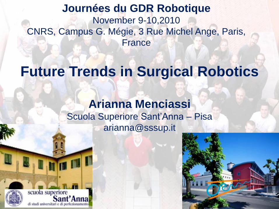
Future Trends in Surgical Robotics
Arianna MenciassiScuola Superiore Sant’Anna – Pisa
Journées du GDR RobotiqueNovember 9-10,2010
CNRS, Campus G. Mégie, 3 Rue Michel Ange, Paris,
France
Outline
The evolution of robotic surgery: state of the art
From external robots to endoluminal robots
Concluding remarks
Outline
The evolution of robotic surgery: state of the art
From external robots to endoluminal robots
Concluding remarks
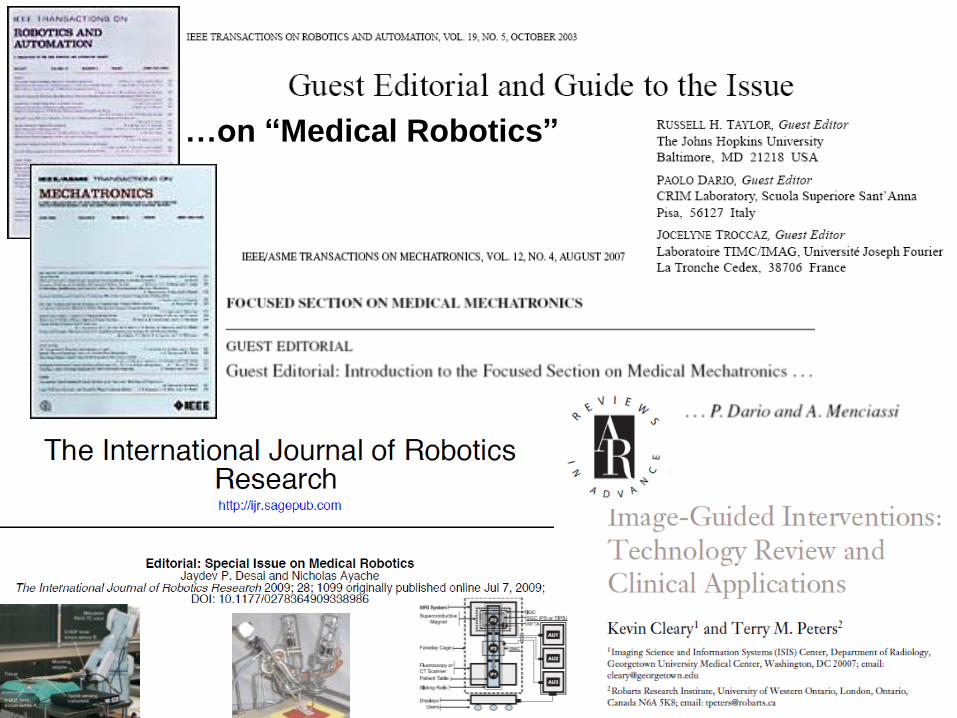
…on “Medical Robotics”
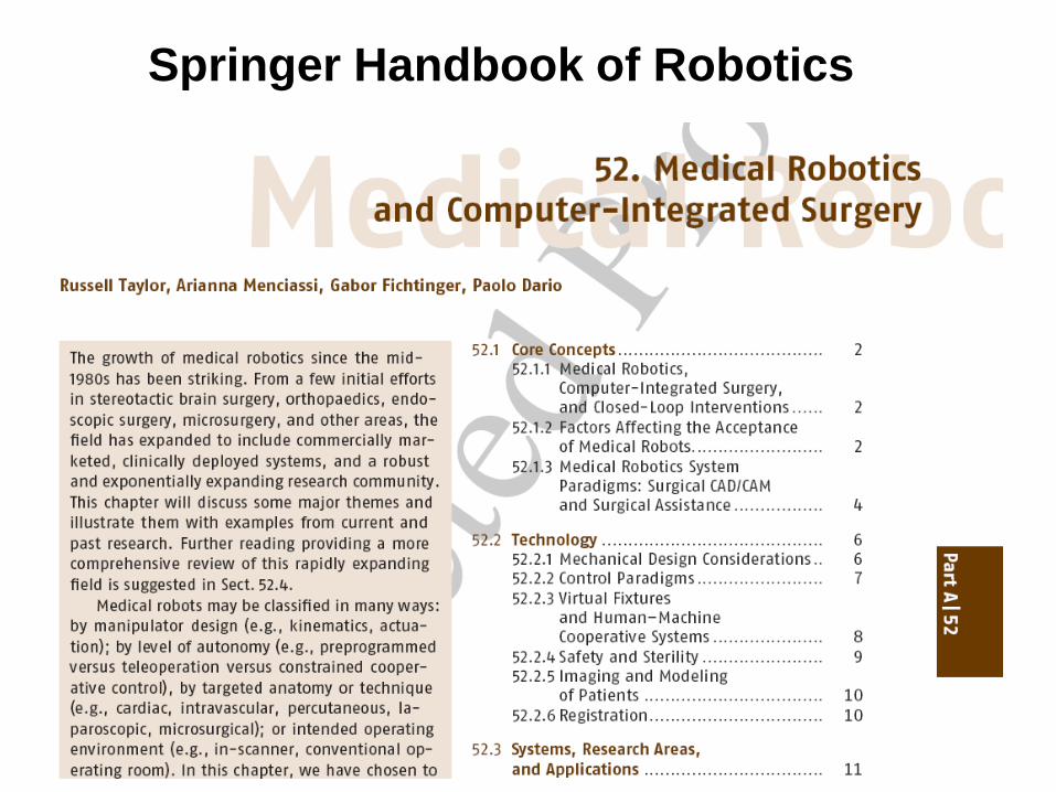
Springer Handbook of Robotics
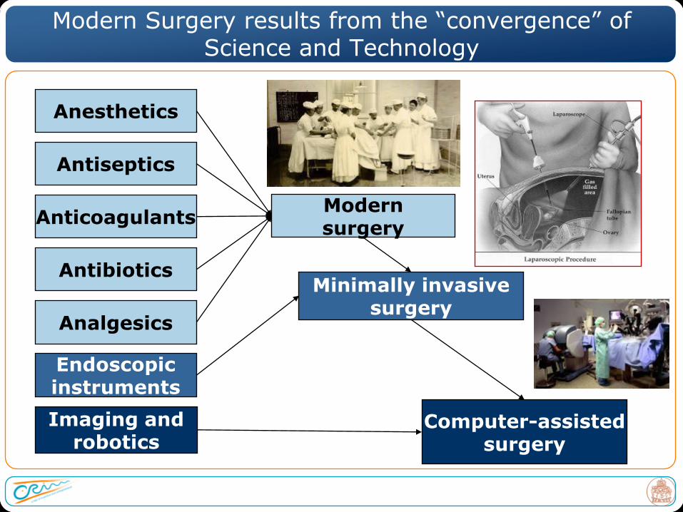
Modern Surgery results from the “convergence” of Science and Technology
Antiseptics
Anesthetics
Antibiotics
Analgesics
AnticoagulantsModernsurgery
Endoscopicinstruments
Minimally invasivesurgery
Imaging androbotics
Computer-assistedsurgery

The Evolution of Surgery
TRADITIONAL TECHNIQUES
ROBOTIC SURGERY
LAPAROSCOPIC SURGERY
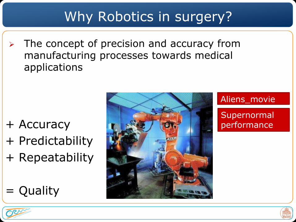
Why Robotics in surgery?
The concept of precision and accuracy from manufacturing processes towards medical applications
Aliens_movie
Supernormalperformance+ Accuracy
+ Predictability
+ Repeatability
= Quality
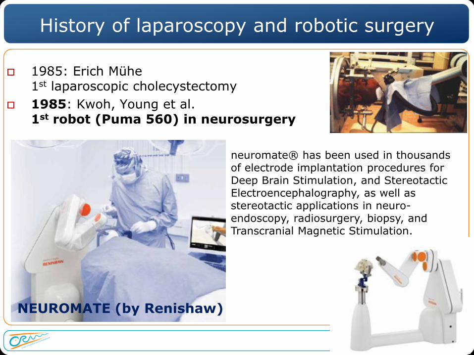
History of laparoscopy and robotic surgery
1985: Erich Mühe1st laparoscopic cholecystectomy
1985: Kwoh, Young et al.1st robot (Puma 560) in neurosurgery
neuromate® has been used in thousands of electrode implantation procedures for Deep Brain Stimulation, and Stereotactic Electroencephalography, as well as stereotactic applications in neuro-endoscopy, radiosurgery, biopsy, and Transcranial Magnetic Stimulation.
NEUROMATE (by Renishaw)
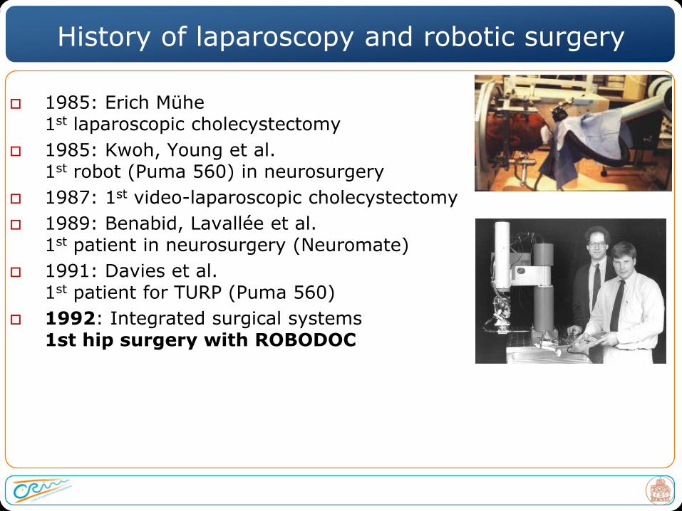
History of laparoscopy and robotic surgery
1985: Erich Mühe1st laparoscopic cholecystectomy
1985: Kwoh, Young et al.1st robot (Puma 560) in neurosurgery
1987: 1st video-laparoscopic cholecystectomy
1989: Benabid, Lavallée et al.1st patient in neurosurgery (Neuromate)
1991: Davies et al.1st patient for TURP (Puma 560)
1992: Integrated surgical systems1st hip surgery with ROBODOC
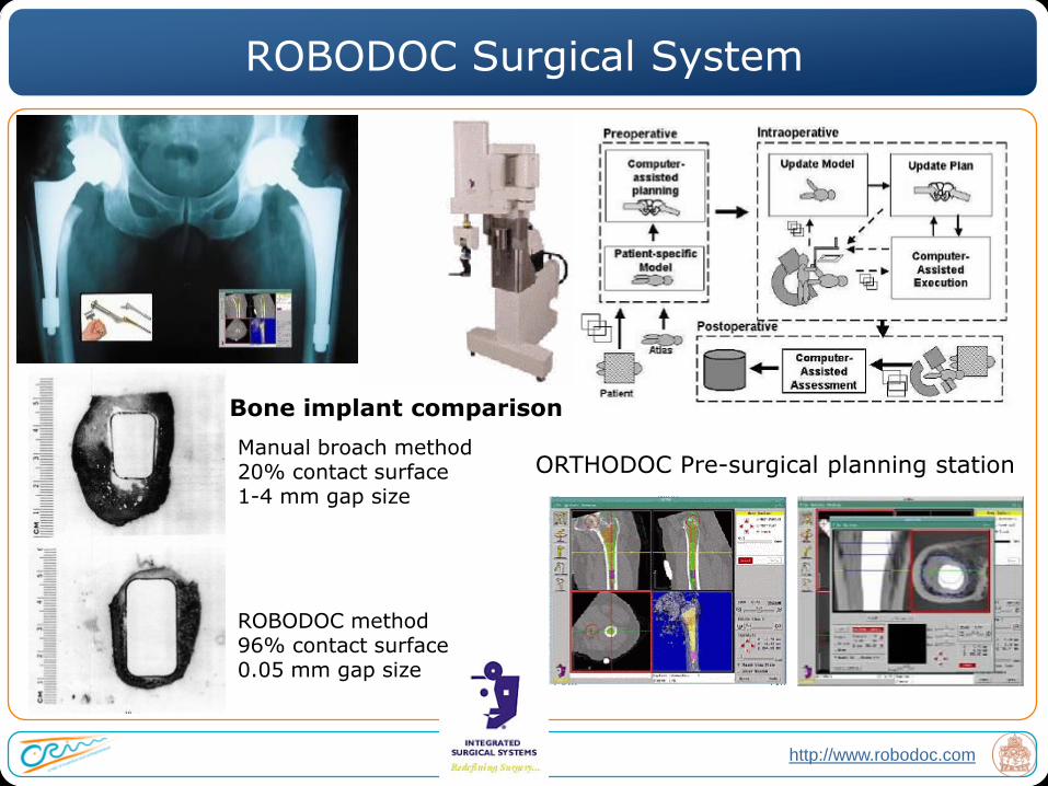
Manual broach method20% contact surface1-4 mm gap size
ROBODOC method96% contact surface0.05 mm gap size
Bone implant comparison
ORTHODOC Pre-surgical planning station
http://www.robodoc.com
ROBODOC Surgical System
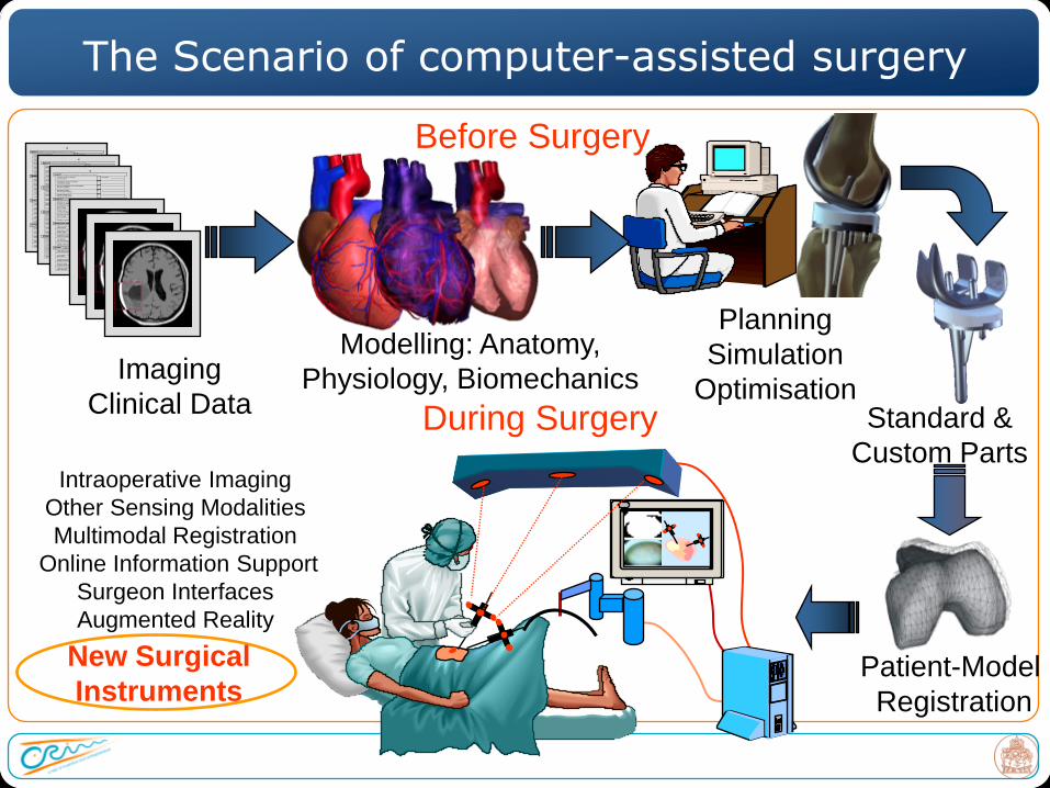
Imaging
Clinical Data
Before Surgery
Modelling: Anatomy,
Physiology, Biomechanics
Planning
Simulation
OptimisationStandard &
Custom Parts
New Surgical
Instruments
Intraoperative Imaging
Other Sensing Modalities
Multimodal Registration
Online Information Support
Surgeon Interfaces
Augmented Reality
Patient-Model
Registration
During Surgery
The Scenario of computer-assisted surgery
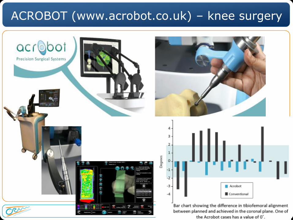
ACROBOT (www.acrobot.co.uk) – knee surgery

History of laparoscopy and robotic surgery
1985: Erich Mühe1st laparoscopic cholecystectomy
1985: Kwoh, Young et al.1st robot (Puma 560) in neurosurgery
1987: 1st video-laparoscopic cholecystectomy
1989: Benabid, Lavallée et al.1st patient in neurosurgery (Neuromate)
1991: Davies et al.1st patient for TURP (Puma 560)
1992: Integrated surgical systems1st hip surgery with ROBODOC
1994: Computer Motion Inc.1st FDA clearance: AESOP laparoscope holder
AESOP, assistant
robot for
laparoscope
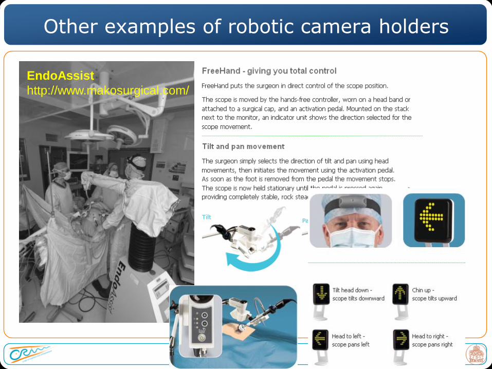
Other examples of robotic camera holders
EndoAssist
http://www.makosurgical.com/
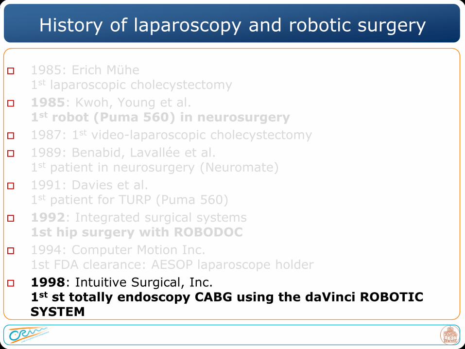
History of laparoscopy and robotic surgery
1985: Erich Mühe1st laparoscopic cholecystectomy
1985: Kwoh, Young et al.1st robot (Puma 560) in neurosurgery
1987: 1st video-laparoscopic cholecystectomy
1989: Benabid, Lavallée et al.1st patient in neurosurgery (Neuromate)
1991: Davies et al.1st patient for TURP (Puma 560)
1992: Integrated surgical systems1st hip surgery with ROBODOC
1994: Computer Motion Inc.1st FDA clearance: AESOP laparoscope holder
1998: Intuitive Surgical, Inc.1st st totally endoscopy CABG using the daVinci ROBOTIC SYSTEM
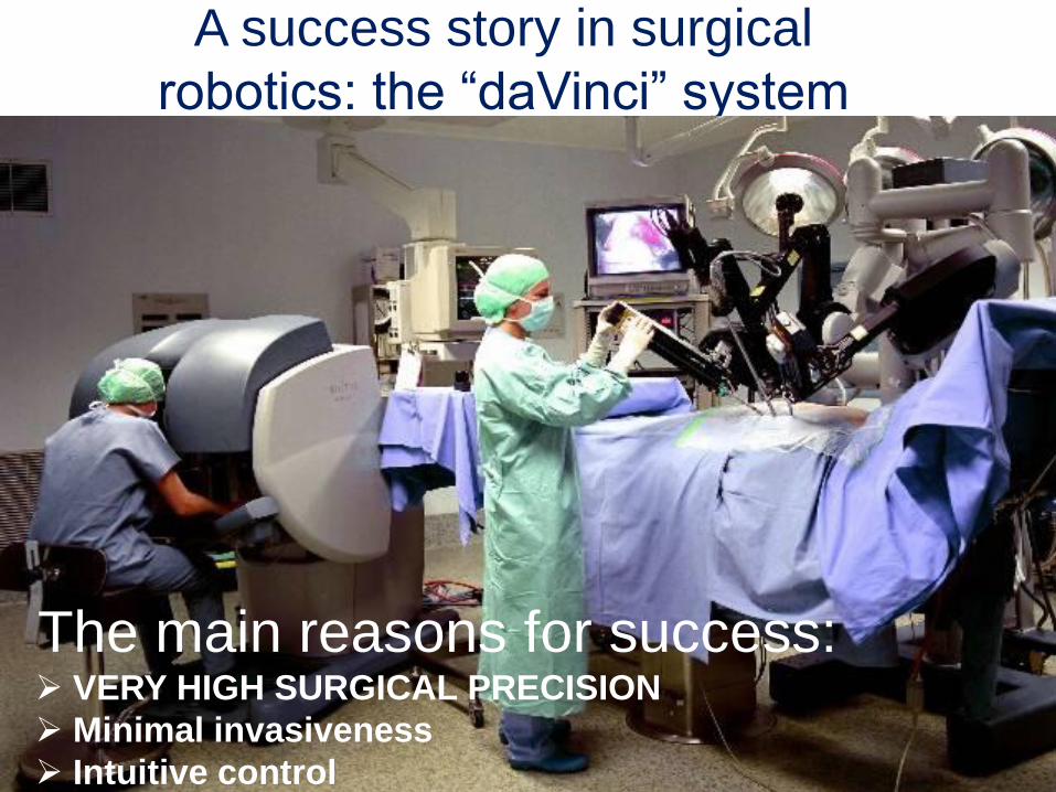
A success story in surgical
robotics: the “daVinci” system
The main reasons for success: VERY HIGH SURGICAL PRECISION
Minimal invasiveness
Intuitive control
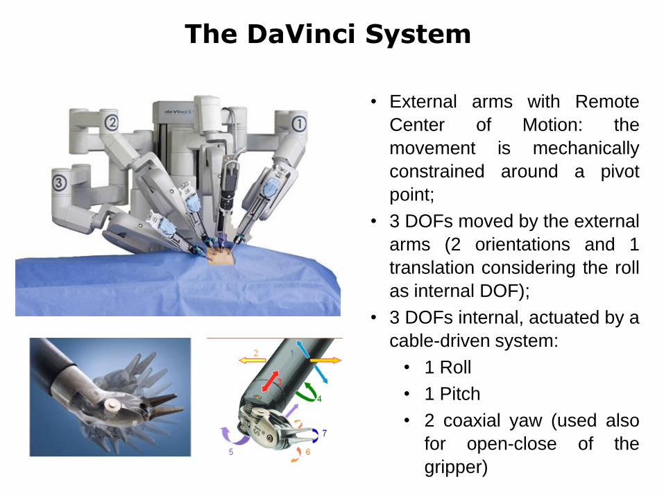
The DaVinci System
• External arms with Remote
Center of Motion: the
movement is mechanically
constrained around a pivot
point;
• 3 DOFs moved by the external
arms (2 orientations and 1
translation considering the roll
as internal DOF);
• 3 DOFs internal, actuated by a
cable-driven system:
• 1 Roll
• 1 Pitch
• 2 coaxial yaw (used also
for open-close of the
gripper)

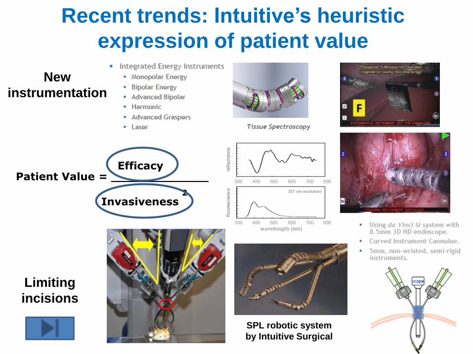
Patient Value = _____________Efficacy
Invasiveness2
SPL robotic system
by Intuitive Surgical
Recent trends: Intuitive’s heuristic
expression of patient value
New
instrumentation
Limiting
incisions
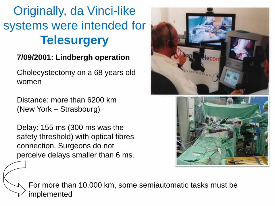
Originally, da Vinci-like
systems were intended for
Telesurgery
7/09/2001: Lindbergh operation
Cholecystectomy on a 68 years old
women
Distance: more than 6200 km
(New York – Strasbourg)
Delay: 155 ms (300 ms was the
safety threshold) with optical fibres
connection. Surgeons do not
perceive delays smaller than 6 ms.
For more than 10.000 km, some semiautomatic tasks must be
implemented

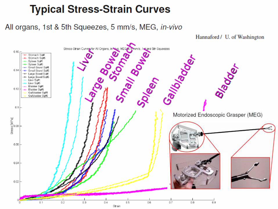
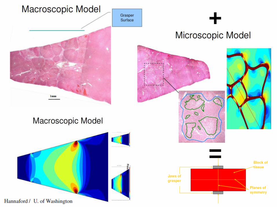
The RAVEN System – Biorobotics Lab Seattle (B. Hannaford)
The RAVEN patient site and the surgeon site

The RAVEN System – Biorobotics Lab Seattle (B. Hannaford)
RavenDemo_Final.mov
Mean network
latency (ms):
approx. 70 ms
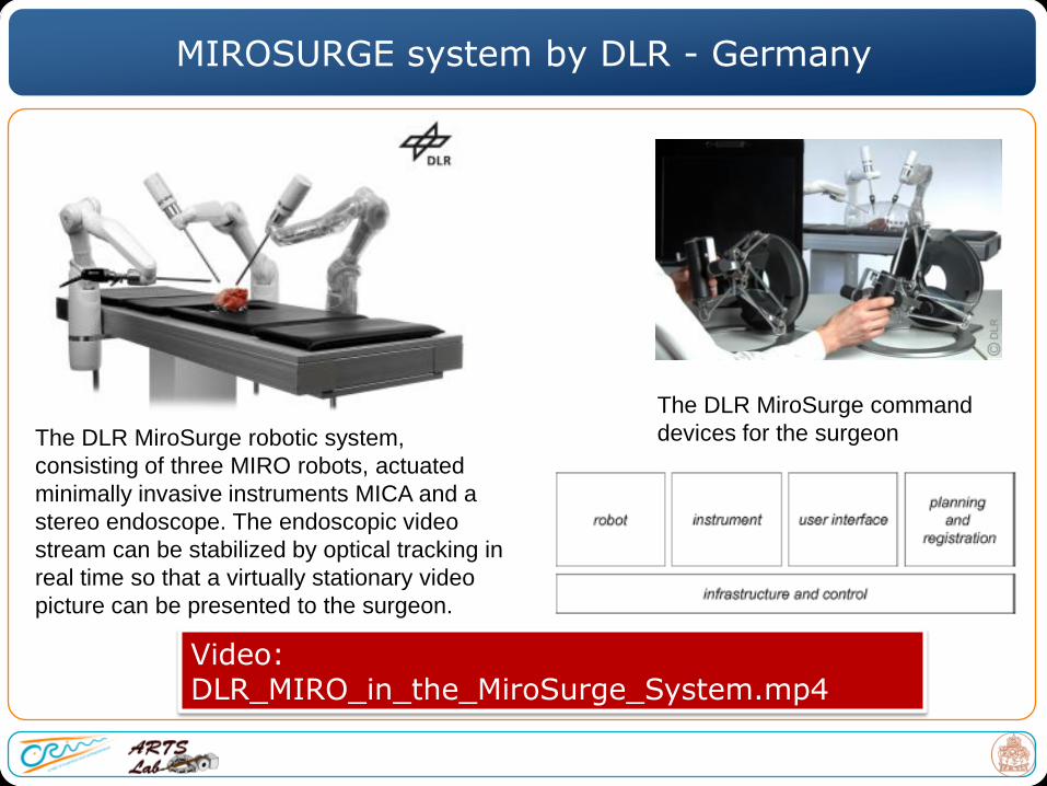
MIROSURGE system by DLR - Germany
The DLR MiroSurge robotic system,
consisting of three MIRO robots, actuated
minimally invasive instruments MICA and a
stereo endoscope. The endoscopic video
stream can be stabilized by optical tracking in
real time so that a virtually stationary video
picture can be presented to the surgeon.
The DLR MiroSurge command
devices for the surgeon
Video: DLR_MIRO_in_the_MiroSurge_System.mp4

MIROSURGE system by DLR - Germany
The DLR MIRO wrist design:
configuration for conventional instruments
(left) ; with MICA dedicated tool (right)
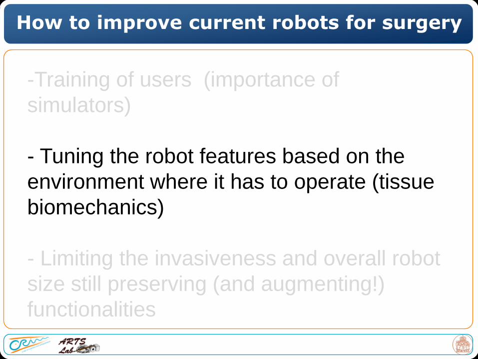
How to improve current robots for surgery
-Training of users (importance of
simulators)
- Tuning the robot features based on the
environment where it has to operate (tissue
biomechanics)
- Limiting the invasiveness and overall robot
size still preserving (and augmenting!)
functionalities
How to improve current robots for surgery
-Training of users (importance of
simulators)
- Tuning the robot features based on the
environment where it has to operate (tissue
biomechanics)
- Limiting the invasiveness and overall robot
size still preserving (and augmenting!)
functionalities
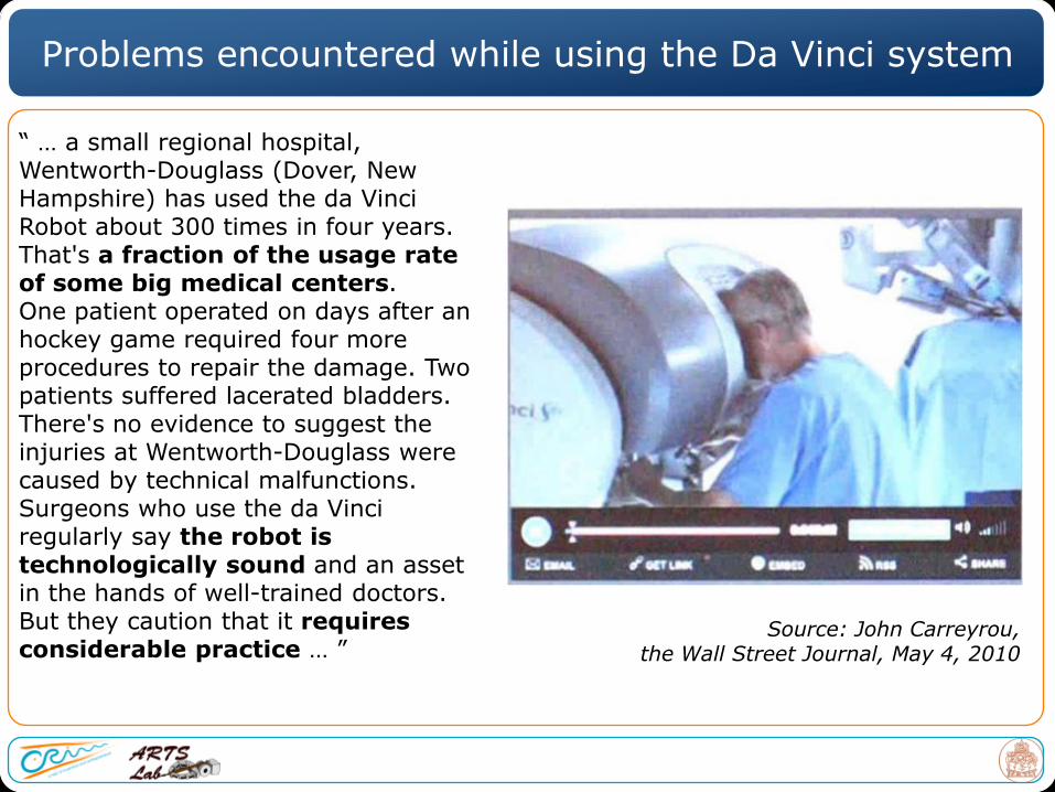
Problems encountered while using the Da Vinci system
Source: John Carreyrou, the Wall Street Journal, May 4, 2010
“ … a small regional hospital, Wentworth-Douglass (Dover, New Hampshire) has used the da Vinci Robot about 300 times in four years. That's a fraction of the usage rate of some big medical centers.One patient operated on days after an hockey game required four more procedures to repair the damage. Two patients suffered lacerated bladders.There's no evidence to suggest the injuries at Wentworth-Douglass were caused by technical malfunctions. Surgeons who use the da Vinci regularly say the robot is technologically sound and an asset in the hands of well-trained doctors. But they caution that it requires considerable practice … ”
31
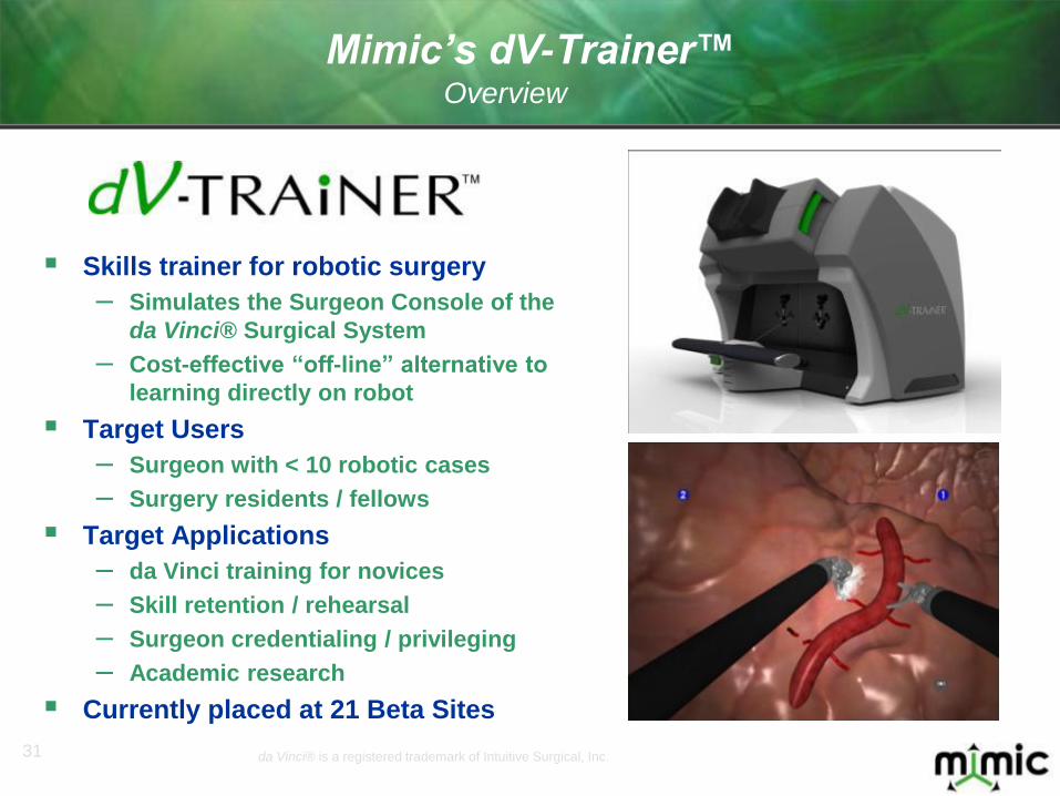
Skills trainer for robotic surgery
– Simulates the Surgeon Console of the
da Vinci® Surgical System
– Cost-effective “off-line” alternative to
learning directly on robot
Target Users
– Surgeon with < 10 robotic cases
– Surgery residents / fellows
Target Applications
– da Vinci training for novices
– Skill retention / rehearsal
– Surgeon credentialing / privileging
– Academic research
Currently placed at 21 Beta Sites
Overview
Mimic’s dV-Trainer™
da Vinci® is a registered trademark of Intuitive Surgical, Inc.
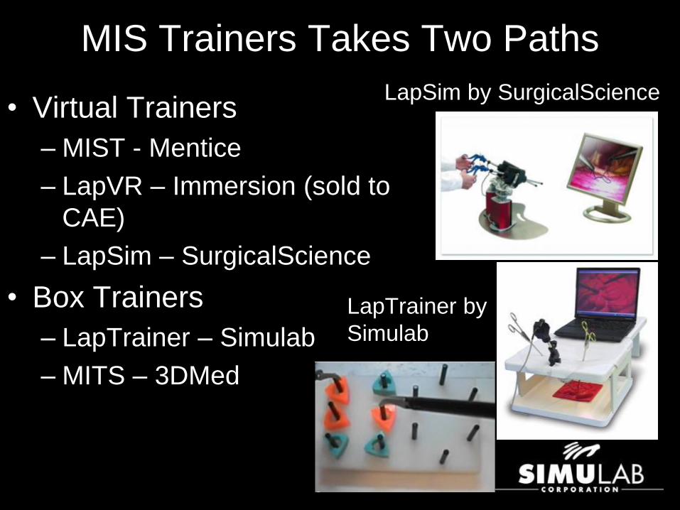
MIS Trainers Takes Two Paths
• Virtual Trainers
– MIST - Mentice
– LapVR – Immersion (sold to
CAE)
– LapSim – SurgicalScience
• Box Trainers
– LapTrainer – Simulab
– MITS – 3DMed
LapSim by SurgicalScience
LapTrainer by
Simulab
How to improve current robots for surgery
-Training of users (importance of
simulators)
- Tuning the robot features based on the
environment where it has to operate (tissue
biomechanics)
- Limiting the invasiveness and overall robot
size still preserving (and augmenting!)
functionalities
+
=
How to improve current robots for surgery
-Training of users (importance of
simulators)
- Tuning the robot features based on the
environment where it has to operate (tissue
biomechanics)
- Limiting the invasiveness and overall robot
size still preserving (and augmenting!)
functionalities
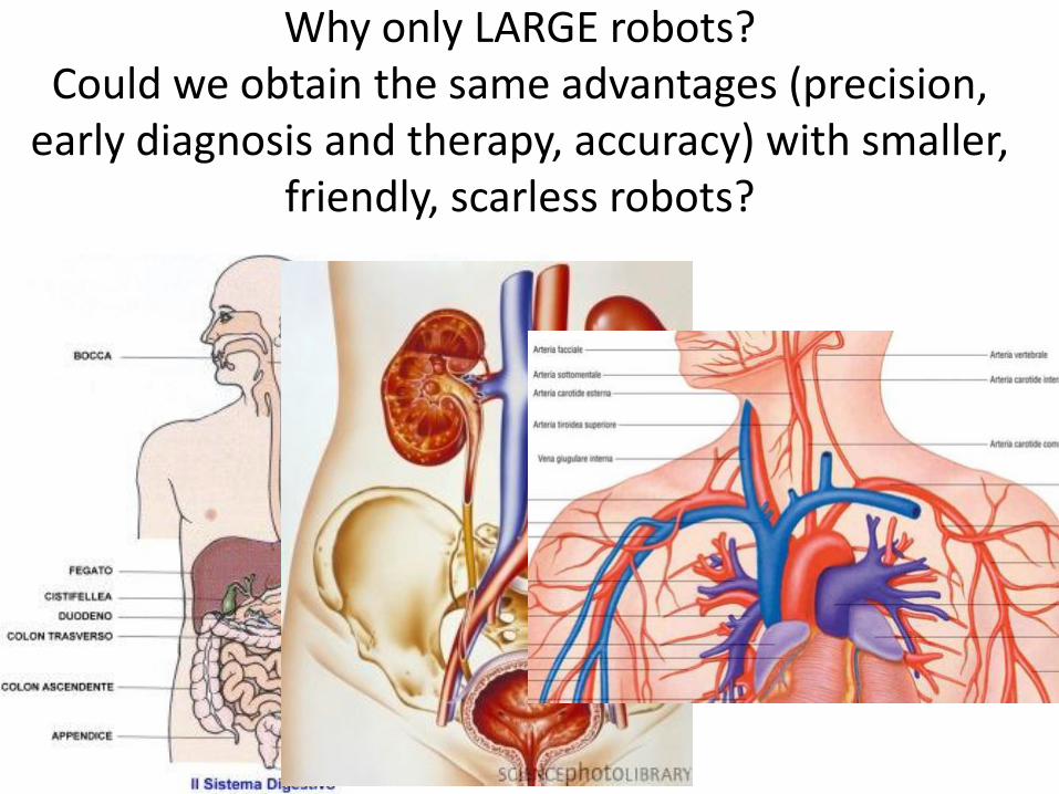
Why only LARGE robots?Could we obtain the same advantages (precision,
early diagnosis and therapy, accuracy) with smaller, friendly, scarless robots?
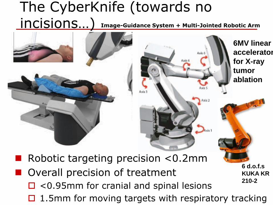
The CyberKnife (towards no incisions…)
Robotic targeting precision <0.2mm
Overall precision of treatment
<0.95mm for cranial and spinal lesions
1.5mm for moving targets with respiratory tracking
6MV linear
accelerator
for X-ray
tumor
ablation
Image-Guidance System + Multi-Jointed Robotic Arm
6 d.o.f.s
KUKA KR
210-2
Outline
The evolution of robotic surgery: state of the art
From external robots to endoluminal robots
Concluding remarks
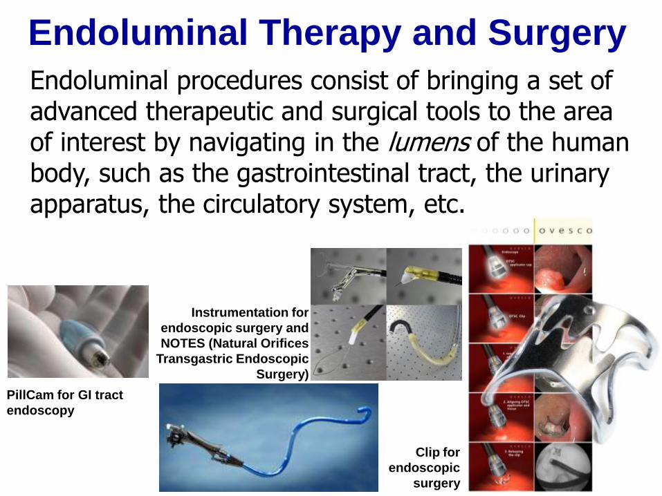
Endoluminal Therapy and Surgery
Endoluminal procedures consist of bringing a set of advanced therapeutic and surgical tools to the area of interest by navigating in the lumens of the human body, such as the gastrointestinal tract, the urinary apparatus, the circulatory system, etc.
PillCam for GI tract
endoscopy
Clip for
endoscopic
surgery
Instrumentation for
endoscopic surgery and
NOTES (Natural Orifices
Transgastric Endoscopic
Surgery)
41
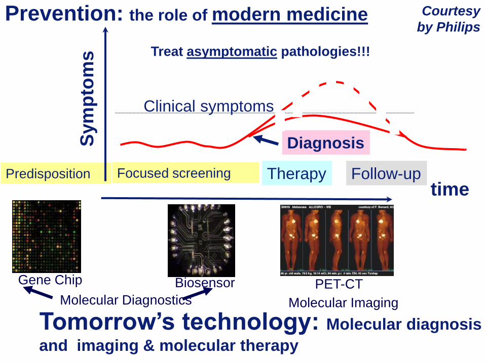
Prevention: the role of modern medicine
Sym
pto
ms
time
Clinical symptoms
Therapy Follow-up
Diagnosis
Predisposition
Tomorrow’s technology: Molecular diagnosis
and imaging & molecular therapy
Gene Chip Biosensor PET-CT
Molecular Diagnostics Molecular Imaging
Focused screening
Treat asymptomatic pathologies!!!
Courtesy
by Philips
Outline
The evolution of robotic surgery: state of the art
From external robots to endoluminal robots
Case 1: endoscopy of the GI tract
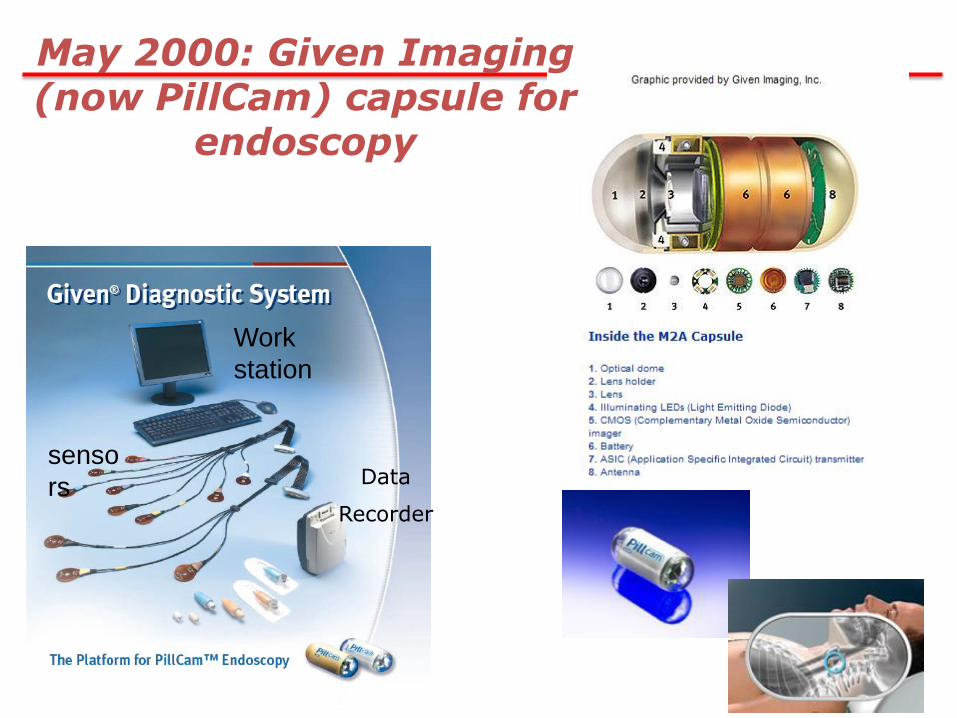
senso
rs
Work
station
Data
Recorder
May 2000: Given Imaging (now PillCam) capsule for
endoscopy
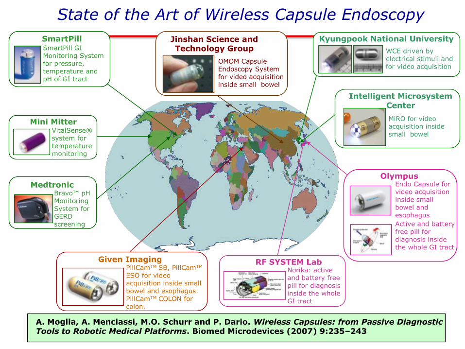
State of the Art of Wireless Capsule Endoscopy
SmartPill SmartPill GI Monitoring System for pressure, temperature and pH of GI tract
Mini Mitter VitalSense® system for temperature monitoring
Medtronic Bravo™ pH Monitoring System for GERD screening
Given ImagingPillCamTM SB, PillCamTM
ESO for video acquisition inside small bowel and esophagus.
PillCamTM COLON for colon.
RF SYSTEM Lab Norika: active and battery free pill for diagnosis inside the whole GI tract
OlympusEndo Capsule for video acquisition inside small bowel and esophagus
Active and battery free pill for diagnosis inside the whole GI tract
Jinshan Science and Technology Group
OMOM Capsule Endoscopy System for video acquisition inside small bowel
Kyungpook National University
WCE driven by electrical stimuli and for video acquisition
Intelligent Microsystem Center
MiRO for video acquisition inside small bowel
A. Moglia, A. Menciassi, M.O. Schurr and P. Dario. Wireless Capsules: from Passive Diagnostic Tools to Robotic Medical Platforms. Biomed Microdevices (2007) 9:235–243
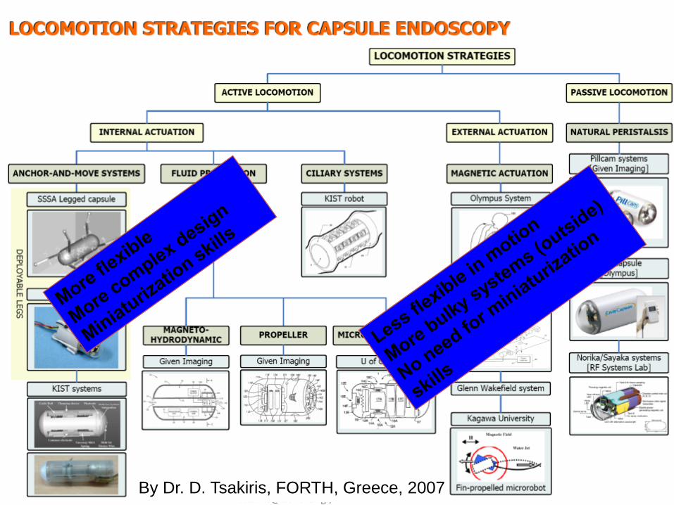
Dr. Dimitris P. Tsakiris, Institute of Computer Science –[email protected], http://www.icsforth.gr/~tsakiri
By Dr. D. Tsakiris, FORTH, Greece, 2007

Internal Locomotion Approach
Locomotion in the Stomach:
The capsule swims in a liquid
environment
Locomotion in the Colon:
The capsule moves by 12
legs
Due to space constraints we decided to pursue different
strategies optimized for the 2 targeted districts:
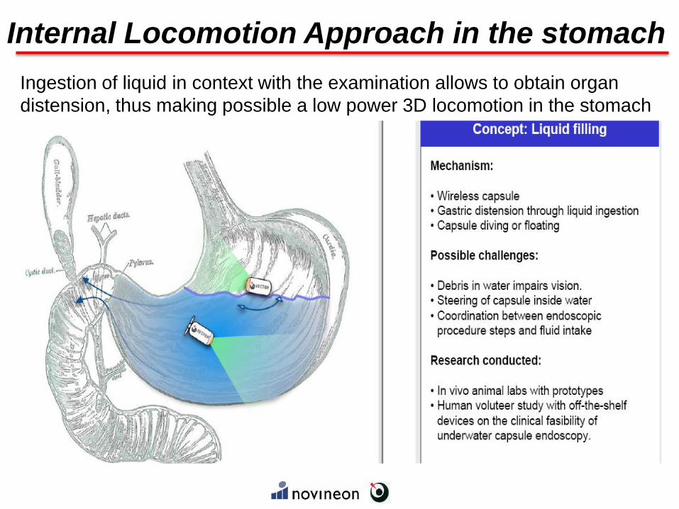
Stomach capsule : swimming locomon
Ingestion of liquid in context with the examination allows to obtain organ
distension, thus making possible a low power 3D locomotion in the stomach
Internal Locomotion Approach in the stomach

Ex vivo test in
stomach of pig filled
with water
Fine control of steering
and speed in 3D
Stomach capsule : swimming locomotion
Driver
Control unit
In the capsule:Control/Telemetry module
TI CC2430 (Zigbee + μC)
4 DC brushless motors
Didel MK04S-24, 2400rps
3.0V, 15mA/V no load
4 propellers in the rear side
1 battery: LP20, Plantraco Ltd., 20mAh,3.7V; or a power module for inductive power supply
External “dongle”:TI CC2430 (Zigbee + μC)
USB front-end
Internal Locomotion Approach in the stomach
[in collaboration with Prof.
Puers, Leuven Univ., Belgium]

Internal Locomotion Approach
Locomotion in the Stomach:
The capsule swims in a liquid
environment
Locomotion in the
Colon:
The capsule moves by
legs
Due to space constraints we decided to pursue different
strategies optimized for the 2 targeted districts:
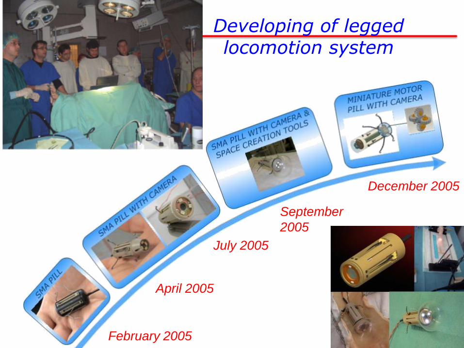
February 2005
April 2005
July 2005
September
2005
December 2005
Developing of legged locomotion system
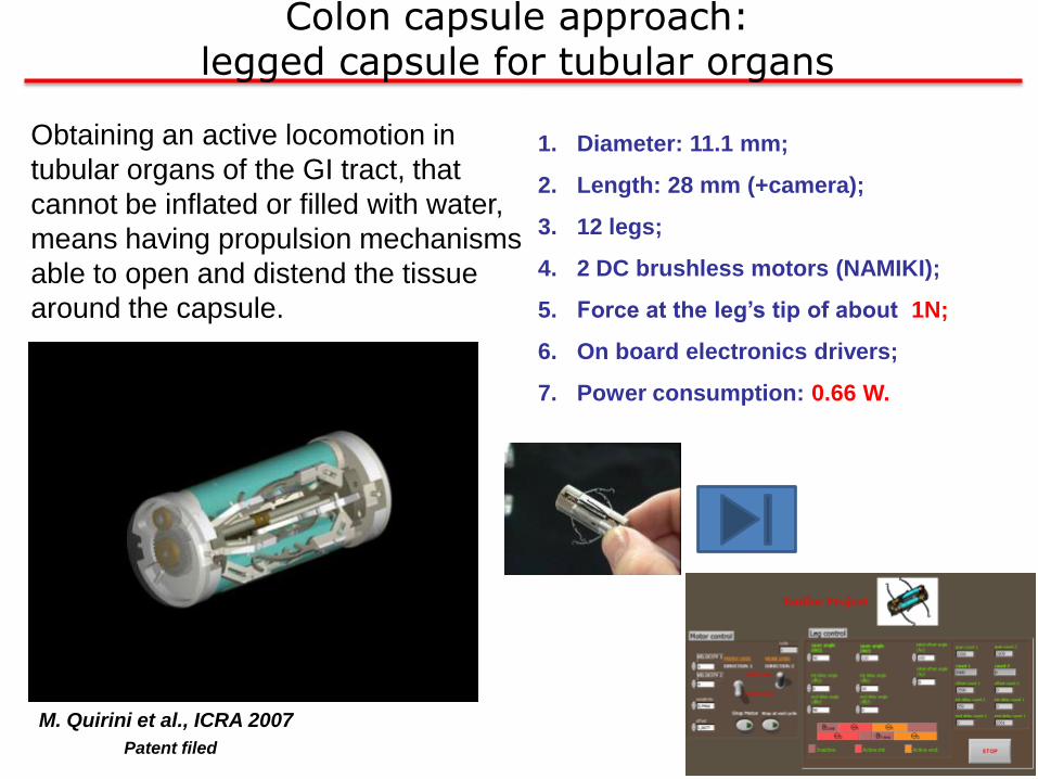
Colon capsule approach: legged capsule for tubular organs
Obtaining an active locomotion in
tubular organs of the GI tract, that
cannot be inflated or filled with water,
means having propulsion mechanisms
able to open and distend the tissue
around the capsule.
M. Quirini et al., ICRA 2007
1. Diameter: 11.1 mm;
2. Length: 28 mm (+camera);
3. 12 legs;
4. 2 DC brushless motors (NAMIKI);
5. Force at the leg’s tip of about 1N;
6. On board electronics drivers;
7. Power consumption: 0.66 W.
Patent filed

Colon capsule approach: legged capsule for tubular organs
M. Quirini, S. Scapellato, A. Menciassi, P. Dario, F. Rieber, C.-N. Ho, S. Schostek, M.O. Schurr, “Feasibility proof of
a legged locomotion capsule for the GI tract”, GASTROINTESTINAL ENDOSCOPY, 67(7), 2008
Simulator test
Colon test (5
cm/min)
By considering the power budget for all the capsule functions (vision,
locomotion, communication), the single capsule approach shows dramatic
limitations: new battery / powering technologies would be necessary!
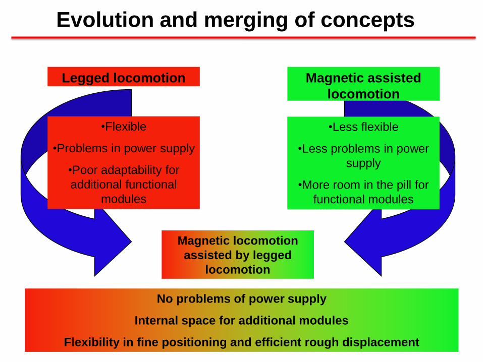
Legged locomotion Magnetic assisted
locomotion
•Less flexible
•Less problems in power
supply
•More room in the pill for
functional modules
•Flexible
•Problems in power supply
•Poor adaptability for
additional functional
modules
Magnetic locomotion
assisted by legged
locomotion
No problems of power supply
Internal space for additional modules
Flexibility in fine positioning and efficient rough displacement
Evolution and merging of concepts
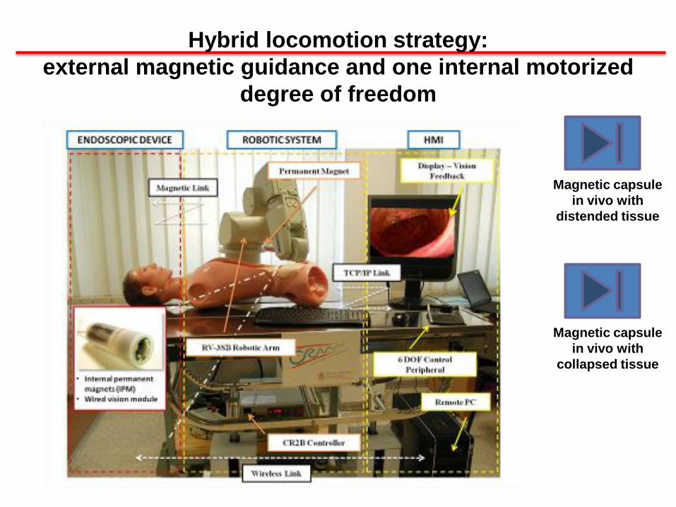
Hybrid locomotion strategy:
external magnetic guidance and one internal motorized
degree of freedom
Magnetic capsule
in vivo with
collapsed tissue
Magnetic capsule
in vivo with
distended tissue
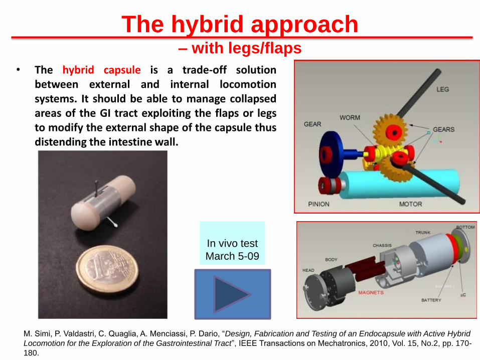
The hybrid approach – with legs/flaps
• The hybrid capsule is a trade-off solutionbetween external and internal locomotionsystems. It should be able to manage collapsedareas of the GI tract exploiting the flaps or legsto modify the external shape of the capsule thusdistending the intestine wall.
M. Simi, P. Valdastri, C. Quaglia, A. Menciassi, P. Dario, “Design, Fabrication and Testing of an Endocapsule with Active Hybrid
Locomotion for the Exploration of the Gastrointestinal Tract”, IEEE Transactions on Mechatronics, 2010, Vol. 15, No.2, pp. 170-
180.
In vivo test
March 5-09
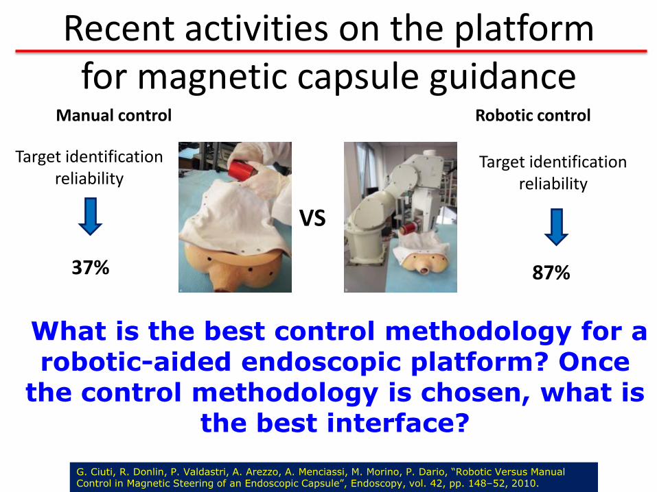
Recent activities on the platform for magnetic capsule guidance
Robotic control
VS
Target identificationreliability
Target identificationreliability
37% 87%
Manual control
G. Ciuti, R. Donlin, P. Valdastri, A. Arezzo, A. Menciassi, M. Morino, P. Dario, “Robotic Versus Manual Control in Magnetic Steering of an Endoscopic Capsule”, Endoscopy, vol. 42, pp. 148–52, 2010.
What is the best control methodology for a robotic-aided endoscopic platform? Once
the control methodology is chosen, what is the best interface?

A comparative evaluation of control methodologies and interfaces for a robotic-aided endoscopic platform
Teleoperated remote control
Human-robot cooperative control
The proposed task consists of the exploration of a segment of ex-vivo colon by steering the capsule by the robotic-
magnetic approach, with different control methodologies and interfaces.
[in collaboration with Dr. A. Arezzo team, Torino University, Italy]
slide31ago
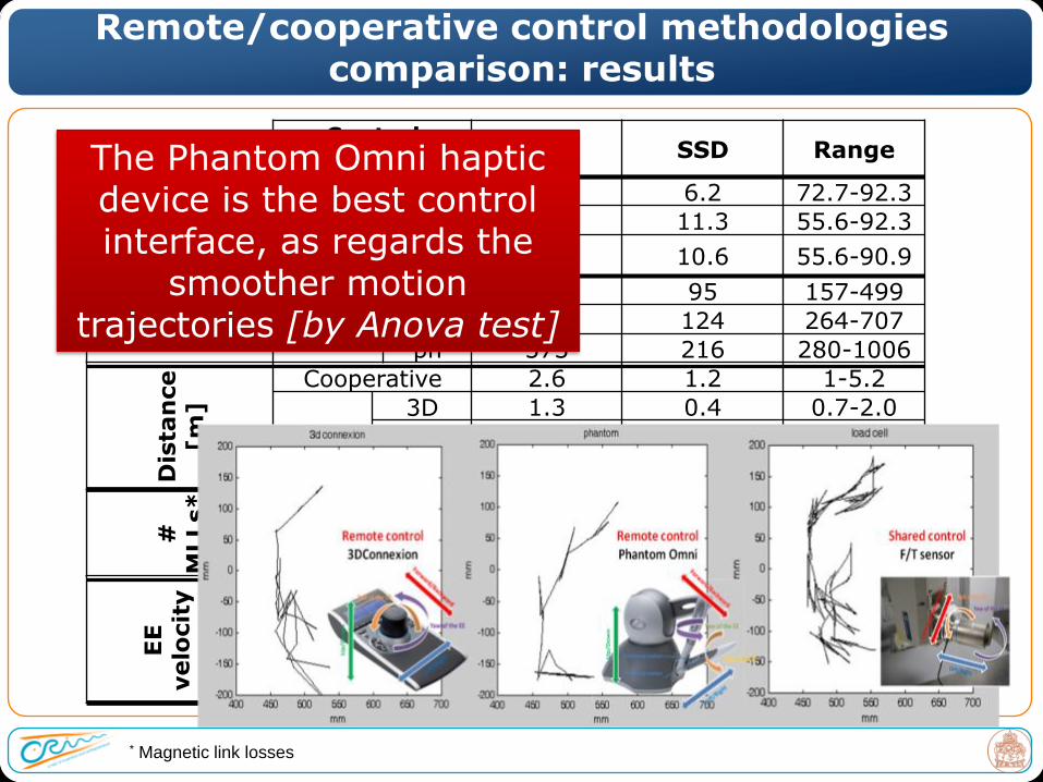
Control Method
Mean SSD RangeM
arker
[%
]Cooperative 84.6 6.2 72.7-92.3
Remote3D 79.6 11.3 55.6-92.3
ph 79.6 10.6 55.6-90.9
Tim
e
[s]
Cooperative 298 95 157-499
Remote3D 440 124 264-707
ph 573 216 280-1006
Dis
tan
ce
[m
]
Cooperative 2.6 1.2 1-5.2
Remote
3D 1.3 0.4 0.7-2.0
ph 1.1 0.5 0.4-2.2
#
MLLs* Cooperative 3.5 1.5 0-6
Remote3D 1.9 1.2 0-4
ph 2.0 1.1 0-4
EE
velo
cit
y
[m
m/
s] Cooperative 8.9 4.16 4.1-16.9
Remote
3D 3.0 0.8 2.0-5.1
ph 1.9 0.7 1.1-3.7
Remote/cooperative control methodologies comparison: results
* Magnetic link losses
The Phantom Omni haptic device is the best control interface, as regards the
smoother motion trajectories [by Anova test]
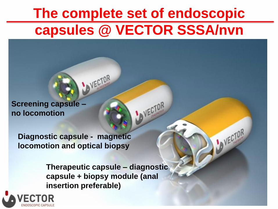
Screening capsule –
no locomotion
Diagnostic capsule - magnetic
locomotion and optical biopsy
Therapeutic capsule – diagnostic
capsule + biopsy module (anal
insertion preferable)
The complete set of endoscopic
capsules @ VECTOR SSSA/nvn
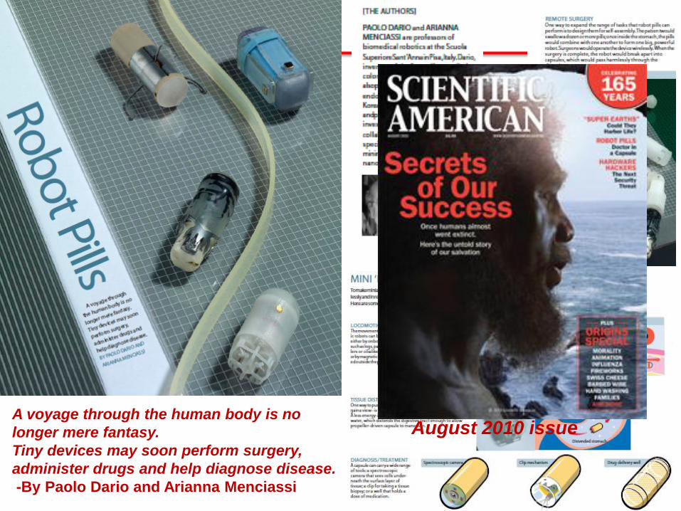
A voyage through the human body is no
longer mere fantasy.
Tiny devices may soon perform surgery,
administer drugs and help diagnose disease.
-By Paolo Dario and Arianna Menciassi
August 2010 issue

Outline
The evolution of robotic surgery: state of the art
From external robots to endoluminal robots
Case 2: reconfigurablesurgical robot with single access
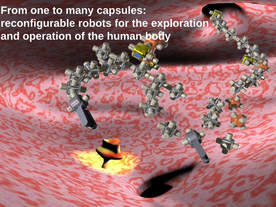
From one to many capsules:
reconfigurable robots for the exploration
and operation of the human body
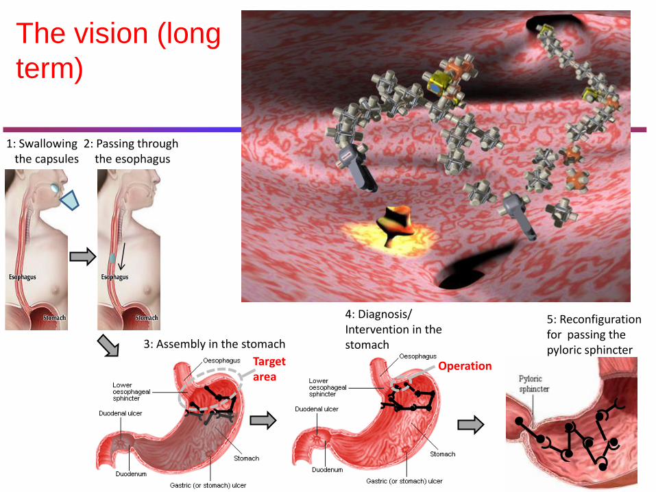
The vision (long
term)
1: Swallowing the capsules
2: Passing through the esophagus
3: Assembly in the stomach
4: Diagnosis/ Intervention in the stomach
Targetarea
Operation
5: Reconfiguration for passing the pyloric sphincter
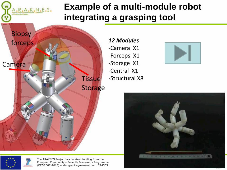
The ARAKNES Project has received funding from the European Community's Seventh Framework Programme (FP7/2007-2013) under grant agreement num. 224565.
Camera
Biopsy forceps
TissueStorage
12 Modules-Camera X1-Forceps X1-Storage X1-Central X1-Structural X8
Example of a multi-module robot
integrating a grasping tool
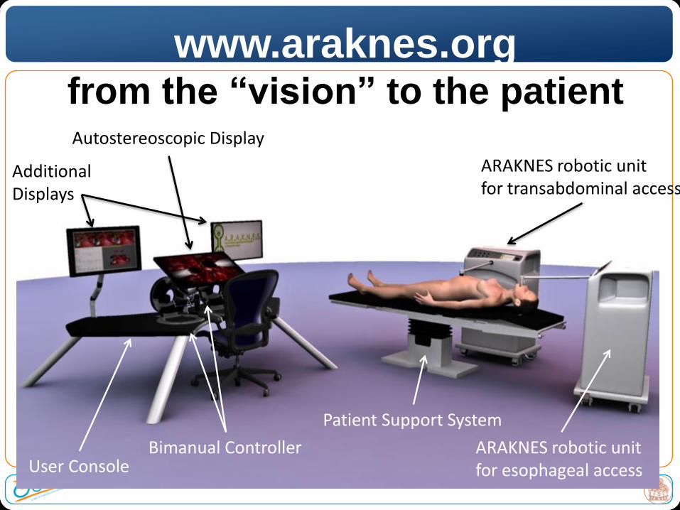
65User Console
Bimanual Controller
Autostereoscopic Display
Additional Displays
Patient Support System
ARAKNES robotic unit for esophageal access
ARAKNES robotic unit for transabdominal access
www.araknes.orgfrom the “vision” to the patient
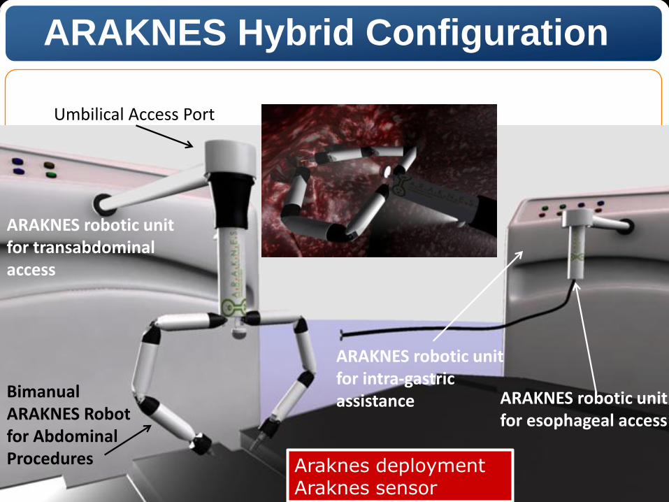
ARAKNES robotic unit for esophageal access
ARAKNES robotic unit for transabdominal access
Bimanual ARAKNES Robotfor Abdominal Procedures
Umbilical Access Port
ARAKNES robotic unit for intra-gastric assistance
ARAKNES Hybrid Configuration
Araknes deploymentAraknes sensor
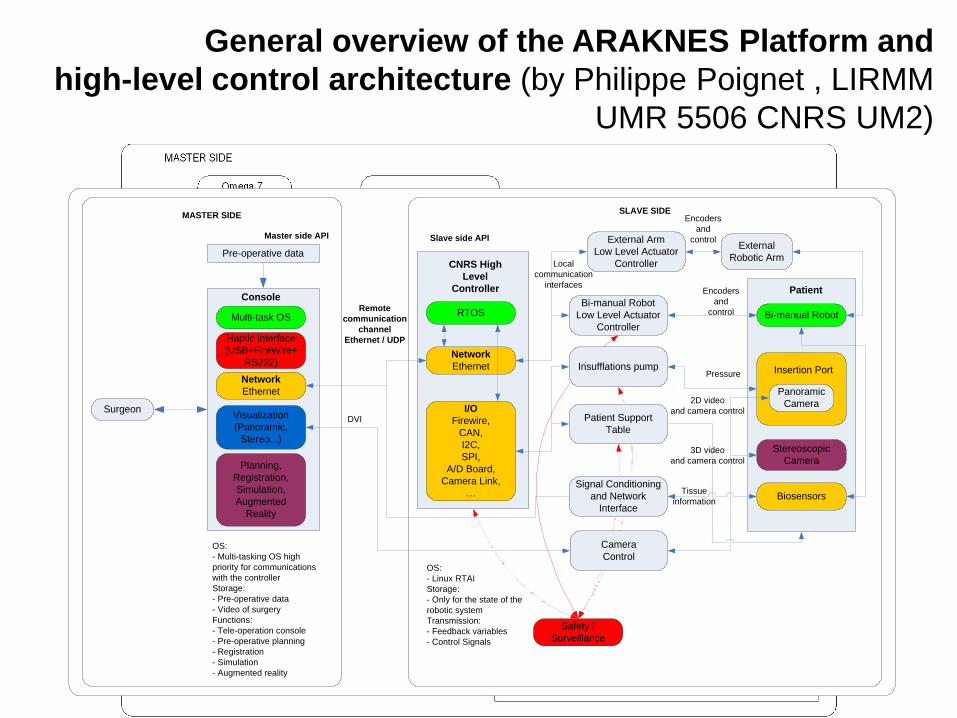
General overview of the ARAKNES Platform and
high-level control architecture (by Philippe Poignet , LIRMM
UMR 5506 CNRS UM2)
Surgeon
Multi-task OS
Haptic interface
(USB+FireWire+
RS232)
Network
Ethernet
Visualization
(Panoramic,
Stereo...)
Planning,
Registration,
Simulation,
Augmented
Reality
Console
OS:
- Multi-tasking OS high
priority for communications
with the controller
Storage:
- Pre-operative data
- Video of surgery
Functions:
- Tele-operation console
- Pre-operative planning
- Registration
- Simulation
- Augmented reality
Master side API
Remote
communication
channel
Ethernet / UDP
Pre-operative data
RTOS
Network
Ethernet
I/O
Firewire,
CAN,
I2C,
SPI,
A/D Board,
Camera Link,
…
CNRS High
Level
Controller
Bi-manual Robot
Insertion Port
Panoramic
Camera
Stereoscopic
Camera
Biosensors
Patient
External
Robotic Arm
Bi-manual Robot
Low Level Actuator
Controller
External Arm
Low Level Actuator
Controller
Insufflations pump
Patient Support
Table
Signal Conditioning
and Network
Interface
Camera
ControlOS:
- Linux RTAI
Storage:
- Only for the state of the
robotic system
Transmission:
- Feedback variables
- Control Signals
Slave side API
Local
communication
interfaces
Encoders
and
control
Encoders
and
control
Pressure
2D video
and camera control
3D video
and camera control
Tissue
information
Safety /
Surveillance
MASTER SIDESLAVE SIDE
DVI
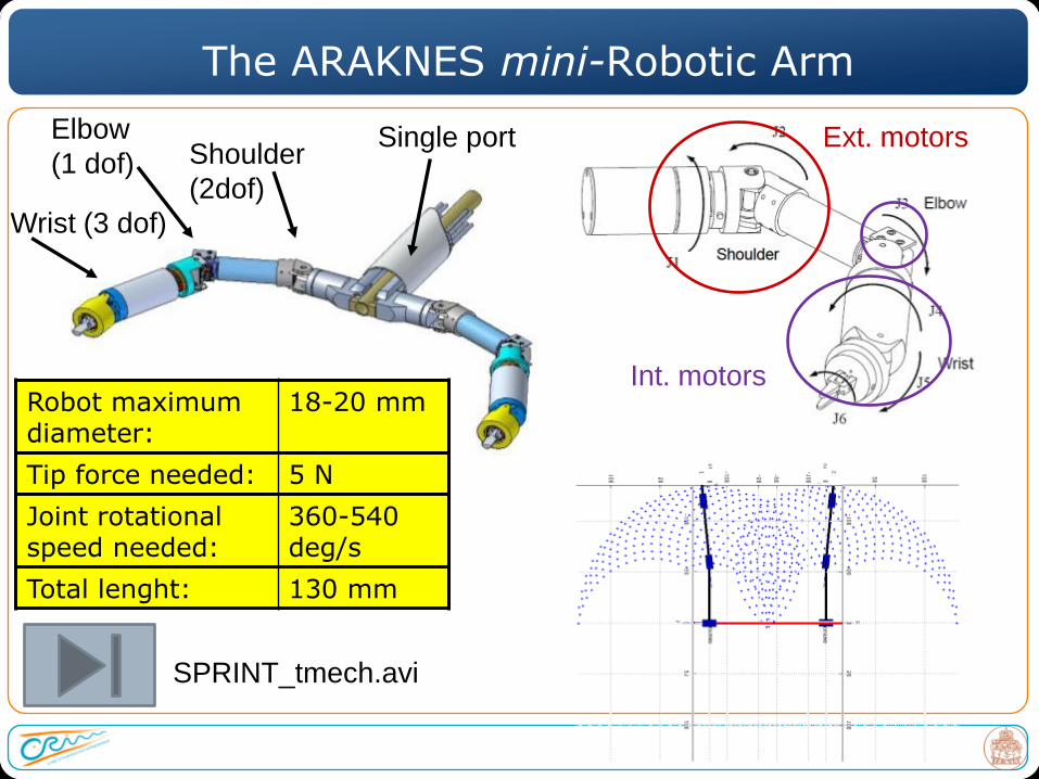
The ARAKNES mini-Robotic Arm
SPRINT_tmech.avi
Shoulder
(2dof)
Elbow
(1 dof)
Wrist (3 dof)
Single port
Robot maximumdiameter:
18-20 mm
Tip force needed: 5 N
Joint rotational speed needed:
360-540 deg/s
Total lenght: 130 mm
Ext. motors
Int. motors
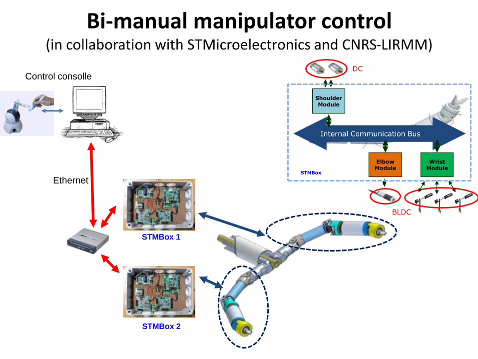
Ethernet
Bi-manual manipulator control (in collaboration with STMicroelectronics and CNRS-LIRMM)
STMBox 1
STMBox 2
Control consolle
Shoulder Module
Elbow Module
Wrist Module
STMBox
Internal Communication Bus
DC
BLDC

From mini to micro: the top-down approach
Example of miniature platform to be used in Single
Port Laparoscopy and NOTES surgery
+ =
procedure
1 robot 2 robotsVideo ICRA2011
ARAKNES User Console
Bimanual Controller
with Haptic Feedback (OMEGA)
Autostereoscopic
Display
Additional
DisplayAdditional
Display
Main Features-Omega based dual haptic interface- Autostereoscopic display
Outline
The evolution of robotic surgery: state of the art
From external robots to endoluminal robots
Concluding remarks

Robotics Surgery: Lessons Learned
Problems to be solved for full acceptance of robots in surgery:
Real application domains and procedures that benefit: finding the unmet clinical needs among the 6301 currently performed surgical procedures
Time of intervention
Time and complexity for set up
Cost/benefit clearly proved
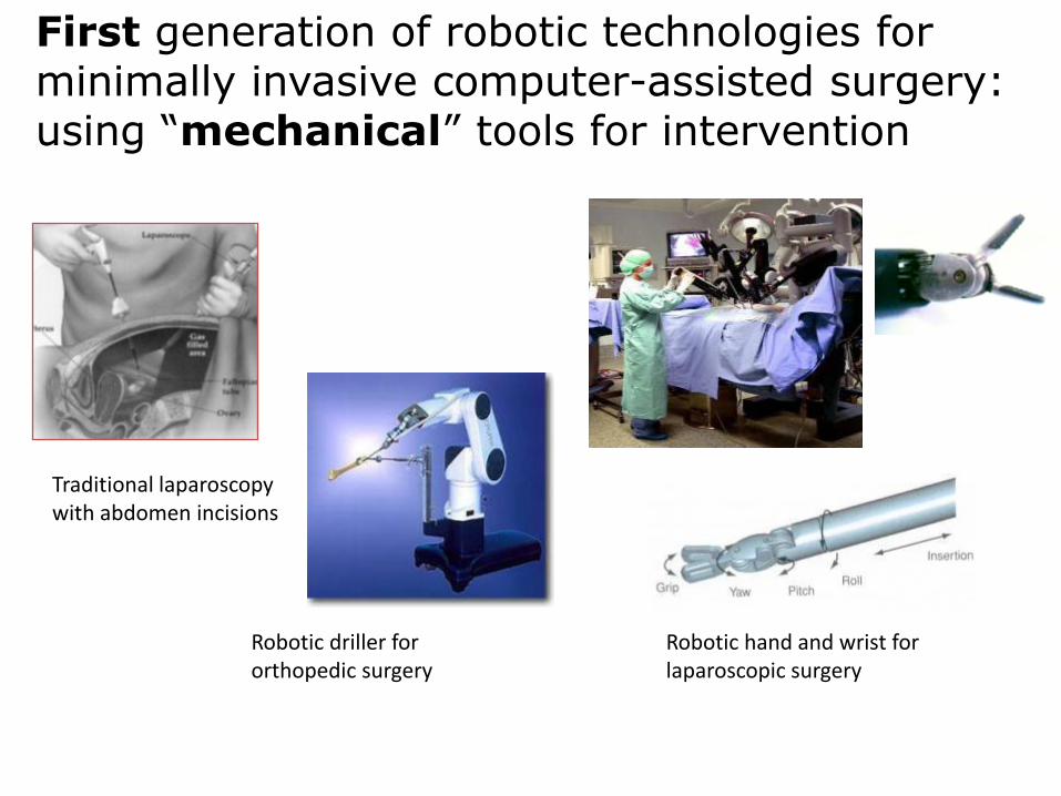
First generation of robotic technologies for minimally invasive computer-assisted surgery: using “mechanical” tools for intervention
Traditional laparoscopy with abdomen incisions
Robotic driller for orthopedic surgery
Robotic hand and wrist for laparoscopic surgery

Second generation of robotic technologies for minimally invasive computer-assisted surgery: using “non contact” tools for navigation and intervention
Surgical procedure for “scarless” delivery of tools/particles inside the abdomen
Robotic radiosurgeryRobotic platform with magnetic guidance for wireless delivery of treatment in the vascular system

76
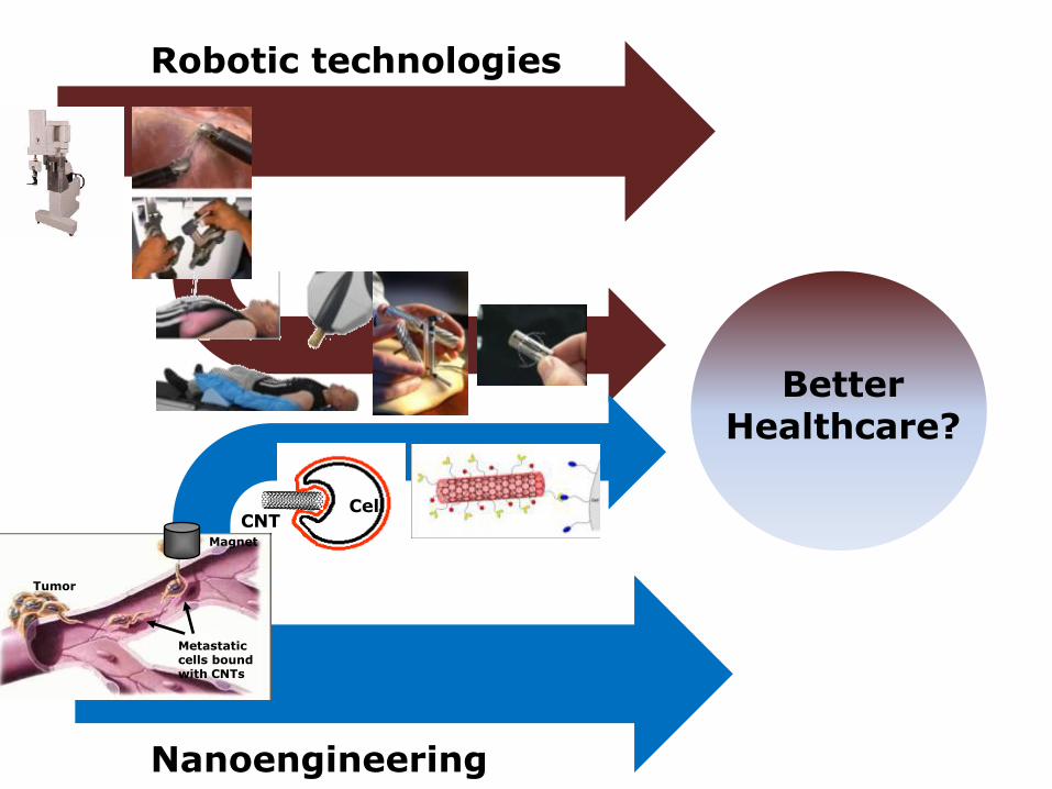
Robotic technologies
Nanoengineering
Better Healthcare?
Magnet
Metastatic cells bound with CNTs
Tumor
CNTCell

Many thanks to … and all the
Biorobotics Team@SSSA
Financial Support from EU, IIT, IMC-Korea