g., exp. - florida state universitygilmer/pdfs/pyridoxamine-pyruvate... · which is assigned to the...
TRANSCRIPT
G I 1 M F R A N I ) K I R S C H
178.
489.
1414.
lowing paper in this issue).
1318.
1 906.
SOC. 75, 653.
Berkeley.
New York, N.Y., McGraw-Hill, p 589.
18A, 433.
Cavins, J . F., and Friedman, M. ( 1 970), Anal. Biochem. 35,
Dempsey, W. B., and Snell, E. E. ( 1 963), Biochemistry 2,
Gilmer, P. J., and Kirsch, J . F. (1977), Biochemistry 16 (fol-
Guirard, B. M., and Snell, E. E. (1971), J . Bacteriol. 108,
Henderson, G. B., and Snell, E. E. (1973), J . Biol. Chem. 248,
Heyl, D., Harris, S. A., and Folkers, K. (1953), J . Am. Chem.
Hodsdon, J. M. (1970), Ph.D. Thesis, University of California,
Jencks, W. P. (1969), Catalysis in Chemistry and Enzymology,
Johnson, R. J., and Metzler, D. E. ( 1 970), Methods Enzymol.
Kolb, H. , Cole, R. D., and Snell, E. E. (1968), Biochemistrj,
7 , 2946.
Soc. Exp . Biol. 32, Abstr. 2205. Kury, P. G., and Kirsch, J . F. ( l973) , Fed. Proc.. Fed. Ani.
Lever, J . E. ( 1 972), J . Biol. Chem. 247, 43 17. Lowry, 0. H. , Rosebrough, N . J., Farr, A . L.. and Randall, K .
Ma, T. S., and Zuazaga, G . ( 1 942), Ind. E'ng. <'he)??., Anal.
Morino, Y., and Snell, E. E. (1967), J . Biol. Chem. 242,
Muhlradt, P. F., and Snell, E. E. (1967), J . Med. Chem. IO,
O'Farrell, P. H . ( 1 975), J . Biol. Chem. 250, 4007. Richards, E. G., Teller, D. C., and Schachman, H . K . (1968),
Snell, E. E., and DiMari. S. J . ( l 970) , Enzymes 2, 335. Suelter, C . H . (1970), Science 168, 789. Wada, H. , and Snell, E. E. ( l962) , J. Biol. Chem. 237, 133. Zarlengo, M. H., Robinson, G. W . , and Burns, R. 0. (1968).
J . (1951), J . Biol. C'hem. 193, 265.
Ed. 14. 280.
2800.
129.
Biochemistry 7 , 1054.
J . Biol Chem. 243, 186.
Pyridoxamine-Pyruvate Transaminase. 2. Temperature- Jump and Stopped-Flow Kinetic Investigation of the Rates and Mechanism of the Reaction of 5'-Deoxypyridoxal with the Enzyme? Penny J. Gilmert and Jack F. Kirsch*
ABSTRACT: The kinetics and mechanism of enzymatic Schiff base formation and hydrolysis were investigated by rapid re- actions measurements of 5'-deoxypyridoxal with pyridox- amine-pyruvate transaminase (EC 2.6.1.30). The dissociation rate constant, koff, was determined as a function of pH over the range pH 7-9 by a stopped-flow method in which the nascent free enzyme was trapped by the potent bisubstrate analogue inhibitor, N-pyridoxyl-L-alanine. The values of kofr increase with pH and are dependent upon a pK, (app) of 8.35 which is assigned to the pyridine nitrogen of the Schiff base formed between 5'-deoxypyridoxal and an t-amino group of the active site lysine. The rate-determining step in the disso- ciation reaction is assigned to the separation of the components
T h e wealth of spectral detail accompanying transformations between the various intermediates involved in pyridoxal phosphate dependent enzyme catalyzed transamination has stimulated important efforts to elucidate the mechanistic de- tails of this reaction by the temperature-jump technique. The prototypic enzyme for these investigations has been aspartate
t From the Department of Biochemistr), University of California, Berkeley, California 94720. Receiced September 28, 1976; reaised man- uscrip receired April 19. 1977. This investigation was sponsored by National Science Foundation Grant No. 35573X. This paper is dedicated to Professor Esmond E. Snell on the occasion of his retirement from the University of California. * Present address: Institute of Molecular Biophysics, Florida State University, Tallahassee, Florida 32306. Supported by a National Institutes of Health Predoctoral Training Program.
of the Michaelis complex by diffusion. A temperature-jump investigation of the pH dependence of the association rate constant, k,,, showed a maximum a t pH 8.15. This is engen- dered by a mechanism involving formation of a productive Michaelis complex only when the active site €-amino group is unprotonated and 5'-deoxypyridoxal is in its neutral zwitter- ionic form. The pK, of the lysine €-amino group has a kineti- cally determined pK, of 8.2. Analysis of the amplitudes of the temperature-jump experiments confirms that the enzyme has 4 active sites per tetramer and gives values of -14.4 kcal/mol and -24.2 eu for the enthalpy and entropy of the association reaction, respectively.
aminotransferase studied by Hammes and Haslam ( 1 968, l969), Giannini et al. ( 1 975), Fasella and Hammes ( 1 967), and Czerlinski and Malkewitz ( 1 965). This enzyme catalyzes the reactions shown in eq I .
L-aspartate + PLP * oxaloacetate + P M P
P M P + a-ketoglutarate * PLP + L-glutamate ( I )
Each of the two equations shown in eq I is itself composed of several steps which include carbinolamine or gem-diamine formation and decomposition, and the interconversion of al- dimine and ketimine forms of the amino acid-B-6 adducts (Snell and DiMari, 1970). These are reflected, for example, in the 1 1 relaxation times observed for the interaction of the
5246 B I O C H E M I S T R Y , V O L . 1 6 , N O . 2 4 , 1 9 7 7

P Y R I D O X A M I N E - P Y R U V A T E T R A N S A M I N A S E K I N E T I C S
PLP’ form of this enzyme with erythro-P-hydroxyaspartic acid (Hammes and Haslam, 1969).
Because of the regenerative nature of the two steps in eq 1, the pyridoxyl phosphate moiety is not consumed and does not normally dissociate from the enzyme during the course of the catalyzed reaction. Indeed studies on the reconstitution of the holoenzyme, combining apoenzyme with P M P or PLP, have shown that this reaction is much slower than the overall cata- lyzed reaction (Snell, 1970; Fonda, 1971). The reconstitution of glutamate decarboxylase from apoenzyme and P L P is also much slower than the catalyzed decarboxylation (O’Leary and Malik, 1971).
Contrastingly PL, or P M in the reverse reaction, is a sub- strate rather than a cofactor for the reaction catalyzed by pyridoxamine-pyruvate transaminase (EC 2.6.1.30) (PPT) (eq 2).
L-alanine + PL pyruvate + P M (2) This enzyme thus provides a tool for the direct study of an enzyme catalyzed Schiff base formation between P L or its analogues and PPT. The foundations for the work reported here were laid by the steady-state and stopped-flow investi- gations of Ayling and Snell(1968a,b) which showed that the catalyzed reaction proceeds by a kinetically ordered mecha- nism in which the pyridoxyl moiety binds to the enzyme before the 3-carbon acid. Of particular value was their discovery that the PL analogue, 5’-deoxypyridoxal (5’-deoxy-PL), functions about as well in transamination as does PL (Ayling and Snell,
CHO
H’ 1968b), because the use of this analogue eliminates the en- zyme-independent hemiacetal formation reaction of PL which occurs within a reaction time which considerably overlaps that characteristic of the PPT-PL association reaction in the ac- cessible concentration range (Ahrens et al., 1970). The only significant side reaction for the 5’-deoxy-PL-PPT system is the much slower hydration reaction of 5’-deoxy-PL which does not seriously interfere with the enzymatic studies. A prelimi- nary account of this research has been presented (Kury and Kirsch, 1973).
Experimental Section Materials and Instrumentation. Those materials and pro-
cedures not described herein are given in the previous paper (Gilmer et al., 1977). N P A was synthesized by the method of lkawa ( 1 967). The recrystallized product, which was dried over phosphorus pentoxide a t the temperature of boiling acetone in an Abderhalden vessel, was assumed to have the molecular weight of the monohydrate (Ikawa, 1967) in calculations for the experimentally determined extinction coefficient (t = 7300 at 308 nm in 0.1 N sodium hydroxide). The temperature-jump experiments were performed in a 1.5-mL cell (7-mm path- length) supplied with the single beam instrument manufac- tured by Messenlagen Studiengesellschaft (Gottingen, West Germany). A Durrum-Gibson stopped-flow spectrophotometer with a 20-mm pathlength cell was used for the stopped-flow experiments.
Temperature-Jump Measurements. The magnitude of the
’ Abbreviations used are: 5’-deoxy-PL, 5’-deoxypyridoxal; NPA, N - pyridoxyl-L-alanine; PPT, pyridoxamine-pyruvate transaminase; HPA, 3-hydroxypyridine-4-aldehyde; PLP, pyridoxal 5’-phosphate; PMP, pyridoxamine 5’-phosphate; PL, pyridoxal; PM, pyridoxamine.
temperature-jump was calibrated by the use of an indicator, phenolpthalein, in a buffer whose pK, is very sensitive to temperature (0.1 M Tris, ionic strength 1.1). It was so deter- mined that the solutions were raised from 17.5 to 23 “C by a 30-kV discharge. The relaxation rate was routinely monitored at 430 nm, the largest peak in the difference spectrum, (Gilmer et al., 1977) where the substrate hydration relaxation ampli- tude is relatively small (maximum change in absorbance at 390 nm) and the enzyme-substrate relaxation is relatively large in amplitude (maximum change in absorbance a t 410 nm). Normally the PPT-5’-deoxy-PL relaxation amplitude was 10 to 15 times larger than that due to the hydration of aldehyde, but, under unfavorable conditions such as a t ([S,]/[E,]) = 2.25 a t p H 9.0, the enzyme-substrate relaxation displayed an am- plitude only twice as large as that due to 5’-deoxy-PL hydra- tion. The amplitude of the hydration relaxation process in- creases markedly as the pH is lowered from 9 to 7 making it more difficult to determine the relaxation time of the en- zyme-5’-deoxy-P1 interaction a t neutral pH.
The absorbance at 430 nm of the enzyme-5’-deoxy-PL so- lution increases, following each addition of 5’-deoxy-PL so that a t constant light intensity less light is received by the photo- multiplier. The optimal response of the photomultiplier is maintained after successive substrate additions by adjusting either the lamp intensity or the slit opening so that the total light on-light off voltage detected by the photomultiplier re- mains constant (Le., a constant change in voltage is maintained between the % T of the solution and 0% T). Repeated tem- perature jumps did not produce a significant change in enzyme activity (e.g., 66 temperature jumps with one enzyme-5’- deoxy-PL solution resulted in only a 1.5% loss in enzyme ac- tivity).
Correction of Calculated Substrate Concentration f o r Depletion by the Slow Reaction. In a temperature-jump ex- periment with PPT a certain amount of the total 5’-deoxy-PL in solution is bound a t nonactive site lysine residues under conditions of [S,] > [E,]. These sites equilibrate in a time range which is much slower than that of the active site (Gilmer et al., 1977) resulting in a reduced concentration of free 5’-deoxy-PL. A correction must therefore be applied in order to calculate the [5’-deoxy-PL] available to react a t the active sites. This is accomplished by calculating the concentration of E’S, from the following coupled equilibria
E‘ E E’S &S &= ES (3 1
where the primed forms represent the non-active site lysine residues. The equilibrium constants for binding 5’-deoxy-PL a t the active and non-active sites were taken from Gilmer et al. (1977).
Recording and Analysis of Temperature-Jump Traces. Relaxation traces were recorded on 35-mm film. The ampli- tudes and relaxation times were obtained either by enlarging the negative and hand-plotting the log transmittance vs. time or by superimposing a synthetic trace of a single exponential on the oscilloscope over the experimental recording. (We thank Mr. Peter Lovely for the construction of the exponential gen- erator.)
Data Analysis. A general nonlinear regression program was used to fit the data in Figures 1 and 4. The slopes of the lines in Figure 3 were fit by linear regression with the ordinate fixed as the values of k,rr determined in the stopped-flow experi- ments.
Results Stopped-Flow Experiments. The enzyme-5’-deoxy-PL
B I O C H E M I S T R Y , V O L . 16, N O . 2 4 , 1 9 7 7 5247
! I 70 75 8 0 8 5 9 0
0--- ’ P H
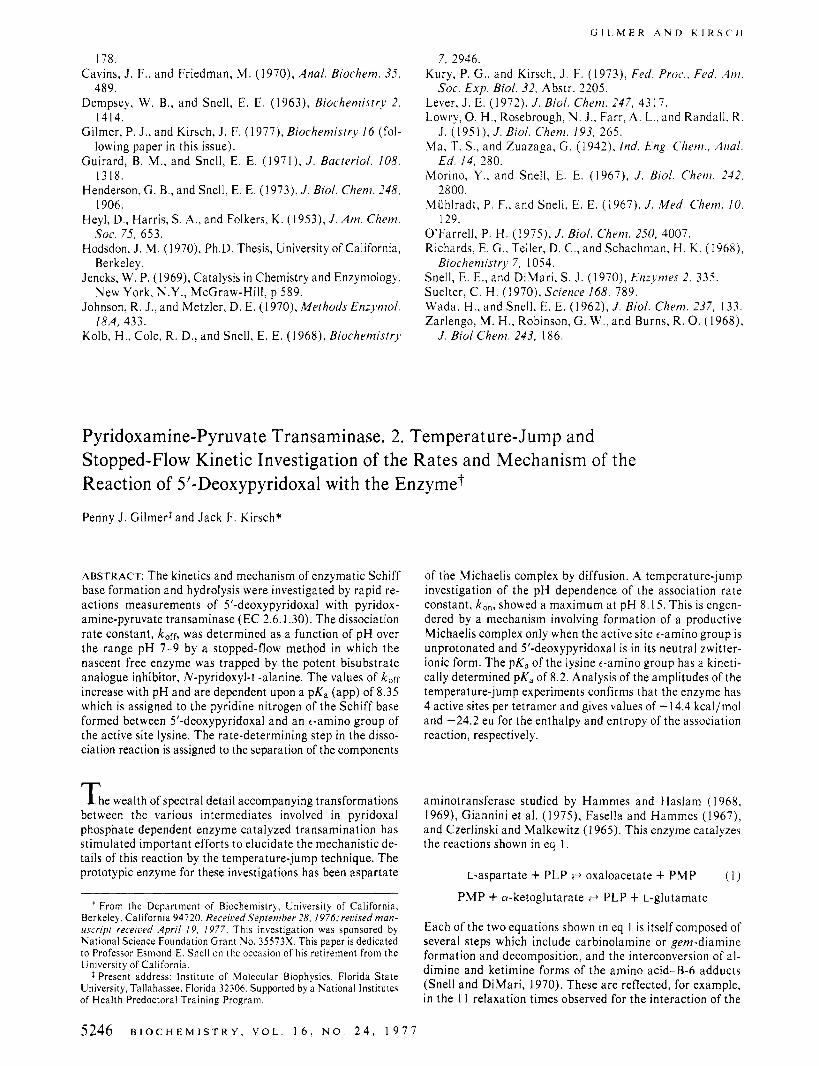
FIGURE I : Rate constants for dissociation of the PPT-5’-deoxy-PL complex, kofr, plotted as a function of pH as determined in stopped-flou experiments. Conditions: 0.05 M potassium pyrophosphate buffer, P = 0.5 at 25 OC. The curve is theoretical based on eq 1 I or 13 with the con- stants given in the text. (Insert) Spectrophotometric records of a typical experiment at 430 nm. (Conditions) Syringe A contained 5’-deoxy-PL ( 1 3 pM) and PPT (26 pM) and syringe B contained NPA (390 PM), pH 8.17. Each horizontal division corresponds to 200 ms and each vertical division to 3.4% of the total transmittance of the sample after reaction. The + Y direction corresponds to a decrease in absorbance.
dissociation rate constants are too slow to be accurately de- termined by the temperature-jump method under the acces- sible conditions; therefore they were measured by a stopped- flow method. In these experiments a mixture of PPT-5’- deoxy-PL complex in one syringe was mixed with N P A in the other in order to trap the newly released enzyme and thus prevent the reverse reaction (eq 4).
hc,.-:
h,,,, PPT-5’deoxy-PL z== 5‘deoxyPL + PPT ( 4 )
f.1st SP.4
PPT-NPA J
The conditions were designed so that virtually all of the 5’- deoxy-PL was bound to the active site of the enzyme before mixing; i.e., [PPT] 2 IOK,and [5’-deoxy-PL] = I/,[PPT]. The concentration of N P A was in large excess over that of total 5’-deoxy-PL so that the rate of combination of N P A with the free enzyme was fast relative to the rate of the reversible re- action of eq 4. The observed first-order rate constants for re- lease of 5’-deoxy-PL were unaffected by varying the concen- tration of NPA over a fourfold range confirming that the trapping was completely effective. The reactions were followed a t 430 nm at 25 O C . The plot of k,ffvs. pH shows that the rate constants decrease as the pH is lowered (Figure I ) .
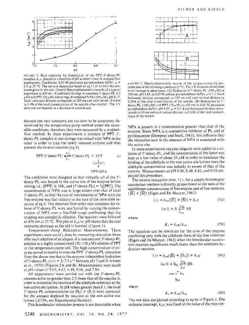
Temperature-Jump Relaxation Measurements. These experiments were usually done by measuring relaxation times after each addition of an aliquot of a concentrated 5’-deoxy-PL solution to a highly concentrated (50-1 20 pM) solution of PPT in the temperature-jump cell. The high concentration of en- zyme served to resolve in time the PPT-5’-deoxy-PL relaxation from the slower one due to the enzyme independent hydration of 5’-deoxy-PL ( 1 17 = 2-7.5 s-I between pH 7 and 9; Ahrens et al., 1970) (Figures 2A and B). Measurements were made a t pH values of 9.03, 8.82, 8.48, 8.06, and 7.84.
All experiments were carried out with the 5’-deoxy-PL concentration no greater than 2.5 times that of the enzyme in order to minimize the reaction of the aldehyde substrate a t the non-active site lysines. At p H values greater than 8.1, the total 5’-deoxy-PL concentrations (at [S,] > [E,] j were corrected for the amount depleted by reaction a t the non-active site lysines (I 15%, see Experimental Section).
This bimolecular relaxation process is not detectable when
F I G U R E 2: Spectrophotometric records of the temperature-jump per- turbations of the following systems at 23 “C. The + Y direction corresponds to an increase in absorbance. (A) Hydration of 5’-deoxy-PL (100 pM) at 390 nm, pH 8.85, in 0.05 M sodium pyrophosphate buffer, P = I . I . Each horizontal division corresponds to 100 ms and each vertical division to 0.28% of the total transmittance of the sample. ( B ) Interaction of 5’- deoxy-PL (100 PM) and PPT (70 pM) at 430 nm in 0.05 M potassium pyrophosphate buffer, pH 9 . 0 3 , ~ = 0,s . Each horizontal division corre- sponds to 10 ms and each vertical division to O.d3% of the total transmit- tance of the sample.
N P A is present in a concentration greater than that of the enzyme. Since N P A is a competitive inhibitor of PL and of pyridoxamine (Dempsey and Snell, 1963), this indicates that the relaxation seen in the absence of N P A is associated with the active site.
I n some experiments enzyme aliquots were added to a so- lution of 5’-deoxy-PL, and the concentration of the latter was kept a t a low value of about 35 p M in order to minimize the binding of the aldehyde to the non-active site lysines since the aldehyde concentration was initially in excess of that of the enzyme. Measurements a t pH 8.06, 8.48, 8.82, and 9.03 em- ployed this procedure.
The inverse relaxation time, 117, for a simple bimolecular association reaction is directly proportional to the sum of the equilibrium concentrations of free enzyme and of free substrate ([E] + [s]) (Eigen and De Maeyer, 1963):
(Sa) 117 = ken( [El + [SI 1 + koff
for E + s 3 ES LoIf
where
Ke = koff/kon (5b)
The equation can be rewritten for the case of the enzyme combining only with the aldehyde form of the free substrate (Eigen and De Maeyer, 1963) when the bimolecular associa- tion reaction equilibrates much faster than the substrate hy- dration reaction.
117 = k0,([E] + [SA]) + kerf
for E + sA 3 ES
(6a)
koff
>low i t K H
S H
where K , = koff/kon (6b)
The raw data are plotted according to eq 6a in Figure 3. The ordinate intercept, koff, was fixed at the value of the rate con-
5248 B I O C H E M I S T R Y , V O L . 1 6 , N O . 2 4 , 1 9 7 7

P Y R I D O X A M I N E - P Y R U V A T E T R A N S A M I N A S E K I N E T I C S
I [E + S&] pM
FLGURk3: Reciprocal relaxation time at 23 OC plotted as a function of (E) + (SA) as calculated from the following values of Kd and K H at each
8.06, 1.05, 0.196; 7.84, 0.71, 0.216. Calculations of [E,] allowed for 10-1 5% inactive protein. The buffers were 0.05 M potassium pyrophos- phate, p = 0.35-0.5.
pH [pH, K d ( p M ) , K H ] : 9.03, 5.8,0.130; 8.82,4.1,0.138; 8.47,2.1,0.160;
stant determined independently in the stopped-flow experi- ments. The values of ([E] + [SA]) were calculated from the independent determinations of Kd and K H (Gilmer et al., 1977) where Kd = [E] [ 5’-deoxy-PL] / [E-5’-deoxy-PL] and KH = [SH] / [SA]. A linear dependence of 1 /T vs. ([E] + [SA]) was observed a t pH values 8.48, 8.82, and 9.03. A leveling off of 1/7 at high values of ([E] + [SA]) was noted at pH 8.06. This might indicate the presence of an intermediate preceding the Schiff base. However, at the lowest pH value examined, 7.84, it was not possible to utilize high enough concentrations of ([E] + [SA]) to detect any possible nonlinearity in the plots because of the smaller amplitudes of the PPT-5’-deoxy-PL relaxation at the lower pH values.
The value of k,, at each pH value was determined from the slopes in Figure 3. A plot of k,, vs. pH indicates that this pa- rameter has a maximal value at pH 8.1 (Figure 4).
Temperature-Jump Amplitudes. There are two basic types of information that can be obtained from temperature-jump kinetic experiments. One is the relaxation time, 7, and the other is the amplitude of the relaxation, 6112.
The enthalpy of association of 5’-deoxy-PL with PPT can be obtained from a plot of the relaxation amplitude according to eq 7 (Guillain and Thusius, 1970)
(7) - ( ~ . ~ O A E ~ ~ A H I ~ ~ T )
R T2 r12 6112/10 =
where 6112/10 is the experimental relaxation amplitude, At12 = ( t s ~ - tsA), AH12 = H ~ B - H s A - HE, d T is the tempera- ture change, and r12, the amplitude factor. The definition of the amplitude factor depends on the mechanism (Winkler, 1969; Thusius, 1972). The amplitude factor for the mechanism shown in eq 6b is given in eq 8.
where Q = [E,] + [St] + (1 + K H ) ( K s ) , P = [Et] [S,], and K H = [sH]/[gA] = 0.13 at pH 9.03. A plot of 6112 (at constant IO) vs. r12 should be linear and pass through the origin. A linear dependence is observed in this plot when the enzyme concen- tration is corrected for 15% inactive protein (Figure 5).
From the slope of Figure 5 one can calculate from eq 7 that the AH12 for the reaction is -14.4 kcal/mol where the ex- perimentalslopeis(3.12f0.11) X 106mVM-’,Zo= 800mV, At12 = ~ S B - tsA = 5350 - 1600 = 3750, d T = 5.5 O C , T =
75 80 8 5 90 PH
FIGURE 4: Dependence of k,,, as determined by temperature-jump ki- netics, on pH. All experiments were performed in 0.05 M potassium py- rophosphate buffer, p = 0.35-0.5 a t 23 O C . The solid line represents the least-squares fit to eq 14 (see Discussion section).
2ol 1
I I
15-
N Y
* 10-
FIGURE 5: Determination of the enthalpy of the PPT-5’-deoxy-PL re- action from amplitudes of relaxation experiments a t pH 9.03, plotted according to eq 7. Conditions as in Figure 6. The value of K , at this pH is 3.5 p M and allowance was made for 15% inactive enzyme.
298 K. The value of AH12 together with that of K , leads to the values of AGlz and AS12 of -7.2 kcal/mol and -24.2 eu, re- spect ively .
The number of enzyme binding sites can be determined from the concentration dependence of the relaxation amplitude values in a titration experiment of enzyme with substrate. Under conditions where [Et] >> K, , the maximum amplitude for a bimolecular association reaction is found when [E,] = [S,] (Winkler, 1969).
Amplitude data from the temperature-jump titration ex- periment at pH 9.03 is plotted together with the theoretical curve calculated from eq 7 and 8, as 6Z12/[Et] (at constant IO) vs. log([St]/[Et]) (Winkler, 1969) where IO is the intensity of the transmitted light and [E,] is the total enzyme monomer concentration (Figure 6). I t was necessary to correct the en-
B I O C H E M I S T R Y , V O L . 1 6 , N O . 2 4 , 1 9 7 7 5249

G I L M E R A N D K I R S C H
1 I 2 3 4
[ S t l / [ E t l
F I G U R E 6: Amplitudes of temperature-jump relaxations for the 5'- deoxy-PL-PPT system at pH 9.03. Conditions: 0.05 M potassium pyro- phosphate, p = 0.5, 23 "C. Reactions were monitored at 430 nm with 10 constant at 800 mV.
zyme concentration for 15% inactive protein2 or else the curve would have had a maximum at [S,]/[EJ = 0.9 which is theo- retically impossible. With this correction the maximum am- plitude is found where PPT monomer concentration is nearly equal to that of 5'-deoxy-PL thus providing additional evidence for four binding sites per tetramer.
Discussion Spectral Properties and pK,'s of Schiff Bases. The proto-
tropic equilibria involving the Schiff bases formed from 5'- deoxy-PL and amines are shown in eq 9
P //N
R R C"H
€ 414~ 7500 E 4 2 0 ~ 5600 E ~ ~ ~ = 4900
Johnson and Metzler (1970) give p K s ~ + = 6.5 and P K ~ B = 1 1.7 for the aldimine formed from 5'-deoxy-PL and leucine.
The spectrum of the aldimine formed between PPT and 5'-deoxy-PL has a peak at 410-420 nm and exhibits no sig- nificant absorption at 340-360 nm from pH 7 to 9 (Gilmer et ai., 1977). This indicates by comparison with the models that there is little of the SB- species present.
Dissociation of 5'-Deoxy-PL f r o m the S'-Deoxy-PL-PPT Complex. The observation that P M and PL combine and dissociate from PPT with nearly the same rate constants a t pH 8.85 (Ayling and Snell, 1968a) suggests that Michaelis com- plex formation and decomposition is rate determining and that subsequent Schiff base formation from the aldehyde derivative contributes only about 700 cal/mol to the stability of the en- zyme-PL complex at this pH. These data are shown in a free energy vs. reaction coordinate diagram in Figure 7. The pH dependence for the rates of reaction of P M with this enzyme has been followed by the fluorescence temperature-jump technique (J. F. Kirsch and H. Winkler, unpublished results). It was observed that koff is pH independent between pH 7 and 9, while k,, decreases with increasing p H and is dependent
I SE'i 1 S E I ISE-1
This may in part be a result of an undetected complex formed from the enzyme and the hydrate form of 5'-deoxy-PL.
' 2 , 1 -1
-121 Reaction Coordinate
F I G U R E 7: Free energy vs. reaction coordinate ( I M standard state) di- agram for the reaction of PL and PM with pyridoxamine-pyruvate transaminase calculated from the steady-state data of Ayling and Snell (1968a) a t pH 8.85. The rate constants are calculated for the total of the ionic forms of each species represented. Assignments of the reacting ionic species are made in the text. The observed rate constants for the coi:ibi- nation of PL or PM with the enzyme ( k , " ) are nearly identical. An addi- tional 700 cal/mol of stability at this pH is provided to the E.PL complex by formation of a covalent Schiff base (E-PL). Covalent bond formation contributes more significantly to the stability at lower values of pH. The height of the latter kinetic barrier (dashed curve) is unknown, but must be smaller than that for the formation of the Michaelis complex. Thus the rate constant for the dissociation of PL or its analogue 5'-deoxy-PL from the enzyme is given by k,rr = k d d c q .
upon a pK, of 8.1 which is assigned to the pyridine nitrogen of PM. This implies that the only form of P M capable of combining with the enzyme is that in which the pyridine ni- trogen is protonated, and that the pK, of this group is raised to a value considerably greater than 9 in the enzyme-PM complex.
Assuming that PM, PL, and 5'-deoxy-PL form essentially the same noncovalent Michaelis complex, any differences in the pH dependence of the dissociation rate constants result from the pH dependence of the equilibrium constant, Keq, for the Schiff base formed from enzyme bound PL or 5'-deoxy- PL, since kerf = kdissKeq (Figure 7).
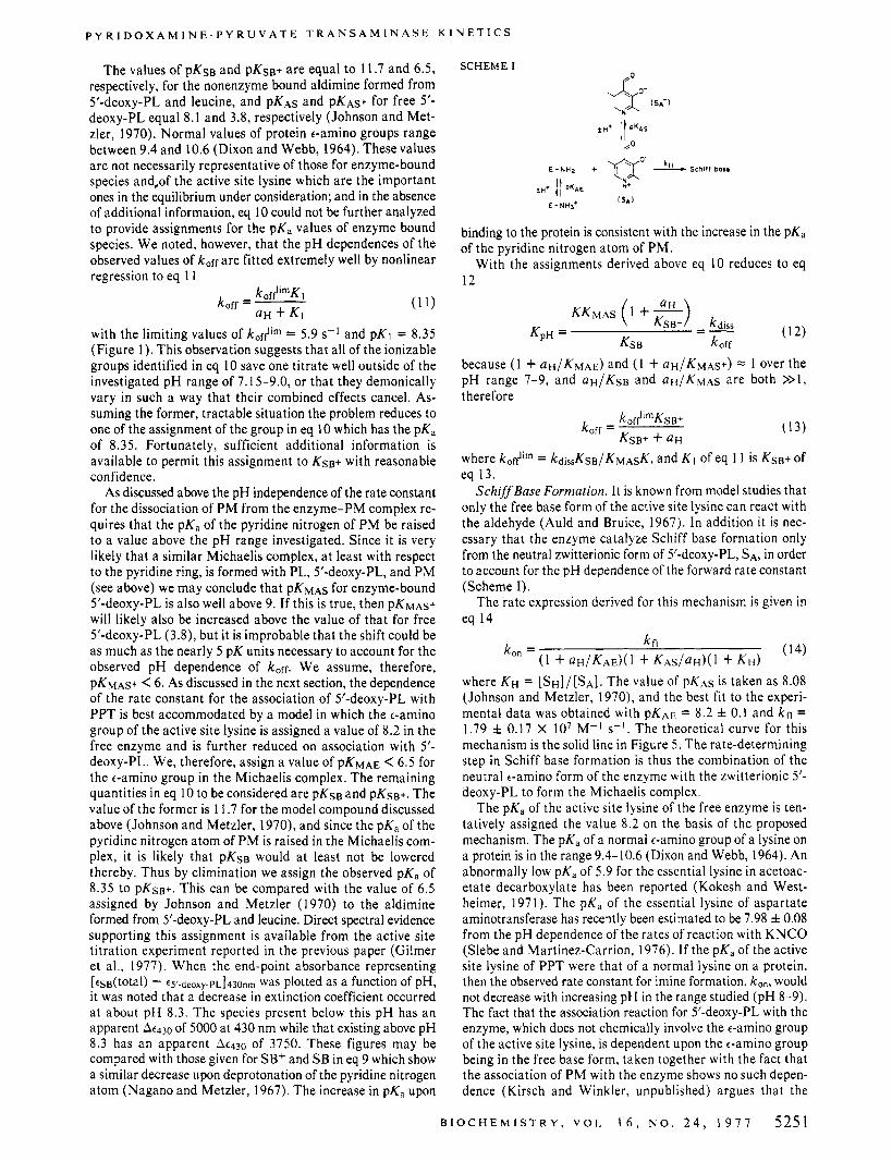
Auld and Bruice ( I 967) have published the results of a valuable study on the pH dependence for the formation of Schiff bases from HPA and alanine which provide a frame- work in which to analyze the equilibrium depicted in Figure 7. Chart I is adapted from their publication and the pH de- pendence of the association constant, K p ~ , is given in eq 10
KP" = Keq-'
CHART I
/ I K M A S
5250 B I O C H E M I S T R Y , V O L . 1 6 , N O . 2 4 , 1 9 7 7
P Y R I D O X A M I N E - P Y R U V A T E T R A N S A M I N A S E K I N E T I C S
The values of PKSB and p K s ~ + are equal to 1 1.7 and 6.5, respectively, for the nonenzyme bound aldimine formed from 5'-deoxy-PL and leucine, and P K A ~ and p K ~ s + for free 5'- deoxy-PL equal 8.1 and 3.8, respectively (Johnson and Met- zler, 1970). Normal values of protein e-amino groups range between 9.4 and 10.6 (Dixon and Webb, 1964). These values are not necessarily representative of those for enzyme-bound species and,of the active site lysine which a re the important ones in the equilibrium under consideration; and in the absence of additional information, eq 10 could not be further analyzed to provide assignments for the pK, values of enzyme bound species. We noted, however, that the p H dependences of the observed values of koff a re fitted extremely well by nonlinear regression to eq 1 1
with the limiting values of kof$im = 5.9 s-l and pK1 = 8.35 (Figure 1 ) . This observation suggests that all of the ionizable groups identified in eq 10 save one titrate well outside of the investigated p H range of 7.15-9.0, or that they demonically vary in such a way that their combined effects cancel. As- suming the former, tractable situation the problem reduces to one of the assignment of the group in eq 10 which has the pK, of 8.35. Fortunately, sufficient additional information is available to permit this assignment to KSB+ with reasonable confidence.
As discussed above the pH independence of the rate constant for the dissociation of P M from the enzyme-PM complex re- quires that the pK, of the pyridine nitrogen of P M be raised to a value above the p H range investigated. Since it is very likely that a similar Michaelis complex, a t least with respect to the pyridine ring, is formed with PL, 5'-deoxy-PL, and P M (see above) we may conclude that P K M A ~ for enzyme-bound 5'-deoxy-PL is also well above 9. If this is true, then PKMAS+ will likely also be increased above the value of that for free 5'-deoxy-PL (3.8), but it is improbable that the shift could be as much as the nearly 5 pK units necessary to account for the observed pH dependence of koff. W e assume, therefore, PKMAS+ < 6. As discussed in the next section, the dependence of the rate constant for the association of 5'-deoxy-PL with PPT is best accommodated by a model in which the t-amino group of the active site lysine is assigned a value of 8.2 in the free enzyme and is further reduced on association with 5'- deoxy-PL. We, therefore, assign a value of PKMAE < 6.5 for the t-amino group in the Michaelis complex. The remaining quantities in eq 10 to be considered are P K ~ B and p K s ~ + . The value of the former is 1 1.7 for the model compound discussed above (Johnson and Metzler, 1970), and since the pK, of the pyridine nitrogen atom of P M is raised in the Michaelis com- plex, it is likely that p K s ~ would a t least not be lowered thereby. Thus by elimination we assign the observed pK, of 8.35 to p K s ~ + . This can be compared with the value of 6.5 assigned by Johnson and Metzler (1970) to the aldimine formed from 5'-deoxy-PL and leucine. Direct spectral evidence supporting this assignment is available from the active site titration experiment reported in the previous paper (Gilmer et al., 1977). When the end-point absorbance representing [ ts~(total) - t5'-deoxy.pL]430nm was plotted as a function of pH, it was noted that a decrease in extinction coefficient occurred a t about pH 8.3. The species present below this p H has an apparent At430 of 5000 at 430 nm while that existing above pH 8.3 has an apparent At430 of 3750. These figures may be compared with those given for SB+ and SB in eq 9 which show a similar decrease upon deprotonation of the pyridine nitrogen atom (Nagano and Metzler, 1967). The increase in pK, upon
SCHEME I
binding to the protein is consistent with the increase in the PKa of the pyridine nitrogen atom of PM.
With the assignments derived above eq 10 reduces to eq 12
KKMAS (1 +"") (12)
because (1 + UH/KMAE) and ( 1 + U H / K M A ~ + ) = 1 over the pH range 7-9, and UH/KSB and UH/KMAS are both >>1, therefore
(13)
KSB+ kdiss =- KpH = KSB koff
koffllrnKSB+ koff = KSB+ QH
where kofflim = kdi,,KsB/KMAsK, and K I of eq 1 1 is KSB+ of eq 13.
SchiffBase Formation. It is known from model studies that only the free base form of the active site lysine can react with the aldehyde (Auld and Bruice, 1967). In addition it is nec- essary that the enzyme catalyze Schiff base formation only from the neutral zwitterionic form of 5'-deoxy-PL, SA, in order to account for the pH dependence of the forward rate constant (Scheme I).
The rate expression derived for this mechanism is given in eq 14
(14) kfl ( 1 + ~ H / K A E ) ( ~ + K A S / ~ H ) ( ~ + KH)
k," =
where KH = [SH]/[SA]. The value of PKAS is taken as 8.08 (Johnson and Metzler, 1970), and the best fit to the experi- mental data was obtained with PKAE = 8.2 f 0.1 and kfl = 1.79 f 0.17 X IO7 M-' S - I . The theoretical curve for this mechanism is the solid line in Figure 5 . The rate-determining step in Schiff base formation is thus the combination of the neutral t-amino form of the enzyme with the zwitterionic 5'- deoxy-PL to form the Michaelis complex.
The pK, of the active site lysine of the free enzyme is ten- tatively assigned the value 8.2 on the basis of the proposed mechanism. The pK, of a normal €-amino group of a lysine on a protein is in the range 9.4-10.6 (Dixon and Webb, 1964). An abnormally low pK, of 5.9 for the essential lysine in acetoac- etate decarboxylate has been reported (Kokesh and West- heimer, 1971). The pK, of the essential lysine of aspartate aminotransferase has recently been estimated to be 7.98 f 0.08 from the pH dependence of the rates of reaction with K N C O (Slebe and Martinez-Carrion, 1976). I f the pK, of the active site lysine of PPT were that of a normal lysine on a protein, then the observed rate constant for imine formation, ken, would not decrease with increasing pH in the range studied (pH 8-9). The fact that the association reaction for 5'-deoxy-PL with the enzyme, which does not chemically involve the €-amino group of the active site lysine, is dependent upon the t-amino group being in the free base form, taken together with the fact that the association of P M with the enzyme shows no such depen- dence (Kirsch and Winkler, unpublished) argues that the
B I O C H E M I S T R Y , V O L . 1 6 , N O . 2 4 , 1 9 7 7 5251
C i l L M E R A N D K I R S C H
TABLE I : Rate Constants for Combination of 5’-Deoxy-PL with PPT at DH 8.85.
Method Steady-state kinetic measurements (Ayling and 0.89 17
Stopped-flow kinetics (Ayling and Snell. 0.72 6.5
Temperature-jump and stopped-flow kinetics I .35 4.5
Snell, 1968b)a
1968b)“
(this studv)b
Sodium pyrophosphate buffers, = 0.5,25 ‘C. Potassium py- roohosohate buffers. u = 0.5. 23 “C.
e-amino group in the Michaelis complex formed from 5’- deoxy-PL is not in a mobile protonic equilibrium. If it were, k,, should not show a dependence on the lysine pK,. For this reason and for those discussed in the previous section, PKMAE is assigned a value <6.5.
We can compare the rate constants obtained in this study with those determined by Ayling and Snell (1968b) a t pH 8.85 (Table I ) . The agreement is substantial.
Comparison of Binding at the Active Site and the Nonactive Site. The fast relaxation due to imine formation a t the active site and the slow reaction of B-6 aldehydes a t the nonactive sites exhibit differing pH rate dependencies and optima for the dissociation constants. The affinity of the substrate for the active site is greatest near pH 7. This is due primarily to the decreased values of koff a t neutral pH (Figure 2). In contrast, binding of 5’-deoxy-PL a t nonactive sites shows little variation with pH (Gilmer et al., 1977).
There are two lines of evidence that indicate that the reac- tion of 5‘-deoxy-PL a t nonactive site lysines does not effect enzymatic activity: (1) Michaelis-Menten kinetics was ob- served i n the steady-state analysis of the enzyme when [5’- deoxy-PL] was as high as 100 pM (the range of concentrations where non-active site lysines are modified (Ayling and Snell, 1968b)); (2) the temperature-jump data indicate the same association and dissociation rate constants for the bimolecular reaction at the active site under conditions when only the active site is modified and when both the active and nonactive sites are modified by aldimine formation. These two types of kinetic behavior support the idea that the nonactive site reactions do not effect the reactivity a t the active site.
I n conclusion it has been demonstrated that the rate of formation of the Schiff base for 5’-deoxy-PL and PPT is con- trolled by an ionizable group of pK, of ca. 8.2 on the enzyme which is presumably the active site lysine and by the pK, of 8. I of the pyridine nitrogen atom of 5’-deoxy-PL. Any enzyme- substrate complexes preceding the aldimine must be relatively unpopulated because intermediates cannot be detected by the temperature-jump experiments except possibly at pH 8.06 (by the nonlinearity in Figure 3 at high values of ([E] + [SA]). The limiting rate constant of 1.79 X lo7 M-I s-l, although below the diffusion-controlled limit, is about equal to that observed for the reaction of P M with PPT (Kirsch and Winkler, un- published results) where no covalent interactions follow the formation of the Michaelis complex. This similarity of values of kfl for P M and 5’-deoxy-PL thus suggests that the rate- determining step in aldimine formation is formation of the ES complex, and from the law of microscopic reversibility it fol- lows that dissociation of PL or 5‘-deoxy-PL from the Michaelis complex is rate determining i n the reverse direction (Figure 7 ) .
ISS’i
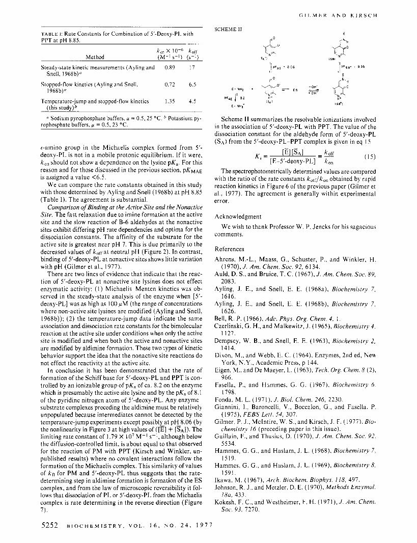
Scheme I 1 summarizes the resolvable ionizations involved in the association of 5’-deoxy-PL with PPT. The value of the dissociation constant for the aldehyde form of 5’-deoxy-PL (SA) from the 5’-deoxy-PL-PPT complex is given in eq 15
E l [ S A I =- kofr K, = [ E-j’-deoxy-PL] k,,
The spectrophotometrically determined values are compared with the ratio of the rate constants koff/kon obtained by rapid reaction kinetics in Figure 6 of the previous paper (Gilmer et a] . , 1977). The agreement is generally within experimental error.
Acknowledgment
comments. We wish to thank Professor W. P. Jencks for his sagacious
References Ahrens, M.-L., Maass, G., Schuster, P., and Winkler, H .
Auld, D. S., and Bruice, T. C. ( l967) , J . A m . Chem. SOC. 89,
Ayling, J . E., and Snell, E. E. (1968a), Biochemistr,v 7 ,
Ayling, J . E., and Snell, E. E. (1968b), Biochemistry 7 ,
Bell, R . P. (1966), Ado. Phys. Org. Chem. 4, 1 . Czerlinski, G. H., and Malkewitz, J. (1965), Biochemistry 4,
Dempsey, W. B., and Snell, E. E. ( 1 963), Biochemistry 2,
Dixon, M., and Webb, E. C. (1964), Enzymes, 2nd ed, Nen
Eigen, M., and De Maeyer, L. (1963), Tech. Org. Chem. 8 (2),
Fasella, P., and Hammes, G . G . (1967), Biochemistry 6 .
Fonda, M. L. ( 1 97 I ), J . Bioi. Chem. 246, 2230. Giannini, I . , Baroncelli, V., Boccalon, G. , and Fasella. P.
Gilmer, P. J . , McIntire, W . S., and Kirsch, J . F. (1977), Bio-
Guillain, F., and Thusius, D. ( l970) , J . A m . Chem. SOC. 92,
Hammes, G. G., and Haslam, J . L. (1968), Biochemistry 7 ,
Hammes, G . G., and Haslam, J . L. (1969), Biochemistry 8,
Ikawa, M. (1967), Arch. Biochem. Biophys. 118, 497. Johnson, R. J., and Metzler, D. E. (l970), Methods Enzyriiol
Kokesh, F. C. , and Westheimer, F. H. (1971), J . A m . Chem.
( 1970), J . Am. Chem. Soc. 92, 6 134.
2083.
1616.
1626.
1127.
1414.
York, N.Y., Academic Press, p 144.
966.
1798.
( 1 9 7 9 , FEBS Lett. 54, 307.
chemistry 16 (preceding paper in this issue).
5534.
1519.
1591.
18a. 433.
SOC. 93, 7270.
5252 B I O C H E M I S T R Y , V O L . 1 6 , N O . 2 4 , 1 9 1 7

A C T I V A T I O N O F H U M A N F A C T O R X
Kury, P. G., and Kirsch, J . F. (1973), Fed. Proc., Fed. Am.
Nagano, K., and Metzler, D. E. ( 1 967), J . Am. Chem. SOC. 89,
O’Leary, M. H., and Malik, J . M. ( l 9 7 l ) , J . Biol. Chem. 246,
Schaleger, L . L., and Long, F. A. (1963), Adc. Phys. Org.
SOC. Exp . Biol. 32, Abstr. 2205.
289 I .
544.
Chem. I , I .
Slebe, J . C., and Martinez-Carrion, M. (1976), J . Biol. Chem.
Snell, E. E. ( l970) , Vitam. Horm. ( N . Y.) 28, 265. Snell, E. E., and Di Mari, S. J . (1 970), Enzymes, 3rd. Ed. 2,
Thusius, D. (1972), J . Am. Chem. SOC. 94, 356. Winkler, R . (1969), Ph.D. Dissertation, Universitat Wien and
2.51, 5663.
335.
Gottingen.
Activation of Human Factor X (Stuart Factor) by a Protease from Russell’s Viper Venom?
Richard G . Di Scipio, Mark A. Hermodson,* and Earl W . Davie*
ABSTRACT: Human Factor X (Stuart factor) is a plasma protein (mol wt 59 000) that participates in the middle phase of blood coagulation. I t is composed of a heavy and a light chain held together by a disulfide bond(s). Factor X is readily converted to an enzyme, factor X,, by a protease from Russell’s viper venom. In this reaction, a specific arginyl-isoleucine bond is cleaved in the amino-terminal region of the heavy chain giving rise to an activation peptide and factor X,. This results in the formation of an Ile-Val-Gly-Gly-Gln-Glu-Cys-Lys- Asp-Gly-Glu-Cys-Pro-Thr-Gln-Ala-Leu- sequence in the heavy chain of the enzyme. No change was observed in the light chain of factor X during the activation reaction. The heavy
F a c t o r x (Stuart factor)’ is one of four known coagulation factors present in plasma that requires vitamin K for its bio- synthesis. During the coagulation process, factor X is converted to an enzyme, factor X,, which in turn converts prothrombin to thrombin [see Davie and Fujikawa (1975) and Suttie and Jackson (1977) for reviews].
In recent years, human factor X has been purified and characterized in several different laboratories (Aronson et al., 1969; Rosenberg et al., 1975; Kosow, 1976; Vician and Tish- koff, 1976; Di Scipio et al., 1977). It is a glycoprotein composed of a heavy and a light chain held together by a disulfide bond(s). In the preqent article, we describe the mechanism of activation of human factor X by a protease from Russell’s viper venom. In this reaction, factor X is converted to factor X, by the cleavage of a specific arginyl-isoleucine bond in the amino-terminal portion of the heavy chain of the precursor molecule. This mechanism of activation is essentially identical with that previously observed for bovine factor X when it is activated by (a) the protease from Russell’s viper venom, (b) trypsin, (c) factor IX, and factor VIII, or (d) factor VI1 and tissue factor (Fujikawa et al., 1972a, 1974, 1975; Jesty and Esnouf, 1973; Jesty and Nemerson, 1974).
~ ~~~
f From the Departments of Biochemistry and Medical Genetics, Uni- versity of Washington, Seattle, Washington 98 195. Receioed May 16, 1977. This work was supported in part by Grants H L 16919 and G M 15253 from the National Institutes of Health.
t Present address: Department of Biochemistry, Purdue University, West Lafayette, Indiana 47907.
I The nomenclature for the various coagulation factors is that recom- mended by an international nomenclature committee (Wright, 1959).
chain of human factor X, also contains the active site sequence of Phe-Cys-Ala-Gly-Tyr-Asp-Thr-Lys-Gin-Glu-Asp-Ala- Cys - Gln- Gly-Asp-SER-Gly- Gly- Pro- His- Val-Thr- Arg- Phe-. The amino-terminal and active site sequences of the heavy chain are homologous with the corresponding amino-terminal and active site sequences of bovine factor X, and a number of other plasma serine proteases. Human factor X, was rapidly inhibited by antithrombin 111 and formed a stable one-to-one molar complex with the inhibitor. These data indicate that the mechanism of activation and inhibition of human factor X are essentially identical with that previously described for bovine factor X.
Experimental Section Materials
Human factor X was purified to homogeneity by a slight modification of the method of Di Scipio et al. (1977). In the present experiments, a single 0-40% ammonium sulfate pre- cipitation was substituted for the sequential 0-10% and 10- 40% ammonium sulfate precipitation. This preparation does not contain two forms of factor X similar to bovine factor XI and factor X2 (Fujikawa et al., 1972b). The protease from Russell’s viper venom that activates factor X (RVV-X)2 was purified from crude Vipera russeli venom by the method of Schiffman et al. (1969) or Kisiel et al. (1976).
Barbital, urethane, imidazole (grade I), morpholino- ethanesulfonic acid (Mes), N-acetylneuraminic acid, Coom- assie brilliant blue R, galactosamine, dithiothreitol, aldolase, bovine serum albumin, bovine carbonic anhydrase, and myo- globin were obtainedfrom Sigma Chemical Co., St. Louis, Mo. Sodium sulfate, sodium arsenate, and 4-vinylpyridine were obtained from J . T . Baker Chemical Co., Phillipsburg, N.J . The 4-vinylpyridine was vacuum distilled before use. Periodic acid was a product of Frederick Smith Chemical Co., Co- lumbus, Ohio. 2-Thiobarbituric acid, 2-mercaptoethanol, N,N-methylenebisacrylamide, iodoacetic acid, and N,N,N,N’-tetraethylenediamine were purchased from, East- man Kodak Co., Rochester, N.Y. Agarose was a product of
Abbreviations used: RVV-X, protease from Russell’s viper venom that activates factor X; DFP, diisopropyl phosphorofluoridate; Tris, tris(hy- droxymethy1)aminomethane; Mes, morpholinoethanesulfonic acid.
B I O C H E M I S T R Y . V O L . 1 6 , N O . 2 4 , 1 9 1 1 5253