gag in immature human immunodeficiency virus type 1 …jvi.asm.org/content/74/20/9381.full.pdf ·...
TRANSCRIPT
JOURNAL OF VIROLOGY,0022-538X/00/$04.0010
Oct. 2000, p. 9381–9387 Vol. 74, No. 20
Copyright © 2000, American Society for Microbiology. All Rights Reserved.
Evidence for a Stable Interaction of gp41 with Pr55Gag in ImmatureHuman Immunodeficiency Virus Type 1 Particles
DONALD J. WYMA, ALEXANDER KOTOV, AND CHRISTOPHER AIKEN*
Department of Microbiology and Immunology, Vanderbilt University School of Medicine, Nashville, Tennessee 37232-2363
Received 6 April 2000/Accepted 14 July 2000
Assembly of infectious human immunodeficiency virus type 1 (HIV-1) virions requires incorporation of theviral envelope glycoproteins gp41 and gp120. Several lines of evidence have suggested that the cytoplasmic tailof the transmembrane glycoprotein, gp41, associates with Pr55Gag in infected cells to facilitate the incorpo-ration of HIV-1 envelope proteins into budding virions. However, direct evidence for an interaction betweengp41 and Pr55Gag in HIV-1 particles has not been reported. To determine whether gp41 is associated withPr55Gag in HIV-1 particles, viral cores were isolated from immature HIV-1 virions by sedimentation throughdetergent. The cores contained a major fraction of the gp41 that was present on untreated virions. Associationof gp41 with cores required the presence of the gp41 cytoplasmic tail. In HIV-1 particles containing a functionalprotease, a mutation that prevents cleavage of Pr55Gag at the matrix-capsid junction was sufficient for thedetergent-resistant association of gp41 with the isolated cores. In addition to gp41, a major fraction ofvirion-associated gp120 was also detected on immature HIV-1 cores. Isolation of cores under conditions knownto disrupt lipid rafts resulted in the removal of a raft-associated protein incorporated into virions but not theHIV-1 envelope proteins. These results provide biochemical evidence for a stable interaction between Pr55Gag
and the cytoplasmic tail of gp41 in immature HIV-1 particles. Moreover, findings in this study suggest that theinteraction of Pr55Gag with gp41 may regulate the function of the envelope proteins during HIV-1 maturation.
The replication cycle of human immunodeficiency virus type1 (HIV-1) culminates in the release of progeny virions from aninfected cell via budding from the plasma membrane. Duringvirion assembly, incorporation of viral envelope (Env) proteinsis essential for the formation of infectious particles. The HIV-1Env complex consists of the surface glycoprotein (SU), gp120,and the transmembrane glycoprotein (TM), gp41, which arenoncovalently associated. Fusion of HIV-1 particles with targetcells is initiated by binding of gp120 to CD4. Secondary en-gagement of a chemokine receptor results in conformationalchanges in gp120, triggering the gp41-mediated fusion of viraland cellular membranes. HIV-1 gp41, like other lentivirus TMproteins, contains an unusually long cytoplasmic tail consistingof 150 amino acids in contrast to the cytoplasmic tails of simpleretrovirus TM proteins, which are approximately 20 to 50amino acids in length (14). Although much has been learnedabout mechanism of HIV-1 fusion, the role of the gp41 cyto-plasmic tail in Env function remains enigmatic.
Several lines of evidence suggest that an interaction betweenthe gp41 cytoplasmic tail and the structural protein precursor,Pr55Gag, occurs during HIV-1 assembly. This possibility wasfirst implied by studies examining virion release from polarizedepithelial cells. Coexpression of Env and Pr55Gag results inbudding of HIV-1 particles exclusively from the basolateralsurface of polarized epithelial cells, while expression ofPr55Gag alone results in the release of particles from bothapical and basolateral sites (17, 23). Second, the matrix (MA)domain of Pr55Gag is required for incorporation of full-lengthEnv, as evidenced by the observations that deletions or pointmutations in MA inhibit the incorporation of full-length HIV-1Env proteins into budding virions (11, 12). These mutants were
rescued by truncating the cytoplasmic tail of gp41 or bypseudotyping virions with a heterologous retroviral Env con-taining a short cytoplasmic tail, suggesting that the MA domainof Pr55Gag is required for accommodating the long cytoplasmicdomain of gp41. A third line of evidence for a gp41-Pr55Gag
interaction is based on the observation that HIV-1 Env ex-pressed in cells undergoes rapid internalization from the cellsurface due to an endocytic motif present in the cytoplasmictail of gp41 (25). Coexpression of Pr55Gag dramatically reducesEnv internalization, suggesting that Pr55Gag binds the TM cy-toplasmic domain and prevents its interaction with the endo-cytic machinery (10). Fourth, a direct interaction between theMA region of Pr55Gag and a glutathione S-transferase fusionprotein containing the cytoplasmic tail of gp41 was observed invitro using recombinant HIV-1 proteins (7). Finally, in simianimmunodeficiency virus, a lentivirus closely related to HIV-1,expression of Env proteins engineered with an endoplasmicreticulum retention motif results in a dramatic decrease in theamount of viral particles released from cells (28). This findingsuggested that Env and Pr55Gag interact soon after translationbut before transport to the plasma membrane. Though theabove data imply an interaction between gp41 and Pr55Gag invirus-infected cells, a direct interaction between Env andPr55Gag in HIV-1 particles has yet to be demonstrated.
In this study, immature HIV-1 virions were treated withdetergent to remove the viral lipid membrane, and the result-ing immature cores were purified by equilibrium sedimentationcentrifugation. Through the isolation of immature HIV-1cores, we demonstrate a detergent-stable interaction of theHIV-1 Env proteins with Pr55Gag. These results provide bio-chemical evidence supporting an interaction between the gp41cytoplasmic tail and MA region of Pr55Gag during HIV-1 as-sembly.
MATERIALS AND METHODS
Cells and viruses. 293T cells (obtained from I. Verma) were cultured at 37°Cin 5% CO2 Dulbecco modified Eagle medium (DMEM; Cellgro) supplemented
* Corresponding author. Mailing address: Department of Microbi-ology and Immunology, Vanderbilt University School of Medicine,A-5301 Medical Center North, Nashville, TN 37232-2363. Phone:(615) 343-7037. Fax: (615) 343-7392. E-mail: [email protected].
9381
on June 22, 2018 by guesthttp://jvi.asm
.org/D
ownloaded from

with fetal bovine serum (10%), penicillin (50 IU/ml), and streptomycin (50mg/ml). Unless otherwise noted, the proviral DNA constructs used for the pro-duction of HIV-1 have been described previously (16, 31) and are as follows: R9,wild-type HIV-1; R9.PR2, protease-defective HIV-1 containing a triple alaninesubstitution in the protease active site; R9Tr712.PR2, protease-defective HIV-1containing a truncation of the N-terminal 144-amino-acid gp41; NLPI.nef1 (5),HIV-1 reporter virus expressing placental alkaline phosphatase (AP); R9.MA-CA, HIV-1 containing a mutation which blocks cleavage at the matrix-capsid(MA-CA) junction of Pr55Gag (described below). R9Tr712.PR2 was created byinserting the SalI-to-BamHI mutation from NLTr712 (31) into the R9.PR2
provirus.R9.MA-CA was created using PCR site-directed mutagenesis. PCR segment
overlap extension, as described previously (13), was used to change a Tyr residueat amino acid 132 of the MA gene to an Ile using the forward primer of59-CACTATAGGAATATTTTGGCTGAC. A silent mutation encoding an SspIrestriction enzyme site was engineered into the primer to aid in screening pos-itive clones. A PCR product containing the Tyr-to-Ile mutation was transferredinto the full-length proviral clone R9 using the restriction enzyme sites BssHIIand SpeI. Functional verification of the mutation was determined by immunoblotanalysis using capsid (CA)-specific antisera of the mutant virions produced fromtransfected 293T cells.
For production of viruses, 293T cells were transfected in 10-cm-diameterdishes with 20 mg of plasmid DNA as previously described (6). Virus-containingsupernatants were collected 48 to 60 h after transfection, clarified by low-speedcentrifugation to remove cellular debris, and passed through a 0.45-mm (pore-size) membrane filter.
Isolation of HIV-1 core structures. Immature cores were isolated using amodification of the “spin-thru” procedure as previously described for the puri-fication of HIV-2 cores (15). Filtered supernatants (60 ml) from transfected 293Tcells were concentrated by centrifugation (120,000 3 g, 3 h, 4°C) through 3 ml of20% (wt/vol) sucrose in STE buffer (10 mM Tris-HCl [pH 7.4], 100 mM NaCl, 1mM EDTA). The pellets were resuspended in a total volume of 0.4 ml of STEbuffer by gentle pipetting, followed by incubation for 4 h at 4°C. Linear densitygradients were prepared by mixing 30 and 70% sucrose in STE buffer in agradient maker and were cooled for 2 h at 4°C. The precooled gradients wereoverlaid with 15% sucrose in STE buffer (0.25 ml) containing 1% Triton X-100.This layer was covered with a barrier layer of STE (0.25 ml) containing 7.5%sucrose in order to prevent premature mixing of the concentrated virions withthe detergent. Concentrated HIV-1 particles were applied to the top of thebarrier layer, and the tubes were centrifuged in a Beckman SW-41 rotor(100,000 3 g, 16 h, 4°C). However, cores from NLPI.nef1 were isolated bytreatment of the concentrated virions with 1% Triton X-100 for 1 h at either 4 or37°C and then layering them directly onto a 30 to 70% linear sucrose gradient.Intact virions were purified in a similar manner but without exposure to deter-gent. Eleven 1-ml fractions were collected from the top of the gradient andassayed for CA protein and gp120 concentration by enzyme-linked immunosor-bent assays (ELISAs). The density of each fraction was determined by measure-ment of the refractive index. Refractive index measurements were converted todensity using the formula r 5 (2.6496ph) 2 2.5323, where r is the density ingrams/milliliter and h is the index of refraction at the measured temperature.
Protein analyses. Gradient fractions were diluted with STE buffer to reducethe density of the fraction and pelleted by ultracentrifugation (100,000 3 g, 20min, 4°C). The supernatants were removed, and the pellets were resuspended in13 sodium dodecyl sulfate (SDS) sample buffer for subsequent immunoblotanalysis. The samples were electrophoresed on a 4 to 20% Tris-HCl acrylamidegel and transferred to polyvinylidene difluoride (PVDF) membranes. Blots wereprobed for HIV-1 and cellular proteins using various antisera, including rabbitpolyclonal anti-CA (obtained from D. Trono), human monoclonal antiserumagainst gp41 (C2F5) and gp120 (C2G12) (obtained from the NIH AIDS Re-search and Reference Program), and rabbit polyclonal anti-cyclophilin A (ob-tained from L. Henderson). Protein bands were revealed by chemiluminescentdetection (SuperSignal; Pierce Chemical Co.) after probing the blots with ap-propriate peroxidase-conjugated secondary antibodies.
Concentrations of virus in the supernatants of transfected 293T cells and in thegradient fractions were determined by a CA-specific ELISA as described previ-ously (29). Concentrations of gp120 in the gradient fractions were also deter-mined by ELISA. For gp120 ELISA, a 96-well plate (Immulon II; Dynex) wascoated with 2 mg (0.1 ml) of the mouse monoclonal gp120 capture antibody(Advanced Bioscience Laboratories) per ml in phosphate-buffered saline (PBS)and incubated overnight at 37°C. The plate was rinsed twice with PBS, followedby the addition of 0.2 ml of PBS containing 5% donor calf serum and incubationat 37°C for 1 h to block the nonspecific binding of proteins. The plate was washedfour times with PBS containing 0.2% Tween 20. Samples were diluted in PBScontaining 10% donor calf serum and 0.5% Triton X-100. Samples (0.1 ml) wereadded, and the plate was incubated for 2 h at 37°C. After four washes, rabbitpolyclonal anti-gp120 primary antibody (1:5,000 dilution; Intracel Co.) wasadded, and the plate was incubated for 1 h at 37°C. After four washes, peroxi-dase-conjugated antibody to rabbit immunoglobulin (1:5,000 dilution; PierceChemical Co.) was added, and the mixture was incubated for 1 h at 37°C. Theplate was developed using TMB peroxidase substrate (Kirkegaard & Perry Lab-oratories), and the absorbance at 450 nm for each well was determined. Thegp120 concentration of each sample was determined by comparison to a standard
curve of recombinant gp120 from HIV-1LAV (obtained from the NIH AIDSResearch and Reagent Program).
Gradient fractions were assayed for the presence of human placental AP(PLAP) by both chemiluminescent and immunoblot analyses. Chemiluminescentactivity assays were performed by adding 10 ml of the unpelleted gradient frac-tion to 50 ml of AP-Substrate (ImmTech, Inc.) and measuring the relativeluminescence in a 96-well luminometer. Immunoblot analysis was performed onthe pelleted gradient fractions as mentioned above using rabbit antiserumagainst human PLAP (obtained from Fitzgerald International Industries, Inc.).
Phospholipid analysis. Radiolabeled immature virions were produced bytransfecting two 10-cm dishes of 293T cells with R9.PR2. Following the removalof the calcium phosphate precipitate, 1 mCi of [32P]orthophosphate was addedin phosphate-free DMEM (3 ml) supplemented with fetal bovine serum (10%,dialyzed against 150 mM NaCl), penicillin (50 IU/ml), and streptomycin (50mg/ml). Labeled virions were filtered, mixed with unlabeled virions, and concen-trated by ultracentrifugation. Concentrated virions were resuspended in PBS,and half of the sample was treated with 1% Triton X-100 at 37°C for 1 h to isolatecores. The remaining half of the virion sample was incubated in parallel in theabsence of detergent at 4°C for 1 h. To separate free lipids from HIV-1 cores, thesamples were pelleted through a 20% sucrose cushion by ultracentrifugation(100,000 3 g, 20 min, 4°C).
Pellets were resuspended in 100 ml of PBS and assayed for Pr55Gag content byp24 ELISA, and lipids were extracted according to the method of Bligh and Dyer(3). The samples were subsequently analyzed by thin-layer chromatography onsilica plates with chloroform-methanol-water (60:35:8 [vol/vol/vol]) as a solvent.Radiolabeled 32P-containing lipids were visualized by autoradiography.
RESULTS
Isolation of immature HIV-1 cores. We and others havereported the isolation and characterization of mature HIV-1cores (16, 30). These particles contained normal quantities ofCA, reverse transcriptase, and integrase but lacked significantquantities of the viral Env proteins. To further examine theinteraction of the HIV-1 Env proteins with HIV-1 particles, wehave isolated immature cores by a similar procedure. Imma-ture HIV-1 particles were harvested from 293T cells trans-fected with an HIV-1 molecular clone encoding an inactiveprotease and were concentrated by ultracentrifugation througha 20% sucrose cushion. Concentrated virions were then lay-ered onto a linear 30 to 70% sucrose gradient containing alayer of 1% Triton X-100 at the top of the gradient. Uponcentrifugation, the immature virions pass through the layer ofdetergent and sediment to their equilibrium density in thesucrose gradient. As a control, intact immature virions wereisolated on a similar 30 to 70% sucrose gradient lacking adetergent layer. One-milliliter fractions were collected fromthe top of gradient, and the Pr55Gag concentration for eachfraction was determined by HIV-1 p24 ELISA. The particles ineach fraction were pelleted in a microultracentrifuge and as-sayed for protein composition by immunoblotting.
Analysis of the gradient fractions revealed that the peak ofPr55Gag for intact virions (Fig. 1A) was located in fraction 5 ofthe gradient, corresponding to a density of 1.16 g/ml, which istypical for retroviral particles. In contrast, the peak of Pr55Gag
for the immature cores (Fig. 1B) was detected in fraction 7 ofthe gradient, corresponding to a density of 1.24 g/ml, which issimilar to that observed for mature HIV-1 cores (16). Theprofiles demonstrate that a 30 to 70% sucrose allowed virionsand cores to be distinguished using physical criteria. Further-more, the sharp increase in density of the isolated cores indi-cated that most, if not all, of the lipid was removed from theviral particles during exposure to detergent. Unlike maturecores, which are recovered at approximately 15% of the inputvirion associated CA (reference 16 and data not shown), theimmature cores proved to be remarkably stable, allowing re-covery of approximately 80% of the input virus as determinedby an ELISA for CA protein.
Detergent-stable association of gp41 with the immaturecore. To test for the association of the HIV-1 Env proteins withimmature HIV-1 cores, Pr55Gag-containing fractions from the
9382 WYMA ET AL. J. VIROL.
on June 22, 2018 by guesthttp://jvi.asm
.org/D
ownloaded from

gradients in Fig. 1 were analyzed by pelleting the particles andimmunoblotting using gp41- and CA-specific antisera. A ma-jority of the gp41 molecules remained associated with the im-mature cores following exposure to 1% Triton X-100 com-pared to intact virions isolated in a similar manner (Fig. 2A,compare lanes 2 and 7). To determine whether brief exposureto detergent was sufficient to disrupt the lipid bilayer of thevirus, immature virions and cores were tested for the presenceof the cellular protein cyclophilin A (CypA). CypA is specifi-cally incorporated into HIV-1 virions but is removed uponisolation of mature HIV-1 cores (30). Reprobing the blot withantiserum specific for CypA revealed a significant depletion ofCypA in immature HIV-1 cores relative to intact virions (Fig.2A, compare lanes 2 and 7). These results, together with theincrease in density of the immature HIV-1 cores, demonstratethat exposure to 1% Triton X-100 effectively disrupts the virallipid envelope and that gp41 remains associated with the im-mature HIV-1 core.
Detergent-stable association of gp120 with the HIV-1 imma-ture core. Assays of spontaneous and CD4-induced gp120shedding from mature HIV-1 particles and Env-expressingcells have shown that the association of gp41 with gp120 isrelatively unstable on laboratory-adapted strains of HIV-1 (18,19). To determine whether gp120 remains associated with gp41on immature cores, these particles were analyzed for gp120content by immunoblot using a gp120-specific antiserum (Fig.2A). Remarkably, a large fraction of the gp120 present onuntreated virions remained associated with the immaturecores. Quantitation of the gp120 associated with the immaturecores was performed by ELISA of gradient fractions from theimmature virions and cores (Fig. 2B and C). Immature HIV-1cores retained approximately 60% of the gp120 present onintact virions (compare fraction 5 in Fig. 2B and fraction 7 inFig. 2C) after normalizing for Pr55Gag content. These resultsdemonstrate that the interaction of gp41 and gp120 on imma-ture virions is highly resistant to exposure to 1% Triton X-100.
Association of HIV-1 Env proteins with immature cores isdependent on the presence of the gp41 cytoplasmic tail. Pre-vious studies have provided strong indirect evidence for aninteraction between the gp41 cytoplasmic tail and Pr55Gag inHIV-1-infected cells. However, this interaction has not beenobserved in HIV-1 particles. To determine whether the gp41cytoplasmic tail is required for the association of Env withimmature HIV-1 cores, we isolated cores from immature viri-ons containing a truncated form of gp41 lacking all but 6 of the150 amino acids of the gp41 cytoplasmic tail. This mutant hasbeen previously shown to incorporate high levels of Env intovirions, probably due to the high level of expression of the Envproteins on the cell surface (31). Virions were produced bytransfection of the PR-defective HIV-1 clone R9Tr712.PR2
encoding the truncated Env protein. As previously observedfor immature HIV-1 particles expressing wild-type Env, thepeak of Pr55Gag for the intact virions was found in fraction 5 of
FIG. 1. Isolation of immature HIV-1 core structures. Immature HIV-1 par-ticles were harvested from 293T cells transiently transfected with the R9.PR2
proviral plasmid and were filtered to remove cellular debris. Concentrated viri-ons were sedimented through a layer of 1% Triton X-100 into a linear 30 to 70%sucrose gradient. Fractions (1 ml) were collected from the top of the gradientand assayed for CA concentration (p24) by ELISA and for density by refrac-tometry. (A) Virions subjected to ultracentrifugation on a control gradient lack-ing detergent. (B) Detergent-treated virions.
FIG. 2. Immunoblot and ELISA analysis of immature HIV-1 virions andcores. (A) The peak Pr55Gag-containing fractions of virions (lanes 1 to 3) andcores (lanes 4 to 6) were diluted with STE buffer, and particles were pelleted byultracentrifugation. Pellets were dissolved in 13 SDS loading dye, subjected toelectrophoresis on a 4 to 20% acrylamide gel, transferred to PVDF membrane,and probed with the indicated antisera. Protein bands were revealed by chemi-luminescent detection after probing with the appropriate HRP-conjugated sec-ondary antiserum. For comparative purposes, twofold serial dilutions of the peakcores fraction (lanes 7 to 9) were analyzed on the same gel. Molecular massmarkers are shown in kilodaltons on the left of each panel. (B and C) Quanti-tation of gp120 associated with immature HIV-1 virions and cores by ELISA.Gradient fractions of HIV-1 immature virions and cores were analyzed for thepresence of gp120 and Pr55Gag by ELISA. The gp120 concentration for eachsample was determined using a standard curve of recombinant HIV-1LAV gp120.(B) Virions subjected to ultracentrifugation on a control gradient lacking deter-gent. (C) Detergent-treated virions.
VOL. 74, 2000 Pr55Gag-gp41 INTERACTION IN HIV-1 PARTICLES 9383
on June 22, 2018 by guesthttp://jvi.asm
.org/D
ownloaded from

the gradient, corresponding to a density of approximately 1.16g/ml (data not shown). The peak of Pr55Gag for the Env mutantimmature cores was found in fraction 7 of the gradient, corre-sponding to a density of approximately 1.24 g/ml (data notshown). The yield of immature cores lacking the cytoplasmictail of gp41 was identical to that of the immature cores con-taining the full-length form of gp41, indicating that the cyto-plasmic tail of gp41 does not influence the stability of theHIV-1 particles (data not shown). The peak Pr55Gag-contain-ing fractions from the virions and cores were concentrated,normalized for Pr55Gag content by CA-specific ELISA, andassayed for gp41 and gp120 by immunoblotting (Fig. 3). Incontrast to immature HIV-1 particles containing full-lengthEnv, detergent treatment of immature R9Tr712.PR2 virionsremoved essentially all of the gp41 and gp120 from immaturevirions lacking the gp41 cytoplasmic tail (Fig. 3, lane 3). Asmall amount of unprocessed Env protein (gp160) remainedassociated with the cores, a result that was occasionally ob-served in repeated experiments. Reprobing the blot with an-ti-CA antiserum confirmed that similar quantities of Pr55Gag
were analyzed, and probing with anti-CypA antiserum verifiedthat the viral lipid envelope was disrupted by exposure to 1%Triton X-100 (Fig. 3A). These results demonstrate that thedetergent-resistant association of the HIV-1 Env proteins withimmature HIV-1 particles requires the gp41 cytoplasmic tail.
Inhibition of cleavage at the MA-CA junction allows reten-tion of the HIV-1 Env proteins on a PR-competent core. Mu-tations in the MA region of Pr55Gag have been reported toblock the incorporation of Env into nascent virions, suggestingthat the MA region of Pr55Gag associates with gp41 duringvirion assembly (12). Structural studies of MA suggest thatupon cleavage from Pr55Gag, the free MA protein assumes adifferent conformation than when part of the full-lengthPr55Gag (27). Based on these observations, we hypothesizedthat inhibiting cleavage at the MA-CA junction, in the contextof an active PR, would be sufficient for retention of gp41 by theisolated cores. To test this hypothesis, we created an MA-CAcleavage site mutant by mutating the P1 position of theMA-CA junction, converting the tyrosine (Tyr) to an isoleu-cine (Ile) codon. This strategy was based on reports that sub-
stitution of residues containing b-branched side chains in theP1 position of a cleavage site has been shown to block cleavageby HIV-1 PR (24).
Intact virions and cores from the MA-CA mutant were iso-lated, and the peak Pr55Gag-containing fractions (virion frac-tions 4-6 and core fractions 6-8) were analyzed for the presenceof Env proteins (Fig. 4A) by immunoblotting. As observed withthe immature cores, a large fraction of the gp41 and gp120present on virions remained associated with the MA-CA cores(Fig. 4A, compare lanes 2 and 7, which contain similaramounts of Gag protein). Reprobing the blot with a CA-spe-cific antiserum verified that cleavage at the MA-CA junction ofPr55Gag was blocked in the mutant virus. The low levels ofCypA detected in the cores fractions confirmed that the virallipid envelope was disrupted by treatment with 1% TritonX-100. The amount of gp120 in the gradient fractions ofMA-CA virions and cores was measured by a gp120-specificELISA (Fig. 4B and C). The peak Pr55Gag-containing fractionof the MA-CA cores (Fig. 4C, fraction 7) retains approximately45% of the gp120 protein that is present on the intact MA-CAvirions (Fig. 4B, fraction 5). These results demonstrate thatblocking cleavage at the MA-CA junction is sufficient for theretention of both HIV-1 Env proteins on the isolated corestructures.
FIG. 3. Immunoblot analysis of immature HIV-1 cores and virions lackingthe cytoplasmic tail of gp41. The peak Pr55Gag-containing fraction of cores (C,lanes 1 and 3) and virions (V, lanes 2 and 4), containing either a full-length formof gp41 (WT) or a truncated form of gp41 lacking the cytoplasmic tail (Tr712),were diluted with STE buffer and pelleted by ultracentrifugation. Pellets weredissolved in 13 SDS loading buffer, subjected to electrophoresis on a 4 to 20%acrylamide gel, and transferred to PVDF membrane, and the blot was probedwith the indicated antisera. Protein bands were revealed by chemiluminescentdetection after incubation of the blot with appropriate HRP-conjugated second-ary antisera. Molecular mass markers are shown in kilodaltons on the left of eachpanel.
FIG. 4. Immunoblot and ELISA analysis of HIV-1 virions and cores inhib-ited for cleavage at the MA-CA junction of Pr55Gag. (A) The peak MA-CA-containing fractions of virions (lanes 1 to 3) and cores (lanes 4 to 6) wereanalyzed by immunoblotting as described in the legend to Fig. 2. (B and C) Thegp120 concentrations of the gradient fractions were determined as described inthe legend to Fig. 2. (B) Analysis of virions. (C) Analysis of cores.
9384 WYMA ET AL. J. VIROL.
on June 22, 2018 by guesthttp://jvi.asm
.org/D
ownloaded from

The detergent-stable association of gp41 with Pr55Gag isindependent of lipid rafts. Recently, it was reported thatHIV-1 assembly occurs in cholesterol-rich membrane microdo-mains known as lipid rafts (22). The lipid composition ofHIV-1 particles is consistent with the presence of lipid rafts (1,2), suggesting that HIV-1 contains raft microdomains withinthe lipid envelope on the virion. A characteristic property oflipid rafts is their insolubility in 1% Triton X-100 at 4°C (26),conditions similar to those used in the isolation of cores. Wetherefore tested the possibility that the HIV-1 Env proteinswere present on immature cores due to the presence of deter-gent-insoluble lipid rafts. For this purpose, we utilized a re-porter virus (NLPI.nef1) that encodes PLAP, a raft-associatedprotein made in the presence of an HIV-1 PR inhibitor toprevent Pr55Gag cleavage and thus maintain the immaturevirion phenotype. PLAP is a glycophosphatidylinositol-an-chored protein that localizes to lipid rafts on cells and is usedas a marker to identify lipid rafts (4). Immature HIV-1 parti-cles were produced transfecting 293T cells with plasmid DNAand culturing in the presence of an HIV-1 PR inhibitor. ThePLAP protein is incorporated into budding virions (data notshown) and was therefore used as a marker for the presence ofrafts on the immature cores. Immature cores were isolated byincubating concentrated virions with 1% Triton X-100 for 1 hat either 4°C, conditions known to preserve lipid rafts, or at37°C, which disrupts lipid rafts (4). The particles were thenpurified by sedimentation centrifugation. Gradient fractionsfrom virions and cores were tested for the presence of PLAPusing a quantitative chemiluminescent activity assay for AP(Fig. 5). Virions treated with 1% Triton X-100 at 4°C exhibiteda peak of PLAP activity that cosedimented with a peak ofPr55Gag in the immature cores (Fig. 5A, fraction 7). In con-trast, cores isolated by treatment of virions with 1% TritonX-100 at 37°C lacked detectable levels of AP activity in thecores (Fig. 5B, fraction 8). It is noteworthy that virions treatedat 37°C sedimented at a slightly higher density, 1.26 g/ml (datanot shown), suggesting that removal of the lipid rafts from thevirions altered the density of the isolated cores. To determinewhether the HIV-1 Env proteins were associated with imma-ture cores independently of the presence of lipid rafts, the peakPr55Gag-containing gradient fraction from each gradient waspelleted and assayed for the presence of Env proteins by im-munoblotting (Fig. 5C). Similar levels of both gp41 and gp120were present on virions following detergent treatment at either4 or 37°C. Reprobing the blot with a PLAP-specific antiserumverified that treatment with detergent at 37°C was sufficient toremove PLAP from the cores. These results indicate that thestable association of the HIV-1 Env proteins with immaturecores is not due to the presence of lipid rafts. Furthermore,these data indicate that the interactions between gp41 andPr55Gag and between gp41 and gp120 are remarkably stable,persisting after treatment with 1% Triton X-100 for 1 h at37°C.
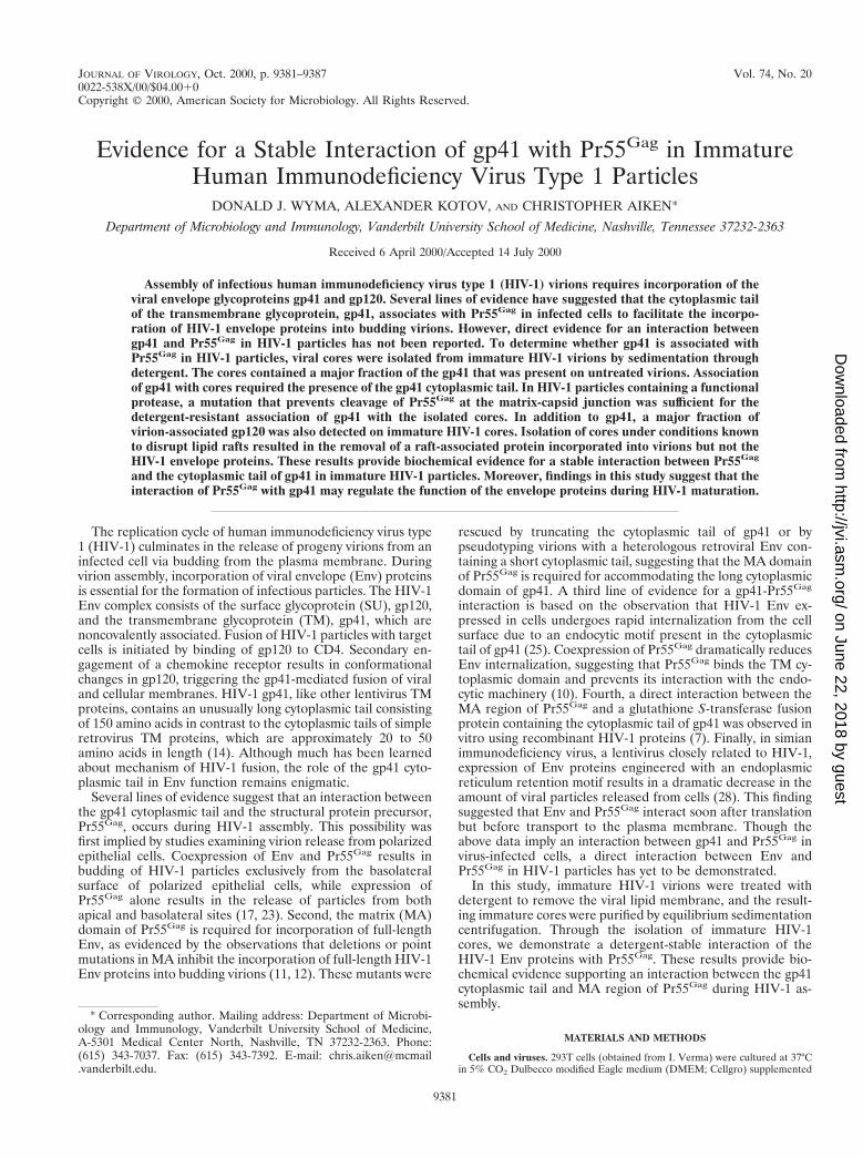
Phospholipid analysis of immature HIV-1 virions and cores.Our results suggested that a direct interaction between thecytoplasmic tail of gp41 and Pr55Gag was responsible for thestable association of Env with immature cores. However, thisconclusion was based on the assumption that the core isolationprocedure efficiently removed the viral lipid envelope. To de-termine the efficiency of lipid removal during Triton X-100treatment of immature HIV-1 particles, we labeled the phos-pholipids of virions by culturing producer cells with[32P]orthophosphate. The 32P-labeled immature virions weretreated with 1% Triton X-100 for 1 h at 37°C, and the resultingcores were pelleted by ultracentrifugation. Lipids were ex-tracted from 32P-labeled cores and control virions and were
analyzed by thin-layer chromatography and visualized by au-toradiography (Fig. 6). Analysis of lipids extracted from similarquantities of intact virions (Fig. 6, lane 1, 30 mg of p24) andcores (Fig. 6, lane 3, 26 mg of p24) demonstrated that treat-ment with 1% Triton X-100 at 37°C for 1 h efficiently removeda majority of the phospholipids from the virions. Comparisonof the cores sample to a 1:50 dilution of lipids extracted fromintact virions (Fig. 6, lane 2) revealed that ,2% of the ana-lyzed phospholipids remained on the isolated cores. We con-clude that the procedure used to isolate immature HIV-1 coresresulted in efficient removal of phospholipids.
DISCUSSION
In this study, we isolated immature cores from PR-defectivevirions with the goal of dissecting the putative interaction ofthe HIV-1 TM protein with Pr55Gag. The inherent stability ofthese immature particles allowed us to recover greater than80% of the initial virus. Based on this result, we conclude thatbiochemical observations made using these particles are likelyto be representative of the whole virion population and not a
FIG. 5. Association of HIV-1 Env proteins on immature HIV-1 cores inde-pendently of the presence of lipid rafts. Immature HIV-1 cores were isolatedfrom a PLAP-encoding virus by incubation for 1 h in detergent at either 4 or37°C, followed by sedimentation centrifugation. Fractions were examined for thepresence of PLAP and HIV-1 Env proteins. (A and B) Levels of PLAP weredetermined by incubation of gradient fractions with an AP substrate and quan-titated on a luminometer as relative units (RU) of luminescence. A CA-specificELISA was used to determine the amount of virus in each gradient fraction. (C)Immunoblot analysis of the peak Pr55Gag-containing gradient fractions fromimmature cores treated with detergent at either 4 or 37°C (lanes 1 and 4,respectively) was performed as described in the legend to Fig. 2. Threefold serialdilutions of the peak Pr55Gag-containing fractions were also analyzed (lanes 2and 3 and lanes 5 and 6).
VOL. 74, 2000 Pr55Gag-gp41 INTERACTION IN HIV-1 PARTICLES 9385
on June 22, 2018 by guesthttp://jvi.asm
.org/D
ownloaded from
consequence of the presence of a minor hyperstable subset ofvirions.
We and others have shown that the HIV-1 Env proteins areefficiently removed from mature HIV-1 particles when coresare isolated by exposure of virions to 1% Triton X-100 (16, 30).In contrast, immature cores isolated by an identical procedurecontained high levels of both gp41 and gp120. HIV-1 assemblywas recently reported to take place in lipid rafts (22), which aremembrane microdomains enriched in cholesterol, glycolipids,and some proteins (26). The isolation conditions we used toisolate HIV-1 cores are known to preserve lipid rafts, andimmature HIV-1 cores also contained a known raft-associatedprotein, PLAP. However, detergent treatment of virions underconditions known to disrupt lipid rafts resulted in the efficientremoval of PLAP but not of the HIV-1 Env proteins. Weconclude that immature HIV-1 virions incorporate lipid raftsduring assembly. Our results further demonstrate that the sta-ble association of HIV-1 Env proteins with immature coresdoes not require the presence of lipid rafts. The interactionsmediating this association are remarkably stable, persistingduring a 1-h exposure to 1% Triton X-100 at 37°C. The obser-vation that gp41 exhibits a detergent-stable association withimmature cores extends previously reported genetic data andrepresents the first biochemical evidence for an interactionbetween gp41 and Pr55Gag in HIV-1 particles.
The precise function of the 150-residue gp41 cytoplasmic tailin HIV-1 replication is not known. In some cell lines, such asMT-4, HIV-1 lacking the gp41 cytoplasmic domain replicatesefficiently, while in others, including peripheral blood mono-
nuclear cells, the TM cytoplasmic tail is required for Env in-corporation (21). In our experiments, HIV-1 particles wereproduced in 293T cells, a cell type that is permissive for virionincorporation of tail-less HIV-1 Env proteins. Our finding, thatthe detergent-stable interaction of gp41 with immature HIV-1particles requires the gp41 cytoplasmic domain, suggests thatthe gp41 tail mediates the strong association of Env proteinswith Pr55Gag in immature HIV-1 particles. We conclude thatthis interaction is probably essential for incorporation of Envin cell types in which the gp41 tail is required for HIV-1replication.
Although removal of the TM cytoplasmic tail demonstratedthat this domain is necessary for the interaction of gp41 withimmature cores, the region of Pr55Gag required for gp41 bind-ing remains to be dissected. Although other retroviruses can-not be pseudotyped by full-length HIV-1 proteins, the MAdomain of HIV-1 Gag is sufficient for the incorporation offull-length HIV-1 Env in particles produced from chimericHIV-1–visna virus Gag proteins (8). Our demonstration thatcores isolated from PR-competent HIV-1 particles containinguncleaved MA-CA precursor retained large quantities of gp41supports the hypothesis that the MA domain of Pr55Gag, whenpart of a larger Gag precursor, binds the gp41 cytoplasmicdomain. It remains to be determined whether other regions ofGag also participate in this interaction.
Although our findings do not formally exclude the possibilityof a third protein mediating the interaction between gp41 andPr55Gag, published genetic data argue against this (20). Such abridging protein must be present at high concentrations withinthe virion, must lose the ability to bridge gp41 and Pr55Gag
when the gp41 tail is mutated, and must regain this ability whencompensatory mutations are introduced in MA. Likewise, ourdata do not completely exclude the possibility of a requirementfor a lipid moiety as being required for the gp41-Pr55Gag as-sociation. However, our lipid analysis demonstrated that morethan 98% of the phospholipid was removed from immaturecores, while approximately half of the Env molecules remainedassociated with immature cores. With these caveats, the moststraightforward interpretation of our data is that the gp41cytoplasmic tail binds directly to the MA region of Pr55Gag inimmature HIV-1 particles.
A surprising outcome of this study was the finding that, inaddition to gp41, gp120 was stably associated with the imma-ture cores. Previous studies employing measurements of gp120shedding from mature HIV-1 particles concluded that thegp120-gp41 interaction is unstable in laboratory-adaptedstrains of HIV-1 (18, 19). We observed that approximately50% of the gp120 remained associated with immature HIV-1cores. This result suggests that the binding of the gp41 cyto-plasmic tail to Pr55Gag stabilizes the gp120-gp41 interaction.We hypothesize that the interaction of Pr55Gag with the cyto-plasmic tail of gp41 regulates the conformation of the Envprotein complex on the immature virion surface, thereby pre-venting entry until the virion has matured. Further studies willbe required to determine whether gp41 binding to Pr55Gag
regulates fusion of HIV-1 particles.
ACKNOWLEDGMENTS
We thank B. Chen for the plasmid pNLPI, V. Bosch for pNLTr712,and D. Trono and L. Henderson for antibodies. We also thank A. D.De Silva and S. Joyce for expert assistance with the phospholipidanalysis. The following reagents were obtained through the NIH AIDSResearch and Reference Reagent Program, Division of AIDS, NIAID,NIH: HIV-1 monoclonal antibodies against gp41 (C2F5) and gp120(C2G12) from Hermann Katinger.
This study was supported by NIH grant AI47056 and a Discovery
FIG. 6. Analysis of phospholipids present immature HIV-1 virions and cores.Immature HIV-1 particles were harvested from 32P-labeled 293T cells transientlytransfected with the R9.PR2 proviral plasmid. The virions were concentrated byultracentrifugation and then treated with 1% Triton X-100 for 1 h at 37°C. Theremaining core structures were separated from free proteins and lipids by pel-leting them through a 20% sucrose cushion. Lipids from virions (V) and cores(C) were extracted by chloroform-methanol (2:1) extraction, analyzed by thin-layer chromatography, and visualized by autoradiography. Lipids were extractedfrom similar quantities of immature virions (lane 1, 30 mg of p24) and immaturecores (lane 3, 26 mg of p24) and analyzed. A 1:50 dilution of the intact virions(lane 2) was analyzed for quantitative comparison. Phosphatidylethanolamine(PE), phosphatidylinositol (PI), and phosphatidylcholine (PC) standards wereanalyzed in parallel.
9386 WYMA ET AL. J. VIROL.
on June 22, 2018 by guesthttp://jvi.asm
.org/D
ownloaded from

Grant from Vanderbilt University. D.J.W. was supported by NIHtraining grant T32 CA09385.
REFERENCES
1. Aloia, R. C., F. C. Jensen, C. C. Curtain, P. W. Mobley, and L. M. Gordon.1988. Lipid composition and fluidity of the human immunodeficiency virus.Proc. Natl. Acad. Sci. USA 85:900–904.
2. Aloia, R. C., H. Tian, and F. C. Jensen. 1993. Lipid composition and fluidityof the human immunodeficiency virus envelope and host cell plasma mem-branes. Proc. Natl. Acad. Sci. USA 90:5181–5185.
3. Bligh, E. G., and W. J. Dyer. 1959. A rapid method of total lipid extractionand purification. Can. J. Biochem. Physiol. 37:911–917.
4. Brown, D. A., and J. K. Rose. 1992. Sorting of GPI-anchored proteins toglycolipid-enriched membrane subdomains during transport to the apical cellsurface. Cell 68:533–544.
5. Chen, B. K., M. B. Feinberg, and D. Baltimore. 1997. The kB sites in thehuman immunodeficiency virus type 1 long terminal repeat enhance virusreplication yet are not absolutely required for viral growth. J. Virol. 71:5495–504.
6. Chen, C., and H. Okayama. 1987. High-efficiency transformation of mam-malian cells by plasmid DNA. Mol. Cell Biol. 7:2745–2752.
7. Cosson, P. 1996. Direct interaction between the envelope and matrix pro-teins of HIV-1. EMBO J. 15:5783–5788.
8. Dorfman, T., F. Mammano, W. A. Haseltine, and H. G. Gottlinger. 1994.Role of the matrix protein in the virion association of the human immuno-deficiency virus type 1 envelope glycoprotein. J. Virol. 68:1689–1696.
9. Dubay, J. W., S. J. Roberts, B. H. Hahn, and E. Hunter. 1992. Truncation ofthe human immunodeficiency virus type 1 glycoprotein cytoplasmic domainblocks virus infectivity. J. Virol. 66:6616–6625.
10. Egan, M. A., L. M. Carruth, J. F. Rowell, X. Yu, and R. F. Siliciano. 1996.Human immunodeficiency virus type 1 envelope protein endocytosis medi-ated by a highly conserved intrinsic internalization signal in the cytoplasmicdomain of gp41 is suppressed in the presence of the Pr55gag precursorprotein. J. Virol. 70:6547–56.
11. Freed, E., and M. A. Martin. 1995. Virion incorporation of envelope glyco-proteins with long but not short cytoplasmic tails is blocked by specific, singleamino acid substitutions in the human immunodeficiency virus type 1 matrix.J. Virol. 69:1984–1989.
12. Freed, E. O., and M. A. Martin. 1996. Domains of the human immunodefi-ciency virus type 1 matrix and gp41 cytoplasmic tail required for envelopeincorporation. J. Virol. 70:341–351.
13. Horton, R. M., Z. L. Cai, S. N. Ho, and L. R. Pease. 1990. Gene splicing byoverlap extension: tailor-made genes using the polymerase chain reaction.BioTechniques 8:528–35.
14. Hunter, E., and R. Swanstrom. 1990. Retrovirus envelope glycoproteins.Curr. Top. Microbiol. Immunol. 157:187–253.
15. Kewalramani, V. N., and M. Emerman. 1996. Vpx association with maturecore structures of HIV-2. Virology 218:159–168.
16. Kotov, A., J. Zhou, P. Flicker, and C. Aiken. 1999. Association of Nef withthe human immunodeficiency virus type 1 core. J. Virol. 73:8824–8830.
17. Lodge, R., J.-P. Lalonde, G. Lemay, and E. A. Cohen. 1997. The membrane-proximal intracytoplasmic tyrosine residue of HIV-1 envelope glycoprotein iscritical for basolateral targeting of viral budding in MDCK cells. EMBO J.16:695–705.
18. McKeating, J. A., A. McKnight, and J. P. Moore. 1991. Differential loss ofenvelope glycoprotein gp120 from virions of human immunodeficiency virustype 1 isolates: effects on infectivity and neutralization. J. Virol. 65:852–860.
19. Moore, J. P., J. A. McKeating, R. A. Weiss, and Q. J. Sattentau. 1990.Dissociation of gp120 from HIV-1 virions induced by soluble CD4. Science250:1139–1142.
20. Murakami, T., and E. O. Freed. 2000. Genetic evidence for an interactionbetween human immunodeficiency virus type 1 matrix and alpha-helix 2 ofthe gp41 cytoplasmic tail. J. Virol. 74:3548–3554.
21. Murakami, T., and E. O. Freed. 2000. The long cytoplasmic tail of gp41 isrequired in a cell type-dependent manner for HIV-1 envelope glycoproteinincorporation into virions. Proc. Natl. Acad. Sci. USA 97:343–348.
22. Nguyen, D. H., and J. E. Hildreth. 2000. Evidence for budding of humanimmunodeficiency virus type 1 selectively from glycolipid-enriched mem-brane lipid rafts. J. Virol. 74:3264–3272.
23. Owens, R. J., J. W. Dubay, E. Hunter, and R. W. Compans. 1991. Humanimmunodeficiency virus envelope protein determines the site of virus releasein polarized epithelial cells. Proc. Natl. Acad. Sci. USA 88:3987–3991.
24. Pettit, S. C., J. Simsic, D. D. Loeb, C. A. Everitt, C. A. Hutchison III, and R.Swanstrom. 1991. Analysis of retroviral protease cleavage sites reveals twotypes of cleavage sites and the structural requirements of the P1 amino acid.J. Biol. Chem. 266:14539–14547.
25. Rowell, J. F., P. E. Stanhope, and R. F. Siliciano. 1995. Endocytosis ofendogenously synthesized HIV-1 envelope protein. Mechanism and role inprocessing for association with class II MHC. J. Immunol. 155:473–488.
26. Simons, K., and E. Ikonen. 1997. Functional rafts in cell membranes. Nature387:569–572.
27. Spearman, P., R. Horton, L. Ratner, and I. Kuli-Zade. 1997. Membranebinding of human immunodeficiency virus type 1 matrix protein in vivosupports a conformational myristyl switch mechanism. J. Virol. 71:6582–6592.
28. Vincent, M. J., L. R. Melsen, A. S. Martin, and R. W. Compans. 1999.Intracellular interaction of simian immunodeficiency virus Gag and Envproteins. J. Virol. 73:8138–8144.
29. Wehrly, K., and B. Chesebro. 1997. p24 antigen capture assay for quantifi-cation of human immunodeficiency virus using readily available inexpensivereagents. Methods 12:288–293.
30. Welker, R., H. Hohenberg, U. Tessmer, C. Huckhagel, and H. G. Krausslich.2000. Biochemical and structural analysis of isolated mature cores of humanimmunodeficiency virus type 1. J. Virol. 74:1168–1177.
31. Wilk, T., T. Pfeiffer, and V. Bosch. 1992. Retained in vitro infectivity andcytopathogenicity of HIV-1 despite truncation of the C-terminal tail of theenv gene product. Virology 189:167–177.
32. Yu, X., X. Yuan, Z. Matsuda, T.-H. Lee, and M. Essex. 1992. The matrixprotein of human immunodeficiency virus type 1 is required for incorpora-tion of viral envelope protein into mature virions. J. Virol. 66:4966–4971.
VOL. 74, 2000 Pr55Gag-gp41 INTERACTION IN HIV-1 PARTICLES 9387
on June 22, 2018 by guesthttp://jvi.asm
.org/D
ownloaded from