excretion of human immunodeficiency virus type 1...
TRANSCRIPT
JOURNAL OF VIROLOGY, Dec. 2008, p. 11526–11535 Vol. 82, No. 230022-538X/08/$08.00�0 doi:10.1128/JVI.01111-08Copyright © 2008, American Society for Microbiology. All Rights Reserved.
Excretion of Human Immunodeficiency Virus Type 1 throughPolarized Epithelium by Immunoglobulin A�
Alison Wright,1 Michael E. Lamm,1 and Yung T. Huang1,2*Institute of Pathology, Case Western Reserve University,1 and University Hospitals Case Medical Center,2 Cleveland, Ohio 44106
Received 26 May 2008/Accepted 22 September 2008
Human immunodeficiency virus (HIV) is transmitted primarily sexually across mucosal surfaces. Afterinfection, HIV propagates initially in the lamina propria below the polarized epithelium and causes extensivedestruction of mucosal T cells. Immunoglobulin A (IgA) antibodies, produced in the lamina propria and thentranscytosed across the mucosal epithelium into the lumen, can be the first line of immune defense againstHIV. Here, we used IgA monoclonal antibodies against HIV envelope proteins to investigate the abilities ofpolarized primate and human epithelial cells to excrete HIV virions from the basolateral to the apical surfacevia polymeric Ig receptor (pIgR)-mediated binding and the internalization of HIV-IgA immune complexes.African green monkey kidney cells expressing pIgR demonstrated HIV excretion that was dependent on the IgAconcentration and the exposure time. Matched IgG antibodies with the same variable regions as the IgAantibodies and IgA antibodies to non-HIV antigens had no HIV excretory function. A mixture of two IgA anti-bodies against gp120 and gp41 showed a synergistic increase in the level of HIV excreted. The capacity for HIVexcretion correlated with the ability of IgA antibodies to bind HIV and of the resulting immune complexes tobind pIgR. Consistent with the epithelial transcytosis of HIV-IgA immune complexes, the colocalization of HIVproteins and HIV-specific IgA was detected intracellularly by confocal microscopy. Our results suggest thepotential of IgA antibodies to excrete HIV from mucosal lamina propria, thereby decreasing the viral burden,access to susceptible cells, and the chronic activation of the immune system.
Human immunodeficiency virus type 1 (HIV-1) is estimatedto have newly infected 4.3 million people worldwide in 2006(41). The transmission of HIV occurs primarily through con-tact with rectal, genital, or intestinal mucosal surfaces (69).Once at the mucosal barrier, free virus and virally infected cellscan enter the body through gaps in the epithelial lining, butboth simian immunodeficiency virus and HIV can also crosswithout apparent damage to the epithelial layer (47, 71). Otherroutes allowing HIV access to mucosal lymphoid cells includetranscytosis across epithelial tight junctions and directlythrough epithelial cells via the galactosyl ceramide receptor, aswell as transepithelial transport by Langerhans cells, dendriticcells, and M cells (2, 5, 8, 37, 49, 70). Human epithelial cellinfection in vitro is enhanced by semen complement (11, 33),and gp340, a protein on genital tract epithelial cells, binds HIVand increases infectivity (71). HIV replication in vitro has beenreported to occur in epithelial cells from the colon, uterus, andoral cavity and salivary gland cells, although the presence androle of epithelial cell infection in vivo are debated (23, 24, 26,29, 62, 63, 65, 75, 76, 84).
Early HIV infection causes significantly more damage tomucosal than to systemic lymphoid tissues, and in both rhesusmacaques and humans, mucosal CD4� T cells are rapidly in-fected and killed within the first few weeks of infection. Thisrapid decline of mucosal T cells is irrespective of the route ofinfection (31, 60, 78). The transmission rate correlates with theviral load and is highest per coital act during the first months
of infection (79). Therefore, methods to reduce early viremiahave implications for lowering transmission rates.
Understanding the interactions of HIV with the main mucosalantibody class, immunoglobulin A (IgA), may help identify meth-ods to decrease viremia. HIV-specific IgA has been detectedpreviously in genital and intestinal secretions, and the IgA col-lected has been shown to neutralize HIV in vitro (17, 18, 44, 61,82). Secretory IgA produced after oral immunization has alsobeen able to neutralize HIV, and lymph node immunization inmacaques can generate protective mucosal immunity (13, 50).The presence of IgA antibodies against HIV can correlate withresistance in sex workers and in uninfected sexual partners ofinfected individuals, and in some instances, the antibodies medi-ate cross-clade protection (7, 18, 19, 44, 45, 57, 58, 64). In contrast,uninfected HIV-exposed individuals have not been shown to haveanti-HIV IgG (32, 52).
To protect from HIV and other microbial pathogens, IgAmediates host defense functions via the polymeric Ig receptor(pIgR) that enables the basolateral endocytosis of IgA and itssubsequent transcytosis through the mucosal epithelium. In-tracellular neutralization is a protective function whereby an-tiviral IgA interferes with virus production via an intraepithe-lial cell action. This IgA function has been shown previouslyfor Sendai virus, measles virus, influenza virus, and rotavirusand for HIV via antibodies against envelope gp160, as well asthe internal proteins reverse transcriptase (RT) and Gag (15,25, 27, 54–56, 68, 81, 83). The ability of basolaterally endocy-tosed IgA antibody to meet apically endocytosed HIV intra-cellularly and prevent the virus from reaching the basolateralcompartment by recycling it to the apical side has also beentermed intracellular neutralization (1, 6, 9, 17, 34, 35, 80),although in our view this phenomenon is more accurately de-
* Corresponding author. Mailing address: Institute of Pathology,Case Western Reserve University, Cleveland, OH 44106. Phone: (216)844-8611. Fax: (216) 368-0495. E-mail: [email protected].
� Published ahead of print on 1 October 2008.
11526
on July 15, 2018 by guesthttp://jvi.asm
.org/D
ownloaded from

scribed as a variant of immune excretion in which alreadyformed IgA antibody-antigen complexes are endocytosed ba-solaterally and transcytosed intact to the apical surface (43, 66,81, 83).
No previous studies have demonstrated the IgA-mediatedexcretion of HIV from the basolateral surface across polarizedepithelial cells to the apical surface. In the present work, weshow that IgA antibodies against HIV envelope proteins werecapable of binding intact HIV virions and mediating theirtransport from the basolateral (serosal) compartments acrosspolarized epithelial cells into the apical (luminal) compart-ments of Transwell chambers. Both monkey kidney cells trans-fected to express pIgR and human cells with endogenous pIgRexpression were permissive for HIV excretion. Of note, twoIgA antibodies against gp120 and gp41 were synergistic.
MATERIALS AND METHODS
Cell culture and viruses. African green monkey kidney cells, Vero C1008(ATCC CRL 1587) cells, human endometrial adenocarcinoma HEC-1A (ATCCHTB-112) cells, and human colorectal adenocarcinoma HT-29 (ATCC HTB-38)cells from the American Type Culture Collection (ATCC, Rockville, MD) wereused to evaluate virus excretion. The Vero C1008 cells had been transfected andstably expressed human pIgR (38). Both pIgR� and pIgR� Vero cells weregrown in Eagle’s minimal essential medium (Invitrogen, Carlsbad, CA) contain-ing 10% fetal bovine serum (FBS; HyClone, Logan, UT) with 0.2 mg/ml Gene-ticin (Invitrogen) added for pIgR� cells. The human cell lines HEC-1A andHT-29, which naturally express pIgR, were grown in McCoy’s medium (Invitro-gen) containing 10% FBS. Cells of the Vero, HEC-1A, and HT-29 lines polarizeeasily and transport IgA efficiently. All cells were grown on tissue culture-treated,0.4-�m-pore-size Transwell polyester membranes (Costar Corp., Cambridge,MA) to allow the formation of polarized monolayers. Polarization was assessedby monitoring electrical resistance between the apical and basal chambers (38).Polarized Vero, HEC-1A, and HT-29 cells had values of 60 to 80, 210 to 270, and120 to 140 � per cm2, respectively. Vero cells were plated at 2 � 105 cells permembrane and used on day 4 to 5. HEC-1A cells plated at 3 � 105 cells permembrane were used between days 5 and 7, and HT-29 cells plated at the samedensity as HEC-1A cells were used on days 7 to 10.
Human T-cell lines CEM-SS and 174 � CEM were obtained from the AIDSResearch and Reference Reagent Program, Division of AIDS, NIAID, NIH, andboth were cultured in RPMI 1640 medium (Invitrogen) with 5% FBS. ProviralDNA (67) from HIV subtype B, an X4 virus (the pKS242 molecular clonesupplied by the AIDS Research and Reference Reagent Program), was used totransfect Vero C1008 cells, and the virus was then propagated in CEM-SS cells.Continuous cultures were maintained, and HIV-containing supernatant was col-lected fresh for each experiment. 174 � CEM cells, which produced moreobvious cytopathic effects (CPE) than CEM-SS cells, were used for determiningvirus infectivity.
MAbs. Hybridomas secreting previously characterized anti-HIV IgG antibod-ies against the gp120 V3 loop (D19 and D47), the CD4 binding site region (D25),the gp41 cluster 1 region (D61), and unknown conformational epitopes (D10 andT33) were generously provided by Patricia Earl (21, 22, 72). Matching IgAmonoclonal antibodies (MAbs) were obtained by repetitive cycles of limitingdilution and spontaneous isotype switching of the IgG hybridomas (10, 40).Control IgA was a MAb against measles virus fusion protein (83). The MAbspecificities were confirmed by Western blotting. The production and purifica-tion of MAbs and the determination of the percentages of IgA oligomers inMAbs were performed as described previously (83).
MAb transcytosis through polarized epithelial cells. Mouse IgA is known tobe efficiently transported by human pIgR (74), and the extent of transport wasassessed for both free MAbs and antibody-virus immune complexes. Purified IgAor IgG (12 �g in 120 �l of medium [100 �g/ml]) with or without HIV wasincubated at 4°C for 3 h. The free MAbs or immune complexes were then addedto the basolateral chambers below polarized pIgR� or pIgR� Vero C1008,HEC-1A, and HT-29 cells for incubation at 37°C. Apical supernatants werecollected after 8 h, and the Ig content was analyzed via enzyme-linked immu-nosorbent assay (ELISA) (39).
Excretion of HIV. When cell membranes were polarized (as assessed by mon-itoring electrical resistance), fresh HIV supernatant from CEM-SS cells, with anaverage copy number of 4 � 109 virions per ml, was prepared. Equal volumes of
virus and IgA or IgG MAb in a total volume of 120 �l were mixed. Between 1 and12 �g of purified IgA or IgG was used, for a concentration range from 8.3 to 100�g/ml. Controls employed IgG antibodies with the same variable (V) regions asthe IgA, as well as IgA antibodies to non-HIV antigens. HIV-antibody mixtureswere added to the basolateral chamber below polarized epithelial cells, with 200�l of medium in the apical chamber. After 2, 4, 8, or 12 h of incubation at either4 or 37°C, apical and basolateral samples were collected and viral RNA wasextracted from 100 �l with a viral RNA kit by following the instructions of themanufacturer (ZYMO, Orange, CA). RT-PCR with a SuperScript One-StepRT-PCR system (Invitrogen) was performed using primers for the V3 region ofthe HIV envelope protein gene (59), and the 320-bp fragment was detected byethidium bromide staining after gel electrophoresis. In some cases, polarized cellmonolayers had either free virus or immune complexes added apically ratherthan basolaterally prior to the 8-h incubation. For those membranes, the baso-lateral medium was sampled and tested for HIV.
To determine the sensitivity of the assay to detect HIV, the same HIV used toform immune complexes was serially diluted and RNA was extracted for RT-PCR. The positive controls for each experiment were made from a 10�6 dilutionof the HIV used to form the immune complexes, and the controls were runconcurrently with the apical samples.
Evaluation of binding of IgA to HIV and of immune complexes to pIgR. Therelative affinities of the MAbs for HIV were examined. ELISA plates (96 well;Becton Dickinson) were coated overnight at 4°C with HIV supernatant in a 50%mixture with 50 mM carbonate buffer (pH 9.6). The plates were blocked with 1%bovine serum albumin–phosphate-buffered saline at room temperature for 5 hand then incubated overnight at 4°C with three dilutions of the MAbs. Alkalinephosphatase-labeled goat anti-mouse IgA (Southern Biotech, Birmingham, AL)was added at room temperature for 5 h before detection with a 4-nitrophenylphosphate disodium salt hexahydrate substrate (Sigma), and the optical densitiesat 405 nm (OD405) were read with a microplate spectrophotometer. The level ofbinding of irrelevant IgA was subtracted to eliminate background effects.
The ability of HIV-IgA immune complexes to bind to recombinant humanpIgR (R&D Systems, Minneapolis, MN) was assessed via ELISA. The pIgR wasbound to ELISA plates at 4 �g/ml (50 �l per well) overnight at 4°C before thewashing of the plates and the addition of immune complexes overnight at 4°C. Apolyclonal mixture of mouse anti-HIV IgG was added for 5 h at room temper-ature before detection with alkaline phosphatase-labeled goat anti-mouse IgG(Southern Biotech). After OD determination, any level of binding of HIV indi-vidually was subtracted to eliminate background effects.
Infectivity of excreted HIV. An antibody capable of conventionally neutralizingpKS242 HIV (D47A) at 0.075 �g/ml and one unable to neutralize at 50 �g/ml(D10A) (39) were used for excretion. To determine whether the excreted HIVwas still infectious, 2 � 105 174 � CEM T cells were added to the apicalcompartment 1 h after HIV-IgA immune complexes were added basolaterally topIgR� Vero membranes. The 174 � CEM cells were centrifuged for 30 min at1,500 � g prior to apical addition to improve infection. Excretion was allowed tooccur for 8 h at 37°C. The 174 � CEM cells were collected and incubated foranother 5 h before being washed three times with Hanks’ balanced salt solution(HBSS; HyClone) to remove unabsorbed virus and p24. The T cells were thenfurther incubated at 37°C and monitored for CPE. Supernatants were sampled atdays 3, 5, and 7 for viral p24 analysis using an HIV-1 p24 antigen kit (Zeptrome-trix, Buffalo, NY).
Epithelial cell monolayers monitored for infection after excretion. Each epi-thelial cell line was examined for possible productive HIV infection. After anexcretion experiment, the epithelial cells were washed apically and basally threetimes with HBSS before being washed with 0.25% trypsin-EDTA (Invitrogen)for 1 min and then three more times with HBSS. The usual growth medium wasadded, cell monolayers were incubated at 37°C, and apical and basal media weresampled at days 3, 7, and 10. The samples were analyzed for viral RNA byRT-PCR and gel electrophoresis for amplicon detection by ethidium bromidestaining.
Determination of excreted virus copy numbers. Apical and basal media werecollected after 8 h of excretion through pIgR� Vero cells. The copy number ofexcreted virus was determined with an Amplicor Monitor HIV-1 test, version 1.5,per the instructions of the manufacturer (Roche, Switzerland).
Statistical determinations. The means � standard deviations (SD) were usedto analyze the results of binding and viral copy number analyses. Significance wasdetermined with unpaired two-tailed t tests by using GraphPad Prism (GraphPadSoftware, San Diego, CA).
Colocalization of IgA and HIV in complexes within polarized epithelial cells.After 8 h of excretion with pIgR� Vero, HEC-1A, and HT-29 cell monolayersand a mixture of HIV and 100 �g/ml D10A/D47A or irrelevant IgA MAb in thebasolateral chambers, the membranes were washed, fixed in 2% paraformalde-
VOL. 82, 2008 HIV EXCRETION ACROSS POLARIZED EPITHELIUM BY IgA 11527
on July 15, 2018 by guesthttp://jvi.asm
.org/D
ownloaded from

hyde, and permeabilized with 0.1% Triton X-100 (39). The HIV proteins werestained with mouse IgG MAbs against gp160 epitopes different from thoseepitopes bound by D10 or D47. Rhodamine-labeled goat anti-mouse IgG wasused to detect HIV proteins (red fluorescence) and fluorescein isothiocyanate-labeled goat anti-mouse IgA (39) was used to stain transcytosing IgA (greenfluorescence). Two-color immunofluorescence confocal microscopy (performedat the Case Western Reserve University/Ireland Comprehensive Cancer Centerconfocal microscopy facility) was used to visualize HIV and IgA antibody withinthe epithelial cells. A model 510 laser scanning confocal microscope with a 100�alpha Plan-Fluar oil immersion objective lens (Zeiss, Thornwood, NY) was usedto detect fluorescence with excitation at 488 nm from an argon laser and at 543nm from a He/Ne laser.
RESULTS
MAbs. The anti-gp120 and -gp41 IgG hybridomas were iso-type switched to IgA with the same V regions to allow com-parison between the matched IgA and IgG antibodies. Table 1lists the MAbs and the percentages of oligomers (only theoligomers bind pIgR and are able to be transcytosed). TheMAbs were more than 90% pure as determined by densitom-etry after sodium dodecyl sulfate-polyacrylamide gel electro-phoresis.
MAb transcytosis through polarized epithelial cells. MAbtranscytosis was assessed at 8 h for each polarized cell line witheach MAb singly or combined to yield a final MAb concentra-tion of 100 �g/ml in the basolateral chamber with or withoutHIV (Table 2). The three pIgR� cell lines (Vero, HEC-1A,
and HT-29) were all capable of transporting IgA but not IgG.The pIgR� Vero cells did not transport IgA: only 22 � 3 ng/mlof D10/47A was detected after 8 h (data not shown). HEC-1Aand HT-29 cells had similar extents of IgA transcytosis, whichwere lower than that for Vero cells. The presence of HIV,leading to the formation of immune complexes, reduced anti-HIV IgA transcytosis modestly (by 12 to 34%); the transcytosisof irrelevant IgA was unaffected by virus.
Detection of HIV by RT-PCR. To measure HIV, serial dilu-tions of the virus prior to the addition of MAb were processedby extracting viral RNA for RT-PCR and electrophoresing theproducts on an agarose gel to visualize a 320-bp amplicon fromthe gp120 gene (Fig. 1A). HIV was detected to the 10�8 dilu-tion. The positive control for each experiment was generatedfrom a 10�6 dilution of the HIV used to make immune com-plexes.
Abilities of different IgA antibodies against gp120 and gp41to excrete HIV. To determine the excretion abilities of differentIgA antibodies (Table 1), each MAb (100 or 300 �g/ml) wasmixed with HIV for 3 h at 4°C to allow the formation ofimmune complexes. The immune complexes were then addedto the basolateral compartments of Transwell chambers belowpolarized pIgR� Vero cells. After 8 h of incubation at 37°C,apical medium was collected and processed to detect viralRNA. At 100 �g/ml, anti-gp41 MAb D10A and anti-gp120MAb D47A showed robust excretion abilities (Fig. 1B). D25A,also active against gp120, excreted HIV at a lower level. Thethree anti-HIV IgA MAbs that did not excrete HIV at 100�g/ml (results for D61A are not shown in Fig. 1B) were testedat 300 �g/ml (Fig. 1C). T33A and D61A still did not excreteHIV, but D19A did. Irrelevant IgA at 300 �g/ml did not ex-crete HIV (data not shown).
IgA binding to HIV. Because of differences in the levels ofHIV excretion among the IgA antibodies, the ability of eachMAb to bind HIV was assessed via ELISA. Three antibodyconcentrations (100, 50, and 10 �g/ml) were analyzed andyielded identical relative binding patterns. The results with 100�g/ml are shown for illustration (Fig. 2A). Increased binding toHIV corresponded with more efficient excretion ability, i.e.,D10A and D47A yielded the most robust excretion (Fig. 1B)and bound best to HIV (Fig. 2A). Interestingly, when D10Aand D47A were combined, IgA binding to HIV was signifi-cantly increased over that of D10A (P � 0.0006) or D47A (P �0.001) individually. D19A and D25A had intermediate bindingto HIV and were capable of virus excretion (Fig. 1), though at
TABLE 1. MAb specificities and percentages of oligomeric IgA
Hybridoma Antigen specificity % OligomericIgAa
D10A gp41 70D10G gp41D19A gp120 43D19G gp120D25A gp120 65D25G gp120T33A gp41 65T33G gp41D47A gp120 64D47G gp120D61A gp41 47D61G gp41Irrelevant IgA Measles virus (F protein) 62Irrelevant IgG Measles virus (F protein)
a The percentage of oligomeric IgA was assessed by size exclusion liquidchromatography.
TABLE 2. MAbs transported across polarized Vero, HEC-1, and HT-29 cells
Cell line Condition
Amt (ng/ml)a in apical supernatant of:
D10A D61A D19A D10/47A D47A D25A T33A IrrelevantIgA IgG
Vero Without HIV 711 � 42 631 � 9 569 � 10 911 � 22 787 � 12 698 � 21 588 � 16 559 � 10 26 � 8With HIV 564 � 37 496 � 9 376 � 12 801 � 25 606 � 31 521 � 20 400 � 14 566 � 5 31 � 9
HEC-1A Without HIV 435 � 19 585 � 16 461 � 33 411 � 12 62 � 4With HIV 328 � 10 515 � 27 389 � 26 396 � 8 58 � 8
HT-29 Without HIV 410 � 22 565 � 11 420 � 10 356 � 6 50 � 6With HIV 352 � 10 464 � 6 367 � 8 349 � 7 51 � 4
a Ig levels in apical supernatant were detected by ELISA after 8 h of transcytosis and are expressed as means � SD.
11528 WRIGHT ET AL. J. VIROL.
on July 15, 2018 by guesthttp://jvi.asm
.org/D
ownloaded from

lower levels than D10A and D47A. The MAbs with the leastHIV binding abilities, T33A and D61A, were incapable ofexcretion even at 300 �g/ml.
Immune complex binding to pIgR. Immune complexes ofHIV and IgA antibody (at a 100-�g/ml final MAb concentra-tion) were tested for the ability to bind to pIgR. After immunecomplex binding to pIgR, the associated HIV was detected byELISA. D10A and D47A complexes bound to the greatestextent (Fig. 2B). When D10A and D47A were combined toyield mixed immune complexes, there was significantly in-creased binding compared to that of D10A (P � 0.00001) orD47A (P � 0.0003) alone. D19A and D25A, which were ca-pable of weak excretion (Fig. 1), yielded similar, moderatelevels of binding to pIgR. T33A and D61A, which boundweakly to HIV but did not excrete HIV, produced immunecomplexes that failed to associate with pIgR.
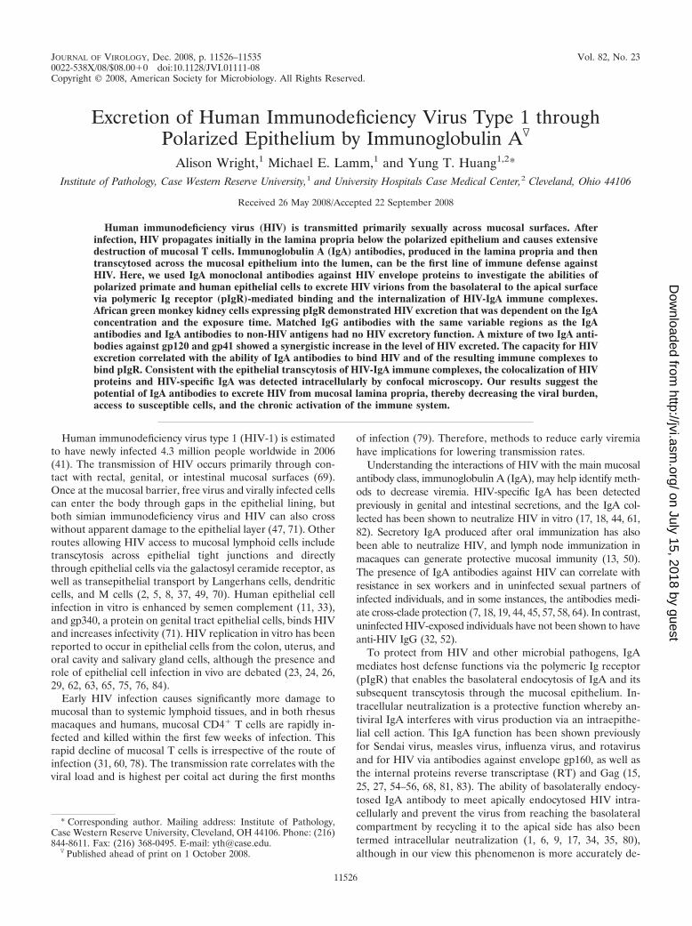
Synergy in HIV excretion. The ability of different IgA anti-bodies to work synergistically in virus excretion was assessed byusing D10A and D47A in combination to form HIV-IgA im-mune complexes. Immune complexes with D10A (50 �g/ml)plus D47A (50 �g/ml) in the basolateral chamber below pIgR�
Vero cells yielded increased levels of HIV excretion comparedto that yielded by each individual IgA at 100 �g/ml. The apicalcopy number of HIV with the combination of D10A and D47A(50 �g/ml each) was 289,000 (�68,000) virions per ml, whereasthat with D10A alone (100 �g/ml) was 96,000 (�17,000) viri-ons per ml and that with D47A alone (100 �g/ml) was 173,000(�59,000) virions per ml (Fig. 3A). Virus excretion with thecombined MAbs was therefore increased 1.7-fold over thatwith D47A alone (P � 0.042) and 3-fold over that with D10A
alone (P � 0.002). Figure 3B shows the viral band intensitiesafter excretion by the individual and combined IgAs. The in-dividual IgAs at 50 �g/ml each yielded the least intense bands,and the bands from 100-�g/ml concentrations of the individualMAbs were less intense than those from the combination ofD10A and D47A. Vero cells without the pIgR receptor wereunable to transport IgA and did not excrete HIV, even with thecombination of D10A and D47A at 100 �g/ml (data notshown). Incubation at 4°C rather than 37°C did not allowexcretion to occur (data not shown).
HIV excretion is IgA antibody concentration dependent. Toobserve HIV excretion as a function of the IgA antibody con-centration, equal volumes of HIV and D10A/D47A at concen-trations ranging from 8.3 to 100 �g/ml were mixed and themixture was placed into the basolateral chambers below polar-ized pIgR� Vero cell monolayers. After 8 h of excretion at37°C, HIV in the apical medium was assessed by the extractionof viral RNA, followed by RT-PCR and gel electrophoresisanalysis of the PCR product. At the lowest MAb concentration(8.3 �g/ml), no viral excretion was detected. At 25 �g/ml,apical HIV was detected, and the extent of virus excretionincreased further according to the IgA concentration (Fig. 4).The amount of virus detected after the addition of 200 �g/mlof IgA was comparable to that detected after the addition of100 �g/ml, indicating a plateau (data not shown). NeitherV-region-matched IgG nor irrelevant IgA at 100 �g/ml ex-creted HIV.
HIV excretion by IgA antibody is time dependent. To deter-mine whether IgA-mediated excretion depends on the dura-tion of basolateral exposure to immune complexes, HIV-IgA
FIG. 1. Transepithelial excretion of HIV through polarized pIgR� Vero cells mediated by different IgA antibodies. (A) Detection of HIV afterfivefold dilutions (10�5 to 10�8) in lanes 2 to 8. Lanes 1 and 9 are DNA ladders (M, molecular size) identifying the 320-bp amplicon location.(B) HIV excretion ability of each IgA at 100 �g/ml. RT-PCR products from apical samples collected after 8 h are shown in lanes 2 to 13. IrrelevantIgA (IgA control [IgA con.]) is in lanes 12 and 13. A positive control (PC; 10�6 dilution of virus) is shown in lane 14 and in lane 8 in panel C.(C) HIV excretion by IgA MAbs at 300 �g/ml.
FIG. 2. Relative abilities of IgA MAbs to bind to HIV and of HIV-IgA immune complexes to bind to pIgR. Results are expressed as means �SD. (A) Binding of MAbs (100 �g/ml) to HIV (n � 3). (B) Binding of immune complexes (100 �g/ml MAb) to pIgR (n � 3).
VOL. 82, 2008 HIV EXCRETION ACROSS POLARIZED EPITHELIUM BY IgA 11529
on July 15, 2018 by guesthttp://jvi.asm
.org/D
ownloaded from
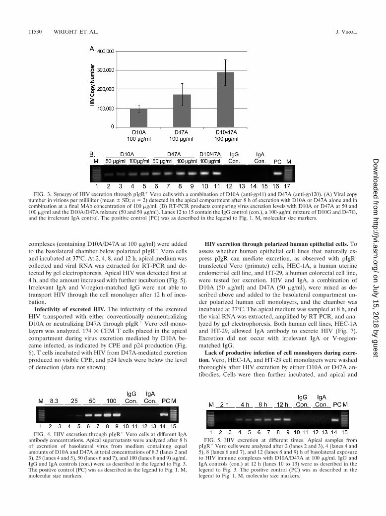
complexes (containing D10A/D47A at 100 �g/ml) were addedto the basolateral chamber below polarized pIgR� Vero cellsand incubated at 37°C. At 2, 4, 8, and 12 h, apical medium wascollected and viral RNA was extracted for RT-PCR and de-tected by gel electrophoresis. Apical HIV was detected first at4 h, and the amount increased with further incubation (Fig. 5).Irrelevant IgA and V-region-matched IgG were not able totransport HIV through the cell monolayer after 12 h of incu-bation.
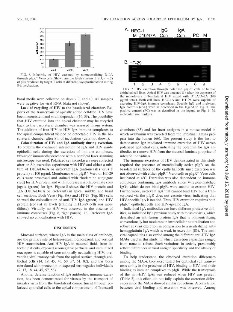
Infectivity of excreted HIV. The infectivity of the excretedHIV transported with either conventionally nonneutralizingD10A or neutralizing D47A through pIgR� Vero cell mono-layers was analyzed. 174 � CEM T cells placed in the apicalcompartment during virus excretion mediated by D10A be-came infected, as indicated by CPE and p24 production (Fig.6). T cells incubated with HIV from D47A-mediated excretionproduced no visible CPE, and p24 levels were below the levelof detection (data not shown).
HIV excretion through polarized human epithelial cells. Toassess whether human epithelial cell lines that naturally ex-press pIgR can mediate excretion, as observed with pIgR-transfected Vero (primate) cells, HEC-1A, a human uterineendometrial cell line, and HT-29, a human colorectal cell line,were tested for excretion. HIV and IgA, a combination ofD10A (50 �g/ml) and D47A (50 �g/ml), were mixed as de-scribed above and added to the basolateral compartment un-der polarized human cell monolayers, and the chamber wasincubated at 37°C. The apical medium was sampled at 8 h, andthe viral RNA was extracted, amplified by RT-PCR, and ana-lyzed by gel electrophoresis. Both human cell lines, HEC-1Aand HT-29, allowed IgA antibody to excrete HIV (Fig. 7).Excretion did not occur with irrelevant IgA or V-region-matched IgG.
Lack of productive infection of cell monolayers during excre-tion. Vero, HEC-1A, and HT-29 cell monolayers were washedthoroughly after HIV excretion by either D10A or D47A an-tibodies. Cells were then further incubated, and apical and
FIG. 5. HIV excretion at different times. Apical samples frompIgR� Vero cells were analyzed after 2 (lanes 2 and 3), 4 (lanes 4 and5), 8 (lanes 6 and 7), and 12 (lanes 8 and 9) h of basolateral exposureto HIV immune complexes with D10A/D47A at 100 �g/ml. IgG andIgA controls (con.) at 12 h (lanes 10 to 13) were as described in thelegend to Fig. 3. The positive control (PC) was as described in thelegend to Fig. 1. M, molecular size markers.
FIG. 3. Synergy of HIV excretion through pIgR� Vero cells with a combination of D10A (anti-gp41) and D47A (anti-gp120). (A) Viral copynumber in virions per milliliter (mean � SD; n � 2) detected in the apical compartment after 8 h of excretion with D10A or D47A alone and incombination at a final MAb concentration of 100 �g/ml. (B) RT-PCR products comparing virus excretion levels with D10A or D47A at 50 and100 �g/ml and the D10A/D47A mixture (50 and 50 �g/ml). Lanes 12 to 15 contain the IgG control (con.), a 100-�g/ml mixture of D10G and D47G,and the irrelevant IgA control. The positive control (PC) was as described in the legend to Fig. 1. M, molecular size markers.
FIG. 4. HIV excretion through pIgR� Vero cells at different IgAantibody concentrations. Apical supernatants were analyzed after 8 hof excretion of basolateral virus from medium containing equalamounts of D10A and D47A at total concentrations of 8.3 (lanes 2 and3), 25 (lanes 4 and 5), 50 (lanes 6 and 7), and 100 (lanes 8 and 9) �g/ml.IgG and IgA controls (con.) were as described in the legend to Fig. 3.The positive control (PC) was as described in the legend to Fig. 1. M,molecular size markers.
11530 WRIGHT ET AL. J. VIROL.
on July 15, 2018 by guesthttp://jvi.asm
.org/D
ownloaded from
basal media were collected on days 3, 7, and 10. All sampleswere negative for viral RNA (data not shown).
Lack of recycling of HIV to the basolateral chamber. Re-ports of the transcytosis of apically added cell-free HIV havebeen inconsistent and strain dependent (16, 33). The possibilitythat HIV excreted into the apical chamber may be recycledback to the basolateral chamber was assessed in our system.The addition of free HIV or HIV-IgA immune complexes tothe apical compartment yielded no detectable HIV in the ba-solateral chamber after 8 h of incubation (data not shown).
Colocalization of HIV and IgA antibody during excretion.To confirm the continued interaction of IgA and HIV insideepithelial cells during the excretion of immune complexes,two-color immunofluorescence with a confocal laser scanningmicroscope was used. Polarized cell monolayers were collectedafter an 8-h excretion experiment with HIV and either a mix-ture of D10A/D47A or irrelevant IgA (anti-measles virus Fprotein) at 100 �g/ml. Membranes with pIgR� Vero or HT-29cells were processed and stained with rhodamine conjugate(red) for HIV protein and with fluorescein isothiocyanate con-jugate (green) for IgA. Figure 8 shows the HIV protein andIgA (D10A/D47A or irrelevant) in apical, middle, and basalcell sections. Both Vero (Fig. 8A) and HT-29 (Fig. 8B) cellsshowed the colocalization of anti-HIV IgA (green) and HIVprotein (red) at all levels (staining in HT-29 cells was morediffuse). Virtually no HIV was observed in the absence ofimmune complexes (Fig. 8, right panels), i.e., irrelevant IgAshowed no colocalization with HIV.
DISCUSSION
Mucosal surfaces, where IgA is the main class of antibody,are the primary site of heterosexual, homosexual, and verticalHIV transmission. Anti-HIV IgA in mucosal fluids from in-fected patients, exposed seronegative partners, and immunizedmacaques is capable of conventionally neutralizing HIV, pre-venting viral transcytosis from the apical surface through epi-thelial cells (14, 19, 45, 46, 50, 57, 61, 82), and has beencorrelated with protection in exposed seronegative individuals(7, 17, 18, 44, 45, 57, 58).
Another defense function of IgA antibodies, immune excre-tion, has been demonstrated for viruses by the transport ofmeasles virus from the basolateral compartment through po-larized epithelial cells to the apical compartment of Transwell
chambers (83) and for inert antigens in a mouse model inwhich ovalbumin was excreted from the intestinal lamina pro-pria into the lumen (66). The present study is the first todemonstrate IgA-mediated immune excretion of HIV acrosspolarized epithelial cells, indicating the potential for IgA an-tibodies to remove HIV from the mucosal laminae propriae ofinfected individuals.
The immune excretion of HIV demonstrated in this studyrequired the presence of metabolically active pIgR on thebasolateral surfaces of the epithelial cells since excretion wasnot observed with either pIgR� Vero cells or pIgR� Vero cellsincubated at 4°C. Excretion was also dependent on immunecomplexes containing IgA antibody since V-region-matchedIgGs, which do not bind pIgR, were unable to excrete HIV.Furthermore, irrelevant IgA that cannot bind HIV but is tran-scytosed via the pIgR does not excrete virus, indicating thatHIV-specific IgA is needed. Thus, HIV excretion requires bothpIgR� epithelial cells and HIV-specific IgA.
Individual IgA antibodies can have different protective abil-ities, as indicated by a previous study with measles virus, whichdescribed an anti-fusion protein IgA that is nonneutralizingconventionally but moderate in intracellular neutralization androbust at virus excretion in comparison to a neutralizing anti-hemagglutinin IgA which is weak in excretion (83). The anti-viral capabilities also varied among the different anti-HIV IgAMAbs used in this study, in which excretion capacities rangedfrom none to robust. Such variations in activity presumablyreflect differences in viral antigen specificity and the affinity ofbinding.
To help understand the observed excretion differencesamong the MAbs, they were tested for epithelial cell transcy-tosis ability in the presence of HIV, binding to HIV, and theirbinding as immune complexes to pIgR. While the transcytosisof the anti-HIV IgAs was reduced when HIV was present(Table 2), this effect did not fully explain the excretion differ-ences since the MAbs showed similar reductions. A correlationbetween viral binding and excretion was observed. Among
FIG. 7. HIV excretion through polarized pIgR� cells of humanepithelial cell lines. Apical HIV was detected 8 h after the exposure ofthe monolayers to basolateral HIV mixed with D10A/D47A (100�g/ml total). Both cell lines, HEC-1A and HT-29, were capable ofexcreting HIV-IgA immune complexes. Specific IgG and irrelevantIgA controls (con.) were as described in the legend to Fig. 3. Thepositive control (PC) was as described in the legend to Fig. 1. M,molecular size markers.
FIG. 6. Infectivity of HIV excreted by nonneutralizing D10Athrough pIgR� Vero cells. Shown are the levels (means � SD; n � 2)of p24 produced by target T cells at different days postinfection during8-h incubations.
VOL. 82, 2008 HIV EXCRETION ACROSS POLARIZED EPITHELIUM BY IgA 11531
on July 15, 2018 by guesthttp://jvi.asm
.org/D
ownloaded from

individual MAbs, D10A (anti-gp41) and D47A (anti-gp120)produced the highest levels of excretion (Fig. 1) and alsobound best both to virus and as immune complexes to pIgR(Fig. 2). D25A (anti-gp120) and D19A (anti-gp120) both hadlower levels of binding to virus and as immune complexes topIgR than D10A and D47A but were still capable of excretingHIV. T33A and D61A (anti-gp41) had the lowest levels of HIVbinding, and their immune complexes showed virtually no
binding to pIgR, in keeping with their failure to excrete virus.In summary, the ability of IgA antibodies to mediate excretionappears to be related to their ability to bind to virus, as well astheir ability in immune complexes to bind pIgR.
After appropriate immunization, the mucosal lamina pro-pria contains a polyclonal population of antibodies, whosecollective properties determine the effectiveness of protection.The results in prior reports have shown the ability of some
FIG. 8. Intracellular colocalization of IgA and HIV protein within polarized epithelial cells after the endocytosis of virus-IgA immunecomplexes observed by confocal immunofluorescence microscopy. Apical, middle, and basal horizontal sections through the cell monolayers after8 h of excretion are shown for the red channel (HIV protein), the green channel (IgA), and merged red and green channels. A yellow to orangesignal in the merged channel indicates colocalization. Vero C1008 pIgR� cells and HT-29 pIgR� cells are shown. Cells transcytosing irrelevant IgAshowed no HIV, while cells transcytosing immune complexes of HIV and specific IgA antibody showed the colocalization of HIV protein and IgAat all levels. Bars, 10 �m.
11532 WRIGHT ET AL. J. VIROL.
on July 15, 2018 by guesthttp://jvi.asm
.org/D
ownloaded from

MAbs to work synergistically to mediate HIV neutralization(3, 48, 51, 53). The potential for antibody synergy in excretionfunction was tested here with IgAs against gp120 (D47A) andgp41 (D10A), and synergy was indeed observed (Fig. 3). Alikely explanation for the increased excretion is the ability ofthese two MAbs when combined to transcytose at higher ratesthan the individual MAbs (Table 2), bind HIV more efficiently(Fig. 2A), and bind better to pIgR when in complexes withHIV (Fig. 2B).
HIV excretion was detected at 25 �g/ml IgA antibody andincreased as the IgA antibody concentration was raised. Theeffect plateaued at 100 �g/ml (Fig. 4), which is roughly consis-tent with the concentration of IgA in human external secre-tions (40) and dog mesenteric lymph (77). These results sug-gest that physiological IgA antibody concentrations maymediate excretion in vivo, especially since the heterogeneouspolyclonal IgA antibodies produced during actively inducedimmune responses would include antibodies of greater affinitythan the MAbs employed in the present study. Furthermore,the extensive daily production of IgA, greater than that of allthe other Ig classes combined (42), allows the mucosae to havenewly synthesized IgA continuously available for the bindingand transport of antigens. Given the enormous surface area ofthe body’s mucosae, especially in the intestinal tract, the on-going excretion of virus has the potential to reduce viremiaand, in the case of HIV, to limit damage to mucosal T cellswhose loss can set the course of future disease (4, 28, 31).
Though IgA-mediated immune excretion removes antigenfrom the lamia propria, it has not been shown to cause theintracellular degradation of antigen (66). D10A antibody, incontrast to D47A, lacks conventional and intracellular neutral-ization activities (39) but is capable of binding and excretingHIV, indicating that excretion function is not directly relatedto neutralization ability. The different neutralizing abilities ofD10A and D47A provided an opportunity to study whetherexcreted virus could remain infectious. Not unexpectedly, neu-tralizing D47A MAb excreted noninfectious virions whileD10A transported infectious virus as indicated by CPE andconfirmed by p24 production by infected T cells (Fig. 6). Otherstudies have shown that the apical-to-basolateral transcytosisof HIV through epithelial cells can be interrupted by IgA orIgM antibody transcytosing from the basolateral compartmentand that virus redirected to the apical side remains infectious(8, 35). Additionally, phagocytes can become infected afteringesting antibody-bound HIV via Fc receptor intake (36, 73),and since epithelial cells may be capable of supporting HIVreplication, it is possible that virus from antibody-mediatedexcretion may infect epithelial cells. However, for the Vero,HEC-1A, and HT-29 cells studied here, productive infectionresulting from excretion was not detected.
HEC-1A, a uterine endometrial cell line, and HT-29, a colo-rectal cell line, are both derived from mucosae where IgAtranscytosis through the lining epithelium occurs. Both thesehuman cell lines express endogenous pIgR and therefore dem-onstrate the potential capacity of human mucosal epithelialcells to excrete HIV from the lamina propria, thereby helpingto protect mucosal T cells.
The vesicular transcytosis of polymeric IgA begins with itsbinding to pIgR, followed by the endocytosis of IgA and pIgRat the basolateral cell membrane before transport through a
series of endosomal compartments and release into the apicalmucosal secretions (42). Confocal microscopy showed the co-localization of transcytosing IgA antibody and HIV within thebasal, middle, and apical portions of polarized epithelial cells,consistent with the established pathway of pIgR-mediatedtransport for apical release (Fig. 8). Colocalization was ob-served for both a pIgR-transfected primate cell line and ahuman cell line with endogenous pIgR expression.
It is apparent that IgA antibodies can manifest a number ofanti-HIV properties. IgA has been shown previously to blockthe apical adherence of HIV to epithelial cells and inhibit theapical-to-basolateral transcytosis of HIV (1, 2, 16). We havepreviously demonstrated that IgA antibodies against the enve-lope proteins gp120 and gp41, as well as the internal proteinsRT and Gag, can mediate HIV neutralization intracellularlyduring their epithelial transcytosis (39, 81). In the presentwork, we provided evidence in vitro for another host defensefunction in which anti-HIV IgA excretes virus across epithelialcells. Determining the significance of our findings for mucosaldefense in vivo must, of course, await future studies, includingexperiments with R5 viruses, which tend to be associated withinitial infection.
Currently, there is considerable interest in the possibilitythat an increase in microbially derived antigens in the intestinalmucosa leads to a detrimental chronic activation of the im-mune system during HIV infection (12, 20, 30). Therefore,mucosal IgA antibodies that can excrete intact HIV and itsindividual components, as well as the products of other mi-crobes, may be significant in attempts to improve the clinicalresponse to HIV exposure. The findings of the present studyand others illustrating the multiple protective functions of anti-HIV IgA would seem to support the rationale for mucosalimmunization strategies against HIV.
ACKNOWLEDGMENTS
We thank the Confocal Microscopy Core in the ComprehensiveCancer Center of Case School of Medicine/University Hospitals ofCleveland (funded by NIH grant no. P30 CA43703-12) for the use oftheir facility.
This research was supported by grants AI-36359 and CA-43703 fromthe NIH.
REFERENCES
1. Alfsen, A., P. Iniguez, E. Bouguyon, and M. Bomsel. 2001. Secretory IgAspecific for a conserved epitope on gp41 envelope glycoprotein inhibitsepithelial transcytosis of HIV-1. J. Immunol. 166:6257–6265.
2. Alfsen, A., H. Yu, A. Magerus-Chatinet, A. Schmitt, and M. Bomsel. 2005.HIV-1-infected blood mononuclear cells form an integrin- and agrin-depen-dent viral synapse to induce efficient HIV-1 transcytosis across epithelial cellmonolayer. Mol. Biol. Cell 16:4267–4279.
3. Allaway, G. P., A. M. Ryder, G. A. Beaudry, and P. J. Maddon. 1993.Synergistic inhibition of HIV-1 envelope-mediated cell fusion by CD4-basedmolecules in combination with antibodies to gp120 or gp41. AIDS Res. Hum.Retrovir. 9:581–587.
4. Ambrose, Z., K. Larsen, J. Thompson, Y. Stevens, E. Finn, S. L. Hu, andM. L. Bosch. 2001. Evidence for early local viral replication and local pro-duction of antiviral immunity upon mucosal simian-human immunodefi-ciency virus SHIV89.6 infection in Macaca nemestrina. J. Virol. 75:8589–8596.
5. Amerongen, H. M., R. Weltzin, C. M. Farnet, P. Michetti, W. A. Haseltine,and M. R. Neutra. 1991. Transepithelial transport of HIV-1 by intestinal Mcells: a mechanism for transmission of AIDS. J. Acquir. Immune Defic.Syndr. 4:760–765.
6. Belec, L., P. D. Ghys, H. Hocini, J. N. Nkengasong, J. Tranchot-Diallo, M. O.Diallo, V. Ettiegne-Traore, C. Maurice, P. Becquart, M. Matta, A. Si-Mohamed, N. Chomont, I. M. Coulibaly, S. Z. Wiktor, and M. D. Kazatch-kine. 2001. Cervicovaginal secretory antibodies to human immunodeficiencyvirus type 1 (HIV-1) that block viral transcytosis through tight epithelial
VOL. 82, 2008 HIV EXCRETION ACROSS POLARIZED EPITHELIUM BY IgA 11533
on July 15, 2018 by guesthttp://jvi.asm
.org/D
ownloaded from

barriers in highly exposed HIV-1-seronegative African women. J. Infect. Dis.184:1412–1422.
7. Beyrer, C., A. W. Artenstein, S. Rugpao, H. Stephens, T. C. VanCott, M. L.Robb, M. Rinkaew, D. L. Birx, C. Khamboonruang, P. A. Zimmerman, K. E.Nelson, C. Natpratan, et al. 1999. Epidemiologic and biologic characteriza-tion of a cohort of human immunodeficiency virus type 1 highly exposed,persistently seronegative female sex workers in northern Thailand. J. Infect.Dis. 179:59–67.
8. Bomsel, M. 1997. Transcytosis of infectious human immunodeficiency virusacross a tight human epithelial cell line barrier. Nat. Med. 3:42–47.
9. Bomsel, M., M. Heyman, H. Hocini, S. Lagaye, L. Belec, C. Dupont, and C.Desgranges. 1998. Intracellular neutralization of HIV transcytosis acrosstight epithelial barriers by anti-HIV envelope protein dIgA or IgM. Immu-nity 9:277–287.
10. Boot, J. H., M. E. Geerts, E. R. De Groot, and L. A. Aarden. 1988. Murinemonoclonal isotype switch variants. Detection with rat monoclonal antibod-ies in ELISA and isolation by sequential sublining. J. Immunol. Methods106:195–202.
11. Bouhlal, H., N. Chomont, N. Haeffner-Cavaillon, M. D. Kazatchkine, L.Belec, and H. Hocini. 2002. Opsonization of HIV-1 by semen complementenhances infection of human epithelial cells. J. Immunol. 169:3301–3306.
12. Brenchley, J. M., D. A. Price, T. W. Schacker, T. E. Asher, G. Silvestri, S.Rao, Z. Kazzaz, E. Bornstein, O. Lambotte, D. Altmann, B. R. Blazar, B.Rodriguez, L. Teixeira-Johnson, A. Landay, J. N. Martin, F. M. Hecht, L. J.Picker, M. M. Lederman, S. G. Deeks, and D. C. Douek. 2006. Microbialtranslocation is a cause of systemic immune activation in chronic HIV in-fection. Nat. Med. 12:1365–1371.
13. Bukawa, H., K. Sekigawa, K. Hamajima, J. Fukushima, Y. Yamada, H.Kiyono, and K. Okuda. 1995. Neutralization of HIV-1 by secretory IgAinduced by oral immunization with a new macromolecular multicomponentpeptide vaccine candidate. Nat. Med. 1:681–685.
14. Burnett, P. R., T. C. VanCott, V. R. Polonis, R. R. Redfield, and D. L. Birx.1994. Serum IgA-mediated neutralization of HIV type 1. J. Immunol. 152:4642–4648.
15. Burns, J. W., M. Siadat-Pajouh, A. A. Krishnaney, and H. B. Greenberg.1996. Protective effect of rotavirus VP6-specific IgA monoclonal antibodiesthat lack neutralizing activity. Science 272:104–107.
16. Chomont, N., H. Hocini, J. C. Gody, H. Bouhlal, P. Becquart, C. Krief-Bouillet, M. Kazatchkine, and L. Belec. 2008. Neutralizing monoclonal an-tibodies to human immunodeficiency virus type 1 do not inhibit viral trans-cytosis through mucosal epithelial cells. Virology 370:246–254.
17. Devito, C., K. Broliden, R. Kaul, L. Svensson, K. Johansen, P. Kiama, J.Kimani, L. Lopalco, S. Piconi, J. J. Bwayo, F. Plummer, M. Clerici, and J.Hinkula. 2000. Mucosal and plasma IgA from HIV-1-exposed uninfectedindividuals inhibit HIV-1 transcytosis across human epithelial cells. J. Im-munol. 165:5170–5176.
18. Devito, C., J. Hinkula, R. Kaul, J. Kimani, P. Kiama, L. Lopalco, C. Barass,S. Piconi, D. Trabattoni, J. J. Bwayo, F. Plummer, M. Clerici, and K.Broliden. 2002. Cross-clade HIV-1-specific neutralizing IgA in mucosal andsystemic compartments of HIV-1-exposed, persistently seronegative sub-jects. J. Acquir. Immune Defic. Syndr. 30:413–420.
19. Devito, C., J. Hinkula, R. Kaul, L. Lopalco, J. J. Bwayo, F. Plummer, M.Clerici, and K. Broliden. 2000. Mucosal and plasma IgA from HIV-exposedseronegative individuals neutralize a primary HIV-1 isolate. AIDS 14:1917–1920.
20. Douek, D. 2007. HIV disease progression: immune activation, microbes, anda leaky gut. Top. HIV Med. 15:114–117.
21. Earl, P. L., C. C. Broder, R. W. Doms, and B. Moss. 1997. Epitope map ofhuman immunodeficiency virus type 1 gp41 derived from 47 monoclonalantibodies produced by immunization with oligomeric envelope protein.J. Virol. 71:2674–2684.
22. Earl, P. L., W. Sugiura, D. C. Montefiori, C. C. Broder, S. A. Lee, C. Wild,J. Lifson, and B. Moss. 2001. Immunogenicity and protective efficacy ofoligomeric human immunodeficiency virus type 1 gp140. J. Virol. 75:645–653.
23. Fantini, J., N. Yahi, S. Baghdiguian, and J. C. Chermann. 1992. Humancolon epithelial cells productively infected with human immunodeficiencyvirus show impaired differentiation and altered secretion. J. Virol. 66:580–585.
24. Fantini, J., N. Yahi, and J. C. Chermann. 1991. Human immunodeficiencyvirus can infect the apical and basolateral surfaces of human colonic epithe-lial cells. Proc. Natl. Acad. Sci. USA 88:9297–9301.
25. Feng, N., J. A. Lawton, J. Gilbert, N. Kuklin, P. Vo, B. V. Prasad, and H. B.Greenberg. 2002. Inhibition of rotavirus replication by a non-neutralizing,rotavirus VP6-specific IgA mAb. J. Clin. Investig. 109:1203–1213.
26. Fleming, S. C., M. S. Kapembwa, T. T. MacDonald, and G. E. Griffin. 1992.Direct in vitro infection of human intestine with HIV-1. AIDS 6:1099–1104.
27. Fujioka, H., S. N. Emancipator, M. Aikawa, D. S. Huang, F. Blatnik, T.Karban, K. DeFife, and M. B. Mazanec. 1998. Immunocytochemical colo-calization of specific immunoglobulin A with Sendai virus protein in infectedpolarized epithelium. J. Exp. Med. 188:1223–1229.
28. George, M. D., E. Reay, S. Sankaran, and S. Dandekar. 2005. Early antiret-
roviral therapy for simian immunodeficiency virus infection leads to mucosalCD4� T-cell restoration and enhanced gene expression regulating mucosalrepair and regeneration. J. Virol. 79:2709–2719.
29. Han, Y., C. L. Ventura, K. P. Black, J. E. Cummins, Jr., S. D. Hall, and S.Jackson. 2000. Productive human immunodeficiency virus-1 infection ofepithelial cell lines of salivary gland origin. Oral Microbiol. Immunol. 15:82–88.
30. Hazenberg, M. D., S. A. Otto, B. H. van Benthem, M. T. Roos, R. A.Coutinho, J. M. Lange, D. Hamann, M. Prins, and F. Miedema. 2003.Persistent immune activation in HIV-1 infection is associated with progres-sion to AIDS. AIDS 17:1881–1888.
31. Hel, Z., J. R. McGhee, and J. Mestecky. 2006. HIV infection: first battledecides the war. Trends Immunol. 27:274–281.
32. Hirbod, T., and K. Broliden. 2007. Mucosal immune responses in the genitaltract of HIV-1-exposed uninfected women. J. Intern. Med. 262:44–58.
33. Hocini, H., P. Becquart, H. Bouhlal, N. Chomont, P. Ancuta, M. D. Kaza-tchkine, and L. Belec. 2001. Active and selective transcytosis of cell-freehuman immunodeficiency virus through a tight polarized monolayer of hu-man endometrial cells. J. Virol. 75:5370–5374.
34. Hocini, H., L. Belec, S. Iscaki, B. Garin, J. Pillot, P. Becquart, and M.Bomsel. 1997. High-level ability of secretory IgA to block HIV type 1 trans-cytosis: contrasting secretory IgA and IgG responses to glycoprotein 160.AIDS Res. Hum. Retrovir. 13:1179–1185.
35. Hocini, H., and M. Bomsel. 1999. Infectious human immunodeficiency viruscan rapidly penetrate a tight human epithelial barrier by transcytosis in aprocess impaired by mucosal immunoglobulins. J. Infect. Dis. 179(Suppl.3):S448–S453.
36. Homsy, J., M. Meyer, M. Tateno, S. Clarkson, and J. A. Levy. 1989. The Fcand not CD4 receptor mediates antibody enhancement of HIV infection inhuman cells. Science 244:1357–1360.
37. Hu, J., B. Murray, M. B. Gardner, and C. J. Miller. 2000. Simian immuno-deficiency virus rapidly penetrates the cervicovaginal mucosa after intravag-inal inoculation and infects intraepithelial dendritic cells. J. Virol. 74:6087–6095.
38. Huang, Y. T., C. J. Miller, V. Wong, H. Fujioka, J. G. Nedrud, and M. E.Lamm. 1999. Replication and budding of simian immunodeficiency virus inpolarized epithelial cells. Virology 257:24–34.
39. Huang, Y. T., A. Wright, X. Gao, L. Kulick, H. Yan, and M. E. Lamm. 2005.Intraepithelial cell neutralization of HIV-1 replication by IgA. J. Immunol.174:4828–4835.
40. Jackson, S., J. Mestecky, Z. Moldoveanu, and P. Spearman. 1999. Collectionand processing of human mucosal secretions, p. 1567–1575. In J. Mestecky,P. L. Ogra, M. E. Lamm, W. Strober, J. Bienenstock, and J. R. McGhee(ed.), Mucosal immunology, 2nd ed. Academic Press, San Diego, CA.
41. Joint United Nations Programme on HIV/AIDS. 30 May 2006, posting date.2006 report on the global AIDS epidemic: a UNAIDS 10th anniversaryspecial edition, p. 7–50. Joint United Nations Programme on HIV/AIDS,Geneva, Switzerland. http://www.unaids.org/en/KnowledgeCentre/HIVData/GlobalReport/2006/default.asp.
42. Kaetzel, C. S., and K. Mostov. 2005. Immunoglobulin transport and the poly-meric immunoglobulin receptor, p. 211–250. In J. Mestecky, M. E. Lamm, W.Strober, J. Bienenstock, J. R. McGhee, and L. Mayer (ed.), Mucosal immunol-ogy, 3rd ed., vol. 1. Elsevier Academic Press, San Diego, CA.
43. Kaetzel, C. S., J. K. Robinson, K. R. Chintalacharuvu, J. P. Vaerman, andM. E. Lamm. 1991. The polymeric immunoglobulin receptor (secretory com-ponent) mediates transport of immune complexes across epithelial cells: alocal defense function for IgA. Proc. Natl. Acad. Sci. USA 88:8796–8800.
44. Kaul, R., F. Plummer, M. Clerici, M. Bomsel, L. Lopalco, and K. Broliden.2001. Mucosal IgA in exposed, uninfected subjects: evidence for a role inprotection against HIV infection. AIDS 15:431–432.
45. Kaul, R., D. Trabattoni, J. J. Bwayo, D. Arienti, A. Zagliani, F. M. Mwangi,C. Kariuki, E. N. Ngugi, K. S. MacDonald, T. B. Ball, M. Clerici, and F. A.Plummer. 1999. HIV-1-specific mucosal IgA in a cohort of HIV-1-resistantKenyan sex workers. AIDS 13:23–29.
46. Kozlowski, P., R. W. Buckheit, Jr., and S. Jackson. 1995. Neutralization ofHIV infection by serum IgA antibodies. Adv. Exp. Med. Biol. 371B:1027–1030.
47. Kozlowski, P. A., and M. R. Neutra. 2003. The role of mucosal immunity inprevention of HIV transmission. Curr. Mol. Med. 3:217–228.
48. Laal, S., S. Burda, M. K. Gorny, S. Karwowska, A. Buchbinder, and S.Zolla-Pazner. 1994. Synergistic neutralization of human immunodeficiencyvirus type 1 by combinations of human monoclonal antibodies. J. Virol.68:4001–4008.
49. Lagaye, S., M. Derrien, E. Menu, C. Coito, E. Tresoldi, P. Mauclere, G.Scarlatti, G. Chaouat, F. Barre-Sinoussi, and M. Bomsel. 2001. Cell-to-cellcontact results in a selective translocation of maternal human immunodefi-ciency virus type 1 quasispecies across a trophoblastic barrier by both trans-cytosis and infection. J. Virol. 75:4780–4791.
50. Lehner, T., Y. Wang, M. Cranage, L. A. Bergmeier, E. Mitchell, L. Tao, G.Hall, M. Dennis, N. Cook, R. Brookes, L. Klavinskis, I. Jones, C. Doyle, andR. Ward. 1996. Protective mucosal immunity elicited by targeted iliac lymph
11534 WRIGHT ET AL. J. VIROL.
on July 15, 2018 by guesthttp://jvi.asm
.org/D
ownloaded from

node immunization with a subunit SIV envelope and core vaccine in ma-caques. Nat. Med. 2:767–775.
51. Li, A., H. Katinger, M. R. Posner, L. Cavacini, S. Zolla-Pazner, M. K. Gorny,J. Sodroski, T. C. Chou, T. W. Baba, and R. M. Ruprecht. 1998. Synergisticneutralization of simian-human immunodeficiency virus SHIV-vpu� by tri-ple and quadruple combinations of human monoclonal antibodies and high-titer anti-human immunodeficiency virus type 1 immunoglobulins. J. Virol.72:3235–3240.
52. Lopalco, L., C. Pastori, A. Cosma, S. E. Burastero, B. Capiluppi, E. Boeri, A.Beretta, A. Lazzarin, and A. G. Siccardi. 2000. Anti-cell antibodies in ex-posed seronegative individuals with HIV type 1-neutralizing activity. AIDSRes. Hum. Retrovir. 16:109–115.
53. Mascola, J. R., M. K. Louder, T. C. VanCott, C. V. Sapan, J. S. Lambert,L. R. Muenz, B. Bunow, D. L. Birx, and M. L. Robb. 1997. Potent andsynergistic neutralization of human immunodeficiency virus (HIV) type 1primary isolates by hyperimmune anti-HIV immunoglobulin combined withmonoclonal antibodies 2F5 and 2G12. J. Virol. 71:7198–7206.
54. Mazanec, M. B., C. L. Coudret, and D. R. Fletcher. 1995. Intracellularneutralization of influenza virus by immunoglobulin A anti-hemagglutininmonoclonal antibodies. J. Virol. 69:1339–1343.
55. Mazanec, M. B., C. S. Kaetzel, M. E. Lamm, D. Fletcher, and J. G. Nedrud.1992. Intracellular neutralization of virus by immunoglobulin A antibodies.Proc. Natl. Acad. Sci. USA 89:6901–6905.
56. Mazanec, M. B., J. G. Nedrud, C. S. Kaetzel, and M. E. Lamm. 1993. Athree-tiered view of the role of IgA in mucosal defense. Immunol. Today14:430–435.
57. Mazzoli, S., L. Lopalco, A. Salvi, D. Trabattoni, S. Lo Caputo, F. Semplici,M. Biasin, C. Ble, A. Cosma, C. Pastori, F. Meacci, F. Mazzotta, M. L. Villa,A. G. Siccardi, and M. Clerici. 1999. Human immunodeficiency virus (HIV)-specific IgA and HIV neutralizing activity in the serum of exposed seroneg-ative partners of HIV-seropositive persons. J. Infect. Dis. 180:871–875.
58. Mazzoli, S., D. Trabattoni, S. Lo Caputo, S. Piconi, C. Ble, F. Meacci, S.Ruzzante, A. Salvi, F. Semplici, R. Longhi, M. L. Fusi, N. Tofani, M. Biasin,M. L. Villa, F. Mazzotta, and M. Clerici. 1997. HIV-specific mucosal andcellular immunity in HIV-seronegative partners of HIV-seropositive individ-uals. Nat. Med. 3:1250–1257.
59. Meng, G., X. Wei, X. Wu, M. T. Sellers, J. M. Decker, Z. Moldoveanu, J. M.Orenstein, M. F. Graham, J. C. Kappes, J. Mestecky, G. M. Shaw, and P. D.Smith. 2002. Primary intestinal epithelial cells selectively transfer R5 HIV-1to CCR5� cells. Nat. Med. 8:150–156.
60. Miller, C. J., Q. Li, K. Abel, E. Y. Kim, Z. M. Ma, S. Wietgrefe, L. LaFranco-Scheuch, L. Compton, L. Duan, M. D. Shore, M. Zupancic, M.Busch, J. Carlis, S. Wolinsky, and A. T. Haase. 2005. Propagation anddissemination of infection after vaginal transmission of simian immunodefi-ciency virus. J. Virol. 79:9217–9227.
61. Moja, P., C. Tranchat, I. Tchou, B. Pozzetto, F. Lucht, C. Desgranges, andC. Genin. 2000. Neutralization of human immunodeficiency virus type 1(HIV-1) mediated by parotid IgA of HIV-1-infected patients. J. Infect. Dis.181:1607–1613.
62. Moore, J. S., S. D. Hall, and S. Jackson. 2002. Cell-associated HIV-1 infec-tion of salivary gland epithelial cell lines. Virology 297:89–97.
63. Moore, J. S., F. Rahemtulla, L. W. Kent, S. D. Hall, M. R. Ikizler, P. F.Wright, H. H. Nguyen, and S. Jackson. 2003. Oral epithelial cells are sus-ceptible to cell-free and cell-associated HIV-1 infection in vitro. Virology313:343–353.
64. Pastori, C., C. Barassi, S. Piconi, R. Longhi, M. L. Villa, A. G. Siccardi, M.Clerici, and L. Lopalco. 2000. HIV neutralizing IgA in exposed seronegativesubjects recognise an epitope within the gp41 coiled-coil pocket. J. Biol.Regul. Homeost. Agents 14:15–21.
65. Qureshi, M. N., C. E. Barr, T. Seshamma, J. Reidy, R. J. Pomerantz, and O.Bagasra. 1995. Infection of oral mucosal cells by human immunodeficiencyvirus type 1 in seropositive persons. J. Infect. Dis. 171:190–193.
66. Robinson, J. K., T. G. Blanchard, A. D. Levine, S. N. Emancipator, and M. E.Lamm. 2001. A mucosal IgA-mediated excretory immune system in vivo.J. Immunol. 166:3688–3692.
67. Sakai, K., X. Y. Ma, I. Gordienko, and D. J. Volsky. 1991. Recombinationalanalysis of a natural noncytopathic human immunodeficiency virus type 1(HIV-1) isolate: role of the vif gene in HIV-1 infection kinetics and cyto-pathicity. J. Virol. 65:5765–5773.
68. Schwartz-Cornil, I., Y. Benureau, H. Greenberg, B. A. Hendrickson, and J.Cohen. 2002. Heterologous protection induced by the inner capsid proteinsof rotavirus requires transcytosis of mucosal immunoglobulins. J. Virol.76:8110–8117.
69. Smith, P. D., and S. M. Wahl. 2005. Immunobiology of mucosal HIV-1infection, p. 1199–1211. In J. Mestecky, M. E. Lamm, W. Strober, J. Bienen-stock, J. R. McGhee, and L. Mayer (ed.), Mucosal immunology, 3rd ed., vol.1. Elsevier Academic Press, San Diego, CA.
70. Spira, A. I., P. A. Marx, B. K. Patterson, J. Mahoney, R. A. Koup, S. M.Wolinsky, and D. D. Ho. 1996. Cellular targets of infection and route of viraldissemination after an intravaginal inoculation of simian immunodeficiencyvirus into rhesus macaques. J. Exp. Med. 183:215–225.
71. Stoddard, E., G. Cannon, H. Ni, K. Kariko, J. Capodici, D. Malamud, andD. Weissman. 2007. gp340 expressed on human genital epithelia binds HIV-1envelope protein and facilitates viral transmission. J. Immunol. 179:3126–3132.
72. Sugiura, W., C. C. Broder, B. Moss, and P. L. Earl. 1999. Characterizationof conformation-dependent anti-gp120 murine monoclonal antibodies pro-duced by immunization with monomeric and oligomeric human immunode-ficiency virus type 1 envelope proteins. Virology 254:257–267.
73. Sullivan, N. J. 2001. Antibody-mediated enhancement of viral disease. Curr.Top. Microbiol. Immunol. 260:145–169.
74. Tamer, C. M., M. E. Lamm, J. K. Robinson, J. F. Piskurich, and C. S.Kaetzel. 1995. Comparative studies of transcytosis and assembly of secretoryIgA in Madin-Darby canine kidney cells expressing human polymeric Igreceptor. J. Immunol. 155:707–714.
75. Tan, X., R. Pearce-Pratt, and D. M. Phillips. 1993. Productive infection of acervical epithelial cell line with human immunodeficiency virus: implicationsfor sexual transmission. J. Virol. 67:6447–6452.
76. Tan, X., and D. M. Phillips. 1996. Cell-mediated infection of cervix derivedepithelial cells with primary isolates of human immunodeficiency virus. Arch.Virol. 141:1177–1189.
77. Vaerman, J. P., and J. F. Heremans. 1970. Origin and molecular size of immu-noglobulin-A in the mesenteric lymph of the dog. Immunology 18:27–38.
78. Veazey, R. S., and A. A. Lackner. 2004. Getting to the guts of HIV patho-genesis. J. Exp. Med. 200:697–700.
79. Wawer, M. J., R. H. Gray, N. K. Sewankambo, D. Serwadda, X. Li, O.Laeyendecker, N. Kiwanuka, G. Kigozi, M. Kiddugavu, T. Lutalo, F. Nal-ugoda, F. Wabwire-Mangen, M. P. Meehan, and T. C. Quinn. 2005. Rates ofHIV-1 transmission per coital act, by stage of HIV-1 infection, in Rakai,Uganda. J. Infect. Dis. 191:1403–1409.
80. Wolbank, S., R. Kunert, G. Stiegler, and H. Katinger. 2003. Characterizationof human class-switched polymeric (immunoglobulin M [IgM] and IgA)anti-human immunodeficiency virus type 1 antibodies 2F5 and 2G12. J. Vi-rol. 77:4095–4103.
81. Wright, A., H. Yan, M. E. Lamm, and Y. T. Huang. 2006. Immunoglobulin Aantibodies against internal HIV-1 proteins neutralize HIV-1 replication in-side epithelial cells. Virology 356:165–170.
82. Wu, X., S. Hall, and S. Jackson. 2003. Tropism-restricted neutralization bysecretory IgA from parotid saliva of HIV type 1-infected individuals. AIDSRes. Hum. Retrovir. 19:275–281.
83. Yan, H., M. E. Lamm, E. Bjorling, and Y. T. Huang. 2002. Multiple functionsof immunoglobulin A in mucosal defense against viruses: an in vitro measlesvirus model. J. Virol. 76:10972–10979.
84. Yeaman, G. R., A. L. Howell, S. Weldon, D. J. Demian, J. E. Collins, D. M.O’Connell, S. N. Asin, C. R. Wira, and M. W. Fanger. 2003. Human immu-nodeficiency virus receptor and coreceptor expression on human uterineepithelial cells: regulation of expression during the menstrual cycle andimplications for human immunodeficiency virus infection. Immunology 109:137–146.
VOL. 82, 2008 HIV EXCRETION ACROSS POLARIZED EPITHELIUM BY IgA 11535
on July 15, 2018 by guesthttp://jvi.asm
.org/D
ownloaded from