synergistic activation of human immunodeficiency virus type .... publication j... · vincent...
TRANSCRIPT

JOURNAL OF VIROLOGY, Nov. 2002, p. 11091–11103 Vol. 76, No. 210022-538X/02/$04.00�0 DOI: 10.1128/JVI.76.21.11091–11103.2002Copyright © 2002, American Society for Microbiology. All Rights Reserved.
Synergistic Activation of Human Immunodeficiency Virus Type 1Promoter Activity by NF-�B and Inhibitors of Deacetylases:
Potential Perspectives for the Development ofTherapeutic Strategies
Vincent Quivy,1 Emmanuelle Adam,1 Yves Collette,2 Dominique Demonte,1 Alain Chariot,3Caroline Vanhulle,1 Ben Berkhout,4 Remy Castellano,2 Yvan de Launoit,5,6
Arsene Burny,1 Jacques Piette,3 Vincent Bours,3 and Carine Van Lint1*Laboratoire de Virologie Moleculaire, Service de Chimie Biologique, Institut de Biologie et de Medecine Moleculaires,
Universite Libre de Bruxelles, 6041 Gosselies,1 Center for Molecular and Cellular Therapy, Universite de Liege,Sart-Tilman, 4000 Liege,3 and Laboratoire de Microbiologie, Faculte de Medecine, Universite Libre de
Bruxelles, 1070 Brussels,5 Belgium; Department of Human Retrovirology, Academic Medical Center,University of Amsterdam, 1105 AZ Amsterdam, The Netherlands4; and INSERM U119,
13009 Marseille,2 and Institut de Biologie de Lille, Institut Pasteur de Lille,UMR 8526 CNRS, 59021 Lille Cedex,6 France
Received 18 March 2002/Accepted 17 July 2002
The transcription factor NF-�B plays a central role in the human immunodeficiency virus type 1 (HIV-1)activation pathway. HIV-1 transcription is also regulated by protein acetylation, since treatment with deacety-lase inhibitors such as trichostatin A (TSA) or sodium butyrate (NaBut) markedly induces HIV-1 transcrip-tional activity of the long terminal repeat (LTR) promoter. Here, we demonstrate that TSA (NaBut) synergizedwith both ectopically expressed p50/p65 and tumor necrosis factor alpha/SF2 (TNF)-induced NF-�B to activatethe LTR. This was confirmed for LTRs from subtypes A through G of the HIV-1 major group, with a positivecorrelation between the number of �B sites present in the LTRs and the amplitude of the TNF-TSA synergism.Mechanistically, TSA (NaBut) delayed the cytoplasmic recovery of the inhibitory protein I�B�. This coincidedwith a prolonged intranuclear presence and DNA binding activity of NF-�B. The physiological relevance of theTNF-TSA (NaBut) synergism was shown on HIV-1 replication in both acutely and latently HIV-infected celllines. Therefore, our results open new therapeutic strategies aimed at decreasing or eliminating the pool oflatently HIV-infected reservoirs by forcing viral expression.
The persistence of latently human immunodeficiency virus(HIV)-infected cellular reservoirs, despite prolonged treat-ment with highly active antiretroviral therapy (HAART), rep-resents the major hurdle to virus eradication. These latentlyinfected cells are a permanent source for virus reactivation andlead to a rebound of the viral load after interruption ofHAART (reviewed in reference 42). Therefore, a greater un-derstanding of the molecular mechanisms regulating viral la-tency and reactivation should lead to rational strategies aimedat purging the latent HIV reservoirs (6). At the cellular level,two major forms of HIV type 1 (HIV-1) latency have beendescribed: preintegration latency and postintegration latency(reviewed in reference 33). Several cell lines selected in vitrohave served as models for studying the latter type of latency. Inthese cell lines, production of viral particles can be induced atthe transcriptional level by a variety of agents, including phor-bol esters and cytokine tumor necrosis factor SF2 (TNF) (16).Several explanations have been proposed for the low level oftranscription observed during postintegration latency, includ-
ing the following. (i) The first explanation involves the site ofintegration of the provirus into the host cell genome and thecellular chromatin environment at this site (26, 60). (ii) Asecond explanation involves the presence of a potentially re-pressive nucleosome (nuc-1) located immediately downstreamof the HIV transcription start site under latency conditions.Nuc-1 is remodeled upon activation of the HIV promoterlocated in its 5� long terminal repeat (LTR) in response to Tat,phorbol esters, and deacetylase inhibitors (12, 54, 58). Nuc-1could pose a unique elongation barrier for the polymerase, andits remodeling could play a significant role in the transcrip-tional reactivation of the HIV promoter from latency (re-viewed in reference 52). (iii) A third explanation involves theabsence of the viral trans-activator Tat, which binds to TAR, anRNA hairpin loop formed at the 5� termini of all nascentHIV-1 transcripts (1). (iv) A fourth explanation involves thepresence of mutations in the integrated provirus, includinginterruption of the Tat-TAR axis (13, 14). (v) A fifth explana-tion involves the absence of the inducible transcription factorNF-�B. The enhancer in the U3 region of the LTR containstwo binding sites for NF-�B, which plays a central role in theactivation pathway of the HIV-1 provirus. Various studies havereported that the NF-�B-binding sites as well as the NF-�Bproteins are critical for LTR promoter activity and importantfor optimal HIV-1 replication (reviewed in reference 44). Re-
* Corresponding author. Mailing address: Universite Libre de Brux-elles, Institut de Biologie et de Medecine Moleculaires, Service deChimie Biologique, Laboratoire de Virologie Moleculaire, Rue desProfs Jeener et Brachet, 12, 6041 Gosselies, Belgium. Phone: 32-2-6509807. Fax: 32-2-650 9800. E-mail: [email protected].
11091

cently, West et al. have shown that p65 stimulates transcrip-tional elongation of the HIV-1 promoter (59).
NF-�B is an inducible transcription factor complex thatplays a role in the expression of a variety of genes involved inimmune and inflammatory responses and cell survival (re-viewed in references 18 and 28). In mammalian cells, there arefive known members of the NF-�B/Rel family: p65 (RelA),c-Rel, RelB, p50, and p52. The most widely studied and mostabundant form of NF-�B is a heterodimer of p50 and p65. Inunstimulated cells, NF-�B is sequestered in the cytoplasm inan inactive form through interaction with inhibitory I�B pro-teins including I�B�, I�B�, and I�Bε. Upon activation ofNF-�B by various stimuli (including inflammatory cytokines[TNF and interleukin-1], bacterial lipopolysaccharides, viralproteins, and mitogens [phorbol esters and UV light]), I�Bsare rapidly phosphorylated by a macromolecular I�B kinasecomplex (22, 27), ubiquitinated, and degraded by the 26S pro-teasome. The released NF-�B then translocates to the nucleus,where it can activate transcription from a wide variety of pro-moters, including that of its own inhibitor, I�B�. The newlysynthesized I�B� enters the nucleus, enhances NF-�B removalfrom DNA, and takes it back to the cytoplasm, thus restoringthe inducible cytoplasmic pool of NF-�B. Thus, the de novoexpression of I�B� proteins, which display nucleocytoplasmicshuttling properties, participates in a negative feedback systemensuring a transient NF-�B transcriptional response (reviewedin references 28 and 32).
There is now strong evidence that both transcriptional acti-vation and silencing are mediated through the recruitment ofenzymes that control protein acetylation. Acetylation of spe-cific lysine residues within nucleosomal histones is closelylinked to chromatin disruption and transcriptional activation inmany genes. Consistent with their role in altering chromatinstructure, many transcriptional coactivators (including GCN5,CBP/p300, p300/CBP-associated factor [P/CAF], and SRC-1)possess intrinsic histone acetyltransferase activity that is criticalfor their function (reviewed in reference 46). Similarly, core-pressor complexes include proteins that have histone deacety-lase (HDAC) activity (reviewed in reference 29). Importantly,reversible acetylation is also a critical posttranslational modi-fication of nonhistone proteins, including general and specifictranscription factors, coactivators, nonhistone structural chro-mosomal proteins, and nuclear import factors. Protein acety-lation regulates many diverse functions, including DNA bind-ing, protein-protein interaction, protein stability, and cellularlocalization (reviewed in references 3 and 31). Hence, acety-lation may rival phosphorylation as a mechanism for the trans-duction of cellular regulatory signals.
In the case of HIV-1, we and others have previously re-ported ample evidence that viral transcription is regulated byprotein acetylation. We have shown that treatment of latentlyHIV-infected cell lines with deacetylase inhibitors [trichostatinA (TSA), trapoxin, and sodium butyrate (NaBut)] induces viraltranscription and nuc-1 remodeling (54; reviewed in reference52). Transcriptional activation of the HIV-1 promoter in re-sponse to TSA has also been demonstrated in ex vivo tran-siently or stably transfected HIV LTR reporter constructs (12,26, 30) and on in vitro chromatin-reconstituted HIV-1 tem-plates (48, 49). Moreover, acetylation of Tat by p300, by
P/CAF, and by human GCN5 is important for its transcrip-tional activity (7, 30).
To better understand the molecular mechanisms regulatingHIV-1 reactivation from latency, we here extended our studieson the TSA inducibility of the viral promoter (LTR) and fo-cused on the functional role of the �B sites in this TSA re-sponse. In this report, we demonstrate that deacetylase inhib-itors (TSA and NaBut) synergized with both ectopicallyexpressed p50/p65 and TNF-induced NF-�B to activate theHIV-1 promoter. This synergism required intact �B sites andwas observed with LTRs from subtypes A through G of theHIV-1 group M (major). TSA and NaBut drugs prolongedTNF-induced NF-�B binding to DNA and the intranuclearpresence of p65. Remarkably, a marked delay in the cytoplas-mic recovery of I�B� coincided with the prolonged bindingactivity and presence of NF-�B in the nucleus. Importantly, thephysiological relevance of the TNF-TSA (NaBut) synergismwas shown both on HIV-1 reactivation in a model cell line forpostintegration latency and on HIV-1 replication in the contextof a de novo viral infection. Therefore, our results open newtherapeutic strategies aimed at forcing viral expression and atcontributing, in the presence of an efficient HAART, to areduction of the pool of latently HIV-infected cellular reser-voirs.
MATERIALS AND METHODS
Cell culture. The U1 and SupT1 cell lines were obtained from the AIDSResearch and Reference Reagent Program (National Institute of Allergy andInfectious Diseases [NIAID], National Institutes of Health [NIH]). The mono-cytic cell lines U937 (reference no. 85011440) and HL60 (reference no.98070106) were obtained from the European Collection of Cell Cultures. All celllines were grown as reported previously (56).
Plasmid constructs. The plasmids pLTR(1-789)-luc, pLTR(292-789)-luc,pLTR(1-531)-luc, pLTR(292-531)-luc, and pLTR(345-531)-luc contain the HIV-1LAI 5� LTR (nucleotides indicated parenthetically) cloned into the reportervector pGL2-Basic (Promega). To construct pLTR(345-531)mut �B-luc, pLTR(345-531)-luc was used as substrate for mutagenesis of the two �B sites (5�-AACTCACTTTCCGCTGCTCACTTTCCA-3�; the mutations are shown in bold-face type, and the �B sites are underlined on the coding strand) by theQuikChange site-directed mutagenesis method (Stratagene).
The pLTR(A, B, C1, D, E, F, G, AG)-luc plasmids were previously described(23).
The plasmids pRSV-p50 and pRSV-p65 were obtained from G. Nabel and N.Perkins through the AIDS Research and Reference Reagent Program (Divisionof AIDS, NIAID, NIH).
Transient transfection and luciferase assays. SupT1 cells were transfectedusing the DEAE-dextran procedure as previously described (56). At 20 h post-transfection, the cells were treated or mock treated with TSA (450 nM when asingle dose was used) (Sigma Chemical Co.), NaBut (5 mM) (Sigma ChemicalCo.), TNF (10 ng/ml; catalog no. 210-TA; R&D Systems) or a combination ofthese drugs. At 42 h posttransfection, cells were lysed and assayed for luciferaseactivity (Promega). Luciferase activities derived from the HIV-1 LTRs werenormalized with respect to protein concentration using the detergent-compatibleprotein assay (Bio-Rad).
EMSAs. Nuclear extracts were prepared, and electrophoretic mobility shiftassays (EMSAs) with the HIV-1 NF-�B probe were performed as previouslydescribed (54). For supershift assays, monoclonal antibody against p52 (catalogno. 05-361; Upstate Biotechnology) and polyclonal antibodies against p50 (cat-alog no. 06-886; Upstate Biotechnology), p65 (catalog no. sc-109X; Santa CruzBiotechnology, Inc.), RelB (catalog no. sc-226X; Santa Cruz Biotechnology,Inc.), and c-rel (catalog no. sc-6955X; Santa Cruz Biotechnology, Inc.) wereadded to the binding reaction at a final concentration of 2 �g/reaction mixture.As loading controls, the same nuclear extracts were tested for binding of Oct-1to an Oct-1 consensus probe (5�-TGTCGAATGCAAATCACTAGAA-3�).
RNase protection assays. Total RNA samples were prepared from SupT1 cellsor U1 cells using the commercial RNAqueous phenol-free total RNA isolationkit (Ambion) from 5 � 106 cells treated or mock treated with TSA or NaBut
11092 QUIVY ET AL. J. VIROL.

or/and TNF during different periods of time. The HIV-1-specific antisense ribo-probe was obtained as previously described (54). An I�B�-specific 32P-labeledantisense riboprobe was synthesized in vitro by transcription of AflIII-restrictedpcDNA3-I�B� with SP6 polymerase according to the protocol provided with theriboprobe in vitro transcription systems (Promega). As control, a glyceraldehyde-3-phosphate dehydrogenase (GAPDH)-specific antisense probe was synthesizedby the same method and used on the same RNA samples.
The RNase protection assays were performed with the RPA II kit (Ambion)according to the manufacturer’s recommendations. Briefly, hybridization reac-tion mixtures (20 �l) containing 12 �g of total cellular RNA and 200,000 cpm ofprobe in hybridization buffer were heated to 95°C for 4 min to denature the RNAand then incubated at 42°C for 16 h. The reaction mixtures were diluted by theaddition of 200 �l of digestion buffer, and the single-stranded sequences weredigested with RNase T1 and RNase A for 1 h at 37°C. Following addition of 300�l of inactivation buffer and ethanol precipitation, the protected RNA fragmentswere analyzed by electrophoresis through 6% urea polyacrylamide gels.
Western blot analysis. Nuclear and cytoplasmic extracts were prepared aspreviously described (in references 37 and 47, respectively). Western blots wererealized (47) with antibodies against the following proteins (each at a 1:1,000dilution): I�B� (catalog no. 06-494; Upstate Biotechnology), I�B� (catalog no.sc-945; Santa Cruz Biotechnology, Inc.), I�Bε (catalog no. sc-7156; Santa CruzBiotechnology, Inc.), and p65 (catalog no. sc-109X; Santa Cruz Biotechnology,Inc.).
HIV-1 infections and CA-p24 measurements. HIV-1 infectious DNA wasprepared from the pHIV (57). Infectious viral stocks and infections were per-formed as previously described (57). One day after infection, the cells weretreated or mock treated with TSA or/and TNF. Every 2 days, aliquots of 200 �lwere removed from the infected cultures and replaced by complete RPMI 1640medium. The aliquots were assayed for CA-p24 antigen concentration by anenzyme-linked immunosorbent assay (Innogenetics) in order to monitor thekinetics of viral replication.
Immunofluorescence confocal microscopy. SupT1 cells were treated or notwith TSA (450 nM) and/or TNF (50 ng/ml). After a 30-min or 2-h treatment, cellswere centrifuged (with cytospin 3; Shandon) and fixed for 5 min with Immuno-histofix (Bio-Rad) at room temperature followed by 6 min with 100% methanolat �20°C. After two washes with phosphate-buffered saline (PBS), the sampleswere saturated with PBS containing 0.5% gelatin and 0.25% bovine serumalbumin for 1 h and stained for 1 h with a 1/100 dilution of an anti-human p65rabbit polyclonal immunoglobulin G (IgG) (C-20; catalog no. sc-372; Santa CruzBiotechnology, Inc.). The samples were then washed three times with PBScontaining 0.2% gelatin and incubated for 1 h with a 1/200 dilution of thesecondary antibody: Alexa-488-coupled goat anti-rabbit IgG (Molecular Probes).The samples were then washed three times in PBS with 0.2% gelatin andmounted for analysis on a Zeiss LSM510 laser scanning confocal microscope.
RESULTS
TSA inducibility of different deleted HIV-1 LTRs. In orderto delineate the LTR sequences responsible for activation ofthe HIV-1 promoter activity in response to TSA, we generateda series of pLTR-luciferase reporter constructs containing var-ious 5� and/or 3� deletions within the viral promoter region(the prototype LAI strain of HIV-1 subtype B). The five re-sulting plasmids were designated pLTR(1-789)-luc (containingthe complete 5� LTR plus the leader region up to the begin-ning of the GAG open reading frame), pLTR(292-789)-luc,pLTR(1-531)-luc, pLTR(292-531)-luc, and pLTR(345-531)-luc, respectively (Fig. 1A). These plasmids were transientlytransfected into the human CD4� T-lymphoid cell line SupT1.Transfected cells were mock treated or treated with increasingconcentrations of TSA (0, 250, 500, and 1,000 nM) and assayedfor luciferase activity. Results presented in Fig. 1B show theTSA inductions for each construct to eliminate the variationsdue to the differences in basal activity observed with the vari-ous deleted LTRs. All LTR constructs were activated by TSAin a dose-dependent manner. pLTR(1-789)-luc and pLTR(292-789)-luc were induced 13.8- to 27.0-fold and 10.9- to 27.6-fold, respectively. This induction by TSA is likely to be ex-
plained by histone hyperacetylation (48, 49, 54, 55) and/or byacetylation and deacetylation phenomena involved in the reg-ulation of transcription factors binding to the LTR.
The constructs pLTR(1-531)-luc, pLTR(292-531)-luc, andpLTR(345-531)-luc presented TSA activations from 3.51- to17.2-fold, from 4.57- to 11.2-fold, and from 5.14- to 11.6-fold,respectively (Fig. 1B), indicating a decrease in TSA inducibilityassociated with the deletion of the 3� region encompassingnucleotides (nt) 532 to 789. A possible explanation for thisdecrease in TSA inducibility would be the absence of the nu-cleosome nuc-1 in the LTR constructs with 3� deletions whenassembled into chromatin, since an important part of the DNAsequence wrapped around nuc-1 was lacking in these con-structs (Fig. 1A).
Importantly, a significant TSA inducibility was still observedwith the smallest LTR (nt 345 to 531), containing the two �Bsites, the three Sp1 sites, the TATA box and the ligand bindingprotein 1 (LBP-1)/YY1 site (Fig. 1A and B). This could beexplained by the recruitment at the level of these sites ofdifferent factors implicated in acetylation and deacetylationevents: TATA-binding protein (TBP)-associated factor II250(34), transcription factor IIE� (TFIIE�) and TFIIF (21), Sp1(10, 50), YY1 (9), and p65 (17, 40).
Synergistic activation of HIV-1 promoter activity by NF-�Band TSA. The HIV-1 NF-�B binding sites confer a high rate oftranscription to the viral promoter in activated T cells andmonocytes/macrophages (reviewed in reference 44). In orderto investigate the functional role of NF-�B in the inducibility ofthe LTR by TSA, SupT1 cells were transiently cotransfectedwith the pLTR(345-531)-luc construct and with increasingamounts (from 6 ng each to 1,600 ng each) of expressionvectors for p50 and p65 (pRSV-p50/pRSV-p65). Cells weretreated with TSA or mock treated and assayed for luciferaseactivity (Table 1). As expected, in the absence of TSA, p50/p65trans-activated the HIV-1 promoter in a dose-dependent man-ner up to 7.66-fold (Table 1, p50/p65-fold activation, rows 2 to13). Treatment of cells with TSA alone resulted in a 51.8-foldactivation of transcription (Table 1, row 1). Remarkably, whencells were both cotransfected with increasing amounts of ex-pression vectors for p50/p65 and treated with TSA, a strongsynergism was observed between the two activators, resultingin transactivations ranging from 95.0- to 2,655-fold (Table 1,p50/p65 plus TSA activation, rows 2 to 13). Transcriptionalactivators synergize when their combination produces a tran-scriptional rate that is greater than the sum of the effectsproduced by the individual activators. Transfection of 1,200 ngeach of pRSV-p50/pRSV-p65 led to a 7.38-fold stimulation oftranscription in the absence of TSA, whereas in presence ofTSA, it led to a 2,655-fold stimulation (Table 1, row 11). Thisamount of transcription is 45-fold greater (45-fold synergism)than the sum of the effects produced by each activator sepa-rately (51.8-fold � 7.38-fold) (Table 1, rows 1 and 11). Thissynergism between p50/p65 and TSA persisted even at satu-rating amounts of p50/p65 proteins (see data for 1,400 and1,600 ng each of cotransfected p50/p65 plasmid DNAs), indi-cating that the observed effect was not the consequence ofincreased p50/p65 expression due to activation of the RSVpromoter by TSA.
Synergistic activation by ectopically expressed p50/p65 andTSA required intact NF-�B binding sites in the HIV-1 proxi-
VOL. 76, 2002 SYNERGISM BETWEEN DEACETYLASE INHIBITORS AND TNF 11093

mal enhancer, since point mutations in these sites eliminatedthis effect [Table 1, pLTR(345-531)mut �B-luc, rows 14 to 26].This implies that the synergistic effect was mediated by inter-actions at the �B sites and not at the otherwise intactLTR(345-531) DNA sequences.
In conclusion, these results demonstrate that TSA synergis-tically enhanced NF-�B-dependent transcriptional activationof the HIV-1 promoter, suggesting that the NF-�B signalingpathway can be functionally regulated by posttranslationalacetylation in vivo.
Synergistic activation of HIV-1 promoter activity by cyto-kine TNF and deacetylase inhibitors. Proinflammatory cyto-kine TNF stimulates the HIV-1 LTR through activation ofNF-�B in both human CD4� T cells and monocytes/macro-phages. To determine whether the synergism between NF-�Band deacetylase inhibitor TSA could be observed in the contextof a physiological activation of the NF-�B pathway, we exam-ined the effect of deacetylase inhibitors, TSA and NaBut, onthe TNF-induced HIV-1 promoter activity. To this end, SupT1cells were transiently transfected with the reporter construct
pLTR(345-531)-luc or pLTR(345-531)mut �B-luc and subse-quently mock treated or treated with TNF alone, TSA alone,NaBut alone, TNF-TSA, or TNF-NaBut. Treatment of SupT1cells with TSA, NaBut, or TNF alone resulted in a 43.5-, 27.2-,or 7.61-fold increase, respectively, of luciferase gene expres-sion driven by the wild-type LTR (Fig. 2). Remarkably, whencells were treated with a combination of TNF-TSA or TNF-NaBut, we observed a 287- or 261-fold increase of luciferaseexpression above the control level obtained in absence of anytreatment, demonstrating an important synergism betweenTNF and deacetylase inhibitors. Mutation in the �B sites[pLTR(345-531)mut �B-luc] eliminated both the activation ofthe LTR by TNF and its synergistic activation by TSA (NaBut)and TNF (Fig. 2). Moreover, results obtained by transienttransfection of the promonocytic cell line HL60 confirmedthose observed with the CD4�-T-lymphoid-cell line SupT1(data not shown). Thus, the synergism between TNF and TSA(NaBut) was thus observed in two cell lines representative ofthe two major cellular targets for HIV-1 infection.
In conclusion, our functional results demonstrate that the
FIG. 1. TSA responsiveness of different HIV-1 LTRs with deletions. (A) Organization of the 5� LTR region of the prototype HIV-1 virus LAI(subtype B). The LTR is composed of the U3, R, and U5 regions. The complete LTR and the leader region (nt 1 to 789) (nt �1 is the start ofU3 in the 5� LTR) control the luciferase reporter gene. The transcription factor binding sites present in the different LTRs with deletions aredetailed for each construct and are aligned with nucleosome positioning (grey ovals). The following transcription factor binding sites are presentin the full-length LTR construct (reviewed in reference 38): two AP1/COUP sites, the nuclear receptor-responsive element (NRRE), one c-Mybsite, three NF-AT sites, one GR site, one USF site, one Ets-1 site, one TCF/LEF site, two �B sites, five Sp1 sites, the TATA box, one LBP-1/YY1site, three AP-1 sites, and one IRF site. (B) SupT1 cells were transiently transfected with 500 ng of each reporter construct containing the differentHIV-1 LTRs with deletions, and cells were treated with increasing concentrations of TSA. Luciferase activities were measured in cell lysates. Tocompare the TSA responsiveness of the different LTRs, basal LTR activity of each construct was arbitrarily set at a value of 1. Results are presentedas histograms indicating the TSA inductions of the constructs with respect to their respective basal activity. Values represent the means of duplicatesamples. An experiment representative of three independent transfections is shown. Variation for a given plasmid between different experimentswas 15% in most cases.
11094 QUIVY ET AL. J. VIROL.

deacetylase inhibitors TSA and NaBut functionally synergizedwith TNF to activate the HIV-1 LTR. The synergism we ob-served between TSA (NaBut) and TNF was strictly dependenton the presence of intact �B sites in the HIV-1 proximalenhancer.
Synergistic activation by TSA and TNF of LTR activity fromthe HIV-1 subtypes A through G of group M. HIV-1 isolateshave been classified into three genetic groups: the major group(M), the outlier group (O), and the non-M, non-O group (N).All groups are thought to have arisen from independent zoo-notic transmissions. The group M isolates, which are respon-sible for more than 99% of all infections, have diversifiedduring their worldwide spread. These isolates have beengrouped according to their genomic sequences and can bedivided into 10 distinct subtypes termed A through J. Althoughthere is some variation in the exact position and in the se-quence of �B sites, two similar sites are present in the LTRs ofmost described HIV-1 isolates, including isolates from sub-types A and B (used in the above experiments and most ex-tensively studied in laboratories) and subtypes D, F, G, andAG (the last of which is a recombinant between subtypes Aand G). On the other hand, subtype C viruses generally contain
three �B sites, whereas subtype E viruses contain one func-tional �B site (23).
To examine the impact on the TNF-TSA synergism of thesedifferences among the various HIV-1 group M subtypes, weperformed transient transfections of SupT1 cells with reporterluciferase constructs containing LTRs from subtypes A, B, C1,D, E, F, G, and AG. Transfected LTRs were assayed for theirresponsiveness to TSA, to TNF and to both agents in combi-nation. Results presented in Fig. 3 show the induction for eachsubtype (obtained by dividing the induced luciferase activitiesof subtype X by the basal activity of this same subtype X) inorder to eliminate subtype-specific differences in basal activityof the LTRs.
LTR activity of each subtype tested was induced by TNFalone from 2.55- to 6.63-fold and by TSA alone from 26.2- to63.6-fold depending on the subtype (Fig. 3). Importantly, TNF-TSA together synergized to activate all the subtype LTRs.Subtype E, containing one �B site, was induced 169-fold byTNF-TSA, corresponding to a 3.7-fold synergism. The sub-types A, B, D, F, G, and AG, each containing two �B sites,presented inductions from 216- to 596-fold, corresponding tosynergisms from 5.1- to 11-fold. Subtype C1, containing three
TABLE 1. Synergistic activation of HIV-1 promoter by NF-�B (p50/p65) and TSAa
Row no. Plasmid Amt of p50/p65b
(ng)
Result of luciferaseassay (RLU) Activation (fold) with: Synergism
(fold)�TSA �TSA p50/p65 p50/p65 � TSA
1 pLTR(345-531)-luc 2.58 133 1.00 51.82 6 2.99 245 1.16 95.0 1.83 25 2.43 530 1.94 205 3.94 50 2.92 811 1.13 314 6.05 100 8.01 2,961 3.11 1,148 216 200 6.15 2,723 2.39 1,055 207 400 11.8 2,631 4.59 1,020 188 600 11.5 4,102 4.45 1,590 289 800 10.9 6,129 4.24 2,376 4310 1,000 14.1 5,809 5.46 2,252 3911 1,200 19.0 6,851 7.38 2,655 4512 1,400 19.7 5,394 7.66 2,091 3513 1,600 18.3 3,189 7.10 1,236 2114 pLTR(345-531)mut �B-luc 1.55 101 1.00 65.015 6 2.02 115 1.31 74.2 1.116 25 1.87 90.0 1.21 58.1 0.917 50 2.05 130 1.32 84.1 1.318 100 3.30 161 2.13 104 1.619 200 1.80 82.5 1.16 53.2 0.820 400 2.10 98.8 1.36 63.7 1.021 600 1.88 70.2 1.21 45.3 0.722 800 1.55 71.6 1.00 46.2 0.723 1,000 1.79 95.7 1.16 61.8 0.924 1,200 1.51 97.8 0.98 61.2 0.925 1,400 1.76 129 1.14 83.2 1.326 1,600 0.97 66.0 0.63 42.6 0.7
a SupT1 cells were transiently cotransfected with 500 ng of either pLTR(345–531)-luc (rows 1 to 13) or pLTR(345–531)mut �B-luc (rows 14 to 26) and increasingamounts of expression vectors for p50 and p65 (from 6 to 1,600 ng of each vector). Cells were mock treated (�TSA) or treated with TSA (�TSA). Luciferase assayresults are represented as relative light units (RLU). For rows 1 to 13, p50/p65 activations were obtained by dividing the RLUs in the �TSA column forpLTR(345-531)-luc by the RLU result obtained for this construct in the absence of both TSA and p50/p65. The p50/p65 � TSA activations were obtained by dividingthe RLUs in the �TSA column for pLTR(345-531)-luc by the RLU result obtained for this construct in the absence of both TSA and p50/p65. Similarly, for rows 14to 26, p50/p65 activations were obtained by dividing the RLUs in the �TSA column for pLTR(345-531)mut �B-luc by the RLU result obtained for this construct inthe absence of both TSA and p50/p65. The p50/p65 � TSA activations were obtained by dividing the RLUs in the �TSA column for pLTR(345-531)mut �B-luc bythe RLU result obtained for this construct in the absence of both TSA and p50/p65. The synergism was determined as previously described (19) using the followingformula: (fold activation by TSA � p50/p65)/(fold activation by TSA alone � fold activation by p50/p65 alone). An experiment representative of five repeatedtransfections is shown.
b Same amount for each.
VOL. 76, 2002 SYNERGISM BETWEEN DEACETYLASE INHIBITORS AND TNF 11095

�B sites, was activated 802-fold by TNF-TSA, corresponding toa 11.8-fold synergism, a synergism three times higher than thatobserved for subtype E (Fig. 3).
From these experiments, we conclude that the synergistictranscriptional activation of the LTR by TNF-TSA is a com-mon feature of HIV-1 subtypes A through G. In addition, wemeasured a positive correlation between the number of �Bsites present in the respective LTRs and the amplitude of thesynergism between TNF and TSA.
Synergistic activation by TNF and TSA of HIV-1 replicationfollowing infection of U937 cells. To assess the biological rel-evance of the TNF-TSA synergism, we tested the effects ofthese drugs on viral replication in the context of an infection byHIV-1. To this end, we infected U937 monocytic cells with anHIV-1 NL4-3 stock. One day after infection, cells were mocktreated or treated with either TSA, TNF, or both activators.HIV-1 replication was monitored by measuring the concentra-tion of CA-p24 gag antigen in the culture supernatants over a15-day period (Fig. 4). Our results indicated that, in absence ofany treatment, infection resulted in progressive virus produc-tion. Following treatment with TSA alone or TNF alone,HIV-1 NL4-3 replicated more efficiently with levels of virusproduction higher than the control level. Importantly, TNF-TSA together synergized to enhance virus production at eachtime point. At day 15, TSA alone, TNF alone, and TNF-TSAincreased CA-p24 levels by two-, three-, and eightfold, respec-tively, above the control level obtained in the absence of anytreatment.
Our data indicate that TNF and TSA synergistically in-creased the replicative capacity of the HIV-1 NL4-3 virus inU937 cells. These results were confirmed in three independentinfection experiments performed in triplicate and were consis-tent with the results of the LTR-luciferase assays.
Thus, while the transcriptional activation of the HIV-1 pro-moter in response to TSA had been previously demonstratedin ex vivo transiently or stably transfected HIV LTR reporterconstructs (26, 30), in latently HIV-infected cell lines (54), andon in vitro chromatin-reconstituted HIV-1 templates (48, 49),the results presented here constitute the first demonstration ofthe activating effect of a deacetylase inhibitor in the context ofa natural HIV-1 infection. Importantly, we demonstrated asynergistic effect of TNF and TSA on the level of HIV-1 rep-lication from a monocytic cell line, a cell lineage substantiallycontributing to simian immunodeficiency virus latent reser-voirs in monkeys (20).
Synergistic activation of HIV-1 transcription and replica-tion by TNF and inhibitors of deacetylases in latently infectedcells. Different culture systems have served as in vitro modelsfor postintegration latency, and the study of these cells hasprovided important insight into the mechanism of transcrip-tional reactivation and pathogenesis of HIV. The U1 mono-cytic cell line (cloned from a population of chronically HIV-1-infected U937 cells) is one of the most-studied models ofpostintegration latency. The inducing effect of TNF on endog-enous HIV-1 replication in U1 cells has been correlated withthe activation of NF-�B binding to the viral enhancer and thestimulation of newly transcribed HIV-1 RNAs (43).
FIG. 2. Synergistic activation of HIV-1 promoter by cytokine TNFand inhibitors of deacetylases, TSA, and NaBut. SupT1 cells weretransiently transfected with 500 ng of the indicated constructs. Cellswere mock treated or treated with TSA, NaBut, TNF, TNF-TSA, orTNF-NaBut. The mock-treated value of the wild-type LTR reporterconstruct was arbitrarily set at a value of 1. Values represent the meansof duplicate samples. A representative experiment of three indepen-dent transfections is shown. Variation for a given plasmid betweendifferent experiments was 15% in most cases.
FIG. 3. Synergistic activation by TSA and TNF of the LTRs fromthe HIV-1 group M subtypes A through G. SupT1 cells were tran-siently transfected with 500 ng of an LTR luciferase construct contain-ing the fragment from nt �147 to �63 of the subtype LTRs (A throughG and AG as indicated). Results are presented as histograms indicat-ing inductions by TSA, TNF, or TNF-TSA with respect to the basalactivity of each subtype LTR construct, which was assigned a value of1. Values represent the means of duplicate samples. An experimentrepresentative of three independent transfections is shown. Variationfor a given plasmid between different experiments was 15% in mostcases.
11096 QUIVY ET AL. J. VIROL.

To study the effect of deacetylase inhibitors on HIV reacti-vation, we treated the latently infected cell line U1 for 24 hwith TSA, TNF, NaBut, TNF-TSA, or TNF-NaBut. Treatmentwith TSA alone, TNF alone, or NaBut alone resulted in in-creases in p24 antigen release of 47.3-, 30.3-, and 118-fold,respectively, confirming the previous observations from ourlaboratory (54) (Fig. 5A). Induction by TNF-TSA and TNF-NaBut caused a 674- and 650-fold activation of virus produc-tion, respectively (Fig. 5A). This synergistic activation by TSA(NaBut) and TNF of virus production in U1 cells took place atthe transcriptional level. Indeed, RNase protection analysiswith an antisense riboprobe corresponding to the HIV LTRshowed that treatment with TSA or NaBut resulted in a 4.3- or6.3-fold increase of HIV-1 transcription, to a degree similar tothat observed following TNF treatment (Fig. 5B and C). Aftertreatment with TNF-TSA and TNF-NaBut, we observed a 42-and 48-fold induction of the steady-state HIV mRNA level,respectively, above the mRNA level measured in the absenceof any treatment. Our data thus demonstrated a synergisticactivation by TNF and deacetylase inhibitors of HIV-1 tran-scription in latently infected U1 cells. As an internal control,RNase protection analysis of the same RNA samples using anantisense riboprobe corresponding to the housekeeping genefor GAPDH showed no change in the level of mRNA follow-ing treatment with any drug alone or in combination (Fig. 5B).
These results demonstrate that the combination of TNF witha deacetylase inhibitor has a synergistic effect on reactivationof HIV-1 transcription and replication in the latently infectedU1 cell line.
Deacetylase inhibitors TSA and NaBut prolong TNF-in-duced NF-�B binding to DNA. To examine the effect ofdeacetylase inhibitors on NF-�B binding to DNA, EMSAswere performed by using as a probe an oligonucleotide corre-sponding to the two �B sites from the prototype virus LAI ofsubtype B (54). This probe was incubated with nuclear extractsprepared from SupT1 cells either mock treated or treated withTSA, TNF, NaBut, TNF-TSA, or TNF-NaBut for differentperiods of time (30 min, 1 h, 2 h, and 4 h) (Fig. 6A). Asexpected, a rapid appearance of NF-�B binding activity wasobserved in response to TNF (Fig. 6A, lane 3). We showed bysupershift assays using antibodies directed against individualmembers of the NF-�B family that these two retarded com-plexes corresponded to p50/p65 heterodimers and to p65/p65homodimers, respectively (Fig. 6B). NF-�B appeared after a30-min treatment and faded away after a 1-h treatment (Fig.6A, lane 9). Treatment of cells with TSA alone or NaBut alonecaused no induction of NF-�B binding activity even after a 4-htreatment (Fig. 6A, lanes 2, 8, 14, and 20 or lanes 4, 10, 16, and22, respectively). A 30-min treatment with TNF-TSA or TNF-NaBut caused an induction of NF-�B binding activity identicalto that obtained with TNF alone (Fig. 6A, lanes 5 or 6).Remarkably, when we compared induction by TNF alone withthe inductions by TNF-TSA or TNF-NaBut at later times (1, 2,and 4 h), we observed that NF-�B binding activity was pro-longed up to 4 h in the presence of TNF-TSA or TNF-NaBut(Fig. 6A, compare lane 9 with lanes 11 and 12, lane 15 withlanes 17 and 18, and lane 21 with lanes 23 and 24). This findingindicated a sustained NF-�B binding to DNA after TNF-TSAor TNF-NaBut versus TNF treatment. TSA (NaBut) did notalter the binding of the constitutively expressed Oct-1 tran-
scription factor in either the presence or absence of TNF (Fig.6A, lower panel).
Taken together, these in vitro binding studies demonstratethat deacetylase inhibitors TSA and NaBut prolonged TNF-induced NF-�B binding to DNA but did not themselves stim-ulate NF-�B binding.
The presence of p65 is sustained in the nuclei of TNF-stimulated SupT1 cells in response to TSA or NaBut. Thesame nuclear extracts used in EMSAs were also examined byWestern blotting with an anti-p65 antibody in order to monitorthe presence of p65 as a function of time in the nucleus aftertreatment with TSA, TNF, NaBut, TNF-TSA, or TNF-NaBut.Immunoblotting revealed sustained nuclear p65 presence afterTNF-TSA (NaBut) versus TNF treatment (Fig. 6C).
To study the effect of deacetylase inhibitors on the subcel-lular distribution of p65, we monitored by confocal microscopythe localization of the endogenous p65 protein during stimu-lation with TSA, TNF, or both in combination (Fig. 6D). Inunstimulated SupT1 cells, p65 was localized predominantly inthe cytoplasmic compartment (Fig. 6D, panel a). Treatmentwith TSA for 30 min or 2 h did not alter this subcellulardistribution (Fig. 6D, panel b or e, respectively). Treatmentwith TNF led after 30 min to the migration of p65 to thenucleus (Fig. 6D, panel c). This was transient, since, following
FIG. 4. Synergistic effect of TSA and TNF on HIV-1 replication inmonocytic cells. U937 cells were infected with an HIV-1 NL4-3 infec-tious stock. One day after infection, cells were mock treated or treatedwith TSA, TNF, or TNF-TSA. Virus replication was monitored atdifferent intervals (every 2 days) by measuring the CA-p24 concentra-tion in the culture supernatants. For each time point, CA-p24 wasquantified from independent triplicate infections and the means of thetriplicate samples are presented. An experiment representative of fourindependent infections performed in triplicate is shown.
VOL. 76, 2002 SYNERGISM BETWEEN DEACETYLASE INHIBITORS AND TNF 11097

2 h of treatment with TNF, we observed the return of thenuclear p65 to the cytoplasm (Fig. 6D, panel f). A 30-mintreatment with TNF-TSA caused a translocation of p65 intothe nucleus identical to that observed with TNF alone (Fig. 6D,compare panels c and d). In contrast, comparison betweenboth treatments at the 2 h-time point indicated that TSA treat-ment prolonged the TNF-induced nuclear translocation of p65(Fig. 6D, compare panels f and g).
These results indicate that the prolonged NF-�B binding toDNA we observed after TNF-TSA (NaBut) versus TNF treat-ment (Fig. 6A) coincided with a prolonged intranuclear pres-ence of p65.
Delay in the cytoplasmic recovery of I�B� in response toTNF-TSA versus TNF treatment. The nuclear expression andaction of NF-�B require signal-coupled phosphorylation anddegradation of the I�B inhibitory proteins, which normallybind and sequester NF-�B in the cytoplasm. The activation ofde novo I�B� gene expression by NF-�B likely plays a key rolein the termination of nuclear NF-�B action, thereby ensuring atransient NF-�B transcriptional response. Therefore, we rea-soned that the prolonged nuclear binding activity and presenceof NF-�B we observed in response to TNF-TSA versus TNFtreatment could result from a delay in cytoplasmic I�B� re-covery.
To test this hypothesis, we monitored the presence of theI�Bs as a function of time in the cytoplasm after treatment withTSA, TNF, or TNF-TSA. Cytoplasmic extracts were preparedfrom SupT1 cells treated with these activators for differentperiods of time (30 min, 1 h, 2 h, and 4 h) and analyzed forI�B�, I�B�, and I�Bε expression by Western blotting. TSAalone did not induce I�B� degradation (Fig. 7A, lanes 2, 6, 10,and 14). As expected, TNF induced a rapid degradation of theI�B� protein (Fig. 7A, lane 3) followed by its recovery, whichwas completed 1 h after stimulation (Fig. 7A, lane 7). AfterTNF-TSA treatment, rapid I�B� degradation was also ob-served (Fig. 7A, lane 4), but in contrast to what we saw withTNF alone, its recovery was delayed up to more than 2 h (Fig.7A, compare lane 7 with lane 8 and lane 11 with lane 12). Nochange in I�B� and I�Bε cytoplasmic concentration was ob-served after any treatment. Similar results were obtained whenexamining the combined effect of TNF and NaBut (data notshown). Moreover, we confirmed the delay in cytoplasmicI�B� recovery in response to TNF-TSA versus TNF treatmentusing cytoplasmic extracts prepared from U1 cells (data notshown).
We analyzed the steady-state level of I�B� mRNA byRNase protection assay using an I�B�-specific antisense ribo-probe and RNA samples prepared from SupT1 cells treated ornot for 2 h with either TNF or TNF-TSA (Fig. 7B). We ob-served similar strong activations of I�B� transcription follow-ing TNF or TNF-TSA treatment (Fig. 7B), thereby indicatingthat TSA did not prevent the TNF-dependent transcriptionalactivation of I�B�. Control RNase protection analysis of the
FIG. 5. Synergistic activation of HIV-1 transcription and virus pro-duction by deacetylase inhibitors and TNF in the latently infected cellline U1. (A) U1 cells were mock treated or treated with TSA, TNF,NaBut, TNF-TSA, or TNF-NaBut. At 24 h posttreatment, viral pro-duction was estimated by measuring CA-p24 antigen concentration insupernatants. The mock-treated value was arbitrarily set at a value of1. Each point is the average of triplicate cultures performed in thesame experiment. The error bars show the standard errors of the mean.A representative experiment out of four independent experiments isshown. (B) RNase protection analysis after a 6 h-treatment of U1 cellswith the same activators as in panel A. To detect HIV-1 RNA, totalRNA samples were incubated with an antisense riboprobe correspond-ing to the HIV-1 LTRs. The figure shows the 3� LTR protected band(bottom panel). As a control, the same RNA samples were incubated
with a specific probe corresponding to the GAPDH gene (top panel).(C) Relative levels of HIV mRNA shown in panel B (bottom) werequantified by radioimaging analysis using an Instant Imager (Packard).The HIV mRNA level in untreated U1 cells was assigned a value of 1.
11098 QUIVY ET AL. J. VIROL.

same RNA samples using a GAPDH riboprobe showed nodifference in the mRNA levels (Fig. 7B).
Our results thus demonstrate a marked delay in the cyto-plasmic recovery of the NF-�B inhibitor, I�B�, after TNF-TSAversus TNF treatment. This delay was not due to a defect in thesteady-state level of I�B� mRNA and coincided with the sus-tained NF-�B binding activity and the sustained intranuclearpresence of p65 that we observed after TNF-TSA versus TNFtreatment by EMSAs, immunoblotting, and confocal micros-
copy. This delay could explain the strong transcriptional syn-ergism we observed between NF-�B and TSA on the HIV-1promoter.
The TNF-TSA synergistic activation of HIV-1 transcriptioncoincides with the prolonged nuclear level of NF-�B. To de-termine whether a temporal correlation existed between thesynergistic transcriptional activation of the HIV-1 promoterand the prolonged nuclear level of NF-�B, we examined byRNase protection the time course of HIV transcription in
FIG. 6. Sustained NF-�B binding to DNA after TNF-TSA (NaBut) versus TNF treatment. (A) EMSA analysis of NF-�B binding activity.Nuclear extracts were prepared from SupT1 cells mock treated or treated with TSA, TNF, NaBut, TNF-TSA, or TNF-NaBut for different periodsof time. An oligonucleotide corresponding to the LTR �B sites was used as probe. As a control for equal loading, the lower panel showscomparability of the various nuclear extracts assessed by EMSA with an Oct-1 consensus probe. (B) Supershift assays. The HIV-1 �B siteoligonucleotide probe was incubated with 5 �g of nuclear extracts from SupT1 cells treated for 2 h with TNF-TSA. Next, antibodies directed againstdifferent members of the NF-�B family (lanes 2 to 6) or purified rabbit IgG as a negative control (lane 1) were added to the binding reaction.(C) Western blot analysis of levels of the p65 protein in SupT1 cells mock treated or treated with TNF and/or TSA (NaBut). The same nuclearextracts used in panel A were fractionated by electrophoresis, and the Western blots were probed with an anti-p65-specific antibody. (D) Immu-nofluorescence analysis of the p65 protein in SupT1 cells mock treated or treated with TNF and/or TSA. Subcellular localization of endogenousp65 was assessed by indirect immunofluorescence with a rabbit polyclonal anti-p65 IgG antibody and a goat anti-rabbit IgG antibody coupled withAlexa-488 (green color). SupT1 cells were left unstimulated or were stimulated with TNF and/or TSA for the indicated periods of time.
VOL. 76, 2002 SYNERGISM BETWEEN DEACETYLASE INHIBITORS AND TNF 11099
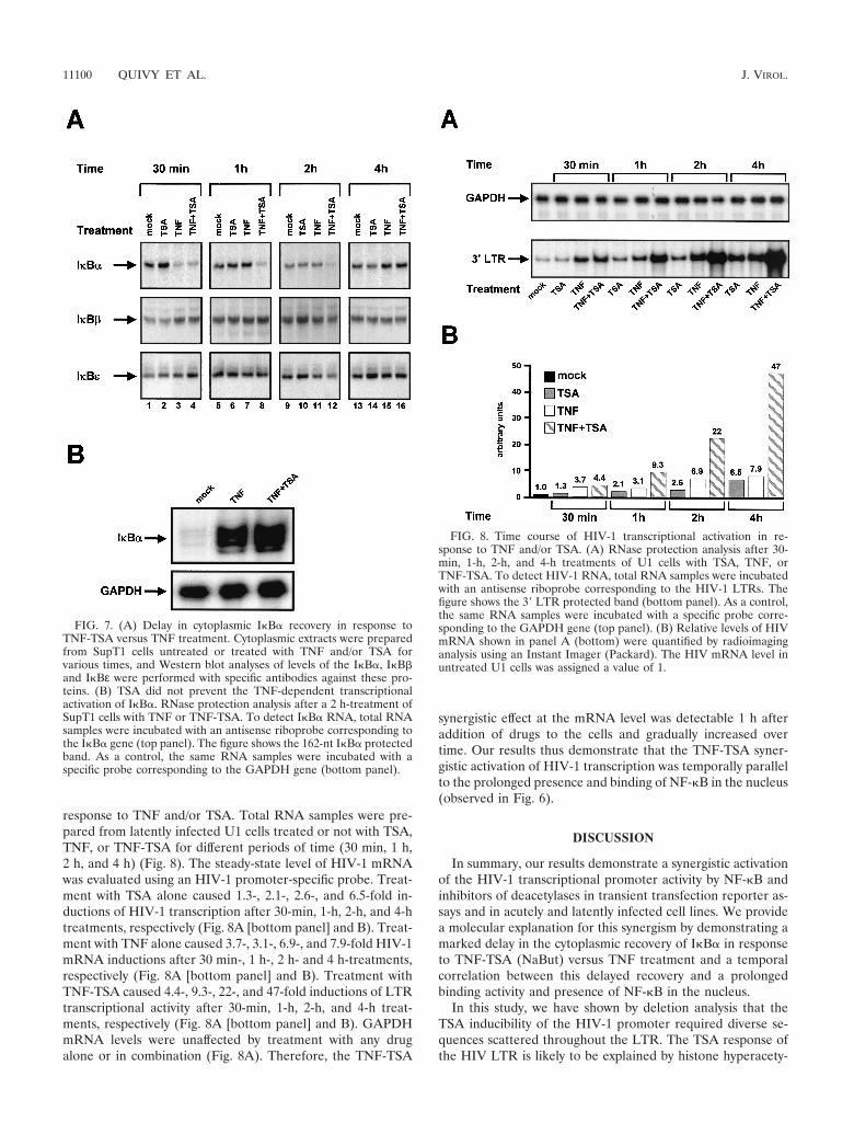
response to TNF and/or TSA. Total RNA samples were pre-pared from latently infected U1 cells treated or not with TSA,TNF, or TNF-TSA for different periods of time (30 min, 1 h,2 h, and 4 h) (Fig. 8). The steady-state level of HIV-1 mRNAwas evaluated using an HIV-1 promoter-specific probe. Treat-ment with TSA alone caused 1.3-, 2.1-, 2.6-, and 6.5-fold in-ductions of HIV-1 transcription after 30-min, 1-h, 2-h, and 4-htreatments, respectively (Fig. 8A [bottom panel] and B). Treat-ment with TNF alone caused 3.7-, 3.1-, 6.9-, and 7.9-fold HIV-1mRNA inductions after 30 min-, 1 h-, 2 h- and 4 h-treatments,respectively (Fig. 8A [bottom panel] and B). Treatment withTNF-TSA caused 4.4-, 9.3-, 22-, and 47-fold inductions of LTRtranscriptional activity after 30-min, 1-h, 2-h, and 4-h treat-ments, respectively (Fig. 8A [bottom panel] and B). GAPDHmRNA levels were unaffected by treatment with any drugalone or in combination (Fig. 8A). Therefore, the TNF-TSA
synergistic effect at the mRNA level was detectable 1 h afteraddition of drugs to the cells and gradually increased overtime. Our results thus demonstrate that the TNF-TSA syner-gistic activation of HIV-1 transcription was temporally parallelto the prolonged presence and binding of NF-�B in the nucleus(observed in Fig. 6).
DISCUSSION
In summary, our results demonstrate a synergistic activationof the HIV-1 transcriptional promoter activity by NF-�B andinhibitors of deacetylases in transient transfection reporter as-says and in acutely and latently infected cell lines. We providea molecular explanation for this synergism by demonstrating amarked delay in the cytoplasmic recovery of I�B� in responseto TNF-TSA (NaBut) versus TNF treatment and a temporalcorrelation between this delayed recovery and a prolongedbinding activity and presence of NF-�B in the nucleus.
In this study, we have shown by deletion analysis that theTSA inducibility of the HIV-1 promoter required diverse se-quences scattered throughout the LTR. The TSA response ofthe HIV LTR is likely to be explained by histone hyperacety-
FIG. 7. (A) Delay in cytoplasmic I�B� recovery in response toTNF-TSA versus TNF treatment. Cytoplasmic extracts were preparedfrom SupT1 cells untreated or treated with TNF and/or TSA forvarious times, and Western blot analyses of levels of the I�B�, I�B�and I�Bε were performed with specific antibodies against these pro-teins. (B) TSA did not prevent the TNF-dependent transcriptionalactivation of I�B�. RNase protection analysis after a 2 h-treatment ofSupT1 cells with TNF or TNF-TSA. To detect I�B� RNA, total RNAsamples were incubated with an antisense riboprobe corresponding tothe I�B� gene (top panel). The figure shows the 162-nt I�B� protectedband. As a control, the same RNA samples were incubated with aspecific probe corresponding to the GAPDH gene (bottom panel).
FIG. 8. Time course of HIV-1 transcriptional activation in re-sponse to TNF and/or TSA. (A) RNase protection analysis after 30-min, 1-h, 2-h, and 4-h treatments of U1 cells with TSA, TNF, orTNF-TSA. To detect HIV-1 RNA, total RNA samples were incubatedwith an antisense riboprobe corresponding to the HIV-1 LTRs. Thefigure shows the 3� LTR protected band (bottom panel). As a control,the same RNA samples were incubated with a specific probe corre-sponding to the GAPDH gene (top panel). (B) Relative levels of HIVmRNA shown in panel A (bottom) were quantified by radioimaginganalysis using an Instant Imager (Packard). The HIV mRNA level inuntreated U1 cells was assigned a value of 1.
11100 QUIVY ET AL. J. VIROL.

lation. Indeed, our laboratory has previously demonstrated bychromatin mapping experiments that a nucleosome (nuc-1)positioned immediately downstream of the transcription startsite is remodeled upon activation of the HIV promoter inresponse to HDAC inhibitors (54). Nuc-1 is likely to be theunique nucleosome target of action of the deacetylases since itis the only nucleosome whose structure or conformation isaffected when deacetylases are inhibited (54). Although we didnot investigate in the present study the chromatin organizationof the transiently transfected LTR templates, we speculatedthat our LTR templates with deletions exhibited nucleosomepositioning similar to that observed in vivo. Indeed, it is knownthat transfected DNA rapidly assembles into minichromo-somes with histones attached (45). Moreover, the DEAE-dex-tran transfection technique we used here allows the typical160-bp DNA ladder characteristic of the physiological nucleo-somal DNA (24). Finally, in vitro chromatin-reconstitutedHIV-1 templates corroborate the native nucleosomal organi-zation found in latently infected cells (48, 49).
Importantly, because of the numerous nonhistone proteinsubstrates for acetylation, the TSA response of the HIV-1promoter could be explained in large part by acetylation anddeacetylation events involved in the regulation of nuclear fac-tors binding to the LTR. On one hand, several of these factors,including AP-1, ligand-bound nuclear hormone receptors, c-Myb, glucocorticoid receptor (GR), NF-AT, E-box bindingproteins, Ets-1, TCF/LEF, NF-�B, Sp1, interferon regulatoryfactor (IRF), and the HIV trans-activator Tat have been shownto interact with acetyltransferases. On the other hand, severaltranscription factors that bind to the LTR, including unligan-ded nuclear hormone receptors, GR, E-box binding proteins,YY1, Sp1, TCF/LEF have been shown to interact with deacety-lases. These factors therefore represent good candidates forthe specific targeting of acetyltransferases and deacetylases tothe HIV promoter, thereby regulating the acetylation level ofhistones (in particular nuc-1 histones) and/or of transcriptionfactor substrates binding to the LTR (such as c-Myb, Sp1, IRF,TFIIE� and TFIIF, and Tat) (43a). The addition and removalof acetyl groups on these histone and nonhistone proteinscould be crucial in controlling transcription initiation and elon-gation.
Thus, the HIV promoter appears to contain numerous cis-regulatory DNA elements involved in the inducibility of theLTR by TSA. As such, the HIV promoter is representative ofa small subset (2%) of cellular genes that have their expres-sion upregulated by deacetylase inhibitors (55) and constitutesa unique regulatory model system to study the complex rela-tionship between acetylation processes and transcriptional ac-tivity.
Importantly, a significant TSA inducibility was still observedwith a reduced LTR (nt 345 to 531), containing the two �Bsites, the three Sp1 sites, the TATA box, and the LBP-1/YY1site (Fig. 1A and B). This could be explained by the recruit-ment at the level of these sites of different factors presentinglinkages with deacetylation and acetylation processes. (i) Atthe Sp1 sites, Sp1 is acetylated in vitro by p300 and interactswith p300, which acts as a coactivator for Sp1-mediated tran-scriptional activation (50). Sp1 has also been shown to interactdirectly with HDAC-1 (10). (ii) At the TATA box, the generaltranscription factors TFIIE� and TFIIF are acetylated in vitro
by P/CAF and p300 (21). The TFIID subunit TBP-associatedfactor II250 is a histone acetyltransferase (34). (iii) At theLBP-1 site, LBP functions as a docking molecule for YY1,which in turn acts by recruiting HDAC-1. This ternary complexrepresses the HIV-1 promoter, probably via the HDAC activitysince this repression is blocked by TSA (9). (iv) At the �Bsites, NF-�B-dependent gene expression requires the func-tion of transcriptional coactivator proteins, including CBP/p300, P/CAF, and SRC-1, which possess acetyltransferaseactivity (17, 35, 40). Moreover, there is some evidence tosuggest that deacetylase inhibitors may function to positivelyregulate NF-�B transcriptional activity (53).
In the present study, we focused on the potential functionalrole of these �B sites in the TSA inducibility of the HIV-1 LTRand demonstrated a strong transcriptional synergism betweendeacetylase inhibitors and NF-�B.
Mechanistically, we showed here by gel retardation assaysthat TSA (or NaBut) prolonged TNF-induced NF-�B DNA-binding activity, whereas TSA (or NaBut) alone caused noinduction of NF-�B. These in vitro binding studies coincidedwith a sustained nuclear p65 presence as revealed by immuno-blotting and confocal immunofluorescence microscopy. Impor-tantly, Western blot analysis also revealed a marked delay inthe cytoplasmic reappearance of the inhibitory protein I�B�after TNF-TSA versus TNF treatment. This delay correlatedtemporally with the sustained binding activity and presence ofNF-�B in the nucleus and was not due to a defect in the I�B�mRNA production. These data therefore provide a molecularmechanism involving I�B� for the functional synergism weobserved between TNF and inhibitors of deacetylases. I�B�plays a pivotal role in the NF-�B signaling pathway. Indeed,the primary level of regulation of NF-�B activity is through itsretention in the cytoplasm through interactions with I�B�.Moreover, the resynthesis of de novo I�B� participates in anegative feedback system ensuring a transient NF-�B tran-scriptional response (reviewed in reference 28). We are cur-rently further investigating the role of TSA in the delayedcytoplasmic reappearance of I�B� reported here. To this end,we are testing whether some proteins involved in the NF-�B/I�B signaling have their expression and/or action modulated byTSA.
The molecular mechanisms mediating the TNF-TSA syner-gism are likely to be highly complex and to implicate phenom-ena other than the delayed cytoplasmic recovery of I�B�. Onone hand, the direct acetylation of Rel family members couldalso intervene in the mechanism of synergistic activation byTNF and TSA. In this regard, we have shown in a separatestudy that the p65 and p50 NF-�B subunits are weakly subjectto reversible acetylation (E. Adam, V. Quivy, and C. Van Lint,unpublished results). In agreement, during the preparation ofthe present work, W. Greene and colleagues reported that p65is acetylated and that this posttranslational modification gov-erns I�B� binding to p65 and the nuclear export of the NF-�Bcomplex (4). On the other hand, deacetylase corepressor pro-teins might be involved in the TNF-TSA synergism. Consistentwith this, we have recently shown by in vitro interaction assaysthat HDAC-1, -3, and -4 can interact directly with the p65 andp50 subunits of NF-�B (Y. de Launoit and C. Van Lint, un-published results). During the preparation of this manuscript,two other groups have separately reported the interaction
VOL. 76, 2002 SYNERGISM BETWEEN DEACETYLASE INHIBITORS AND TNF 11101

of p65 either with HDAC-1 (2) or with HDAC-3 (4). TheseHDACs could repress expression of NF-�B-regulated genes bymaintaining histones and/or other proteins in a deacetylatedstate. TSA or NaBut, which inhibit the HDAC activity, wouldincrease NF-�B-dependent transcription by alleviating thechromatin- and/or factor-mediated block to transcriptional ac-tivation.
The application of HAART has resulted in a major reduc-tion of virus loads in individuals tolerating the regimen andcomplying with its requirements, a stabilization of the clinicalcourse, and a significant decline in mortality. Nonetheless, thepersistence of HIV reservoirs has posed a sobering challengeto the long-term control or eradication of HIV in infectedindividuals receiving HAART (reviewed in reference 42).These latently infected cells are a permanent source for reac-tivation and lead to a rebound of viral load levels after inter-ruption of HAART (15, 61). Activators of HIV expressioncombined with HAART could lead to the elimination of thelatently infected cells and to the eradication of the infection.Indeed, it is likely that the latently infected cells die uponreactivation of virus (39) and that HAART prevents spread ofreleased virus to adjacent cells (5). In this report, we demon-strate a synergistic effect of TNF and TSA (NaBut) for HIV-1reactivation in the U1 cell line, a postintegration latency cellculture model. It is important to note that an array of cyto-kines, including the proinflammatory cytokines TNF and inter-leukin-1 (inducers of NF-�B), are already copiously expressedin the microenvironment of the lymphoid tissues, which harborlatent viral reservoirs (36). Therefore, our results suggest thatthe use of deacetylases inhibitors in the treatment of HIVinfection may represent a valuable approach for purging thelatently infected reservoirs in HAART-treated individuals.These deacetylase inhibitors would synergize with the TNFalready present at increased level in the serum of the HIV-infected individuals. The possible clinical use of deacetylaseinhibitors raises several issues. First, these drugs do not act ina cell-specific manner. Second, this class of agents is safelyadministered for other diseases, including beta chain hemoglo-binopathies (such as beta-thalassemia and sickle cell anemia)(8, 11) and epilepsy and bipolar disorders (25, 41, 51). Third,an increasing number of non-B HIV-1 subtype infections arecurrently diagnosed. Here, we have shown that, in addition tothe prototypical subtype B LTR, the LTRs from subtypes Athrough G of the HIV-1 group M were also activated syner-gistically by TSA and TNF, and the amplitude of the synergismcorrelated with the number of �B sites in the respective LTRs,which varies from one (subtype E) to three (subtype C). Basedon our results, we propose the administration of deacetylaseinhibitor(s) together with continuous HAART as a new poten-tial therapeutic perspective to decrease in a subtype-nonspe-cific manner the pool of latent HIV reservoirs.
In conclusion, the results described in this study provide newinsights into HIV-1 transcriptional regulation and more gen-erally into the molecular mechanisms of NF-�B-mediatedtransactivation.
ACKNOWLEDGMENTS
V.Q. and E.A. contributed equally to this work.We thank Francoise Bex for her help in the confocal microscopy
analysis and Fabrice Moore for his help in the transfection assays. The
following reagents were obtained through the AIDS Research andReference Reagent Program, NIAID, NIH: pRSV-p50 and pRSV-p65from G. Nabel and N. Perkins, the U1 cell line from T. Folks, and theSupT1 cell line from J. Hoxie. We are grateful to L. Vanhamme and C.Calomme for critical reading of the manuscript.
This work was supported by grants to C.V.L. from the Fonds Na-tional de la Recherche Scientifique (FNRS, Belgium), the Televie-Program, the Universite Libre de Bruxelles (ULB), the InternationaleBrachet Stiftung, the CGRI-INSERM cooperation, the Region Wal-lonne-Commission Europeenne FEDER, the Agence Nationale deRecherches sur le SIDA (ANRS, France), and the Theyskens-MineurFoundation. V.Q. is an Aspirant of the FNRS. A.C. is a ChercheurQualifie of the FNRS. J.P. and V.B. are Directeurs de Recherches ofthe FNRS. C.V.L. is a Maıtre de Recherches of the FNRS. E.A. andD.D. are supported by postdoctoral fellowships from the ULB (ARCprogram 98/03-224) and the Region Wallonne (grant 991/4202), re-spectively. R.C. is supported by a fellowship from the Agence Natio-nale de Recherches sur le SIDA (France).
REFERENCES
1. Adams, M., L. Sharmeen, J. Kimpton, J. M. Romeo, J. V. Garcia, B. M.Peterlin, M. Groudine, and M. Emerman. 1994. Cellular latency in humanimmunodeficiency virus-infected individuals with high CD4 levels can bedetected by the presence of promoter-proximal transcripts. Proc. Natl. Acad.Sci. USA 91:3862–3866.
2. Ashburner, B. P., S. D. Westerheide, and A. S. Baldwin, Jr. 2001. The p65(RelA) subunit of NF-�B interacts with the histone deacetylase (HDAC)corepressors HDAC1 and HDAC2 to negatively regulate gene expression.Mol. Cell. Biol. 21:7065–7077.
3. Chen, H., M. Tini, and R. M. Evans. 2001. HATs on and beyond chromatin.Curr. Opin. Cell Biol. 13:218–224.
4. Chen, L., W. Fischle, E. Verdin, and W. C. Greene. 2001. Duration of nuclearNF-�B action regulated by reversible acetylation. Science 293:1653–1657.
5. Chun, T. W., D. Engel, S. B. Mizell, L. A. Ehler, and A. S. Fauci. 1998.Induction of HIV-1 replication in latently infected CD4� T cells using acombination of cytokines. J. Exp. Med. 188:83–91.
6. Cohen, J. 1998. Exploring how to get at and eradicate hidden HIV. Science279:1854–1855.
7. Col, E., C. Caron, D. Seigneurin-Berny, J. Gracia, A. Favier, and S. Khoch-bin. 2001. The histone acetyltransferase, hGCN5, interacts with and acety-lates the HIV transactivator, Tat. J. Biol. Chem. 276:28179–28184.
8. Collins, A. F., H. A. Pearson, P. Giardina, K. T. McDonagh, S. W. Brusilow,and G. J. Dover. 1995. Oral sodium phenylbutyrate therapy in homozygousbeta thalassemia: a clinical trial. Blood 85:43–49.
9. Coull, J. J., F. Romerio, J. M. Sun, J. L. Volker, K. M. Galvin, J. R. Davie,Y. Shi, U. Hansen, and D. M. Margolis. 2000. The human factors YY1 andLSF repress the human immunodeficiency virus type 1 long terminal repeatvia recruitment of histone deacetylase 1. J. Virol. 74:6790–6799.
10. Doetzlhofer, A., H. Rotheneder, G. Lagger, M. Koranda, V. Kurtev, G.Brosch, E. Wintersberger, and C. Seiser. 1999. Histone deacetylase 1 canrepress transcription by binding to Sp1. Mol. Cell. Biol. 19:5504–5511.
11. Dover, G. J., S. Brusilow, and S. Charache. 1994. Induction of fetal hemo-globin production in subjects with sickle cell anemia by oral sodium phenyl-butyrate. Blood 84:339–343.
12. el Kharroubi, A., G. Piras, R. Zensen, and M. A. Martin. 1998. Transcrip-tional activation of the integrated chromatin-associated human immunode-ficiency virus type 1 promoter. Mol. Cell. Biol. 18:2535–2544.
13. Emiliani, S., W. Fischle, M. Ott, C. Van Lint, C. A. Amella, and E. Verdin.1998. Mutations in the tat gene are responsible for human immunodeficiencyvirus type 1 postintegration latency in the U1 cell line. J. Virol. 72:1666–1670.
14. Emiliani, S., C. Van Lint, W. Fischle, P. Paras, Jr., M. Ott, J. Brady, and E.Verdin. 1996. A point mutation in the HIV-1 Tat responsive element isassociated with postintegration latency. Proc. Natl. Acad. Sci. USA 93:6377–6381.
15. Finzi, D., M. Hermankova, T. Pierson, L. M. Carruth, C. Buck, R. E.Chaisson, T. C. Quinn, K. Chadwick, J. Margolick, R. Brookmeyer, J. Gal-lant, M. Markowitz, D. D. Ho, D. D. Richman, and R. F. Siliciano. 1997.Identification of a reservoir for HIV-1 in patients on highly active antiret-roviral therapy. Science 278:1295–1300.
16. Folks, T. M., K. A. Clouse, J. Justement, A. Rabson, E. Duh, J. H. Kehrl, andA. S. Fauci. 1989. Tumor necrosis factor alpha induces expression of humanimmunodeficiency virus in a chronically infected T-cell clone. Proc. Natl.Acad. Sci. USA 86:2365–2368.
17. Gerritsen, M. E., A. J. Williams, A. S. Neish, S. Moore, Y. Shi, and T.Collins. 1997. CREB-binding protein/p300 are transcriptional coactivators ofp65. Proc. Natl. Acad. Sci. USA 94:2927–2932.
18. Ghosh, S., M. J. May, and E. B. Kopp. 1998. NF-kappa B and Rel proteins:evolutionarily conserved mediators of immune responses. Annu. Rev. Im-munol. 16:225–260.
11102 QUIVY ET AL. J. VIROL.

19. Herschlag, D., and F. B. Johnson. 1993. Synergism in transcriptional activa-tion: a kinetic view. Genes Dev. 7:173–179.
20. Igarashi, T., C. R. Brown, Y. Endo, A. Buckler-White, R. Plishka, N. Bischof-berger, V. Hirsch, and M. A. Martin. 2001. Macrophage are the principalreservoir and sustain high virus loads in rhesus macaques after the depletionof CD4� T cells by a highly pathogenic simian immunodeficiency virus/HIVtype 1 chimera (SHIV): implications for HIV-1 infections of humans. Proc.Natl. Acad. Sci. USA 98:658–663.
21. Imhof, A., X. J. Yang, V. V. Ogryzko, Y. Nakatani, A. P. Wolffe, and H. Ge.1997. Acetylation of general transcription factors by histone acetyltrans-ferases. Curr. Biol. 7:689–692.
22. Israel, A. 2000. The IKK complex: an integrator of all signals that activateNF-�B? Trends Cell Biol. 10:129–133.
23. Jeeninga, R. E., M. Hoogenkamp, M. Armand-Ugon, M. de Baar, K. Verhoef,and B. Berkhout. 2000. Functional differences between the long terminalrepeat transcriptional promoters of human immunodeficiency virus type 1subtypes A through G. J. Virol. 74:3740–3751.
24. Jeong, S., and A. Stein. 1994. Micrococcal nuclease digestion of nucleireveals extended nucleosome ladders having anomalous DNA lengths forchromatin assembled on non-replicating plasmids in transfected cells. Nu-cleic Acids Res. 22:370–375.
25. Johannessen, C. U. 2000. Mechanisms of action of valproate: a commentary.Neurochem. Int. 37:103–110.
26. Jordan, A., P. Defechereux, and E. Verdin. 2001. The site of HIV-1 integra-tion in the human genome determines basal transcriptional activity andresponse to Tat transactivation. EMBO J. 20:1726–1738.
27. Karin, M. 1999. The beginning of the end: I�B kinase (IKK) and NF-�Bactivation. J. Biol. Chem. 274:27339–27342.
28. Karin, M., and Y. Ben Neriah. 2000. Phosphorylation meets ubiquitination:the control of NF-�B activity. Annu. Rev. Immunol. 18:621–663.
29. Khochbin, S., and H. Y. Kao. 2001. Histone deacetylase complexes: func-tional entities or molecular reservoirs. FEBS Lett. 494:141–144.
30. Kiernan, R. E., C. Vanhulle, L. Schiltz, E. Adam, H. Xiao, F. Maudoux, C.Calomme, A. Burny, Y. Nakatani, K. T. Jeang, M. Benkirane, and C. VanLint. 1999. HIV-1 tat transcriptional activity is regulated by acetylation.EMBO J. 18:6106–6118.
31. Kouzarides, T. 2000. Acetylation: a regulatory modification to rival phos-phorylation? EMBO J. 19:1176–1179.
32. Li, N., and M. Karin. 2000. Signaling pathways leading to nuclear factor-kappa B activation. Methods Enzymol. 319:273–279.
33. McCune, J. M. 1995. Viral latency in HIV disease. Cell 82:183–188.34. Mizzen, C. A., X. J. Yang, T. Kokubo, J. E. Brownell, A. J. Bannister, T.
Owen-Hughes, J. Workman, L. Wang, S. L. Berger, T. Kouzarides, Y. Na-katani, and C. D. Allis. 1996. The TAF(II)250 subunit of TFIID has histoneacetyltransferase activity. Cell 87:1261–1270.
35. Na, S. Y., S. K. Lee, S. J. Han, H. S. Choi, S. Y. Im, and J. W. Lee. 1998.Steroid receptor coactivator-1 interacts with the p50 subunit and coactivatesnuclear factor �B-mediated transactivations. J. Biol. Chem. 273:10831–10834.
36. Navikas, V., J. Link, C. Persson, T. Olsson, B. Hojeberg, A. Ljungdahl, H.Link, and B. Wahren. 1995. Increased mRNA expression of IL-6, IL-10,TNF-alpha, and perforin in blood mononuclear cells in human HIV infec-tion. J. Acquir. Immune Defic. Syndr. Hum. Retrovirol. 9:484–489.
37. Osborn, L., S. Kunkel, and G. J. Nabel. 1989. Tumor necrosis factor alphaand interleukin 1 stimulate the human immunodeficiency virus enhancer byactivation of the nuclear factor kappa B. Proc. Natl. Acad. Sci. USA 86:2336–2340.
38. Pereira, L. A., K. Bentley, A. Peeters, M. J. Churchill, and N. J. Deacon.2000. A compilation of cellular transcription factor interactions with theHIV-1 LTR promoter. Nucleic Acids Res. 28:663–668.
39. Perelson, A. S., P. Essunger, Y. Cao, M. Vesanen, A. Hurley, K. Saksela, M.Markowitz, and D. D. Ho. 1997. Decay characteristics of HIV-1-infectedcompartments during combination therapy. Nature 387:188–191.
40. Perkins, N. D., L. K. Felzien, J. C. Betts, K. Leung, D. H. Beach, and G. J.Nabel. 1997. Regulation of NF-�B by cyclin-dependent kinases associatedwith the p300 coactivator. Science 275:523–527.
41. Phiel, C. J., F. Zhang, E. Y. Huang, M. G. Guenther, M. A. Lazar, and P. S.Klein. 2001. Histone deacetylase is a direct target of valproic acid, a potent
anticonvulsant, mood stabilizer, and teratogen. J. Biol. Chem. 276:36734–36741.
42. Pierson, T., J. McArthur, and R. F. Siliciano. 2000. Reservoirs for HIV-1:mechanisms for viral persistence in the presence of antiviral immune re-sponses and antiretroviral therapy. Annu. Rev. Immunol. 18:665–708.
43. Poli, G., A. Kinter, J. S. Justement, J. H. Kehrl, P. Bressler, S. Stanley, andA. S. Fauci. 1990. Tumor necrosis factor alpha functions in an autocrinemanner in the induction of human immunodeficiency virus expression. Proc.Natl. Acad. Sci. USA 87:782–785.
43a.Quivy, V., and C. Van Lint. 2002. Diversity of acetylation targets and roles intranscriptional regulation: the HIV-1 promoter as a model system. Biochem.Pharmacol. 64:925–934.
44. Rabson, A. B., and H. C. Lin. 2000. NF-kappa B and HIV: linking viral andimmune activation. Adv. Pharmacol. 48:161–207.
45. Reeves, R., C. M. Gorman, and B. Howard. 1985. Minichromosome assemblyof non-integrated plasmid DNA transfected into mammalian cells. NucleicAcids Res. 13:3599–3615.
46. Roth, S. Y., J. M. Denu, and C. D. Allis. 2001. Histone acetyltransferases.Annu. Rev. Biochem. 70:81–120.
47. Schoonbroodt, S., V. Ferreira, M. Best-Belpomme, J. R. Boelaert, S. Le-grand-Poels, M. Korner, and J. Piette. 2000. Crucial role of the amino-terminal tyrosine residue 42 and the carboxyl-terminal PEST domain of Ikappa B alpha in NF-kappa B activation by an oxidative stress. J. Immunol.164:4292–4300.
48. Sheridan, P. L., T. P. Mayall, E. Verdin, and K. A. Jones. 1997. Histoneacetyltransferases regulate HIV-1 enhancer activity in vitro. Genes Dev.11:3327–3340.
49. Steger, D. J., A. Eberharter, S. John, P. A. Grant, and J. L. Workman. 1998.Purified histone acetyltransferase complexes stimulate HIV-1 transcriptionfrom preassembled nucleosomal arrays. Proc. Natl. Acad. Sci. USA 95:12924–12929.
50. Suzuki, T., A. Kimura, R. Nagai, and M. Horikoshi. 2000. Regulation ofinteraction of the acetyltransferase region of p300 and the DNA-bindingdomain of Sp1 on and through DNA binding. Genes Cells 5:29–41.
51. Tunnicliff, G. 1999. Actions of sodium valproate on the central nervoussystem. J. Physiol. Pharmacol. 50:347–365.
52. Van Lint, C. 2000. Role of chromatin in HIV-1 transcriptional regulation.Adv. Pharmacol. 48:121–160.
53. Vanden Berghe, W., K. De Bosscher, E. Boone, S. Plaisance, and G. Haege-man. 2098. 1999. The nuclear factor-�B engages CBP/p300 and histoneacetyltransferase activity for transcriptional activation of the interleukin-6gene promoter. J. Biol. Chem. 274:32091–32093.
54. Van Lint, C., S. Emiliani, M. Ott, and E. Verdin. 1996. Transcriptionalactivation and chromatin remodeling of the HIV-1 promoter in response tohistone acetylation. EMBO J. 15:1112–1120.
55. Van Lint, C., S. Emiliani, and E. Verdin. 1996. The expression of a smallfraction of cellular genes is changed in response to histone hyperacetylation.Gene Expr. 5:245–253.
56. Van Lint, C., J. Ghysdael, P. Paras, Jr., A. Burny, and E. Verdin. 1994. Atranscriptional regulatory element is associated with a nuclease-hypersensi-tive site in the pol gene of human immunodeficiency virus type 1. J. Virol.68:2632–2648.
57. Van Lint, C., C. A. Amella, S. Emiliani, M. John, T. Jie, and E. Verdin. 1997.Transcription factor binding sites downstream of the human immunodefi-ciency virus type 1 transcription start site are important for virus infectivity.J. Virol. 71:6113–6127.
58. Verdin, E., P. Paras, Jr., and C. Van Lint. 1993. Chromatin disruption in thepromoter of human immunodeficiency virus type 1 during transcriptionalactivation. EMBO J. 12:3249–3259.
59. West, M. J., A. D. Lowe, and J. Karn. 2001. Activation of human immuno-deficiency virus transcription in T cells revisited: NF-�B p65 stimulatestranscriptional elongation. J. Virol. 75:8524–8537.
60. Winslow, B. J., R. J. Pomerantz, O. Bagasra, and D. Trono. 1993. HIV-1latency due to the site of proviral integration. Virology 196:849–854.
61. Wong, J. K., M. Hezareh, H. F. Gunthard, D. V. Havlir, C. C. Ignacio, C. A.Spina, and D. D. Richman. 1997. Recovery of replication-competent HIVdespite prolonged suppression of plasma viremia. Science 278:1291–1295.
VOL. 76, 2002 SYNERGISM BETWEEN DEACETYLASE INHIBITORS AND TNF 11103