functional surfaces of the human immunodeficiency …jvi.asm.org/content/77/9/5439.full.pdf ·...
TRANSCRIPT

JOURNAL OF VIROLOGY, May 2003, p. 5439–5450 Vol. 77, No. 90022-538X/03/$08.00�0 DOI: 10.1128/JVI.77.9.5439–5450.2003Copyright © 2003, American Society for Microbiology. All Rights Reserved.
Functional Surfaces of the Human Immunodeficiency Virus Type 1Capsid Protein
Uta K. von Schwedler, Kirsten M. Stray, Jennifer E. Garrus, and Wesley I. Sundquist*Department of Biochemistry, University of Utah, Salt Lake City, Utah 84132
Received 10 July 2002/Accepted 6 February 2003
The human immunodeficiency virus type 1 initially assembles and buds as an immature particle that isorganized by the viral Gag polyprotein. Gag is then proteolyzed to produce the smaller capsid protein CA,which forms the central conical capsid that surrounds the RNA genome in the mature, infectious virus. Todefine CA surfaces that function at different stages of the viral life cycle, a total of 48 different alanine-scanningsurface mutations in CA were tested for their effects on Gag protein expression, processing, particle productionand morphology, capsid assembly, and infectivity. The 27 detrimental mutations fall into three classes: 13mutations significantly diminished or altered particle production, 9 mutations failed to assemble normalcapsids, and 5 mutations supported normal viral assembly but were nevertheless reduced more than 20-fold ininfectivity. The locations of the assembly-defective mutations implicate three different CA surfaces in immatureparticle assembly: one surface encompasses helices 4 to 6 in the CA N-terminal domain (NTD), a secondsurrounds the crystallographically defined CA dimer interface in the C-terminal domain (CTD), and a thirdsurrounds the loop preceding helix 8 at the base of the CTD. Mature capsid formation required a distinctsurface encompassing helices 1 to 3 in the NTD, in good agreement with a recent structural model for the viralcapsid. Finally, the identification of replication-defective mutants with normal viral assembly phenotypesindicates that CA also performs important nonstructural functions at early stages of the viral life cycle.
Several stages of the retroviral life cycle, including virionassembly, budding, maturation, entry, uncoating, and replica-tion, occur within large, multicomponent particles that areorganized by the structural Gag polyprotein or its proteolyticproducts (see references 31, 41, 54, 79, 85, 90 and 97 forreviews). Late in the infectious cycle, unprocessed human im-munodeficiency virus type 1 (HIV-1) Gag molecules assemblebeneath the plasma membrane and then bud from the cell asimmature, enveloped virions. These immature particles areroughly spherical but lack global symmetry elements and ex-hibit a range of diameters (�100 to 140 nm), indicating thatthey are not strictly regular objects (33, 96, 99, 101). Therod-shaped Gag molecules associate on the membrane withtheir long axes projecting radially toward the center of theimmature virion. Each HIV-1 Gag molecule is composed of aseries of distinct domains (denoted MA, CA, NC, and p6, fromthe N to C terminus), as well as two small spacer peptides (SP1and SP2) (Fig. 1B). The myristoylated MA domain associateswith the membrane, and the CA and NC polypeptides formshells of successively smaller radii (96). Although the lateralinteractions between adjacent Gag molecules in the immatureparticle lattice are not yet understood in detail, there are in-dications that the lattice may be constructed from Gag trimers(46, 66, 74).
As the virus buds, HIV-1 Gag is sequentially processed atfive different sites by the viral protease, releasing the MA, CA,and NC polypeptides as discrete proteins. This triggers a majorstructural rearrangement (maturation) in which NC and the
RNA genome condense into a complex at the center of thevirion, CA assembles into a conical shell (the capsid) thatsurrounds the NC/RNA complex, and MA remains associatedwith the viral membrane (Fig. 1A). Maturation is required forinfectivity and presumably converts the virion from a particlethat can assemble and bud into a particle that can disassembleand replicate in a new host cell (94).
Previous studies of retroviral Gag proteins have revealed theinvolvement of several distinct elements at different stages ofimmature particle production (31, 41, 54, 79, 85, 90, 97). Themajor signal(s) for trafficking and retention of HIV-1 Gag atthe plasma membrane (the M domain), reside within MA (26,68, 70, 80, 104), and signal(s) required for late stages of particlerelease (the L domain) are located within the p6 polypeptide(14, 32, 42, 60, 72). In contrast, the regions of Gag that par-ticipate in spherical particle assembly appear to be larger andmore complex, presumably because Gag proteins associatealong their lengths and may therefore make multiple contactswith neighboring molecules. Nevertheless, genetic analyseshave revealed that the most important determinant(s) for Gagassembly span the C-terminal domain of CA, SP1, and NC(which contains the I, or interaction, domain) (31, 41). The Idomain within NC appears to facilitate assembly primarily bytethering Gag molecules together via nonspecific RNA bindinginteractions (10, 18, 19, 22, 102).
High-resolution structures of the mature HIV-1 MA, CA,and NC proteins have been determined (reviewed in reference89), and the structures of these proteins appear to be con-served across retroviruses. In particular, CA proteins fromHIV-1 (4, 34, 38, 64), equine infectious anemia virus (47),human T-cell leukemia virus type I (20, 49), and Rous sarcomavirus (13, 51) all adopt similar two-domain structures, withtheir helical N- and C-terminal domains (NTD and CTD)
* Corresponding author. Mailing address: Department of Biochem-istry, University of Utah, 20 N 1900 East, Salt Lake City, UT 84132-3201. Phone: (801) 585-5402. Fax: (801) 581-7959. E-mail: [email protected].
5439
on June 15, 2018 by guesthttp://jvi.asm
.org/D
ownloaded from
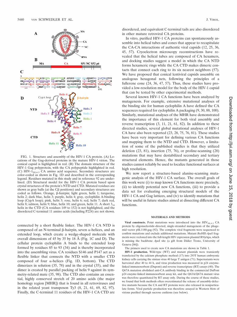
connected by a short flexible linker. The HIV-1 CA NTD iscomposed of an N-terminal �-hairpin, seven �-helices, and anextended loop, which create a wedge-shaped molecule withoverall dimensions of 45 by 35 by 18 A (Fig. 1C and D). Thecellular protein cyclophilin A binds to the extended loopformed by residues 85 to 93 (34) and is thereby incorporatedinto the assembling virus. CA residues S146 and P147 act as aflexible linker that connects the NTD with a smaller CTDcomposed of four �-helices (Fig. 1D, bottom). The CTDdimerizes in solution (35, 78) and in the crystal (35), and thedimer is created by parallel packing of helix 9 against its sym-metry-related mate (35, 98). The CTD also contains an essen-tial, highly conserved stretch of 20 amino acids (the majorhomology region [MHR]) that is found in all retroviruses andin the related yeast transposon Ty3 (8, 21, 61, 69, 82, 97).Finally, the C-terminal 11 residues of the HIV-1 CA CTD are
disordered, and equivalent C-terminal tails are also disorderedin other mature retroviral CA proteins.
In vitro, purified HIV-1 CA proteins can spontaneously as-semble into helical tubes and cones that appear to recapitulatethe CA-CA interactions of authentic viral capsids (12, 25, 36,45, 57). Cryoelectron microscopy reconstructions have re-vealed that the helical tubes are composed of CA hexamers,and docking studies suggest a model in which the CA NTDforms hexameric rings while the CA CTD makes dimeric con-tacts that connect each ring to its six nearest neighbors (57).We have proposed that conical lentiviral capsids assemble onanalogous hexagonal nets, following the principles of afullerene cone (24, 36, 47, 57). Thus, these studies have pro-vided a low-resolution model for the body of the HIV-1 capsidthat can be tested by other experimental methods.
Several known HIV-1 CA functions have been analyzed bymutagenesis. For example, extensive mutational analyses ofthe binding site for human cyclophilin A have defined the CAsequences required for cyclophilin A packaging (9, 30, 88, 100).Similarly, mutational analyses of the MHR have demonstratedthe importance of this element for both viral assembly andreverse transcription (3, 11, 21, 61, 82). In addition to thesedirected studies, several global mutational analyses of HIV-1CA have also been reported (23, 28, 75, 76, 81). These studieshave been very important for defining various CA functionsand mapping them to the NTD and CTD. However, a limita-tion of some of the published studies is that they utilizeddeletion (23, 81), insertion (75, 76), or proline-scanning (28)mutations that may have destabilized secondary and tertiarystructural elements. Hence, the mutants generated in thosestudies cannot reliably be used to localize functional surfaces athigh resolution.
We now report a structure-based alanine-scanning muta-genic analysis of the HIV-1 CA surface. The overall goals ofthis work were (i) to map known CA functions more precisely,(ii) to identify potential new CA functions, (iii) to provide adata set for evaluating emerging structural models of theHIV-1 CA and Gag lattices, and (iv) to identify mutations thatwill be useful in future studies aimed at dissecting different CAfunctions.
MATERIALS AND METHODS
Viral constructs. Point mutations were introduced into the HIVNL4-3 CAprotein by oligonucleotide-directed, single-stranded mutagenesis of the phage-mid vector pSL1180-gag (92). The complete viral fragments were sequenced toconfirm mutations and exclude additional mutations. Mutant BssHII-ApaI frag-ments were recloned into the full-length HIV expression plasmid R9�Apa, whichis missing the backbone ApaI site (a gift from Didier Trono, University ofGeneva [86]).
The primers used to create new CA mutations are shown in Table 1.HIV-1 production. Wild-type (WT) and mutant plasmids were transiently
transfected by the calcium phosphate method (17) into 293T human embryonickidney cells carrying the simian virus 40 large T antigen (71). Supernatants wereharvested after 40 to 44 h, and virus production was measured in p24 enzyme-linked immunosorbent (Dupont) and reverse transcriptase (RT) assays (40). TheQ67A mutation abolished anti-CA antibody binding in the commercial DuPontp24 enzyme-linked immunosorbent assay kit, and the Q63A/Q67A mutant viruswas therefore quantitated by RT assay only. During the course of these studies,we found that both methods often overestimated the release of assembly-defec-tive mutants because the CA and RT proteins were also released in nonparticu-late forms. Viral particle production was therefore assayed in Western blots ofvirions purified through sucrose cushions (see below).
FIG. 1. Structure and assembly of the HIV-1 CA protein. (A) Lo-cations of the Gag-derived proteins in the mature HIV-1 virion. Theconical capsid is highlighted in red. (B) The domain structure of theHIV-1 Gag polyprotein, with the CA polypeptide highlighted in red.(C) HIV-1NL4-3 CA amino acid sequence. Secondary structures arecolor-coded as shown in Fig. 1D and described in the correspondinglegend. Residues mutated in this study and in reference 92 are under-lined. (D) Structural model for the HIV-1 CA protein based uponcrystal structures of the protein’s NTD and CTD. Mutated residues areshown as gray balls (at the C� positions) and secondary structures arecoded as follows. Orange, �-hairpin; light green, helix 1; turquoise,helix 2; dark blue, helix 3; purple, helix 4; gray, cyclophilin A-bindingloop (CypA loop); pink, helix 5; rose, helix 6; red, helix 7; dark red,helix 8; salmon, helix 9; blue, helix 10; and green, helix 11. A short 310helix in the CTD (CA residues 149 to 152) is not highlighted, and thedisordered C-terminal 11 amino acids (including P224) are not shown.
5440 VON SCHWEDLER ET AL. J. VIROL.
on June 15, 2018 by guesthttp://jvi.asm
.org/D
ownloaded from

Viral infectivity. Viral infectivities were quantitated in multinucleate activationof galactosidase indicator cell (MAGIC) assays by using P4 HeLa.CD4.LTR.�-gal indicator cells (16), which stain blue with 5-bromo-4-chloro-3-indolyl-�-D-galactopyranoside (X-Gal) after HIV-1 infection and Tat protein expression(50). Infected cells were counted after 48 h, and relative infectivities werequantitated as the number of blue cells per nanogram of p24 of input virus (orunits of RT activity for the Q63A/Q67A mutant) and compared with that of WT.Infective titers for the WT virus were typically 2.5 � 105 infectious units/ml. Thedata in Table 2 are the averaged results of at least three repetitions for eachmutant; viral titers reduced more than 100-fold were labeled 0. Growth curves(K158A, K158D, K158Q, M185A, D197A, D197N) were performed for up to 100days in CEM-ss T cells after normalization for CA protein (92).
Western blotting. Transfected 293T cells were harvested directly into radio-immunoprecipitation assay buffer (10 mM TrisCl [pH 7.0], 150 mM NaCl, 1%NP-40, 0.1% sodium dodecyl sulfate [SDS]) for Western blotting, whereas virus-containing supernatants were pelleted through 20% sucrose and then resus-pended in 25 �l of SDS sample buffer. Proteins from 2 �l of pelleted virions wereseparated by 10 to 15% SDS-polyacrylamide gel electrophoresis, transferred,blotted with antisera, and detected by chemiluminescence (91). The primaryantibodies used were rabbit anti-MA at 1:50,000 from Didier Trono, rabbitanti-CA (nos. 40 and 49) at 1:2,000 from Hans-Georg Krausslich , mouse mono-clonal anti-RT mAb21 at 1:500 (obtained from Stephen Hughes through the
AIDS Research and Reference Reagent Program) (27), and human monoclonalanti-Env gp41 2F5 at 1:1,000 (obtained from Hermann Katinger through theAIDS Reagent Program, cat. no. 1475) (73).
At least three independent Western blots were performed for each mutant.Figure 2 and Table 2 show representative data and were fully reproducible,except that (i) the intensities of the unprocessed p55 Gag bands varied betweenexperiments, (ii) CA degradation products of �23 and �18 kDa were sometimesobserved, particularly in the K70A and E128A/R132A mutants (see Fig. 2), and(iii) the slight reduction in particle production with the T54A/N57A mutationapparent in Fig. 2 was not reproducible.
Transmission EM. Free viral particles were concentrated for electron micros-copy (EM) studies either by pelleting through 20% sucrose (as described forWestern blotting) or by adjustment of the supernatant to 1� fixative (2.5%glutaraldehyde–1% paraformaldehyde in sodium cacodylate buffer) and 20%fetal calf serum and pelleting for 90 min in a microcentrifuge. Pelleted virionswere resuspended in 1� EM fixative and processed for EM as described previ-ously (92). Cellular vesicles present in these preparations (6, 39) served ascarriers through this procedure, particularly for mutants with lower particleyields. To visualize cell-associated virions, transfected 293T cells were fixed in 1�EM fixative for 30 min without a rinse and processed the same way, except thatdehydration was in EtOH and embedding was in Spurr’s plastic. Transfectionand preparation of K158A-transfected HeLa cells for EM was performed as
TABLE 1. Primers used to create new CA mutations
HIV CA mutant 5�-3� (forward) sequence (mutated codons underlined) Altered restriction site
Q4A TACCCCTATAGTCGCGAACCTCCAGGG NruIQ13A GGCAAATGGTGCATGCGGCCATATCACCTAG SphIR18A/N21A CATATCACCTGCAACTTTAGCTGCATGGGTA NsiI destroyedP38A CCAGAAGTAATCGCGATGTTTTCAGC NruIE45A CATGTTTTCAGCCTTATCGGCAGGAGCCACC BglIT54A CACAAGATTTGAATGCCATGCTAAAC BsmIT54A/N57A AGATTTAATGCCATGCTAGCCACAGTGGG NheIQ63A/Q67A GGGGGGACATGCAGCAGCCATGGCAATGTTAAAAG DsaIK70A CCATGCAAATGCTAGCAGAGACCATC NheIE71A CATGCAAATGCTTAAGGCGACCATCAAT AflIIN74A GTTAAAAGAGACGATCGCTGAGGAAGCTG PvuIE75A/E76A GACCATCAATGCGGCGGCCGCAGAATGGG NotIA78D/E79A/R82A GAGGAAGCTGACGCATGGGATGCATTGCATCCAG PstI destroyedR100A/S102A GAGAGAACCAGCGGGAGCGGACATAGCAGG BsrBIT107A/T108A CATAGCAGGAGCTGCTAGTACCCTTC SpeI destroyedT110A/Q112A GAACTACTAGCGCCCTTGCGGAACAAATAG SpeI destroyedG116A GAACAAATAGCATGGATGACA NoneT119A CAAATAGGATGGATGGCACATAATCC NoneE128A/R132Aa CCCAGTAGGAGCAATCTATAAAGCTTGGATAATCC HindIIIL136D TAAAAGATGGATAATCGATGGATTAAATAAAATAG ClaIN139A GATAATCCTGGGACTCGCGAAAATAGTAAG NruIR143A AATAAAATAGTGGCCATGTATAGCCC MscID152A TACCAGCATTTTAGCCATAAGACAAG BsmI destroyedK158A ACAAGGACCAGCTGAACCCTTTAG PvuIIK158D ACAAGGACCAGATGAACCCTTTA NoneK158R GACAAGGACCTCGAGAACCCTTTA XhoIK158Q ACAAGGACCACAAGAACCCTTA NoneD163A CCCTTTAGAGCCTACGTAGACCGA SnaBIK170A CCGATTCTATGCGACGCTAAGAGCC HgaIQ176A AAGAGCCGAGGCAGCTTCACAAG HindIII destroyedE180A CGAGCAAGCTACACAAGCGGTAAAAAAT HindIII destroyedW184A AGGTAAAAAATGCGATGACAGAAACGTTGTTGGTCCAA Psp 1406IM185A AGGTAAAAAATTGGGCGACAGAAACGTTGTTGGTCCAA Psp 1406IQ192A CTTGTTGGTCGCGAATGCGAACC NruID197A TGCGAACCCAGCATGCAAGACTATTTT SphID197N TGCGAACCCAAACTGTAAGACTAT XhoID197E CAAAATGCGAATCCGGAGTGGAAGACTATTTTAAAAGC BbsIK203A GATTGTAAGACAATATTAGCAGCATTGGGA SspIP207A AAGCATTGGGCGCCGGAGCGACAC EheIE212A CAGGAGCGACGCTAGCAGAAATGATGAC NheIQ219A TGACAGCATGCGCAGGAGTGGGGG FspIP224A GAGTGGGGGGAGCGGGCCATAAAG BsrBI
a R132A was also inadvertently made as a single mutant, but it was fully infectious and showed a phenotype similar to that of the double mutant, so it is not listedseparately.
VOL. 77, 2003 ALANINE SCANNING MUTAGENESIS OF HIV-1 CA 5441
on June 15, 2018 by guesthttp://jvi.asm
.org/D
ownloaded from

TABLE 2. Viral phenotypes of HIV-1 CA mutations
Mutation Location in domaina Particleproductionb Infectivityc
Presence ofconicalcapsidsd
Comments Color codee
NTD mutationsQ4A �-hairpin � 1.9 1.4x2 � GreenQ7A/Q9Af �-hairpin � 10 3.5x2 � GreenQ13A �-hairpin � 2.5 0.9x2 � GreenR18A/N21Ag Helix 1 � 0 Multiple capsids YellowA22Df Helix 1 � 0 YellowE28A/E29Af Helix 1 � 0 YellowP38A Helix 2 � 33 16x2 � Capsid assembly but reduced infectivity BlueM39Df Helix 2 � 0 YellowA42Df Helix 2 � 0 YellowE45A Helix 2 � 29 3.2x2 � Capsid assembly but reduced infectivity BlueD51Af Hx 3 Pro1 salt
bridge� 0 Yellow
T54A Helix 3 � 10 5x2 � GreenT54A/N57A Helix 3 � 80 9x2 Few conical capsids YellowQ63A/Q67A Helix 4 � 34 14x2 � Capsid assembly but reduced infectivity BlueK70A Helix 4 � 21 8x2 Few conical capsids YellowE71A Helix 4 � 14 4x2 � GreenN74A Helix 4 � 5.2 1x2 � GreenE75A/E76A Helix 4 2 0 Gag assembly defect, aberrant capsids RedA78D/E79A/R82A Helix 4 � 3.3 0.8x2 � GreenR100A/S102A Helix 5 2 0 Gag assembly defect RedT107A/108A Helix 5/6 2 0 Gag assembly defect RedT110A/Q112A Helix 6 2 0 Gag assembly defect RedG116A Helix 6 � 1.8 0.1x1 � GreenT119A Helix 6 � 1.2 0.0x1 � GreenE128A/R132A Helix 7 � 6.2 0.7x2 � GreenL136D Helix 7 � 91 13x2 Few cones YellowN139A Helix 7 � 1.2 0.2x2 � GreenR143A Helix 7 � 5.4 4.5x2 � Green
CTD mutationsD152A Interdomain linker � 6.2 3x2 � GreenK158A Turn (MHR) 0 Gag assembly defect RedK158D Turn (MHR) 2 0 Gag assembly defect RedK158Q Turn (MHR) 2 0 Gag assembly defect RedK158R Turn (MHR) � 3.4 0.5x2 � RedD163A Helix 8 (MHR) � 15 7x2 � GreenK170A Helix 8 (MHR) � 0 � Capsid assembly but no infectivity BlueQ176A Helix 8/9 � 2.7 0.6x2 � GreenE180A Helix 9 � 13 3x2 � GreenW184A Hx 9 dimer interface 2 0 Gag and capsid assembly defects RedM185A Hx 9 dimer interface 2 0 Gag and capsid assembly defects RedQ192A Helix 10 � 2.8 0.7x2 � GreenD197A Helix 10 0 Gag assembly defect, aberrant capsids RedD197E Helix 10 2 0 ND Gag assembly defect RedD197N Helix 10 2 0 Gag assembly defect, aberrant capsids RedK203A Helix 10 � 0 � Capsid assembly but reduced infectivity BlueP207A Turn � 1.3 0.3x2 � GreenE212A Helix 11 � 3.1 1.8x2 � GreenQ219A Helix 11 � 14 12x2 � GreenP224A Unstructured 2 0 Gag assembly defect, aberrant capsids Red
a Position in the secondary structure of HIV-1 CA, as derived from crystal structures of the protein’s NTD and CTD (see Fig. 1).b Detected in Western blots of pelleted virions from the supernatant of transfected 293T cells. �, similar to WT; 2, consistently lower than WT; , undetectable.c Infectivity of virus released into the supernatant of transfected cells was analyzed in MAGIC assays and is reported as fold reduced (2) or fold increased (1)
standard deviation relative to that of the WT virus (typically �2.5 � 105 infectious units/ml). Any infectivity less than 1/100 that of the WT is called 0.d Conical capsids were assayed by transmission EM of thin sections of pelleted virions as described in Materials and Methods. A minimum of 10 fields of virions from
at least two different grids at � 30,000 to 60,000 magnification were screened for each mutant (see Fig. 3A, overview panels). Each field typically contained at least40 virions, distinguished from microvesicles by their electron-dense cores. For mutants with reduced virus yields, at least 100 identifiable virions with electron-densecores or immature Gag rings were screened. �, conical capsids frequency of at least 2 cones per field; , fewer than 5 normal cones in 10 fields of virions; , nodiscernible conical capsids in 10 fields or highly aberrant particles and capsid shapes.
e Color codes for the different mutant phenotypes are as described in the legends for Fig. 4B and C.f These six point mutants were described previously (see reference 92) and are included here for completeness.g The primary defect in R18A/N21A assembly appeared to be in capsid formation, but this mutation also reduced particle production modestly and sometimes
produced enlarged virions with multiple capsids, suggesting a slight Gag assembly defect as well (see Fig. 3). ND, not done.
5442 VON SCHWEDLER ET AL. J. VIROL.
on June 15, 2018 by guesthttp://jvi.asm
.org/D
ownloaded from

described previously (95). Electron micrographs were collected on a HitachiH-7100 transmission electron microscope at magnifications of �30,000 to100,000.
RESULTS
Experimental design. An ensemble of 48 single and doublepoint mutations, encompassing 54 different amino acids, wasdesigned to sample the entire surface of the monomeric CAmolecule (omitting the cyclophilin A binding loop). The sidechains of all mutated residues were solvent exposed and madeno significant intramolecular interactions in the CA crystalstructures, thereby minimizing the chance of structural pertur-bations beyond the mutation site. Residues were typically mu-tated to alanine to remove side chain atoms while retainingphi/psi restraints (exceptions are given in Tables 1 and 2). Thedesired mutations were introduced into the CA gene of pro-viral HIV-1NL4-3 within the R9 �Apa plasmid and transfectedinto 293T producer cells, and viral phenotypes were tested asdescribed in Materials and Methods. A complete list of the CAmutations tested, their locations within the CA structure, andtheir viral phenotypes is presented in Table 2 and Fig. 1C andD.
Viral infectivity. Infectivities of mutant viruses were ana-lyzed in single-cycle MAGIC assays (Table 2 and Fig. 4A). Ofthe 48 mutants tested, 21 retained substantial infectivity (with-in 20-fold that of the WT), 6 had significantly reduced infec-tivity (20- to 100-fold), and 21 were noninfectious (�100-foldreduced). Even among the mutants that retained substantialinfectivity, however, all but five were reduced at least twofoldin infectivity. Reductions in viral replication generally reflected
slightly lower particle yields and fewer ideal mature particles(see below).
To confirm the relevance of the MAGIC results to spreadingviral infections, we also tested the ability of several represen-tative CA mutants to replicate in cultured T cells. As expected,mutant viruses that were noninfectious in the MAGIC assay(e.g., K158A, K158D, K158Q, M185A, D197A, and D197N)also failed to replicate detectably in cultured CEM cells (notshown). In contrast, a prototypic infectious mutant (Q7A/Q9A) which was reduced only 10-fold in the MAGIC assayreplicated to the same peak level as WT virus, but with a delayof 4 days (92). Thus, there is a reasonable correlation betweenthe MAGIC and viral replication assays, although some mu-tants, particularly those with modest virus assembly defects inthe 293T overexpression system, have even more severe phe-notypes in peripheral blood mononuclear cells or T cells (29).
Gag expression, processing, and particle production. Viralprotein expression in transfected 293T cells was analyzed inWestern blots of cytoplasmic extracts. Steady-state Gag expres-sion levels were not significantly affected by any of the muta-tions except D197A (not shown). However, a more conserva-tive mutation at the same site (D197N) resulted in normalcytoplasmic Gag protein levels. All CA proteins with detrimen-tal mutations were also tested for expression in Escherichiacoli. They were expressed at normal levels as soluble proteins,with the exception of CA E75A/E76A, which was reduced insolubility but nevertheless could be purified and was shown toassemble normally into helical cylinders (B. K. Ganser, U. K.von Schwedler, K. Stray, and W. I. Sundquist, submitted forpublication). We therefore conclude that altered Gag expres-
FIG. 2. Western blot analysis of HIV-1 particle production. Western blots of sucrose-pelleted virions, incubated with anti-MA and anti-CA(top), anti-Env TM (middle), and anti-RT (bottom) antibodies. , mock transfected control. Molecular weight markers are shown at left. Mutantswith reduced particle yields are marked with an asterisk.
VOL. 77, 2003 ALANINE SCANNING MUTAGENESIS OF HIV-1 CA 5443
on June 15, 2018 by guesthttp://jvi.asm
.org/D
ownloaded from
sion or stability does not account for any of the phenotypes weobserved.
Gag processing, particle release, and viral protein packagingwere monitored by Western blotting of sucrose cushion-pel-leted virions from culture supernatants (Fig. 2). Fully pro-cessed MA and CA proteins were detectable in all virions,indicating that none of the mutations blocked Gag processing(although K70A and E128A/R132A did increase the levels ofGag processing intermediates; see Fig. 2, upper panel). All ofthe mutant viruses also packaged processed Pol (RT p51) andEnv (TM gp41) proteins (Fig. 2, bottom), and the levels ofthese proteins generally paralleled Gag protein levels, indicat-ing that the virion stoichiometries of RT and Env were notgrossly altered by any of the mutations. Levels of pelletablevirus were significantly reduced or eliminated by mutations atnine different sites, namely, E75A/E76A, R100A/S102A,T107A/T108A, T110A/Q112A, K158A(D,Q), W184A (in someexperiments), M185A, D197A(N), and P224A. These muta-tions presumably reduced immature particle production or sta-bility by adversely affecting proper Gag assembly.
Virion morphology. Viral particle morphology was analyzedin EM images of pelleted, thin-sectioned virions (Fig. 3). Mu-tations at four of nine sites that reduced overall particle releasealso grossly altered particle morphology (E75A/E76A, K158A,K158D, K158Q, D197A, D197N, and P224A). These aberrantparticles were enlarged (up to fivefold in diameter), irregularlyshaped, and frequently contained multiple capsids that couldbe tubular and kinked (Fig. 3B). Normal-sized virions withconical capsids were very rarely observed in this mutant class.
The unusual sizes and shapes of these mutant viruses againsuggested defective Gag lattice interactions, and EM images ofHeLa and 293T cells expressing the E75A/E76A, K158A,D197A, and P224A mutants further supported this idea (Fig.3C and data not shown). In all mutants and cell types, electron-dense patches of associated Gag molecules accumulated be-neath the plasma membrane but lacked the proper curvaturenecessary to create closed particles. Moreover, when particleswere released, they frequently exhibited discontinuities in theGag layer (Fig. 3C). We therefore conclude that accumulationsof Gag patches beneath the plasma membrane, reductions inparticle release, and enlarged virions likely represent differentmanifestations of the same basic defect, i.e., the inability of theassembling Gag lattice to assemble, curve, and close properly.
Viral maturation. Nine mutations in the N-terminal domainof CA were classified as defective in viral maturation becausethey supported normal levels of particle release but eithereliminated (A22D, E28A/E29A, M39D, A42D, and D51A) orreduced (R18A/N21A, T54A/N57A, K70A, and L136D) theefficiency of capsid formation (Fig. 3A and 4B and Table 2).All of these mutations also reduced infectivity more than 20-fold.
Although conical capsids predominate in WT HIV-1, cap-sids can adopt a range of different shapes, including cylinders(2, 37, 53, 65, 93), and it is therefore difficult to define a typicalHIV-1 capsid morphology (see Fig. 3A, overviews). Neverthe-less, the noninfectious R18A/N21A mutation clearly produceda range of abnormally shaped and multiple capsids in additionto reducing the overall efficiency of capsid assembly (Fig. 3A).
Other defects. Five CA mutations supported normal or al-most normal capsid assembly (see footnote d to Table 2) but
were nevertheless noninfectious (P38A, E45A, Q63A/Q67A,K170A, and K203A, colored blue in Fig. 4B and C).
DISCUSSION
The importance of the CA protein in HIV-1 replication isunderscored by our finding that over half (27 of 48) of our CAsurface point mutations reduced viral infectivity at least 20-fold, and nearly all (43 of 48) reduced infectivity at least two-fold. Thus, most of the CA surface cannot tolerate substitutionwithout a reduction in viral fitness. This presumably reflectsthe fact that CA must engage in a series of different interac-tions as the virion assembles, matures, and disassembles. Theeffects of various CA mutations at different stages of the virallife cycle are discussed below.
Immature particle assembly. Nine mutations significantlydiminished or altered immature particle production, presum-ably by disrupting important interactions during Gag assembly.These mutations mapped to three different CA surfaces,namely, (i) the base of the CA CTD surrounding the looppreceding helix 8, (ii) the crystallographic C-terminal dimerinterface (helix 9), and (iii) a surface surrounding helix 5 in theCA NTD. These observations suggest that CA makes at leastthree different kinds of interactions during HIV particle as-sembly (discussed in turn below).
A Gag assembly element at the base of the CA CTD. Thebase of CA contains determinant(s) that are critical for retro-viral Gag assembly (21, 41, 82), and our most detrimental Gagassembly mutations map to residues in this region (K158A/D/Q, D197A/E/N, and P224A) (see Fig. 4B). All three muta-tions reduce particle production severely and also alter theshape of particles that are produced. These mutations do notblock Gag association altogether, as sheets of Gag moleculescan be seen assembling beneath the plasma membrane (Fig.3C); rather, they appear to disrupt important Gag-Gag inter-actions that are essential for creating a tight, closed immatureparticle lattice.
The K158 side chain is critical for Gag assembly, and Ala,Asp, and Asn mutations, but not Arg mutations, at this posi-tion all impair virion release. The exposed K158 side chainextends away from the type I �-turn in the center of the MHRin the mature HIV-1 CA structure, and mutations at this po-sition do not reduce mature CA protein assembly in vitro(B. K. Ganser, et al., submitted). We therefore presume thatthis residue either makes an intramolecular contact in a dis-tinct, immature CA conformation or, alternatively, contactsanother protein. Although the lysine side chain is poorly con-served in other retroviruses, mutation of the equivalent gluta-mate residue in MPMV CA (to Tyr or Asn) also blocks infec-tivity, albeit with only modestly impaired virion assembly (82).
HIV-1 CA residue Asp197 also makes critical Gag assemblycontact(s) and cannot be mutated to Ala, Asn, or even Gluwithout impairing virion release. Asp197 is located near the Nterminus of helix 10, which is outside the continuous MHRmotif, but the Asp197 and Lys158 side chains project out in thesame direction from the bottom of the CA CTD and are only5 A apart. Moreover, the two residues seem to make function-ally analogous contacts as judged by their similar phenotypes.P224A also impairs Gag assembly; it is located in the disor-
5444 VON SCHWEDLER ET AL. J. VIROL.
on June 15, 2018 by guesthttp://jvi.asm
.org/D
ownloaded from
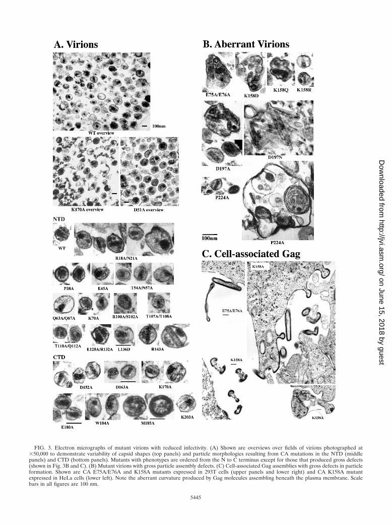
FIG. 3. Electron micrographs of mutant virions with reduced infectivity. (A) Shown are overviews over fields of virions photographed at�50,000 to demonstrate variability of capsid shapes (top panels) and particle morphologies resulting from CA mutations in the NTD (middlepanels) and CTD (bottom panels). Mutants with phenotypes are ordered from the N to C terminus except for those that produced gross defects(shown in Fig. 3B and C). (B) Mutant virions with gross particle assembly defects. (C) Cell-associated Gag assemblies with gross defects in particleformation. Shown are CA E75A/E76A and K158A mutants expressed in 293T cells (upper panels and lower right) and CA K158A mutantexpressed in HeLa cells (lower left). Note the aberrant curvature produced by Gag molecules assembling beneath the plasma membrane. Scalebars in all figures are 100 nm.
5445
on June 15, 2018 by guesthttp://jvi.asm
.org/D
ownloaded from
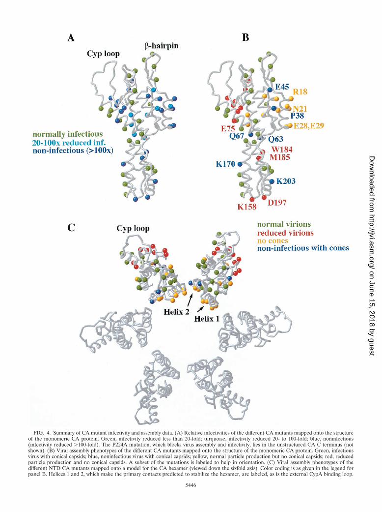
FIG. 4. Summary of CA mutant infectivity and assembly data. (A) Relative infectivities of the different CA mutants mapped onto the structureof the monomeric CA protein. Green, infectivity reduced less than 20-fold; turquoise, infectivity reduced 20- to 100-fold; blue, noninfectious(infectivity reduced �100-fold). The P224A mutation, which blocks virus assembly and infectivity, lies in the unstructured CA C terminus (notshown). (B) Viral assembly phenotypes of the different CA mutants mapped onto the structure of the monomeric CA protein. Green, infectiousvirus with conical capsids; blue, noninfectious virus with conical capsids; yellow, normal particle production but no conical capsids; red, reducedparticle production and no conical capsids. A subset of the mutations is labeled to help in orientation. (C) Viral assembly phenotypes of thedifferent NTD CA mutants mapped onto a model for the CA hexamer (viewed down the sixfold axis). Color coding is as given in the legend forpanel B. Helices 1 and 2, which make the primary contacts predicted to stabilize the hexamer, are labeled, as is the external CypA binding loop.
5446
on June 15, 2018 by guesthttp://jvi.asm
.org/D
ownloaded from

dered C-terminal tail in structures of the mature HIV-1 CACTD.
We and others have observed similar Gag assembly defectsfor additional mutations in retroviral CA CTD residues (11,59, 63; O.W. Pornillos and K. M. Stray, personal communica-tion; D. Muriaux and A. Rein, personal communication), aswell as for mutations in the SP1 spacer peptide (1, 43, 55, 58)and in NC (5, 18). We therefore speculate that these down-stream elements may interact with the base of the CA CTDdomain to create a continuous structure that is critical forimmature Gag lattice assembly (but which may be unstablefollowing Gag proteolysis or in the absence of lattice contacts).Indeed, there is evidence that the CA-SP1 junction forms ahelix that is lost upon Gag proteolysis (1, 58, 59). Formation ofa continuous Gag structure spanning the CA CTD-SP1-NCjunctions and involving the MHR fold (11) would explain thecommon phenotypes of mutations in this region, and it mightalso provide a mechanism for coupling RNA packaging withviral assembly and/or for ensuring incorporation of intact Gagmolecules.
The CA dimer interface in Gag assembly. The CA CTDdimerizes in solution (35, 78) and crystallizes as a symmetricaldimer created by parallel packing of CA helix 9 against itssymmetry-related mate (98). One goal of our study was to testwhether or not the crystallographically characterized CA CTDdimer interface functions during particle assembly and matu-ration. Residues W184 and M185 are buried in the core of thedimer interface, and alanine mutations at either position blockCA dimerization in vitro (35). In addition, several pairs ofresidues on the periphery of the dimer interface make favor-able hydrophilic contacts across the dyad (e.g., D152–E180 andK203–Q192). However, their energetic contributions to CAdimerization are not known.
Mutations in peripheral dimer interface residues (D152A,E180A, and Q192A) reduced infectivity (3- to 13-fold) but didnot obviously alter the morphology of either immature or ma-ture particles. These modest phenotypes may reflect the factthat surface side chain salt bridges often contribute very littleto overall protein stability (83, 84). However, mutations in thetwo core dimer interface residues (W184A and M185A) mea-surably diminished (but did not eliminate) immature particleproduction. These same mutations also reduced intermolecu-lar Gag-Gag interactions in vitro (10). Taken together, thesedata suggest that the CA dimer interface may form and con-tribute to the stability of the immature Gag lattice but is notabsolutely required for particle assembly. An important rolefor Gag dimerization in HIV-1 assembly is also suggested bythe fact that defects in particle release caused by deletion ofthe NC protein can be rescued by dimeric leucine zipper motifs(2, 48, 103).
The partial defect of the CA dimer interface mutants inimmature particle assembly complicates evaluation of possibledownstream role(s) of the dimer in mature capsid assembly.Nevertheless, both mutations in the core of the dimer interfaceinhibited CA assembly in vitro (B. K. Ganser, et al., submitted)and completely blocked mature capsid assembly and viral in-fectivity in cultured virus, supporting the idea that this inter-face is also essential at later stage(s) of the assembly-matura-tion pathway.
A Gag assembly element in the CA NTD. Studies from sev-eral laboratories indicate that the NTD of CA may not play anessential role in immature particle assembly (reviewed in ref-erence 41). Indeed, Gag proteins lacking this entire domaincan assemble and bud from cells (2, 7, 67, 77). Nevertheless, wefound that a series of mutations in CA helices 4 to 6 (E75A/E76A, R100A/S102A, T107A/T108A, and T110A/Q112A) re-duced viral particle production, abolished capsid formation,and blocked viral infectivity. A similar phenotype was reportedfor an Ala mutation of the Pro99 residue that precedes helix 5(28, 52). Despite strong viral phenotypes, however, none of ourmutations in this region affected CA cylinder or cone forma-tion in vitro (B. K. Ganser et al., submitted). Taken together,these data indicate that Gag assembly is sensitive to the com-position of this CA NTD surface (at least when the CA NTDis present), and they define a previously unrecognized contig-uous CA surface located beneath the cyclophilin A bindingloop that participates in Gag assembly. We speculate that thisregion of CA may form a weak Gag-Gag interface or, alterna-tively, bind a cellular factor necessary for efficient assembly invivo.
Capsid assembly elements. Strikingly, all of the mutant viri-ons that retained infectivity formed conical capsids, whereas allmutants lacking cones were severely reduced in infectivity(�20-fold). This correlation strongly supports the long-stand-ing, but still unproven, assumption that the proper assembly ofa mature conical capsid is essential for viral infectivity. Boththe NTD and CTD of HIV-1 CA perform essential structuralroles in the mature viral capsid (41), and we have proposedthat this is because the NTD forms hexameric rings while theCTD links the hexamers into a continuous lattice (57). Con-sistent with this model, two mutations in the CA CTD dimerinterface blocked conical capsid formation (W184A andM185A, discussed above), as did a series of mutations in theCA NTD. The latter mutants clustered about N-terminal he-lices 1 to 3 (mutants R18A/N21A, A22D, E28A/E29A, M39D,A42D, D51A, and N57A; two exceptions were K70A [helix 4]and L136D [helix 7]) (Table 2 and Fig. 4B). These mutantslikely affected capsid assembly (and not prior steps) becausemany had no effect on particle release and because most di-minished CA tube and cone assembly in vitro (B. K. Ganser etal., submitted).
Our mutational analysis agrees well with the structuralmodel for the CA hexamer in which helices 1 and 2 form theprimary intermolecular contacts at the center of the CA hex-amer (Fig. 4C). Helix 1/2 residues R18, A22, E28, E29, M39,and A42 are positioned so they could make intermolecularcontacts in the CA hexamer, and mutations in each blockcapsid assembly. An exception to this correlation is residuePro38 (helix 2), which also appears positioned to make inter-molecular contacts but can be mutated to Ala without blockingcapsid assembly (albeit with a 27-fold reduction in infectivity).However, a P38L mutation does block capsid assembly (28),indicating that the CA hexamer likely cannot accommodatelarger side chains at this position. Similarly, an F40A mutationis also defective in CA assembly (87).
Mutation R18A/N21A (at the top of helix 1) was unique inthat it frequently caused assembly of multiple aberrant capsidswithin the same enlarged virions, including tubes, cones, andpossibly spheres (which are difficult to discern unambiguously
VOL. 77, 2003 ALANINE SCANNING MUTAGENESIS OF HIV-1 CA 5447
on June 15, 2018 by guesthttp://jvi.asm
.org/D
ownloaded from

in thin section). The assembly phenotype of this CA mutant invitro was similarly aberrant (B. K. Ganser et al., submitted).We suggest that this mutation alters the proper distribution ofCA pentamers and hexamers required to form a cone (B. K.Ganser et al., submitted).
Helix 3 serves to buttress the first two CA helices and alsoanchors the �-hairpin, and this may explain the diminishedcapsid assembly of the D51A and N57A mutants. In contrast,mutations in residues within the N-terminal �-hairpin thatprecedes helices 1 and 2 (Q4A, Q7A/Q9A, and Q13A) had noeffect on cone formation and only modest effects on infectivity(�10-fold), consistent with other experiments indicating thatthe hairpin itself does not make essential contacts in the as-sembled capsids (44). Several alternative models for the CAhexamer that have been proposed recently (56, 62) are dis-cussed in detail elsewhere (B. K. Ganser et al., submitted).
Other defects. Five mutant viruses (P38A, E45A, Q63A/Q67A, K170A, and K203A) were not obviously defective inassembly, maturation, or TM and RT packaging yet were sig-nificantly reduced in infectivity (�20-fold). These mutants aretherefore of interest for understanding how CA performs itsessential nonstructural function(s).
Further studies have revealed that these mutations all pack-age normal levels of viral RNA and exhibit normal levels en-dogenous reverse transcription activity (29). With the excep-tion of E45A, however, all of these mutations appeared toreduce viral capsid stability as judged by the fact that intactcores from these mutants could not be isolated (29). Intrigu-ingly, the E45A mutation actually appeared to stabilize theviral capsid because this mutation increased yields of isolatedcores and reduced the rate of core dissociation in vitro. Thereduced infectivity of all of these mutants suggests that properdisassembly of a capsid of normal stability is an essential earlyevent in viral replication. Thus, our studies are consistent withthe idea that the principal role of the capsid is to organize theviral replication complex for ordered uncoating and replicationupon viral entry (8, 11, 15, 29, 87). Further support for this ideacomes from the phenotype of the Q63A/Q67A mutant, whichis actually reverse transcribed more rapidly than the WT virusupon entry yet is very poorly infectious–perhaps because itdoes not follow the proper viral uncoating pathway.
In summary, our mutational analyses have identified a seriesof residues in both domains of CA that help to establish theimmature Gag lattice and to build the mature viral capsid. Ourstudies also support the idea that an essential function of theviral capsid is to organize the viral RNA and its associatedenzymes for ordered disassembly and reverse transcriptionupon entry of the new host cell.
ACKNOWLEDGMENTS
We thank Victor Klishko for technical assistance with sample prep-aration and EM, Miguel Knochel and Ryan Brady for assistance withmutagenesis, Su (Sam) Li and Felix Vajdos for help with Fig. 1 and 4,Klaus Wiegers, Gabriel Rutter, and Hans-Georg Krausslich for elec-tron microscopy of HeLa cells transfected with mutants, and ChrisHill, Hans-Georg Krausslich, and Volker Vogt for helpful discussionsand suggestions.
This work was supported by an NIH grant (to W.I.S.).
REFERENCES
1. Accola, M. A., S. Hoglund, and H. G. Gottlinger. 1998. A putative alpha-helical structure which overlaps the capsid-p2 boundary in the human
immunodeficiency virus type 1 Gag precursor is crucial for viral particleassembly. J. Virol. 72:2072–2078.
2. Accola, M. A., B. Strack, and H. G. Gottlinger. 2000. Efficient particleproduction by minimal Gag constructs which retain the carboxy-terminaldomain of human immunodeficiency virus type 1 capsid-p2 and a lateassembly domain. J. Virol. 74:5395–5402.
3. Alin, K., and S. P. Goff. 1996. Amino acid substitutions in the CA proteinof Moloney murine leukemia virus that block early events in infection.Virology 222:339–351.
4. Berthet-Colominas, C., S. Monaco, A. Novelli, G. Sibai, F. Mallet, and S.Cusack. 1999. Head-to-tail dimers and interdomain flexibility revealed bythe crystal structure of HIV-1 capsid protein (p24) complexed with a mono-clonal antibody Fab. EMBO J. 18:1124–1136.
5. Berthoux, L., C. Pechoux, M. Ottmann, G. Morel, and J. L. Darlix. 1997.Mutations in the N-terminal domain of human immunodeficiency virus type1 nucleocapsid protein affect virion core structure and proviral DNA syn-thesis. J. Virol. 71:6973–6981.
6. Bess, J. W., Jr., R. J. Gorelick, W. J. Bosche, L. E. Henderson, and L. O.Arthur. 1997. Microvesicles are a source of contaminating cellular proteinsfound in purified HIV-1 preparations. Virology 230:134–144.
7. Borsetti, A., A. Ohagen, and H. G. Gottlinger. 1998. The C-terminal half ofthe human immunodeficiency virus type 1 Gag precursor is sufficient forefficient particle assembly. J. Virol. 72:9313–9317.
8. Bowzard, J. B., J. W. Wills, and R. C. Craven. 2001. Second-site suppressorsof Rous sarcoma virus Ca mutations: evidence for interdomain interactions.J. Virol. 75:6850–6856.
9. Braaten, D., C. Aberham, E. K. Franke, L. Yin, W. Phares, and J. Luban.1996. Cyclosporine A-resistant human immunodeficiency virus type 1 mu-tants demonstrate that Gag encodes the functional target of cyclophilin A.J. Virol. 70:5170–5176.
10. Burniston, M. T., A. Cimarelli, J. Colgan, S. P. Curtis, and J. Luban. 1999.Human immunodeficiency virus type 1 Gag polyprotein multimerizationrequires the nucleocapsid domain and RNA and is promoted by the capsid-dimer interface and the basic region of matrix protein. J. Virol. 73:8527–8540.
11. Cairns, T. M., and R. C. Craven. 2001. Viral DNA synthesis defects inassembly-competent Rous sarcoma virus CA mutants. J. Virol. 75:242–250.
12. Campbell, S., and V. M. Vogt. 1995. Self-assembly in vitro of purifiedCA-NC proteins from Rous sarcoma virus and human immunodeficiencyvirus type 1. J. Virol. 69:6487–6497.
13. Campos-Olivas, R., J. L. Newman, and M. F. Summers. 2000. Solutionstructure and dynamics of the Rous sarcoma virus capsid protein andcomparison with capsid proteins of other retroviruses. J. Mol. Biol. 296:633–649.
14. Carter, C. A. 2002. Tsg101: HIV-1’s ticket to ride. Trends Microbiol. 10:203–205.
15. Cartier, C., P. Sivard, C. Tranchat, D. Decimo, C. Desgranges, and V.Boyer. 1999. Identification of three major phosphorylation sites withinHIV-1 capsid. Role of phosphorylation during the early steps of infection.J. Biol. Chem. 274:19434–19440.
16. Charneau, P., G. Mirambeau, P. Roux, S. Paulous, H. Buc, and F. Clavel.1994. HIV-1 reverse transcription. A termination step at the center of thegenome. J. Mol. Biol. 241:651–662.
17. Chen, C., and H. Okayama. 1987. High-efficiency transformation of mam-malian cells by plasmid DNA. Mol. Cell. Biol. 7:2745–2752.
18. Cimarelli, A., S. Sandin, S. Hoglund, and J. Luban. 2000. Basic residues inhuman immunodeficiency virus type 1 nucleocapsid promote virion assem-bly via interaction with RNA. J. Virol. 74:3046–3057.
19. Cimarelli, A., S. Sandin, S. Hoglund, and J. Luban. 2000. Rescue of mul-tiple viral functions by a second-site suppressor of a human immunodefi-ciency virus type 1 nucleocapsid mutation. J. Virol. 74:4273–4283.
20. Cornilescu, C. C., F. Bouamr, X. Yao, C. Carter, and N. Tjandra. 2001.Structural analysis of the N-terminal domain of the human T-cell leukemiavirus capsid protein. J. Mol. Biol. 306:783–797.
21. Craven, R. C., A. E. Leure-duPree, R. A. Weldon, Jr., and J. W. Wills. 1995.Genetic analysis of the major homology region of the Rous sarcoma virusGag protein. J. Virol. 69:4213–4227.
22. Dawson, L., and X. F. Yu. 1998. The role of nucleocapsid of HIV-1 in virusassembly. Virology 251:141–157.
23. Dorfman, T., A. Bukovsky, A. Ohagen, S. Hoglund, and H. G. Gottlinger.1994. Functional domains of the capsid protein of human immunodefi-ciency virus type 1. J. Virol. 68:8180–8187.
24. Ebbesen, T. 1998. Cones and tubes: geometry in the chemistry of carbon.Acc. Chem. Res. 31:558–566.
25. Ehrlich, L. S., B. E. Agresta, and C. A. Carter. 1992. Assembly of recom-binant human immunodeficiency virus type 1 capsid protein in vitro. J. Vi-rol. 66:4874–4883.
26. Facke, M., A. Janetzko, R. L. Shoeman, and H. G. Krausslich. 1993. A largedeletion in the matrix domain of the human immunodeficiency virus gaggene redirects virus particle assembly from the plasma membrane to theendoplasmic reticulum. J. Virol. 67:4972–4980.
27. Ferris, A. L., A. Hizi, S. D. Showalter, S. Pichuantes, L. Babe, C. S. Craik,
5448 VON SCHWEDLER ET AL. J. VIROL.
on June 15, 2018 by guesthttp://jvi.asm
.org/D
ownloaded from

and S. H. Hughes. 1990. Immunologic and proteolytic analysis of HIV-1reverse transcriptase structure. Virology 175:456–464.
28. Fitzon, T., B. Leschonsky, K. Bieler, C. Paulus, J. Schroder, H. Wolf, andR. Wagner. 2000. Proline residues in the HIV-1 NH2-terminal capsid do-main: structure determinants for proper core assembly and subsequentsteps of early replication. Virology 268:294–307.
29. Forshey, B. M., U. von Schwedler, W. I. Sundquist, and C. Aiken. 2002.Formation of a human immunodeficiency virus type 1 core of optimalstability is crucial for viral replication. J. Virol. 76:5667–5677.
30. Franke, E. K., H. E. Yuan, and J. Luban. 1994. Specific incorporation ofcyclophilin A into HIV-1 virions. Nature 372:359–362.
31. Freed, E. O. 1998. HIV-1 gag proteins: diverse functions in the virus lifecycle. Virology 251:1–15.
32. Freed, E. O. 2002. Viral late domains. J. Virol. 76:4679–4687.33. Fuller, S. D., T. Wilk, B. E. Gowen, H. G. Krausslich, and V. M. Vogt. 1997.
Cryo-electron microscopy reveals ordered domains in the immature HIV-1particle. Curr. Biol. 7:729–738.
34. Gamble, T. R., F. F. Vajdos, S. Yoo, D. K. Worthylake, M. Houseweart, W. I.Sundquist, and C. P. Hill. 1996. Crystal structure of human cyclophilin Abound to the amino-terminal domain of HIV-1 capsid. Cell 87:1285–1294.
35. Gamble, T. R., S. Yoo, F. F. Vajdos, U. K. von Schwedler, D. K. Worthylake,H. Wang, J. P. McCutcheon, W. I. Sundquist, and C. P. Hill. 1997. Struc-ture of the carboxyl-terminal dimerization domain of the HIV-1 capsidprotein. Science 278:849–853.
36. Ganser, B. K., S. Li, V. Y. Klishko, J. T. Finch, and W. I. Sundquist. 1999.Assembly and analysis of conical models for the HIV-1 core. Science 283:80–83.
37. Gelderblom, H. R., E. H. Hausmann, M. Ozel, G. Pauli, and M. A. Koch.1987. Fine structure of human immunodeficiency virus (HIV) and immu-nolocalization of structural proteins. Virology 156:171–176.
38. Gitti, R. K., B. M. Lee, J. Walker, M. F. Summers, S. Yoo, and W. I.Sundquist. 1996. Structure of the amino-terminal core domain of theHIV-1 capsid protein. Science 273:231–235.
39. Gluschankof, P., I. Mondor, H. R. Gelderblom, and Q. J. Sattentau. 1997.Cell membrane vesicles are a major contaminant of gradient-enriched hu-man immunodeficiency virus type-1 preparations. Virology 230:125–133.
40. Goff, S., P. Traktman, and D. Baltimore. 1981. Isolation and properties ofMoloney murine leukemia virus mutants: use of a rapid assay for release ofvirion reverse transcriptase. J. Virol. 38:239–248.
41. Gottlinger, H. G. 2001. The HIV-1 assembly machine. AIDS 15:S13–S20.42. Gottlinger, H. G., T. Dorfman, J. G. Sodroski, and W. A. Haseltine. 1991.
Effect of mutations affecting the p6 gag protein on human immunodefi-ciency virus particle release. Proc. Natl. Acad. Sci. USA 88:3195–3199.
43. Gottlinger, H. G., J. G. Sodroski, and W. A. Haseltine. 1989. Role of capsidprecursor processing and myristoylation in morphogenesis and infectivity ofhuman immunodeficiency virus type 1. Proc. Natl. Acad. Sci. USA 86:5781–5785.
44. Gross, I., H. Hohenberg, C. Huckhagel, and H. G. Krausslich. 1998. N-Terminal extension of human immunodeficiency virus capsid protein con-verts the in vitro assembly phenotype from tubular to spherical particles.J. Virol. 72:4798–4810.
45. Gross, I., H. Hohenberg, and H. G. Krausslich. 1997. In vitro assemblyproperties of purified bacterially expressed capsid proteins of human im-munodeficiency virus. Eur. J. Biochem. 249:592–600.
46. Hill, C. P., D. Worthylake, D. P. Bancroft, A. M. Christensen, and W. I.Sundquist. 1996. Crystal structures of the trimeric human immunodefi-ciency virus type 1 matrix protein: implications for membrane associationand assembly. Proc. Natl. Acad. Sci. USA 93:3099–3104.
47. Jin, Z., L. Jin, D. L. Peterson, and C. L. Lawson. 1999. Model for lentiviruscapsid core assembly based on crystal dimers of EIAV p26. J. Mol. Biol.286:83–93.
48. Johnson, M. C., H. M. Scobie, Y. M. Ma, and V. M. Vogt. 2002. Nucleicacid-independent retrovirus assembly can be driven by dimerization. J. Vi-rol. 76:11177–11185.
49. Khorasanizadeh, S., R. Campos-Olivas, and M. F. Summers. 1999. Solu-tion structure of the capsid protein from the human T-cell leukemia virustype-I. J. Mol. Biol. 291:491–505.
50. Kimpton, J., and M. Emerman. 1992. Detection of replication-competentand pseudotyped human immunodeficiency virus with a sensitive cell lineon the basis of activation of an integrated beta-galactosidase gene. J. Virol.66:2232–2239.
51. Kingston, R. L., T. Fitzon-Ostendorp, E. Z. Eisenmesser, G. W. Schatz,V. M. Vogt, C. B. Post, and M. G. Rossmann. 2000. Structure and self-association of the rous sarcoma virus capsid protein. Structure Fold. Des.8:617–628.
52. Kong, L. B., D. An, B. Ackerson, J. Canon, O. Rey, I. S. Chen, P. Krogstad,and P. L. Stewart. 1998. Cryoelectron microscopic examination of humanimmunodeficiency virus type 1 virions with mutations in the cyclophilin Abinding loop. J. Virol. 72:4403–4407.
53. Kotov, A., J. Zhou, P. Flicker, and C. Aiken. 1999. Association of Nef withthe human immunodeficiency virus type 1 core. J. Virol. 73:8824–8830.
54. Krausslich, H. G. (ed.). 1996. Morphogenesis and maturation of retrovi-ruses. Springer-Verlag, Berlin, Germany.
55. Krausslich, H. G., M. Facke, A. M. Heuser, J. Konvalinka, and H. Zentgraf.1995. The spacer peptide between human immunodeficiency virus capsidand nucleocapsid proteins is essential for ordered assembly and viral infec-tivity. J. Virol. 69:3407–3419.
56. Lanman, J., T. T. Lam, S. Barnes, M. Sakalian, M. R. Emmett, A. G.Marshall, and P. E. Prevelige. 2003. Identification of novel interactions inHIV-1 capsid protein assembly by high-resolution mass spectrometry. J.Mol. Biol. 325:759–772.
57. Li, S., C. P. Hill, W. I. Sundquist, and J. T. Finch. 2000. Image reconstruc-tions of helical assemblies of the HIV-1 CA protein. Nature 407:409–413.
58. Liang, C., J. Hu, R. S. Russell, A. Roldan, L. Kleiman, and M. A. Wainberg.2002. Characterization of a putative �-helix across the capsid-SP1 boundarythat is critical for the multimerization of human immunodeficiency virustype 1 Gag. J. Virol. 76:11729–11737.
59. Liang, C., J. Hu, J. B. Whitney, L. Kleiman, and M. A. Wainberg. 2003. Astructurally disordered region at the C terminus of capsid plays essentialroles in multimerization and membrane binding of the Gag protein ofhuman immunodeficiency virus type 1. J. Virol. 77:1772–1783.
60. Luban, J. 2001. HIV-1 and Ebola virus: the getaway driver nabbed. Nat.Med. 7:1278–1280.
61. Mammano, F., A. Ohagen, S. Hoglund, and H. G. Gottlinger. 1994. Role ofthe major homology region of human immunodeficiency virus type 1 invirion morphogenesis. J. Virol. 68:4927–4936.
62. Mayo, K., D. Huseby, J. McDermott, B. Arvidson, L. Finlay, and E. Barklis.2003. Retrovirus capsid protein assembly arrangements. J. Mol. Biol. 325:225–237.
63. McDermott, J., L. Farrell, R. Ross, and E. Barklis. 1996. Structural analysisof human immunodeficiency virus type 1 Gag protein interactions, usingcysteine-specific reagents. J. Virol. 70:5106–5114.
64. Momany, C., L. C. Kovari, A. J. Prongay, W. Keller, R. K. Gitti, B. M. Lee,A. E. Gorbalenya, L. Tong, J. McClure, L. S. Ehrlich, M. F. Summers, C.Carter, and M. G. Rossmann. 1996. Crystal structure of dimeric HIV-1capsid protein. Nat. Struct. Biol. 3:763–770.
65. Nakai, M., and T. Goto. 1996. Ultrastructure and morphogenesis of humanimmunodeficiency virus. J. Electron Microsc. (Tokyo) 45:247–257.
66. Nermut, M. V., P. Bron, D. Thomas, M. Rumlova, T. Ruml, and E. Hunter.2002. Molecular organization of Mason-Pfizer monkey virus capsids assem-bled from Gag polyprotein in Escherichia coli. J. Virol. 76:4321–4330.
67. Ono, A., D. Demirov, and E. O. Freed. 2000. Relationship between humanimmunodeficiency virus type 1 Gag multimerization and membrane bind-ing. J. Virol. 74:5142–5150.
68. Ono, A., and E. O. Freed. 1999. Binding of human immunodeficiency virustype 1 Gag to membrane: role of the matrix amino terminus. J. Virol.73:4136–4144.
69. Orlinsky, K. J., J. Gu, M. Hoyt, S. Sandmeyer, and T. M. Menees. 1996.Mutations in the Ty3 major homology region affect multiple steps in Ty3retrotransposition. J. Virol. 70:3440–3448.
70. Paillart, J. C., and H. G. Gottlinger. 1999. Opposing effects of humanimmunodeficiency virus type 1 matrix mutations support a myristyl switchmodel of Gag membrane targeting. J. Virol. 73:2604–2612.
71. Pear, W. S., G. P. Nolan, M. L. Scott, and D. Baltimore. 1993. Productionof high-titer helper-free retroviruses by transient transfection. Proc. Natl.Acad. Sci. USA 90:8392–8396.
72. Perez, O. D., and G. P. Nolan. 2001. Resistance is futile. Assimilation ofcellular machinery by HIV-1. Immunity 15:687–690.
73. Purtscher, M., A. Trkola, G. Gruber, A. Buchacher, R. Predl, F. Steindl, C.Tauer, R. Berger, N. Barrett, A. Jungbauer, et al. 1994. A broadly neutral-izing human monoclonal antibody against gp41 of human immunodefi-ciency virus type 1. AIDS Res. Hum. Retrovir. 10:1651–1658.
74. Rao, Z., A. S. Belyaev, E. Fry, P. Roy, I. M. Jones, and D. I. Stuart. 1995.Crystal structure of SIV matrix antigen and implications for virus assembly.Nature 378:743–747.
75. Reicin, A. S., A. Ohagen, L. Yin, S. Hoglund, and S. P. Goff. 1996. The roleof Gag in human immunodeficiency virus type 1 virion morphogenesis andearly steps of the viral life cycle. J. Virol. 70:8645–8652.
76. Reicin, A. S., S. Paik, R. D. Berkowitz, J. Luban, I. Lowy, and S. P. Goff.1995. Linker insertion mutations in the human immunodeficiency virus type1 gag gene: effects on virion particle assembly, release, and infectivity.J. Virol. 69:642–650.
77. Reil, H., A. A. Bukovsky, H. R. Gelderblom, and H. G. Gottlinger. 1998.Efficient HIV-1 replication can occur in the absence of the viral matrixprotein. EMBO J. 17:2699–2708.
78. Rose, S., P. Hensley, D. J. O’Shannessy, J. Culp, C. Debouck, and I.Chaiken. 1992. Characterization of HIV-1 p24 self-association using ana-lytical affinity chromatography. Proteins 13:112–119.
79. Sakalian, M., and E. Hunter. 1998. Molecular events in the assembly ofretrovirus particles. Adv. Exp. Med. Biol. 440:329–339.
80. Spearman, P., R. Horton, L. Ratner, and I. Kuli-Zade. 1997. Membranebinding of human immunodeficiency virus type 1 matrix protein in vivo
VOL. 77, 2003 ALANINE SCANNING MUTAGENESIS OF HIV-1 CA 5449
on June 15, 2018 by guesthttp://jvi.asm
.org/D
ownloaded from

supports a conformational myristyl switch mechanism. J. Virol. 71:6582–6592.
81. Srinivasakumar, N., M. L. Hammarskjold, and D. Rekosh. 1995. Charac-terization of deletion mutations in the capsid region of human immunode-ficiency virus type 1 that affect particle formation and Gag-Pol precursorincorporation. J. Virol. 69:6106–6114.
82. Strambio-de-Castillia, C., and E. Hunter. 1992. Mutational analysis of themajor homology region of Mason-Pfizer monkey virus by use of saturationmutagenesis. J. Virol. 66:7021–7032.
83. Strop, P., and S. L. Mayo. 2000. Contribution of surface salt bridges toprotein stability. Biochemistry 39:1251–1255.
84. Sun, D. P., U. Sauer, H. Nicholson, and B. W. Matthews. 1991. Contribu-tions of engineered surface salt bridges to the stability of T4 lysozymedetermined by directed mutagenesis. Biochemistry 30:7142–7153.
85. Swanstrom, R., and J. W. Wills. 1997. Synthesis, assembly, and processingof viral proteins. In J. M. Coffin, S. H. Hughes, and H. E. Varmus (ed.),Retroviruses. Cold Spring Harbor Press, Plainview, N.Y.
86. Swingler, S., P. Gallay, D. Camaur, J. Song, A. Abo, and D. Trono. 1997.The Nef protein of human immunodeficiency virus type 1 enhances serinephosphorylation of the viral matrix. J. Virol. 71:4372–4377.
87. Tang, S., T. Murakami, B. E. Agresta, S. Campbell, E. O. Freed, and J. G.Levin. 2001. Human immunodeficiency virus type 1 N-terminal capsid mu-tants that exhibit aberrant core morphology and are blocked in initiation ofreverse transcription in infected cells. J. Virol. 75:9357–9366.
88. Thali, M., A. Bukovsky, E. Kondo, B. Rosenwirth, C. T. Walsh, J. Sodroski,and H. G. Gottlinger. 1994. Functional association of cyclophilin A withHIV-1 virions. Nature 372:363–365.
89. Turner, B. G., and M. F. Summers. 1999. Structural biology of HIV. J. Mol.Biol. 285:1–32.
90. Vogt, V. M. 1997. Retroviral virions and genomes, p. 27–70. In J. M. Coffin,S. H. Hughes, and H. E. Varmus (ed.), Retroviruses. Cold Spring HarborPress, Plainview, N.Y.
91. von Schwedler, U., J. Song, C. Aiken, and D. Trono. 1993. Vif is crucial forhuman immunodeficiency virus type 1 proviral DNA synthesis in infectedcells. J. Virol. 67:4945–4955.
92. von Schwedler, U. K., T. L. Stemmler, V. Y. Klishko, S. Li, K. H. Albertine,D. R. Davis, and W. I. Sundquist. 1998. Proteolytic refolding of the HIV-1capsid protein amino-terminus facilitates viral core assembly. EMBO J.17:1555–1568.
93. Welker, R., H. Hohenberg, U. Tessmer, C. Huckhagel, and H. G. Krauss-lich. 2000. Biochemical and structural analysis of isolated mature cores ofhuman immunodeficiency virus type 1. J. Virol. 74:1168–1177.
94. Wiegers, K., G. Rutter, H. Kottler, U. Tessmer, H. Hohenberg, and H. G.Krausslich. 1998. Sequential steps in human immunodeficiency virus par-ticle maturation revealed by alterations of individual Gag polyprotein cleav-age sites. J. Virol. 72:2846–2854.
95. Wiegers, K., G. Rutter, U. Schubert, M. Grattinger, and H. G. Krausslich.1999. Cyclophilin A incorporation is not required for human immunodefi-ciency virus type 1 particle maturation and does not destabilize the maturecapsid. Virology 257:261–274.
96. Wilk, T., I. Gross, B. E. Gowen, T. Rutten, F. de Haas, R. Welker, H. G.Krausslich, P. Boulanger, and S. D. Fuller. 2001. Organization of immaturehuman immunodeficiency virus type 1. J. Virol. 75:759–771.
97. Wills, J. W., and R. C. Craven. 1991. Form, function, and use of retroviralgag proteins. AIDS 5:639–654.
98. Worthylake, D. K., H. Wang, S. Yoo, W. I. Sundquist, and C. P. Hill. 1999.Structures of the HIV-1 capsid protein dimerization domain at 2.6 A res-olution. Acta Crystallogr. D Biol. Crystallogr. 55:85–92.
99. Yeager, M., E. M. Wilson-Kubalek, S. G. Weiner, P. O. Brown, and A. Rein.1998. Supramolecular organization of immature and mature murine leuke-mia virus revealed by electron cryo-microscopy: implications for retroviralassembly mechanisms. Proc. Natl. Acad. Sci. USA 95:7299–7304.
100. Yoo, S., D. G. Myszka, C. Yeh, M. McMurray, C. P. Hill, and W. I.Sundquist. 1997. Molecular recognition in the HIV-1 capsid/cyclophilin Acomplex. J. Mol. Biol. 269:780–795.
101. Yu, F., S. M. Joshi, Y. M. Ma, R. L. Kingston, M. N. Simon, and V. M. Vogt.2001. Characterization of Rous sarcoma virus Gag particles assembled invitro. J. Virol. 75:2753–2764.
102. Zabransky, A., E. Hunter, and M. Sakalian. 2002. Identification of a min-imal HIV-1 gag domain sufficient for self-association. Virology 294:141–150.
103. Zhang, Y., H. Qian, Z. Love, and E. Barklis. 1998. Analysis of the assemblyfunction of the human immunodeficiency virus type 1 Gag protein nucleo-capsid domain. J. Virol. 72:1782–1789.
104. Zhou, W., and M. D. Resh. 1996. Differential membrane binding of thehuman immunodeficiency virus type 1 matrix protein. J. Virol. 70:8540–8548.
5450 VON SCHWEDLER ET AL. J. VIROL.
on June 15, 2018 by guesthttp://jvi.asm
.org/D
ownloaded from