gamma carboxyglutamic acid-containing protein primary...
TRANSCRIPT
Serum Bone GammaCarboxyglutamic Acid-containing Protein in PrimaryHyperparathyroidism and in Malignant HypercalcemiaComparison with Bone Histomorphometry
P. D. Delmas, B. Demiaux, L. Malaval, M. C. Chapuy, C. Edouard, and P. J. MeunierClinique de Rhumatologie and Institut Nationale de la Sante et de la RechercheMedicale U. 234, H6pital Edouard Herriot, Lyon, France
Abstract
Serum bone y-carboxyglutamic acid-containing (Gla) protein(sBGP), a sensitive and specific marker of bone turnover, was
measured in 25 patients with primary hyperparathyroidism andin 24 patients with bone metastases with or without hypercal-cemia. Despite similar levels of hypercalcemia, sBGP was in-creased in primary hyperparathyroidism (14.2±9.6 ng/ml, P< 0.001), was decreased in malignant hypercalcemia (3.1±2.8ng/ml, P < 0.001), and was normal in patients with bone me-
tastases without hypercalcemia (6.6±2.7 ng/ml). In primary hy-perparathyroidism, sBGP was correlated with serum immuno-reactive parathyroid hormone (r = 0.90), calcium (r = 0.73),and with the adenoma weight (r = 0.79). After parathyroidec-tomy, sBGP slowly returned to normal values within 2-6 mo,suggesting that sBGP reflects increased bone turnover ratherthan a direct effect of parathyroid hormone on BGPsynthesisat the cell level. An iliac crest biopsy was performed in 11 patientswith primary hyperparathyroidism and in 9 cancer patients in a
noninvaded area. sBGPwas significantly correlated with all pa-
rameters reflecting bone formation but not with bone resorption.Patients with bone metastases were analyzed according to thepresence or the absence of hypercalcemia. In contrast to nor-
mocalcemic patients who had normal sBGP, hypercalcemic pa-tients had decreased sBGP (P < 0.001) and a lower bone for-mation at the cellular level (P < 0.05). Thus, biochemical andhistological data suggest that an unknown humoral factor mightbe responsible for this uncoupling between increased resorptionand decreased formation. This uncoupling, rather than local re-
lease of calcium by the metastatic process, might be responsiblefor hypercalcemia in patients with bone metastases.
Introduction
Primary hyperparathyroidism and cancer are the most frequentcauses of hypercalcemia. In patients with primary hyperpara-thyroidism, increased bone turnover is a common finding. Iliaccrest biopsy analysis shows an increase of both osteoclastic boneresorption and osteoblastic bone formation (1-6) and imbalancebetween both plays a role in the mechanism of hypercalcemia.In patients with malignant hypercalcemia related to solid tumors,
Address correspondence to Dr. Delmas, Hopital Edouard Herriot, Pav-illon E, Place d'Arsonval, 69374 Lyon, France.
Receivedfor publication 10 December 1984 and in revisedform 14November 1985.
two mechanisms are commonly implicated according to thepresence or the absence of bone metastases (7, 8). In the absenceof bone metastases, hypercalcemia occurs as the result of secre-tion by the tumor of humoral calcemic factors. These putativefactors have been shown to induce a marked uncoupling betweenincreased bone resorption and decreased bone formation, whichaccounts for bone loss (9). Amongpatients with cancer-associatedhypercalcemia, those with bone metastases constitute the largestgroup but they have received little attention in the literature.Hypercalcemia is usually attributed to local release of skeletalcalcium caused by the metastatic process (8, 10), but the possiblerole of humoral factors has been suggested even in the presenceof bone metastases (7). Increased osteoclastic bone resorption isa common finding (1 1, 12), but the level of bone formation hasnot been investigated systematically. Assessment of total boneturnover by noninvasive methods is crucial in order to answerthis question, but available markers are insufficient. Serum al-kaline phosphatase (sAP)', an index of bone formation, and uri-nary hydroxyproline (uOH-Prol), an index of bone resorption,are not specific for bone tissue. They may be increased in situ-ations with no abnormality of bone.
We have studied this problem by measuring serum bone,y-carboxyglutamic acid-containing (Gla) protein (sBGP)', alsocalled osteocalcin (13, 14). BGPis the most abundant noncol-lagenous protein of bone and it is specific for skeletal tissue. Itis synthesized by bone cells and circulates in blood (15, 16). Welack accurate estimates of the fraction which is incorporatedinto bone and of the fraction released into the circulation, butsBGPhas been found to be a sensitive indicator of bone turnoverin patients with a variety of metabolic bone diseases (17-19),including primary hyperparathyroidism (17, 20). Using a sen-sitive radioimmunoassay (21), one of us has previously shownthat sBGP is increased in patients with primary hyperparathy-roidism and decreased in patients with hypoparathyroidism (22).Recently, we have shown that sBGPis a specific marker of boneformation but not resorption in patients with postmenopausalosteoporosis (23), a finding recently confirmed by others in pa-tients with renal osteodystrophy (24).
In a prospective study, we have measured sBGP in a groupof 25 patients with primary hyperparathyroidism before andafter surgery and in 9 patients with hypercalcemia related tobone metastases. sBGPvalues were correlated with other markersof bone turnover (sAP and uOH-Prol) and with markers of para-thyroid hormone secretion. Biochemical measurements werecompared with histological measurement of bone turnover oW
1. Abbreviations used in this paper: BGP, bone y-carboxyglutamic acid-containing (Gla) protein; PTH, parathyroid hormone; sAP, serum alkalinephosphatase; sBGP, serum BGP; sCa, serum calcium; sP, serum phos-phate; sPTH, serum PTH; uCa, urinary calcium; uOH-Prol, urinary hy-droxyproline.
Bone-Gla-protein in Hyperparathyroidism and Malignant Hypercalcemia 985
J. Clin. Invest.© The American Society for Clinical Investigation, Inc.0021-9738/86/03/0985/07 $ 1.00Volume 77, March 1986, 985-991
tained from iliac crest biopsies. Data were also compared withthose obtained in 15 patients having bone metastases withouthypercalcemia.
MethodsSubjects. Westudied 25 consecutive patients (21 women, 55±1 1-yr-old;4 men, 55±12-yr-old) admitted to the Rheumatology and MetabolicBone Disease Unit for primary hyperparathyroidism. In 21 of them thediagnosis was confirmed by surgical removal of a parathyroid adenoma.In the remaining four the diagnosis was based on clinical and laboratorycriteria including hypercalcemia and an elevated level of serum immu-noreactive parathyroid hormone (sPTH). 18 patients had asymptomatichypercalcemia, 5 had a history of urolithiasis, one had vertebralcompression fractures, and one patient had severe hypercalcemia. Renalfunction was normal in all patients except one who had a moderateincrease of serum creatinine (148 Mmol/liter, normal < 110 ,mol/liter).
Wealso studied a group of nine patients (51±8-yr-old) with hyper-calcemia related to bone metastases. These were consecutive patientsadmitted to the Rheumatology and Metabolic Bone Disease Unit forbone metastases and hypercalcemia. Clinical features are indicated inTable I. sBGPwas measured before therapy of hypercalcemia and primarytumor was discovered at the time of hypercalcemia. Therefore none ofthe patients had previously received chemotherapy.
A control cancer group comprised of patients with bone metastaseswithout hypercalcemia was studied. They were 15 women (50±13-yr-old) with breast cancer that were admitted to the Oncology Unit (Dr.M. Clavel, Centre Lion Berard, Lyon, France). X-ray examinationshowed that bone metastases were of the lytic type in six cases and ofthe mixed type in two cases. Bone metastases were detected only by bonescan in five cases and by bone biopsy in two cases. The number of bonemetastases per patient was similar in the group of patients with hyper-calcemia and in the group of patients without hypercalcemia (5±4 and5±5, respectively, NS). Thirteen patients were being treated at the timeof the study (Tamoxifen in 11 patients, ovariectomy in two cases, ad-renalectomy in one case, two to three courses of chemotherapy in threecases).
Biochemistry. Weobtained fasting morning blood samples for de-terminations of serum concentrations of calcium (sCa), phosphate (sP),sAP, BGP, and PTH. 24-h urine collection was obtained during two tothree consecutive days for measurement of calcium (uCa), total uOH-Prol, and in some cases for cyclic AMP(cAMP). sBGP was measuredwith an assay similar to the radioimmunoassay described by Price andNishimoto (16) with a modification of previously published methods(21). This assay uses antiserum (R 102 M) directed against bovine BGPand bovine BGPas a standard and tracer. After a 4-h incubation at37°C, antibody-bound and free '25I-labeled BGPare separated by the
double-antibody method using a complex of sheep anti-rabbit IgG andpolyethylene glycol (precipitating reagent; Cie. ORIS Industrie, Bagnols,France). The sensitivity of the assay is 0.05 ng of bovine BGPper tubeand all values for standards and samples were determined in triplicate.Dilution curves of serum samples from controls and from patients witheither primary hyperparathyroidism or malignant hypercalcemia werefound to be parallel. Control values were obtained in 107 age-matchednormal women (60±19-yr-old, sBGP = 6.4±2.2 ng/ml) and in 23 age-matched normal men (53±9-yr-old, sBGP = 5.7±1.9 ng/ml). sCa wasmeasured by a complexometric method (Corning Glass Works, CorningScience Products, Corning, NY) and in patients with malignant hyper-calcemia, serum calcium was corrected for serum albumin (25). sP andsAP were measured by a colorimetric method and uOH-Prol was deter-mined with the method of Kivirikko et al. (26). sPTH was measuredwith a commercial double-antibody radioimmunoassay (PTH-RIA-100;Cie. ORIS Industrie) that uses a rabbit antiserum directed to bovinePTHwith major immunochemical specificity for the carboxy-terminalregion of the molecule. cAMPwas measured in urine by a solid-phaseradioimmunoassay (Immunotech; Luminy, Case 915, Marseille, France).Control values for sCa, sP, sAP, sPTH, uCa, uOH-Prol, and cAMPwereobtained in 30 normal adults, 40-60-yr-old.
Bone histomorphometry. A transiliac bone biopsy was performedwith a 7.5-mm trephine in 11 patients with primary hyperparathyroidismwho gave informed consent. In these patients, the severity of the diseaseassessed by biochemistry and by bone X-rays did not differ from thegeneral population of 25 patients with primary hyperparathyroidism.In 11 patients with bone metastases with or without hypercalcemia, atransiliac bone biopsy was obtained in a noninvaded area as assessed byX-rays and bone scan after informed consent as part of a prospectivestudy on bone metastases. Bone marrow examination showed metastatictumoral cells on two biopsies that were excluded in order to rule out thedirect local effect of metastases on bone remodeling. Weanalyzed thefollowing parameters in undecalcified bone biopsy specimens on serialsection, 8 jsm thick, stained with Solochrome Cyanin or by Goldner'sprocess (27, 28): relative resorption surfaces (percent), osteoclast number(per millimeter of trabecular surfaces), relative osteoid volume (percent),relative osteoid surfaces (percent), and the osteoid seam width measureddirectly according to the method of Kragstrup et al. (29). Histomor-phometric data were compared with values obtained in age-matchedcontrols (10 women, 5 men) who all had violent deaths.
Statistical analysis. Correlations between biochemical measurementsand histomorphometric data were assessed by using linear regressionanalysis and Spearman's coefficient of rank correlation. Analysis of linearregression was used to determine the relation between the biochemicalmeasurements. Differences between patients and controls were analyzedby the unpaired Student's t test; variations of sBGP values with timewere analyzed by the paired Student's t test.
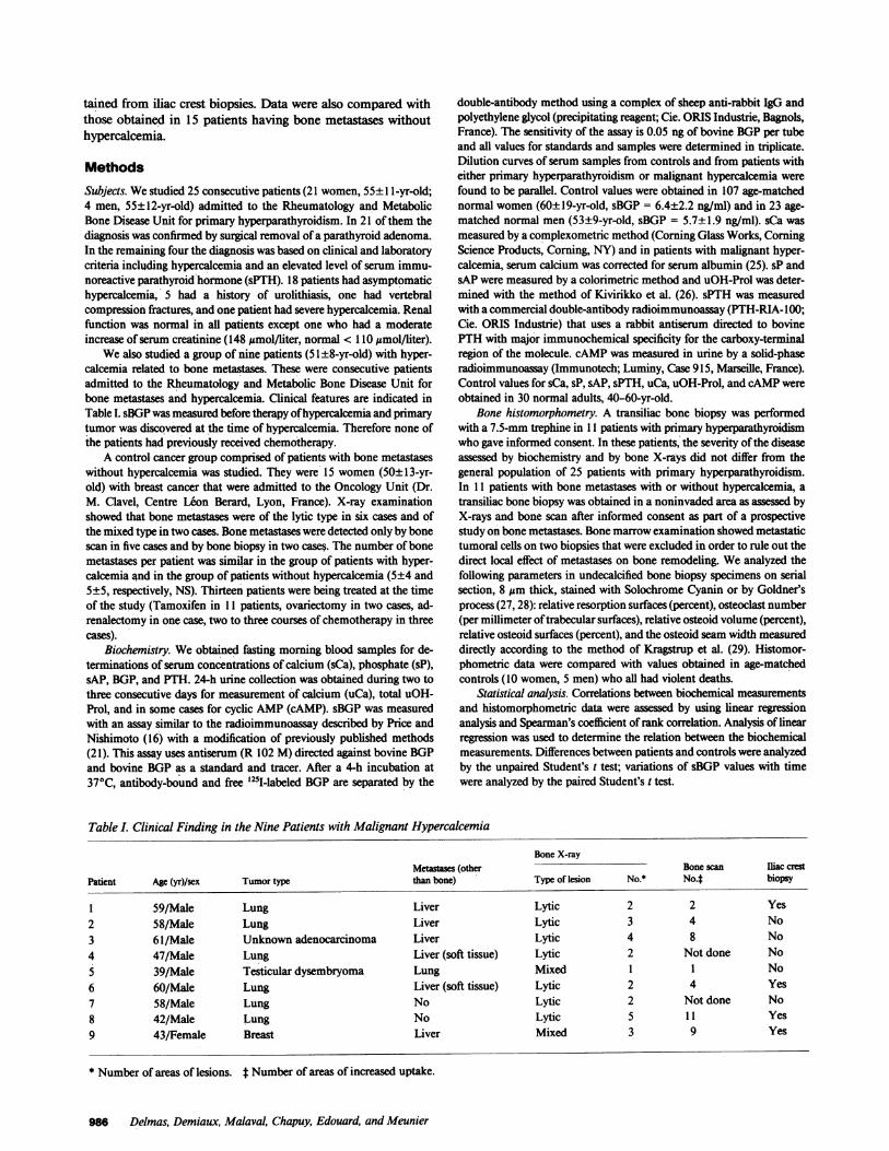
Table I. Clinical Finding in the Nine Patients with Malignant Hypercalcemia
Bone X-rayMetastases (other Bone scan Iliac crest
Patient Age (yr)/sex Tumor type than bone) Type of lesion No.* No.4 biopsy
1 59/Male Lung Liver Lytic 2 2 Yes2 58/Male Lung Liver Lytic 3 4 No3 61/Male Unknown adenocarcinoma Liver Lytic 4 8 No4 47/Male Lung Liver (soft tissue) Lytic 2 Not done No5 39/Male Testicular dysembryoma Lung Mixed I 1 No6 60/Male Lung Liver (soft tissue) Lytic 2 4 Yes7 58/Male Lung No Lytic 2 Not done No8 42/Male Lung No Lytic 5 11 Yes9 43/Female Breast Liver Mixed 3 9 Yes
* Number of areas of lesions. t Number of areas of increased uptake.
986 Delmas, Demiaux, Malaval, Chapuy, Edouard, and Meunier

Table II. Biochemical Data (±SD) in Patients with Primary Hyperparathyroidismand in Patients with Bone Metastases with or without Hypercalcemia
Bone metastases
Hyperparathyroidism With hypercalcemia Without hypercalcemia Normal
n= 25 n = 9 n = IS n = 30-130
Blood chemistryBGP(ng/ml) 14.2±9.6§ 3.1±2.6§1¶ 6.6±2.7 6.2±2.1Calcium (mg/ml) 11.6±1.2§ 12.3±2.5$§ 9.2±0.4 9.7±0.3Phosphate (mg/dl) 2.8±0.4§ 3.8±0.5 3.4±0.3 3.7±0.5Alkaline phosphatase (Bodansky U/liter) 4.6±1.8§ (23)* 13.3±7.5§1" 4.8±3.1§ 3.0±1.5PTH (mIU/ml) 8.4±9.0§ 2.8±0.6 (7) 2.8±0.7 2.8±0.9
Urine chemistry/24-hCalcium (mg) 366±147§ (22) 480±153§1 168±122 207±35Hydroxyproline (mg) 83±57§ (23) 126±46§"¶ 50±33 (14) 38±16cAMP(pmol) 7.84±5.8 (20) 6.8 3.08±1.25
* Number of observations. t 12.5±2.5 after correction for serum albumin. § P < 0.05-0.001 vs. normal. 1' P < 0.05-0.001 vs. primary hyper-parathyroidism. ¶ P < 0.01-0.001 vs. bone metastases without hypercalcemia.
Results
Biochemical data in untreated patients (Tables II and III). De-spite similar values of sCa, sBGPwas significantly increased inpatients with primary hyperparathyroidism and significantly de-creased in patients with malignant hypercalcemia. Mean sBGPwas normal in patients with bone metastases without hypercal-cemia (Fig. 1). A similar pattern was observed after logarithmictransformation of sBGP values. As expected, mean sP was de-creased and mean values of sPTH, sAP, uCa, and uOH-Prolwere increased in patients with primary hyperparathyroidism.In contrast to sBGPthat was elevated in 17 of the 25 patients,sAP and uOH-Prol were elevated in 7 and 10 patients, respec-tively. Seven patients, in whoma parathyroid adenoma was sub-
Table III. Correlations between Biochemical Parametersin Patients with Primary Hyperparathyroidism
n r P
sBGP vs.sPTH 24 0.90 <0.001sCa 25 0.73 <0.001Adenoma wt 14 0.79 <0.001sP 25 0.18 NSsAP 24 0.17 NSuCa 22 0.33 NSuOH-Prol 23 0.30 NS
sPTH vs.sCa 24 0.88 <0.001
sAP vs.sCa 24 0.03 NSsPTH 24 0.02 NSuOH-Prol 23 0.46 <0.05
uOH-Prol vs.sCa 23 0.27 NSsPTH 23 0.29 NS
sequently removed, had normal values of sPTH; three of themhad a marked elevation of sBGP, whereas sAP was normal. sBGPwas highly correlated with sCa, sPTH, and the adenoma weight,but not with sAP and uOH-Prol. In contrast to sBGP, sAP anduOH-Prol did not reflect PTH secretion as assessed by sPTH,adenoma weight, and sCa. In patients with malignant hyper-calcemia there was a marked increase of sAP because of hepaticmetastases in two-thirds of the cases. sPTH was normal or lowin all patients. uCa and uOH-Prol were significantly higher inmalignant hypercalcemia than in controls and than in bone me-tastases without hypercalcemia.
Bone histomorphometry (Table IV and V). In patients withprimary hyperparathyroidism there was a marked increase of
.
BM
Figure 1. Individual sBGPvalues in 25 patients with primary hyper-parathyroidism (PHP) and in 24 patients having bone metastases withhypercalcemia (BM + HCa) or without hypercalcemia (BM).
Bone-Gla-protein in Hyperparathyroidism and Malignant Hypercalcemia 987
Ssqr_+O1l
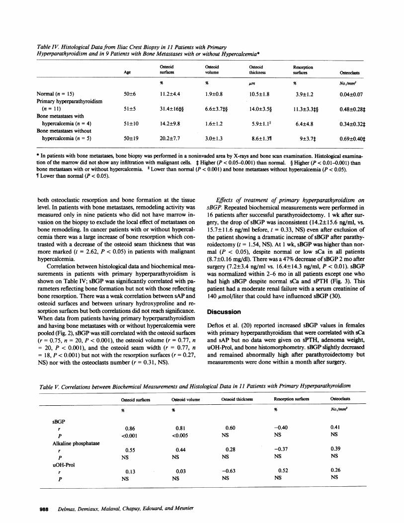
Table IV. Histological Data from Iliac Crest Biopsy in 1I Patients with PrimaryHyperparathyroidism and in 9 Patients with Bone Metastases with or without Hypercalcemia*
Osteoid Osteoid Osteoid ResorptionAge surfaces volume thickness surfaces Osteoclasts
% % 'Um % No./mm2
Normal (n = 15) 50±6 11.2±4.4 1.9±0.8 10.5±1.8 3.9±1.2 0.04±0.07Primary hyperparathyroidism
(n = 11) 51±5 31.4±16#§ 6.6±3.7t§ 14.0±3.5§ 11.3±3.3t§ 0.48±0.28tBone metastases with
hypercalcemia (n = 4) 51±10 14.2±9.8 1.6±1.2 5.9±1.1"1 6.4±4.8 0.34±0.32tBone metastases without
hypercalcemia (n = 5) 50±19 20.2±7.7 3.0±1.3 8.6±1.31 9±3.7$ 0.69±0.40t
* In patients with bone metastases, bone biopsy was performed in a noninvaded area by X-rays and bone scan examination. Histological examina-tion of the marrow did not show any infiltration with malignant cells. * Higher (P < 0.05-0.001) than normal. § Higher (P < 0.01-0.001) thanbone metastases with or without hypercalcemia. 11 Lower than normal (P < 0.001) and bone metastases without hypercalcemia (P < 0.05).¶ Lower than normal (P < 0.05).
both osteoclastic resorption and bone formation at the tissuelevel. In patients with bone metastases, remodeling activity wasmeasured only in nine patients who did not have marrow in-vasion on the biopsy to exclude the local effect of metastases onbone remodeling. In cancer patients with or without hypercal-cemia there was a large increase of bone resorption which con-trasted with a decrease of the osteoid seam thickness that wasmore marked (t = 2.62, P < 0.05) in patients with malignanthypercalcemia.
Correlation between histological data and biochemical mea-surements in patients with primary hyperparathyroidism isshown on Table IV; sBGPwas significantly correlated with pa-rameters reflecting bone formation but not with those reflectingbone resorption. There was a weak correlation between sAP andosteoid surfaces and between urinary hydroxyproline and re-sorption surfaces but both correlations did not reach significance.When data from patients having primary hyperparathyroidismand having bone metastases with or without hypercalcemia werepooled (Fig. 2), sBGPwas still correlated with the osteoid surfaces(r = 0.75, n = 20, P < 0.001), the osteoid volume (r = 0.77, n= 20, P < 0.001), and the osteoid seam width (r = 0.77, n= 18, P < 0.001) but not with the resorption surfaces (r = 0.27,NS) nor with the osteoclasts number (r = 0.31, NS).
Effects of treatment of primary hyperparathyroidism onsBGP. Repeated biochemical measurements were performed in16 patients after successful parathyroidectomy. 1 wk after sur-gery, the drop of sBGPwas inconsistent (14.2±15.6 ng/ml, vs.15.7±11.6 ng/ml before, t = 0.33, NS) even after exclusion ofthe patient showing a dramatic increase of sBGPafter parathy-roidectomy (t = 1.54, NS). At 1 wk, sBGPwas higher than nor-mal (P < 0.05), despite normal or low sCa in all patients(8.7±0.16 mg/dl). There was a 47%decrease of sBGP2 moaftersurgery (7.2±3.4 ng/ml vs. 16.4±14.3 ng/ml, P < 0.01). sBGPwas normalized within 2-6 mo in all patients except one whohad high sBGP despite normal sCa and sPTH (Fig. 3). Thispatient had a moderate renal failure with a serum creatinine of140 ,umol/liter that could have influenced sBGP (30).
Discussion
Deftos et al. (20) reported increased sBGP values in femaleswith primary hyperparathyroidism that were correlated with sCaand sAP but no data were given on sPTH, adenoma weight,uOH-Prol, and bone histomorphometry. sBGPslightly decreasedand remained abnormally high after parathyroidectomy butmeasurements were done within a month after surgery.
Table V. Correlations between Biochemical Measurements and Histological Data in 1I Patients with Primary Hyperparathyroidism
Osteoid surfaces Osteoid volume Osteoid thickness Resorption surfaces Osteoclasts
9to 9% 9% No./mM2
sBGPr 0.86 0.81 0.60 -0.40 0.41P <0.001 <0.005 NS NS NS
Alkaline phosphataser 0.55 0.44 0.28 -0.37 0.39P NS NS NS NS NS
uOH-Prolr 0.13 0.03 -0.63 0.52 0.26P NS NS NS NS NS
988 Delmas, Demiaux, Malaval, Chapuy, Edouard, and Meunier

0
*-*@0
*
r:0.27ns
*
I05 lb Is 20 %
mSorption swfac
Figure 2. Correlation between sBGPand either the osteoid volume (left)or the resorption surfaces (right) inpatients with primary hyperparathy-roidism (.) and in patients havingbone metastases with (o) or without(*) hypercalcemia. The rectanglesrepresent the normal range.
Our finding of increased sBGP values in patients with pri-mary hyperparathyroidism and the highly significant correlationof sBGP with either sPTH, sCa, or adenoma weight indicatethat sBGPis a sensitive marker of PTHsecretion in this disease.These data are consistent with our previous findings that in nor-
mal women, there is an increase in bone turnover with agingwith a significant correlation between sBGPand sPTH (21). Thefact that sBGPgoes slowly back to normal (within 2-6 mo) afterremoval of parathyroid adenoma suggests that sBGPvalues re-
flect increased bone turnover at the tissue level rather than a
direct effect of PTH on BGPsynthesis and/or secretion at thecell level. It is interesting to note that this delay for the nor-
malization of BGPis in agreement with the quantum remodeling
sBGP,
theory, as the lifespan of the remodeling activity of a bone struc-tural unit ranges from 4 to 6 mo (31).
An important question is whether sBGP reflects bone for-mation, bone resorption, or both. Experimental studies in therat using warfarin (32) and diphosphonates (33) strongly suggestthat rat sBGPoriginates from new cellular synthesis rather thanfrom release from bone matrix during bone resorption. Wehavepreviously correlated sBGPwith quantitative measurements on
iliac crest biopsy in 26 patients with postmenopausal osteopo-rosis, and we have found that sBGPis a specific marker of boneformation but does not reflect bone resorption (19). In this studysimilar findings were observed in patients with primary hyper-parathyroidism, indicating that sBGP reflects bone formation
Figure 3. Individual values of sBGPbefore and after parathyroidectomy
s AVn (PTX).
Bone-Gla-protein in Hyperparathyroidism and Malignant Hypercalcemia 989
S BGP _-nglml 0 15
10
5
r 0.77
p C 0.001
sBGPng/ml
0
*0:::
..
whether bone turnover is low, normal, or high. BGPreleasedfrom bone during resorption is probably degraded into smallfragments that are not recognized by our antiserum. However,as pointed out earlier, we have no precise information aboutwhat controls the fraction of BGPreleased into the circulationand whether this fraction varies in different disease states.Therefore, no conclusion about the function of BGPcan bedrawn from this study.
Patients with bone metastases had evidence of increased boneresorption that was higher in patients with hypercalcemia. Incontrast to normocalcemic patients who had normal sBGP, hy-percalcemic patients had a low sBGP and osteoid seams thatwere thinner than normocalcemic patients and than controls,both indicating a decreased bone formation. This uncouplingbetween an increased bone resorption and a decreased osteo-blastic activity was probably responsible, at least in part, for thehypercalcemia, but its cause is unknown. Because the bone bi-opsy was performed in all cases in a nonmetastatic area, thishistological pattern cannot be explained by a local effect of tumorcells. As described earlier, it should be noted that the numberof bone metastases was not different in patients with or withouthypercalcemia. The fact that sBGP was dramatically reducedfurther supports the hypothesis that an unknown systemic factormight be responsible for the inhibition of bone formation inmalignant hypercalcemia. In fact, Stewart has shown that tu-moral factors are likely to contribute to hypercalcemia in manypatients with metastatic bone disease (7) and it has been recentlyshown that the degree of hypercalcemia is not related to theextent of metastatic disease detected by bone scans in patientswith bone metastases (34). It should be stressed that the sameuncoupling between high resorption and low formation has beenreported in patients with humoral hypercalcemia of malignancywithout any bone metastases (9). Conversely, these patients werein poor health and some degree of immobilization might haveinfluenced, at least in part, the histological pattern. Furtherstudies are required comparing sBGPand bone histomorphom-etry in a larger sample of patients, including patients with typicalhumoral hypercalcemia and patients with hypercalcemia relatedto hematologic disorders. Nevertheless, our data indicate thatlow sBGP in patients with hypercalcemia and bone metastasesreflects a low bone formation.
For clinical purposes, the question is to know if sBGPmightbe useful for the diagnosis of the etiology of hypercalcemia. Incontrast to most PTH assays, sBGP can be measured within a
day. Therefore its measurement might be useful for investigatingpatients with dramatic hypercalcemia, as sBGPwill be very highin cases of primary hyperparathyroidism, whereas it will be nor-mal or low in cases of malignant hypercalcemia related to bonemetastases. In patients with mild, asymptomatic hypercalcemiadue to primary hyperparathyroidism, sBGPwas clearly elevatedin three patients who had normal sPTH values, suggesting thatBGPassay might be useful in such patients when a very sensitivePTH assay is not available.
Bone symptoms, including bone pain and fractures, havebecome uncommon clinical features in patients with primaryhyperparathyroidism (35, 36). Evaluating bone turnover with a
specific and noninvasive method might be valuable in asymp-tomatic patients to decide if surgical or medical treatment shouldbe applied and for the follow-up of patients who do not undergoparathyroidectomy. Further studies should be performed to bet-ter define the potential benefit of sBGP measurement in theassessment of bone status in primary hyperparathyroidism.
Acknowledgments
Wethank Drs. M. Arlot, E. Saubier (Hotel-Dieu, Lyon), and M. Clavelfor their collaboration. The work was supported in part by Cie. ORISIndustrie (contract R098 520 and R098 562).
References
1. Riggs, B. L., P. J. Kelly, J. Jowsey, and F. R. Keating. 1965. Skeletalalterations in hyperparathyroidism: determination of bone formation,resorption and morphologic changes by microradiography. J. Clin. En-docrinol. Metab. 25:777-782.
2. Byers, P. D., and R. Smith. 1971. Quantitative histology of bonein hyperparathyroidism: its relation to clinical features, X-ray and bio-chemistry. Q. J. Med. 40:471-480.
3. Meunier, P. J., G. Vignon, J. Bernard, C. Edouard, and P. Cour-pron. 1973. Quantitative bone histology as applied to the diagnosis ofhyperparathyroid states. In Clinical Aspects of Metabolic Bone Diseases.B. Frame, A. M. Parfitt, and H. Duncan, editors. Excerpta Medica, Am-sterdam. 215-221.
4. Mosekilde, L., and F. Melsen. 1978. A tetracycline-based histo-morphometric evaluation of bone resorption and bone turnover in hy-perthyroidism and hyperparathyroidism. Acta Med. Scand. 204:97-106.
5. Parfitt, A. M. 1976. The action of parathyroid hormone on bone.Relation to bone remodeling and turnover, calcium homeostasis andmetabolic bone disease. III. PTHand osteoblasts, the relationship betweenbone turnover and bone loss, and the state of the bones in primaryhyperparathyroidism. Metab. Clin. Exp. 25:1033-1069.
6. Charhon, S. A., C. M. Edouard, M. E. Arlot, and P. J. Meunier.1982. Effects of parathyroid hormone on remodeling of iliac trabecularbone packets in patients with primary hyperparathyroidism. Clin. Orthop.Relat. Res. 162:255-263.
7. Stewart, A. F., R. Horst, L. J. Deftos, E. C. Cadman, R. Lang,and A. E. Broadus. 1980. Biochemical evaluation of patients with cancer-associated hypercalcemia. N. Engl. J. Med. 303:1377-1383.
8. Mundy, G. R., K. J. Ibbotson, S. M. D'Souza, E. L. Simpson,J. W. Jacobs, and T. J. Martin. 1984. The hypercalcemia of cancer. N.Engl. J. Med. 310:1718-1727.
9. Stewart, A. F., A. Vignery, A. Silvergate, N. D. Ravin, V. Livolsi,A. E. Broadus, and R. Baron. 1982. Quantitative bone histomorphometryin humoral hypercalcemia of malignancy: uncoupling of bone cell activity.J. Clin. Endocrinol. Metab. 55:219-227.
10. Besarb, A., and J. F. Caro. 1978. Mechanisms of hypercalcemiain malignancy. Cancer. 41:2276-2285.
11. Sharp, C. F., R. K. Rude, S. B. Oldham, R. Terry, and F. R.Singer. 1982. Abnormal bone and parathyroid histology in carcinomapatients with pseudo hyperparathyroidism. Cancer (Phil.). 49:1449-1557.
12. McDonnel, G. D., C. R. Dunstan, R. A. Evans, J. N. Carter, E.Hills, S. Y. P. Wong, and D. R. McNeil. 1982. Quantitative bone histologyin the hypercalcemia of malignant disease. J. Clin. Endocrinol. Metab.55:1066-1072.
13. Hauschka, P. V., J. B. Lian, and P. M. Gallop. 1975. Directidentification of the calcium-binding amino acid, ycarboxyglutamate,in mineralized tissue. Proc. Natl. Acad. Sci. USA. 72:3925-3929.
14. Price, P. A., A. S. Otsuka, J. W. Poser, J. Kristaponis, and N.Raman. 1976. Characterization of a y carboxyglutamic acid-containingprotein from bone. Proc. Natl. Acad. Sci. USA. 73:1447-1451.
15. Nishimoto, S. K., and P. A. Price. 1980. Secretion of the vitaminK dependent protein of bone by rat osteosarcoma cells. J. Biol. Chem.255:6579-6583.
16. Price, P. A., and J. K. Nishimoto. 1980. Radioimmunoassay forthe vitamin K-dependent protein of bone and its discovery in plasma.Proc. Natl. Acad. Sci. USA. 77:2234-2238.
17. Price, P. A., J. G. Parthemore, and L. J. Deftos. 1980. Newbiochemical marker for bone metabolism. J. Clin. Invest. 66:878-883.
18. Gundberg, C. M., J. B. Lian, P. M. Gallop, and J. J. Steinberg.1983. Urinary y-carboxyglutamic acid and serum osteocalcin as bone
990 Delmas, Demiaux, Malaval, Chapuy, Edouard, and Meunier

markers: studies in osteoporosis and Paget's disease. J. Clin. Endocrinol.Metab. 57:1221-1225.
19. Slovik, D. M., C. M. Gundberg, R. M. Neer, and J. B. Lian.1984. Clinical evaluation of bone turnover by serum osteocalcin mea-surements in a hospital setting. J. Clin. Endocrinol. Metab. 59:228-230.
20. Deftos, L. J., J. G. Parthemore, and P. A. Price. 1982. Changesin plasma bone gla-protein during treatment of bone disease. CalcifTissue Int. 34:121-124.
21. Delmas, P. D., D. Stenner, H. W. Wahner, K. G. Mann, andB. L. Riggs. 1983. Increase in serum bone -y-carboxyglutamic acid proteinwith aging in women. Implications for the mechanism of age-relatedbone loss. J. Clin. Invest. 71:1316-1321.
22. Delmas, P. D., H. W. Wahner, K. G. Mann, and B. L. Riggs.1983. Assessment of bone turnover in post menopausal osteoporosis bymeasurement of serum bone gla-protein. J. Lab. Clin. Med. 102:470-476.
23. Brown, J. P., P. D. Delmas, L. Malaval, C. Edouard, M. C.Chapuy, and P. J. Meunier. 1984. Serum bone gla-protein: a specificmarker for bone formation in postmenopausal osteoporosis. Lancet. i:1091-1093.
24. Malluche, H. H., M. C. Faugere, P. Fanti, and P. A. Price. 1984.Plasma levels of bone gla-protein reflect bone formation in patients onchronic maintenance dialysis. Kidney Int. 26:869-874.
25. Payne, R. B., A. J. Little, R. B. Williams, and J. R. Milner. 1973.Interpretation of serum calcium in patients with abnormal serum proteins.Br. Med. J. 4:643-646.
26. Kivirikko, K. I., 0. Laitinen, and D. J. Prockop. 1967. Modifi-cation of a specific assay for hydroxyproline in urine. Analytical Biochem.19:249-255.
27. Meunier, P. J., P. Courpron, C. Edouard, J. Bernard, J. P. Brin-guier, and G. Vignon. 1973. Physiological senile involution and patho-
logical rarefaction of bone. Quantitative and comparative histologicaldata. Clin. Endocrinol. Metab. 2:239-256.
28. Meunier, P. J., J. M. Coindre, C. Edouard, and M. E. Arlot.1980. Bone histomorphometry in Paget's disease. Quantitative and dy-namic analysis of pagetic and nonpagetic bone tissue. Arthritis Rheum.23:1095-1103.
29. Kragstrup, J., F. Melsen, and L. Mosekilde. 1983. Thickness oflamellae in normal human iliac trabecular bone. Metab. Bone Dis. Rel.Res. 4:291-295.
30. Delmas, P. D., D. M. Wilson, K. G. Mann, and B. L. Riggs.1983. Effect of renal function on plasma levels of bone gla-protein. J.Clin. Endocrinol. Metab. 57:1028-1030.
31. Frost, H. M. 1973. Bone Remodeling and Its Relationship toMetabolic Bone Disease. Charles C. Thomas, Springfield, IL.
32. Price, P. A., M. K. Williamson, and J. W. Lothringer. 1981.Origin of the vitamin K dependent bone protein found in plasma andits clearance by kidney and bone. J. Biol. Chem. 256:12760-12766.
33. Price, P. A., M. K. Williamson, and S. A. Baukol. 1981. Thevitamin K-dependent bone protein and the biological response of boneto 1,25 dihydroxyvitamin D3. In The Chemistry and Biology of Min-eralized Connective Tissues. A. Veis, editor. Elsevier/North Holland,Amsterdam. 327-335.
34. Ralston, S. H., I. Fogelman, M. D. Gardiner, and I. T. Boyle.1984. Relative contribution of humoral and metastatic factors to patho-genesis of hypercalcemia in malignancy. Br. Med. J. 288:1405-1409.
35. Heath, H., S. F. Hodgson, and M. A. Kennedy. 1980. Primaryhyperparathyroidism: incidence, morbidity, and potential economic im-pact in a community. N. Engl. J. Med. 302:189-193.
36. Mundy, G. R., D. H. Cove, and R. Fisken. 1980. Primary hy-perparathyroidism: changes in the pattern of clinical presentation. Lancet.i:1317-1320.
Bone-Gla-protein in Hyperparathyroidism and Malignant Hypercalcemia 991