metal binding sites of a y-carboxyglutamic acid-rich fragment of
TRANSCRIPT

Vol 254, No. 24, Issue of December 25, pp. 12521-12530, 1979 Prmted m U S A
Metal Binding Sites of a y-Carboxyglutamic Acid-rich Fragment of Bovine Prothrombin*
(Received for publication, July 2, 1979)
Barbara C. Furie,.$ Michael Blumenstein, and Bruce Furieg
From the Division of Hematology-Oncology, Department of Medicine and the Department of Biochemistry and Pharmacology, Tufts-New England Medical Center and Tufts University School ofMedicine, Boston, Massachusetts 02111
The metal binding sites of a y-carboxyglutamic acid- rich fragment derived from bovine prothrombin were examined using paramagnetic lanthanide ions to eval- uate the role of y-carboxyglutamic acid residues in metal binding. A y-carboxyglutamic acid-rich peptide, fragment 12-44, was isolated from a tryptic digest of prothrombin. Using lS3Gd(III), fragment 12-44 was found to contain one high affinity metal binding site (& = 0.55 pM) and four to six lower affinity metal binding sites (& - 4 to 8 PM). The S-carboxymethyl derivative of fragment 12-44, in which the disulfide bond in fragment 12-44 was reduced and alkylated, contained no high affinity metal binding site and four or five lower affinity sites (KD = 8 PM). The effects of paramagnetic lanthanide ions on fragment 12-44 and its S-carboxymethyl derivative were studied by natural abundance 13C NMR spectroscopy. The 13C NMR spec- trum of fragment 12-44 was recorded at 67.88 MHz and the resonances were assigned by comparison to the chemical shift of carbon resonances of amino acids and peptides previously studied. The proximity between bound metal ions and carbon atoms in fragment 12-44 was estimated using Gd(III), based upon the strategy that the magnitude of the change in the transverse relaxation rate of resonances of carbon nuclei induced by bound metal ions is related in part to the interatomic distances between bound metal and carbon nuclei. Ti- tration of fragment 12-44 with Gd(II1) resulted in the selective broadening of the y-carboxyl carbon, C,, Cp, and C, resonances of y-carboxyglutamic acid, and the C, of the arginines. S-Carboxymethyl fragment 12-44, which lacked the high affinity metal binding site, showed markedly decreased perturbation of the C, of the a&nine residues upon titration with Gd(II1). These studies indicate that y-carboxyglutamic acid residues in prothrombin fragment 12-44 participate in metal liganding. A high affinity metal binding site in fragment 12-44 is in close proximity of Arg 16 and Arg 25 and is stabilized by the disulfide bond. On the basis of these
* This research was supported by Grants HL-18834 and HL-21543 from the National Institutes of Health and Grant-In-Aid 76-996 from the American Heart Association and its Massachusetts affiiiate. The NMR facility of the Francis Bitter National Magnet Laboratory at the Massachusetts Institute of Technology, which includes the Bruker HX 270 spectrometer, is supported by Grant RR 00995 from the National Institutes of Health and Contract C670 from the National Science Foundation. The Bruker WP 60 NMR spectrometer was obtained in part with National Science Foundation Instrument Grant PCM 77-00884. Preliminarv renorts of this work have been nresented at the Symposium on Calcium Binding Proteins, Ithaca,‘N. Y., in June, 1977 (1) and the meeting of the American Society of Biological Chemists in Atlanta, Ga., in June, 1978 (2).
$ Recipient of Research Career Development Award lK04 HL 00235 from the National Institutes of Health.
5 Established Investigator of the American Heart Association and its Massachusetts affiiiate.
data, a model of the metal binding sites is proposed in which the high affinity site is composed of two y-car- boxyglutamic acid residues which participate in intra- molecular metal-dependent bridging of two regions of the polypeptide chain. The lower affinity metal binding sites, formed by single or paired adjacent y-carboxyglu- tamic acid residues, then may participate in intermo- lecular metal-dependent protein l protein or protein l
membrane complex formation.
Prothrombin is one of four vitamin K-dependent plasma zymogens which participate in blood coagulation. Bovine pro- thrombin has a molecular weight of 70,000, a known amino acid sequence, and 10 y-carboxyglutamic acid residues near the NH2 terminus (3). The structural role of y-carboxyglu- tamic acid is only recently being clarified. Comparison of the metal binding properties of prothrombin and a form of pro- thrombin (abnormal prothrombin) lacking y-carboxyglutamic acid has suggested that y-carboxyglutamic acid may partici- pate in metal liganding (4,5). We have recently demonstrated that y-carboxyglutamic acid binds metal ions and possesses the unique structural features to interact strongly with half of the primary coordination sphere of the metal ion (6). Thus, the other half of the coordination sphere is available to interact with either another y-carboxyglutamic acid or other metal ligands. y-Carboxyglutamic acid residues on proteins can be perceived as specialized amino acids which participate in either intramolecular or intermolecular metal-dependent bridging to stabilize tertiary or quaternary structure.
The metal binding properties of the vitamin K-dependent blood coagulation proteins have been extensively studied (for review, see Ref. 7). Using lanthanide ions as probes of the metal binding sites of Ca(I1) binding proteins, we have iden- tified two high affinity metal binding sites and a larger number of lower affinity metal binding sites on bovine Factor X and prothrombin (8,9). The structures of these metal binding sites with specific reference to y-carboxyglutamic acid have not been established.
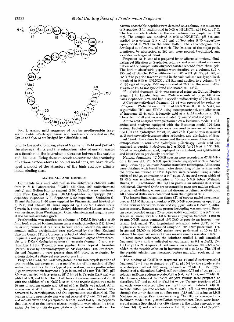
In the current investigation we describe the interaction of lanthanide ions with a y-carboxyglutamic acid-rich fragment of prothrombin, fragment 12-44.’ Fragment 12-44, with a known amino acid sequence, contains 8 y-carboxyglutamic acid residues and has a molecular weight of about 4100 (Fig. 1). This fragment binds metal ions (10) and is of a molecular size amenable to study by natural abundance “C! nuclear magnetic resonance spectroscopy. Paramagnetic lanthanide ions, with ionic radii and ligand specificity similar to Ca(II),
’ The fragment numbers refer to a polypeptide fragment of pro- thrombin where the first number (X) is the NH2-terminal residue and where the second number (Y) is the COOH-terminal residue.
12521
by guest on April 12, 2018
http://ww
w.jbc.org/
Dow
nloaded from
12522 Metal Binding Sites of a Prothrombin Fragment
NH: -
r Phe
20
FIG. 1. Amino acid sequence of bovine prothrombin frag- ment 12-44. y-Carboxyglutamic acid residues are indicated as Gla; Cys 18 and Cys 23 are bridged by a disulfide bond.
bind to the metal binding sites of fragment 12-44 and perturb the chemical shifts and the relaxation rates of carbon nuclei as a function of the interatomic distance between the carbon and the metal. Using these methods to estimate the proximity of various carbon atoms to bound metal ions, we have devel- oped a model of the structure of the high and low affinity metal binding sites.
MATERIALS AND METHODS
Lanthanide ions were obtained as the anhydrous chloride salts from K & K Laboratories. ‘53GdC1, (33 Ci/g, 99% radiochemical purity) and Bolton-Hunter reagent (1500 Ci/mol) were purchased from New England Nuclear. DEAE-Sephadex, sulfopropyl (SP)- Sephadex, Sephadex G-75, Sephadex G-50 (superfine), Sephadex G- 25, and Sephadex G-10 were supplied by Pharmacia, and Bio-Gel P- 2, P-30, and Chelex 100 were supplied by Bio-Rad Laboratories. Trypsin (L-l-tosylamido-2-phenylethyl chloromethyl ketone-treated) was purchased from Worthington. Other chemicals and reagents were of the highest available grade.
Prothrombin was purified on columns of DEAE-Sephadex A-50 from fresh titrated bovine plasma using standard methods (11). Blood collection, removal of red cells, barium citrate adsorption, and am- monium sulfate precipitation were performed by the New England Enzyme Center (Tufts University School of Medicine). Prothrombin fragment 1 was prepared by applying a thrombin digest of prothrom- bin to a DEAE-Sephadex column to separate fragment 1 and pre- thrombin 1 (11). Thrombin was purified from Topical Thrombin (Parke-Davis) by chromatography on SP-Sephadex (12). Prothrom- bin and fragment 1 were greater than 95% pure, as evaluated by sodium dodecyl sulfate-gel electrophoresis (13).
Fragment 12-44, the y-carboxyglutamic acid-rich tryptic peptide of prothrombin, was prepared by a modification of the method of Nel- sestuen and Suttie (10). In a typical preparation, bovine prothrombin (3 g) or prothrombin fragment 1 (1 g) in 222 ml of 1 mu Tris-HCl, pH 8.5, was digested with trypsin at 25°C for 24 h. Trypsin (18.5 mg) was added at 0, 3, and 18 h. The pH of the solution was maintained at 8.0 with the addition of 1 M NaOH. At 24 h, the tryptic digest was made 20 mM in sodium citrate and 9.6 ml of 1 M Bach was added. After incubation at 4°C for 30 min, the precipitate which formed was harvested by centrimgation and the supernatant was discarded. The barium citrate precipitate was washed twice at 4°C with 222 ml of 10 mM sodium citrate and precipitated with 9.6 ml of BaC12. The peptides that absorbed to the barium citrate precipitate were eluted by tritu- rating the barium citrate precipitate with 1 M sodium sulfate. The
barium-absorbable peptides were desalted on a column (4.0 x 150 cm) of Sephadex G-10 equilibrated in 0.05 M NHdHCOa, pH 8.0, at 25°C. The fraction which eluted in the void volume was lyophilized (120 mg). The sample was dissolved in 0.05 M NH4HC03, pH 8.0, and applied to a column (2.5 x 150 cm) of Sephadex G-75 (superfine) equilibrated at 25°C in the same buffer. The chromatogram was developed at a flow rate of 4.0 ml/h. The fractions of the major peak, monitored by absorption at 280 nm, were pooled, lyophilized, and identified as fragment 12-44.
Fragment 12-44 was also prepared by an alternate method, elimi- nating gel filtration on Sephadex columns and concomitant contami- nation of the sample with oligosaccharides leeched from these gels. The barium-absorbable peptides were desalted on a column (1.5 X 150 cm) of Bio-Gel P-2 equilibrated in 0.05 M NHdHCO:s, pH 8.0, at 25°C. The peptide fraction eluted in the void volume was lyophilized, dissolved in 0.05 M NH4HCOs, pH 8.0, and applied to a column (1.5 x 180 cm) of Bio-Gel P-30 equilibrated at 25°C in the same buffer. Fragment 12-44 was lyophilized and stored at -15’C.
‘251-labeled fragment 12-44 was prepared using the Bolton-Hunter reagent (14). Labeled fragment 12-44 was isolated by gel fdtration using Sephadex G-25 and had a specific radioactivity of 720 Ci/mol.
S-Carboxymethylated fragment 12-44 was prepared by reduction of fragment 12-44 (38 mg) in 12 ml of 0.5 M Tris-HCl, 0.5 M NaCl, 5.5 M guanidine HCl, and EDTA using mercaptoethanol, and alkylation of fragment 12-44 with iodoacetic acid at a 1:175 molar ratio (15). The extent of alkylation was evaluated by amino acid analysis.
Amino acid analyses were performed on a Beckman model 119CL amino acid analyzer equipped with the Beckman model 126 data system. Protein hydrolysates were prepared in evacuated tubes in 6 M HCl and hydrolyzed for 24, 48, and 72 h. Cystine was measured as S-carboxymethylcysteine after reduction and alkylation of frag- ment 12-44. The values for serine and threonine were determined by extrapolation to zero time hydrolysis. y-Carboxyglutamic acid was analyzed in peptide hydrolyzed in 2 M KOH for 22 h at 1lO’C (16). or,-y-Carboxyglutamic acid, employed as a standard, was synthesized and identified as previously described (6).
Natural abundance 13C NMR spectra were recorded at 67.88 MHz on a Bruker HX 270 NMR spectrometer equipped with a Nicolet computer using pulse-mode Fourier transform techniques. All spectra were obtained with broad band noise decoupling of the protons with the probe maintained at 22°C. Spectra were recorded using a pulse width of 19.5 as, equivalent to a 90’ pulse. A spectral sweep width of 15 KHz was employed. Samples in lo-mm NMR tubes (Wilmad, Buena, N. J.) contained 10% D20 to provide an internal deuterium lock signal. Chemical shifts are presented in parts per million relative to tetramethylsilane, where internal dioxane is defined as 66.88 ppm. All chemical shifts were computed from the digitized data.
The longitudinal relaxation times (Tl) of carbon nuclei were meas- ured at 15.1 MHz using a Bruker WP60 NMR spectrometer operating in the Fourier transform mode and equipped with a Nicolet quadra- ture accessory. Random noise proton decoupling was employed. Spec- tra were recorded using a 20-ps pulse width equivalent to a 90” pulse. A spectral sweep width of 4.0 KHz was employed. Samples (1 ml) in lo-mm NMR tubes contained 10% D20 to provide an internal deu- terium lock signal. The longitudinal relaxation rates (l/Z’l) of the aliphatic carbons were obtained using the 180”.7-90” pulse train (17). In general 75,000 to 100,000 pulses were performed at 10 to 12 7 values. The standard error of these measurements was about 10%.
Unless stated otherwise, the solutions studied (1 ml) contained fragment 12-44 at the indicated concentration in 0.1 M NaCI, 10% D20 at pH 5.39. Aliquots of lanthanide ion solutions (10 mM) were added to the peptide solution in increments of 5 to 25 ,ul. The pH of the peptide solution was measured before and after each metal ion addition.
The binding of Gd(II1) to fragment 12-44 and S-carboxymethyl fragment 12-44 was evaluated at 23” at pH 5.4 by the steady state rate dialysis method (18) using radioactive ls3Gd(III). The upper chamber of a siliconized dialysis cell contained 0.75 ml of the peptide solution in 25 mM sodium acetate, 0.25 M NaCl (pH 5.4), and ‘53Gd(III). Membranes, obtained as Fisher dialyzer tubing, were prepared as described (19) and stored in distilled water. Twenty fractions of 2.8 ml each were collected after each addition of unlabeled Gd(II1). Acetate buffer (25 mM acetate, 0.25 M NaCl, pH 5.4) was pumped through the lower chamber at a flow rate of 2.8 ml/min using an LKB Multiperpex pump. Fractions containing ls3Gd(III) were assayed in a Beckman model 8000 y scintillation spectrometer. Data were inter- preted using a Scatchard plot (20) where c is the molar concentration of free Gd(II1) and r is the moles of Gd(II1) bound/m01 of peptide.
by guest on April 12, 2018
http://ww
w.jbc.org/
Dow
nloaded from

Metal Binding Sites of a Prothrombin Fragment 12523
The relative occupancy of the high and low affinity metal binding sites was calculated using the affinities and stoichiometries deter- mined experimentally (21). Based upon two classes of sites,
where V is the molar ratio of bound metal to peptide, N, and Nr are the number of sites in Class 1 and Class 2, KI and K1 are the association constants for the metal binding sites of Class 1 and Class 2, and C is the free metal concentration. The fraction (F) of a particular class of site i which is occupied by metal may be expressed as
RESULTS
Purification of Prothrombin Fragment 12-44-Prothrom- bin or prothrombin fragment 1 was digested with trypsin, and fragment 12-44 in the hydrolysate adsorbed to and eluted from barium citrate. After desalting the peptide preparation on a column of Sephadex G-10, the peptide fractions contain- ing fragment 12-44 were purified on a column of Sephadex G- 75. A typical chromatogram is shown in Fig. 2. The major peak represents fragment 12-44. Fragment 12-44 was pre- pared in approximately 50% yield, based upon the theoretical quantity expected from the amount of prothrombin or frag- ment 1 digested. In preparations using Bio-Gel P-2 and P-30 columns fragment 12-44 was obtained in similar yield. An E& of 13.6 was employed for fragment 12-44, based upon the extinction coefficient of the single tryptophan residue.
24
2.c
I8
I6
: 14
z a 1.2
IO
08
06
VOLUME (ml )
FIG. 2. Purification of fragment 12-44 by gel filtration. The tryptic digest of prothrombin, desalted on a column of Sephadex G- 10, was applied to a column (2.5 X 150 cm) of Sephadex G-75 (superfine) equilibrated in 0.05 M NH,HCO.I, pH 8.0. Flow rate, 4.0 ml/h; temperature, 25°C.
TABLE I Amino acid composition ofprothrombin fragment 12-44
Values are molar ratios of amino acids, based upon comparison to leucine.
Acid hydrolysis Alkaline hydrolysis
Aspartic acid 2.08 (2)” 2.15 (2)” Threonine 0.96 (1) Serine 2.91 (3)
Glutamic acid 8.52 (8) 0.50 (0) Proline 1.28 (1) Glycine 1.13 (1) Alanine 4.10 (5) Half-cystine” 1.87 (2) Valine 0.12 (0) Methionine 0.08 (0) Isoleucine 0.04 (0) Leucine 4.00 (4) 4.00 (4) Tyrosine 0.13 (0) Phenylalanine 2.03 (2) Tryptophan N.Q.’ Lysine 0.51 (1) Histidine 0.05 (0) Arginine 1.96 (2)
_ y-Carboxyglutamic acid 0.00 (0) 7.35 (8)
“ Number in parentheses is number of residues expected. ’ Measured as carboxymethylcysteine. ’ Present but not quantitated.
The amino acid compositions of the acid and the alkaline hydrolysates of the preparation of fragment 12-44 are shown in Table I. The amino acid composition obtained was in good agreement with that expected from the amino acid sequence. The low values of alanine and lysine are unexplained, although in other analyses the alanine composition approached that expected from the sequence data. The glutamic acid observed in the acid hydrolysate was derived from y-carboxyglutamic acid.
Metal Binding Properties-The interaction of Gd(II1) and fragment 12-44 was examined by steady state rate dialysis at pH 5.4 and 23°C using ‘“,‘Gd(III). A dialysis membrane was employed which was permeable to Gd(II1) and which did not bind Gd(II1) at the metal concentrations employed. Further- more, fragment 12-44, labeled with I”? using the Hunter- Bolton reagent, did not dialyze across the membrane in de- tectable quantities in the presence or absence of 1 pM GdCL. The binding of ‘““Gd(II1) (0.4 to 70 PM) to unlabeled fragment 12-44 (3.6 pM) was evaluated using the method of Colowick and Womack (18) to determine the concentration of free and peptide-bound Gd(II1). These data were interpreted using a Scatchard plot (Fig. 3A). A single high affinity metal binding site which bound Gd(II1) with a dissociation constant, KD, of 0.55 PM was observed. Multiple lower affinity sites were less well defined. As a first approximation, about 4 to 6 lower affinity metal binding sites were estimated which bound Gd(II1) with a K,, of between 4 and 8 pM. These affinities are similar to those determined for the two classes of sites on intact prothrombin.
Similar experiments were performed with S-carboxymethyl fragment 12-44 (Fig. 3B). This fragment 12-44 derivative has no high affinity metal binding sites. However, four to six lower affinity metal binding sites which bound Gd(II1) with a KI, of about 8 PM were detected.
These results suggest that the low affinity metal binding sites are single or paired adjacent y-carboxyglutamic acid residues. These sites have a K,, of about 4 to 8 pM, while the y-carboxyglutamic acid. metal complex, studied previously (6), has a K, and Kz which were observed as an average KJ, of about 35 to 50 PM. Within experimental error and interpretive ambiguities, these values are similar. It would follow, then,
by guest on April 12, 2018
http://ww
w.jbc.org/
Dow
nloaded from

12524 Metal Binding Sites of a Prothrombin Fragment
that the high affinity metal binding site, with a KD of 0.55 PM, is composed of more metal ligands than are contained in a single y-carboxyglutamic acid residue. The formation of this site requires an intact disulfide bond and, as such, involves maintenance of a necessary geometry between two metal ligands. On the basis of studies of the geometry of the ternary complex of free y-carboxyglutamic acid and a single metal ion (6), we can propose that the high affinity site is formed by two y-carboxyglutamic acid residues which bind a single metal ion. An upper limit of the KD may be estimated at approxi- mately 2 nM, on the basis of the energy of binding of Gd(II1) to y-carboxyglutamic acid (6). However, the actual binding energy would be roughly equivalent to the sum of the energy of interaction of the metal ion with each y-carboxyglutamic
A so- I
0
*o-
50- .
FIG. 3. Interaction of “%d(III) with fragment 12-44 and S- carboxymethyl fragment 12-44. Scatchard plot of data obtained from steady state rate dialysis experiments performed at 23°C using 3.5 PM prothrombin fragment 12-44, ‘53GdCh, and 0.4 to 70 PM GdC4 in 25 mu imidaxole, 0.15 M NaCl, pH 5.4. A, fragment 12-44, B, S- carboxymethyl fragment 12-44.
acid residue minus the energy associated with the metal- induced conformational rearrangement of the polypeptide chain.
It should be emphasized that, although the Scatchard anal- ysis of the metal binding data is consistent with two classes of sites, we cannot eliminate the possibility of a third class of metal binding sites with intermediate affinity. Specifically, the two Gla-Gla sequences found in this peptide may have metal binding properties which differ from a single y-carboxyglu- tamic acid residue.
13C NMR Spectrum of Prothrombin Fragment 12-M-The 13C NMR spectrum of fragment 12-44 at 67.88 MHz is shown in Fig. 4. The resonances are numbered sequentially from the downfield to upfield peaks. In this experiment fragment 12-44 was prepared by gel filtration on polyacrylamide gels (Bio- Gel) instead of Sephadex to eliminate contaminating oligosac- charides from the preparation. Tentative assignments of the carbon resonances in fragment 12-44 were made on the basis of known chemical shift values of carbon nuclei in free amino acids and peptides and are presented in Table I (22, 23). The chemical shift values of carbon resonances of y-carboxyglu- tamic acid are 178.5, 178.0, 174.8, 54.3, 55.9, and 31.4 ppm for the yl-carboxyl carbon, p-carboxyl carbon, cu-carboxyl carbon, C,, C,, and Cp (6). On this basis we have assigned the large envelope (peak 2), centered at 177 ppm, to include resonances of the yl- and yz-carboxyl carbons of the 8 y-carboxyglutamic acid residues. The large envelope at 53.5 ppm (peak 23-25) contains, among other resonances, those of the eight C, and C, carbons of y-carboxyglutamic acid. The envelope at 31.6 ppm (peak 38) is assigned to the eight CB carbon resonances of y-carboxyglutamic acid. The C, resonances of the 2 arginine residues at 157.2 ppm are labeled as peak 5. Tentative assign- ments of the resonances in the spectrum fragment 12-44 are given in Table II.
Estimates of the longitudinal relaxation rate, l/1;, for the phenylalanine aromatic carbons and the (Y carbon envelope, including the LX and y carbons of y-carboxyglutamic acid residues, were made for fragment 12-44 and S-carboxymeth- ylated fragment 12-44 (see below). These carbons were cho- sen, rather than the y-carboxyl carbons, because the relaxation mechanism involves the directly bonded hydrogen and allows
FIG. 4. Natural abundance “C NMR spectrum of fragment 12-44. Pulses, 21,600, repeat time, 1.5 s; temperature, 22°C. Chemical shifta, relative to tetramethylsilane, of the numbered resonances were obtained from the digitized data and are presented in Table II. The sample of fragment 12-44 was prepared by gel fdtration on polyacrylamide gels to avoid contamination of the peptide with oligosaccharide. Fragment 12-44,6 mu, in 10% D20,O.l M NaCl.
by guest on April 12, 2018
http://ww
w.jbc.org/
Dow
nloaded from

Metal Binding Sites of a Prothrombin Fragment 12525
TABLE II
Chemical shift values of the resonances in the 13C NMR spectrum of fragment 12-44
The numbers in parentheses indicate the number of residues of each amino acid in this peptide. Each y-carboxyglutamic acid residue contains two v-carboxvl carbons.
Ped Tentative assignment Chemical shift
1 2
10 11 12 13 14 15 16 17 18 19 20 21 22 23. 24 25.
26 27 28
29 I
30 31 32 33 1 34 35
36 37 38 39 40 41 42 43 44 45 46 47
y-carboxyl C, Gla (8) ,&carboxyl C, Asp (1) Carbonyl C (32) Carbonyl C, Gly (1)
Cc, A&(2) CL Phe (2) Ci;, Gee, Pie (2) C&t, CSZ, Phe (2)
r Cr, Phe (2) C,, Trp (1)
CL, Trp (1)
CD, Trp (1) C,a, Trp (1)
CO, Trp (1) CL, Tw (1) CSS, Trp (1) Cg, Thr (1) C/i, Ser (3) CL,, Thr (0, C,, Pro (1)
I CL Ser (3) C,,, Phe (2)
I
Cc,, Gla (8); C,, Gla (8) CL Arg (2); CL, LYS (1) CL, IA’ (4 )
C<,, Asp (1)
I
c<,, CYS (2) CL Trp (1) Cc,, Ala (5) G,, Asn (1)
C,,. Trp (1) C,,, Ala (5) G, Pro (1) CL Gly (1); CS, Arg (2)
i C,b Leu (4)
C/J, LYS (1)
-I CR, ASP (1); CD, Phe (2)
c/r, CYS (2) Cl,, Asn (1)
Ch Gla (8) CO, Pro (1); CP, LYS (1) Gc, Am (2) C, LYE (1); CD, Trp (1)
C,, Leu (4); C,, Pro (1); C,, Arg (2) G, Leu (4) Cc, Leu (4) c,, LYS (1) C,, Thr (1) CJ, Ala (5)
PPm
176.7-177.4
172.1-175.2 157.2 136.4 129.4 129.0
127.5 127.4 125.0 122.2 119.7 118.6 112.3 109.3
67.4 61.3 59.7 56.3 56.0 55.7
54.3 53.7
53.4 53.2
52.0 51.5 50.5
50.2
48.0 41.1
39.9
38.6 37.3 36.5 33.1 31.6 28.6 27.4 27.3 24.6 22.6 21.0 19.2
17.8 16.8
for unambiguous calculation of the 7,. These measurements were performed at 15.1 MHz using peptide rendered metal- free using Chelex 100.’ The Tl of the aromatic envelope in fragment 12-44 and S-carboxymethyl fragment 12-44 was 0.09 and 0.10 s, respectively. The Tl of the ai carbon envelope was 0.05 s in fragment 12-44 and 0.09 in S-carboxymethyl fragment 12-44. Assuming that the relaxation times of these carbons can be interpreted in terms of a dipole-dipole interaction with
’ The estimate of the correlation time was based upon measure- ments on the metal-free peptide. Experiments involving the binary complex of fragment 12-44 and diamagnetic La(II1) were precluded because of precipitation.
the directly bonded hydrogens and that the rotational motion is isotropic, a 7c of about 1 x lo-’ s can be approximated for both fragment 12-44 and S-carboxymethylated fragment 12- 44. This value of 7, represents an upper limit since the rota- tional correlation time of the y-carboxyl carbons (Gla) and C, (Arg) may be shorter.
To estimate the relative proximity of the carbon nuclei in fragment 12-44 to bound metal ions, the effect of paramag- netic lanthanide ions on the 13C NMR spectrum of fragment 12-44 was determined. The magnitude of the perturbation of a carbon nucleus induced by each bound paramagnetic metal ion depends on the following considerations: 1) the distance between the metal ion bound in a specific metal binding site and the carbon nucleus; 2) the rate of chemical exchange of the carbon nucleus into and out of the paramagnetic environ- ment; 3) the mole fraction of a specific metal binding site occupied by a paramagnetic metal ion when the moles of binding sites of varying affinity for metal ions exceeds the moles of metal ions; 4) for Pr(II1) and Eu(III), the orientation factor relating the geometry of the metal ion and the asym- metric peptide ligand; 5) the correlation time, T?.
Effect of Lanthanide Ions on the 13C NMR Spectrum of Fragment 1%44-The effects of Eu(III), Pr(III), Gd(III), and La(II1) on carbon nuclei of fragment 12-44 were examined at 67.88 MHz at 22°C. In solutions containing 3 mM Eu(II1) and 12 mM peptide, perturbations due to Eu(II1) were evident (Fig. 5~). These included an upfield shift of 120 to 200 Hz of components of the carboxyl envelope (peak 2), a small upfield shift of the C, of the arginines (peak 5), a shift of the center of 23-25 (C, and C,, Gla) and peak 38 (CL{, Gla). In solutions containing 11 mM Pr(II1) and 20 mu peptide, the carboxyl carbon resonances (peak 2) were shifted downfield about 70 Hz (Fig. 5b). The envelope 23-25 was broadened, with a significant downfield shift obscuring the resolution between 19-21 and 22-25. In solutions containing 0.05 IIIM Gd(II1) and 10 mM fragment 12-44, marked broadening of resonances of carbons in close proximity of bound metal ions were observed (Fig. 5d). Resonances perturbed included peaks 2, 5, 30, 37, 26, 32-33, 38, and 23-25; these are discussed below. We also evaluated the interaction of diamagnetic La(II1) (4 mM) with fragment 12-44 (12 InM). As shown in Fig. 5e, no detectable changes were induced in the 13C NMR spectrum by La(II1). Thus, the perturbations induced by Eu(III), Pr(III), and Gd(II1) are due to paramagnetic effects. These results would suggest that the nuclei perturbed by these metals are in close proximity of the bound metals.
In order to predict the magnitude of the shifts of carbon resonances in fragment 12-44 induced by lanthanide ions, parallel studies of the magnitude of shifts of the carboxyl carbon resonances of methylmalonic acid bound to Eu(II1) and Pr(II1) were performed.” While these model studies yielded predicted shifts similar to those observed in fragment 12-44 when Pr(II1) was employed, the shifts of the y-carboxyl carbons observed in the Eu(II1) *fragment 12-44 complex were larger than those expected. This discrepancy may relate to the absence of axial symmetry in the peptide. metal complex with regard to its magnetic properties. The implications of such nonaxial magnetic properties on the chemical shift in- duced by lanthanide ion shift reagents has been discussed by Horrocks (24). To eliminate consideration of the relation of the carbon nucleus relative to the principal magnetic axes defined by the magnetic susceptibility tensor of the metal in the binding site, further studies were performed with Gd(II1). With Gd(III), the paramagnetic broadening of a nucleus is a
’ B. C. Furie, M. Blumenstein, and B. Furie, unpublished observa- tions.
by guest on April 12, 2018
http://ww
w.jbc.org/
Dow
nloaded from

12526 Metal Binding Sites of a Prothrombin Fragment
FIG. 5. Effect of paramagnetic lan- thanide ions on the 13C! NMR spec- trum. Fragment 12-44: (a) fragment 12- 44, 10 mM in 10% DQO, 0.1 M NaCl, pH 5.39; (b) fragment 12-44, 20 miv in 10% D20, 0.1 M NaCl, 11 ItIM PrC13, pH 5.39; (c) fragment 12-44, 12 mM in 10% D20, 0.1 M NaCl, 3 mM EuCl$, pH 5.39; (d) fragment 12-44, 10 mM in 10% D20, 0.1 M NaCl, 0.05 mM GdCls, pH 5.39; (e) fragment 12-44, 9 mM in 10% DZO, 0.1 M NaCl, 4 mM La& pH 5.39.
function of the internuclear distance alone and does not include angular considerations.
Effect of Gd(IIJ on the 13C NMR Spectrum of Fragment 12-44-Gd(II1) caused marked broadening of resonances of carbon nuclei near the bound metal ion. Low molar ratios of Gd(II1) to fragment 12-44 were associated with significant perturbations of the 13C NMR spectrum. The titration of fragment 12-44 with Gd(II1) is shown in Fig. 6. The spectrum of fragment 12-44 (11 mM) in 10% DzO, 0.1 M NaCl, pH 5.39, was recorded at 67.88 MHz at 22°C (Panel a). Subsequent spectra were obtained after the addition of Gd(II1) to fragment 12-44 to yield Gd(II1) concentrations of 0.0012, 0.0062, 0.021, 0.046,0.096,0.29,0.49,0.69,0.89, 1.09, 1.59,2.05,3.05,4.05, and 6.05 mM. At molar ratios of 0.004 (Panel b), Gd(II1) appeared to have no effect on the carbon resonances except for a decrease in the amplitude of peak 2, representing the y-car- boxy1 carbon resonances. At a ratio of 0.038 (Panel c), the y- carboxyl carbon envelope is markedly reduced in amplitude, peak 5 is about 50% of its original amplitude, and peaks 32-33, 38, and 23-25 are broadened. At a ratio of 0.088 the line widths of peak 2 and peak 5 were about 140 and 40 Hz, respectively (Panel d) . In addition to peaks 32-33,38, and 23-25, peaks 30,
37, and 26 became significantly broadened. At a ratio of 0.17 (Panel e), exaggeration of the above perturbations were ob- served. The line widths of peak 2 and peak 5 were about 240 and 70 Hz, respectively. Of significance is that the broadening of the resonances appeared selective. For instance, the reso- nances of the aromatic carbons (Phe 41, Phe 29, and Trp 42) were unperturbed and the carbonyl carbons were minimally affected.
These data indicate that the y-carboxyl carbon, C,, C/c, and C!, of y-carboxyglutamic acid residues, and the C, of the arginine residues are in close proximity of bound Gd(II1) ions. Using affinities and stoichiometries of fragment 12-44 metal binding that were determined experimentally, we can calcu- late that about 60% of the Gd(II1) is bound in the single high affinity site and about 40% of the remaining Gd(II1) is distrib- uted among the low affinity sites. The line broadening that is observed is consistent with this calculation. Using the Gd(III)- y-carboxyl carbon distance of 3.2 A measured in the metal. y-carboxyglutamic acid complex (6), an upper limit of TV of 1 X lo-” as measured, and the Solomon-Bloembergen equation (25, 26), we can estimate that the upper limit of the bound line width of the y-carboxy carbon of y-carboxyglutamic acid.
by guest on April 12, 2018
http://ww
w.jbc.org/
Dow
nloaded from

Metal Binding Sites of a Prothrombin Fragment 12527
FIG. 6. Titration of fragment 12- 44 with Gd(II1). Fragment 12-44 (11 mM) in 10% D20, 0.1 M NaCl, pH 5.39. Pulses, 22,ooO, temperature, 22°C; repeat time, 1.5 s; GdCl, concentrations were: a, 0 mM; b, 0.046 mM; c, 0.50 mM; d, 1.1 mM;
e, 2.0 rnM.
Gd(II1) in fragment 12-44 is about 17,200 Hz. Three illustra- tive models may be considered in which the high affinity site is in slow exchange and the low affinity sites in fast exchange with Gd(II1) relative to the NMR time scale. First, if the 8 y- carboxyglutamic acid residues are equivalent low affinity metal binding sites in fragment 12-44 (which they are not), we would predict that at the molar ratio of Gd(III)/peptide employed in Fig. 6, d and e, the upper limits of the y-carboxyl carbon line width would be 190 and 370 Hz, respectively. I f significant internal motion occurred, these line widths would be appreciably smaller. I f 60% of the Gd(II1) occupies the high affinity site, resonances of y-carboxyl carbons of participating residues will be broadened beyond detection. About 40% of the Gd(II1) would be distributed among the lower affinity sites. Thus, at the molar ratios of Gd(III)/peptide of 0.088 and 0.17 studied, the line width of the y-carboxyl carbon would be about 120 and 234 Hz. These data, derived for the model based upon the rate dialysis experiments, closely approximate the observed line widths. Third, if most of the Gd(II1) is associated with the high affinity site and minimal Gd(II1) is associated with the lower affinity sites, minimal perturbation of the observed y-carboxyl carbon spectrum would be ex-
pected. This is, of course, not the case. The observed line widths were 140 and 240 Hz at these molar ratios. These data are fully consistent with the binding model postulated on the basis of the rate dialysis experiments. However, due to the uncertainty of the predicted line widths mentioned above, these data do not constitute independent evidence for this model. The NMR data do, however, preclude the model in which most of the metal is associated with the high affinity site. Further, they indicate that all of the y-carboxyglutamic acid residues are involved in metal binding. Given this model, is it Gd(II1) in the single high affinity site or Gd(II1) in the lower affinity sites which perturbs the C, of the arginine residues? If the latter, Gd(II1) would be distributed evenly among the lower affinity sites. To yield the line width of the C, of arginine that were observed, the bound line width of the C, (Arg) in the metal. peptide complex would have to be about 5700 Hz. Using the Solomon-Bloembergen equation and the experimentally determined 7e, an upper limit of 3.9 A can be placed on the Gd(III)-C, distance. If this were the case, we would have to assume that arginine participates in metal liganding. In fact, metal binding experiments employing L-
Arg-n-Gla-n-Gla-O-methyl ester have indicated that arginine
by guest on April 12, 2018
http://ww
w.jbc.org/
Dow
nloaded from

12528 Metal Binding Sites of a Prothrombin Fragment
has no apparent role in metal binding (27). We then must consider that the C, of arginine is perturbed by Gd(II1) bound in the single high affinity site. By performing the same cal- culations, a bound line width of the C, in the Gd(II1). peptide complex may be estimated to be 760 Hz. This is equivalent to an upper limit of 5.5 A between Gd(II1) bound in the high affinity site and the C, of the arginine residues. This appears to be a more plausible explanation of the specific perturbation of the C, carbons.
The selective perturbation of the C, of the arginine residues was used to localize 1 or more of the y-carboxyglutamic acid residues which could participate in the formation of the high affinity metal binding sites. Specifically, Gla 15 and Gla 26 (Fig. 1) were considered since a metal bound by these residues could be in close proximity of the C, of the arginines. It would appear, however, that this site would require an intact disul- fide bond. To evaluate the contribution of the disulfide bond in stabilizing the structure of the high affinity site, which in turn contains a bound Gd(II1) in close proximity of the C, of the arginines, the disulfide bond was cleaved by reduction and alkylation. If the high affinity sites were near these arginine residues, elimination of the site should reduce the perturba- tion of these carbon nuclei.
Effect of Gd(III) on S-Carboxymethyl Fragment 12-44- The effect of Gd(II1) on S-carboxymethyl fragment 12-44 was compared to the effect of Gd(II1) on fragment 12-44. As shown in Fig. 3B, the S-carboxymethyl derivative of the peptide lacked the high affinity metal binding site. Titration of S- carboxymethyl fragment 12-44 with Gd(II1) altered the spec- trum, shown in Fig. 7. The spectrum of S-carboxymethyl fragment 12-44 (8 mM) in 10% D20, 0.1 M NaCl, pH 5.39, was
recorded at 67.88 MHz at 22°C (Panel a). Subsequent spectra were obtained at various Gd(II1) concentrations of 0.028, 0.271, 0.390, 0.509, 0.627 and 1.2 mM. Panels b and c (Fig. 7), with metal and peptide at molar ratios of 0.088 and 0.179, respectively, may be compared directly to Panels d and e in Fig. 6, in which the molar ratios of metal/peptide are similar. Although peak 2 was altered (approximately 250 and 500 Hz in Panels b and c, respectively), small changes in peak 5 were observed. Line widths of about 10 and 25 Hz were seen in Panels b and c, respectively. The y-carboxyl carbon line widths observed are similar to those that might be predicted for S-carboxymethyl fragment 12-44, which contains four to six metal bindings sites of equivalent affinity (Fig. 3B). Based upon an upper limit of the 7c of 1 x lo-’ s measured experi- mentally for S-carboxymethyl fragment 12-44 and a Gd(III)- y-carboxyl carbon distance of 3.2 A, an upper limit of the bound line width for the y-carboxyl carbon of y-carboxyglu- tamic acid complexed to Gd(II1) in the S-carboxymethyl fragment 12-44*Gd(III) complex may be calculated as 17,200 Hz. From the number of binding sites, we can estimate that at molar ratios of Gd(III)/peptide of 0.088 and 0.179, an upper limit of the observed y-carboxyl carbon line width of 300 and 615 Hz, respectively, would be expected. This is in reasonable agreement with the observed line widths (250 and 500 Hz, respectively) obtained experimentally. Thus, it would appear that the bound Gd(II1) is distributed evenly among the metal binding sites of the peptide.
If we consider that this peptide contains four to six binding sites with equal occupancy by Gd(III), an estimate of the bound C, line widths can be made. Based upon the fractional occupancy of the sites, the experimentally observed line widths of the C, of the arginines, and the molar ratio of Gd(III)/peptide, we can estimate this bound line width to be about 650 Hz. This is similar to the bound line width of the C, (Arg) in fragment 12-44. This would indicate that the Gd(III)- C,(Arg) distance is similar in both fragment 12-44 and S- carboxymethyl fragment 12-44. It would thus appear that the differences in the observed line width of C,(Arg) in fragment 12-44 and S-carboxymethyl fragment 12-44 induced by Gd(II1) are not due to differences in the metal-C, distance but due to changes in the bound Gd(II1) concentration in the vicinity of the C, of the arginines. In fragment 12-44, 60% of the Gd(II1) is bound in the high affinity site in close proximity of the C,(Arg). In S-carboxymethyl fragment 12-44, which lacks the high affinity site, only 20% of the Gd(II1) is bound in a site in close proximity to a C,(Arg). This interpretation allows us to conclude that the high affinity site is in close proximity of the 2 arginines.
In summation, the results of the rate dialysis studies indi- cate that fragment 12-44 contains two classes of metal binding sites. A model has been proposed in which a high affinity site is composed of 2 y-carboxyglutamic acid residues bound to a single metal ion. In the low affinity sites a metal ion is bound to a single y-carboxyglutamic acid residue. Although the NMR data alone do not provide direct evidence for two classes of metal binding sites, they are consistent with this model, considering the relative occupancy of the two classes of sites. When interpreted within the context of the model, the NMR data suggest that the high affinity metal binding site is close to the 2 arginine residues in fragment 12-44. These results are consistent with the interpretation that the high affinity site is stabilized by the disulfide bond.
DISCUSSION FIG. 7. Titration of S-carboxymethyl fragment 12-44 with
Gd(II1). Fragment 12-44 (8 mM) in 10% DzO, 0.1 M NaCl, pH 5.39. Metal ions play a central role in hemostasis (28). Among Pulses, 20000; temperature, 22’C; repeat time, 1.5 s. GdClj concentra- other functions, calcium is required in the zymogen activation tion was: a, 0 mM; b, 0.8 InM; c, 1.4 mM. of the vitamin K-dependent clotting proteins. These proteins
by guest on April 12, 2018
http://ww
w.jbc.org/
Dow
nloaded from

Metal Binding Sites of a Prothrombin Fragment
bind metal ions, with structural and functional implications (7). Using trivalent lanthanide ions as substitutes for Ca(II), we have established that Factor X and prothrombin contain two high affinity metal binding sites and a larger number of lower affinity metal binding sites which bind Gd(II1) with a binding constant, KD, of 0.1 to 1 pM and about 10 to 20 pM,
respectively (8, 9). These two classes of metal binding sites differ not only in their affinity for metal ions but in their metal ion specificity, their effect on tertiary structure, and their participation in protein-protein or protein-membrane interaction (29-32). In the absence of vitamin K or the pres- ence of vitamin K antagonists, an abnormal form of prothrom- bin circulates in plasma which lacks y-carboxyglutamic acid and which does not bind metal ions (4,5). These observations have implicated y-carboxyglutamic acid in the vitamin K- dependent proteins as participants in metal binding. y-Carl boxyglutamic acid has unique structural features which confer important metal binding properties on proteins (6, 33). Most significantly y-carboxyglutamic acid binds to half of the fist coordination sphere of metal ions, exposing the other half of the coordination sphere to another y-carboxyglutamic acid residue or other metal ligands. As previously suggested from the stoichiometry of ligand-metal binding (6), y-carboxyglu- tamic acid may participate in forming intramolecular or inter- molecular metal bridges.
In the current study we have attempted to define the structural features of the high and lower affinity metal binding sites of the vitamin K-dependent proteins. Specifically, we have examined the effect of paramagnetic lanthanide ions on the natural abundance 13C! NMR spectrum of a 33-residue y- carboxyglutamic acid-rich peptide isolated from bovine pro- thrombin. 13C NMR spectroscopy permits the detection of classes of carbon atoms in the amino acid residues of fragment 12-44 and, to some extent, the detection of individual atoms. In this study, we have been able to obtain qualitative infor- mation concerning the relative proximity of y-carboxyglu- tamic acid residues in this peptide and bound metal ions. However, the presence of multiple binding sites, the separa- tion of these sites into two separate classes on the basis of metal binding affinity, and the presumed difference in the chemical exchange rate of these classes of metal binding sites with trivalent lanthanide ions complicate analysis of the ob- served phenomena.
This peptide, fragment 12-44, contains a single high affinity metal binding site which binds Gd(II1) with an affinity similar to the high affinity sites of Factor X and prothrombin. The integrity of this site requires an intact disulfide bond, as do the sites on Factor X and prothrombin (34, 35). Furthermore, this peptide has a large number of lower affinity sites. The affinity of these sites for Gd(II1) is approximately the same as that of the lower affinity sites of Factor X (8) and prothrombin (9) and that of free y-carboxyglutamic acid (6). It will follow that the lower affinity sites are single or paired adjacent y- carboxyglutamic acid residues.
One explanation of the binding energy involved in the interaction of lanthanide ions and the high affinity site is that this metal binding is likely composed of multiple y-carboxy- glutamic acid residues which interact with a single metal ion. Based upon the geometry of the metal. y-carboxyglutamic acid complex and the stoichiometry of 2 mol of y-carboxyglu- tamic acid/l mol of lanthanide ions measured using free y- carboxyglutamic acid, we have suggested that 2 y-carboxyglu- tamic acid residues form the high affinity metal binding site (6). Since C, carbons of Arg 16 and Arg 25 are in close proximity of the metal ion bound in the high affinity site, Gla 15 and Gla 26 may represent the two participants in the formation of the high affinity site (Fig. 1). Significant stability
I20
FIG. 8. CPK model of prothrombin fragment 12-44. In this model the high affinity metal binding site, formed by Gla 15 and Gla 26, is occupied by a single metal ion. The C, carbon of the arginines is indicated by the arrow. The disulfide bond is indicated as S-S.
of the tertiary structure would be conferred by the disulfide bond connecting Cys 18 and Cys 23. This may explain how a small peptide such as fragment 12-44 possesses a binding site which binds metal ligands with the same affinity as native prothrombin. Indeed, when the disulfide bond is reduced and alkylated, no high affinity site can be detected and paramag- netic perturbation of the C, carbons of arginine is markedly reduced.
Based upon these hypotheses, we have constructed a Corey- Pauling-Koltun model of fragment 12-44 (Fig. 8). In this model Gla 15 and Gla 26 interact with a single metal ion and represent the high affinity metal binding site. The metal- carboxyl carbon distances employed are those measured pre- viously by paramagnetic relaxation enhancement studies (6). Each of the two y-carboxyglutamic acid residues in the high affinity metal binding site bind half of the coordination sphere of the metal ion. Arg 16 and Arg 25 can be oriented, one above and one below the binding site, such that their respective C, carbons are in close proximity of the bound metal. Due to the disulfide bond, the positions of Gla 15 and Gla 26 in the model are somewhat defined, with only limited conformational flex- ibility. The other 6 y-carboxyglutamic acid residues may par- ticipate by themselves or with an adjacent y-carboxyglutamic acid to form the low affinity metal binding sites.
These two classes of binding sites likely have separate structural properties and functional roles. The lower affinity metal binding sites involve y-carboxyglutamic acid residues which may bridge the protein via metal ions to either mem- brane surfaces or the surfaces of other proteins. Thus, these intermolecular bridges may participate in metal-dependent
by guest on April 12, 2018
http://ww
w.jbc.org/
Dow
nloaded from

Metal Binding Sites of a Prothrombin Fragment
protein-protein or protein-membrane interactions. The metal ion bound in the high affinity metal binding site forms an intramolecular bridge joining two polypeptide chains. Unlike a disulfide bond, this bridge is formed noncovalently and reversibly. In the unoccupied state, Gla 15 and Gla 26 would repel each other, with implications for the tertiary structure of this region. In the presence of metal ions, a significant alteration in the tertiary structure of this region should occur. Studies of prothrombin and prothrombin fragment 1 using fluorescence techniques and circular dichroism have been interpreted to indicate that there are metal ion-induced changes in the three-dimensional structure of prothrombin (29, 30,36). Our studies using conformation specific antibodies directed against the native format of fragment 12-44 have indicated that changes in the three dimensional structure of the y-carboxyglutamic acid-rich 12-44 region of prothrombin accompany metal binding (37, 38).
REFERENCES
1. Furie, B. C., Blumenstein, M., and Furie, B. (1977) Calcium Binding Proteins and Calcium Function (Wasserman, R. H., et al., eds) pp. 361-363, Elsevicr-North Holland, Amsterdam
2. Furie, B. C., Blumenstein, M., and Furie, B. (1978) Fed. Proc. 37, 1292
3. Davie, E. W., and Hanahan, D. J. (1977) in The Plasma Proteins (Putnam, F. W., ed) pp. 421-544, Academic Press, New York
4. Stenflo, J., and Ganrot, P. 0. (1973) Biochem. Biophys. Res. Commun. 50,98-104
5. Nelsestuen, G. L., and Suttie, J. W. (1972) Biochemistry 11,4961- 4964
6. Sperling, R., Furie, B. C., Blumenstein, M., Keyt, B., and Furie, B. (1978) J. Biol. Chem. 253, 3898-3906
7. Nemerson, Y., and Furie, B. (1980) CRC Crit. Rev. Biochem., in press
8. Furie, B. C., and Furie, B. (1975) J. Biol. Chem. 250, 601-608 9. Furie, B. C., Mann, K. G., and Furie, B. (1976) J. Biol. Chem.
251,3235-3241 10. Nelsestuen, G. L., and Suttie, J. W. (1973) Proc. Natl. Acad. Sci.
U. S. A. 70, 3366-3370 11. Mann, K. G. (1976) Methods Enzymol. 45, 123-156
12. 13. 14.
15. 16. 17.
18.
19. 20. 21.
22.
23.
24. 25. 26. 27.
28.
29. 30.
31.
32.
33.
34.
35.
36. 37.
38.
Lundblad, R. L. (1971) Biochemistry 10,2501-2506 Laemmli, U. K. (1970) Nature 227, 680-685 Bolton, A. E., and Hunter, W. M. (1973) Biochem. J. 133, 529-
539 Gurd, F. R. N. (1967) Methods Enzymol. 11, 532-541 Haushka, P. (1977) Anal. Biochem. 80,212-223 Farrar, T., and Becker, E. II. (1971) Pulse and Fourier Transform
NMR, pp. 20-22, Academic Press, New York Colowick, S. P., and Womack, F. C. (1969) J. Biol. Chem. 244,
774-777 McPhie, P. (1971) Methods Enzymol. 22, 25 Scatchard, G. (1949) Ann. N. Y. Acad. Sci. 51, 660-672 Fletcher, J. E., and Spector, A. A. (1977) Mol. Pharmacol. 13,
387-399 Dwek, R. A. (1973) Nuclear Magnetic Resonance in Biochemis-
try, Clarendon Press, Oxford Gurd, F. R. N., and Keim, P. (1973) Methods Enzymol. 27, 836-
911 Horrocks, W. D., Jr. (1974) J. Amer. Chem. Sot. 96, 3022-3024 Solomon, I. (1955) Physiol. Rev. 99, 559 Bloembergen, N. (1957) J. Chem. Physiol. 27, 572 Robertson, P., Jr., Koehler, K. A., and Hiskey, R. G. (1979)
Biochem. Biophys. Res. Commun. 86, 265-270‘ Furie. B.. and Furie. B. C. (1979) Chemistry and Phvsioloev of Y” ,
the Plasma Proteins (Bing, D. H., ed) pp. 185-192, Pergamon Press, New York
Nelsestuen, G. L. (1976) J. Biol. Chem. 251, 5648-5656 Prendergast, F. G., and Mann, K. G. (1977) J. Biol. Chem. 252,
840-850 Nelsestuen, G. L., Broderius, M., and Martin, G. (1976) J. Biol.
Chem. 251,6886-6893 Nelsestuen, G. L., and Broderius, M. (1977) Biochemistry 16,
4172-4177 Robertson, P., Jr., Hiskey, R. G., and Koehler, K. A. (1978) J.
Biol. Chem. 253, 5880-5883 Nelsestuen, G. L., Broderius, M., Zytkovicz, T. H., and Howard,
J. B. (1975) Biochem. Biophys. Res. Commun. 65, 233 Henrickson, R. A., and Jackson, C. M. (1975) Arch. Biochem.
Biophys. 170,149-159 Bloom, J., and Mann, K. G. (1978) Biochemistry 17, 4430-4438 Furie, B., Provost, K. L., Blanchard, R. A., and Furie, B. C. (1978)
J. Biol. Chem. 253,8980-8987 Furie, B., and Furie, B. C. (1979) J. Biol. Chem. 254,9766-9771
by guest on April 12, 2018
http://ww
w.jbc.org/
Dow
nloaded from

B C Furie, M Blumenstein and B Furieprothrombin.
Metal binding sites of a gamma-carboxyglutamic acid-rich fragment of bovine
1979, 254:12521-12530.J. Biol. Chem.
http://www.jbc.org/content/254/24/12521Access the most updated version of this article at
Alerts:
When a correction for this article is posted•
When this article is cited•
to choose from all of JBC's e-mail alertsClick here
http://www.jbc.org/content/254/24/12521.full.html#ref-list-1
This article cites 0 references, 0 of which can be accessed free at
by guest on April 12, 2018
http://ww
w.jbc.org/
Dow
nloaded from