gastrointestinal nursing
TRANSCRIPT

GastrointestinalNursing
Graeme Smith andRoger Watson

Gastrointestinal Nursing
GNA01 7/3/05, 5:33 PM1

GNA01 7/3/05, 5:33 PM2

Gastrointestinal Nursing
Graeme D SmithandRoger Watson
BlackwellScience
GNA01 7/3/05, 5:33 PM3
© 2005 by Blackwell Science Ltd, a Blackwell Publishing company
Editorial offices:Blackwell Science Ltd, 9600 Garsington Road, Oxford OX4 2DQ, UK
Tel: +44 (0) 1865 776868Blackwell Publishing Inc., 350 Main Street, Malden, MA 02148–5020, USA
Tel: +1 781 388 8250Blackwell Science Asia Pty Ltd, 550 Swanston Street, Carlton, Victoria 3053,Australia
Tel: +61 (0)3 8359 1011
The right of the Author to be identified as the Author of this Work has beenasserted in accordance with the Copyright, Designs and Patents Act 1988.
All rights reserved. No part of this publication may be reproduced, stored ina retrieval system, or transmitted, in any form or by any means, electronic,mechanical, photocopying, recording or otherwise, except as permitted bythe UK Copyright, Designs and Patents Act 1988, without the prior permissionof the publisher.
First published 2005
Library of Congress Cataloging-in-Publication DataSmith, Graeme D.
Gastrointestinal nursing / Graeme D. Smith and Roger Watson.p. ; cm.
Includes bibliographical references and index.ISBN-13: 978-0-632-05294-3 (pbk. : alk. paper)ISBN-10: 0-632-05294-5 (pbk. : alk. paper)
1. Gastrointestinal system—Diseases—Nursing.[DNLM: 1. Gastrointestinal Diseases—nursing. 2. Digestive System.
WY 156.5 S648g 2005] I. Watson, Roger, 1955– II. Title.
RC802.S615 2005616.3′3′0231—dc222004024235
ISBN-10: 0-632-05294-5ISBN-13: 978-0632-05294-3
A catalogue record for this title is available from the British Library
Set in 10/12.5pt Palatinoby Graphicraft Limited, Hong KongPrinted and bound in Indiaby Replika Press Pvt Ltd, Kundli
The publisher’s policy is to use permanent paper from mills that operatea sustainable forestry policy, and which has been manufactured from pulpprocessed using acid-free and elementary chlorine-free practices. Furthermore,the publisher ensures that the text paper and cover board used have metacceptable environmental accreditation standards.
For further information on Blackwell Publishing, visit our website:www.blackwellnursing.com
GNA01 7/3/05, 5:33 PM4

Contents
Foreword vii
1 Introduction 1
Section 1 Structure, Function and Disorders of theGastrointestinal Tract 11
2 An Overview of the Gastrointestinal Tract 13
3 The Oesophagus 24
4 The Stomach 38
5 The Small Intestine 58
6 The Large Intestine 75
7 The Liver 106
8 The Biliary System 115
9 The Pancreas 124
Section 2 Essential Aspects of Gastroenterology 135
10 Diagnostic Procedures and Tests in Gastroenterology 137
11 Gastrointestinal Emergencies 151
12 Pharmacology in Gastroenterology 158
Section 3 Living with Gastrointestinal Disorders 165
13 The Role of Psychosocial Factors in Gastroenterology 167
14 Quality of Life in Gastroenterology 179
Glossary 189
Useful Addresses 193
Appendix 196
Index 205
v
GNA01 7/3/05, 5:33 PM5

GNA01 7/3/05, 5:33 PM6

Foreword
In 1991 I was presenter of the BBC’s Watchdog programme, married to my co-presenter John Stapleton with a two-year-old son. Life was good. I had neverheard of bowel cancer, had no idea that it was the second biggest cancer killerin the UK. So I had no worries that the subtle symptom I had spotted intermit-tently – just a bit of rectal bleeding – might be serious.
When my GP reassured me that it was ‘nothing to worry about’ at my age,‘probably piles’, I believed him. It was a terrible shock to discover nearly ayear later, through my persistence, that I had advanced bowel cancer, in thelymph nodes. Luckily I survived and have spent much of the last seven yearsworking with leading colorectal specialist doctors and nurses on ways to savelives and improve quality of life for bowel cancer patients. I now appreciatehow complex our insides are, and how difficult it can be to diagnose and treatdigestive disorders. I also appreciate how vital well-trained, supportive nursescan be at every stage of the patient’s journey.
I’ve learned a lot from reading this book and really recommend it to nurseswith an interest in gastrointestinal diseases and conditions.
Lynn Faulds WoodBowel Cancer Campaign
Chairman of European Cancer Patient Coalition
vii
GNA01 7/3/05, 5:33 PM7

GNA01 7/3/05, 5:33 PM8

Introduction 1
Chapter 1
Introduction
Chapter objectives
After reading this chapter you should be able to:
• Understand the scope of nursing practice within the gastrointestinalsetting.
• Describe the general responsibilities of the gastrointestinal nurse.
• Identify the specific role of the gastrointestinal nurse practitioners andnurse endoscopists.
• Relate the responsibilities of the nurse in the gastrointestinal setting toNMC policy.
Introduction
Over the last 20 years there have been many changes within the scope of practicein gastrointestinal nursing. In particular, the development of endoscopic equip-ment has resulted in the demand for skilled nurses not only to look after patientsin this area but also to perform endoscopic procedures. Historically, nurseswere required to attend patients whilst the doctor conducted the procedure.
This changed significantly in the United Kingdom with junior doctors’ hoursbeing reduced (http://www.doh.gov.uk/juniordoctors/ accessed 8 May 2004).The UKCC confirmed role extension in nursing with a timely document TheScope of Professional Practice in 1992. This verified nurses as personally account-able for their own clinical decision-making and allowed for the development ofnursing practice roles. The implication of this publication has been far-reachingin the speciality of gastrointestinal nursing, especially with the developmentof nurse consultants (NHSE 1999), clinical nurse specialists, nurse practitionersand nurse endoscopists over the last 10 years.
Nurses now commonly perform diagnostic tests and prescribe specific med-ications in gastroenterology which were previously the enclave of the medicalfraternity (http://www.doh.gov.uk/supplementaryprescribing/ accessed 8 May2004; NMC 2002a). Additionally, with an increased understanding of organicgastrointestinal conditions and the widespread recognition of the need for
1
GNC01 7/3/05, 5:22 PM1

2 Chapter 1
psychosocial support for gastrointestinal patients, in areas such as inflammatorybowel disease, advanced gastrointestinal nurse consultants, nurse specialistsand nurse practitioners have evolved to deal with holistic patient care in theseconditions.
The scope of gastrointestinal nursing
Gastrointestinal nursing is a distinct specialism within nursing in which nurseswork alongside their medical and surgical colleagues in gastroenterology. There-fore gastroenterology nurses work with a wide range of patients from thosesuffering from minor and acute gastrointestinal disorders through chronic con-ditions to those requiring major surgery and treatment for malignant disease.Gastrointestinal nurses therefore support patients with distressing symptomsand those requiring endoscopic examination (nurses increasingly performingthese themselves) and provide perioperative support.
At present the gastrointestinal nurse may work in a variety of locationsranging from hospital ward to endoscopy unit, outpatient setting and in thecommunity. The role of specific nurses depends upon their basic nursing back-ground, specialised formal education and clinical experiences.
The question as to what distinguishes a gastrointestinal nurse from othernurses requires attention. Gastrointestinal nursing can be defined as the nursingcare of patients with established or suspected gastrointestinal conditions. Thepractice of gastrointestinal nursing requires application of the nursing processand includes nursing diagnosis. Several disciplines contribute to the basis ofgastrointestinal nursing practice, including biological sciences, microbiology,behavioural sciences, communication skills and ethics. The work of Benner(1984) described the development of practice from novice to expert in nursing.The question arises of what constitutes expertise in gastrointestinal nursing.All gastrointestinal nurses will have had a grounding in the above-mentioneddisciplines in the preregistration programmes and it is this platform that isbuilt upon within the specialism of gastrointestinal nursing. One of the maindifferences between an experienced gastrointestinal nurse and a general nurselies in their use of information when making judgements. Expertise developsas the gastrointestinal nurse practitioner begins to accumulate many similarinstances of personal clinical experiences about particular care issues and for-mulates them into a body of experiential knowledge that is generalisable toother situations and the development of evidence-based practice.
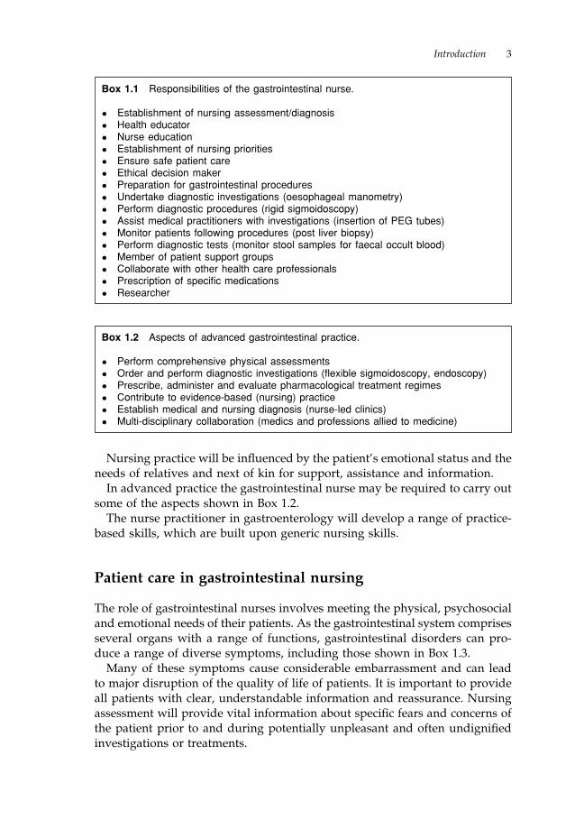
Gastrointestinal nurses therefore assume responsibility for assessing, plan-ning, implementing and evaluating nursing care for gastrointestinal patients,whether in the paediatric or adult setting. Generally, they are professionallyautonomous in the clinical setting, documentation, teaching and research andcare of equipment. These factors will have a direct effect upon the quality ofnursing care provided. Additionally, responsibilities of the present-day gastroin-testinal nurse may include those shown in Box 1.1.
GNC01 7/3/05, 5:22 PM2
Introduction 3
Box 1.1 Responsibilities of the gastrointestinal nurse.
• Establishment of nursing assessment/diagnosis
• Health educator
• Nurse education
• Establishment of nursing priorities
• Ensure safe patient care
• Ethical decision maker
• Preparation for gastrointestinal procedures
• Undertake diagnostic investigations (oesophageal manometry)
• Perform diagnostic procedures (rigid sigmoidoscopy)
• Assist medical practitioners with investigations (insertion of PEG tubes)
• Monitor patients following procedures (post liver biopsy)
• Perform diagnostic tests (monitor stool samples for faecal occult blood)
• Member of patient support groups
• Collaborate with other health care professionals
• Prescription of specific medications
• Researcher
Box 1.2 Aspects of advanced gastrointestinal practice.
• Perform comprehensive physical assessments
• Order and perform diagnostic investigations (flexible sigmoidoscopy, endoscopy)
• Prescribe, administer and evaluate pharmacological treatment regimes
• Contribute to evidence-based (nursing) practice
• Establish medical and nursing diagnosis (nurse-led clinics)
• Multi-disciplinary collaboration (medics and professions allied to medicine)
Nursing practice will be influenced by the patient’s emotional status and theneeds of relatives and next of kin for support, assistance and information.
In advanced practice the gastrointestinal nurse may be required to carry outsome of the aspects shown in Box 1.2.
The nurse practitioner in gastroenterology will develop a range of practice-based skills, which are built upon generic nursing skills.
Patient care in gastrointestinal nursing
The role of gastrointestinal nurses involves meeting the physical, psychosocialand emotional needs of their patients. As the gastrointestinal system comprisesseveral organs with a range of functions, gastrointestinal disorders can pro-duce a range of diverse symptoms, including those shown in Box 1.3.
Many of these symptoms cause considerable embarrassment and can leadto major disruption of the quality of life of patients. It is important to provideall patients with clear, understandable information and reassurance. Nursingassessment will provide vital information about specific fears and concerns ofthe patient prior to and during potentially unpleasant and often undignifiedinvestigations or treatments.
GNC01 7/3/05, 5:22 PM3

4 Chapter 1
Box 1.5 Procedural documentation.
• Nature of procedure
• Staff involved in procedure
• Equipment used in procedure (i.e. endoscope log number)
• Medication and fluids administered during procedure
• Unusual events
• Vital observations throughout procedure
• Type of specimen/biopsy obtained
• Post-procedural assessment
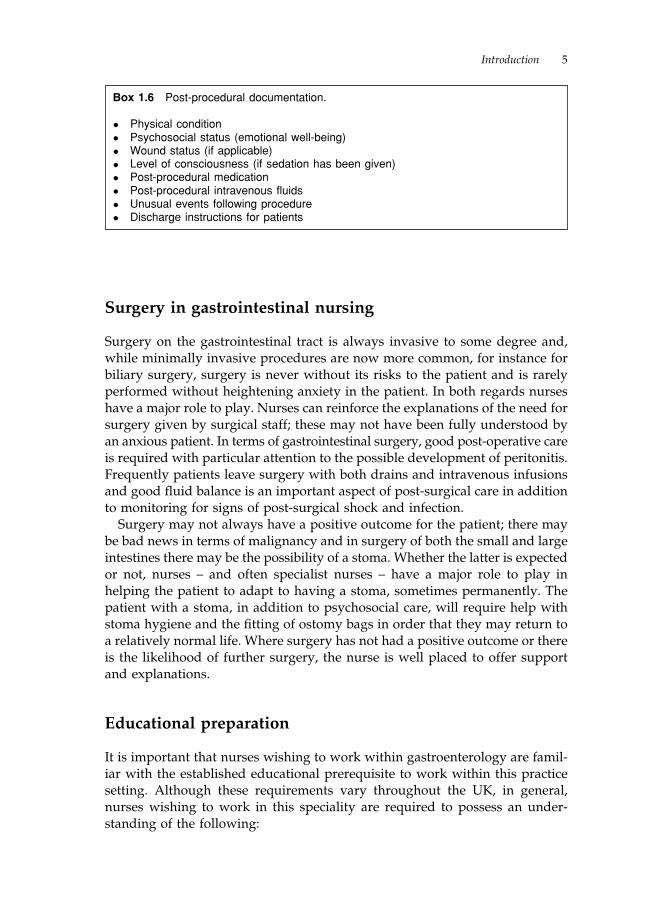
If a patient requires sedation during a procedure, such as endoscopy, thegastrointestinal nurse should be on hand to assess the patient’s response to thesedation and the procedure and intervene where necessary. Patient monitor-ing continues for the nurse after the procedure, as patients will often requiretime to recover from the possible effects of sedation or from the potentialcomplications that may be related to treatment or investigation of gastrointes-tinal conditions. Another responsibility relates to the documentation of nurs-ing practice via records, care plans and reports (NMC 2002b). Documentationrequirements may vary from one hospital to the next according to specificinstitutional policies. For the purpose of this text documentation is examinedfor a gastrointestinal outpatient at three specific stages of the patient journey,pre-procedural, procedural and post-procedural. Pre-procedural documentationis summarised in Box 1.4, procedural documentation in Box 1.5 and severalelements of post-procedural documentation in Box 1.6.
Box 1.4 Pre-procedural documentation in gastroenterology.
Pre-procedural documentation includes:
• Presenting gastrointestinal complaint/symptoms
• Patient vital observations
• Physical assessment of patient
• Psychosocial assessment of the patient (i.e. levels of anxiety)
• Current medications
• Past medical history
• Risk factors (i.e. previous allergic reactions)/anaesthetic history
• Prophilactic medication (i.e. antibiotic pre-ERCP)
• Consent for treatment/investigation
Box 1.3 Gastrointestinal symptoms.
• Abdominal pain
• Anorexia
• Weight loss
• Dysphagia
• Dyspepsia
• Vomiting
• Diarrhoea
• Constipation
GNC01 7/3/05, 5:22 PM4
Introduction 5
Surgery in gastrointestinal nursing
Surgery on the gastrointestinal tract is always invasive to some degree and,while minimally invasive procedures are now more common, for instance forbiliary surgery, surgery is never without its risks to the patient and is rarelyperformed without heightening anxiety in the patient. In both regards nurseshave a major role to play. Nurses can reinforce the explanations of the need forsurgery given by surgical staff; these may not have been fully understood byan anxious patient. In terms of gastrointestinal surgery, good post-operative careis required with particular attention to the possible development of peritonitis.Frequently patients leave surgery with both drains and intravenous infusionsand good fluid balance is an important aspect of post-surgical care in additionto monitoring for signs of post-surgical shock and infection.
Surgery may not always have a positive outcome for the patient; there maybe bad news in terms of malignancy and in surgery of both the small and largeintestines there may be the possibility of a stoma. Whether the latter is expectedor not, nurses – and often specialist nurses – have a major role to play inhelping the patient to adapt to having a stoma, sometimes permanently. Thepatient with a stoma, in addition to psychosocial care, will require help withstoma hygiene and the fitting of ostomy bags in order that they may return toa relatively normal life. Where surgery has not had a positive outcome or thereis the likelihood of further surgery, the nurse is well placed to offer supportand explanations.
Educational preparation
It is important that nurses wishing to work within gastroenterology are famil-iar with the established educational prerequisite to work within this practicesetting. Although these requirements vary throughout the UK, in general,nurses wishing to work in this speciality are required to possess an under-standing of the following:
Box 1.6 Post-procedural documentation.
• Physical condition
• Psychosocial status (emotional well-being)
• Wound status (if applicable)
• Level of consciousness (if sedation has been given)
• Post-procedural medication
• Post-procedural intravenous fluids
• Unusual events following procedure
• Discharge instructions for patients
GNC01 7/3/05, 5:22 PM5

6 Chapter 1
• normal anatomy and physiology of the gastrointestinal tract
• pathophysiology related to common gastrointestinal conditions
• pharmacology in gastrointestinal medicine
• behavioural sciences
• counselling skills and communication
Education and research in gastrointestinal nursing
Gastrointestinal nurses have a responsibility as educators. This educationalrole covers nursing students, trained and untrained nursing staff. The devel-opment of advanced nurse practitioners and specialists in gastrointestinalnursing has led to nurses being involved in medical education and the teach-ing of other professionals who are allied to medicine. Nurse specialists ininflammatory bowel disease who disseminate both their academic and clinicalknowledge in presentations, papers and abstracts are a good example of thiswidening educational role of gastrointestinal nurses. Through presentation atprofessional meetings, such as the British Society of Gastroenterology or theRoyal College of Nursing Gastroenterology and Stoma Care Nurses Forum,nurses meet the responsibility of expanding current knowledge. Related toeducation is research in gastrointestinal nursing. Nurses who embark uponresearch are required to have a sound knowledge of research techniques; thisfacilitates critical evaluation of published materials.
To achieve these responsibilities it is imperative for the gastrointestinal nurseto have a thorough understanding of normal gastrointestinal physiology andpathophysiology in common gastrointestinal conditions, and an understandingof the rationale behind investigation techniques and treatment regimes. Thisbook will provide the gastrointestinal nurse with the appropriate informationto assess, plan, implement and evaluate nursing care.
Gastrointestinal nursing: what this text adds
This introduction will help you to understand the purpose of this book andhow to get the best out of it. It is written for a wide range of nurses: at one endof the spectrum for nurses who may have an interest in entering gastroentero-logy as a speciality, and at the other end for nurses working in the specialitywho may wish to develop further into one of many roles such as nurseendoscopist, nurse practitioner or nurse consultant. These roles are develop-ing in the UK at the time of writing and there has been a great demand forsuch a book to ensure at least a common level of knowledge in this area ofwork, for which many nurses will have had no special education or training.
From our experience as nurse educators we know that many nurses at pre-registration level, whether on diploma or degree programmes, struggle with thesubjects of anatomy and physiology. There are many reasons for this, including
GNC01 7/3/05, 5:22 PM6

Introduction 7
the fact that nursing students may enter university with a very poor backgroundin the life sciences. However, we also acknowledge our failings as teachers. Inaddition, there will be many nurses working in gastroenterology who havenever been exposed to the appropriate level of teaching in the life sciencesbecause they trained prior to the nursing educational reforms of the 1990s.
Structure, function and disorders of thegastrointestinal tract
For the above reasons, therefore, a significant proportion of this book isconcerned with the structure and function of the gastrointestinal tract and thedisorders which arise. The management of these disorders is described and,while we have tried to emphasise aspects of nursing where these are unique,our general approach has been to present medical, surgical and nursing man-agement without differentiation. As already stated, with the possible excep-tion of nurses who want to find out more about the speciality, this book ismainly directed at those in the speciality. Even those outside the speciality willbe registered nurses and to list repeatedly aspects of nursing care which aregeneric to all patient groups would be unnecessary. This book is designed tofill gaps in the essential knowledge needed to nurse in this area; knowledgeand experience of nursing generally is assumed. Furthermore, nurses workas part of a multidisciplinary team and to specify their part is unrealistic,particularly when the boundaries between nursing, medicine and surgery arebeing blurred by advanced nursing practice.
As far as possible, the chapters in Section 1 follow the pattern described inFigure 1.1.
Essential aspects of gastroenterology
After an overview of the gastrointestinal tract, each chapter takes one regionof the tract and covers the anatomy and physiology, the range of disorderswith causes and then describes the management of the disorder includingmedical, surgical and nursing care.
The chapters on the regions of the tract should all enable the reader to:
• Describe the region of the tract in anatomical terms
• Understand the physiological function of the region
• Identify the main disorders, and
• Relate the anatomy, physiology and disorders to nursing practice.
Section 2 covers essential aspects of gastroenterology and these include diag-nostic tests, emergencies and pharmacology. Nurses are increasingly involvedin the diagnostic aspects of gastroenterology, specifically nurse endoscopists
GNC01 7/3/05, 5:22 PM7

8 Chapter 1
Figure 1.1 Schematic structure of the text.
and Chapter 10 considers endoscopy in some detail. Clearly, nurses are usuallyfirst on the scene in a gastrointestinal emergency. Some emergencies, such ashaemorrhage, are common to several regions of the tract, therefore a rangeof emergencies is covered in Chapter 11. While specific drugs are mentionedthroughout the book, the pharmacological aspects of gastroenterology are con-sidered overall in Chapter 12. In addition to knowing which drugs are pre-scribed for which conditions and being able to list side-effects and interactions,nurses are increasingly required to understand how drugs work and to havea deeper knowledge of pharmacology, and this is commensurate with thedevelopment of nurse prescribers, especially in gastroenterology.
Living with gastrointestinal disorders
The final section of the book, Section 3, looks at living with gastrointestinaldisorders. Chapter 13 covers the psychosocial aspects both as causes and con-sequences of gastrointestinal disorders, especially inflammatory bowel dis-ease, and Chapter 14 looks at the impact of gastrointestinal disorders on qualityof life. Clearly, there is a strong link between the two chapters. In commonwith developments in endoscopy and nurses prescribing, these are areas wherenursing roles are extending with the development of nurse counsellors andthe use of alternative treatments such as hypnotherapy.
Region of GI tract
Anatomy and Physiology
Disorder Disorder Disorder
Causes Causes Causes
Management Management Management
IncorporatingMedical, Surgical and Nursing
GNC01 7/3/05, 5:22 PM8

Introduction 9
Professional guidelines
Nurses are governed by a professional regulatory body called the Nursing andMidwifery Council (NMC), which was established in April 2002 and replacedthe United Kingdom Central Council for Nursing, Midwifery and HealthVisiting (UKCC). Wherever possible we refer readers to appropriate NMCdocuments. Where UKCC guidelines have not been superseded by NMC guide-lines readers are referred to the relevant UKCC guidelines. The Appendixcontains the latest version, at the time of publication, of the NMC Code ofProfessional Conduct reproduced with permission of the NMC.
FURTHER INFORMATION
This book is designed to be a stand-alone text. However, readers who wish to invest-
igate specific aspects of structure and function or specific gastrointestinal disorders
are referred to a range of standard texts, listed below. In addition, at the end of each
chapter, specific references will be provided to relevant sections of these texts. To provide
an evidence base for gastrointestinal nursing, appropriate sources such as websites,
textbooks and journals are referred to in the text.
The following books were consulted in the preparation of this text:
Alexander, M., Fawcett, J.N. and Runciman, P. (2000) Nursing Practice: Hospital and
Home – the Adult. Churchill Livingstone, Edinburgh.
Brooker, C. and Nicol, M. (2003) Nursing Adults: the Practice of Caring. Mosby, London.
Clancy, J. and McVicar, A.J. (1998) Nursing Care: a Homeostatic Casebook. Arnold, London.
Clancy, J. and McVicar, A.J. (2002) Physiology and Anatomy: a Homeostatic Approach, 2nd
edition. Arnold, London.
Clancy, J., McVicar, A.J. and Baird, N. (2002) Perioperative Practice: Fundamentals of
Homeostasis. Routledge, London.
Haslett, C., Chilvers, E.R., Boon, N.A. and Colledge, N.R. (2002) Davidson’s Principles
and Practice of Medicine, 19th edition. Churchill Livingstone, Edinburgh.
Higgins, C. (2000) Understanding Laboratory Investigations: A Text for Nurses and Healthcare
Professionals. Blackwell Publishing, Oxford.
Hinchliff, S., Montague, S. and Watson, R. (1996) Physiology for Nursing Practice, 2nd
edition. Baillière Tindall, London.
Kindlen, S. (2003) Physiology for Health Care and Nursing. Churchill Livingstone,
Edinburgh.
Kumar, P. and Clark, M. (2002) Clinical Medicine, 4th edition. Saunders, Edinburgh.
McKenry, L.M. and Salerno, E. (1998) Pharmacology for Nursing, 20th edition. Mosby,
St Louis.
Watson, R. (1999) Essential Science for Nursing Students: An Introductory Text. Baillière
Tindall, London.
Watson, R. (2000) Anatomy and Physiology for Nurses, 11th edition. Baillière Tindall,
London.
GNC01 7/3/05, 5:22 PM9

10 Chapter 1
CONCLUSION AND ACKNOWLEDGEMENTS
We are responding to a demand for a book such as this and take full responsibility for
any deficiencies. The book would not have been written without the support of Dr
Kelvin Palmer, Dr Helen J. Dallal, Miss Tonks Fawcett and Ms Rosemary Patterson, or
without the patience and support of Beth Knight at Blackwell Publishing. Anonymous
reviewers also played a significant role in shaping the book. Special thanks to Linda S.
Smith for indexing and to Gillian Kidd for her artwork. We hope this book is found
useful by a wide range of nurses in gastroenterology and we will also be very glad to
receive any feedback for future editions.
REFERENCES
Benner, P. (1984) From Novice to Expert: Excellence and Power in Clinical Nuring Practice.
Addison-Wesley, Massachusetts.
NHSE (National Health Service Executive) (1999) Nurse, Midwifery and Health Visitor
Consultants, HSC 1999/217 Department of Health, London.
NMC (2002a) Guidelines for the Administration of Medicines. Nursing and Midwifery
Council, London.
NMC (2002b) Guidelines for Records and Record Keeping. Nursing and Midwifery Coun-
cil, London.
GNC01 7/3/05, 5:22 PM10

An Overview of the Gastrointestinal Tract 11
Section 1
Structure, Function and Disorders ofthe Gastrointestinal Tract
GNC02 7/3/05, 5:33 PM11

12 Chapter 2
GNC02 7/3/05, 5:33 PM12

An Overview of the Gastrointestinal Tract 13
Chapter 2
An Overview of theGastrointestinal Tract
Chapter objectives
After reading this chapter you should be able to:
• Describe the general features of the gastrointestinal tract.
• Understand the range of functions of the gastrointestinal tract.
Introduction
The adult gastrointestinal tract consists of a continuous fibromuscular tubethat extends from the mouth to the anus. The tract is in contact with theexternal environment at both ends.
The gastrointestinal system consists of the digestive tract (mouth, oeso-phagus, stomach and intestines) in association with the accessory digestiveglands (salivary glands, pancreas and biliary system). The overall function ofthe digestive system is to transfer the nutrients in food from the externalenvironment to the internal environment. Once in the body, the nutrients canbe distributed to the cells of the body via the circulation. The waste material ofdigestion is also excreted via the circulation. Nutrients, water and salts areabsorbed from digested food and all products that cannot be absorbed areretained in the digestive tract until they are eliminated. The gastrointestinaltract is regulated by both the autonomic nervous system and hormonal mech-anisms, which act in conjunction with a variety of gastrointestinal peptides(hormones, neurocrines or paracrines).
In this chapter the general principles and the basic mechanisms involvedin the overall function of the digestive system will be examined. Figure 2.1illustrates both the component organs of the gastrointestinal tract and theaccessory organs that are required for the digestive system to function.
13
GNC02 7/3/05, 5:33 PM13

14 Chapter 2
Figure 2.1 Component organs and accessory organs of the gastrointestinal tract.Reproduced with permission from Watson (2000).
Box 2.1 Generalised layers of the gastrointestinal tract.
• mucosa
• submucosa
• muscularis
• serosa (fibrous outer layer)
Structure of the gastrointestinal tract
The digestive tract wall consists of four structural layers (see Box 2.1). Thesefour layers are present in all areas of the tract from the oesophagus to theanus, with some functional adaptations throughout (Figure 2.2).
The mucosa is the innermost layer, that is, the layer nearest to the lumen ofthe tube, and it exhibits a great deal of variation throughout the tract. Mucusstratified epithelial cells line the lumen (except in the oesophagus), and it isfrom this layer that all glands develop. Mucus secreting cells are situatedthroughout the epithelium. These cells are subjected to a tremendous amountof frictional wear and tear. The epithelial cells lie on a sheet of connective
GNC02 7/3/05, 5:33 PM14

An Overview of the Gastrointestinal Tract 15
Figure 2.2 Four structural layers of digestive tract wall. Reproduced with permissionfrom Hinchliff et al. (1996).
tissue called the lamina propria. Distal to this there is a thin layer of muscletissue called the muscularis mucosa. Throughout the mucosa are patches oflymphoid tissue, which provide a defensive function.
The submucosa lies distal to the mucosa and consists of loose connectivetissue, which supports blood vessels, lymphatics and nerve fibres.
The muscularis layer, as its name suggests, is formed of muscle fibres. Themuscle fibres in the gastrointestinal tract are referred to as smooth, involun-tary, unstriated or visceral muscle fibres.
The serosa is the outermost, protective layer, formed of connective andsquamous tissue. The serosa contains blood vessels, neurones and lymphatics.
Blood supply
An adequate supply of blood to the digestive system is essential to serve thenormal metabolic functions and also to provide a route for nutrients to getfrom the digestive tract to the systemic circulation. The arteries supplyingthe abdominal organs of the digestive system are the coeliac and superiorand inferior mesenteric arteries. The coeliac artery branches to give rise to thegastric, splenic and hepatic arteries that provide blood to the stomach, pan-creas, spleen and liver. The mesenteric arteries supply the intestines.
The branches of the main arteries, which supply the gastrointestinal tract, giverise to smaller branches, which penetrate the organs. These smaller branchesdivide to give rise to an extensive network of arterioles in the submucosa.These in turn lead to mucosal arterioles, which supply blood to the capillaries.
Venous blood from the stomach, pancreas, spleen and liver is collectedtogether and routed through the liver via the hepatic portal vein. Blood from
GNC02 7/3/05, 5:33 PM15

16 Chapter 2
the remainder of the digestive tract (oesophagus and rectum) escapes thehepatic filter and drains directly into the venous system.
PHYSIOLOGY OF THE DIGESTIVE SYSTEM
The unique physiological processes that take place in the digestive system aredigestion, absorption, secretion, motility and excretion.
Digestion is the process whereby large food molecules are broken down tosmaller ones. Food is ingested as large pieces of matter containing substancessuch as protein and starch which are unable to cross the cell membranes ofthe gut epithelium. Before these complex molecules can be utilised they aredegraded to smaller molecules, such as glucose and amino acids, which can beabsorbed from the gastrointestinal system into the bloodstream.
The mixture of ingested material and secretions in the gastrointestinal tractcontains water, minerals and vitamins as well as fats, carbohydrates and pro-teins. The products of digestion, other small dissolved molecules, ions andwater are transported across the epithelial cell membranes, mainly in the smallintestine. This is the process of absorption. The transported molecules enterthe blood or lymph for circulation to the tissues. This process is central to thedigestive system, and the other physiological processes of the gastrointestinaltract, such as elimination, subserve it.
Food which is ingested travels along the gastrointestinal tract to theappropriate sites for mixing, digestion and absorption to occur. Most of thegastrointestinal tract is lined by two layers of smooth muscle; contraction ofthis muscle mixes the contents of the lumen and moves them through thetract. Motility in the digestive system is under neural and hormonal control.
Exocrine glands secrete enzymes, ions, water, mucins and other substancesinto the digestive tract. The glands are situated within the gastrointestinaltract, in the walls of the stomach, small intestine and large intestine, or outsideit in the case of salivary glands, pancreas and liver. Secretion in all regions ofthe gastrointestinal tract is controlled by nerves and hormones.
Peptides of the gastrointestinal tract
Some of the functions of the gastrointestinal tract are regulated by peptides,derivatives of amino acids and a variety of mediators released from nerves.These functions include contraction and relaxation of the smooth muscle walland the sphincters (physiological ‘gatekeepers’ of the gastrointestinal tract);secretion of enzymes for digestion; secretion of fluids and electrolytes; andgrowth of the tissues of the gastrointestinal tract.
All gastrointestinal hormones are peptides, i.e. small molecules comprisingup to 50 amino acids. It is important, however, to realise that not all peptidesfound in the digestive tract are hormones. The gastrointestinal peptides can be
GNC02 7/3/05, 5:33 PM16

An Overview of the Gastrointestinal Tract 17
divided into hormones, paracrines and neurocrines, depending on the methodby which the peptide is delivered to its target site.
Hormones are peptides released from endocrine cells of the gastrointestinaltract. They are secreted into the portal circulation, pass through the liver andenter the systemic circulation. The systemic circulation then delivers the hor-mone to target cells with receptors for that specific hormone. These target cellsmay be in the gastrointestinal tract itself (e.g. gastrin acts upon the parietalcells of the stomach to stimulate acid secretion), or the target cells may belocated in another region of the body (e.g. gastric inhibitory peptide acts uponthe β cells of the pancreas to cause insulin secretion). Several criteria must bemet for a substance to qualify as a gastrointestinal hormone:
• The substance must be secreted in response to a physiological stimulusand carried in the bloodstream to a distant site, where it produces a physio-logical action.
• The hormone’s action must be independent of any neural activity.
• The hormone must have been isolated, purified and chemically identified.
Four gastrointestinal peptides are classified as hormones: gastrin, cholecysto-kinin (CCK), secretin and gastric inhibitory peptide (GIP). These are discussedin more detail in Chapters 4 and 8.
Most paracrines, with the exception of histamine, are peptides secreted byendocrine cells of the gastrointestinal tract. In contrast to hormones, however,the paracrines act locally within the same tissue that secretes them. Para-crine substances reach their target cells by diffusing short distances throughinterstitial tissue, or they are carried short distances in capillaries. The onlygastrointestinal peptide with a known paracrine function is somatostatin, whichhas an inhibitory effect throughout the gastrointestinal tract in that it reducesmotility and secretion of digestive juices.
Neurocrines are synthesised in neurones of the gastrointestinal tract and arereleased in response to an action potential. Following release, the neurocrinediffuses across the synapse and acts upon its target cell. There are manyneurocrines in the gastrointestinal tract including acetylcholine, noradrenalinand vasoactive intestinal peptide (VIP). The sources and actions of theseneurocrines are summarised in Table 2.1.
Innervation of the gastrointestinal tract
The gastrointestinal tract is also regulated by the autonomic nervous system,which has an intrinsic component and an extrinsic component. The extrinsiccomponent is the sympathetic and parasympathetic innervation of the gastro-intestinal tract. The intrinsic component is called the enteric nervous system.The enteric system is wholly contained within the wall of the gastrointestinaltract in the submucosal and myenteric plexuses.
GNC02 7/3/05, 5:33 PM17

18 Chapter 2
Parasympathetic innervation
Parasympathetic nervous innervation is supplied by both the vagus nerveand the pelvic nerve. The pattern of parasympathetic innervation is consistentwith its function. The vagus nerve innervates the upper portions of the gastroin-testinal tract (upper third of oesophagus, wall of stomach, small intestine andascending colon), whilst the pelvic nerve innervates the lower portions of thesystem (striated muscle of external anal canal and walls of the transverse,descending and sigmoid colons).
Postganglionic neurons of the parasympathetic nervous system are classifiedas either cholinergic or peptidergic. Cholinergic neurons release acetylcholineas the main neurotransmitter and peptidergic neurons release one of severalpeptides, including VIP.
Sympathetic innervation
Preganglionic cholinergic fibres of the sympathetic nervous system synapse inganglia outside the gastrointestinal tract. Four sympathetic ganglia serve thegastrointestinal tract: coeliac, superior mesenteric, inferior mesenteric andhypogastric. Postganglionic fibres, which are adrenergic, leave the sympatheticganglia and synapse on ganglia in the myenteric and submucosal plexus, orthey directly innervate smooth muscle, endocrine or secretory cells.
Intrinsic innervation
The intrinsic or enteric nervous system can direct all functions of the gastroin-testinal tract, even in the absence of extrinsic innervation. The enteric nervoussystem is located in the myenteric and submucosal plexus and controls thesecretory, contractile and endocrine functions of the gastrointestinal tract.
Table 2.1 Neurocrines of the enteric nervous system.
Neurocrine Source Action
Acetylcholine Cholinergic neurons Contraction of smooth muscleRelaxation of sphincters↑ Salivary secretion↑ Gastric secretion↑ Pancreatic secretion
Noradrenalin Adrenergic neurons Relaxation of smooth wallContraction of sphincters↑ Salivary secretion
Vasoactive intestinal peptide Neurons of mucosa Relaxation of smooth muscle(VIP) ↑ Intestinal secretion
↑ Pancreatic secretion
↑ = increase in production of secretion.
GNC02 7/3/05, 5:33 PM18

An Overview of the Gastrointestinal Tract 19
Motility (movement) in the gastrointestinal tract
Motility refers to contraction and relaxation of the walls and sphincters of thegastrointestinal tract. Motility involves the grinding and mixing of ingestedfood in preparation for digestion and absorption; it then propels the food alongthe gastrointestinal tract. Smooth muscle in the gastrointestinal tract enables:
• Contractile tone to be maintained even in the absence of food.
• Activity to be increased and decreased as necessary.
• The tract to distend to accommodate different volumes.
The control of motility and secretion of smooth muscle in the gastrointestinaltract is by neural, hormonal and paracrine mechanisms. The neural control isvia the extrinsic nerves of the autonomic nervous system. In most instancesthe mediators of neural or hormonal control are peptides.
Skeletal muscle which is present in the pharynx, upper section of the oeso-phagus and the external anal sphincter is under voluntary control and isinvolved in the initiation of swallowing and the final stage of defaecation.
Digestion and absorption
Digestion and absorption are the ultimate functions of the gastrointestinaltract. Digestion is the chemical breakdown of ingested foods into absorbablemolecules. The digestive enzymes are secreted in salivary, gastric, and pancre-atic juices as well as the mucosa of the small intestine.
Absorption is the movement of nutrients, water and electrolytes from thelumen of the intestine into the blood system. There are two distinct paths forabsorption, a cellular path and a paracellular path. In the cellular path, thesubstance must cross the gastrointestinal luminal membrane, enter the intest-inal epithelial cell, and then be extruded from the cell into the blood. Paracellularabsorption involves the movement of substances across the tight junctionsbetween the intestinal epithelial cells, through the lateral intercellular spaces,and then into the blood.
The structure of the intestinal mucosa is ideally suited for absorption oflarge quantities of nutrients. Structural features called villi and microvilliincrease the surface area of the small intestine, maximising the exposure ofnutrients to digestive enzymes and to the absorptive surface.
The epithelial cells of the small intestine have the highest turnover rate ofany cells in the body. They are replaced every 3–6 days.
Constituents of food
The body needs food to provide energy. Vitamins and minerals are necessaryto maintain good health. The main food groups are carbohydrate, fat and
GNC02 7/3/05, 5:33 PM19

20 Chapter 2
protein. In addition, water is essential to replace fluid, which is continuallybeing lost by the body. There are in total six essential foodstuffs with whichthe body must be constantly supplied, each of which are examined herein turn.
Essential foodstuffs are:
• protein
• carbohydrate
• fat
• water
• mineral salts
• vitamins
These foodstuffs must be digested and absorbed. Food must therefore be ofsuch a nature that it can be digested, i.e. broken down by digestive juices intosubstances that can pass into the bloodstream, and be carried to various tissuesfor their use.
CarbohydratesThe foods required for energy and heat within the body are called carbohyd-rate foods because they contain carbon, hydrogen and oxygen. Carbohydratesinclude sugar and starch and they are the chief source of body fuel.
FatsFatty foods provide energy and heat, which serve as body fuel. Fats alsoprovide food stores, the adipose tissue of the body and protective coveringsfor some organs. It is recommended that dietary fat should account for lessthan 35% of total energy intake.
ProteinsProteins are the most complicated foodstuffs, containing nitrogen in additionto hydrogen, carbon, oxygen and in some cases sulphur and phosphorus.They are called nitrogenous foodstuffs as they are the only ones which containthe element nitrogen. Protein is necessary in the diet to build and replace theprotoplasm of body cells. Proteins are composed of polypeptides, derivedfrom amino acids. There are 20 amino acids, although each protein containsonly some of these. Humans require approximately 0.75 g of protein per kg ofbody weight per day.
WaterWater is essential for life as it forms nearly two-thirds of the human body andis present in most of the foods we eat. The average amount of water in thehuman body is about 45 litres (30 litres inside the cells – intracellular – and 15litres outside the cells – extracellular – i.e. tissue fluid in the plasma). The mainfunctions of water in the body are:
GNC02 7/3/05, 5:33 PM20

An Overview of the Gastrointestinal Tract 21
• excretion of waste products
• making digestive and lubricating fluids
• building of body tissues and body fluids
• temperature control (i.e. evaporation of sweat)
If the body is depleted of fluid the signs and symptoms of dehydration mayappear; these include:
• thirst
• dry mouth
• slack elastic skin
• sunken eyes
• low blood pressure
The body requires 2–3 litres of water every day and the amount of fluid takeninto the body must be balanced by the output.
Mineral saltsMineral salts are essential for normal metabolism. Salts are produced by theaction of an acid on a mineral.
An electrolyte is a dissolved salt (a mineral salt), which is capable of con-ducting electricity. The two major electrolytes are sodium (Na+) and potassium(K+). The concentration of Na+ is high in extracellular sites and low within cells(intracellular). In contrast K+ is low in tissue fluids and high within cells. Acorrect balance of electrolytes is essential for normal function of body tissuesand fluids.
Sodium is present in all tissues; it exists as sodium chloride in a concentra-tion of nine grams per litre (0.9%) in all extracellular fluids. Sodium is derivedfrom our food, particularly animal foodstuffs, and from the rock salt used incooking.
Potassium is present in all tissue cells, where it replaces the sodium of bloodand tissue fluids and the source of the positively charged ions. Potassium isobtained from food, particularly plant foodstuffs.
Calcium is present in all tissues, particularly in bone, teeth and blood, and isnecessary for the functioning of nerves and for muscle tone. It is obtainedchiefly from milk, cheese, eggs and green vegetables. Adults require 400–500 mgdaily.
Iron is essential for the formation of the haemoglobin in red blood cells. It isobtained from green vegetables, particularly spinach and cabbage, egg yolkand red meats. Men require 10 mg daily; women require more, 10–15 mg daily,because of menstrual blood loss.
Iodine is required for the formation of thyroxin by the thyroid gland. It isobtained from seafood and is also present in green vegetables.
Calcium, iron and iodine are the only minerals which may be insufficient inthe diet.
GNC02 7/3/05, 5:33 PM21

22 Chapter 2
VitaminsVitamins are also essential to normal health because they are necessary for arange of metabolic functions; they are of no value to the body as a fuel or asbuilding material. Vitamins are present in small quantities in living foodstuffand are only required in minute traces each day. Vitamins are classified as fatsoluble and water soluble; vitamins A, D, E and K are fat soluble, the othervitamins including B and C are soluble in water.
Vitamin A is present in all fatty foods, e.g. milk, cheese and fish liver oils. Itcan be made in the body from a substance called carotene, which is present incarrots and tomatoes. Vitamin A is needed for normal function of the retinaand to fight infection. Correspondingly a lack of vitamin A causes visual loss,stunted growth and a lowered resistance to infection.
Vitamin D is found in dairy produce and also in fatty fish such as herring.Cod liver oil and halibut liver oil are very rich in vitamin D. Vitamin D canalso be built up in the body; the ultraviolet rays from the sun act on a fattysubstance in the skin called ergosterol, which produces vitamin D. Vitamin Dis necessary, with calcium, for the formation of bone. Lack of vitamin D and/or calcium leads to rickets in childhood, osteomalacia and osteoporosis inadults.
Vitamin E is present in vegetable oils and is found in egg yolk and milk.Vitamin E is necessary for normal functions of the nervous system, reproduc-tion and muscle development.
Vitamin K is fat soluble and can be obtained from green vegetables andliver. It is required for the production of blood clotting factors in the liver.Vitamin K is synthesised in the intestine by colonic bacterial action.
Vitamin B is a complex of several closely related compounds. These arefound particularly in the husks and germs of cereals and pulses, in yeast andyeast extracts and to a lesser extent in vegetables, fruit, milk, eggs and meat.The chief factors in the vitamin B complex are:
• Vitamin B1 (thiamine) is essential for carbohydrate metabolism and con-trols the nutrition of nerve cells.
• Vitamin B2 (riboflavin) is essential for the proper functioning of cellenzymes.
• Vitamin B6 (pyridoxine) is necessary for protein metabolism.
• Vitamin B12 (cyanocobalamin) is the anti-anaemic substance or factorabsorbed by the villi of the small intestine and stored in the liver. It issatisfactorily absorbed only in the presence of intrinsic factor produced bythe cells in the lining of the stomach. Vitamin B12 is essential for the properdevelopment of red cells in the red bone marrow and of nervous tissue.
Folic acid is required in the body for the maturation of red blood cells. It isderived from green vegetables.
Vitamin C (ascorbic acid) is water soluble and is found in citrus fruits(oranges, grapefruits and lemons), green vegetables and potatoes. Vitamin C is
GNC02 7/3/05, 5:33 PM22

An Overview of the Gastrointestinal Tract 23
important in tissue respiratory activity, wound repair and resistance to infec-tion, and it affects the condition of capillary walls. Lack of vitamin C causesscurvy, a condition which used to be common in sailors on long sea voyages.Scurvy is occasionally seen today in older people who have not been feedingthemselves properly.
Roughage is an indigestible part of food. It remains in the bowel and stimu-lates it to empty itself. Roughage is the fibrous part of food, giving it bulk andstimulating bowel action, thus preventing constipation.
Conclusion
From this general overview of the gastrointestinal tract it can be seen that itis important to several crucial functions in the body and ones with which weare very familiar: eating, drinking and elimination. The two main activites ofthe gastrointestinal tract – motility and absorption, both necessary for thedigestive functions of the tract – are generally insensible. However, patholo-gical conditions of the gastrointestinal tract affect these activities, leading todisorders in the functions of the tract with attendant discomfort, poor nutri-tion, embarrassment and distress. The nursing role, in addition to supportingthe medical and surgical interventions related to the gastrointestinal tract, is tosupport patients through the consequences of disorders of the tract and forthis to be possible, at least a working knowledge of the anatomy, physiologyand disorders of the tract is required. The following chapters take you throughthe tract from the oesophagus to the rectum and include the ancillary organsof digestion (liver, pancreas and gall bladder).
BACKGROUND READING
Additional reading to support the material in this chapter can be found in the relevant
sections of the following texts:
Clancy, J. and McVicar, A.J. (2002) Physiology and Anatomy: a Homeostatic Approach, 2nd
edition. Arnold, London (Chapter 10).
Hinchliff, S., Montague, S. and Watson, R. (1996) Physiology for Nursing Practice, 2nd
edition. Baillière Tindall, London (Chapter 5.1).
Kindlen, S. (2003) Physiology for Health Care and Nursing. Churchill Livingstone, Edin-
burgh (Chapter 9).
Watson, R. (1999) Essential Science for Nursing Students: An Introductory Text. Baillière
Tindall, London (Chapter 2).
Watson, R. (2000) Anatomy and Physiology for Nurses, 11th edition. Baillière Tindall,
London (Section V).
GNC02 7/3/05, 5:33 PM23

24 Chapter 3
Chapter 3
The Oesophagus
Chapter objectives
After reading this chapter you should be able to:
• Describe the anatomy and physiology of the oesophagus, including thelower and upper oesophageal sphincters.
• Understand motility in the oesophagus.
• Identify the range of clinical conditions associated with the oesophagus.
• Relate the corresponding pathophysiology, diagnosis and treatment fordisorders of the oesophagus to nursing practice.
ANATOMY AND PHYSIOLOGY
The oesophagus is a thin-walled, muscular tube, approximately 25 cm inlength and 2–3 cm in diameter in adults. An endoscopic view of a normaloesophagus is shown in Plate 1 (see colour plate section).
Control of swallowing (deglutition)
Swallowing can be divided into three phases:
• voluntary
• pharyngeal
• oesophageal
Voluntary phaseThe tongue moves food backwards and upwards into the back of the mouth.This is initiated voluntarily but once it has been initiated it cannot be stoppedvoluntarily, it is a classical ‘all or none’ reflex.
Pharyngeal phaseAs the food moves into the pharynx, it activates pressure receptors in thepharynx. These receptors send impulses via the trigeminal and glossopharyn-
24
GNC03 7/3/05, 5:32 PM24

The Oesophagus 25
geal nerves to the brain stem swallowing centre. Each impulse serves as atrigger for the swallowing reflex. This causes the elevation of the soft palate,which seals the nasal cavity and prevents food entering it. The swallowingcentre inhibits respiration by raising the larynx and closing the glottis to preventfood entering the trachea. The upper oesophageal sphincter, which is closedat rest, opens during swallowing to allow the bolus of food to pass into theoesophagus. Immediately after the bolus has passed, the sphincter closes again,resealing the junction. The pharyngeal phase of swallowing lasts approxim-ately one second (Figure 3.1).
Oesophageal phaseAs the peristaltic waves commence in the oesophagus, the muscle of the loweroesophageal sphincter relaxes, opening the sphincter and allowing the foodbolus to enter the stomach. The lower oesophageal sphincter muscle thencontracts to reseal the junction. The lower oesophageal sphincter remains closedin the absence of peristalsis, preventing the reflux of the acidic contents of thestomach into the oesophagus (Figure 3.2).
Figure 3.1 Pharyngeal phase of swallowing. Reproduced by permission of HodderArnold from Clancy and McVicar (2002).
Figure 3.2 Oesophageal phase of swallowing. Reproduced by permission of HodderArnold from Clancy and McVicar (2002).
GNC03 7/3/05, 5:32 PM25

26 Chapter 3
Anatomical arrangement of the oesophagus
The structure of the oesophagus is made up of three layers: the mucosa,submucosa and the muscularis. Unlike the bulk of the gastrointestinal tract,the oesophagus is not surrounded by serosa.
The innermost mucosal layer is composed of stratified squamous epithelium.Beneath the epithelium is the lamina propria, which is composed of connect-ive tissue. At the upper and lower ends of the oesophagus the mucosa con-tains mucus-producing glands. A thin band of smooth muscle, the muscularismucosa, separates the lamina propria from the underlying mucosa. The sub-mucosa is the middle layer of the oesophagus and contains loose connectivetissue with both elastic and fibrous components, as well as blood vessels andnerve fibres.
The arrangement of the muscularis layer in the oesophagus is similar to thatof the rest of the digestive tract in that there is an inner circular layer and anouter longitudinal layer. Between the two muscular layers lies the intermus-cular Auerbech’s nerve plexus. However, only the lower two-thirds of theoesophagus is surrounded by smooth muscle. The upper third is surroundedby skeletal muscle.
The muscle tissue in the middle of the oesophagus is a transition zone ofboth skeletal and smooth muscles and this area covers as much as 35–45% ofthe length of the oesophagus.
At the upper end of the oesophagus is the upper oesophageal sphincter(hypopharyngeal sphincter). It is composed of skeletal cricopharyngeal mus-cle, which is a thickening of the circular muscular layer. At the lower end ofthe oesophagus, at the junction between the oesophagus and the stomach, isthe lower oesophageal sphincter (gastro-oesophageal sphincter or cardiacsphincter); it comprises the distal 2–3 cm of the oesophagus. The lower oesopha-geal sphincter controls the passage of ingested food to the stomach.
Blood supply to the oesophagus
The oesophagus receives arterial blood via the oesophageal arteries of theaorta, the inferior thyroid artery and the left gastric artery. Blood is returnedto the venous system by way of the left gastric, thyroid and azygous veins.
Neural control of the oesophagus
The oesophagus receives both sympathetic and parasympathetic innervation.The swallowing centre in the medulla oblongata initiates peristaltic contractionsvia the vagus (Xth crainial) nerve, which innervates the skeletal muscles in theupper oesophagus. The smooth muscle is innervated indirectly by neurons inthe vagus nerve, which synapse with neurons in the myenteric plexus. The
GNC03 7/3/05, 5:32 PM26

The Oesophagus 27
lower part of the oesophagus is believed to receive both sympathetic andparasympathetic innervation.
Motility in the oesophagus
When food is not being ingested, both oesophageal sphincters remain closed.The presence of a bolus of food in the pharynx opens the upper oesophagealsphincter and allows the food to pass into the oesophagus. A peristaltic waveconsists of a wave of contraction of the circular muscle, followed by a wave ofrelaxation. These waves of contraction pass along the walls of the oesophagusand move foodstuff towards the stomach. Each peristaltic wave takes aboutnine seconds to travel the length of the oesophagus. Therefore it is not prim-arily gravity, but peristalsis, that moves food towards the stomach, althoughgravity does assist the process. Secondary peristalsis, which arises from withinthe oesophagus, is controlled by sympathetic stimulation.
DISEASES OF THE OESOPHAGUS
Pathophysiology of the oesophagus arises due to disorders of the skeletalmuscle or the smooth muscle. Important disorders of the oesophagus includegastro-oesophageal reflux disease (GORD), motility disorders and gastriccancer.
Gastro-oesophageal reflux disease
Gastro-oesophageal reflux disease (GORD) is the most common cause of indi-gestion, affecting up to 30% of the general population. GORD develops whengastric or duodenal contents flow back into the oesophagus. Oesophagealreflux is only considered a pathological condition when it causes undesirablesymptoms.
Clinical features of GORD
The most common symptoms of oesophageal reflux are dyspepsia, heartburnand regurgitation, which can be provoked by bending, straining or lying down.Waterbrash, which is salivation due to reflex salivary gland stimulation asacid enters the gullet, is often present. A history of weight gain is common.Some patients are woken at night by choking as refluxed fluid irritates thelarynx. Other less common symptoms include dysphagia (difficulty swallow-ing), odynophagia (pain on swallowing), and symptoms of anaemia. A smallnumber of patients present with atypical chest pain, which may be severe, canmimic angina and is probably due to reflux-induced oesophageal spasm.
GNC03 7/3/05, 5:32 PM27

28 Chapter 3
Pathophysiology of GORD
Occasional episodes of GORD are common in health, particularly after eating.Gastro-oesophageal reflux disease develops when the oesophageal mucosa isexposed to gastric contents for prolonged periods of time, resulting in symp-toms and, in a small proportion of cases, this leads to oesophagitis.
Abnormalities of the lower oesophageal sphincter related to GORDIn health the lower oesophageal sphincter is tonically contracted, relaxing onlyduring swallowing. Some patients with GORD have reduced lower oesopha-geal sphincter tone, permitting reflux when intra-abdominal pressure rises. Inothers basal sphincter tone is normal but reflux occurs in response to frequentepisodes of inappropriate sphincter relaxation.
Hiatus herniaA hiatal hernia occurs when part of the stomach protrudes through the dia-phragm and into the thoracic cavity. Such hernias are extremely common inolder people and more common in women than in men. A hiatus hernia causesreflux because the pressure gradient between the abdominal and thoracic cavit-ies, which normally pinches the hiatus, is lost. In addition the oblique anglebetween the cardia and oesophagus disappears. Many patients who have largehiatus hernias develop reflux symptoms, but the relationship between thepresence of a hernia and symptoms is poor. Hiatus hernias are very commonin individuals who have no symptoms, and some symptomatic patients haveonly a very small or no hernia.
Important features of a hiatus hernia include:
• Occur in 30% of the population over the age of 50 years.
• Often asymptomatic.
• Heartburn and regurgitation can occur.
• Gastric volumes may complicate large hernias.
The role of gastric contents in GORDGastric acid is the most important oesophageal irritant and there is a closerelationship between acid exposure time and symptoms. Alkaline reflux, dueto bile reflux following gastric surgery, is of uncertain importance.
Increased intra-abdominal pressurePregnancy and obesity are established predisposing causes. Weight losscommonly improves symptoms and patients should be encouraged to avoidtight-fitting garments.
Dietary and environmental factorsDietary fat, chocolate, alcohol and beverages such as tea and coffee relaxthe lower oesophageal sphincter and may provoke symptoms. There is little
GNC03 7/3/05, 5:32 PM28

The Oesophagus 29
evidence to incriminate smoking or non-steroidal anti-inflammatory drugs(NSAIDs) as causes of gastro-oesophageal reflux disease.
Delayed oesophageal clearanceDefective oesophageal peristaltic activity can be seen in patients who haveGORD. Poor oesophageal clearance leads to increased exposure to acid fromthe stomach.
Complications of GORD
OesophagitisReflux oesophagitis is a chronic inflammatory process mediated by gastricacid and pepsin from the stomach as well as bile from the duodenum, whichcan result in ulceration of the mucosa and secondary fibrosis in the muscularwall. A range of endoscopic findings, from mild redness to severe bleedingulceration with stricture formation, is recognised. There is a poor correlationbetween symptoms and histological and endoscopic findings. A normalendoscopy and normal oesophageal histology are perfectly compatible withsignificant gastro-oesophageal reflux disease. Plate 2 shows an endoscopic viewof mild oesophagitis.
Other causes of oesophagitis: infectious diseasesViruses, bacteria, fungi and mycobacterium can all cause oesophageal infec-tion. The most common of these are candida. Oesophageal candidiasis occursin debilitated patients and those taking broad-spectrum antibiotics or cytotoxicdrugs. It is a particular problem in AIDS patients, who are also susceptible toa spectrum of oesophageal infections. Oesophageal candidiasis rarely developsin patients who do not have an underlying disease such as diabetes, immunedeficiency or malignancy. The main symptoms of oesophageal candidiasis aredysphagia and odynophagia. Severe infection of the gullet can destroy oesopha-geal innervation, causing abnormal motility.
CorrosivesAccidental or suicidal ingestion of highly alkaline or acidic substances mayresult in injury to the oesophagus. The most common symptom is odynophagia,but patients may also complain of dysphagia and chest pain. Ingestion ofcaustic compounds is followed by painful burns of the mouth and pharynxand by extensive erosive oesophagitis. At the time of presentation, manage-ment is conservative, based upon analgesia and nutritional support. Vomitingshould be avoided and endoscopy should not be done at this stage because ofthe high risk of oesophageal perforation. Following the acute phase, a bariumswallow and X-ray examination is performed to demonstrate the extent ofstricture formation. Endoscopic dilation is usually necessary, although it isdifficult and hazardous because strictures are often long, tortuous and easilyperforated.
GNC03 7/3/05, 5:32 PM29

30 Chapter 3
Barrett’s oesophagusBarrett’s oesophagus is defined as epithelial metaplasia in which the normalsquamous epithelium of the oesophagus is replaced by one or more of thefollowing types of columnar epithelium: a specialised columnar epithelium, ajunctional type of epithelium; and/or a gastric type of epithelium. Barrett’soesophagus is thought to be a consequence of chronic gastro-oesophagealreflux.
Diagnosis of Barrett’s oesophagus is made by endoscopic visualisation ofthe oesophageal mucosa, supported by examination of tissue biopsies. Barrett’soesophagus is recognised endoscopically as confluent areas or fingers of pink,gastric-like mucosa extending from the cardia of the stomach into the oesopha-gus. The prevalence of adenocarcinoma in patients with Barrett’s oesophagusis reported to be in the region of 30 to 50 times that of the general population(Clark et al. 2000). Consequently patients discovered to have Barrett’s changesduring endoscopy are considered for endoscopic surveillance programmes.Patients with moderate dysplasia should undergo repeated biopsies at 6 to12-monthly intervals. Patients found to have severe dysplasia usually haveassociated cancer and are usually referred for oesophageal surgery.
AnaemiaIron deficiency anaemia occurs as a consequence of chronic, insiduous bloodloss and can result from longstanding oesophagitis.
Benign oesophageal strictureAn oesophageal stricture is an abnormal formation of fibrous tissue that isusually at the lower end of the oesophagus. Fibrous strictures develop as aconsequence of longstanding oesophagitis. Most patients are older and havepoor oesophageal peristaltic activity. Progressive dysphagia is the most com-mon clinical feature. Diagnosis is made by endoscopy and biopsies of thestricture are taken to exclude malignancy. Treatment of strictures may involvethe use of weighted bougies, pneumatic balloon dilators or graduated plasticSavary-Gillard dilators. An endoscopic balloon dilation of a benign oesophagealstricture is shown in Plate 3.
Subsequent treatment usually involves long-term therapy with a protonpump inhibitor drug (i.e. omeprazole or lansoprazole) which should be pre-scribed to reduce the risk of recurrent oesophagitis and stricture formation.The patient should be advised to chew food thoroughly and it is also import-ant to ensure that dentition is adequate.
Investigations for GORD
Investigation is advisable if patients present in middle or late age, if symptomsare atypical or if a complication is suspected. Endoscopy is the investigationof choice. This is done to exclude other upper gastrointestinal diseases that
GNC03 7/3/05, 5:32 PM30

The Oesophagus 31
can mimic gastro-oesophageal reflux, and to identify complications. A normalendoscopy in a patient with compatible symptoms should not preclude treat-ment for gastro-oesophageal reflux disease.
When, despite endoscopy, the diagnosis is unclear or if surgical interventionis under consideration, 24-hour pH monitoring is indicated (see Chapter 9).This involves tethering a slim catheter with a terminal radiotelemetry pH-sensitive probe above the gastro-oesophageal junction. The intraluminal pH isrecorded whilst the patient undergoes normal activities, and episodes of painare noted and related to pH. A pH of less than 4 for more than 4% of the studytime is diagnostic of reflux disease.
Management of GORD
The first-line nursing of patients with GORD should relate to behaviour modi-fication and nurses should encourage the following recommendations:
• weight loss
• avoidance of tight-fitting garments
• avoidance of dietary items which the patient finds worsens symptoms
• elevation of the bed-head in those who experience nocturnal symptoms
• avoidance of late meals
• cessation of smoking
Antacids, which are said to produce a protective mucosal ‘raft’ over the oeso-phageal mucosa, are taken with considerable symptomatic benefit by mostpatients. H2 receptor antagonist drugs, which reduce gastric acid secretion,help symptoms without healing oesophagitis. They are well tolerated and thetiming of medication and dosage should be tailored to individual need.
Proton pump inhibitors are the treatment of choice for severe symptomsand for complicated reflux disease. These drugs irreversibly inhibit theproton pump, reducing the transport of hydrogen (H+) ions out of parietalcells. Symptoms almost invariably resolve and oesophagitis heals in the major-ity of patients. Recurrence of symptoms is almost inevitable when therapy isstopped, and some patients require lifelong treatment.
Patients who fail to respond to medical therapy, those who are unwilling totake long-term proton pump inhibitors and those whose major symptom issevere regurgitation are considered for anti-reflux surgery.
Evidence-based guidelines for the management of GORD have been pub-lished by the Scottish Intercollegiate Guidelines Network (SIGN) (2003) andthe British Society of Gastroenterology (BSG) (2002).
Mallory-Weiss tearA Mallory-Weiss tear is a mucosal tear at the gastro-oesophageal junction. It isassociated with complications of GORD, prolonged forceful vomiting, alcohol
GNC03 7/3/05, 5:32 PM31

32 Chapter 3
abuse and trauma. Typically patients present with vomiting of bright redblood. The amount of blood lost is usually fairly small and these patients aregenerally treated conservatively, allowing the bleeding to stop spontaneously.Profuse bleeding may require to be controlled endoscopically using a coagu-lating heater probe.
Motility disorders
Oesophageal motility disorders can be classified as either primary or secondaryif the oesophageal abnormalities are features of a more generalised condition.Primary oesophageal motility disorders include achalasia and oesophagealspasm.
Achalasia
Achalasia is a condition that involves chronic and progressive obstruction tothe passage of contents through the lower oesophageal sphincter. It is charac-terised by defective peristalsis in the oesophagus combined with an elevatedlower oesophageal sphincter pressure which fails to relax in response to theoesophageal swallowing wave.
Clinical features of achalasiaPatients with achalasia present with dysphagia to solids and liquids, some-times associated with pain, regurgitation and weight loss. It is an unusualdisease affecting 1 in 100 000 of Western populations (Dent 2000). It usuallydevelops in middle or late adult life and the aetiology is unknown. Dysphagiadevelops slowly, and is initially intermittent. It is worse for solids and is easedby drinking liquids, standing and moving around after eating. Heartburn doesnot occur, since the closed oesophageal sphincter prevents gastro-oesophagealreflux. Some patients experience episodes of severe chest pain due to oeso-phageal spasm (‘vigorous achalasia’), although this disappears as the body ofthe oesophagus loses peristaltic activity. Patients may worry that this pain isrelated to heart disease and gastrointestinal nurses should be in the position toprovide reassurance and support in such situations.
Investigations for achalasiaA chest radiograph may be abnormal in late disease, with widening of themediastinum from gross oesophageal dilation and features of aspiration pneu-monia. A barium swallow will display narrowing of the lower oesophagus. Inlate disease the oesophageal body is dilated, with absence of peristalsis.Manometry is diagnostic and may demonstrate the failure of relaxation in thelower oesophagus. The nurse should provide the patient with clear informa-tion prior to this investigation to allay potential concerns.
GNC03 7/3/05, 5:32 PM32

The Oesophagus 33
Management of achalsiaSymptoms of achalasia may be minimised by eating slowly, chewing well,drinking fluids with meals, and sitting up whilst eating.
Endoscopic treatmentForceful pneumatic dilation using an endoscopically positioned balloon dis-rupts the oesophageal sphincter and improves symptoms in 80% of patients.Some patients require more than one dilation but those requiring frequentdilation are best treated surgically. Endoscopically directed injection of botu-linum toxin into the lower oesophageal sphincter induces clinical remission,but late relapse is common.
Surgical treatmentSurgical myotomy (‘Heller’s operation’) is carried out by open operation or bya laparoscopic approach and is an extremely effective, although more invasive,option. Both pneumatic dilation and myotomy may be complicated by gastro-oesophageal reflux, and this can lead to severe oesophagitis because oesopha-geal clearance is so poor in these patients. For this reason Heller’s myotomy issometimes accompanied by an anti-reflux operation. Acid-suppressing drugtherapy, using a proton pump inhibitor, is often necessary following surgicalor endoscopic intervention for achalasia.
Evidence-based guidelines for the management of achalasia have been pub-lished in the American Journal of Gastroenterology (1999).
Other oesophageal motility disorders
Diffuse oesophageal spasmDiffuse oesophageal spasm is a motility disorder of unknown cause and patho-physiology. Diffuse oesophageal spasm usually presents in late middle age withepisodic chest pain, which may mimic angina, but is sometimes accompaniedby transient dysphagia. Some cases occur in response to gastro-oesophagealreflux. Treatment is based upon the use of proton pump inhibitors when gastro-oesophageal reflux is present. Results of drug therapy are often disappointingand the alternatives of pneumatic dilation and surgical myotomy are also poor.
Benign oesophageal strictureAn oesophageal stricture is an abnormal formation of fibrous tissue in theoesophagus. Progressive dysphagia is the most common clinical feature. Toexclude malignancy as the cause of the stricture, endoscopic examination andmultiple biopsies are required. A benign oesophageal stricture is usually theconsequence of GORD and occurs most often in older patients who have pooroesophageal clearance.
Oesophageal varicesSee Chapter 7, The Liver.
GNC03 7/3/05, 5:32 PM33

34 Chapter 3
Tumours of the oesophagus
Carcinoma of the oesophagus affects approximately five per 100 000 of thepopulation per annum (Clark et al. 2000). Two-thirds of oesophageal tumoursare squamous carcinomas and the rest are adenocarcinomas.
A number of aetiological factors are known to be associated with oesopha-geal tumours. The most frequent of these relate to heavy alcohol intake andsmoking; acid reflux associated with chronic oesophagitis is also postulated asa causative factor.
Squamous cancer
Squamous cancer can arise in any part of the oesophagus from the post-cricoidregion to the cardia. Almost all tumours above the lower third of the oesophagusare squamous cancers.
Adenocarcinoma
This arises in the lower third of the oesophagus from Barrett’s oesophagus orfrom the cardia of the stomach. The incidence of this tumour is increasing,possibly because of the high prevalence of gastro-oesophageal reflux andBarrett’s oesophagus in Western populations.
Clinical features of oesophageal cancer
Most patients have a history of progressive, painless dysphagia for solid foods.Others present acutely because of food bolus obstruction. In late stages weightloss is often extreme; chest pain or hoarseness suggest mediastinal invasion.Fistulation between the oesophagus and the trachea or bronchial tree leads tocoughing after swallowing, pneumonia and pleural effusion. Physical signsmay be absent but even at initial presentation cachexia, cervical lymphadeno-pathy or other evidence of metastatic spread are common.
Investigations for oesophageal cancer
The investigation of choice is upper gastrointestinal endoscopy with cytologyand biopsy. A barium swallow demonstrates the site and length of the stric-ture but adds little useful information.
Once a diagnosis has been achieved, investigations are carried out to stagethe tumour and define operability. A chest radiograph and abdominal ultra-sound may show obvious metastases.
Management of oesophageal cancer
Survival rates in oesophageal cancer are very poor (Clark et al. 2000); only 6%of patients with oesophageal cancer survive for five years following diagnosis.
GNC03 7/3/05, 5:32 PM34

The Oesophagus 35
In patients with disease with no metastatic spread outside the oesophagus thefive-year survival rate rises to between 15 and 20%. For disease confined to theoesophagus, surgery should be considered in fit patients. Surgery alone curesonly 5–10%, but a combination of preoperative radiotherapy, chemotherapyand radical surgery is associated with five-year survival of 30–50% in selectedpatients. Approximately 90% of the patients have extensive disease at pre-sentation; in these, treatment is palliative and based upon relief of dysphagiaand pain. Surgically performed palliative procedures include excision andreconstruction or bypass. Endoscopically directed tumour ablation using lasertherapy or alcohol injection and insertion of stents are the major methodsof improving swallowing. Palliative radiotherapy may induce shrinkage ofboth squamous cancers and adenocarcinomas but risks worsening strictureformation and exacerbating dysphagia.
Psychological care is important for patients who have an inoperable tumouror an oesophageal obstruction. Nursing staff will be involved in providingnutritional support and ensuring patients receive appropriate analgesia tooptimise health-related quality of life.
Evidence-based guidelines for the management of oesophageal cancer havebeen published by Allum et al. (2002).
Benign tumours
The commonest benign tumour is stromal cell tumour (leiomyoma). This isusually asymptomatic but may cause bleeding or dysphagia.
Conclusion
The function of the oesophagus is simply to act as a channel for the end of theprocess of swallowing and the beginning of digestion in the stomach, and itparticipates in this process for only a few seconds. However, problems in theoesophagus can cause long-term misery and discomfort. The problems of theoesophagus are extrinsic and intrinsic to the oesophagus. The extrinsic prob-lems such as GORD are the result of the acidic contents of the stomach cominginto contact with the mucosa of the oesophagus, which if prolonged leads tocomplications such as inflammation and cytological changes. Oesophagealvarices (see Chapter 7) also arise extrinsically as a result of increased pressurein the hepatic portal system, which is a consequence of liver failure. Intrinsicchanges range from problems of motility to benign and malignant tumours.These conditions are especially distressing as they result in difficulty withswallowing, one of the most fundamental aspects of our existence associatedwith the ingestion of food. Malignancy of the oesophagus is especially worry-ing due to its poor prognosis.
The nurse has a range of responsibilities with regard to patients who havedisorders of the oesophagus. These range from psychosocial support through
GNC03 7/3/05, 5:32 PM35

36 Chapter 3
the provision of simple and accurate explanations – augmenting those of themedical profession – to patients with any disorder of the oesophagus. How-ever, the nurse can also be an invaluable source of advice on lifestyle changesthat patients can make to alleviate mild symptoms. Oesophageal investigationsinvolve endoscopy, often with the patient sedated but conscious, and the nursehas a crucial role in supporting such patients with the provision of informa-tion and accompanying the patient throughout the procedure. Nurses are alsoincreasingly developing expertise as advanced and independent practitionersin this area of gastroenterology. Finally, in the case of life-threatening condi-tions such as oesophageal malignancy, the nurse’s role extends beyond theusual support of the patient to those around the patient such as friends andfamily.
BACKGROUND READING
Additional reading to support the material in this chapter can be found in the relevant
sections of the following texts:
Alexander, M., Fawcett, J.N. and Runciman, P. (2000) Nursing Practice: Hospital and
Home – the Adult. Churchill Livingstone, Edinburgh (Chapter 4).
Brooker, C. and Nicol, M. (2003) Nursing Adults: the Practice of Caring. Mosby, London
(Chapter 22).
Clancy, J. and McVicar, A.J. (2002) Physiology and Anatomy: a Homeostatic Approach,
2nd edition. Arnold, London (Chapter 10).
Clancy, J., McVicar, A.J. and Baird, N. (2002) Perioperative Practice: Fundamentals of
Homeostasis. Routledge, London (Chapter 2).
Haslett, C., Chilvers, E.R., Boon, N.A. and Colledge, N.R. (2002) Davidson’s Principles
and Practice of Medicine, 19th edition. Churchill Livingstone, Edinburgh (Chapter
17).
Hinchliff, S., Montague, S. and Watson, R. (1996) Physiology for Nursing Practice, 2nd
edition. Baillière Tindall, London (Chapter 5.1).
Kindlen, S. (2003) Physiology for Health Care and Nursing. Churchill Livingstone, Edin-
burgh (Chapter 9).
Kumar, P. and Clark, M. (2002) Clinical Medicine, 4th edition. Saunders, Edinburgh
(Chapter 6).
McKenry, L.M. and Salerno, E. (1998) Pharmacology for Nursing, 20th edition. Mosby, St
Louis (Unit 11).
Watson, R. (2000) Anatomy and Physiology for Nurses, 11th edition. Baillière Tindall,
London (Chapter 20).
EVIDENCE-BASED GUIDELINES
Allum, W.H., Griffin, S.M., Watson, A. and Colin-Jones, D. (2002) Guidelines for the
management of oesophageal and gastric cancer. Gut, 50, Suppl V, v1–v23 (http://
www.bsg.org.uk/pdf_word_docs/ogcancer.pdf accessed 8 May 2004).
GNC03 7/3/05, 5:32 PM36

The Oesophagus 37
American College of Gastroenterology (1999) Diagnosis and management of achalasia.
American Journal of Gastroenterology, 94, 3406–12 (http://www.guideline.gov/sum-
mary/summary.aspx?doc_id=2197 accessed 8 May 2004).
BSG (2002) Dyspepsia Management Guidelines. British Society of Gastroenterology, Lon-
don (http://www.bsg.org.uk/clinical_prac/guidelines/dyspepsia.htm accessed 8 May
2004).
SIGN (2003) Dyspepia: a National Clinical Guideline. Scottish Intercollegiate Guidelines
Network, Edinburgh (http://www.sign.ac.uk/pdf/sign68.pdf accessed 8 May 2004).
REFERENCES
Clark, M.L., Talbot, I.C., Thomas, H.J.W. and Williams, C.B. (2000) Tumours of the
gastrointestinal tract. In: Concise Oxford Textbook of Medicine (Leadingham, J.G.G. and
Worrell, D.A., eds), pp. 577–84. Oxford University Press, Oxford.
Dent, J. (2000) Diseases of the oesophagus. In: Concise Oxford Textbook of Medicine
(Leadingham, J.G.G. and Worrell, D.A., eds), pp. 526–30. Oxford University Press,
Oxford.
GNC03 7/3/05, 5:32 PM37

38 Chapter 4
Chapter 4
The Stomach
Chapter objectives
After reading this chapter you should be able to:
• Describe the anatomy and physiology of the stomach.
• Understand the motor and secretory functions of the stomach.
• Identify the range of clinical conditions associated with the stomach.
• Relate the corresponding pathophysiology, diagnosis and treatment ofselected conditions of the stomach to nursing practice.
ANATOMY AND PHYSIOLOGY
The stomach is a dilated segment of the digestive tract located between theoesophagus and the small intestine (see Figure 4.1). The physiological pro-cesses of the stomach involve a complex combination of exocrine secretionsand smooth muscle contractions. The stomach serves as a reservoir for food anddigestive secretions. It mixes and delivers chyme to the small intestine andoriginates the signals for hunger and satiety.
The stomach is divided into five regions:
• cardia
• fundus
• body
• antrum
• pylorus
The portion of the stomach that immediately adjoins the oesophagus is thecardia. The gastric fundus is the dome-shaped part of the stomach that ex-tends to the left above the cardia. Below the fundus is the gastric body, whichis located on the upper lateral border of the stomach.
Below the body and extending to the pylorus is the antrum. The upperlateral border of the stomach is called the lesser curvature, and the lowerlateral border is called the greater curvature.
38
GNC04 7/3/05, 5:31 PM38

The Stomach 39
Figure 4.1 Anatomy of the stomach. Reproduced with permission of Hodder Arnold fromClancy and McVicar (2002).
The entry of food into the stomach is controlled by the lower oesophagealsphincter, or cardiac sphincter, which lies at the distal end of the oesophgausand regulates the opening and closing of the oesophageal lumen.
At the distal end of the stomach is the pylorus. This has a thick, muscularwall that forms the pyloric sphincter, which controls the movement of stomachcontents into the first part of the small intestine, the duodenum.
The gastric mucosa lines the interior of the stomach. Folds, known as rugae,are present on the inner surface of the stomach. The rugae allow the stomachto distend and hold a large quantity of foodstuff without any substantialincrease in pressure. This allows the stomach to control the rate at which foodenters the duodenum.
The structure of the stomach wall is similar to that throughout the rest of thedigestive tract (see Chapter 2), except that the stomach has an oblique layer ofmuscle in addition to the circular and longitudinal layers in the muscularismucosa. This additional layer facilitates distension of the stomach and storageof food. The external circular muscle layer is not evenly distributed through-out the stomach; it is relatively thin in the fundus and body regions andthicker in the antrum where strong muscular contractions aid the mechanicalmixing of foods. The lining of the stomach is covered with a protective layer ofcolumnar epithelial cells and these cells secrete mucus and alkaline fluid toprotect the stomach from the acidic contents of the stomach.
Numerous gastric pits penetrate the surface of the stomach. These gastricpits are short ducts into which deep-lying gastric glands empty their secre-tions. These secretions enter the stomach via the neck of these ducts.
GNC04 7/3/05, 5:31 PM39

40 Chapter 4
Secretory mucosa of the stomach
The mucosa of the stomach can be considered as two distinct regions: theupper region comprising the fundus and the body of the stomach, the oxynticglandular region, and the lower antral and pre-pyloric region, which secretesthe hormone gastrin. The secretory cells of the oxyntic region produce gastricjuice. The major secretory cells present in this area are parietal (or oxyntic) cells,which secrete hydrochloric acid and intrinsic factor, which is a glycoproteinnecessary for the absorption of vitamin B12. The chief (or peptic) cells secretepepsinogen, the precursor of the proteolytic enzyme pepsin.
The antrum and pylorus contain the pyloric glands; these glands containmucous cells, which secrete mucus and pepsinogens, and G cells, which secretegastrin. The pyloric and cardiac glands also have enterochromaffin cells, whichsecrete serotonin. The gastric mucosa also contains nine different types ofendocrine cells, which secrete hormonal products such as glucagons andsomatostatin. Both parietal and pyloric glands secrete bicarbonate.
Composition of gastric juice
The adult human secretes approximately two litres of gastric juice per day.Eating food stimulates the stomach to secrete gastric juice. Gastric juice isisotonic with blood plasma but the concentrations of its various constituentsvary with the rate of flow: the higher the rate the more acid the juice. Follow-ing ingestion of a meal the chyme becomes more acidic; the acidity can reachpH 2.0. Maximum acid secretion can be induced by an injection of histamine;this procedure is used clinically to assess the secretory function of the stomach.Absence of hydrochloric acid secretion is known as achlorhydria and is seenwhen oxyntic cells have been damaged. This is usually associated with a lackof intrinsic factor, resulting in vitamin B12 deficiency and pernicious anaemia.
Motility
The movements of the stomach serve two basic functions: mixing and grind-ing of food, which takes place in the distal areas, and the controlled emptyingof the gastric contents into the small intestine.
When a meal is ingested, weak peristaltic waves begin in the body of thestomach, pushing food into the antrum. Gradually these contractions becomemore intense, especially in the antral region. This contractile activity is respons-ible for the churning of food material and mixing with gastric juice. Thus,solid materials are progressively reduced to a semi-fluid material called chyme.Normally, peristalsis moves chyme slowly towards the pylorus at a rate ofabout three waves per minute.
Only a small proportion of chyme that is advanced towards the pylorusmoves through the pyloric sphincter and into the duodenum. The rest is pro-
GNC04 7/3/05, 5:31 PM40

The Stomach 41
pelled backward, colliding with larger food particles still present in the an-trum and body of the stomach. Particles have to be smaller than 1 mm indiameter to pass into the duodenum. About 6–10 ml of chyme are emptiedinto the duodenum each minute. The physiological function of this sphincteris to allow carefully regulated emptying of gastric contents, whilst preventingregurgitation of duodenal contents into the stomach. This is of vital import-ance as the lining of the stomach may be damaged by the presence of intest-inal juices (including bile). Gastric emptying is controlled by neural impulses,by the composition of the chyme and by the presence of hormones secreted inthe small intestine.
Absorption in the stomach
Very few substances are absorbed in the stomach and the stomach is virtuallyimpermeable to water. Aspirin and alcohol are the main substances that canbe absorbed. Alcohol is lipid soluble and aspirin becomes more lipid solublein the acidic environment of the stomach.
Control of gastric secretion
There are three phases of stimulation: the cephalic, gastric and intestinal phases.
Cephalic phaseDuring the cephalic phase, gastric acid and pepsinogen secretion is activatedby the thought, sight, smell and taste of food. It is mediated entirely by thevagus nerve; if vagal innervation of the stomach is ligated, the cephalic phaseis abolished. This response to the sight or smell of food is a conditioned reflex,a learned response based on previous experiences of food.
Gastric phaseThe gastric phase, as the name suggests, occurs when food is in the stomachand the presence of food stimulates the release of gastric juice. The control ofsecretion of gastric juice involves both intrinsic and extrinsic nerves, hormonessuch as gastrin and paracrine mediators such as histamine.
Intestinal phaseThe intestinal phase occurs when food enters the duodenum and the secretionand motility is inhibited by hormonal and neural mechanisms.
Hormonal control of gastric secretionGastrin is secreted from G cells in the stomach and stimulates the secretion ofgastric juice, generally preparing the gastrointestinal tract for the digestionand absorption of food.
Gastrin acts upon a variety of cell types that possess specific receptors (i.e.H2 receptors). One such cell is the oxyntic (parietal) cell in the stomach. The
GNC04 7/3/05, 5:31 PM41

42 Chapter 4
secretion of gastric acid and intrinsic factor by the oxyntic (parietal) cells isnormally regulated in parallel, such that the stimulation of gastric acid isaccompanied by an increase of intrinsic factor. Gastrin stimulates acid secre-tion by two mechanisms: directly through the oxyntic (parietal) cells and indir-ectly through the stimulation of the enterochromaffin cell to release histamine,which acts upon H2 receptors in the oxyntic (parietal) cells to produce moreacid.
Neural control of gastric secretionThe stomach is controlled by the intrinsic nerves in the internal nerve plexiof the enteric nervous system and by the extrinsic nerve fibres in the vagusnerve and sympathetic nerves. In general, cholinergic fibres stimulate gastricsecretion and motility and adrenergic fibres have the opposite effect, generallyinhibiting secretion and motility.
Acetylcholine released from the cholinergic fibres in local nerves stimulatesoxyntic (parietal) cells to release acid, or G cells to secrete gastrin. Some fibresin the vagus nerve also contain gastrin releasing peptide (GRP). GRP releasedfrom the nerves in the stomach stimulates gastrin release from the G cells. Thisinteraction of the neural and gastrin mechanisms facilitates a rapid response tofood ingestion.
Inhibitory control of acid secretionGastric acid secretion is blocked if the contents of the stomach become tooacidic (pH 3.0 or lower). Inhibition is indirect and is exerted via inhibition ofgastrin release. This negative feedback mechanism prevents the gastric andduodenal contents becoming too acidic. If the acidity of the stomach dropsbelow pH 2.0 it is virtually impossible to stimulate gastrin release by anymeans. Numerous peptides inhibit acid secretion; Figure 4.2 gives a summaryof gastrointestinal peptides that inhibit acid production in the stomach.
Other inhibitory factorsThe hormone secretin also profoundly inhibits gastrin release and gastric acidsecretion. It is released from the duodenum in response to presence of chymein the first part of the small intestine. The most potent stimulus for secretinrelease is acid in the duodenum. Secretin inhibits the secretion of gastrin fromG cells and the secretion of acid from the oxyntic (parietal) cells. Other peptidesthat inhibit gastric acid secretion are gastric inhibitory peptide (GIP), releasedin response to fatty foodstuffs in the duodenum, and vasoactive intestinalpeptide (VIP).
Control of pepsinogen secretionPepsin is a co-factor in the acid-induced ulceration of the stomach and theduodenum. Its precursor, pepsinogen, is released from the chief cells in re-sponse to acetylcholine, as well as by a number of gastrointestinal hormones.Acetylcholine acts upon muscarinic receptors on the chief cell membrane where
GNC04 7/3/05, 5:31 PM42

The Stomach 43
Figure 4.2 Summary of gastrointestinal peptides. Reproduced with permission fromHinchliff et al. (1996).
H+ ions stimulate the release of pepsinogen. Gastrin stimulates pepsinogendirectly via receptors; however, the most potent effect of gastrin upon pep-sinogen secretion is its indirect action via acid secretion.
PATHOPHYSIOLOGY OF THE STOMACH
Pathophysiological conditions that are seen in the stomach include gastritis,gastric and duodenal ulceration and tumours of the stomach.
Gastritis
Gastritis means inflammation of the gastric mucosa. It is caused by irritants,such as gastric acid, bile reflux, medications or toxins. Gastritis is often associ-ated with an impairment of natural protective mechanisms. It can be classifiedaccording to its inflammatory pattern as acute (erosive, haemorrhagic gastritis)or chronic (non-erosive gastritis).
Acute gastritis
This may involve the gastric body and antrum of the stomach and is oftenerosive and haemorrhagic. The response of the gastric mucosa to trauma issimilar to that in other tissues with the release of an array of physiologicallyactive substances. Erythema and oedema are predominant features.
GNC04 7/3/05, 5:32 PM43

44 Chapter 4
Causes of acute gastritisAcute gastritis often produces no symptoms but may cause dyspepsia, anor-exia, nausea or vomiting, haematemesis or melaena. Many cases resolve quicklyand do not merit investigation; in others endoscopy and biopsy may be neces-sary to exclude peptic ulcer, cancer or bleeding. Treatment should be directedto the underlying cause. Acute gastritis almost always responds to conservat-ive therapy with oral antacids.
Chronic gastritis
Chronic gastritis is common in adults and may be associated with a numberof conditions including gastric ulcers and Helicobacter pylori (HP). It usuallyinvolves the gastric body and antrum of the stomach. Most patients are asymp-tomatic and do not require any treatment. At present there is no indication forwidespread use of HP eradication therapy in patients with chronic gastritisbut without evidence of peptic ulcer disease.
Chronic gastritis can be classified as:
• Type A (autoimmune)
• Type B (bacterial infection)
• Type C (reflux gastritis)
Type A: autoimmune chronic gastritis (ACG)ACG involves the body of the stomach but does not affect the antral regionand results from autoimmune activity against parietal cells. The histologicalfeatures are diffuse chronic inflammation, atrophy and loss of fundi glands,intestinal metaplasia and sometimes hyperplasia of enterochromaffin-like (ECL)cells. In some patients the degree of gastric atrophy is severe and loss ofintrinsic factor secretion leads to pernicious anaemia. The gastritis itself isusually asymptomatic but some patients have evidence of other organ-specificautoimmunity, particularly thyroid disease. There is a fourfold increase in therisk of gastric cancer development.
Type B: bacterial infectionHP infection is present in about 90% of Type B gastritis cases. It provokes anacute inflammatory response. Type B gastritis can affect the entire stomach.
Type C: reflux gastritisReflux gastritis is caused by the regurgitation of duodenal contents into thestomach through the pylorus. It may be present with dyspepsia and biliousvomiting.
Peptic ulcer disease
Duodenal ulcer and gastric ulcer are often grouped together as peptic ulcers.The term ‘peptic ulcer’ refers to an ulcer in the lower oesophagus, stomach or
GNC04 7/3/05, 5:32 PM44

The Stomach 45
duodenum. Ulcers in the stomach or duodenum may be acute or chronic; bothpenetrate the muscularis mucosae but the acute ulcer shows no evidence offibrosis. Erosions do not penetrate the muscularis mucosae.
When the normal balance is upset between the factors which promotemucosal injury (gastric acid, pepsin, bile acids and ingested substances) andthose that protect the mucosa (intact epithelium, mucus lining, bicarbonatesecretion), peptic ulceration can occur. The development of ulceration may bemechanical, chemical, infectious or ischaemic in nature. It is thought that insome patients decreased pyloric sphincter pressure may permit the reflux ofduodenal material into the stomach; the presence of such material disrupts thegastric mucosal barrier, exposing it to acid. Subsequent histamine release leadsto oedema and mucosal erosion.
Clinical features of peptic ulcers
The most common clinical presentation is recurrent abdominal pain, which islocalised to the epigastric region, is related to the intake of food and has anepisodic pattern.
Pain is referred to the epigastrium and is often so sharply localised thatthe patient can indicate its site with two or three fingers (the ‘pointingsign’). Pain occurs intermittently during the day, often when the stomach isempty, such that the patient identifies it as ‘hunger pain’ and achieves reliefby eating. Night pain is common in duodenal ulceration and can disruptsleep.
In general, ulcer pain is relieved by food, milk or antacids and by belchingand vomiting. Relief by vomiting is more typical of gastric ulcer than of duo-denal ulcer; some patients learn to induce vomiting to gain pain relief. Painis episodic and may last for several weeks at a time. Between episodes thepatient feels perfectly well. Other symptoms, especially during episodes ofpain, include waterbrash, heartburn, loss of appetite and vomiting. Occasion-ally the only symptoms are anorexia and nausea, or a sense of undue repletionafter meals. In some patients the ulcer is completely ‘silent’, presenting for thefirst time with anaemia from chronic undetected blood loss, as an abrupthaematemesis or as acute perforation; in other patients there is recurrent acutebleeding without ulcer pain between the attacks.
Although the prevalence of peptic ulcer is decreasing in many Westerncommunities, it still affects approximately 10% of all adults at some time intheir lives. The male to female ratio for duodenal ulcer varies from 5:1 to 2:1,whilst that for a gastric ulcer is 2:1 or less.
Typical risk factors for gastric ulcer disease include the following:
• Helicobacter pylori (HP) infection
• chronic usage of non-steroidal anti-inflammatory drugs (NSAIDs)
• familial history of gastric ulceration
• cigarette smoking
• stress and personality type
GNC04 7/3/05, 5:32 PM45

46 Chapter 4
Helicobacter pylori
In recent years evidence has accumulated that infection of the gastric mucosaby the bacterium H. pylori is a causative factor in the development of ulcerdisease. These spiral Gram-negative bacteria are found in the stomach of over80% of patients with gastric or duodenal ulcers. Up to 90% of the populationare infected with H. pylori by adult life in the Western world. The vast majorityof individuals with a presence of the HP bacteria are usually healthy andasymptomatic and only a minority develop clinical disease (www.helico.comaccessed 8 May 2004).
Pathophysiolgy of HP infectionThe HP bacterium has many flagella attached to one end, making it highlymobile. It has adapted to the acidic nature of the stomach by burrowingbeneath the mucosa of the stomach. Here the surface pH is close to neutraland any acidity is buffered by the organism’s production of the enzyme urease.Urease produces ammonia from urea and raises the pH around the bacterium.Although it is non-invasive, the bacterium stimulates chronic gastritis by pro-voking a local inflammatory response in the underlying epithelium due torelease of a range of cytotoxins. Once infection in the antrum is established, thereis depletion of antral somatostatin and stimulation of gastrin release from G-cells. The subsequent increased flow of blood to the region (hypergastrinaemia)stimulates acid production by the parietal cells, leading to duodenal mucosaldamage. Consequences of H. pylori infection include gastritis and gastric andduodenal ulcers.
Diagnosis of HP infectionMultiple methods are available for the detection of HP infection. Non-invasivemethods include serology for antibody or antigen detection. Invasive proced-ures include biopsy during endoscopy for histology or rapid urease testing.These procedures are further examined in Chapter 10. For the latest informa-tion on HP consult the Heliobacter Foundation (www.helico.com accessed8 May 2004).
Non-steroidal anti-inflammatory drugs (NSAIDs)
NSAIDs, such as indomethacin and ibuprofen, are effective anti-inflammatoryagents which damage the gastric mucosal barrier and are important causalfactors in gastric ulcers (Misiewicz and Punder 2000). These drugs also reducethe integrity of the duodenal mucosa but are probably responsible for only asmall proportion of duodenal ulcers. They greatly increase the risk of bleedingor perforation from pre-existing gastric and duodenal ulcers.
Risk factors for NSAID-induced ulcers have been identified and should beconsidered when reviewing a patient’s medication, although the availibility ofover-the-counter NSAID preparations should be recognised. Risk factors forNSAID-induced ulcers include:
GNC04 7/3/05, 5:32 PM46

The Stomach 47
• age > 60 years
• past history of peptic ulcer
• past history of adverse event with NSAIDs
• concurrent use of corticosteroids
• high-dose or multiple NSAIDs
Management of NSAID ulcersIf possible the offending NSAID drug should be discontinued. If an NSAIDmust be used then one with a lower risk of complications, e.g. ibuprofen,should be used at the lowest effective dose. Co-prescription of a proton pumpinhibitor will heal most, but not all, ulcers.
Heredity
Patients with peptic ulcer often have a family history of ulcers, particularly inthe case of duodenal ulcers in individuals under the age of 20. Links withblood group O and non-secretor status have been observed but the pathogenicsignificance of these findings is uncertain.
Smoking
Smoking gives an increased risk of gastric ulcer and, to a lesser extent,duodenal ulcer. Once the ulcer has formed it is more likely to cause com-plications and less likely to heal on standard treatment regimens if smokingcontinues. Individuals with gastric ulceration should be encouraged to ceasesmoking.
Acid-pepsin versus mucosal resistance
An ulcer forms when there is an imbalance between aggressive factors, i.e.from the digestive power of gastric acid and pepsin, and defensive factors, i.e.the ability of the gastric and duodenal mucosa to resist this digestive power.This mucosal resistance constitutes the gastric mucosal barrier. Ulcers occuronly in the presence of acid and pepsin; they are never found in achlorhydricpatients such as those with pernicious anaemia. On the other hand, severeintractable peptic ulceration nearly always occurs in patients with Zollinger-Ellison syndrome, which is characterised by very high acid secretion.
Investigation of peptic ulcer
Diagnosis can be made by double-contrast barium meal examination or byendoscopic investigation. Endoscopy is the preferred investigation because itis more accurate and has the enormous advantage that suspicious lesions andHP status can be concurrently evaluated by biopsy. For those with a duodenalulcer seen at barium meal, urea breath testing will accurately define the HP
GNC04 7/3/05, 5:32 PM47

48 Chapter 4
status. In gastric ulcer disease endoscopy must be repeated after suitable treat-ment to confirm that the ulcer has healed.
Management of peptic ulcer disease
HP eradication is the cornerstone of modern therapy for peptic ulcers, as thiswill successfully prevent relapse and eliminate the need for long-term therapyin the majority of patients. All patients with proven acute or chronic duodenalulcer disease, and those with gastric ulcers who are HP-positive, should beoffered eradication therapy as first-line treatment. Treatment is based upon aproton pump inhibitor taken simultaneously with two antibiotics for a periodof seven days. Compliance, side-effects and metronidazole resistance influ-ence the success of therapy.
Nurses involved in the care of patients with peptic ulcer disease mayinclude behavioural modification techniques by incorporating the followingrecommendations:
• Cease smoking.
• NSAIDs should be avoided.
• Alcohol should be taken in moderation.
• No specific dietary advice is required.
At present, the most frequently used medical treatment for the suppressionof gastric acid secretion in peptic ulcer disease is the administration of protonpump inhibitors. These inhibitors are effective in decreasing gastric acid byinhibiting the enzyme responsible for the final step of acid secretion, hydrogen-potassium adenosinetriphosphatase (ATPase) in the parietal cell membrane.After a few days of treatment, virtual achlorhydria is achieved and rapid heal-ing of both gastric and duodenal ulcers follows. Omeprazole and lansoprazoleare important components of HP eradication regimens. Proton pump inhibitorsare also much more effective than H2 antagonists for the healing and mainten-ance of reflux oesophagitis.
Other drugs available for the short-term management of peptic ulcersymptoms and other dyspeptic disorders include antacids and histamine H2
receptor antagonists. These can be purchased without prescription in the UK.Antacids act to neutralise gastric acid and strengthen the gastric mucosal
barrier. They are widely available for self-medication and are used for relief ofminor dyspeptic symptoms. Calcium compounds cause constipation, whilemagnesium-containing agents cause diarrhoea.
Reduction of acid secretion in peptic ulcer disease can be effected by ant-agonising the action of histamine. Using histamine H2 receptor antagonists,symptoms remit promptly, usually within days of starting treatment, and 80%of duodenal ulcers will heal after four weeks. These drugs do not inhibit acidsecretion to the same degree as the proton pump inhibitors but are useful forthe short-term management of reflux oesophagitis.
GNC04 7/3/05, 5:32 PM48

The Stomach 49
The effectiveness of diet manipulation in ulcer healing is unknown. Foodsknown to increase acid secretion include milk, alcohol and coffee.
Surgical management of peptic ulcer diseaseSurgery was once the definitive treatment for peptic ulcer disease. However,the development of effective acid-suppressing drugs has made electivesurgery for peptic ulcer disease a rare event. Emergency gastric surgery isindicated for patients who do not adhere to medical therapy, and for pepticulcers which are refractive to medical therapy, or in extreme circumstances ofuncontrolled haemorrhage from a gastric ulcer. Three surgical procedures areindicated for the treatment of peptic ulcer disease: partial gastrectomy withgastroduodenostomy, partial gastrectomy with gastrojejunostomy, and highlyselective vagotomy.
Complications of peptic ulcer disease
The main complications of peptic ulcer disease are perforation, gastric outletobstruction and bleeding.
PerforationWhen perforation occurs, the contents of the stomach escape into the peritonealcavity, leading to peritonitis. Perforation occurs more commonly in duodenalthan in gastric ulcers, and usually in ulcers on the anterior wall.
Although perforation may be the first sign of an ulcer, there is commonly ahistory of recurrent epigastric pain. The most striking symptom is sudden,severe pain; its distribution follows the spread of the gastric contents overthe peritoneum. Pain initially develops in the upper abdomen and rapidlybecomes degeneralised; shoulder tip pain is due to irritation of the diaphragm.The pain is accompanied by shallow respiration due to limitation of dia-phragmatic movements, and by shock. The abdomen is held immobile andthere is generalised ‘board-like’ rigidity. Vomiting is common. After somehours symptoms improve, although abdominal rigidity remains. Later thepatient’s condition deteriorates as general peritonitis develops.
After initial resuscitation the acute perforation is usually treated surgically,either by simple closure or by closure combined with vagotomy and drainage.Patients who are found to be HP-positive should undergo eradication therapy.Perforation is more common in men and the outlook is worst for olderpatients with large perforations (Misiewicz and Punder 2000).
Gastric outlet obstructionAnother complication of peptic ulcer disease is gastric outlet obstruction.Obstruction of the pyloric sphincter at the outlet of the stomach blocks theflow of gastric contents into the duodenum. Nausea, vomiting, gastric painand abdominal distension are the cardinal features of such obstruction. Thegastric pain is aggravated by eating and many patients with gastric outlet
GNC04 7/3/05, 5:32 PM49

50 Chapter 4
obstruction may become anorexic. Loss of gastric contents leads to dehydra-tion with low serum sodium, chloride and potassium, and raised bicarbonateand urea concentrations. Nasogastric aspiration of food residue or at least 200 mlof fluid from the stomach after an overnight fast suggests the diagnosis.
Endoscopy should be performed after the stomach has been emptied by awide-bore nasogastric tube in order to confirm or refute organic obstruction.Barium studies are rarely advisable because they cannot usually distinguishbetween peptic ulcer or cancer. Moreover, barium remains in the stomach andis difficult to remove.
Treatment begins with restoration of fluid and electrolyte balance, correc-tion of nutritional deficiences and decompression of the stomach. Nasogastricsuction and intravenous correction of dehydration and metabolic acidosis areundertaken. In some patients ulcer healing with proton pump inhibitors relievespyloric oedema and surgery is not required. In other patients vagotomy andgastroenterostomy are necessary, although they are best carried out after sevendays of gastric aspiration, which enables the stomach to return to normal size.
Gastrointestinal bleedingPeptic ulceration is the leading cause of upper gastrointestinal bleeding,accounting for half of all cases. The specific nursing management of patientswith an upper gastrointestinal bleed is addressed in Chapter 11.
Stress ulcers
Stress ulcers are gastric mucosal stress erosions that are associated with aserious illness. They appear to be associated with the following situations:
• severe trauma
• extreme burns
• intracranial trauma (Cushing’s ulcer)
• gastric erosive drugs
The only symptom of a stress ulcer is an upper gastrointestinal bleed. Treat-ment in stress ulcers is focused upon control of the bleeding, correcting theshock and treating the underlying disorder.
Zollinger-Ellison syndrome
In this rare disorder a gastrin secreting neuroendocrine tumour of the pancreas(‘gastrinoma’) is present with recurrent duodenal ulceration and diarrhoea. Itaccounts for only about 0.1% of all cases of duodenal ulceration (Misiewiczand Punder 2000). The syndrome occurs in either sex at any age, although itis most common between 30 and 50 years. Patients with Zollinger-Ellisonsyndrome present with pain as the predominant symptom from peptic ulcerdisease; they also have diarrhoea and steatorrhoea.
GNC04 7/3/05, 5:32 PM50

The Stomach 51
Peptic ulcers are often multiple and severe and there is a poor response tostandard ulcer medical therapy. The history is usually short; bleeding andperforations are common.
Pathophysiology of Zollinger-Ellison syndromeTumours mostly occur in the pancreatic head or tail (Misiewicz and Punder2000). The gastrinoma secretes large amounts of gastrin, which stimulates theparietal cells of the stomach to secrete acid to their maximal capacity andincreases the parietal cell mass three to sixfold. The acid output may be sogreat that it reaches the upper small intestine, reducing the luminal pH to 2or less. Pancreatic lipase is inactivated and bile acids are precipitated.
An increased serum gastrin is the most specific and reliable test for Zollinger-Ellinson syndrome. Gastrin levels are grossly elevated (ten to one thousand-fold). Tumour localisation is often attempted using a CT scan.
Management of Zollinger-Ellison syndromeApproximately 30% of small and single tumours can be localised and resectedbut many tumours are multifocal. Some patients present with metastatic diseaseand surgery is inappropriate. Advances in medical treatment have made totalgastrectomy unnecessary, and in the majority of patients continuous therapywith proton pump inhibitors heals ulcers and alleviates diarrhoea. Larger doses(60–80 mg daily) than those used to treat duodenal ulcer are required.
Functional disorders of the stomach
Non-ulcer dyspepsia (functional dyspepsia)
Symptoms that mimic an ulcer in patients who have no objective evidence ofan ulcer are termed non-ulcer dyspepsia. Dyspepsia is a rather vague termthat relates to symptoms of epigastric discomfort, burning or pain, nausea,bloating and belching. The condition of non-ulcer dyspepsia probably covers aspectrum of mucosal, motility and psychiatric disorders. The symptoms causedby motor dysfunction are analogous to the motility disturbances that occur inirritable bowel syndrome; indeed, these disorders commonly occur together.
Clinical features of non-ulcer dyspepsiaIn general, patients are usually young (< 40 years) and women are affectedmore than men. Abdominal pain is associated with a variable combination ofother ‘dyspeptic’ symptoms, the commonest being nausea and bloating aftermeals. Morning symptoms are characteristic and pain or nausea may occur onwaking. Direct enquiry may elicit symptoms suggestive of colonic dysmotility,such as pellet-like stools or a sense of incomplete rectal evacuation ondefaecation. Peptic ulcer disease must be considered, whilst in older subjectsintra-abdominal malignancy is a prime concern.
GNC04 7/3/05, 5:32 PM51

52 Chapter 4
Symptoms may appear disproportionate to clinical well-being and there isno weight loss. Patients often appear anxious and distraught and it is sometimespossible to detect psychological symptoms. A drug history should be takenand the possibility of psychological morbidity should be considered in nursingassessment. In many cases a simple history will often suggest the diagnosisbut in older subjects an endoscopy is necessary to exclude mucosal disease.
Management of non-ulcer dyspepsiaExplanation and reassurance are essential for patients with non-ulcer dyspep-sia. Possible psychological factors should be explored and the concept of psy-chological influences on gut function should be explained. Lifestyle advicemay be necessary; cigarette smoking and alcohol abuse should be discouragedand sensible dietary advice provided.
Drug treatment should only be used when symptoms are intolerable.Antacids are sometimes helpful. Prokinetic drugs, such as metoclopramide orcisapride, may be given before meals if nausea, vomiting or bloating are prom-inent. H2-receptor antagonist drugs may be tried if night pain or heartburn aretroublesome. Symptoms which can be associated with an identifiable cause ofstress may resolve with appropriate nursing support or counselling. Somepatients have major chronic psychological disorders resulting in persistentor recurrent symptoms and may require formal psychotherapy.
Evidence-based guidelines for the management of non-ulcer dyspepsia havebeen written by Vakil (2001).
Vomiting
Vomiting is part of the protective role of the stomach as it protects the bodyfrom the ingestion of toxic substances. This role augments the other mainprotective mechanisms of the stomach, acid and pepsin secretion and mucinsecretion, which protect the columnar epithelium.
Vomiting or emesis is the forceful ejection of gastric contents and some-times duodenal contents, through the mouth. It is a reflex which is usuallypreceded by a feeling of nausea. Vomiting is usually accompanied by salivation,sweating, pallor, a fall in blood pressure, increased heart rate and irregularbreathing. The vomiting reflex is controlled by the vomiting centre in themedulla oblongata and the reflex involves stimulation of the respiratory andabdominal skeletal muscles as well as the smooth muscle of the gastrointestinaltract.
Psychogenic vomiting may occur in anxiety neurosis. It starts usually onwakening, or immediately after breakfast; only rarely does it occur later in theday. The disorder is probably a reaction to facing up to the worries of every-day life, for example in the young it can be due to school phobia. There maybe retching alone or the vomiting of gastric secretions or food. Although psy-chogenic vomiting may occur regularly over long periods, there is little or noweight loss.
GNC04 7/3/05, 5:32 PM52
The Stomach 53
In all patients it is essential to address any underlying psychological circum-stances. Tranquilisers and anti-emetic drugs have only a secondary place inthe management of a patient with vomiting.
Tumours of the stomach
Gastric carcinoma
Gastric cancer is exceptional, in that its incidence has been declining for thepast 20 years whilst other gastrointestinal tumours have increased in frequency.Despite this, it remains a common cause of death in the Western world and isextremely common in China, Japan and parts of South America. Variations inpopulations are believed to be due to local environmental, mainly dietary,factors. Gastric cancer is more common in men and the incidence rises sharplyafter 50 years of age. Ninety-seven per cent of gastric cancers are adenocar-cinomas; the remaining 3% are lymphomas, carcinoid tumours or sarcomas(Clark et al. 2000).
Helicobacter pylori, which is known to predispose to peptic ulcer disease, hasalso been linked to gastric carcinoma. HP infection may be responsible for 60–70% of cases and acquisition of infection at an early age may be important.Although the majority of HP-infected individuals have normal or increasedacid secretion, a few become hypo- or achlorhydric and these people are thoughtto be at greatest risk (www.helico.com accessed 8 May 2004).
Recognised dietary factors associated with gastric carcinoma include foodsthat are rich in salted, smoked or pickled foods (such as vegetables, fish andmeat) and the consumption of nitrites and nitrates are also associated withcancer risk. Carcinogenic N-nitroso-compounds are formed from nitrates bythe action of nitrite-reducing bacteria which colonise the achlorhydric stomach.A diet which is lacking in fresh fruit and vegetables as well as vitamins A andC may also be a contributing factor. Other recognised risk factors includesmoking and heavy alcohol intake.
The clinical signs and symptoms of gastric cancer include epigastric discom-fort, vomiting and presence of faecal occult blood. Depending on the locationof the lesion, patients can present with a variety of symptoms including:
• unexplained weight loss
• early satiety or anorexia
• anaemia
• abdominal mass
• gastric outlet obstruction
• vomiting and nausea
• ascites
• enlarged lymph nodes
GNC04 7/3/05, 5:32 PM53

54 Chapter 4
Examination may reveal no abnormalities, but signs of weight loss, anaemia ora palpable epigastric mass are not infrequent. Jaundice or ascites may signifymetastatic spread. Occasionally tumour spread occurs to the supraclavicularlymph nodes, umbilicus or ovaries. Metastases occur most commonly in theliver, lungs, peritoneum, bone and marrow.
Most adenocarcinomas arise from mucus-secreting cells in the base of thegastric crypts. Most develop on a background of chronic atrophic gastritiswith intestinal metaplasia and dysplasia.
Approximately half of all gastric cancers occur in the antral region of thestomach and 20–30% are situated in the gastric body, often on the greatercurve (Clark et al. 2000). About 20% occur in the cardia and this type of tumouris becoming more common.
Early gastric cancer is defined as cancer confined to the mucosa or submucosa,regardless of lymph node involvement. It is often recognised in Japan, wherewidespread use of endoscopic screening is practised. Most patients in theWestern world present with advanced gastric cancer.
Diagnosis and staging of gastric cancerGastric cancer may be diagnosed by upper gastrointestinal endoscopy, CT(computerised tomography) scan, plain stomach or chest X-ray films anddouble-contrast upper gastrointestinal series. Upper gastrointestinal endoscopyis the investigation of choice and should be performed promptly in any patientwith ‘alarm symptoms’ such as unexplained weight loss, rectal bleeding and afamily history of gastric carcinoma. Once the diagnosis is made, further imagingis necessary for accurate staging and assessment of the tumour for resectability.Endoscopic ultrasound will demonstrate whether the lesion has penetratedthe submucosa or not and will also allow for visualisation of infiltrated lymphnodes. CT scans may not demonstrate small involved lymph nodes, but willshow evidence of intra-abdominal spread or liver metastases. Even with thesetechniques, laparoscopy may be required to determine whether the tumour isresectable.
Treatment of gastric cancerSurgery is the treatment of choice for curable lesions. Unfortunately manygastric cancers present at an advanced stage; patients may have an enlargedliver and associated jaundice – due to blood-borne metastases. Treatment foradvanced cancer is largely palliative. It involves surgical resection of the tum-our to relieve obstruction or dysphagia, or to control chronic bleeding. Follow-ing surgery, combination chemotherapy may be used alone or in conjunctionwith localised radiotherapy. Complications of gastric cancer, such as perfora-tion and obstruction, should be managed as and when they occur.
Carcinomas at the cardia of the stomach may require endoscopic dilation, lasertherapy or insertion of expandable metallic stents to allow adequate swallowing.
There are many nursing responsibilities in the care of the patient with aterminal illness including: meeting the emotional needs of the individual, pro-viding adequate nutrition, and management of the patient’s pain.
GNC04 7/3/05, 5:32 PM54
The Stomach 55
Resection offers the only hope of cure and this can be achieved in 80–90% ofpatients with early gastric cancer. For the majority of patients who presentwith locally advanced disease, tumours of the distal stomach require partialgastrectomy, while cancers of the proximal body or cardia may requireoesophagogastrectomy. Extensive lymph node resection may also increase sur-vival rates but carries greater morbidity. Even for those who cannot be cured,palliative resection may be safely performed in patients with low morbidityand may be necessary if features such as gastric outlet obstruction are present.
PrognosisApart from patients with early gastric cancer the prognosis remains verypoor, with less than 10% surviving five years (Clark et al. 2000). Even after anapparently curative resection, five-year survival is only around 20%. Thus thebest hope for improved survival lies in greater detection of tumours at anearlier stage. The low incidence of gastric carcinoma in many Western coun-tries makes widespread endoscopic screening impractical, but urgent referraland investigation of patients with new-onset dyspepsia over the age of 45, orthose with ‘alarm’ symptoms, is essential.
Evidence-based guidelines for the management of gastric cancer have beenpublished by Allum et al. (2002).
Gastric polyps
Gastric polyps are defined as any restricted, discrete stomach tumour and theyare relatively uncommon. They develop most often after the age of 55. Gastricpolyps are more common in patients with achlorhydria, atrophic gastric,pernicious anaemia or gastric cancer, or patients who have undergone gastricresection. Unless a polyp bleeds it does not usually cause symptoms. Gastricpolyps are usually detected on endoscopy and are treated by endoscopicpolypectomy.
Conclusion
The stomach plays a crucial role in digestion by breaking down food intoparticles which are sufficiently small to continue the journey through theremainder of the digestive tract. This mechanical breakdown also increases thesurface area of the food available for chemical breakdown, which is initiatedin the stomach by the release of the enzyme pepsin. The stomach also acts asan effective ‘acid bath’ achieving very high acidity and thereby killing mostpathogens which may be ingested. However, the digestive functions of thestomach, which mainly rely on the high acidity in the stomach, can also lead toproblems when things go wrong. The stomach has an effective defence againstthe high acidity of its contents, which would corrode other tissues of the body,in the shape of the copious mucus, which is also secreted by the stomach.When this mucus becomes disrupted or deficient, the stomach wall itself is left
GNC04 7/3/05, 5:32 PM55

56 Chapter 4
vulnerable to corrosion by its acidic contents. The acid contents of the stomachcan, as we have seen, cause problems in the oesophagus, which leads to thestomach, and also in the duodenum, which receives the contents of the stom-ach as they enter the small intestine. The control of digestion in the stomach isinitiated by the presence or even the thought of food; the stomach partlycontrols digestion itself and the presence of food in the intestine also exertscontrol.
The disorders of the stomach are all in some way related to the acidity of thestomach contents. These disorders range from the discomfort of gastritis throughthe pain and potential threat to life of ulcerative conditions. Cancer of thestomach may share some of the aetiology of the ulcerative conditions in thatthe Helicobacter pylori bacterium is associated with it.
In common with the disorders of the oesophagus, disorders of the stomachoften require endoscopic examination, among other procedures. The perform-ance of these procedures and the outcome for the patients are often distressingand nurses have a major role to play in explaining to the patient what is goingto happen and accompanying them during any investigations. The options fordisorders of the stomach – depending upon the diagnosis – range from drugtherapy to major surgery, therefore the patient with a disorder of the stomachwill require considerable support from nursing staff, including information toreinforce the medical diagnosis and, where appropriate, advice about lifestylechange. In the case of advanced stomach cancer a realistic assessment of theoutcome of surgery will need to be reinforced, with attention paid to thesignificant others of the patient.
BACKGROUND READING
Additional reading to support the material in this chapter can be found in the relevant
sections of the following texts:
Alexander, M., Fawcett, J.N. and Runciman, P. (2000) Nursing Practice: Hospital and
Home – the Adult. Churchill Livingstone, Edinburgh (Chapter 4).
Brooker, C. and Nicol, M. (2003) Nursing Adults: the Practice of Caring. Mosby, London
(Chapter 22).
Clancy, J. and McVicar, A.J. (2002) Physiology and Anatomy: a Homeostatic Approach, 2nd
edition. Arnold, London (Chapter 10).
Clancy, J., McVicar, A.J. and Baird, N. (2002) Perioperative Practice: Fundamentals of
Homeostasis. Routledge, London (Chapter 2).
Haslett, C., Chilvers, E.R., Boon, N.A. and Colledge, N.R. (2002) Davidson’s Principles
and Practice of Medicine, 19th edition. Churchill Livingstone, Edinburgh (Chapter 17).
Hinchliff, S., Montague, S. and Watson, R. (1996) Physiology for Nursing Practice, 2nd
edition. Baillière Tindall, London (Chapter 5.1).
Kindlen, S. (2003) Physiology for Health Care and Nursing. Churchill Livingstone, Edin-
burgh (Chapter 20).
Kumar, P. and Clark, M. (2002) Clinical Medicine, 4th edition. Saunders, Edinburgh
(Chapter 6).
GNC04 7/3/05, 5:32 PM56

The Stomach 57
McKenry, L.M. and Salerno, E. (1998) Pharmacology for Nursing, 20th edition. Mosby, St
Louis (Unit 11).
Watson, R. (2000) Anatomy and Physiology for Nurses, 11th edition. Baillière Tindall,
London (Chapter 20).
EVIDENCE-BASED GUIDELINES
Allum, W.H., Griffin, S.M., Watson, A. and Colin-Jones, D. (2002) Guidelines for the
management of oesophageal and gastric cancer. Gut, 50, Suppl V, v1–v23 (http://
www.bsg.org.uk/pdf_word_docs/ogcancer.pdf accessed 8 May 2004).
Vakil, N. (2001) Dyspepsia, non-ulcer dyspepsia and Helicobacter pylori. Reviews in
Gastroenterological Disorders, 1, 139–146 (http://www.medreviews.com/pdfs/articles/
RIGD_13_139.pdf accessed 8 May 2004).
REFERENCES
Clark, M.L., Talbot, I.C., Thomas, H.J.W. and Williams, C.B. (2000) Tumours of the
gastrointestinal tract. In: Concise Oxford Textbook of Medicine (Leadingham, J.G.G. and
Worrell, D.A., eds), pp. 577–84. Oxford University Press, Oxford.
Misiewicz, J.J. and Punder, R.E. (2000) Peptic ulceration. In: Concise Oxford Textbook of
Medicine (Leadingham, J.G.G. and Worrell, D.A., eds), pp. 530–37. Oxford University
Press, Oxford.
GNC04 7/3/05, 5:32 PM57

58 Chapter 5
Chapter 5
The Small Intestine
Chapter objectives
After reading this chapter you should be able to:
• Describe the anatomy and physiology of the small intestine.
• Understand the physiology of intestinal absorption, secretion andmotility.
• Identify the major disorders of the small intestine.
• Relate the pathophysiology, diagnosis and treatment of disorders of thesmall intestine to nursing care.
ANATOMY AND PHYSIOLOGY
The small intestine is a convoluted tube, with two concentric layers of smoothmuscle, that extends from the pyloric sphincter to its junction with the largeintestine at the ileo-caecal valve. It is approximately 6 m long with a 3.5 cmdiameter and lies in the central and lower part of the abdominal cavity. Thesmall intestines consist of three sections:
• duodenum
• jejunum
• ileum
The first 30 cm of the small intestine is the C-shaped muscular duodenum,which begins at the pyloric sphincter in the stomach. The common bile ductempties into the duodenum at the ampulla of vater.
After the duodenum, the proximal two-fifths of the small bowel (2.5 m) isknown as the jejunum. The distal three-fifths of the small intestine, knownas the ileum (3.5 m), extends from the jejunum to the ileo-caecal valve. Thisimportant physiological valve controls the flow of chyme into the large intest-ine and prevents backflow or reflux into the small intestine. No anatomicalfeature separates the jejunum from the ileum and their structure is consistentwith that of the duodenum.
58
GNC05 7/3/05, 5:31 PM58

The Small Intestine 59
The surface of the duodenum is folded. These folds are known as plicaecirculares (circular folds). However, there is a gradual decease in the diameter,thickness of the wall and number of folds, with distance from the duodenum.The folds are virtually absent in the terminal portion of the ileum. The mucosaof the small intestine is covered with tiny projections, known as villi. Thesevilli become less numerous, smaller and more finger-like with distance fromthe duodenum. Numerous lymphatic nodes, called Peyer’s patches, are presentin the mucosa and submucosa in the ileum. These are circular, aggregatedlymph nodes that participate in the body’s immune response and synthesiseantibodies.
The terminal junction of the ileum joins the large intestine at the ileo-caecalvalve. It is approximately 4 cm long in an adult and consists of a ring ofthickened smooth muscle. Relaxation and contraction of this sphincter con-trols the rate of entry of chyme into the colon. The ileo-caecal valve may alsohave an important role in preventing the movement of bacteria from the largebowel into the ileum. This sphincter is normally closed, but when peristalsistakes place in the terminal ileum, distension of the region causes a reflexrelaxation of the sphincter muscle. This allows a small amount of chyme toenter the large intestine. The rate of entry into the colon is appropriately slowto allow salt and water absorption from the chyme before the next portion ofchyme enters.
The wall of the small intestine is composed of the following layers:
• Serosa: an outer layer composed of peritoneum and connective tissue.
• Muscularis: containing outer longitudinal and inner circular layers ofmuscle, separated by the myenteric plexus nerve network.
• Submucosa: connective tissue, which contains blood vessels, lymphatictissue and a submucosal nerve plexus.
• Mucosa: inner mucosa layer.
The surface of the small intestine forms a series of circular folds whichincrease the surface area available for absorption of nutrients. The surfacehas a velvety appearance owing to the presence of fine hair-like projectionscalled villi, each containing a lymph vessel (lacteal) and blood vessels. Eachvillus is lined with simple columnar epithelial cells. Below the epithelium thelamina propria separates the mucosa from the submucosa. A brush borderconsisting of multiple microvilli covers the surface of each columnar cell. Themucosa is supplied with simple, tubular-type glands which secrete intestinaljuice.
The villus is regarded as a unit of absorption. Its length can vary between0.5 and 1 mm, depending on the location in the small intestine. The structureof the villus is shown in Figure 5.1. Each villus contains a blood capillary net-work and a blind-ended lacteal, which is a lymph vessel. The villus is coveredby simple columnar epithelium. Most of these cells have numerous cytoplasmicextensions at the luminal surface, known as microvilli. The microvillus surface
GNC05 7/3/05, 5:31 PM59

60 Chapter 5
Figure 5.1 Structure of the villus. Reproduced with permission from Hinchliff et al.(1996).
of the small intestine is known as the brush border. The presence of the mucosalfolds, the villi and the microvilli increases the surface area and therefore theabsorptive capacity of the small bowel by approximately five hundredfold.
The mucosa of the small intestine is simple columnar epithelium. Four ma-jor cell types are present:
• Absorptive cells, which produce digestive enzymes and absorb nutrientsfrom digested food.
• Goblet cells, which produce protective mucus that lubricates the surfaceand protects it from mechanical damage.
• Granular cells, which protect the intestinal epithelium from bacteria.
• Endocrine cells, which produce peptide hormones that regulate secretionand motility in the gastrointestinal tract, liver and pancreas.
Two types of gland are present in the duodenum. At the base of the villi aretubular invaginations that reach almost to the muscularis layer; these are knownas the intestinal crypts or crypts of Lieberkuhn. The submucosa of the duode-num contains coiled compound tubular mucous glands, known as the glandsof Brunner, which secrete an alkaline fluid rich in mucus. These are morenumerous in the proximal region of the duodenum.
GNC05 7/3/05, 5:31 PM60

The Small Intestine 61
Blood supply to the small intestine
The duodenum receives arterial blood from the hepatic artery, whereas therest of the small intestine blood is derived from the superior mesenteric artery.Numerous arterial branches form an extensive network in the submucosallayer, which supplies the wall of the small intestine. Venous blood from theentire small bowel drains through the superior mesenteric vein.
Nerves, hormones and local paracrine factors control the intestinal circula-tion. Stimulation of sympathetic nerves causes vasoconstriction and reducedblood flow, enabling a redistribution of blood away from the small intestine.In the blood vessels of the villi, this vasoconstriction effect is short-lived owingto the presence of vasodilator metabolites, such as adenosine, which accumu-late during the vasoconstrictor response. Blood flow to the small intestineincreases by 50–300% during ingestion of food and this is termed functionalhyperaemia. Distension of the small intestine walls and substances present inchyme also stimulate blood flow.
Absorption in the small intestine
The primary function of the small intestine is the absorption of nutrients fromchyme. It receives up to eight litres of chyme per day and passes only 500–1000 ml to the large intestine. The remaining fluid is absorbed by the columnarcells of the villous epithelium. Most substances are absorbed in the proximalsmall intestine. The duodenum is the primary site of iron and calcium absorp-tion, and the jejunum is the site where absorption of fats, carbohydrates andproteins takes place. Finally, only a few substances such as vitamin B12 andbile salts are actively absorbed in the ileum. There are a number of barriers totransport from the intestinal lumen to the blood: the luminal plasma mem-brane, the cell’s interior, the intercellular space, the basement membrane of thecapillary and the cell membranes of the endothelial cell of the capillary orlymphatic vessels.
There are five basic mechanisms for absorption to take place effectivelyin the small intestine: hydrolysis, non-ionic movement, passive diffusion,facilitated diffusion and active transport.
Absorption of water in the small intestine
Water transport in the gastrointestinal tract is largely a function of the smallintestine. It is an example of passive diffusion across the wall of the smallintestine. The stomach is almost impermeable to water but the small intestineis highly permeable; therefore the transport of water in the small intestine canoccur both from the lumen to the blood, or from the blood to the lumen. Nettransport of water is achieved by the osmotic gradient and it will occur inwhatever direction the osmotic forces dictate. Water will be secreted into the
GNC05 7/3/05, 5:31 PM61

62 Chapter 5
lumen if the chyme is hypertonic to the plasma, and absorbed into the blood ifit is hypotonic. The chyme entering the duodenum from the pylorus of thestomach is normally hypertonic in nature.
Absorption of electrolytes in the small intestine
Sodium is absorbed along the length of the small bowel but mainly in thejejunum via active transport. Magnesium, phosphate and potassium are ab-sorbed throughout the small intestine.
Absorption of most water-soluble vitamins takes place by diffusion. Theexception is vitamin B12, which combines with intrinsic factor (produced bythe parietal cells in the stomach) for active transport and is mainly absorbed inthe terminal ileum.
Carbohydrates are broken down by digestive enzymes in the intestine intosimple sugars (glucose, galactose and fructose), which are then absorbed intothe bloodstream via the intestinal mucosa, using either active transport orfacilitated diffusion. Proteins are hydrolysed by proteolytic digestive enzymesinto amino acids, which are absorbed by active transport.
Fats are emulsified and then broken down into glycerol, fatty acids andglycerides, primarily by the enzyme pancreatic lipase.
Control of absorption in the small intestine
Various factors are involved in the control of water and electrolyte absorptionby the cells near the tip of the villi. These include endocrine, paracrine andnervous influences. Glucocorticoids stimulate electrolyte and water absorptionin both the small and large intestines. Somatostatin stimulates electrolyte andwater absorption in the ileum. Absorption can be inhibited by inflammatorymediators such as histamine and prostaglandins, which are released from cellsof the gastrointestinal immune system.
Intestinal secretion
In addition to the absorptive functions of the small intestine, the cells of thesmall intestine secrete digestive juices, mucus and a variety of hormones. Thisalkaline intestinal juice contains electrolytes, mucus and water and is secretedthroughout the length of the small intestine. The small intestine also receives avariety of secretions from the pancreas and the liver.
The microvilli on the brush border contain the peptidases and disaccharidesthat are required for digestion of proteins and carbohydrates. Brunner’s glands,which are located in the proximal duodenum, secrete a clear, alkaline (pH 8.2–9.0), viscous fluid that acts as a protective layer from gastric acid secretionsin the duodenal mucosa. Goblet cells located on and between the villi on themucosal lining secrete a protective mucus.
GNC05 7/3/05, 5:31 PM62

The Small Intestine 63
Between two and three litres of watery fluid are produced by the crypts ofLieberkuhn each day. This fluid contains a carrier substance for the absorptionof nutrients when chyme comes in contact with the villi. There are a variety ofendocrine cells located in these crypts, which produce a number of peptidesand hormones, including cholecystokinin, secretin, gastrin inhibitory peptide,somatastatin, vasoactive intestinal peptide and serotonin.
Control of intestinal secretions
Secretion in the small intestine can be controlled by hormones, paracrine factorsand nervous activity. Hormones and paracrine factors such as gastrin, serotoninand prostaglandins stimulate the epithelial cells directly. The cells are innerv-ated by secretomotor neurones, mainly the ganglia in the submucosal plexus,but also via ganglia in the myenteric plexus. The submucosal neurones releaseacetylcholine, vasoactive inhibitory peptide and serotonin, to stimulate secretion.
Parasympathetic nerves innervate nerves in the enteric nerve plexi. Theyenhance secretion by releasing acetylcholine onto the neurones in the plexi.Parasympathetic tone contributes to the basal secretion in the small intestine.
Reflexes triggered by distension of the small intestinal lumen, and the pres-ence of various substances (i.e. glucose, bile salts, acid, alcohol) in the intestinalchyme, stimulate secretion.
There are two ways that noradrenalin has an inhibitory effect upon intestinalsecretion. First, it acts directly upon the epithelial cells, and second it actsupon the neurones in the submucosal ganglia to inhibit secretory nerves thatstimulate the epithelial cells.
Motility in the small intestine
The smooth muscle lining the small intestine performs two functions. First,it is responsible for a thorough mixing of digestive juices arriving from thepancreas and liver via the common bile duct with the chyme received fromthe stomach. Second, it is responsible for moving the contents, usually slowlybut occasionally rapidly, along the 6 m from the stomach to the ileo-caecalvalve. This movement enables one meal to make way for the next. It is vitallyimportant that food is retained in each part of the small intestine for sufficienttime to allow for mixing, digestion and absorption of food.
Within the small bowel, three types of movement contribute to the mixingof chyme:
• Concentric, segmenting contractions. Segmentation helps to mix the secre-tions of the small intestine with the chyme particles.
• Peristaltic waves or short, propulsive contractions. These slowly push thechyme in the direction of the ileo-caecal valve. Peristaltic waves are strong-est in the proximal portion of the small bowel.
GNC05 7/3/05, 5:31 PM63

64 Chapter 5
• The continuous shortening and lengthening of the villi constantly stirs theintestinal contents.
As chyme approaches the large bowel, contractions in the ileum increase.
Control of motility in the small intestine
Motility in the small intestine is under physiological control via severalfactors, including stretch, extrinsic autonomic nerves, intrinsic nerves of theintramural plexi paracrine factors and circulating hormones.
Neural control of motility in the small intestineActivation of the intrinsic nerves in the intramural plexi can control segmenta-tion and short peristaltic waves by influencing the basal electrical rhythm,in the absence of hormones or extrinsic nerves. Segmentation and peristalsisare increased by activation of parasympathetic nerves, and inhibited by stimu-lation of sympathetic nerves. Activation of the sympathetic nervous system,in response to stress for example, results in the release of adrenalin into thecirculation, which inhibits intestinal motility. Sympathetic activation alsoresults in vasoconstriction of the blood vessels in the small intestine.
There are many other transmitters, in addition to acetylcholine and catecho-lamines (adrenalin and noradrenalin), which can influence motility in the smallbowel. These include peptides, amines and nucleotides. The peptides includevasoactive intestinal peptide and somatostatin.
Hormonal control of motility in the small intestineGastrin, which is released into the blood in response to the presence of peptidesin the stomach, and secretin and cholecystokinin, released into the blood inresponse to the presence of fats and acids in intestinal chyme, all increaseintestinal motility. Motilin, a peptide released from the walls of the smallintestine into the blood when the intestinal chyme becomes alkaline, increasesintestinal motility. Another peptide which is released in the presence of chymein the small intestine is enteroglucagon. It is released in response to particlesof glucose and fat in the chyme. This hormone inhibits peristalsis, and its roleallows additional time for absorption of glucose and fat before the chymereaches the ileo-caecal valve.
Reflex control of motility in the small intestineActivation of pressure receptors by the distension of the intestinal walls isinvolved in the reflex control of intestinal motility. A bolus of food placed inthe small intestine will cause smooth muscle behind it to contract, and in frontof it to relax. When food is present in the stomach, motility increases in theileum and the ileo-caecal sphincter relaxes. This is known as the gastro-ilealreflex. This reflex appears to be mainly under the control of external nerves
GNC05 7/3/05, 5:31 PM64

The Small Intestine 65
to the mucosa of the intestine; however, gastrin released into the blood inresponse to chyme in the stomach may augment this reflex.
PATHOPHYSIOLOGY OF THE SMALL BOWEL
The presence of pathological conditions in the small intestine can affect ab-sorption of nutrients of the affected region. It is therefore important to con-sider the location of the disease to understand fully the impact it can haveupon nutrient absorption. Duodenal disease may result in deficiencies of cal-cium and iron, whereas a diseased jejunum can reduce absorption of fats,carbohydrates and proteins. Vitamin B12 deficiency and bile acid malabsorp-tion can result in ileal disease.
Important pathological conditions of the small intestine that will be consid-ered in this section include duodenal ulcer disease, bacterial and viral infec-tions, Crohn’s disease, vitamin B12 deficiency, small bowel carcinomas andmalabsorptive conditions, including coeliac disease.
Duodenal ulcers
Peptic ulcers can develop in the oesophagus, stomach or duodenum. Around80% of all peptic ulcers are duodenal ulcers (Misiewicz and Punder 2000).Duodenal ulcers are most common in men aged between 20 and 50 years andin individuals who have type O blood. It is now well recognised that Helicobacterpylori, a spiral Gram-negative bacteria, is present in the stomach of over 80%of individuals who present with gastric or duodenal ulcers. The presence ofHelicobacter pylori leads to impairment of the function of the protective mucosalmembrane.
In peptic ulcer disease, erosion of the affected mucosa can lead to haemor-rhage, perforation and peritonitis. The typical clinical presentation of duode-nal ulcers includes gnawing or burning epigastric pain occurring shortly aftermeals, heartburn and nocturnal pain. The epigastric pain can be exacerbatedby certain foods (i.e. fatty food) but relieved by others (i.e. milk).
Crohn’s disease
Crohn’s disease is a chronic inflammatory condition, characterised by periodsof remission and exacerbation. It can affect any part of the gastrointestinaltract but occurs most commonly in the terminal ileum. Patients with Crohn’sdisease develop classical symptoms of diarrhoea and abdominal pain, oftenassociated with weight loss. Crohn’s disease and ulcerative colitis are oftengrouped together under the term inflammatory bowel disease (IBD). The
GNC05 7/3/05, 5:31 PM65

66 Chapter 5
presentation and clinical management of these chronic intestinal disorders areexamined in Chapter 6.
Coeliac disease
Coeliac disease is a condition which affects the mucosa of the small bowel dueto an abnormal reaction to gluten, a protein found in wheat, oats, rye andbarley. The presence of gluten causes malabsorption in the proximal smallbowel due to atrophy of the villi and a decrease in the activity and amount ofenzymes present in the surface epithelium. Injury to the intestinal villi appearsto be due to an abnormal immune response to gliadin, a component of gluten.The exact cause of coeliac disease remains unknown but it is thought to resultfrom a combination of genetic predisposition and environmental factors. Al-though the disease may present at any age, it is most commonly seen between30 and 40.
Coeliac disease occurs worldwide but is commoner in northern Europe. Theprevalence in the UK is 1 in 2000–8000 but reaches 1 in 300 of the populationin parts of Ireland ( Jewell 2000). Many mild cases are probably undiagnosedand screening studies of asymptomatic populations suggest a prevalence of 1in 300 throughout northern Europe.
Clinical presentation of coeliac disease
The clinical presentation of coeliac disease is highly variable, depending onthe severity and extent of small bowel involvement. Symptoms include recur-rent attacks of diarrhoea, steatorrhoea, abdominal distension, flatulence andstomach cramps. Coeliac disease is associated with autoimmune disorders(thyroid disease, insulin-dependent diabetes mellitus, IgA deficiency, Down’ssyndrome and inflammatory bowel disease). The pathology of coeliac diseasealso varies considerably; in severe cases the mucosa looks flat with completeloss of surface villi. Histology shows ‘subtotal villous atrophy’, accompaniedby crypt hyperplasia and an accumulation of plasma cells and lymphocytes inthe lamina propria. In cases with less severe atrophy the changes are milderand a few patients may show only partial villous with an increase in theintraepithelial lymphocyte count.
Diagnosis of coeliac disease
Diagnosis is usually made by small bowel biopsy showing evidence of villousatrophy. Elevated blood levels of antigliadin and antiendomysium antibodiesare detectable in most untreated cases. These antibodies are a valuable ‘screen-ing’ test in patients with diarrhoea but they are not a substitute for smallbowel biopsy and become negative with successful treatment.
GNC05 7/3/05, 5:31 PM66

The Small Intestine 67
Other investigations are usually unnecessary. Barium follow-through X-raysshow dilated loops of bowel, coarse or diminished folds and sometimesflocculation of contrast. Sugar tests of intestinal permeability are abnormaland a modest degree of fat malabsorption is usual.
Management of coeliac disease
The management of coeliac disease involves a permanent gluten-free diet, andthe vast majority of patients show a marked improvement following dietarychange. As gluten is present in wheat, rye, barley and oats this imposes severedietary restrictions on the patient, which must be fully explained. Rice andpotatoes are satisfactory sources of complex carbohydrate.
Initially, frequent dietary counselling is required to make sure the diet isbeing observed, as the most common reason for failure to improve withdietary treatment is accidental or unrecognised gluten ingestion. In addition todiet, supportive therapy may include iron supplements, vitamin B12 and folicacid. In rare cases patients are refractory to standard treatment and requirecorticosteroids to induce remission.
Ideally, patients should undergo repeat jejunal biopsy after six months ofgluten-free diet to ensure that the small bowel lesion has returned to normal,but this may not be necessary in the majority of patients in whom there is adramatic clinical improvement.
Nursing priorities in coeliac disease relate to the planning and assessment ofcare which is focused upon nutritional impairment, diarrhoea and associatedfeelings of anxiety and embarrassment.
Evidence-based guidelines for the management of coeliac disease have beenpublished by the British Society of Gastroenterology (BSG 2002).
Complications of coeliac disease
This condition brings with it an increased risk of malignancy T-cell lymphoma.Patients can develop ulcerative jejuno-ileitis characterised by deep ulcers inthe jejunum with malabsorption. Fever, pain, obstruction or perforation maysupervene. Metabolic bone disease is common in patients with long-standing,poorly controlled coeliac disease and is a source of considerable morbidity.This complication is less common in patients who adhere strictly to a gluten-free diet.
Malabsorptive disorders of the small intestine
Tropical sprue
As the name suggests, this disease is found in the tropics. Tropical sprue is achronic, progressive, malabsorptive disorder. It is similar to coeliac disease in
GNC05 7/3/05, 5:31 PM67

68 Chapter 5
that villous atrophy is present but symptoms do not respond to a gluten-freediet. The epidemiological pattern and occasional epidemics suggest that aninfective agent or agents may be involved. Although no single bacteriumhas been isolated, the condition often begins after an acute diarrhoeal illness.Diarrhoea, abdominal distension, anorexia, fatigue and weight loss are themost common symptoms. In visitors to the tropics the onset of severe diar-rhoea may be sudden and accompanied by fever. Tropical sprue is treatedwith broad-spectrum antibiotics as small bowel overgrowth with E. coli,enterobacterium and Klebsiella are often seen. Resulting nutritional deficiencesmay be corrected with folic acid supplements.
Evidence-based guidelines for the management of a variety of malabsorptivedisorders have been published by the British Society of Gastroenterology (BSG2003).
Bacterial overgrowth
The small intestine normally supports a large number of bacterial flora. Theseare normally kept in check by intestinal peristalsis, the acidity of chymeleaving the stomach, and the secretion of immunoglobulins into the intestinallumen by mucosal cells. If one or more of these factors is reduced, bacterialovergrowth may result in malabsorption. Diagnosis is by aspiration of thecontents of the jejunum, which will reveal increased numbers of both aerobicand anaerobic organisms. If bacterial overgrowth is present, enzymes in thebacteria can inactivate bile acids, leading to fat malabsorption. The gut floramay catabolise ingested proteins, metabolise sugars and bind vitamin B12.Bacterial overgrowth is treated with antibiotics.
Intestinal resection
The long-term effects of small bowel resection depend on the site and amountof intestine resected and vary from trivial to life-threatening. Following ilealresection, vitamin B12 and bile salt malabsorption usually develops. Unabsorbedbile salts pass into the colon, stimulating water and electrolyte secretion andresulting in diarrhoea. If hepatic synthesis of new bile salts cannot keep pacewith faecal losses, then fat malabsorption occurs. Another consequence isthe formation of lithogenic bile and gallstones. Renal calculi, rich in oxalate,develop. Normally, oxalate in the colon is bound to and precipitated bycalcium. Unabsorbed bile salts preferentially bind calcium, leaving free oxalateto be absorbed with subsequent development of urinary oxalate calculi.
Patients have urgent watery diarrhoea or mild steatorrhoea. Contrast stud-ies of the small bowel and tests of B12 and bile acid absorption are usefulinvestigations. Parenteral vitamin B12 supplementation is necessary. Diarrhoeausually responds well to cholestyramine, a resin which binds bile salts in theintestinal lumen. Aluminium hydroxide may also do this in those unable totolerate cholestyramine.
GNC05 7/3/05, 5:31 PM68

The Small Intestine 69
Bacterial and viral infections
Improved sanitation has led to a decreased prevalence of infectious enterocolitisin the developed world. However, infectious enterocolitis accounts for up to50% of all deaths before the age of five worldwide, and over 12 000 deathseach day in the children of developing countries. The small bowel maybecome infected by any of the following types of agents.
Enterotoxigenic bacteria can produce enterotoxins that stimulate the activesecretion of electrolytes into the lumen of the small intestine. This resultsin watery diarrhoea, which is associated with fever. Examples of enterotox-igenic bacteria are Bacillus cereus, Clostridium perfingens and Staphylococcusaureus.
Penetrating bacteria may invade the mucosal lining of the distal small intest-ine. They often cause extraintestinal disease, sepsis and fever. Salmonella typhiis a common example of penetrating invasive bacteria which damage the in-testinal mucosa of the distal small bowel. These often produce scant, bloody,mucoid stools, with fever and faecal polymorphnuclear leukocytes. Examplesof invasive bacteria that affect the lining of the small intestine are the Salmo-nella species, Shigella species, Escherichia coli, Clostridium difficile and Vibriocholerae.
Viruses, such as rotavirus and adenovirus, may invade the small bowelmucosa, resulting in diarrhoea and malabsorption.
Parasitic diseases of the small intestine
There are a number of parasitic diseases that affect the small intestine, includ-ing giardiasis and cryptosporidiosis.
Giardiasis
Giardiasis is caused by the protozoa Giardia lamblia and is often associatedwith ingestion of contaminated food or water. The parasite can present in twoforms: cysts and trophozoites. After a cyst has been ingested orally or nasallyit matures, and once in the stomach it releases trophozoites. These adhere tothe mucosal lining of the proximal small intestine and initiate an inflammatoryresponse. Most adults with giardiasis are asymptomatic, although non-bloodydiarrhoea, headaches, nausea and vomiting can result. Diagnosis is by stoolculture for the Giardia antigen or by small bowel biopsy examination.
Cryptosporidiosis
Cryptosporidiosis is caused by the sporozoa Cryptosporidium. The colon andthe small intestine are the most common sites for infection, but Cryptosporidiumhas been found in all areas of the digestive and respiratory tracts. Transmission
GNC05 7/3/05, 5:31 PM69

70 Chapter 5
is usually by the faecal–oral route. The severity of the infection ranges fromdiarrhoeal episodes, which last for up to four weeks in immunocompetenthosts, to death in individuals who are immunocompromised. Before the 1980svery few cases of cryptosporidiosis had been recorded in humans. However,because immunocompromised patients are susceptible to Cryptosporidium,the increase in AIDS has caused a noteworthy rise in the number of casesreported.
Miscellaneous disorders of the small intestine
Protein losing enteropathy
This term is used when there is excessive loss of protein into the gut lumen,sufficient to cause hypoproteinaemia. Less than 10% of plasma protein isnormally lost from the gastrointestinal tract. Protein-losing enteropathy occursin many gut disorders but is most common in those where ulceration occurs.In other disorders protein loss results from increased mucosal permeabilityor obstruction of intestinal lymphatic vessels. Patients present with peripheraloedema and hypoproteinaemia in the presence of normal liver function andwithout proteinuria. There may also be features of the underlying cause.
Adverse food reactions
Adverse food reactions are common and are subdivided into food intoleranceand food allergy, the former being much more common.
Food intolerance
This is adverse reactions to food that are not immune-mediated and resultfrom a wide range of mechanisms. Contaminants in food, preservatives andlactase deficiency may all be involved.
Lactose intoleranceLactose intolerence may be as a result of lactase deficiency secondary to adisease process, such as coeliac disease or Crohn’s disease. It may also resultfrom decreased time of exposure to the intestinal mucosa, such as in shortbowel syndrome or dumping syndrome.
Human milk contains around 200 mmol/l of lactose, which is normallydigested to glucose and galactose by the brush border enzyme lactase prior toabsorption. In most people lactase deficiency is completely asymptomatic.However, some complain of colicky pain, abdominal distension, increasedflatus and diarrhoea after ingesting milk or milk products. Irritable bowelsyndrome is often suspected but the diagnosis is suggested by clinical
GNC05 7/3/05, 5:31 PM70

The Small Intestine 71
improvement on lactose withdrawal. The lactose hydrogen breath test is auseful non-invasive confirmatory test.
Patients with lactose intolerance should avoid milk and milk products,including cheeses and butters. Some sufferers are able to tolerate small amountsof milk without symptoms.
Food allergiesFood allergies are immune-mediated disorders due to antibodies and hyper-sensitivity reactions. Up to 20% of the population perceive themselves to sufferfrom food allergy but only 1–2% of adults have genuine food allergies (Heaney2000). The most common culprits are peanuts, milk, eggs and shellfish.
Clinical manifestations occur immediately on exposure and range from trivialto life-threatening or even fatal anaphylaxis. Allergic gastroenteropathy hasfeatures similar to eosinophilic gastroenteritis, while gastrointestinal anaphylaxisconsists of nausea, vomiting, diarrhoea and sometimes cardiovascular andrespiratory collapse. Fatal reactions to trace amounts of peanuts are welldocumented.
The diagnosis of food allergy is difficult to prove or refute. Skin prick testsand measurements of antigen-specific IgE antibodies in serum have limitedpredictive value.
Treatment of proven food allergy consists of detailed patient educationand awareness, strict elimination of the offending antigen and in some casesantihistamines. Anaphylaxis should be treated as a medical emergencywith resuscitation, airway support and intravenous adrenaline. Subsequentlypatients should wear an information bracelet and be taught to carry and usea preloaded adrenaline syringe.
Small bowel tumours
Benign or malignant neoplasms of the small bowel make up less than 5% of allgastrointestinal tumours. Small bowel tumours may be primary or secondarycarcinomas, carcinoids or lymphomas (Clark et al. 2000). Presenting symptomsmay include small bowel obstruction, abdominal pain, gastrointestinal bleed-ing and weight loss. The patient’s stools may contain occult blood. Diagnosisis by blood results, barium contrast X-rays, endoscopy and biopsy examina-tion or CT scan.
Benign tumoursThe most common benign tumours are adenomas, leiomyomas and lipomas.Adenomas are most often found in the periampullary region and are usuallyasymptomatic, although occult bleeding or obstruction due to intussusceptionmay occur. Transformation to adenocarcinoma is rare. Multiple adenomas arecommon in the duodenum of patients with familial adenomatous polyposis(FAP) who require regular endoscopic surveillance.
GNC05 7/3/05, 5:31 PM71

72 Chapter 5
Primary/secondary carcinomasMost primary carcinomas of the small bowel are adenocarcinomas and occurin the duodenum. Symptoms include pain, vomiting, anorexia and malaise.Diagnosis is made most commonly by barium X-ray.
Carcinoid tumoursCarcinoid tumours are derived from enterochromaffin cells and are mostcommon in the ileum. Carcinoid tumours also occur in the rectum and inthe appendix; the latter are usually benign. Overall, these tumours are lessaggressive than carcinomas and their growth is usually slow. Treatment for acarcinoid tumour is by surgical resection. The treatment of patients withcarcinoid syndrome is palliative because hepatic metastases have occurred,although prolonged survival is common. Surgical removal of the primarytumour is usually attempted and the hepatic metastases are often excised asreduction of tumour mass improves symptoms.
LymphomasLymphomas of the gastrointestinal tract may be primary, with or withoutinvolvement of the adjacent nodes, or secondary, having spread from else-where. Non-Hodgkin’s lymphoma may involve the gastrointestinal tract aspart of more generalised disease or may rarely arise in the gut, with the smallintestine being most commonly affected. Lymphomas occur with increasedfrequency in patients with coeliac disease, AIDS and other immuno-deficiencystates. Colicky abdominal pain, obstruction and weight loss are the usual pre-senting features and perforation is also occasionally seen.
The diagnosis is made by small bowel biopsy, radiological contrast studiesand CT scan. Surgical resection, where possible, is the treatment of choice,with radiotherapy and combination chemotherapy reserved for those withadvanced disease. The prognosis depends largely on the state at diagnosis, celltype and patient age.
Often metastatic tumours require palliative surgery to relieve obstructionand bleeding. Radiation and chemotherapy of small bowel tumours have beenshown to be largely ineffective.
Conclusion
The small intestine receives the contents of the stomach and also, at theduodeum, secretions from the gall bladder and the pancreas. The proximity ofthe duodenum to the stomach means that this is the prime site for pepticulcers; the stomach is well protected against the acidity of its contents but theduodenum is less well adapted to this high acidity. The small intestine is themajor site of absorption in the gastrointestinal tract and disorders of the smallintestine tend to have a deleterious effect on absorption.
GNC05 7/3/05, 5:31 PM72

The Small Intestine 73
One of the first events in the small intestine is the neutralisation of the acidcontents of the stomach as they enter the duodenum, by the copious secretionof sodium bicarbonate from the pancreas. Apart from the importance of thisprocess to digestion in the small intestine, which takes place in a neutralto slightly alkaline environment, there is another consequence which canbe deleterious to the intestine. Any bacteria, viruses or parasites which havesurvived the acidic environment of the stomach may have the opportunity togrow in the less hostile environment of the small intestine and this means thatthe small intestine is the prime site in the gastrointestinal tract for infectiousand parasitic diseases. These are all debilitating and, in some cases, life-threat-ening disorders. The small intestine is also the site where food intolerance andfood allergies are manifested.
Nursing care of patients with disorders of the small intestine will involvethe usual reinforcement of medical diagnoses. Patients with acute gastroin-testinal infections may require intensive care and will require attention to fluidand electrolyte levels as well as nutritional status if they are unable to eatnormally. Advice on lifestyle change will be a major feature of nursing care forpatients who have intolerant and allergic conditions.
BACKGROUND READING
Additional reading to support the material in this chapter can be found in the relevant
sections of the following texts:
Alexander, M., Fawcett, J.N. and Runciman, P. (2000) Nursing Practice: Hospital and
Home – the Adult. Churchill Livingstone, Edinburgh (Chapter 4).
Brooker, C. and Nicol, M. (2003) Nursing Adults: the Practice of Caring. Mosby, London
(Chapter 22).
Clancy, J. and McVicar, A.J. (2002) Physiology and Anatomy: a Homeostatic Approach, 2nd
edition. Arnold, London (Chapter 10).
Clancy, J., McVicar, A.J. and Baird, N. (2002) Perioperative Practice: Fundamentals of
Homeostasis. Routledge, London (Chapter 2).
Haslett, C., Chilvers, E.R., Boon, N.A. and Colledge, N.R. (2002) Davidson’s Principles
and Practice of Medicine, 19th edition. Churchill Livingstone, Edinburgh (Chapter 17).
Hinchliff, S., Montague, S. and Watson, R. (1996) Physiology for Nursing Practice, 2nd
edition. Baillière Tindall, London (Chapter 5.1).
Kindlen, S. (2003) Physiology for Health Care and Nursing. Churchill Livingstone, Edin-
burgh (Chapter 9).
Kumar, P. and Clark, M. (2002) Clinical Medicine, 4th edition. Saunders, Edinburgh
(Chapter 6).
McKenry, L.M. and Salerno, E. (1998) Pharmacology for Nursing, 20th edition. Mosby, St
Louis (Unit 11).
Watson, R. (2000) Anatomy and Physiology for Nurses, 11th edition. Baillière Tindall,
London (Chapter 20).
GNC05 7/3/05, 5:31 PM73

74 Chapter 5
EVIDENCE-BASED GUIDELINES
BSG (2002) Interim Guidelines for the Management of Patients with Coeliac Disease (http://
www.bsg.org.uk/clinical_prac/guidelines/coeliac.htm accessed 8 May 2004).
BSG (2003) Guidelines for the Investigation of Chronic Diarrhoea (Tests for Malabsorption),
2nd edition (http://www.bsg.org.uk/clinical_prac/guidelines/chronic_diarr.htm
accessed 8 May 2004).
REFERENCES
Clark, M.L., Talbot, I.C., Thomas, H.J.W. and Williams, C.B. (2000) Tumours of the
gastrointestinal tract. In: Concise Oxford Textbook of Medicine (Leadingham, J.G.G. and
Worrell, D.A., eds), pp. 577–84. Oxford University Press, Oxford.
Heaney, M.R. (2000) Immune disorders of the gastrointestinal tract. In: Concise Oxford
Textbook of Medicine (Leadingham, J.G.G. and Worrell, D.A., eds), pp. 591–95.
Oxford University Press, Oxford.
Jewell, D.P. (2000) Coeliac disease. In: Concise Oxford Textbook of Medicine (Leadingham,
J.G.G. and Worrell, D.A., eds), pp. 577–84. Oxford University Press, Oxford.
Misiewicz, J.J. and Punder, R.E. (2000) Peptic ulceration. In: Concise Oxford Textbook of
Medicine (Leadingham, J.G.G. and Worrell, D.A., eds), pp. 530–37. Oxford University
Press, Oxford.
GNC05 7/3/05, 5:31 PM74

The Large Intestine 75
Chapter 6
The Large Intestine
Chapter objectives
After reading this chapter you should be able to:
• Describe the normal anatomy and physiology of the large intestine.
• Understand the physiology of absorption, secretion and motility in thelarge intestine.
• Identify the range of disorders of the large intestine.
• Relate the pathophysiology, diagnosis and treatment of a number ofclinical disorders of the large intestine to nursing care.
ANATOMY AND PHYSIOLOGY
The last 150 cm of the digestive tract are the colon. It is a tube of approxim-ately 4–6 cm in diameter and it extends from the ileo-caecal valve to the anus.The arrangement of the large intestine and its associated structures is shownin Figure 6.1.
The large intestine can be divided up into various regions: the caecum, theascending colon, the transverse colon, the descending colon, the sigmoid colonand the rectum. The rectum is the portion beyond the sigmoid colon.
Consistent with other regions of the digestive tract, the wall of the largebowel is composed of four distinct layers: the mucosa, the submucosa, themuscularis and the serosa.
In the large intestine the outer longitudinal smooth muscle layer is arrangedin three prominent bands, known as taeniea coli. These are shorter than in thesmall bowel, causing the mucosal lining to pucker and form small sacs calledhaustra. The size and shape of haustra vary with the state of contraction of thecircular and longitudinal muscular layers. These muscular bands are shorterthan the longitudinal muscle bands of the colon. Together these two featuresprovide a sacculated appearance to the bowel. Auerbach’s plexus is locatedbetween the circular and longitudinal muscle layers in the large bowel.
The large bowel has no villi, only the presence of projections. The luminalsurface appears much smoother than that of the small bowel. One consequence
75
GNC06 7/3/05, 5:29 PM75

76 Chapter 6
Figure 6.1 Arrangement of the large intestine and its associated structures. Reproducedwith permission from Hinchliff et al. (1996).
of this structural difference is that the surface area of the large intestine is onlyone-thirtieth of that in the small intestine.
At the distal end of the ileum, a flap known as the ileo-caecal valve acts as aphysiological sphincter both to control the passage of intestinal contents fromthe small intestine to the large intestine and to prevent any reflux of bacteriafrom the colon back into the small bowel. The pressure of the ileo-caecal valveis about 20 mm Hg above normal colonic pressure in an average adult.
The caecum forms a blind-ended pouch that is wider in diameter than itis long and which sits below the junction of the small and large intestines.Attached to the end of the caecum is the vermiform appendix. The appendix isa thin, tubular finger-like projection that can range from 5 to 20 cm in lengthand is about 8 mm in diameter. It has no known digestive role in humans.
Immediately above the caecum is the ascending (right) colon, which passesup the right-hand side of the abdomen to the lower part of the liver, where itbends left at the hepatic flexure (right colic flexure). The transverse colon travelsacross the abdomen from right to left, from the hepatic flexure to the splenicflexure (left colic flexure). The descending colon runs down the left-hand side ofthe abdomen from the spleen to the iliac crest, where it forms the sigmoid colon.
The last portion of the large intestine is the rectum. It is about 15 cm long inadults. The distal portion of the rectum forms the anal canal, beginning wherethe rectum narrows markedly. The surface of the upper anal canal exhibits anumber of folds, known as anal (or rectal) folds. Depressions between analcolumns are known as anal sinuses. These sinuses end abruptly at the lower endof the columns, in a region known as the dentate line, where there are small
GNC06 7/3/05, 5:29 PM76

The Large Intestine 77
crescent-shaped folds of mucosa around the wall. These folds form the analvalve. The anal canal is surrounded by sphincter muscles that control the releaseof faecal material. The internal anal sphincter is a thickening of the circular layerof the muscularis externa. The external sphincter is composed of skeletal muscle.
The arrangement of the anal sphincters is shown in Figure 6.2.
Blood supply to the large intestine
The right-hand side of the large intestine receives its blood from the branchesof the superior mesenteric artery, and the left-hand side and lower portionsreceive blood from the inferior mesenteric artery. Venous blood from the largebowel is drained mainly through the superior and inferior mesenteric veins.
The rectum and anal canal receive arterial blood from the haemorrhoidalartery, which branches from the inferior mesenteric artery. The rectum alsoreceives arterial blood from branches of the hypogastric artery.
Nerve suppy to the large intestine
The ascending colon, and most of the transverse colon, are innervated by theparasympathetic vagus nerve. The remaining segments receive parasympatheticinnervation via branches of the sacral nerves and sympathetic innervation via
Figure 6.2 Arrangement of the anal sphincters. This material is used by permission ofJohn Wiley & Sons, Inc. From G.J. Tortora et al. Principles of Anatomy and Physiology,8th edition (1996).
GNC06 7/3/05, 5:29 PM77

78 Chapter 6
the spinal nerves. Stimulation of parasympathetic nerves increases intestinalcontraction and the production of mucus. Conversely stimulation of sympatheticnerve fibres inhibits colonic secretions and motility and stimulates the rectalsphincter.
Afferent sensory nerve fibres are present in the mucosa, submucosa andmuscular layers of the colon. The colon is insensitive to painful stimuli but issensitive to changes in pressure. Stretching of the colon as a result of over-distension can result in abdominal pain, but removal of lesions, such as colonicpolyps, can be achieved painlessly without anaesthetic.
Functions of the large intestine
The main functions of the large bowel are secretion of a mucinous fluid;the storage and movement of intestinal contents; absorption of water, someelectrolytes and bile acids; and excretion of mucus and bicarbonate.
Secretion in the large intestine
The large intestine secretes a thick mucinous secretion, which has a high con-tent of water, mucus, potassium and bicarbonate ions. The alkaline mucuslubricates the intestinal walls, protects the mucosa from acidic bacterial actionand helps lubricate the passage of stool.
Secretion in the colon is stimulated by distension and by the mechanicalirritation of the colonic walls. Secretomotor neurones from the submucosaland myenteric plexus stimulate secretion through the release of acetylcholineand vasoactive intestinal peptide (VIP). Stimulation of the parasympatheticpelvic nerves also results in secretion. Stimulation of the sympathetic nervessuppresses secretion in the large intestine due to the release of adrenalin andsomatostatin. One clinical implication of this is the use of somatostatin ana-logues in the treatment of secretory diarrhoea.
Motility in the large intestine
The motor function of the large bowel serves both to mix the contents of thelumen and to propel them towards the anus. The chyme enters the colon in asemi-liquid state, but water is absorbed from it and residual matter graduallybecomes more solid as it travels through the large bowel. A major function ofthe rectum is to store faecal material. The passage of food through the stomachand small intestine usually takes less than 12 hours, but a meal residue canremain in the large bowel for a week or more.
There are two basic patterns of movement in the colon:
• Periodic, uncoordinated tonic contractions or segmentations of the circularmuscles. This type of segmental motility is known as haustration. Therectum is more active in segmental contraction than the colon.
GNC06 7/3/05, 5:29 PM78

The Large Intestine 79
• Propulsion of chyme towards the anus by segmental propulsion, peristal-sis and peristaltic mass movements. Spontaneous movements occur threeto four times a day when the colon is full and distended.
When the movements of the sigmoid colon move the faeces into the rectum,the urge to defaecate is stimulated, causing peristaltic waves in the rectum andrelaxation of the internal followed by the external anal sphincter. If relaxationof the anal sphincters is not sufficient to allow defaecation, a Valsalva man-oeuvre may assist the process. A Valsalva manoeuvre aids the defaecationprocess by contraction of the diaphragm, thoracic and abdominal muscles.
Control of motility in the large intestineMotility in the colon is controlled by intrinsic nerves of the intramural plexiand by extrinsic autonomic nerves. Intrinsic nerves, which release acetylcho-line, stimulate motility. Extrinsic nerves, which release vasoactive intestinalpeptide and nitric oxide, inhibit it. The importance of the intrinsic nerve plexito the normal functioning of the large intestine is illustrated by the clinicalcondition Hirshsprung’s disease, which is characterised by an absence of in-tramural ganglion cells in a region of the colon.
Extrinsic autonomic nerves are also involved in the control of the colon.They synapse with neurones in the plexi to modulate the effects of the intrin-sic innervation, and innervate colonic smooth muscle directly. Stimulation ofthe parasympathetic nerves increases motility, whereas stimulation of the sym-pathetic nerves inhibits motility.
Reflex control of the large intestine
Distension of certain regions of the colon can result in relaxation in otherparts. This is known as the colono-colic reflex. Additionally other regionsof the digestive tract can reflexly influence colonic motility. The presence offood in the stomach can have a marked influence upon motility in the largeintestine; this is known as the gastro-colic reflex and occurs three to four timesa day. The gastro-colic reflex is dependent upon the parasympathetic innerva-tion of the colon and the presence of the hormones gastrin and cholecystokinin.
Defaecation
Faeces
In the human adult approximately 150 g of faecal material is eliminated eachday. Of this, two-thirds are water and one-third solid. The solid componentsof faeces comprise undigested cellulose, intestinal bacteria, cell debris, bilepigments and some salts. There is a high content of potassium ions in faecesowing to the secretion of the ions from the colonic walls. The brown colourof faeces is due to the presence of the bile pigment stercobilinogen. The odour
GNC06 7/3/05, 5:29 PM79

80 Chapter 6
is caused by products of protein catabolism and bacterial fermentation inthe gut.
Defaecation is a reflex response to the sudden distension of the walls of therectum. It is a result of mass movements in the colon moving faecal materialtowards it.
The defaecation reflex has four components:
• increased activity in the sigmoid colon
• distension of the wall of the rectum
• reflex contraction of the rectum
• relaxation and opening of the internal and external anal sphincters.
Control of defaecation is basically an intrinsic reflex mediated by impulses inthe internal nerve plexi, which is reinforced by an autonomic reflex trans-mitted in the spinal cord. However, higher centres are also involved in theregulation of defaecation. When faeces enter the rectum, distension of the wallactivates pressure receptors. These send afferent signals through the myentericplexus to initiate peristaltic waves of muscular contraction in the descendingand sigmoid regions of the bowel and the rectum. These waves force faecestowards the anus. As the waves approach the anus, the internal sphinctersare inhibited and they relax. The external anal sphincter is innervated bysomatic motor nerves and therefore is under voluntary control. If the externalsphincter is relaxed voluntarily when the faeces are propelled towards it,defaecation will occur. This is an intrinsic reflex involving parasympatheticnerve fibres in the pelvic nerves, which innervate the terminal colon.
Impact of emotions on defaecation
The effect of emotions upon colonic motility has been studied in several condi-tions of the large bowel. Patients with irritable bowel syndrome commonlyreport exacerbation of symptoms at times of emotional stress.
Absorption and digestion in the large intestine
Of the 1000–2000 ml of semi-liquid chyme that enter the colon each day, only150–200 ml of this material are evacuated in stool. The main reason for thisrelates to the efficient removal of water in the colon, up to 80–90% from thematerial that enters the colon.
Electrolytes and water
The absorption of ions and water occurs mainly in the proximal region of thecolon. The net absorption of sodium and chloride occurs via exchange mech-anisms, similar to the process in the ileum. The neural control of absorption
GNC06 7/3/05, 5:29 PM80

The Large Intestine 81
of water and ions is via the enteric nerve plexi. Hormonal control of waterabsorption in the colon is achieved by production of aldosterone. Anti-diuretichormone (vasopressin) decreases water absorption in this region.
Intestinal bacteria
Most of the microbial cells that colonise the body reside in the large intestine.A large number of bacteria are excreted in human faeces and more than 99%of these bacteria are non-sporing, anerobes. Apart from these anerobic bacteria,lactobacilli and coliforms also reside in the large bowel. These bacteria syn-thesise vitamins which are required by the body: vitamins of the B complex(thiamine, riboflavin and vitamin B12) and vitamin K. The production of vit-amin K is of particular importance but it is deficient in the average diet and isrequired for normal blood clotting. Vitamin K is fat-soluble and is thereforeeasily absorbed in the large bowel. Other vitamins are absorbed by passivediffusion in the colon, and a small number of vitamins which have moved tothe small intestine via reflux may be absorbed in this region.
Intestinal bacteria also display digestive functions. They are involved in theconversion of primary bile acids to secondary bile acids and in the deconjugationof conjugated bile acids. Colonic bacteria also convert bilirubins to colourlessderivatives known as urobilinogens, which are absorbed directly into the blood.These pigments are mostly excreted in the bile but some are excreted in theurine. Urobilinogen remaining in the gut is partially reoxidised to strercobil-inogen, the reddish-brown pigment responsible for the colour of faeces.
PATHOPHYSIOLOGY OF THE LARGE INTESTINE
Among the diseases and disorders that affect the large bowel are intestinalpolyps, irritable bowel syndrome, diverticular disease, inflammatory boweldisease and tumours of the colon.
Colonic polyps
Colonic polyps are elevated areas above the mucosal surface into the lumenof the bowel. Polyps range in appearance from tiny translucent and almostinvisible bumps only 1–2 mm in diameter to those having a head 1–3 cmin diameter on a vascular stalk. They may be single or may occur togetherin small numbers, or in hundreds. Diagnosis is achieved by colonoscopyor proctosigmoidoscopy. Most polyps are asymptomatic, but some patientsexperience intermittent bleeding which can cause mild anaemia. Very largepolyps can result in excessive mucus production, altered bowel habit andabdominal pain.
GNC06 7/3/05, 5:29 PM81

82 Chapter 6
Non-neoplastic polyps make up the vast majority of epithelial polyps foundin the large intestine (about 90%); these increase in frequency with age (Clarket al. 2000). Endoscopic removal of polyps (polypectomy) greatly reduces therisk of development of cancer.
Colonic diverticulosis
Definitions related to colonic diverticulosis:
• Diverticulum: an outpouching of the wall of the colon.
• Diverticulosis: the presence of diverticula.
• Diverticulitis: more than one diverticula are inflamed.
• Diverticular disease: inflammed diverticula.
Diverticular disease
Diverticular disease is a general term which encompasses diverticulosis anddiverticulitis. Diverticular disease has a high prevalence in the Western world,affecting up to 50% of all adults over the age of 70 (Mortensen and Kettlewell2000). It is characterised by herniation at weak points on the intestinal mucosaand submucosa. A diverticulum typically has a narrow neck that contains allfour mucosal layers, and a spherical sac that contains intestinal serosa andmucosa only. Ninety-five per cent of diverticular disease patients have diver-ticula of the sigmoid colon. Endoscopic presentation of diverticular disease isshown in Plate 6.
Contributing factors for diverticular disease include:
• hypertrophy (enlargement of tissue) of segments of the colon’s circularmuscle
• age-related atrophy or weakness in the bowel muscles
• increased intracolonic pressure
• chronic constipation and straining
• irregular, uncoordinated bowel contractions
• lack of dietary fibre
• obesity
Possible complications of diverticular disease include ruptured or inflameddiverticulum; abcess formation around the diverticulum; oedema and spasmrelated to inflammation; erosion of an artery or vein; and colonic fibrosis andnarrowing.
Treatment of mildly symptomatic diverticular disease usually involves thediet, most often in the form of a high-fibre diet, and the use of bulk-forminglaxatives to help maintain a regular, soft stool. In extreme acute attacks ofdiverticular disease, bed rest, antibiotics and analgesia may be required.
GNC06 7/3/05, 5:29 PM82

The Large Intestine 83
Diverticulosis
Diverticulosis is uncomplicated diverticular disease. Patients with diverticulosisshow no signs of infection; most are asymptomatic. When symptoms arereported, they commonly include diffuse abdominal pain that may be chronicor intermittent and is affected by eating or bowel evacuation, and constipa-tion alternating with diarrhoea. Diagnosis of diverticulosis is confirmed bycolonoscopy or barium enema.
Diverticulitis
Diverticulitis is an inflammation in the wall of the diverticulum which occursmost often in the sigmoid colon. Common symptoms of diverticulitis are fever,nausea and vomiting, left lower quadrant abdominal pain and constipation.
Parasitic infections of the large intestine
There are a number of parasitic diseases that affect the large intestine, includ-ing amebiasis and trypanosomiasis.
Amebiasis
Amebiasis is a form of colitis caused by the protozoan Entamoeba histolytica. Itis mainly transmitted by faecal contamination of water or food. Ingested cystsform trophozoites in the small bowel. The amoebae penetrate host tissuescausing necrosis without inflammation. Carriers of Entamoeba histolytica areoften asymptomatic but may suffer from amoebic diarrhoea. Diagnosis ofamebiasis requires demonstration of Entamoeba histolytica trophozoites and cystsin stool samples. Amebiasis is usually treated with dramatic effect with oralantibiotics, most often diodohydroxyquin.
Trypanosomiasis
Trypanosomiasis is a chronic illness caused by the protozoan Trypanosoma.Trypanosoma cruzi causes Chagas’ disease, which is transmitted primarily bythe reduviid bug’s bite but may also be related to blood transfusion or contam-inated food. Symptoms of Chagas’ disease are anorexia; nausea and vomiting;generalised lymphadenopathy; diarrhoea; cardiac arrhythmias; congestive heartfailure; and frequently death. Symptomatic Chagas’ disease is largely confinedto rural South America. Diagnosis requires identification of Trypanosoma cruziin tissues or blood. Treatment involves prolonged use of antibiotics. Surgerymay be required to treat the megacolon (massively enlarged colon).
Further information on parasitic disorders has been written by Francis et al.(2003).
GNC06 7/3/05, 5:30 PM83

84 Chapter 6
Intestinal obstruction
Congenital or acquired intestinal obstructions may occur in either the largeor small intestines. The source of obstruction may be neurogenic, vascular ormechanical.
Neurogenic obstruction (intestinal pseudo-obstruction)
In neurogenic obstruction, there is no mechanical blockage of the intestine.Obstruction results from ineffective intestinal peristalsis. Clinical symptomsinclude pain from intestinal distension, and patients may be cachexic. Diagnosisis made by observation of abdominal distension, gastric aspiration and lack offlatus. Neurogenic obstruction is treated by nasogastric suction and intravenousfluid administration to correct electrolyte imbalances.
Vascular obstruction
A vascular obstruction occurs in the large bowel when an artheroscleroticnarrowing interrupts the blood supply to the bowel. This narrowing inhibitsperistalsis and can lead to life-threatening intestinal ischaemia in less than onehour. Symptoms include abdominal pain, vomiting, intractable constipationand abdominal distension. Intervention strategies for patients with vascularintestinal obstruction involve the following:
• Regulation of fluids and electrolytes (intravenously).
• Administration of analgesia and anti-emetics.
• Restoration of bowel patency.
• Use of nasogastric suction to relieve abdominal distension.
• Administration of antibiotics to treat bacterial growth.
After abdominal distension has been controlled, surgical intervention may berequired depending on the source of the obstruction.
Mechanical obstruction
Mechanical obstructions that are related to congenital defects include stenosis,hernia and aganglionic megacolon (Hirschprung’s disease). Hirschprung’sdisease is a familial disease affecting 1 in 5000 live births (Wright and Walker-Smith 2000). It results from absent intramural ganglion cells in the nerve plexus,most commonly in a segment of the distal colon. As a result the internal analsphincter fails to relax and constipation and vomiting can develop shortlyafter birth.
Hirschprung’s disease is due to a defect during embryonic development. Itis diagnosed by barium enema and anorectal manometry. Treatment involvessurgery to remove or counterbalance the obstructing effect of the diseasedsegment.
GNC06 7/3/05, 5:30 PM84

The Large Intestine 85
Inflammatory bowel disease
Introduction
Crohn’s disease and ulcerative colitis are idiopathic chronic diseases. Althoughthey are generally recognised as distinct clinical syndromes they are veryclosely related illnesses and are commonly grouped together under theterm inflammatory bowel disease (IBD). In this section nursing care will beexamined without distinction between Crohn’s disease and ulcerative colitis.Current best practice in IBD has been published by the BSG (1996).
The prevalence of Crohn’s disease varies between geographical locations. Itaffects 5 in 100 000 of the population in Northern Europe, the United States ofAmerica and Australia but appears less common in other areas of the world. Itis likely that 20 000–30 000 individuals are affected in the UK (www.bsg.org.ukaccessed 8 May 2000).
Crohn’s disease occurs more frequently among Caucasians than those ofAsian and African origins. The incidence of Crohn’s disease in British Asiansis higher than in natives from the Indian sub-continent. Similarly, among Afro-Caribbeans the incidence of Crohn’s disease is greater in black British WestIndians than in native Africans. Crohn’s disease is three to eight times morecommon among Jews than non-Jews and is more common among whites thannon-whites.
These findings, however, must be interpreted with caution as access to healthcare and diagnostic facilities vary between regions and may vary for populationsin the same region. Nonetheless they do suggest that a modernised industrial-ised environment may contain aetiological factors and co-factors for IBD.
The frequency with which Crohn’s disease has been diagnosed rose steadilyfrom the 1950s for about 20 years. This may have been due to increased diag-nostic awareness as well as a true increase in disease frequency; the diseasefrequency has stabilised in the last 20 years.
Crohn’s disease can present at any age although it most commonly developsbetween 15 and 30 years of age. It occurs equally commonly in men andwomen and, at least in the UK, is unaffected by social class.
Ulcerative colitis affects 10 in 100 000 of the general population and is nearlytwice as common as Crohn’s disease. There are no associations with race,gender and social class. The median age of incidence lies between 15 and 30years with a second peak between 55 and 70 years, although no age is exempt(www.bsg.org.uk accessed 8 May 2004).
Predisposing factors
There are many suggested predisposing factors related to the development ofIBD. The tendency for IBD to occur in populations living in Northern Europeand North America has inevitably suggested a causal link with dietary habits,particularly with dietary fibre and sugar consumption.
GNC06 7/3/05, 5:30 PM85

86 Chapter 6
Since IBD tends to develop in young adults, factors occurring in childhoodhave been proposed; breast feeding exerts a protective influence, but whetherprotection is a direct consequence of breast feeding or whether it arises fromassociated factors is unknown. One factor may be smoking and there is a largebody of evidence indicating that patients with ulcerative colitis are likely to benon-smokers, whereas those with Crohn’s disease tend to be smokers. Patientswith Crohn’s disease who do smoke have more relapses, hospital admissions,surgery and higher blood leukocyte counts than non-smokers. Thus smokingmay be a risk factor for the development of Crohn’s disease and is associatedwith more severe disease.
Studies have shown that both Crohn’s disease and ulcerative colitis occurmore commonly than expected by chance within families. For example, a studyin Wales showed that the prevalence of IBD in siblings of patients affected byCrohn’s disease was almost 30 times greater than the community prevalence.In addition, when one of the diseases occurs in one family member, the samedisease is more likely to manifest itself within the family. This pattern couldarise from either common, predisposing genetic factors within the family orcommon environmental influences.
Aetiology and pathogenesis
Notwithstanding the genetic and environmental predisposing factors, theaetiology of both Crohn’s disease and ulcerative colitis remains unknown.It is still debated whether Crohn’s disease and ulcerative colitis are separatediseases or whether they represent a continuous spectrum of inflammatorybowel disease.
There is continuing speculation that Crohn’s disease may be related in someway to mycobacterial infection, although immunological and therapeutic stud-ies do not show evidence of mycobacterial infection in the majority of cases.
The pathogenesis of the disease is also unclear. The major hypothesis of thepathogenesis of inflammatory bowel disease relates to abnormal immuneresponses and dysfunctional immune-regulation within the bowel wall. Psy-chosocial factors related to IBD are addressed in Chapter 13.
Ulcerative colitis
Ulcerative colitis is an inflammatory disorder of the colonic mucosa. The dis-ease is characterised by a chronic relapsing and remitting course. The domin-ant symptom in ulcerative colitis is diarrhoea, which is usually, but not always,associated with blood and/or mucus in the stool. The endoscopic appearanceof ulcerative colitis is shown in Plate 4.
Onset of ulcerative colitis is usually gradual, but it can be abrupt and theremay be a previous history of episodic diarrhoea. Loose bowel movements area consequence of the inflamed rectum, and bowel frequency is related to the
GNC06 7/3/05, 5:30 PM86

The Large Intestine 87
severity of the disease. Plate 5 shows an endoscopic view of ulcerative colitisin the rectum.
Abdominal pain is not a prominent symptom for most patients with ulcerat-ive colitis, but mild colicky pain or lower abdominal discomfort relieved bydefaecation may be present in some patients. In severe disease, patients canpresent with fever, weight loss, malaise and lethargy. Weight loss is largelydue to diminished food intake secondary to anorexia. Systemic features ofanaemia, such as shortness of breath and ankle swelling, may also be present.Extra-intestinal manifestations of ulcerative colitis affect 10–20% of patients;these include primary sclerosing cholangitis, erythema nodosum, pyodermagangrenosum, iritis and arthritis (Jewell 2000a).
Ulcerative colitis commonly follows a chronic intermittent course, markedby long periods of quiescence interspersed with acute attacks lasting for weeksor months. The reason for these relapses is usually unknown but several causat-ive factors have been postulated, including seasonality, drug ingestion andemotional stress.
Diagnosis is based upon a history of chronic bowel disorder with sigmoido-scopic appearances of granularity, friability and bleeding confirmed by his-tological examination of rectal biopsies. The extent of the disease is defined bycolonoscopy or air contrast barium enema.
Disease severity in ulcerative colitis
Severe ulcerative colitis is defined as the passage of six or more bloody stoolsdaily with systemic disturbances including fever, tachycardia, anaemia or anelevated erythrocyte sedimentation rate (ESR). Mild disease consists of four orless stools per day with little or no blood, the absence of systemic illness or anelevated erythrocyte sedimentation rate. Moderate disease is classified as be-ing intermediate between mild and severe (see Table 6.1).
Complications of ulcerative colitis
Toxic dilatationToxic dilatation is defined as dilatation (> 6 cm) of the colon associated withsevere, fulminant disease. It is a feature of pan-colitis and most commonly
Table 6.1 Disease severity in ulcerative colitis (Truelove and Witts 1955).
Feature Mild Moderate Severe
Motions a day < 4 4–6 > 6Rectal bleeding Small Moderate Large amountsTemperature Apyrexial Intermediate > 37.8°CPulse rate Normal Intermediate > 90 beats a minuteHaemoglobin > 11 g/dl Intermediate < 10.5 g/dlESR < 30 mm/h Intermediate > 30 mm/h
GNC06 7/3/05, 5:30 PM87

88 Chapter 6
occurs at the first presentation of the disease. Features include severe bloodydiarrhoea, abdominal pain, pyrexia and tachycardia. Without prompt treat-ment, toxic dilatation may lead to perforation and generalised peritonitis.
Colonic cancerPatients with extensive colitis have an increased risk for the developmentof colonic cancer, although the magnitude of risk is as yet undefined. Prior tothe development of cancer, colonic biopsies may show dysplasia, and for thisreason screening colonoscopy is undertaken in patients with extensive colitisof more than 10 years’ duration.
Massive haemorrhageMassive haemorrhage may be life-threatening, but is rare. More frequently,bleeding is chronic and insidiously leads to anaemia.
Perianal diseaseAlthough perianal abscess and fistula may develop in ulcerative colitis, theyare more common in Crohn’s disease.
Medical management of ulcerative colitis
The major medical therapy for active disease is to treat with corticosteroids, suchas prednisolone, and when the acute disease has settled, to maintain remissionwith compounds containing 5-aminosalicylic acid (5-ASA). Azathioprine isused as a ‘steroid sparing agent’. Anti-diarrhoea agents, anti-spasmodics, andanalgesics are also sometimes used to reduce symptoms without affectingdisease activity.
CorticosteroidsSteroids have a wide range of actions in the human body, one of which is ageneral ability to reduce inflammation. Two steroids in particular, hydrocor-tisone and prednisolone, have been used with good effect in the management ofIBD for many years. Systemic, intravenous steroids are given for very severeor fulminant relapses of ulcerative colitis. Oral steroids are used for slightly lesssevere exacerbations. Active rectal and sigmoid colitis is treated using cortico-steroids in liquid or foam enema preparations. These are relatively convenientto use and have few side-effects. Corticosteroid use is associated with unpleasantside-effects including weight gain, acne, fluid retention and psychiatric symp-toms such as mood swings. In the long term they also cause metabolic bonedisease and hypertension. It is for these reasons that their use must be closelymonitored and limited to acute exacerbations. There is no evidence that theymaintain remission, and long-term use of corticosteroids is therefore avoided.
5-Aminosalicylic acid compoundsOnly 4% of untreated patients with ulcerative colitis failed to have a furtherattack of their disease in the 15 years after onset. Maintenance of remission is
GNC06 7/3/05, 5:30 PM88

The Large Intestine 89
therefore an important aspect of the long-term management (Jewell 2000a).Over 50 years ago it was noted that the medication, sulphasalazine, used inrheumatological conditions was efficacious in IBD. Sulphasalazine is brokendown by colonic bacteria to release an active component, 5-ASA. Consequently,the active drug is only released in the colon. Newer compounds such as osalaz-ine and mesalazine deliver 5-ASA to the colon without the carrier molecule(sulphonamide). Sulphasalazine is one of the mainstays of maintenance therapyin ulcerative colitis. It reduces the frequency of recurrent attacks and is effectiveover many years. It is also effective in the treatment of mildly active disease.
The reported dose-related side-effects of sulphasalazine include nausea,vomiting, diarrhoea, azospermia and headaches and occur in up to 20% ofpatients. In cases of sulphasalazine intolerance, hypersensitivity or male infer-tility the use of mesalazine or olsalazine is indicated. These drugs are appre-ciably more expensive than sulphasalazine, but are much better tolerated( Jewell 2000a).
Immunosuppressive therapyAzathioprine and 6-mercaptopurine have been used extensively in IBD. Theyhave been mostly used in Crohn’s disease but have also been shown to beeffective in the treatment of active ulcerative colitis. They act by dampingdown immune reactions, probably by indirectly blocking the synthesis of DNAand by direct action on types of white blood cells associated with inflamma-tion. However, their action is slow and they have to be taken for over threemonths for benefit. Immunosuppresive therapies are particularly helpful inproducing remission and preventing relapse in IBD.
The main reluctance to use immunosuppressive drugs is primarily relatedto their potential toxicity, including bone marrow suppression, hepatotoxicityand acute pancreatitis. Therefore patients must be closely monitored usinglaboratory tests. Fear concerning their mutogenic and teratogenic potential hasbeen eliminated.
Antibiotic therapyAntibiotics have long been viewed as a potential treatment option in inflam-matory bowel disease in specific clinical situations. Metronidazole is a usefulagent for managing peri-anal disease and may also suppress Crohn’s diseaseactivity. Other antibiotics have a role in treating septic complications.
Surgical management of ulcerative colitis
The majority of patients with ulcerative colitis can be successfully managed bycareful medical management. A minority still require surgical treatment eitherfor fulminating disease, because they are unresponsive or have side-effectsfrom medical treatment, or because quality of life is poor.
Indications for surgery during an acute attack include the presence of amajor complication such as perforation or acute dilatation, failure to improve
GNC06 7/3/05, 5:30 PM89

90 Chapter 6
with five days of intensive corticosteroid treatment and reactivation of colitisafter the completion of an intravenous corticosteroid regime.
In chronic disease the most common indications are frequent attacks ofcolitis, not adequately controlled by long-term 5-ASA acid therapy, continuoussymptoms in spite of all forms of medical therapy, the development of acomplication such as perianal disease or a serious systemic complication suchas pyoderma gangrenosum.
Surgery is sometimes indicated to prevent colonic cancer. It is establishedthat patients with ulcerative colitis have a higher incidence of colorectal cancerthan the general population and this may be heralded by cytological changeswithin colonic biopsy specimens (dysplasia).
The standard operative procedure is a total colectomy with an ileostomy.More recently colectomy and formation of an ileo-anal pouch has become theoperation of choice for selected patients, since this overcomes the need for apermanent ileostomy.
Crohn’s disease
Crohn’s disease is characterised by chronic transmural granulomatous inflam-mation, with a tendency to form complications such as fistulae or strictures.Crohn’s disease can affect any area of the gastrointestinal tract, often indiscontinuity. The nature and presentation will vary according to the site,the extent of the macroscopic change and presence of complications. Crohn’sdisease most commonly affects the terminal ileum and proximal colon. Somepatients have isolated small bowel or exclusively colonic involvement.
Diagnosis of Crohn’s disease
Diagnosis of Crohn’s disease, as with ulcerative colitis, is based upon theclinical history of the patient in conjunction with physical findings, laboratorydata and endoscopic and radiological examinations. In most patients a goodquality barium follow-through or barium enema examination will identify thecharacteristic features and define the site of macroscopic disease.
Types of Crohn’s disease
Colonic diseaseThe commonest symptoms of extensive colonic involvement are diarrhoeaand general malaise, often associated with anorexia and weight loss. Unlikethe pattern in small intestinal Crohn’s disease, obstructive symptoms areuncommon, though vague persistent abdominal discomfort is often a feature.Left-sided disease is commoner in older patients and symptoms can mimicdiverticular disease, with attacks of pain in the left lower abdominal quadrant
GNC06 7/3/05, 5:30 PM90

The Large Intestine 91
and intermittent diarrhoea. Crohn’s colitis may be complicated by colonicperforation leading to an acute abdominal emergency.
Perianal diseasePerianal disease is present in more than two-thirds of patients with Crohn’sdisease, though it is often painless and asymptomatic. Perianal disease onlybecomes painful when there is local abscess formation or active anal fissure.
Small bowel diseaseIleal disease presents with abdominal pain, diarrhoea and an abdominal mass.Severe symptoms with general malaise, anorexia, weight loss and peripheraloedema, together with a low serum albumin, may occur. Occasionally patientspresent with fever and right lower quadrant abdominal pain as a result oflocalised peritonitis.
Gastroduodenal Crohn’s diseaseCrohn’s disease affecting the gastroduodenal region occurs in fewer than 5%of patients and is usually associated with macroscopic disease in other parts ofthe gastrointestinal tract. Symptoms may resemble that of peptic ulcer diseaseand include epigastric pain, nausea and postprandial vomiting.
Extra-intestinal manifestations in Crohn’s disease
Extra-intestinal manifestations of IBD occur in approximately 15% of all Crohn’sdisease patients but in up to 30% of those with colonic disease ( Jewell 2000b).The extraintestinal features present in Crohn’s disease are similar to thoseexperienced in ulcerative colitis. They include erythema nodosum, peripheralarthritis and ocular lesions. Less commonly (< 5%), patients report pyodermagangrenosum, primary sclerosing cholangitis, renal complications and anky-losing spondylitis.
Some of these manifestations occur during active phases of the disease andrespond to treatment of the bowel disorder, others (e.g. ankylosing spondylitisand hepatic complications) appear completely unrelated to disease activity.
Assessment of disease activity in Crohn’s disease
Objective scoring of disease activity is important in the assessment of severityof disease and response to treatment. The most commonly used activity assess-ment tool is the Crohn’s Disease Activity Index (CDAI), which is summarisedin Table 6.2. Symptoms and physical and laboratory manifestations of Crohn’sdisease are recorded and assigned a weight. Disease activity is determinedfrom the overall score derived, as shown in Table 6.3. The Harvey–BradshawIndex and Dutch Activity Index are also widely used in clinical studies (Harveyand Bradshaw 1980). The measurement of health-related quality of life inCrohn’s disease is discussed in Chapter 14.
GNC06 7/3/05, 5:30 PM91

92 Chapter 6
Table 6.2 Crohn’s Disease Activity Index.
(x2) 1 Number of liquid or very soft stools in 1 week.(x5) 2 Sum of 7 daily pain ratings: 0 = none, 1 = mild, 2 = moderate, 3 = severe.(x7) 3 Sum of daily ratings of general well-being: 0 = generally well, 1 = slightly
below par, 2 = poor, 3 = very poor.(x20) 4 Symptoms or findings presumed related to Crohn’s disease:
(a) Arthritis/arthralgia(b) Skin/mouth lesions, pyoderma gangrenosa/erythema nodosum(c) Iritis/uveitis(d) Anal fissure, fistula or perirectal abscess(e) Other bowel-related fistula (e.g. enterovesicle)(f) Fever over 37.8°C.
(x30) 5 Use of loperamide or other opiate for diarrhoea: 0 = no, 1 = yes.(x10) 6 Abdominal mass: 0 = absence; 0.4 = questionable; 1 = present.(x6) 7 47 minus haematocrit (males); 42 minus haematocrit (females)(x1) 8 100 × [minus (body weight/standard weight)]
These eight criteria comprise the CDAI. Weighting for each in brackets.
Table 6.3 Disease activity and CDAI score.
Mild disease Moderate disease Severe disease
Score recorded < 150 150–250 > 250
Complications of Crohn’s disease
Oral lesionsAphthous ulceration is the most common oral complication of Crohn’s dis-ease. Ulcers can be extensive and painful enough to impair nutrition. Theyoften occur in association with intestinal disease and respond to treatmentdirected at the bowel.
Small bowel obstructionIn Crohn’s disease small bowel obstruction results from strictures due to fibrosis,with superimposed spasm, inflammation or intestinal adhesions. Patients withCrohn’s disease present most commonly with partial small bowel obstruc-tions, which usually respond to medical treatment. Intravenous hydration,nasogastric suction, corticosteroids and parenteral nutrition often lead to promptresolution of symptoms.
Abscess formationAbscesses result from either local perforation proximal to a stricture, a pen-etrating ulcer or inflammatory change in locally involved lymph nodes. Thiscomplication leads to pain with anorexia, weight loss, fluctuating fever andgeneral malaise.
GNC06 7/3/05, 5:30 PM92

The Large Intestine 93
FistulaeThe transmural inflammatory nature of Crohn’s disease predisposes to theformation of a fistula. The presence of fistulae is usually indicative of activedisease and this complication may respond to the medical treatment thatis aimed at the Crohn’s disease. Enterocutaneous fistulae commonly occurafter the incision and drainage of a local abscess and may follow surgery.Spontaneous enterocutaneous fistulae are rare, but can arise in associationwith recurrent disease.
Entero-enteric fistulae usually occur between adjacent loops of small bowel.They do not usually cause specific symptoms and therefore do not affectclinical management, which is determined by the nature and severity of theunderlying disease. Entero-enteric fistulae may occasionally give rise to blindloop syndrome and malabsorption.
HaemorrhageMassive haemorrhage is a rare but important complication of Crohn’s disease.It may occur from ulcers proximal to a tight stricture, owing to erosion of amajor artery.
Management of Crohn’s disease
The treatment of Crohn’s disease encompasses a multi-disciplinary approachincluding drug therapy, dietary manipulation, replacement of nutritional deficitsand surgery. An holistic approach involving psychosocial aspects is importantin improving quality of life and this is addressed in detail in this study.
Drug therapy
Drugs used in the treatment of Crohn’s disease can be broadly grouped intoanti-inflammatory compounds, drugs that may act by affecting immuneresponses, anti-bacterial drugs and symptomatic treatments.
Anti-inflammatory drugs
SulphasalazineSulphasalazine has been widely used in the medical therapy of Crohn’s dis-ease. Unfortunately, sulphasalazine is less than ideal as many patients sufferside-effects (abdominal discomfort, nausea, vomiting and headache). Todaythere are newer aminosalicylates, particularly Pentasa®, which are released inthe ileum as well as the colon and that may have some value in preventingrelapse of small bowel disease.
CorticosteroidsThe main role of corticosteroids in the medical mangement of Crohn’sdisease is to suppress acute inflammation of the gut. Currently hydrocortisone,
GNC06 7/3/05, 5:30 PM93

94 Chapter 6
prednisolone or methylprednisolone are used for severely ill patients in highdoses equivalent to 60–80 mg of prednisolone a day. Oral prednisolone indoses of 40–60 mg a day is effective at achieving remission in less severely illpatients. Corticosteroids are of no benefit, however, at maintaining remissionin Crohn’s disease.
The new steroid preparation, budesonide, is as effective as prednisolone intreating active disease, but because it is then efficiently removed from thecirculation by hepatic metabolism it has few steroid-related complications.
Topical corticosteroids available as foams, suppositories and enemas are auseful treatment for inflammation of the distal colon, anal canal and perianalskin.
Combination therapyIt has been demonstrated that remission can be induced more readily bysulphasalazine combined with prednisolone than by sulphasalzine alone. Com-bination therapy therefore is commonly employed for the induction of remis-sion in Crohn’s disease and may be necessary in some cases for the maintenanceof remission.
Immunosupressants
Azathioprine and 6-mercaptopurine6-Mercaptopurine (6-MP) is a purine antagonist, which interferes with nucleicacid synthesis. Azathioprine is largely converted to 6-MP in the body and bothdrugs have similar clinical effects. Both drugs act slowly over several monthsand exert a steroid-sparing and anti-inflammatory effect in patients with chronicactive Crohn’s disease. Azathioprine is used extensively in Crohn’s diseasepatients who have reported side-effects whilst using steroids, or for those whorelapse rapidly when steroids are reduced. Azathioprine also appears to haveuses in maintaining remission in Crohn’s disease. Reluctance to use thesedrugs is primarily related to their potential toxicity and in one large controlledstudy about one in ten patients reported that they were unable to take thesedrugs because of side-effects.
Antibiotic therapyAntibiotic therapy may reduce secondary infection and reduce the antigenicstimulus of enteric bacteria. Metronidazole has a marked antibacterial actionagainst anaerobic organisms, such as Clostridium difficile. Treatment of Crohn’sdisease with metronidazole as a primary therapy has shown it to be superiorto placebo and as effective as sulphasalazine.
Ciprofloxacin is a second antibiotic that, anecdotally, appears to be useful inthe treatment of Crohn’s disease. Fistulae and perianal symptoms may resolveand it is suggested that ciprofloxacin may function via an immunological mech-anism as well as its role as an antibiotic.
GNC06 7/3/05, 5:30 PM94

The Large Intestine 95
Monoclonal antibodiesAntibodies are an important part of the body’s defence mechanism. Of greatimportance in IBD has been the production of monoclonal antibodies to someof the very damaging inflammatory agents (cytokines) that are overproducedand that cause uncontrolled inflammation. In particular, scientists have becomeinterested in the cytokine, tumour necrosis factor alpha (TNFα). Preliminaryfindings have shown infliximab, a TNFα inhibitor, can be effective in the treat-ment of Crohn’s disease. Although presently restricted to use in severe Crohn’sdisease, infliximab may provide scope for further treatments in the future.
Nutrition
Malnutrition is a major problem that frequently complicates IBD patients of allages. Extensive disease, fistula formation and surgical resection can all con-tribute to inadequate absorption of essential nutrients. Nutrient losses in IBDoccur by way of protein exudation from the inflamed gut, iron loss by bleedingand mineral loss through diarrhoea. Other factors potentially contributing tomalnutrition include steroid therapy and increased nutritional requirementscaused by fever, malabsorption, internal fistulae or inflammatory activity.
General measures of management imply a balanced diet with sufficient calor-ies, including protein, to maintain weight, and adequate vitamins and mineralsto sustain healing and correct or prevent deficiencies. Exceptional measuresare those necessary to maintain nutrition in an acutely ill patient; normallyadministered via enteral or parenteral routes, these treatments are widelyutilised to control disease activity in IBD.
Total parenteral nutrition (TPN) has been demonstrated to be effective incontrolling the disease activity and complications of Crohn’s disease. Elementaldiets have also been shown to alleviate disease activity in addition to improv-ing nutritional status. Elemental diets are liquid diets that contain all the nutri-ents that the human body requires. These nutrients come in a digested form toplace minimal stress on the digestive system. Elemental diets supply completenutritional needs whilst resting the digestive system.
The lower cost and reduced risk of complications with elemental diet favourits use over TPN in patients with Crohn’s disease. The only circumstances inwhich TPN therapy is favoured are limited to patients with a very short gut orwhen there is near complete obstruction. Elemental diet has been found to beless effective in treatment of patients with ulcerative colitis.
The mechanisms by which such nutritional therapies improve diseaseactivity are unclear but may involve the intestinal adaptive response to‘bowel rest’, immunologic effects and nutritional factors. Although nutritionaltherapies are effective as a method of inducing remission in Crohn’s disease,relatively little is known about their use for the maintenance of long-termremission. Various diets, including ‘low residue’ and ‘high-fibre low-refinedsugar’ diets have been employed in IBD without any proven benefits.
GNC06 7/3/05, 5:30 PM95

96 Chapter 6
Surgical management of Crohn’s disease
Eighty per cent of patients with Crohn’s disease require at least one operationand some require three or four surgical procedures. It has been reported thatbetween two and four operations are necessary in most patients.
Not all surgical operations for Crohn’s disease involve major intestinalresection. Surgical management may be required simply to drain an abscess,assess painful disease under anaesthesia, excise a fistulous track, refashion astoma or construct a stoma without resecting the bowel. Obstructed bowelmay be rectified by stricturoplasty.
Patients with Crohn’s disease face a lifetime disorder, which may have meta-bolic consequences, in which surgical intervention is common. Patients mayhave to face the prospect of an intestinal stoma and may be worried about theinfluence of their disease or the effect of surgical treatment on their social,sexual and family lives. These psychosocial issues are given further con-sideration in Chapter 13. Surgical treatment, however, is an important andestablished method of treatment. Furthermore, there is no evidence that therole of surgery is diminishing, despite the advances of drug therapy.
Indications for surgerySurgical interventions for Crohn’s disease are usually performed for the com-plications associated with the disease. The most common indication is obstruc-tion, particularly in the small bowel, but also in the duodenum, colon andrectum. In Crohn’s disease abscesses and fistulae are also important indica-tions for surgical intervention. Extensive Crohn’s colitis that has failed to re-spond to medical management is the most common indication for colectomy,and progressive destructive perianal disease may require proctectomy. Lesscommonly, surgery is required for acute intestinal haemorrhage, perforationof the small or large bowel, acute fulminating colitis, growth retardation in theadolescent patient and malignant change.
Pseudomembranous colitis
Pseudomembranous colitis is defined as an acute inflammation of the mucosa,with the formation of pseudomembranous plaques overlying an area of super-ficial ulceration. It is also called pseudomembranous enteritis and clostridiumdifficile associated colitis. These conditions are part of the same disease spec-trum, which result from disturbance of the normal intestinal lining.
The majority of cases of psuedomembranous colitis involve toxins producedby clostridium difficile, which is a Gram-positive, anaerobic bacteria capable ofproducing many toxic factors. Infection is usually hospital-acquired and be-comes established when the normal colonic bacterial flora is disrupted byantibiotic treatment. It should be suspected in patients who develop diarrhoeawithin six weeks of antibiotic therapy.
GNC06 7/3/05, 5:30 PM96

The Large Intestine 97
Symptoms of psuedomembranous colitis resemble acute ulcerative colitisand range from mild diarrhoea to fulminant colitis, including profuse andwatery diarrhoea, abdominal pain and fever. Complications include toxicmegacolon, perforation and severe electrolye imbalance.
Management of psuedomembranous colitis
Management usually involves the discontinuation of the offending drug. Sup-portive therapy with intravenous fluids and bowel rest is often required.Patients with psuedomembranous colitis should be treated with antibiotics.Preventive measures include the responsible use of antibiotics and improvedward hygiene and disinfection policies.
Radiation enteritis
Radiotherapy may have an effect upon the epithelial lining of the intestine.Radiation enteritis occurs during radiotherapy treatment and is characterisedby symptoms of nausea, cramp-like pains and altered bowel habit. The extentof injury relates to the size and timing of radiation dosage and can be con-trolled by using anti-emetics and anti-diarrhoeal agents. Symptoms usuallyresolve within six weeks of final dose of radiation but some patients candevelop chronic radiation enteritis.
Conclusion
Crohn’s disease and ulcerative colitis are potentially serious and evenlife-threatening chronic conditions that have remitting and relapsing natures.The causes of these conditions remain obscure and the search for environ-mental factors has only made limited progress. Lack of understanding maylead to distress, anxiety, exclusion, loss of self-esteem and impaired health-related quality of life for people with IBD. Further information for sufferers ofCrohn’s disease is available from Patient UK (http://www.patient.co.uk/showdoc.asp?doc=222 accessed 8 May 2004).
Irritable bowel syndrome
Irritable bowel syndrome (IBS) is one of the most common gastrointestinaldisorders in medical practice and can account for approximately 50% of refer-rals to gastroenterology outpatient clinics. It is estimated that 10–15% of thegeneral population may have IBS, and it affects females more than males(Smith 2003). It is one of a group of clinically diverse conditions known asfunctional gastrointestinal (GI) disorders. The symptoms of IBS appear to be
GNC06 7/3/05, 5:30 PM97

98 Chapter 6
due to dysfunction of the intestine and are therefore described as functional,as their occurrence cannot be explained by any underlying biochemicalor structural abnormality. Other functional disorders of the GI tract includefunctional abdominal pain, functional abdominal bloating/distension andfunctional constipation.
Classical IBS is a chronic condition characterised by a range of symptomsincluding abdominal pain and altered bowel function. Pain may occuranywhere in the abdomen but is most typical in the lower abdomen. Boweldisturbances are classically described as alternating between constipationand diarrhoea, although two-thirds of patients have a predominant pattern ofconstipation or diarrhoea.
Patients with IBS commonly report a wide variety of non-colonic symptomssuch as dyspepsia, backache and gynaecological and urinary symptoms. Tired-ness and lethargy are also very common.
Diagnosis of IBS
The symptoms of IBS may be chronic or recurrent and can vary betweenpatients in nature and severity. Diagnosis must be based on the presence ofkey symptoms and IBS is diagnosed positively on the basis of symptom criteriaand the exclusion of organic GI illnesses. Symptom-based diagnostic criteriahave been used to define IBS for some time, initially the Manning criteria inthe 1970s followed by Rome I criteria in 1992. The most recent criteria are theRome II guidelines, which are summarised in Box 6.1.
IBS can affect many aspects of an individual’s life – work, leisure, travel andrelationships – and these effects have a detrimental effect upon the patient’shealth-related quality of life.
Aetiology of IBS
There is no universal agreement about the aetiology of IBS; it has been specu-lated that trigger factors could include stress, lifestyle, candida, prolonged useof antibiotics, post-gastroenteritis, emotional trauma or a combination of thesefactors.
Box 6.1 Rome II criteria for diagnosing IBS.
These criteria state that, within the preceding 12 months there should be at least 12consecutive weeks of abdominal discomfort or pain that has two of the following threefeatures:
• relieved with defecation; and/or
• onset associated with a change in frequency of stool; and/or
• onset associated with a change in form of stool.
GNC06 7/3/05, 5:30 PM98

The Large Intestine 99
Pathophysiology of IBS
The underlying cause of IBS is poorly understood because there are no objectiveor biochemical disease markers. Consequently, treatment options are oftenfocused on the relief of specific symptoms. Several proposed mechanisms toexplain IBS symptoms include:
• abnormal perception of gastrointestinal events
• altered intestinal motility
• inflammation due to infection
• reduced gastrointestinal compliance
There is clearly a close relationship between the central nervous system andthe gut, referred to as the brain/gut axis. Gut function at the end organ levelis controlled by a very intricate nerve supply, the enteric nervous system.The nerve fibres of the enteric system that line the gut transmit messages ofsensations and pain to higher centres in the brain via the afferent arm of theautonomic nervous system.
Management of IBS
Pharmacological treatmentsDespite the development of several new drugs, current pharmacological treat-ments have limited value in the management of IBS. Pharmacological optionstend to focus upon the predominant IBS symptom(s) and generally treatmentis empirical; patients may need to receive a number of different agents. Drugscurrently recommended by the British Society of Gastroenterology (2000) aresummarised in Table 6.4.
Non-pharmacological interventions: psychologicalAlthough it is unlikely that psychological factors cause IBS, they appear toexert a strong influence on some patients with the conditions. Disturbances ofmood such as anxiety and depression have also been shown to influencegastrointestinal functional and to occur commonly in IBS patients.
A clear relationship has been established between psychiatric illness,psychosocial morbidity and IBS in patients who seek medical help. Compared
Table 6.4 Drugs for management of IBS.
Symptom Drug
Abdominal pain Anti-spasmodics (mebeverine, alverine citrate)Tricyclic anti-depressants (amitriptyline)5-HT receptor antagonists/agonists (tegaserod)
Diarrhoea Anti-diarrhoeal (loperamide, codeine, cholestyramine)Constipation Dietary fibre
GNC06 7/3/05, 5:30 PM99

100 Chapter 6
with healthy volunteers, IBS patients have higher scores for anxiety, hostilefeelings, sadness, depression and interpersonal sensitivity as well as sleepdisturbance. There is, however, some difficulty in interpreting the implicationsof the co-morbidity between IBS and psychiatric disorders such as anxiety anddepression. For example, although anxiety, via the autonomic nervous system,has direct effects upon the gastrointestinal tract and may lead to exacerbationof pain, it is also reasonable to suggest that the symptom of abdominal pain initself may lead to increased feelings of anxiety. Thus, anxiety may be a causeor a consequence of the symptoms of IBS. Phrases such as, ‘I can’t stomachthat’ or ‘gut feeling’ highlight the very significant role the gut plays as avehicle of somatic expression. An understanding of the role of psychosocialfactors is therefore required to optimise the nursing care of the IBS patient.
Relaxation therapy, biofeedback, cognitive therapy and gut-directed hypno-therapy are examples of psychological therapies in IBS. These therapies areusually provided by clinical psychologists; however it should be recognisedthat medical and nursing staff can train to provide psychological therapies. Atpresent there are no specific nurse training programmes for psychologicaltherapies to treat IBS patients. It should be recognised that the availability ofpsychological therapies is limited in the National Health Service. Best practiceguidelines for IBS have been published by the BSG (2000).
Colorectal cancer
Colorectal cancer is the second most common type of cancer in adults in theWestern world. In the UK the incidence is 60 in 100 000. Colorectal canceroccurs most frequently in persons over the age of 50 (Clark et al. 2000).Approximately 95% of intestinal cancers are adenocarcinomas.
Environmental factors account for 80–90% of colorectal cancers. Risk factorsinclude:
• a family history of colonic cancer
• increasing age
• a diet high in fat and carbohydrate and low in fruit and fibre
• ulcerative colitis for more than seven years
Early detection of colorectal cancer is important; patients with carcinoma lim-ited to the bowel wall have a 75% 5-year survival rate, compared to 5% forpatients with disseminated disease (Clark et al. 2000).
Clinical features of colorectal cancer
Clinical signs and symptoms of colorectal cancer vary depending on the siteof the carcinoma. Tumours in the right side of the colon may result in anaemiafrom occult bleeding, dull abdominal pain, anorexia and weight loss, lethargyand indigestion. In tumours of the left colon, fresh rectal bleeding is common
GNC06 7/3/05, 5:30 PM100

The Large Intestine 101
and altered bowel habits, including constipation, difficulty passing stool andabdominal distension are regularly noted.
Clinical investigations
Ideally, colonoscopy is the investigation of choice to detect colorectal cancer.It is more sensitive than barium enema. Endoscopic appearance of a rectaltumour is shown in Plate 7. In addition, lesions can be biopsied and removedduring colonoscopy. Examination with rigid sigmoidoscopy will only detectone-third of tumours. Faecal occult blood (FOB) stool testing is widely advoc-ated in individuals over the age of 50 in the USA to increase the number ofearly cancers detected.
Management of colorectal cancer
SurgeryTreatment of colorectal cancer depends on the stage of the disease. Mostpatients with colonic cancer require either curative or palliative surgery, suchas bowel resection or stoma formation. Surgery will involve the resectionof the tumour and associated blood and lymph vessels. In intestinal cancer,patients may require the formation of permanent or temporary colostomy.Formation of a colostomy involves making an outside opening, or stoma, onthe abdomen using a section of the colon.
Stoma careThe nurse who has charge of stoma patients has a responsibility for theirnursing care in the immediate pre- and post-operative period, to help themadjust to their new situation and to overcome potential psychological prob-lems, such as difficulties with body-image, as well as providing emotionalsupport, encouragement and advice which may be required. Such patientsrequire conscientious stoma nursing care, with priority given to protecting theskin and to controlling odour and faecal drainage.
The growing awareness of this particular field of nursing has seen the devel-opment of nurse specialists in stoma care. Stoma nurses work closely with allmembers of nursing and medical teams to ensure the successful rehabilitationof stoma patients.
Adjuvant therapyNearly two-thirds of patients with colorectal cancer have lymph node or dis-ease spread at presentation and are, therefore, beyond cure with surgery alone.Additional interventions include radiation therapy and chemotherapy.
Prevention and screening of colorectal cancer
There is evidence to suggest that in many instances colorectal cancer maybe preventable. Primary prevention relates to dietary and lifestyle advice.
GNC06 7/3/05, 5:30 PM101

102 Chapter 6
Secondary prevention aims to detect and remove tumours at the earliestpossible stage. Colonoscopy is the gold standard but is expensive and requiresexpertise. Flexible sigmoidoscopy is an alternative option. Nurse endoscopistsnow commonly perform diagnostic flexible sigmoidoscopy. Widespread screen-ing of faecal occult blood (FOB) is an important screening tool.
Best practice guidelines in screening for colorectal cancer have beenpublished by BSG (2002).
Anorectal disorders
Clinical symptoms of anorectal disorders include rectal pain, rectal bleedingand altered bowel function. Causes include haemorrhoids, anorectal abscess,fistula and rectal prolapse.
Haemorrhoids
There are numerous veins in the anal canal; these are longitudinal and thin-walled. The veins can become dilated and convoluted to produce a conditionknown as haemorrhoids or piles. They may be divided into two main groups,internal and external. Internal haemorrhoids bulge into the rectal lumenabove the internal anal sphincter. External haemorrhoids protrude below theexternal anal sphincter. Symptoms of haemorrhoids include bright red rectalbleeding, iron deficiency anaemia and rectal pain. Diagnosis can be made byvisual examination or proctoscopy. Medical interventions for patients withhaemorrhoids involve surgical procedures, such as rubber band ligation andhaemorrhoidectomy. Haemorrhoids which do not prolapse through the analsphincter can be treated by local injection.
The role of the nurse involves the provision of dietary advice; a high fibrediet and adequate fluid intake are recommended. Patients may benefit fromwarm compresses and analgesic ointments.
Following surgical treatment the patient may complain of severe pain andrequire analgesia. The nurse should monitor the patient for reactionary haem-orrhage, recording the rate and volume of the pulse. Frequent observation ofanal dressing and pad will detect any bleeding.
Anorectal abscess
Anorectal abscess occurs when infection causes localised accumulation of pusin the tissue spaces around the anorectum. Patients with Crohn’s disease areparticularly susceptible to abscess formation. The patient with an anorectalabscess presents with throbbing pain in the anorectum, which can increasewhen seated. Management involves surgical drainage of the abscess followedby treatment with antibiotics and analgesia. One complication of an anorectalabscess is anorectal fistula.
GNC06 7/3/05, 5:30 PM102

The Large Intestine 103
Anorectal fistula
This term describes a hollow, fibrous tract which leads from the anal canal tothe perianal skin. The two main types of anorectal fistulas are an intersphinctericfistula and a transsphincteric fistula. The most predominant symptom of ananorectal fistula is a purulent drainage of pus, blood and mucus. Other symp-toms include pruritus, pain and odour. The source of a fistula can be detectedon sigmoidoscopy and require surgical management.
Rectal prolapse
This condition is likely to result from continual straining to defaecate, or incases of chronic diarrhoea. Prolapse may also result from overrelaxed analsphincters or weak pelvic muscles. Symptoms include a protruding rectalmass that is apparent on defaecation, mucus discharge, rectal bleeding andfaecal incontinence. Treatment is mainly to remedy the cause of abnormalbowel function and to reduce the prolapse. This condition may be preventedby avoiding constipation and prolonged straining. The nurse should pro-vide dietary advice and encourage adequate intake of fluids. In severecases injection treatment with sclerosant may be required. Surgery is rarelyperformed.
Conclusion
The large intestine is the ultimate reservoir for the waste products of digestionand the site of final absorption of water from foodstuffs. Whilst patients maynot be aware of the structure and function of their large intestine, they will beaware of its periodic function of defaecation and also as the site of productionof gases from the bacterial action on some of the waste products of digestion.While disturbances to other regions of the gastrointestinal tract may not benoticed immediately, disturbances to the defaecatory functions of the largeintestine, such as diarrhoea and constipation, will be noticed rapidly and cancause a great deal of suffering and embarrassment. Eating foods which causethe production of excessive gases in the large intestine can lead to feelingbloated and even to discomfort and pain – the usual cause of the ‘sore stomach’often reported by children. The large intestine is also sensitive to emotionalchanges, for instance, the urge to defaecate which accompanies the ‘fight orflight’ response to stressful stimuli. In addition, there is an increasing publicawareness of cancer of the large intestine.
The range of disorders of the large intestine is wide, from localised, butpotentially dangerous, changes such as polyps, and age-related but relativelybenign conditions such as diverticular disease. However, the large intestine isalso the site of the chronic and debilitating conditions called inflammatorybowel disease. These are especially problematic due to the fact that their causes
GNC06 7/3/05, 5:30 PM103

104 Chapter 6
are unknown and the effectiveness of treatments is limited. As such, the inflam-matory bowel diseases bring the patient into contact with the health serviceson a long-term basis – essentially from diagnosis until the end of life – andnursing is of prime importance in providing information and support. Thepsychosocial aspects of gastrointestinal disorders are the subject of Chapter 13and more specific aspects of nursing care are covered there.
BACKGROUND READING
Additional reading to support the material in this chapter can be found in the relevant
sections of the following texts:
Alexander, M., Fawcett, J.N. and Runciman, P. (2000) Nursing Practice: Hospital and
Home – the Adult. Churchill Livingstone, Edinburgh (Chapter 4).
Brooker, C. and Nicol, M. (2003) Nursing Adults: the Practice of Caring. Mosby, London
(Chapter 22).
Clancy, J. and McVicar, A.J. (2002) Physiology and Anatomy: a Homeostatic Approach, 2nd
edition. Arnold, London (Chapter 10).
Clancy, J., McVicar, A.J. and Baird, N. (2002) Perioperative Practice: Fundamentals of
Homeostasis. Routledge, London (Chapter 2).
Haslett, C., Chilvers, E.R., Boon, N.A. and Colledge, N.R. (2002) Davidson’s Principles
and Practice of Medicine, 19th edition. Churchill Livingstone, Edinburgh (Chapter 17).
Hinchliff, S., Montague, S. and Watson, R. (1996) Physiology for Nursing Practice, 2nd
edition. Baillière Tindall, London (Chapter 5.1).
Kindlen, S. (2003) Physiology for Health Care and Nursing. Churchill Livingstone, Edin-
burgh (Chapter 9).
Kumar, P. and Clark, M. (2002) Clinical Medicine, 4th edition. Saunders, Edinburgh
(Chapter 6).
McKenry, L.M. and Salerno, E. (1998) Pharmacology for Nursing, 20th edition. Mosby, St
Louis (Unit 11).
Watson, R. (2000) Anatomy and Physiology for Nurses, 11th edition. Baillière Tindall,
London (Chapter 20).
BEST PRACTICE GUIDELINES
BSG (1996) Inflammatory Bowel Disease (http://www.bsg.org.uk/clinical_prac/guide-
lines/mainibd.htm accessed 8 May 2004).
BSG (2000) Guidelines for the Management of the Irritable Bowel Syndrome (http://
www.bsg.org.uk/clinical_prac/guidelines/man_ibs.htm accessed 8 May 2004).
BSG (2002) Guidelines for Colorectal Cancer Screening in High Risk Groups (http://
www.bsg.org.uk/clinical_prac/guidelines/colorectal.htm accessed 8 May 2004).
Francis, J., Barrett, S.P. and Chiodini, P.L. (2003) Best practice guidelines for the exam-
ination of specimens for the diagnosis of parasitic infections in routine diagnostic
laboratories. Journal of Clinical Pathology, 56, 888–91 (http://jcp.bmjjournals.com/cgi/
reprint/56/12/888.pdf accessed 8 May 2004).
GNC06 7/3/05, 5:31 PM104

The Large Intestine 105
REFERENCES
Clark, M.L., Talbot, I.C., Thomas, H.J.W. and Williams, C.B. (2000) Tumours of the
gastrointestinal tract. In: Concise Oxford Textbook of Medicine (Leadingham, J.G.G. and
Worrell, D.A., eds), pp. 577–84. Oxford University Press, Oxford.
Harvey, R.G. and Bradshaw, J.M. (1980) A simple index of Crohn’s disease activity. The
Lancet, 1, 514.
Jewell, D.P. (2000a) Ulcerative colitis. In: Concise Oxford Textbook of Medicine (Lead-
ingham, J.G.G. and Worrell, D.A., eds), pp. 564–8. Oxford University Press, Oxford.
Jewell, D.P. (2000b) Crohn’s disease. In: Concise Oxford Textbook of Medicine (Leadingham,
J.G.G. and Worrell, D.A., eds), pp. 559–63. Oxford University Press, Oxford.
Mortensen, N. and Kettlewell, M.G.W. (2000) Colonic diverticular disease. In: Concise
Oxford Textbook of Medicine (Leadingham, J.G.G. and Worrell, D.A., eds), pp. 574–5.
Oxford University Press, Oxford.
Smith, G.D. (2003) IBS: nursing management and psychological therapies. Gastrointestinal
Nursing, 1(7), 24–9.
Truelove, S.C. and Witts, L.J. (1955) Cortisone in ulcerative colitis, final report on thera-
peutic trial. British Medical Journal, 2, 1041–8.
Wright, V.M. and Walker-Smith, J.A. (2000) Congenital abnormalities of the gastro-
intestinal tract. In: Concise Oxford Textbook of Medicine (Leadingham, J.G.G. and Worrell,
D.A., eds), pp. 576–7. Oxford University Press, Oxford.
GNC06 7/3/05, 5:31 PM105

106 Chapter 7
Chapter 7
The Liver
Chapter objectives
After reading this chapter you should be able to:
• Describe the normal anatomy and physiology of the liver.
• Understand the normal physiology of the liver with respect to bileformation, metabolism, vitamin storage, coagulation and detoxification.
• Identify the range of clinical conditions of the liver.
• Relate the corresponding pathophysiology, diagnosis and treatment ofthe disorders of the liver to nursing practice.
ANATOMY AND PHYSIOLOGY
The liver is the largest gland in the body and is described as a gland due toits secretory function; it produces bile, which is stored in the gall bladderand subsequently released into the duodenum where it has the function ofemulsifying fat – breaking it down physically, as opposed to chemically – inorder to prepare it for digestion by lipases secreted by the pancreas and thesmall intestine.
However the liver, which is located in the upper right area of the abdomen(see Figure 7.1), serves many other functions, which are mainly related toits metabolic activity. The liver could also be described as the powerhouse ofthe body due to its high level of metabolic activity. This metabolic activityinvolves the breakdown of fat and glucose, the synthesis and storage of glyco-gen, the breakdown of amino acids to form urea – the means whereby thebody gets rid of excess nitrogen – and the synthesis of proteins. In addition,the liver has a crucial role to play in the breakdown of drugs and toxins in thebody, often processing them into less active or less harmful substances forsubsequent excretion at the kidneys. Clearly, any pathology of the liver hasprofound consequences for the rest of the body. The high metabolic activity ofthe liver, whereby adenosine triphosphate (ATP) is synthesised and brokendown, also generates heat and, at rest, the liver is primarily the site in thebody where heat is generated.
106
GNC07 7/3/05, 5:29 PM106

The Liver 107
Figure 7.1 Position of the liver in the abdomen. Reproduced with permission fromHinchliff et al. (1996).
The liver is composed of highly metabolically active cells called hepatocytesand these are arranged in lobules, with the gross structure of the liver com-posed of two lobes. Being metabolically active means that the liver requiresan adequate supply of oxygenated blood, which is provided by the hepaticartery. Waste substances generated in the liver are removed to the generalcirculation for excretion by the hepatic vein. However, the liver is unique inhaving two blood supplies and the second blood supply comes via the hepaticportal veins, which take nutrients from the gastrointestinal tract to the liver.
All of the above – the hepatocytes, the hepatic arterial blood, the hepaticvenous blood and the portal blood – are in close proximity in the liver andat a microscopic level in the lobules where arterioles and interlobular veinsbring oxygenated blood and nutrients, respectively, from the gastrointestinaltract to the hepatocytes. At the centre of the lobules there is a central interlobularvein, which removes deoxygenated blood and waste products to the venouscirculation. Separated from the blood by hepatocytes are the bile canaliculi,which collect bile. Bile is composed of the breakdown products of red cells,which are processed in the liver. Bile is composed of water, bile pigments andbile salts. About a litre of bile is produced daily and this trickles down the bilecanaliculi into the hepatic ducts, to the common hepatic duct and into the gallbladder via the cystic duct. The function of the gall bladder is to concentratebile by the removal of water; this occurs in the presence of fatty foods in theduodenum, which stimulates the release of the hormone cholecystokinin.Cholecystokinin leads to contraction of the gall bladder and the relaxationof the sphincter of Oddi and the release of bile into the duodenum via theampulla of Vater.
GNC07 7/3/05, 5:29 PM107

108 Chapter 7
PATHOPHYSIOLOGY OF THE LIVER
The liver is essential to life and has a remarkable capacity to regenerate.Damage to the liver may be acute or chronic. The major causes of liver failureare:
• excessive alcohol
• adverse drug reactions
• viral infections
• biliary obstruction
Pathological conditions which affect the liver are cirrhosis, acute hepatitis,chronic hepatitis and liver tumours.
Cirrhosis
Cirrhosis of the liver is a diffuse and irreversible process associated with pro-gressive liver cell injury and disordered liver architecture. It results in twomajor events: failure of liver-cell function and portal hypertension.
Cirrhosis is characterised by nodules of hepatocytes separated by interven-ing fibrosis. It may present as micronodular (nodules of less than 3 mm indiameter), macronodular (nodules of more than 3 mm in diameter) or mixednodules.
The process of regeneration alters the existing vasculature, resulting in im-paired blood flow leading ultimately to hepatic insufficiency.
There are three major aetiological types of cirrhosis.
Alcoholic cirrhosis
Alcoholic cirrhosis is also known as micronodular or portal cirrhosis. It ac-counts for up to half of all patients. Prognosis in alcoholic cirrhosis dependsupon the patient’s ability to abstain from alcohol and the degree of hepaticinjury at diagnosis.
Primary biliary cirrhosis
Primary biliary cirrhosis and sclerosing cholangitis are immune-related bileduct injuries. These are diseases that predominantly affect middle-aged women.Liver disease in this case is primarily cholestatic, which leads to jaundice,steatorrhea and death from hepatic failure.
Best practice guidelines for the management of biliary cirrhosis have beenpublished by Heathcote (2000).
GNC07 7/3/05, 5:29 PM108
The Liver 109
Postnecrotic cirrhosis
Postnecrotic cirrhosis is caused by hepatic necrosis and can be related tohepatitis, metabolic liver disease and infection. In patients with this type ofcirrhosis, the liver can become smaller and distorted.
Physical signs and symptoms of cirrhosis are weight loss, jaundice, anorexia,abdominal pain and bruising. Diagnosis of cirrhosis requires histological confir-mation of altered hepatic architecture seen on liver biopsy. The only therapeuticoption available for advanced cirrhosis is liver transplantation.
Complications of cirrhosis
Complications of liver cirrhosis include:
• portal hypertension
• oesophageal varices
• ascites
• hepatic encephalopathy
Portal hypertensionWhen liver blockage leads to increased portal vein resistance and back-flow, portal vein pressure increases. Symptoms of portal hypertensioninclude splenomegaly (enlargement of the spleen), oesophageal varices andhaemorrhoids.
Oesophageal varicesAlthough varices may present in other regions of the digestive tract, theydevelop most commonly in the submucosal veins of the distal oesophagusand in the stomach. Oesophageal varices are related to portal hypertension,often associated with alcoholic cirrhosis, but may also be seen in patientswith chronic hepatitis and portal vein thrombosis. Although the presenceof varices may be asymptomatic and variceal bleeding is painless, there is ahigh risk of rupture in the distal oesophageal veins, which causes potentiallylife-threatening bleeding. In an acute variceal bleed the patient vomits brightred blood and can develop signs and symptoms of hypovolaemia. Upperendoscopy is used to diagnose, evaluate and treat oesophageal varices (seeChapter 10).
Gastric varicesPortal hypertension may lead to the development of collateral circulation withthe formation of varices that carry blood away from the portal circulation.Oesophageal varices are the most clinically significant type; however, manypatients with oesophageal varices also have gastric varices. Patients who bleedfrom gastric varices appear to rebleed and have a high mortality rate. Acomplete upper endoscopy is required to diagnose variceal bleeding.
GNC07 7/3/05, 5:29 PM109

110 Chapter 7
Best practice guidelines for the management of variceal bleeding have beenpublished by BSG (2000).
AscitesAscites (an abnormal collection of serous fluid in the peritoneal cavity) may bedue to intravascular pressure in the capillaries of the liver or hypoproteinaemia.The nurse can partly control the formation of ascites in the cirrhotic patient byreduction in dietary fluid and sodium, bed rest and through the administra-tion of diuretics.
Hepatic encephalopathyHepatic encephalopathy is related to large amounts of ammonia within braintissue. Ammonia is normally produced by the breakdown of protein inthe bowel; this is metabolised by the healthy individual to produce urea. Inpatients with portal hypertension, the blood cannot pass through the liverand the ammonia therefore heads into the general circulation and flows to thebrain. Hepatic encephalopathy is classified as a neuropsychiatric complica-tion of liver disease and the main symptoms are confusion and irrationalbehaviour. It is treated by rectifying the patient’s pH and electrolyte levels,restricting dietary protein and preventing gastrointestinal bleeding.
Hepatitis
Hepatitis is defined as an inflammation of the liver accompanied by liverdamage. It can be due to infections, usually viral (hepatitis A and B), ordamage by drugs or alcohol. Hepatitis can be acute or chronic.
The most prevalent type of hepatitis is viral hepatitis. It may be caused byhepatitis virus A, B, C, D, E or G. Hepatitis may also result from alcohol abuse,it may be drug-induced or may result from autoimmune disease.
Common forms of hepatitis:
• hepatitis A (infectious hepatitis)
• hepatitis B (serum hepatitis)
• hepatitis C (parenterally transmitted)
• hepatitis D
Hepatitis A
Hepatitis A virus is the most common type of hepatitis (Williamson 2003). It isgenerally a mild disease and is spread by the faecal oral route, either throughoral anal sexual practices or by contaminated food or water. Hepatitis A usu-ally presents sub-clinically. Symptoms when present include low-grade fever,fatigue, nausea and malaise, followed by dark urine and light stools. Physicalfindings include jaundice and tender hepatomegaly (enlarged liver).
GNC07 7/3/05, 5:29 PM110

The Liver 111
Hepatitis B
Hepatitis B virus is a DNA virus that is transmitted via contaminated blood,semen and saliva. Infection with hepatitis B may lead to a chronic liver state,liver cancer or cirrhosis. Fortunately, protection against hepatitis B is availablein the form of immunisation.
In uncomplicated cases of hepatitis B, presentation of jaundice and hepatom-egaly is accompanied by several symptoms including dark urine, pale stools,lethargy and nausea. Hepatitis B is diagnosed by the presence of hepatitis Bsurface antibody in blood serum. The vast majority (95%) of individuals whoacquire hepatitis B infection recover uneventfully. Treatment involves rest fromstrenuous activity, nutritionally balanced diet, electrolyte management and, ifrequired, subcutaneous vitamin K can be administered. Patients should bestrongly advised to avoid alcohol. Complications of hepatitis B infectioninclude chronic hepatitis, which develops in 10% of patients, and in the mostsevere cases fulminant hepatic failure, which is usually fatal (Williamson 2003).
Chronic hepatitisChronic hepatitis is defined as hepatic inflammation continuing without im-provement for longer than six months. A prolonged period of lethargy, jaun-dice and fluid retention, in conjunction with laboratory findings such as lowserum albumin, high gamma globulin and a prolonged prothrombin time,support a diagnosis of chronic hepatitis.
Fulminant hepatic failureFulminant hepatic failure is defined as massive liver cell death following thedevelopment of acute hepatitis. Patients with fulminant disease are confusedand have ascites, oedema, coagulation problems and a shrinking liver. There isa high (> 80%) mortality rate and the only treatment is liver transplantation.
Hepatitis C
Hepatitis C is associated with extrahepatic disease. It may be transmittedparenterally, from mother to baby or through sexual contact. In the Westernworld most infection is associated with high-risk activities such as intravenousdrug use and multiple blood transfusions. The incubation period for hepat-itis C is 6–8 weeks and infection is asymptomatic in over 90% of cases. Com-plications of hepatitis C include chronic hepatitis, cirrhosis and liver cancer.
Best practice guidelines for the management of hepatitis C have beenpublished by BSG (2001).
Hepatitis D
Hepatitis D is caused by a defective RNA virus. It is found worldwide and inthe Western world is closely associated with high-risk activity (intravenousdrug use). Transmission is usually parenteral.
GNC07 7/3/05, 5:29 PM111

112 Chapter 7
Alcoholic hepatitis
This is inflammation of the liver caused by alcohol ingestion. Liver biopsy inpatients with a history of alcohol abuse demonstrates liver cell degeneration,necrosis and fatty deposits. Symptoms of alcoholic hepatitis include right upperquadrant abdominal pain, fever, vomiting, anorexia and dark urine. Alcoholichepatitis does not usually present with jaundice. Physical signs includehepatomegaly (enlarged liver), spenomegaly (enlarged spleen) and signs ofcirrhosis. Treatment is based upon support care and abstinence from alcohol.
Drug-induced hepatitis
The liver plays a central role in the metabolism and excretion of drugs. Atleast 10% of all adverse reactions to drugs affect the liver. These reactionscan range from asymptomatic to fulminant hepatic failure. Drugs can causedamage to hepatocytes, which is indistinguishable from viral hepatitis andcholestatsis. The mechanism of drug-induced damage includes direct toxicityto hepatocytes, the conversion of a drug into toxic metabolite and a drug-induced autoimmune reaction.
Diagnosis of drug-induced hepatitis is made on the basis of a careful drughistory, clinical signs and improvement if the patient stops the offending drug.
Liver tumours
Benign and malignant tumours may arise in the liver from the hepatocytes,bile duct epithelium or supporting hepatic tissue. With the exception ofhepatocellular carcinoma, primary malignant tumours of the liver are rare, butthe liver is frequently the site of metastatic tumours from other areas in thebody (lungs, breast and bronchus). Predisposing factors for primary cancerousliver tumours include a history of cirrhosis, hepatitis B, hepatitis C, alcoholabuse and exposure to hepatotoxic chemicals.
Most patients with liver cancer are asymptomatic until the disease is welladvanced. Presenting complaints include:
• right upper quadrant pain
• weight loss
• general lethargy
Physical signs include an enlarged and tender liver. Diagnosis is made by liverbiopsy, ultrasound and CT scans.
Treatment of liver tumours
Much of the treatment for hepatic tumours is of necessity only palliative dueto the advanced stage of the primary lesion when metastases are present.
GNC07 7/3/05, 5:29 PM112
The Liver 113
Guidelines for the management of liver cancer have been published by BSG(2003).
Conclusion
The liver is one of the vital organs of the body, and while we are generallyunaware of its function we are very soon aware of any dysfunction. Its import-ance as a synthetic and storage organ, in addition to its prime importance inproducing body heat, is evident through its regenerative capacity. However,some infections and the abuse of some substances, principally alcohol, cannotbe sustained indefinitely and ultimately liver failure will ensue. Cancer in theliver is often secondary to cancer elsewhere in the body and the liver’s copiousblood supplies (peripheral and portal circulations) and functional proximity tothe lymphatic system mean that it is a prime site for metastases. These areinvariably fatal.
A patient with liver failure will feel very ill indeed and, due to poor pro-gnosis from liver failure, will be very distressed. The patient may alsobe disfigured by jaundice, which can be quite frank and alarming in extremeliver failure, and by ascites. In addition to the palliative measures whichcan be administered to patients with liver failure, a significant aspect of carewill involve psychosocial support of the patient and family, and nurses havea major role to play in this regard.
BACKGROUND READING
Additional reading to support the material in this chapter can be found in the relevant
sections of the following texts:
Alexander, M., Fawcett, J.N. and Runciman, P. (2000) Nursing Practice: Hospital and
Home – the Adult. Churchill Livingstone, Edinburgh (Chapter 4).
Brooker, C. and Nicol, M. (2003) Nursing Adults: The Practice of Caring. Mosby, London
(Chapter 23).
Clancy, J. and McVicar, A.J. (2002) Physiology and Anatomy: a Homeostatic Approach, 2nd
edition. Arnold, London (Chapter 10).
Haslett, C., Chilvers, E.R., Boon, N.A. and Colledge, N.R. (2002) Davidson’s Principles
and Practice of Medicine, 19th edition. Churchill Livingstone, Edinburgh (Chapter 18).
Higgins, C. (2000) Understanding Laboratory Investigations: a Text for Nurses and Healthcare
Professionals. Blackwell Publishing, Oxford (Chapter 10).
Hinchliff, S., Montague, S. and Watson, R. (1996) Physiology for Nursing Practice, 2nd
edition. Baillière Tindall, London (Chapter 5.2).
Kindlen, S. (2003) Physiology for Health Care and Nursing. Churchill Livingstone, Edin-
burgh (Chapter 10).
Kumar, P. and Clark, M. (2002) Clinical Medicine, 4th edition. Saunders, Edinburgh
(Chapter 7).
GNC07 7/3/05, 5:29 PM113

114 Chapter 7
McKenry, L.M. and Salerno, E. (1998) Pharmacology for Nursing, 20th edition. Mosby, St
Louis.
Watson, R. (2000) Anatomy and Physiology for Nurses, 11th edition. Baillière Tindall,
London (Chapter 21).
BEST PRACTICE GUIDELINES
BSG (2000) UK Guidelines on the Management of Variceal Haemorrhage in Cirrhotic Patients
(http://www.bsg.org.uk/clinical_prac/guidelines/man_variceal.htm accessed 8 May
2004).
BSG (2001) Clinical Guidelines on the Management of Hepatitis C (http://www.bsg.org.uk/
clinical_prac/guidelines/hep_c.htm accessed 8 May 2004).
BSG (2003) Guidelines for the Diagnosis and Treatment of Hepatocellular Carcinoma (HCC) in
Adults (http://www.bsg.org.uk/clinical_prac/guidelines/hcc.htm accessed 8 May
2004).
Heathcote, E.J. (2000) Management of primary biliary cirrhosis. The American Associ-
ation for the Study of Liver Diseases practice guidelines. Hepatology, 31, 1005–13.
REFERENCES
Williamson, L. (2003) Nursing patients with hepatic, biliary and pancreatic disorders.
In: Nursing Adults (Brooker, C. and Nicol, M., eds), pp. 621–51. Mosby, London.
GNC07 7/3/05, 5:29 PM114

The Biliary System 115
Chapter 8
The Biliary System
Chapter objectives
After reading this chapter you should be able to:
• Describe the normal anatomy and physiology of the biliary system,including the gall bladder and its associated duct system.
• Understand the motility and secretory functions of the gall bladder.
• Identify a range of clinical conditions of the biliary system.
• Relate the pathophysiology, diagnosis, and treatment options of biliaryconditions to nursing practice.
ANATOMY AND PHYSIOLOGY
The biliary system consists of the gall bladder and its associated duct system –the hepatic, cystic and common bile ducts.
Gall bladder
The gall bladder is a pear-shaped sac. In the human adult it is approximately8 cm long and 4 cm wide, but is capable of considerable distension. The gallbladder is lined by a mucous membrane, which is thrown into many folds (orrugae) when the gall bladder contracts. When distended the gall bladder hasa capacity of up to 50 ml.
The gall bladder is attached to the undersurface of the liver by connectivetissue, the peritoneum and blood vessels (see Figure 8.1). The four anatomicaldivisions of the gall bladder are:
• a distal blind sac, or fundus
• a funnel-shaped body, which connects the fundus to the infundibulum
• the infundibulum, which connects the body to the neck
• the neck
115
GNC08 7/3/05, 5:28 PM115

116 Chapter 8
The wall of the gall bladder is composed of three distinct layers: the mucousmembrane, the muscularis and the serosa. The epithelium of the mucous mem-brane is composed of high columnar cells with basally located nuclei. Aroundthe mucous membrane is a thin coat of smooth muscle, the mucularis externa.Most of the smooth muscle runs obliquely but some runs circularly and somelongitudinally. Outside the muscle layer is an outer coat of fibroconnectivetissue, the serosa, which is covered by the peritoneum.
At the neck of the gall bladder the mucous membrane is thrown into a spiralfold, which is composed mainly of smooth muscle. This tissue extends into thecystic duct and is known as the spiral valve. It is thought the spiral valve hasa role in preventing sudden filling or emptying of the gall bladder.
Blood supply to the biliary system is via the hepatic artery. Venous blood isdrained through the cystic vein.
Functions of the gall bladder
The functions of the gall bladder are to store and concentrate bile, and todeliver it to the duodenum during a meal.
MotilityAs it emerges from the gall bladder, the cystic duct combines with the com-mon hepatic duct to form the common bile duct, which joins with the mainpancreatic duct to form the ampulla of Vater.
During their passage through the duodenal wall, the common bile duct, thepancreatic duct and the ampulla of Vater are surrounded by an arrangementof smooth muscles, the spincter of Oddi. The closure of this sphincter preventsbile from entering the duodenum. This closure results in formed bile beingdiverted into the gall bladder. The main stimulus for the relaxation of thesphincter muscle is cholecystokinin (CCK). Thus when levels of CCK increasein the blood during a meal, the gall bladder contracts, the sphincter of Oddi
Figure 8.1 Attachment of the gall bladder to the liver. Reproduced with permission fromHinchliff et al. (1996).
GNC08 7/3/05, 5:28 PM116
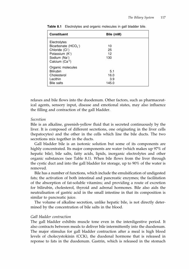
The Biliary System 117
relaxes and bile flows into the duodenum. Other factors, such as pharmaceut-ical agents, sensory input, disease and emotional states, may also influencethe filling and contraction of the gall bladder.
SecretionBile is an alkaline, greenish-yellow fluid that is secreted continuously by theliver. It is composed of different secretions, one originating in the liver cells(hepatocytes) and the other in the cells which line the bile ducts. The twosecretions mix together in the ducts.
Gall bladder bile is an isotonic solution but some of its components arehighly concentrated. Its major components are water (which makes up 97% ofhepatic bile), bile salts, fatty acids, lipids, inorganic electrolytes and otherorganic substances (see Table 8.1). When bile flows from the liver throughthe cystic duct and into the gall bladder for storage, up to 90% of the water isremoved.
Bile has a number of functions, which include the emulsification of undigestedfats; the activation of both intestinal and pancreatic enzymes; the facilitationof the absorption of fat-soluble vitamins; and providing a route of excretionfor bilirubin, cholesterol, thyroid and adrenal hormones. Bile also aids theneutralisation of gastric acid in the small intestine in that its composition issimilar to pancreatic juice.
The volume of alkaline secretion, unlike hepatic bile, is not directly deter-mined by the concentration of bile salts in the blood.
Gall bladder contractionThe gall bladder exhibits muscle tone even in the interdigestive period. Italso contracts between meals to deliver bile intermittently into the duodenum.The major stimulus for gall bladder contraction after a meal is high bloodlevels of cholecystokinin (CCK), the duodenal hormone that is released inreponse to fats in the duodenum. Gastrin, which is released in the stomach
Table 8.1 Electrolytes and organic molecules in gall bladder bile.
Constituent Bile (mM)
ElectrolytesBicarbonate (HCO3
−) 10Chloride (Cl−) 25Potassium (K+) 12Sodium (Na+) 130Calcium (Ca+2)
Organic moleculesBilirubin 5.1Cholesterol 16.0Lecithin 3.9Bile salts 145.0
GNC08 7/3/05, 5:28 PM117

118 Chapter 8
antrum in response to peptides in the food, also stimulates gall bladdercontraction.
Vasoactive inhibitory peptide (VIP), pancreatic polypeptide and stimula-tion of the sympathetic nerves to the gall bladder, all cause gall bladderrelaxation. Bile acids, by means of feedback control, also inhibit gall bladdercontraction.
Bile pigments in the gastrointestinal tract
After delivery to the intestines most conjugated bilirubin is excreted in thefaeces. This is due to the intestinal mucosa not being very permeable to theconjugated bile pigment. However, some bilirubin may be deconjugated bythe action of bacteria in the intestines and the free bilirubin formed can bepartially absorbed. Intestinal bacteria can also convert bilirubin to colourlessderivatives known as urobilinogens, which can be absorbed into the portalblood. These are then secreted in bile and urine. Urobilinogen remainingin the intestines is partially reoxidised to stercobilinogen, the reddish-brownpigment responsible for the colour of faeces.
JaundiceFailure of the body to excrete bile pigments results in accumulation of pig-ments in the blood plasma, termed hyperbilirubinaemia. This can result froma disruption of the flow of bile through the common bile duct or hepatic ductsby obstruction, i.e. gallstones, allowing bile to build up in the blood.
Bile pigments, mainly bilirubin, are deposited in the skin, mucous mem-branes and sclera, causing a yellow discoloration commonly known as jaun-dice. Jaundice is usually persistent and accompanied by severe pruritus(itching). The itching can be all over the body and the patient can scratch untiltheir skin bleeds. Nurses should make every effort to relieve the itching; anti-histamines are commonly prescribed but these can cause sedation. Effectiverelief can be achieved with the use of calamine lotion.
Jaundice is obvious when the plasma bilirubin concentration exceeds34 µmols/litre. Jaundice can result from excessive breakdown of red bloodcells (haemolysis) and haemoglobin to release bilirubin. Consequently theliver’s capacity to excrete is overwhelmed; this is known as prehepatic orhaemolytic jaundice. The bilirubin present in the plasma in this form ofjaundice is largely unconjugated as it has not been taken up and conjugated bythe liver.
Other common causes of jaundice include decreased uptake of bilirubin intohepatocytes, defective intracellular coagulation, or disturbed secretion into thebile canaliculi. Jaundice is most often seen in acute hepatitis and is known asintracellular or hepatocellular jaundice.
Gallstones can block the intrahepatic and extrahepatic bile ducts. This canresult in jaundice as the bile is refluxed into the blood. This type of jaundice isreferred to as obstructive or posthepatic jaundice.
GNC08 7/3/05, 5:28 PM118

The Biliary System 119
PATHOPHYSIOLOGY OF THE GALL BLADDER
Gall bladder diseases can either be short-term and easily manageable or longer-term and debilitating. Generally diseases of the biliary system present duringmiddle age. In individuals aged 20–50, diseases of the biliary tract are sixtimes more common in women than in men, but over the age of 50 the incid-ence in men and women becomes equal (Summerfield 2000).
Biliary tract disorders that are examined in this section include cholelithiasis,cholecystitis, choledocholithiasis, cholangitis and carcinoma.
Gallstones (cholelithiasis)
Gallstones are the presence of stones or calculi in the gall bladder. There aretwo types of gallstone: cholesterol stones and pigment stones. Cholesterolstones make up three-quarters of all gallstones. To understand the mechanismsof gallstone formation it is important to know their composition.
Cholesterol stones
Cholesterol stones contain cholesterol, calcium salts, bile acids, fatty acids,proteins and phospholipids. If the concentration of bile acids or phospholipidsdrops, cholesterol will not be held in micelles. This results in the bile becomingsupersaturated with cholesterol. When the cholesterol is not soluble in thissupersaturated system, it forms microcrystals. These microcrystals grow andcluster with one another and with other bile constituents to form gallstones.
Cholesterol gallstones are more prone to develop when there is a high ratioof cholesterol to bile acids or lecithin in the bile. This can be due to highcholesterol levels related to a high fat diet. Gallstones also form when there isa reduction in bile acid secretion, as a consequence of bile acid malabsorptionor reduced lecithin secretion.
Risk factors for the formation of both cholesterol and pigment stones areshown in Table 8.2.
Table 8.2 Risk factors for formation of gallstones.
Cholesterol gallstones Pigment gallstones
Increasing age Increasing ageFemale sex Chronic haemolysisPregnancy/use of oral contraceptive Alcohol abuse/cirrhosisObesity Biliary infectionGall bladder staisis Total parenteral nutritionSpinal cord injury Gall bladder stasis
GNC08 7/3/05, 5:28 PM119

120 Chapter 8
Pigment stones
Pigment stones are far less common than cholesterol stones. They are usuallysmall, only a few millimetres in diameter, and are either dark brown or black.When they occur they are usually multiple. They contain between 40% and90% pigment and less than 20% cholesterol. Pigment stones account for be-tween 20% and 25% of all gallstones.
Acute cholecystitis
This occurs when gallstones irritate the mucous membrane of the gall bladder,which then becomes inflammed. The patient usually presents with an acuteillness, which is severe in nature. The patient may be in shock due to pain andvomiting. The nurse may also notice pyrexia and tachycardia, due to infection.Treatment, cholecystectomy (surgical removal of the gall bladder), is oftenperformed laprascopically, with good effect. The benefits of laprascopic sur-gery include:
• reduced hospital stay
• minor surgical wound
• earlier mobilisation
• reduced risk of surgical complications
• reduced need for opioid analgesia
• quick return to normal lifestyle
If gallstones are only present in the common bile duct they can be removedvia endoscopic retrograde cholangiopancreatography (ERCP). The nursingpriorities of care before surgery in acute cholecystitis include:
• Pain control (intramuscular opiate analgesia at regular intervals).
• Hourly monitoring of patient’s vital signs until they become stable.
• Fasting the patient and commencing intravenous fluids.
• Intravenous administration of a broad-spectrum antibiotic.
• In the presence of vomiting, a naso-gastric tube may be passed and anantiemetic given.
• Careful monitoring of electrolyte and fluid balance.
Best practice guidelines for the management of acute cholecystitis related toacute pancreatitis have been produced by Glazer and Mann (1998).
Choledocholithiasis
The presence of stones in the common bile duct or hepatic duct is calledcholedocholithiasis. This condition arises when stones passed out of the gall
GNC08 7/3/05, 5:28 PM120

The Biliary System 121
bladder become lodged in the common bile duct or hepatic duct and preventthe flow of bile into the duodenum. Patients may be asymptomatic or canpresent with biliary colic, right upper quadrant abdominal pain, obstructivejaundice, acute gallstone pancreatitis and cholangitis. Therapeutic ERCP is thepreferred method of medical treatment.
Cholangitis
Cholangitis is a bacterial infection of the bile duct that is associated withobstruction of the common bile duct. Infection may lead to widespreadinflammation, which can develop into fibrosis and stenosis of the bile duct.Patients with mild cholangitis report fever spikes, chills, dark urine and ab-dominal pain. In more severe presentation cholangitis can be devastating, withtoxic sepsis, shock and mental impairment. Prognosis in severe cholangitisis poor.
Primary sclerosing cholangitis
Primary sclerosing cholangitis is an inflammatory process that results inmultiple strictures of bile ducts, which lead to chronic cholestatic liverdisease. The cause of primary sclerosing cholangitis is unknown. However,up to 75% of patients with primary sclerosing cholangitis have ulcerativecolitis. Clinical presentation includes fatigue, pruritis (itch), jaundice andabnormal liver function tests (see Chapter 7). Diagnosis is by ERCP and liverbiopsy examination. Asymptomatic primary sclerosing cholangitis requiresno treatment.
Carcinoma of the biliary tract
Gall bladder cancer
Cancer of the biliary tract usually involves the gall bladder. Gall bladder can-cer is usually an adenocarcinoma and occurs far more frequently in womenthan men. Symptoms usually involve:
• right upper quadrant abdominal pain
• nausea and vomiting
• substantial weight loss
• anorexia
Approximately 80% of gall bladder tumours also have gallstones. Diagnosisis made by ERCP, ultrasound, cholecystogram or cholangiogram. The greatmajority of these cases can only be treated palliatively (Summerfield 2000).
GNC08 7/3/05, 5:28 PM121

122 Chapter 8
Carcinoma of the extrahepatic biliary ducts
Tumours of the biliary ducts are extremely rare, usually associated with long-standing primary sclerosing cholangitis. Adenocarcinoma is the most commonform of cancer in the biliary tree. Often the patient presents with painlessobstructive jaundice. Other features of biliary cancer include:
• substantial weight loss
• pyrexia
• pruritis
• nausea and vomiting
Patients with established biliary cancer rarely survive more than a few months,and treatment is usually palliative. Even with surgical resection and radio-therapy, prognosis is extremely poor.
Conclusion
The biliary system is intimately related in terms of structure and function tothe liver and shares a common entrance to the gastrointestinal tract at theduodenum. The biliary system merely concentrates and stores a liver product:bile. While important in fat breakdown, bile is not essential for life. On theother hand, disorders of the biliary system can cause considerable sufferingand also disfigurement in the form of jaundice, if the outflow of bile from thegall bladder is blocked.
The structure and function of the biliary system is relatively easy to under-stand and in turn to explain to patients who may be puzzled at the suddenonset of extreme pain, as in the case of obstruction or inflammation of the gallbladder. The concept of referred pain is also important in any explanation ofsymptoms to patients. In extreme cases, patients will also wonder why andhow they can survive without a gall bladder if it has to be surgically removed.In all of the above, nurses can play a key role in offering information andsupport to patients with disorders of the biliary system. Likewise, while jaun-dice can be very distressing and uncomfortable for patients, they can be reas-sured, in the absence of any liver pathology, that the situation can be resolvedeffectively.
BACKGROUND READING
Additional reading to support the material in this chapter can be found in the relevant
sections of the following texts:
Alexander, M., Fawcett, J.N. and Runciman, P. (2000) Nursing Practice: Hospital and
Home – the Adult. Churchill Livingstone, Edinburgh (Chapter 4).
GNC08 7/3/05, 5:28 PM122

The Biliary System 123
Brooker, C. and Nicol, M. (2003) Nursing Adults: the Practice of Caring. Mosby, London
(Chapter 23).
Clancy, J. and McVicar, A.J. (2002) Physiology and Anatomy: a Homeostatic Approach, 2nd
edition. Arnold, London (Chapter 10).
Clancy, J., McVicar, A.J. and Baird, N. (2002) Perioperative Practice: Fundamentals of
Homeostasis. Routledge, London (Section 2).
Haslett, C., Chilvers, E.R., Boon, N.A. and Colledge, N.R. (2002) Davidson’s Principles
and Practice of Medicine, 19th edition. Churchill Livingstone, Edinburgh (Chapter 18).
Higgins, C. (2000) Understanding Laboratory Investigations: a Text for Nurses and Healthcare
Professionals. Blackwell Publishing, Oxford (Chapter 10).
Hinchliff, S., Montague, S. and Watson, R. (1996) Physiology for Nursing Practice, 2nd
edition. Baillière Tindall, London (Chapter 5.2).
Kindlen, S. (2003) Physiology for Health Care and Nursing. Churchill Livingstone, Edin-
burgh (Chapter 9).
Kumar, P. and Clark, M. (2002) Clinical Medicine, 4th edition. Saunder, Edinburgh
(Chapter 7).
McKenry, L.M. and Salerno, E. (1998) Pharmacology for Nursing, 20th edition. Mosby, St
Louis (Chapter 40).
Watson, R. (2000) Anatomy and Physiology for Nurses, 11th edition. Baillière Tindall,
London (Chapter 21).
BEST PRACTICE GUIDELINES
Glazer, G. and Mann, M.V. (1998) United Kingdom guidelines for the management of
acute pancreatitis. Gut, 42, S1–S13.
REFERENCES
Summerfield, J.A. (2000) Diseases of the gallbladder and biliary tree. In: Concise
Oxford Textbook of Medicine (Leadingham, J.G.G. and Worrell, D.A., eds), pp. 609–13.
Oxford University Press, Oxford.
GNC08 7/3/05, 5:28 PM123

124 Chapter 9
Chapter 9
The Pancreas
Chapter objectives
After reading this chapter you should be able to:
• Describe the normal anatomy and physiology of the pancreas.
• Understand the normal physiological functions of the pancreas, includ-ing both endocrine and exocrine secretions.
• Identify the range of clinical conditions specific to the exocrine functionof the pancreas.
• Relate the corresponding pathophysiology, diagnosis and treatment ofdisorders of the pancreas to nursing practice.
ANATOMY AND PHYSIOLOGY
The pancreas is both an endocrine and an exocrine gland. It is a wedge-shapedelongated gland which lies in the abdominal cavity. It is approximately 15 cmlong and 5 cm wide and it weighs about 80–90 g. Structurally the pancreas canbe divided into three regions: the head, which lies over the vena cava in theC-shaped curve of the duodenum; the body, which lies behind the duodenum;and the tail, which is situated under the spleen (see Figure 9.1).
The pancreatic duct (duct of Wirsung) runs the whole length of the pancreasfrom left to right and joins the common bile duct. Pancreatic juice emptiesfrom this duct into the duodenum via the ampulla of Vater.
The pancreas is composed of two types of cell, exocrine and endocrine cells.
Exocrine tissue
Pyramidal acinar cells are exocrine cells that compose the bulk of pancreatictissue. Groups of acinar cells form an acinus, and groups of acini form grape-like lobules. The acini secrete the digestive enzymes of the pancreatic juice.The nucleus of the acinar cell is situated at the base of the cell. The cytoplasmin the basal region can be stained with a dye to show the presence of roughendoplasmic reticulum, the site of digestive enzyme production. Small mito-
124
GNC09 7/3/05, 5:26 PM124

The Pancreas 125
Figure 9.1 Structure of the pancreas. Reproduced with permission from Hinchliff et al.(1996).
chondria are situated throughout the acinar cell, and the apical portion of thecell contains the Golgi body, and numerous zymogen granules, which containthe pancreatic enzymes and enzyme precursors.
Each exocrine unit of the pancreas consists of a terminal acinar portion anda duct. The duct that drains from the acinus is known as an intercalated duct,which empties into larger intralobular ducts. The intralobular ducts in eachlobule of the pancreas drain into larger extralobular ducts, which empty thesecretions of the pancreas into the main collecting duct, the pancreatic duct(see Figure 9.2).
Small mucous glands situated within the connective tissue surrounding thepancreatic duct secrete mucus directly into the duct.
The exocrine units of the pancreas surround the islets of Langerhan, theendocrine units of the pancreas.
Endocrine pancreas
Endocrine cells, or the islets of Langerhan, make up 1% of the pancreatic cells.They are most numerous in the tail region of the pancreas. They consist of
GNC09 7/3/05, 5:26 PM125

126 Chapter 9
Figure 9.2 Ducts of the pancreas. Reproduced with permission from ChurchillLivingstone. From S. Rutishauser, Physiology and Anatomy: A Basis for Nursing andHealth Care (1994).
clusters of cells surrounded by pancreatic acini and vary in size. As with allendocrine tissue, the hormones they produce are secreted directly into the blood.The major endocrine cells of the pancreas are alpha, beta and delta cells, whichsecrete glucagons, insulin and somatostatin, respectively. Glucagon (alpha cells)and insulin (beta cells) are taken up by the local blood vessels to act systemically.
When the blood sugar level falls below normal levels, the alpha cells arestimulated to secrete glucagon, which accelerates the conversion of glycogento glucose in the liver. When the blood sugar level is above normal, the betacells secrete insulin, which promotes both the metabolism of glucose by tissuecells and the conversion of glucose to glycogen, which is then stored in the liver.
Somatostatin (produced in the delta cells) acts locally in a paracrine mannerto inhibit the secretions of alpha and beta cells.
Blood supply to the pancreas
Oxygenated blood is supplied to the pancreas by the branches of the coeliac,splenic and superior mesenteric arteries. The acini and ducts are surrounded
GNC09 7/3/05, 5:26 PM126

The Pancreas 127
by separate capillary beds. Blood drains from the pancreas via the portal veinto the liver or into the splenic circulation by the superior mesenteric andsplenic veins.
Nerve supply to the pancreas
Cholinergic preganglionic fibres of the vagus nerve enter the pancreas. Theysynapse with postganglionic cholinergic nerve fibres, which lie in pancreatictissue and innervate both acinar and islet cells. Post-ganglionic sympatheticnerves from the coeliac and superior mesenteric plexi innervate vascular flowand control pain sensations and enzyme secretion in the pancreas. Parasym-pathetic fibres control exocrine and endocrine functions.
Pancreatic juice
The exocrine acinar cells secrete between 500 and 1000 ml of pancreatic juiceeach day. Pancreatic juice has two types of secretion, an enzyme-rich portionand an aqueous alkaline-rich portion. It is a colourless fluid and has a pHof 8.1–8.3. It consists of water, bicarbonate, sodium, potassium, chloride andcalcium. The function of the alkaline nature of pancreatic juice, together withother alkaline secretions, namely bile and intestinal juice, is to neutralise theacidic chyme arriving from the stomach.
This neutralising function is important for three main reasons:
• Pancreatic enzymes require a neutral or slightly alkaline environment foroptimal activity.
• The absorption of fat depends upon the formation of micelles in the intes-tinal lumen, a process which will only take place at a neutral or slightlyalkaline pH.
• It protects the intestinal mucosa because excess acid in the duodenum candamage the mucosa and lead to ulceration.
Pancreatic juice enters the duodenum with secretions of the biliary system atthe ampulla of Vater.
Pancreatic enzymes
There are three main types of enzyme present in pancreatic juice:
• Amylases, which break down carbohydrates into glucose and maltose.
• Lipases, which are important in the early stages of fat breakdown.
• Proteases, including trypsinogen, the precursor of proteolytic trypsin.
GNC09 7/3/05, 5:26 PM127

128 Chapter 9
These enzymes are released from the pancreatic acinar cells and are involvedin the digestion of foodstuffs. The acinar cells contain zymogen granules, inwhich enzyme or enzyme precursor proteins are stored. The enzyme precursorsproduced by the acinar cells include those of the proteolytic enzymes, trypsinand chymotrypsin. The release of enzymes as inactive precursors ensures thatthe activated enzymes do not autodigest the lining of pancreatic tissue.
Control of pancreatic juice secretion
The control of exocrine secretion of the acinar cells of the pancreas is viapeptides, such as the hormones secretin and cholecystokinin, and somatostatin,which acts mainly as a paracrine factor, and via neurotransmitters.
Hormonal controlThe main hormones involved in stimulating secretion of pancreatic juice aresecretin, which stimulates the secretion of the alkaline aqueous component,and cholecystokinin (CCK), which stimulates the secretion of the enzyme com-ponent. These hormones are produced in the duodenal mucosa in response tofood constituents in duodenal chyme. As the secretion of pancreatic juice isunder the control of two separate regulatory mechanisms, the composition ofthe juice can vary with regard to its enzyme protein content.
Somatostatin, which is present in the delta cells in the islets of Langerhanof the pancreas, is a powerful inhibitor of pancreatic secretion. It acts in aparacrine manner to inhibit the release of the exocrine alkaline and enzymesecretions, as well as the pancreatic hormones insulin and glucagons. In addi-tion, somatostatin inhibits the release of a number of gastrointestinal hormones,including CCK, secretin and gastrin.
Neural controlThe nervous control of pancreatic secretions is via both sympathetic andparasympathetic nerves. Stimulation of cholinergic fibres in the vagus nerveenhances the rate of both alkaline and enzyme fluid. Stimulation of sympa-thetic nerves inhibits secretion owing to vasoconstriction of blood vessels inthe gland, which leads to a decreased volume of juice secreted. At rest, thepancreas secretes enzymes at about 15% of the maximal rate and alkaline juiceat less than 2%.
The control of secretion of pancreatic juice during a meal depends upon thevolume and composition of the food ingested. Pancreatic secretions occur inthe following three phases.
Cephalic phaseThe sight or smell of food, or other sensory stimuli associated with the impend-ing arrival of food, elicits increased pancreatic secretion via a conditioned reflex.The presence of food in the mouth stimulates secretion via a non-conditioned
GNC09 7/3/05, 5:26 PM128

The Pancreas 129
reflex. The control during this phase is therefore nervous and is mediated byimpulses in cholinergic fibres in the vagus nerve. This results in a modestoutput of enzyme-rich pancreatic juice, containing very little bicarbonate.
Gastric phaseThe presence of food in the stomach stimulates the secretion of pancreaticjuice via a hormonal mechanism. Activation of chemical receptors in thewalls of the stomach causes the release of gastrin from G-cells into the localcirculation. Distension of mechanoreceptors in the stomach also stimulatessecretion. This phase results in a moderate production of pancreatic juice thatis rich in enzymes but low in bicarbonate.
Intestinal phaseThe intestinal phase is probably the most important phase in response to food.Food material in the duodenum stimulates both the alkaline and enzyme-richcomponents of pancreatic juice. The alkaline component of pancreatic juiceis secreted in response to the presence of acid in the duodenum. Acid alsostimulates the release of secretin from cells in the intestine and this hormonestimulates duct cells in the pancreas to secrete alkaline fluid. The enzyme-richjuice released during the intestinal phase is in response to fats and peptides infood. These cause the release of CCK from the walls of the duodenum into thebloodstream. CCK stimulates acinar cells to secrete enzymes. Trypsin in theduodenum inhibits the release of enzymes via the inhibition of CCK; this is afeedback control mechanism which limits the quantity of enzymes in the smallintestine and may have a protective function.
In the absence of pancreatic enzymes, up to 35% of dietary fat and proteinmay be assimilated using less efficient pathways of digestion, such as salivaryamylase and the enzymes for the brush border of the gut mucosa. It is estim-ated that pancreatic enzyme must fall below 10% of normal before maldigestionor malabsorption occurs.
PATHOPHYSIOLOGY OF THE EXOCRINE PANCREAS
Disorders of the exocrine function of the pancreas are an important cause ofmalabsorption because of the central role of the pancreas in the digestion of fatand protein. Conditions that affect the pancreas include acute and chronicpancreatitis, pancreatic fistulas and neoplasms of the pancreas.
Pancreatitis
Pancreatitis is inflammation of the pancreas that generally results from theobstruction of the pancreatic duct. Pancreatitis may be acute or chronic.
GNC09 7/3/05, 5:26 PM129

130 Chapter 9
Acute pancreatitis
Acute pancreatitis is a medical emergency, resulting in auto-digestion of thepancreas. It is most commonly caused by excessive alcohol abuse, abdominaltrauma, trauma during endoscopic retrograde cholangiopancreatography(ERCP) and bile duct stones. Inappropriate activation of digestive enzymes inthe pancreas can result in destruction of the organ with potentially catastrophicconsequences. Symptoms of acute pancreatitis include:
• severe upper abdominal pain, which can radiate into the back
• nausea and vomiting
• abdomen tender to palpation
• hypovolaemia and hypotension
• elevated serum amylase and urine amylase
There is no specific test for acute pancreatitis. Diagnosis is usually made bymeasurement of serum amylase, which is normally greatly elevated in acutepancreatitis. Other diagnostic procedures include ERCP and endoscopicultrasonography.
The key to treatment of acute pancreatitis is prompt and rapid intravenousfluid replacement aimed at haemodynamic stabilisation, correction of meta-bolic abnormalities and resting the pancreas. Patients with impaired islet cellfunction receive insulin to control diabetes.
Best practice guidelines, as outlined above, for the management of acutepancreatitis have been produced by BSG (1998) and by Toouli et al. (2002).
Nursing management of the patient with acute pancreatitis involves thefollowing:
• Ensuring adequate pain relief.
• Withdrawal of all food and fluids and nasogastric suctioning to rest thedigestive system.
• Bed rest and temperature-controlling measures to reduce metabolic rate toa minimum, which will reduce the secretion of pancreatic enzymes.
• Intravenous electrolyte and fluid replacement.
• Provision of enzyme and vitamin supplements.
• In severe cases, peritoneal lavage may be required.
• Supporting the patient in maintaining personal hygiene.
• Monitoring blood sugar levels in order to detect secondary diabetes.
Investigative procedures for pancreatic disease are often extensive and surgeryin the majority of cases only offers palliation. The gastrointestinal nurse has animportant role to play in helping both the patient and relatives deal with thisdifficult time.
The nursing care of patients with acute pancreatitis will vary according tothe severity of the attack. Acute pancreatitis is usually treated conservatively
GNC09 7/3/05, 5:27 PM130

The Pancreas 131
as an operation would increase mortality. Complications of acute pancreatitisinclude infection, respiratory distress, pseudocyst, abscess or pancreatic fistulaformation.
Chronic pancreatitis
Chronic pancreatitis is related to acute pancreatitis, usually caused by excess-ive long-standing alcohol abuse. In chronic pancreatitis the destruction of thepancreas is a slow and progressive disorder (Toskes 2000). It is seen most com-monly in male patients aged between 40 and 60 who normally present with:
• prolonged ill-health (abdominal pain, vomiting and nausea)
• weight loss and malabsorption
• steatorrhea (a condition where faeces have a high fat content)
• diabetes mellitus (as a result of islet cell destruction)
The destruction of pancreatic tissue results in secondary calcification in theorgan and cystic changes due to obstruction of drainage of the small ducts inthe gland. Blood and pancreatic function tests are used to confirm a diagnosisof chronic pancreatitis. ERCP may be used to determine the extent of disease.
Successful management of chronic pancreatitis largely relies on the patientabstaining from alcohol, and on pain relief, nutritional support and oralreplacement of digestive enzymes.
Pancreatic pseudocysts
Pseudocysts are localised collections of pancreatic secretions and are a complic-ation of both acute and chronic pancreatitis. They may be single or multipleand are 5–10 cm in diameter. Pseudocysts may produce abdominal pain and,more rarely, haemorrhage, infection and peritonitis. Abdominal ultrasono-graphy, endoscopic ultrasonography and CT scan are the best methods ofidentifying a pseudocyst or pancreatic abscess.
Pancreatic fistula
Pancreatic fistula may follow trauma to the gland or occur as a complicationfollowing pancreatic surgery. The management of pancreatic fistula requiresboth a nursing and medical management, as the release of pancreatic enzymesonto the skin will quickly cause excoriation. The skin may be protected by theapplication of barrier creams. Fluid loss and electrolyte balance need to becorrected, either by oral or intravenous route, and drainage from deep in thewound may be carried out by insertion of a drain. About 75% of pancreaticfistulas close spontaneously within six months. If a fistula should fail to closeit may require surgical intervention.
GNC09 7/3/05, 5:27 PM131

132 Chapter 9
Pancreatic cancer
Tumours, wherever they present, are classified as benign or malignant. In thepancreas most tumours are malignant and most commonly affect the exocrineportion of the pancreas. Over 90% of malignant tumours are adenocarcinomas.Pancreatic cancer most often develops in persons aged between 60 and 80.Potential risk factors for the development of a pancreatic tumour include chronicpancreatitis, smoking and a high fat diet. Of pancreatic carcinomas, over 60%occur in the head of the pancreas, 15–25% in the body and 5% in the tail (Russell2000). Symptoms are most common in tumours of the head of pancreas andtypically result in dull abdominal pain, weight loss and obstructive jaundice.Obstruction and/or metastasis may also cause signs and symptom of liverdamage (dark urine and light stools). ERCP is the most efficient test for detec-tion of pancreatic cancer. The prognosis for pancreatic cancer is very poor;median survival time is only 2–3 months following diagnosis (Russell 2000).Therefore therapy in pancreatic cancer is primarily palliative and supportive.Most patients present with metastatic disease and are not suitable for curativesurgery. The nurse should aim to help the patient reduce the impact of associ-ated symptoms by ensuring pain control and relief from nausea and vomiting.
Surgery for pancreatic cancer
The majority of patients with pancreatic cancer have disease in the head or theneck of the gland. The only curative surgical procedure is a pancreaticoduod-enectomy, also known as a Whipple’s procedure. In this operation the pan-creatic head, neck and body are resected, along with the distal biliary treeand gall bladder. Outcomes in managing pancreatic cancer surgically haveimproved recently; if surgery is performed before nodal spread takes place,survival rates increase.
Conclusion
Sharing a common route of entry for secretions to the duodenum with the biliarysystem, the pancreas is intimately anatomically located to several organs includ-ing the stomach, liver and spleen. The pancreas has both exocrine activities andendocrine activities as it produces digestive secretions and hormones, whichcontrol carbohydrate metabolism. Disorders of the pancreas are serious, usuallyresulting in both acute and chronic pancreatitis. It is the digestive nature of theorgan which leads to problems as the proteolytic enzymes, which it contains,come into contact with surrounding tissues. The intimate anatomical associa-tion with several other organs can lead to damage to these organs as well as tothe pancreas. Good nursing care is required in terms of pain relief and man-aging shock in patients with pancreatitis, in addition to support of patientswho may have cancer of the pancreas or who may have been abusing alcohol.
GNC09 7/3/05, 5:27 PM132

The Pancreas 133
BACKGROUND READING
Additional reading to support the material in this chapter can be found in the relevant
sections of the following texts:
Alexander, M., Fawcett, J.N. and Runciman, P. (2000) Nursing Practice: Hospital and
Home – the Adult. Churchill Livingstone, Edinburgh (Chapter 4).
Brooker, C. and Nicol, M. (2003) Nursing Adults: the Practice of Caring. Mosby, London
(Chapter 23).
Clancy, J. and McVicar, A.J. (2002) Physiology and Anatomy: a Homeostatic Approach, 2nd
edition. Arnold, London (Chapter 10).
Haslett, C., Chilvers, E.R., Boon, N.A. and Colledge, N.R. (2002) Davidson’s Principles
and Practice of Medicine, 19th edition. Churchill Livingstone, Edinburgh (Chapter 18).
Higgins, C. (2000) Understanding Laboratory Investigations: a Text for Nurses and Healthcare
Professionals. Blackwell Publishing, Oxford (Chapter 11).
Hinchliff, S., Montague, S. and Watson, R. (1996) Physiology for Nursing Practice, 2nd
edition. Baillière Tindall, London (Chapter 5.2).
Kindlen, S. (2003) Physiology for Health Care and Nursing. Churchill Livingstone, Edin-
burgh (Chapter 9).
Kumar, P. and Clark, M. (2002) Clinical Medicine, 4th edition. Saunders, Edinburgh
(Chapter 7).
McKenry, L.M. and Salerno, E. (1998) Pharmacology for Nursing, 20th edition. Mosby, St
Louis (Chapter 40).
Watson, R. (2000) Anatomy and Physiology for Nurses, 11th edition. Baillière Tindall,
London (Chapter 21).
BEST PRACTICE GUIDELINES
BSG (1998) United Kingdom Guidelines for the Management of Acute Pancreatitis (http://
www.bsg.org.uk/clinical_prac/guidelines/acute_pan.htm accessed 8 May 2004).
Toouli, J., Brooke-Smith, M., Bassi, C., Carr-Locke, D., Telford, J., Freeny, P., Imrie, C.
and Tandon, R. (2002) Guidelines for the management of acute pancreatitis. Journal of
Gastroenterology and Hepatology, 17, S15–S39.
REFERENCES
Russell, R.C.G. (2000) Tumours of the pancreas. In: Concise Oxford Textbook of Medicine
(Leadingham, J.G.G. and Worrell, D.A., eds), pp. 606–8. Oxford University Press,
Oxford.
Toskes, P.P. (2000) Chronic pancreatitis. In: Concise Oxford Textbook of Medicine
(Leadingham, J.G.G. and Worrell, D.A., eds), pp. 603–6. Oxford University Press,
Oxford.
GNC09 7/3/05, 5:27 PM133

134 Chapter 9
GNC09 7/3/05, 5:27 PM134

Diagnostic Procedures and Tests in Gastroenterology 135
Section 2
Essential Aspects of Gastroenterology
GNC10 7/3/05, 5:26 PM135

136 Chapter 10
GNC10 7/3/05, 5:26 PM136

Diagnostic Procedures and Tests in Gastroenterology 137
Chapter 10
Diagnostic Procedures and Tests inGastroenterology
The material in this chapter is presented in two sections: the first considersendoscopy and the second considers a range of tests used in gastroenterology.
Chapter objectives
After reading this chapter you should be able to:
• Describe the main diagnostic procedures and equipment used in gastro-enterology.
• Understand the rationale behind diagnostic procedures.
• Identify the role of appropriate investigations for the range ofgastrointestinal disorders.
• Relate diagnostic procedures to clinical nursing practice.
ENDOSCOPY
Gastrointestinal endoscopy involves the direct visual examination of the lu-men of the gastrointestinal tract. It is a relatively safe and effective way ofevaluating the appearance and integrity of the gastrointestinal mucosa for thepurpose of diagnosis and provides access for therapeutic procedures. The tech-niques for endoscopic investigations of the upper and lower gastrointestinaltract include gastroscopy (oesophogastroduodenoscopy), ERCP (endoscopicretrograde cholangiopancreatography), EUS (endoscopic ultrasonography),colonoscopy and flexible and rigid sigmoidoscopy.
Nurse endoscopists
The British Society of Gastroenterology (BSG) Working Party (1994) gavesupport to the establishment of nurse endoscopists. Their report highlightedthe potential role for nurses to perform gastrointestinal endoscopy as long asadequate training for all procedures is available (BSG 1994). The BSG proposed
137
GNC10 7/3/05, 5:26 PM137

138 Chapter 10
the role of the nurse endoscopist to complement and increase the capacity ofexisting medical endoscopy teams.
The NMC document The Scope of Professional Practice (NMC 2002) sup-ports nurses in role development, for example carrying out gastrointestinalendoscopy. In addition, the nursing profession in the UK established the frame-works necessary to allow the development of nursing roles into areas oncedominated by the medical profession (DoH 1997).
Because of the risks associated with therapeutic endoscopy the BSG work-ing party considered that the nurse endoscopist’s work should be confined todiagnostic gastroscopy with or without biopsy, and flexible sigmoidoscopywith or without biopsy. All therapeutic procedures, ERCP and colonoscopyare excluded from the endoscopist’s remit. Understandable concern has beenexpressed by nurses with respect to the potential legal implications ofendoscopic practice and the need for full medico-legal cover in the event ofcomplications. Damage to teeth, bridges, crowns and other expensive dentaltreatments may be a potential source of litigation. The working party aimed totackle these philosophical and practical issues and summarised:
• There is an increasing demand from within both the medical and nursingprofessions to perform endoscopy.
• Medico-legally, nurses may perform an endoscopy provided they havereceived appropriate training and are adequately supervised by a respon-sible consultant.
• Careful patient selection is essential to exclude high-risk patients and thoserequiring therapeutic procedures.
• A designated medical endoscopist should be immediately available dur-ing nurse endoscopy sessions.
• The nurse should be responsible for obtaining consent from the patientbefore endoscopy and for discussing the findings with the patient after theprocedure (BSG 1999).
• The nurse should be responsible for preparing and signing the endoscopyreport.
• Regular records and audit of the nurse endoscopist’s work should take place.
• Continuing education is essential, with regular opportunities to attendendoscopy courses and meetings.
Under these described circumstances the BSG working party gave support tothe proposal that nurses perform endoscopy.
Current best practice guidelines on sedation for endoscopy are published bythe BSG (2003).
Endoscopy is clearly an area in which nurses are being encouraged toextend their practice into invasive and diagnostic techniques. Such proceduresare anxiety-provoking for patients, who remain conscious throughout, andnurses are uniquely positioned to provide the technical expertise and thepsychosocial support required. The extension of practice into endoscopy dem-
GNC10 7/3/05, 5:26 PM138

Diagnostic Procedures and Tests in Gastroenterology 139
onstrates two aspects of nursing which are increasingly coming to the fore: theextent to which nurses require detailed knowledge of anatomy, physiologyand pathology and the issue of accountability.
In terms of the knowledge required, nothing less than a deep knowledgeof the relevant areas of the gastrointestinal tract is required. Nurses cannotsimply carry out such procedures by rote; they need to know what is normaland what is abnormal and if they are to provide the holistic care which isincreasingly expected of them, they need to be sufficiently educated in thewhole field of gastroenterology in order to explain procedures fully to patientsand to discuss likely diagnoses and outcomes.
In terms of accountability, the issue of education is also raised: how can anurse perform a procedure, with attendant risks and the potential for psycho-logical morbidity, without being fully aware of the significance of each actionthey are performing? If nurses are to carry out advanced procedures under anextended role, they must be willing and able to be accountable for all aspectsof the procedures they are undertaking. This chapter outlines the likely roles andresponsibilities of nurse endoscopists and it is hoped that those who seek thisrole will extend their knowledge at the same time as extending their practice.
Types of endoscopes
Depending upon the area of the gastrointestinal tract that is required to beseen there are a range of endoscopes that can be used:
• A flexible end-viewing or side-viewing (oblique) endoscope – to view theoesophagus, stomach and proximal duodenum.
• A colonoscope, which is a 120–180 cm flexible endoscope – to view theentire lower gastrointestinal tract.
• A flexible sigmoidoscope, which may be up to 65 cm in length – to exam-ine the rectum and the sigmoid and descending colon.
• An anoscope, which is a rigid plastic or metal speculum – for inspection ofthe anal canal.
• A proctosigmoidoscope, which is a 15–25 cm rigid endoscope – to exam-ine the rectum and sigmoid colon.
Although all these instruments are designed for specific uses, all endoscopeshave several common features, which include the following:
• A flexible insertion tube which is usually 8–12 mm in diameter. This inser-tion tube contains air, water and biopsy channels, and fibre bundles.
• An umbilical cord that extends from the control head and leads to thelight source.
• An optic system, which consists of fibreoptic bundles that conduct lightthrough the shaft and transmit the images to the eye. In video endoscope,
GNC10 7/3/05, 5:26 PM139

140 Chapter 10
the optic system consists of a one-piece video camera that transmitsimages directly to a television screen with no need for fibreoptics.
• A control head that houses the lenses, controls for manoeuvring the tip upand down and from side to side, and valves that regulate irrigation, air orcarbon dioxide insufflation and suction.
• Channels for air and water flow.
• A suction/biopsy channel that allows the passage of accessory instruments,such as biopsy forceps, cytology brushes, polypectomy snares, laser fibresor stents. The suction channel also allows for suctioning fluid thatobstructs the endoscopist’s vision.
Gastroscopy
Gastroscopy, or oesophogastroduodenoscopy, uses a flexible endoscope lessthan 10 mm in diameter, which is passed directly into the upper gastrointestinaltract. The entire oesophagus and stomach and proximal duodenum can bevisualised. Larger diameter gastroscopes with larger suction channels are usedfor therapeutic procedures.
Gastroscopy allows the physician or nurse endoscopist to diagnose anddocument gastrointestinal disorders through the use of direct vision and videophotography.
Diagnostic gastroscopy is generally indicated for evaluating:
• dysphagia or odynophagia
• dyspepsia
• gastro-oesophageal reflux symptoms unresponsive to medical therapy
• haematemisis/melaena
• oesophageal or gastric varices
• oesophagitis or gastritis
• gastric and peptic ulcers
• chronic abdominal pain
• suspected polyps and cancer
• removal of ingested foreign bodies
• acute injury following caustic ingestion
• management of achalasia
Gastroscopy is generally not indicated for evaluating:
• symptoms which are considered functional in origin
• metastatic adenocarcinoma
Prior to gastroscopy, patients require thorough medical and nursing assess-ment with special attention given to any history of drug reactions, bleedingdisorders or associated cardiac, pulmonary, renal or hepatic disease.
GNC10 7/3/05, 5:26 PM140

Diagnostic Procedures and Tests in Gastroenterology 141
Nursing care during gastroscopy
During gastroscopy the nursing responsibilities include the maintenance ofthe patient’s oral airway, suctioning secretions and regurgitated material whennecessary from the pharynx. Nasal oxygen should be used to treat hypoxia,and resuscitation equipment should be immediately available in the eventof cardiopulmonary complications. The nurse should monitor the patient’soxygen saturation and vital signs regularly and observe the patient for signsof bleeding, vomiting, change in vital signs, pain and abdominal distension.Patients should be fasted for at least four hours prior to gastroscopy, longer ifthey have eaten a large meal.
Although gastroscopy is generally regarded as a safe procedure, adverseevents can occur, including:
• respiratory depression
• effects of sedation
• perforation of the oesophagus, stomach or duodenum
• haemorrhage related to trauma and perforation
• pulmonary aspiration of blood or secretions, or regurgitation of gastriccontents
• cardiac arrhythmia
• infection
• allergic reaction to the anaesthetic or intravenous medications
The rate of adverse events and complications increases when therapeuticmanoeuvres are performed. Patients at the highest risk during gastroscopy areolder people and those with advanced cardiac, pulmonary, hepatic or centralnervous system disease.
Endoscopic retrograde cholangiopancreatography (ERCP)
ERCP uses a combination of endoscopic and radiological techniques to visualisethe biliary and pancreatic ducts. It involves the injection of a contrast materialinto the biliary and pancreatic systems followed by radiological screening.Two different contrasts are used: low iodine for the common bile duct andhigher iodine contrast for the pancreatic duct.
ERCP is indicated for the following:
• Signs and symptoms of pancreatic malignancy.
• Evaluation of acute, recurrent pancreatitis.
• Removal of retained common bile duct stones.
• Unexplained chronic abdominal pain of suspected biliary or pancreaticorigin.
GNC10 7/3/05, 5:26 PM141

142 Chapter 10
• Evaluation of patients with jaundice.
• Pre-operative or post-operative evaluation to detect common duct stonesin patients who undergo laparoscopic cholecystectomy.
Prior to ERCP, patients should be fasted for six hours. Barium studies shouldnot be conducted within 72 hours preceding ERCP because the residual bariumcan obstruct the view of the contrast medium in the ducts. As ERCP can be alonger procedure than gastroscopy, higher levels of sedation and analgesia areusually required.
The patient should be positioned in either the prone or left lateral positionprior to the procedure. A side-viewing duodenoscope is passed by theendoscopist into the second part of the duodenum. Glucagon may be injectedintravenously to suppress duodenal peristalsis and thereby aid visualisation.When the scope is in the correct location to view the ampulla of Vater, thepatient is moved into the prone position. The endoscopist passes a plasticcannula through the endoscope and manoeuvres it into the orifice of the am-pulla. Once the cannula is in the pancreatic duct or the common bile duct, theradiocontrast material is injected. The patient should be closely monitored forany reaction to the radiocontrast dye.
Nursing responsibilities during ERCP
The main responsibilities of the nurse during ERCP are:
• Assess, monitor, and document oxygen saturation and vital signs.
• Observe the patient for signs of abdominal distension.
• Maintain the patient’s airway until the gag reflex returns.
• Observe for signs of pancreatitis, low-grade fever, pain, vomiting andtachycardia.
• Administer antibiotics as prescribed. ERCP may introduce infection lead-ing to cholangitis, therefore prophylactic antibiotics are given to patientswith biliary or pancreatic stasis.
Following the procedure, the patient’s temperature should be checked everyfour hours for 48 hours. The most significant complications associated withERCP are pancreatitis and sepsis.
Injury to the pancreas can result from chemical, enzymatic, mechanical,microbiological or hydrostatic factors. In patients with partial obstruction ofeither the pancreatic or common bile duct, a frequent complication of ERCP isbiliary sepsis. The introduction of infection into a stagnant duct system canresult in cholangitis and septicaemia. Additional potential complications in-clude aspiration, bleeding, perforation, respiratory depression or arrest andcardiac arrythmias or arrest. Acute pancreatitis is caused in about 1% of ERCPs(Imrie 2000).
GNC10 7/3/05, 5:26 PM142

Diagnostic Procedures and Tests in Gastroenterology 143
Proctosigmoidoscopy
Proctosigmoidoscopy is an examination of the rectum and sigmoid colon usinga proctoscope, a rigid tube with a detachable, disposable end which is used forone patient only and then discarded. The proctoscope is 25–30 cm in lengthand approximately 1.5 cm in diameter. Proctosigmoidoscopy gives valuableinformation about the anal canal and rectum, but as the instrument is rigid itcannot be used to view above the splenic flexure. Indications for procto-sigmoidoscopy include:
• melaena
• persistent diarrhoea
• passage of mucus and pus with stool
• bacteriology or histological studies
• rectal pain
Preparation involves a hypertonic phosphate enema on the morning of theprocedure, to clean out the large bowel. Sedation is rarely required forproctosigmoidoscopy. The patient is asked to lie on his or her left side withknees drawn up to the chest, the scope is gently inserted, the obturator isremoved, and air is pumped through the protosigmoidoscope to inflate therectum. The examiner will note the colour and friability of the rectal mucosa,and bleeding sites and ulcers. Throughout the procedure the nurse shouldmonitor the patient’s vital signs, abdominal distension, pain tolerance and thecondition of the skin.
Flexible sigmoidoscopy
Flexible sigmoidoscopy employs a flexible instrument to examine therectum, sigmoid and a variable length of more proximal colon. The flexiblesigmoidoscope measures up to 65 cm in length and its flexibility allowsthe endoscopist to reach the descending colon in up to 80% of patients. Oneadditional benefit of flexible sigmoidoscopy is that it is better tolerated thanrigid protosigmoidoscopy in many patients. Flexible sigmoidoscopy is gener-ally indicated for:
• Screening of asymptomatic patients at risk of colonic cancer.
• Evaluation of suspected distal colonic disease when there is no indicationfor colonoscopy.
• Evaluation of the colon in conjunction with radiological examinations.
• Patients with a family history of colorectal cancer.
• Flexible sigmoidoscopy is generally not indicated when colonoscopy isindicated.
GNC10 7/3/05, 5:26 PM143

144 Chapter 10
Contraindications for flexible sigmoidoscopy are similar to those listed forcolonoscopy and should be avoided in documented acute diverticulitis.
Colonoscopy
Colonoscopy gives information about the lower gastrointestinal tract andis performed to examine the entire colon and rectum, almost always undersedation. Modern colonoscopes are similar in design to upper gastrointestinalendoscopes but are longer, ranging from 1.2 to 1.8 m.
Diagnostic colonoscopy is indicated in the following situations:
• Evaluation of active or occult lower gastrointestinal bleeding.
• Unexplained faecal occult blood.
• Unexplained iron deficiency anaemia.
• Confirmation of suspected polyps or strictures.
• Surveillance for colorectal cancer.
• Screening of patients with family history of colorectal cancer.
• Diagnosis and evaluation of chronic inflammatory bowel disease.
• Surveillance of long-term ulcerative colitis patients.
• Treatment of a bleeding lesion (e.g. laser or injection therapy).
• Palliative treatment of neoplasm (e.g. laser or stenting).
• Foreign body removal.
Colonoscopy is generally not indicated in the following circumstances:
• Acute diarrhoea.
• Routine follow-up of inflammatory bowel disease (except cancer surveil-lance in long-standing ulcerative colitis).
• Upper gastrointestinal bleed or melaena with a demonstrated uppergastrointestinal source.
Preparation of the bowel to enable a successful colonoscopy is vital. The pre-sence of small particles of faecal matter may obscure the endoscopist’s view ofthe colonic mucosa, therefore thorough bowel preparation is essential to avoidthe patient having repeat colonoscopies. Bowel preparation is achieved byrestricting diet to a low residue or clear fluid intake the day before the proced-ure. There are a range of purgatives available for patients to take in advanceof their procedure; this usually involves administration of large volumes (severallitres) of an isoomolar electrolyte lavage solution. The patient may benefitfrom being informed that the preparation can be the most unpleasant part ofthe procedure, but that it is vital for a successful examination.
The nurse will have several responsibilities in the patient preparation forcolonoscopy, including:
GNC10 7/3/05, 5:26 PM144

Diagnostic Procedures and Tests in Gastroenterology 145
• Recording of baseline vital signs so that abnormalities can be detectedduring the procedure.
• Obtaining informed consent.
• Giving the patient the opportunity to urinate prior to the procedure.
• Fasting the patient for several hours before the procedure to prevent therisk of aspiration during sedation.
The nurse caring for the patient following a colonoscopy should be aware thatthe patient will still be drowsy after the procedure and should be allowed tosleep until their sedation wears off before being offered a drink. Vital signs(pulse, blood pressure and rate of respiration) must be recorded regularly(every 15 minutes) following the procedure to allow detection of signs of anyabnormalities, noting risk of perforation to the bowel or oversedation. Majorcomplications occur in less than 1% of patients undergoing colonoscopy. Thetwo main complications, perforation and haemorrhage, usually occur after theremoval of polyps – polypectomy. It is important that the nurse is aware ofany signs of complications, such as bleeding, vomiting, abdominal pain andrigidity, and that these are recorded.
Small bowel enteroscopy
Enteroscopy allows the visualisation of a greater extent of the small bowelthan gastroscopy. There are two types of enteroscopes available: the pushenteroscope, which allows limited tissue sampling and therapy, and the sondeenteroscope, which potentially allows the entire small bowel to be seen with-out the possibility of therapeutic intervention.
Enteroscopy is generally indicated for:
• Evaluation of the source of gastrointestinal bleeding not identified bygastroscopy or colonoscopy.
• Evaluation of an abnormal radiological image of the small bowel.
• Localisation of known or suspected small bowel lesions.
• Therapy (with push enteroscope) of small bowel lesions.
If the source of bleeding can be identified with gastroscopy or colonoscopy,enteroscopy is generally not indicated.
Endoscopic ultrasound
Advances in technology have led to the combination of endoscopes with ultra-sonography to enhance visualisation of the gastrointestinal tract. This techniqueis called endoscopic ultrasound (EUS) and allows for better quality resolution,
GNC10 7/3/05, 5:26 PM145

146 Chapter 10
which enhances evaluation of structures. The walls of the oesophagus, stomach,duodenum and colon can be visualised, as well as the structure of severaladjacent organs.
The general indications for EUS include:
• Staging tumours of the gastrointestinal tract, pancreas and bile ducts.
• Evaluating abnormalities of the gastrointestinal tract wall or adjacentstructures.
• Sampling of tissue lesions within, or adjacent to, the wall of the gastroin-testinal tract.
• Evaluation of abnormalities of the pancreas and biliary tree.
• Providing endoscopic therapy under ultrasonographic guidance.
Although developing, the role of EUS, as compared with endoscopic andradiological examinations, has a greater ability to detect lesions in the wallof the gastrointestinal tract.
DIAGNOSTIC TESTS AND PROCEDURES INGASTROENTEROLOGY
The remainder of this chapter examines a number of tests and procedures thatnurses will encounter in gastrointestinal medicine. These tests and proceduresare available to confirm or disprove diagnoses. Endoscopic procedures havebeen covered earlier in this chapter. Radiological studies and tests involvingthe analysis of blood, urine and faeces are reviewed in this section.
It is vital for the nurse to understand the role of tests and investigationsthat are undertaken in gastroenterology as the nurse will be involved inthe preparation and education of patients. Research has shown that patientanxiety can be reduced and patient compliance improved if nurses provideappropriate information and education prior to a procedure.
Radiological investigations
Radiological studies such as barium swallow, barium enema and gastrointes-tinal upper series are used in gastroenterology to detect abnormalities of thegastrointestinal tract. Barium studies are in general more easily tolerated thanendoscopic investigations and have fewer complications; however, endoscopyhas a greater diagnostic potential.
Most radiological studies use barium as a contrast medium. This is a chalky,non-allergenic substance that allows for X-ray examination of the oesophagus,stomach and lower gastrointestinal tract. The barium preparation is usuallyadministered orally or rectally and its use is contraindicated in patients withsuspected gastrointestinal obstruction or perforation. The nurse should caution
GNC10 7/3/05, 5:26 PM146

Diagnostic Procedures and Tests in Gastroenterology 147
the patient to report failure to pass the barium within 48 hours so that appro-priate measures, an enema or laxative, can be initiated to avoid constipationor obstruction.
Barium swallow
Barium swallow allows for radiological examination of the oesophagus. Thisprocedure can reveal the presence or absence of strictures, motility disorders,ulceration and foreign bodies. As a diagnostic tool, barium swallow is lesssensitive than endoscopy; if possible, barium swallow should be avoided inpatients with dysphagia.
Upper gastrointestinal series
Radiological examination of the stomach and small intestine involves inges-tion of a barium solution followed by an upper gastrointestinal series withsmall bowel follow-through. This procedure allows for diagnosis of diverticula,strictures, hiatus hernia, motility disorders and tumours. Upper gastrointestinalseries is also used in the examination of Crohn’s disease and malabsorptionsyndromes.
Barium enema
Barium enema allows radiological examination of the colon. It is a commontest used for the detection of polyps, diverticula and structural changes inthe colon, and may be used for the diagnosis of colorectal cancer and inflam-matory bowel diseases.
Abdominal ultrasound
Abdominal ultrasound is a non-invasive procedure that can indicate thesize and shape of organs. It can detect any abnormalities of structures orspaces within the abdominal cavity through the transmission of sound waves.Abdominal ultrasound may be useful for initial detection of gallstones, asit involves minimal patient preparation and no radiation. Ultrasound investi-gations can also be used to diagnose acute cholecystitis.
Computed tomography (CT) scan
CT scanners have been used since the 1970s and produce computerisedimages of the body in a series of cross-sectional views. CT scanning is anon-invasive, radiological scanning technique that involves the measurementof differences in tissue density to reflect tissue configuration. CT scanning
GNC10 7/3/05, 5:26 PM147

148 Chapter 10
produces a three-dimensional image which is more detailed and accurate thanimages produced by abdominal ultrasound. CT scans are commonly used fordetection of malignancy in the gall bladder and pancreas.
Blood tests in gastrointestinal medicine
This is a brief overview of the more commonly used blood tests. Referenceranges are provided in Table 10.1.
Haemoglobin/haematocrit
Haemoglobin is the oxygen-carrying pigment of red cells, and haematocritrefers to the volume percentage of erythrocytes in whole blood. Levels of bothhaemoglobin and haematocrit are influenced by pathology in the gastrointes-tinal tract and will decrease when a patient is anaemic, such as during agastrointestinal bleed.
Platelet count
Platelet count is a count of the number of platelets present in blood. A lowplatelet count would be indicative of impaired clotting mechanisms.
Prothrombin level/time
Prothrombin is a glycoprotein present in blood plasma that is normallyconverted to thrombin. Impaired absorption of vitamin K from the intestinecan result in decreased prothrombin levels. Low prothrombin levels are seenin a patient’s liver and in small bowel disease. Prothrombin time relates tothe measurement of blood clotting. Prolonged prothrombin times are seen inpatients who are unable to absorb vitamin K, and in individuals with poornutrition, liver disease and blood disorders.
Table 10.1 Approximate reference ranges.
Red blood cell (RBC) Males 4.5–6.5 × 10¹²/lFemales 3.9–5.6 × 10¹²/l
Haemoglobin (Hb) Males 13.5–17.5 g/dlFemales 11.5–15.5 g/dl
Haematocrit (Ht) Males 40–52%Females 36–48%
Mean cell haemoglobin concentration (MCHC) 20–35 g/dl
Critical valuesHaemoglobin < 7.0 g/dl or > 20.0 g/dl
GNC10 7/3/05, 5:26 PM148

Diagnostic Procedures and Tests in Gastroenterology 149
Liver function tests
Liver function tests may be carried out on blood, urine and faeces to detect thelevels of substances manufactured by the liver as well as to estimate otherfunctions of the liver (i.e. excretion). Liver function tests are usually defined asthe measurement of serum levels of:
• albumin
• bilirubin
• aminotransferase
• alkaline phosphotase
• γ-glutamyltransferase
Serum albumin
Serum albumin is a good indicator of chronic liver disease. It is synthesised bythe liver and its levels fall in chronic liver disease.
Conclusion
Nurses working in gastroenterology are increasingly being enabled, throughspecialist and nationally recognised courses, to undertake training and extendtheir practice as nurse endoscopists. These developments are in line withUK government policy, which seeks to extend the clinical capabilities of nursesgenerally and also in line with the development of the nurse consultantrole.
Extending nursing practice into invasive and diagnostic procedures such asendoscopy raises several issues for nurses, including accountability, relation-ships with the medical and surgical professions and the knowledge baserequired for such practice. It is already clear that nurses are able to carry outendoscopy safely and it is likely that there will be a major expansion in thisrole. In addition to being able to undertake this devolved medical procedure,nurses are ideally placed to undertake it as these invasive procedures areassociated with a great deal of anxiety in patients regarding the procedureitself and the possible outcome in terms of diagnosis – especially of malignantconditions. The nurse endoscopist, in addition to performing the endoscopy,is able to offer support through detailed and clear explanations to the patientof what they are likely to experience, and for minor conditions such as haem-orrhoids can carry out certain treatments and offer ongoing advice about dietand lifestyle which may help to alleviate further problems. Nurses should alsohave a thorough understanding of the medical and surgical roles in relation tothe possible outcomes of endoscopy or the need for further, more invasiveprocedures which they are not competent to carry out.
GNC10 7/3/05, 5:26 PM149

150 Chapter 10
However, the most important aspect of the preparation of nurse endoscopistsis the knowledge of the relevant anatomy and physiology of the gastrointestinaltract that is required. This must be beyond the superficial aspects of structure,function and control and must incorporate the pathology and pathophysiologyof gastrointestinal disorder to ensure that nurses can offer the right kind ofsupport to patients and liaison with medical and surgical colleagues, and canbe truly accountable for every aspect of their extended role.
BACKGROUND READING
Additional reading to support the material in this chapter can be found in the relevant
sections of the following texts:
Alexander, M., Fawcett, J.N. and Runciman, P. (2000) Nursing Practice: Hospital and
Home – the Adult. Churchill Livingstone, Edinburgh (Chapter 4).
Brooker, C. and Nicol, M. (2003) Nursing Adults: the Practice of Caring. Mosby, London
(Chapter 22).
Clancy, J., McVicar, A.J. and Baird, N. (2002) Perioperative Practice: Fundamentals of
Homeostasis. Routledge, London (Chapter 2).
Haslett, C., Chilvers, E.R., Boon, N.A. and Colledge, N.R. (2002) Davidson’s Principles
and Practice of Medicine, 19th edition. Churchill Livingstone, Edinburgh (Chapter 17).
Higgins, C. (2000) Understanding Laboratory Investigations: a Text for Nurses and Healthcare
Professionals. Blackwell Publishing, Oxford.
Walsh, M. (2002) Clinical Nursing and Related Sciences, 6th edition. Ballière Tindall,
London (Chapter 9).
BEST PRACTICE GUIDELINES
BSG (2003) Safety and Sedation During Endoscopic Procedures (http://www.bsg.org.uk/
clinical_prac/guidelines/sedation.htm accessed 8 May 2004).
REFERENCES
British Society of Gastroenterology Working Party (1994) The Nurse Endoscopist.
British Society of Gastroenterology, Glasgow (http://www.bsg.org.uk/clinical_prac/
guidelines/nurse_endo.htm accessed 8 May 2004).
BSG (1999) Guidelines for Informed Consent for Endoscopic Procedures (http://
www.bsg.org.uk/clinical_prac/guidelines/consent.htm accessed 8 May 2004).
DoH (1997) The Extending Role of the Clinical Nurse. Legal Implications and Training
Requirements, Vol. 22 (HO 77). Department of Health, London.
Imrie, C.W. (2000) Acute pancreatitis. In: Concise Oxford Textbook of Medicine (Leading-
ham, J.G.G. and Worrell, D.A., eds), pp. 600–603. Oxford University Press, Oxford.
NMC (2002) The Scope of Professional Practice. Nursing and Midwifery Council, London.
GNC10 7/3/05, 5:26 PM150
Gastrointestinal Emergencies 151
Chapter 11
Gastrointestinal Emergencies
Chapter objectives
After reading this chapter you should be able to:
• Describe the underlying pathophysiology of common gastrointestinalemergencies.
• Understand the cause and risk factors of gastrointestinal emergencies.
• Identify the main symptoms of complications.
• Relate the measures that are required to treat gastrointestinal emergen-cies, and the relevant nursing practice.
Perforation of the gastrointestinal tract
Perforation of the gastrointestinal tract can occur in the oesophagus, stomachor duodenum. It can be the result of trauma, underlying pathology, ingestionof a foreign body, increased intraoesophageal pressure or mechanical traumaduring upper endoscopy. Oesophageal perforation during therapeutic upperendoscopy most commonly occurs during dilatation of oesophageal strictures.It can also be the result of instrument trauma during an endoscopic procedure,caused by the instrument, dilators or biopsy forceps. Gastric perforation is lesscommon than oesophageal perforation and is usually related to peptic ulcerdisease.
Clinical presentation of gastrointestinal perforation
The most common presenting symptom is pain. This pain and other presentingsigns and symptoms are dictated by the site and severity of the perforation.
Oesophageal perforation
If the perforation is in the upper third of the oesophagus the patient will havedysphagia, stiffness of the neck and tenderness in the region affected. Perfora-tion in the thoracic oesophagus can result in sub-sternal or epigastric pain that
151
GNC11 7/3/05, 5:25 PM151

152 Chapter 11
is increased with respirations and movement. In the distal region of the oeso-phagus a perforation may result in shoulder pain, dyspnea, severe back andabdominal pain, tachycardia, cyanosis and hypotension.
Sudden chest pain after vomiting is the cardinal symptom when the distalposterior oesophageal wall tears longitudinally in a spontaneous perforation,called Boorhaeve’s syndrome.
Duodenal perforation
After a duodenal perforation the vital signs may remain stable for a time. Thisis usually followed by the experience of sudden, local, abdominal pain. Duo-denal perforation can lead to the development of peritonitis. The patient’sabdomen becomes rigid and fever develops.
Perforation of the lower gastrointestinal tract
Lower gastrointestinal perforations can be either spontaneous or mechanical.Conditions such as inflammatory bowel disease, ischaemic colitis, adhesions,strictures, diverticular disease or malignancy are believed to put patientsat higher risk. Lower gastrointestinal perforations occur most often frommechanical trauma related to the manipulation of the endoscopic instrumentduring colonoscopy.
Pneumatic perforation may result from overdistension with insufflatedair. The risk of perforation is almost doubled in polypectomy with dia-gnostic colonoscopy and is usually related to electrical injury of the bowelwall. Several predisposing factors can increase the risk of post-polypectomyperforation, often related to large polyps. Signs of a lower gastrointestinalperforation include sudden, severe, abdominal pain accompanied by signsof peritonitis, abdominal distension, malaise, a change in vital signs and fever.Perforation of the colon may be seen by the endoscopist or diagnosed by aradiographer using plain abdominal radiography with the patient in thesupine position.
Gastrointestinal haemorrhage
Frank gastrointestinal bleeding is very distressing for patients, especiallyfrom the upper tract, and the nurse has an important role to play in remainingcalm and reassuring the patient about any action that needs to be taken. This,combined with good first-aid skills, will save lives and lead to effective treat-ment of any underlying gastrointestinal conditions.
In general, bright red blood comes from low in the tract, and dark, alteredblood from higher up the gastrointestinal tract. However, profuse bleedingfrom the upper gut may produce too much blood to be altered on its waythrough the tract and it may appear red.
GNC11 7/3/05, 5:25 PM152

Gastrointestinal Emergencies 153
Box 11.1 Some common causes of upper gastrointestinal bleed in adults.
• Duodenal and gastric ulceration
• Oesophagitis
• Gastritis
• Duodenitis
• Oesophageal varices
• Mallory-Weiss tear
• Carcinoma
Gastrointestinal haemorrhage may be associated with an underlying clin-ical condition or trauma or may result as a rare complication of diagnosticendoscopy.
Complications of gastrointestinal haemorrhage include:
• anaemia from blood loss
• exsanguination from rapid massive intravascular blood loss
• hypovolaemic shock from severe volume depletion
• aspiration in upper gastrointestinal bleed
• myocardial or cerebral infarction from acute haemoglobin depletion
• disseminated intravascular coagulation from shock and clotting factor loss
• peritonitis and sepsis from bowel rupture
Upper gastrointestinal haemorrhage
Acute upper gastrointestinal bleed is one of the most common medical emer-gencies. The most common causes of upper gastrointestinal bleeding are listedin Box 11.1. Peptic ulceration accounts for up to a half of all cases of uppergastrointestinal bleeding. These ulcers are defects in the gastrointestinal mucosawhich relate to the activity of pepsin and gastric acid. The three most commoncategories of ulcers are ulcers related to Helicobacter pylori infection, ulcerscaused by NSAIDs (i.e. aspirin) and stress ulcers.
Upper gastrointestinal bleeding is usually indicated by haematemesis,hypotension, tachycardia and melaena.
Precipitating factors for upper gastrointestinal bleeds include:
• traumatisation of recently bleeding oesophageal varices
• oesophagitis
• gastritis
• duodenitis
• mucosal tear in patients with strictures
• Mallory-Weiss (cardio-oesophageal) tear
• peptic ulcer disease
GNC11 7/3/05, 5:25 PM153

154 Chapter 11
In an acute upper gastrointestinal bleed, upper endoscopy may reveal stig-mata of recent haemorrhage, such as a visible vessel, fresh blood clot or activebleeding. If no signs of recent haemorrhage are visible, medical therapy maybe sufficient. In patients with continued bleeding or stigmata of recent haem-orrhage, therapeutic endoscopy may be indicated.
Resuscitation of upper gastrointestinal bleedingThe main initial priority in management of a patient with a gastrointestinalbleed is to resuscitate the patient and preserve oxygenation of vital organs.The patient will be assessed for clues to the cause of the haemorrhage and forsigns of shock; these are discussed below.
Best practice guidelines for the management of non-variceal haemorrhagehave been published by BSG (2002).
Lower gastrointestinal bleeding
Common causes of lower gastrointestinal bleeding are:
• diverticulosis
• inflammatory bowel disease
• colonic angiodysplasia
• haemorrhoids
Massive bleeding from the lower gastrointestinal tract is relatively uncom-mon. The most common causes of lower gastrointestinal bleeding are minoranorectal conditions, such as haemorroids. Other common causes of lowergastrointestinal bleeding include colorectal polyps, colorectal carcinoma andulcerative colitis. Drug therapy can also cause rectal bleeding (usually occult);in particular, non-steroidal anti-inflammatory drugs (NSAIDs) can lead to smallbowel enteropathy and anti-coagulant therapy may provoke bleeding fromminor mucosal lesions or malignancies. Lower gastrointestinal bleed is associ-ated with symptoms of increased pulse rate, decreased blood pressure, weak-ness and black stools.
Colonoscopy is essential to determine the site of bleeding in a lowergastrointestinal bleed. This will allow exclusion of angiodysplasia and willprevent inappropriate surgery being performed.
Nurses involved in the assessment of a patient with a suspectedgastrointestinal haemorrhage may undertake the following:
• History of presenting symptoms, including haemetemesis, pain and ab-dominal tenderness.
• History of concomitant heart, lung, renal, liver or central nervous system(CNS) disease.
• Recording of vital signs.
• Physical examination (including rectal examination).
GNC11 7/3/05, 5:25 PM154

Gastrointestinal Emergencies 155
Investigations for lower gastrointestinal bleed include:
• Rectal examination: digital examination to palpate rectal lesions.
• Proctoscopy: the distal rectum can be examined for haemorrhoids.
• Sigmoidoscopy: flexible or rigid sigmoidoscopy can examine the sigmoidcolon.
• Barium enema: to detect mucosal lesions.
• Angiography: to demonstrate vascular abnormalities, e.g. angiodysplasia.
• Colonoscopy: allows for early diagnosis and treatment of polyps.
Management of a gastrointestinal bleed
The initial management of the patient with an upper gastrointestinal bleedshould focus on maintaining the airway, providing oxygenation, establishingintravenous access, maintaining adequate fluid volume, stabilising the patient’scondition, stopping the source of bleeding and commencing appropriate therapyto prevent rebleeding. Prompt endoscopic examination may be indicated tomake a diagnosis and stop bleeding.
Shock
Shock is defined as a condition of acute peripheral circulatory failure causedby derangement of circulatory control or loss of circulatory fluid. It is charac-terised by decreased tissue perfusion and increased peripheral vasoconstriction.In gastroenterology, the two most common forms of shock are hypovolaemicshock and bacterial (septic) shock.
Hypovolaemic shock
In hypovolaemic shock, the loss of circulating volume may be the consequenceof loss of blood or plasma, intestinal obstruction, nephrotic syndrome, dehy-dration or trauma. Hypovolaemia causes the body to adapt to the volume lossby decreasing urinary output and vasoconstriction of peripheral blood vessels.Glycogen breakdown increases, insulin is suppressed and hyperglycaemiacan occur.
Common signs and symptoms of hypovolaemic shock are:
• tachycardia
• restlessness
• increased anxiety
• pale, cool, clammy skin
• decreased capillary refill time (noted by pressing on the fingernail)
In the early stages of hypovolaemic shock hypertension may occur. Hypoten-sion is an advanced sign. The main principles of treatment in the hypovolaemic
GNC11 7/3/05, 5:25 PM155

156 Chapter 11
patient focus on restoring tissue perfusion and reducing peripheral vasocon-striction. The nurse should ensure that the patient is positioned in a way thatpromotes blood supply to the brain and venous return to the rest of the body.
Intravenous fluids should be administered to maintain the patency of thevascular system, to restore volume and an oxygen transport system and toremove wastes. The fluid given may be blood, plasma, plasma expander ora balanced salt solution, the choice of fluid depending on the patient’shaematocrit levels.
Steroids may be given to decrease peripheral resistance, increase tissueperfusion, increase venous return and have a positive effect on heart muscle.Metabolic acidosis (the depletion of the body’s alkali reserve, with resultingdisturbance to the acid-base balance) is generally corrected with the adminis-tration of appropriate fluids.
Septic shock
Septic shock is produced by direct invasion of the bloodstream by micro-organisms or their toxins. Septic shock is usually associated with peritonitis,infections, bowel surgery, trauma or haemorrhage.
Initial signs of septic shock are:
• rigor/chills
• fever
• warm, flushed skin
• hypotension
• increased cardiac output
Without treatment, septic shock can rapidly progress towards the symptomsexhibited in hypovolaemic shock. Management involves removal of the cause,antibiotic therapy and prevention of system failure.
Conclusion
Emergencies arising from the gastrointestinal tract invariably involve bleedingand the possible consequence of shock. It is also clear that such emergenciescommonly occur during routine gastrointestinal investigations involvingendoscopy. The increasing development of nurse endoscopists means thatnurses may increasingly, albeit inadvertently, be the cause of a gastrointestinalbleed and they need to be able to recognise the signs and take first-aid action.
GNC11 7/3/05, 5:25 PM156

Gastrointestinal Emergencies 157
BACKGROUND READING
Additional reading to support the material in this chapter can be found in the relevant
sections of the following texts:
Haslett, C., Chilvers, E.R., Boon, N.A. and Colledge, N.R. (2002) Davidson’s Principles
and Practice of Medicine, 19th edition. Churchill Livingstone, Edinburgh (Chapter 17).
Kumar, P. and Clark, M. (2002) Clinical Medicine, 4th edition. Saunders, Edinburgh
(Chapters 6 and 7).
BEST PRACTICE GUIDELINES
BSG (2002) Guidelines for Non-variceal Upper Gastrointestinal Haemorrhage
(http://www.bsg.org.uk/clinical_prac/guidelines/nonvariceal.htm accessed 8 May
2004).
GNC11 7/3/05, 5:25 PM157

158 Chapter 12
Chapter 12
Pharmacology in Gastroenterology
Chapter objectives
After reading this chapter you should be able to:
• Understand the principal types of drugs used in gastroenterology,especially:
• The use of histamine H2-receptor antagonists and proton pumpinhibitors in the treatment of ulcer disease.
• The use of antacid preparations in the treatment of GORD anddyspepsia.
• The use of digestive enzymes in medical therapy.
• Pharmaceutical management of constipation and diarrhoea.
• Pharmaceutical management of inflammatory bowel disease andirritable bowel syndrome.
Drugs are prescribed to gastroenterology patients for a variety of reasons, toalter gastrointestinal secretion and motility. Pharmaceutical agents in gastro-enterology may be administered orally, parenterally or topically. The route ofadministration is usually determined by the required speed of onset of thedrug’s effect, patient comfort and safety considerations, and the organ tar-geted by the drug. When the gastrointestinal system is the target of drugtherapy, oral administration is often the most effective.
Administration of drugs
Oral medication
Oral medications can take the form of tablets, capsules, syrups, elixirs, liquids,suspensions, powders or granules. Most oral medications are swallowed bymouth, but they may also be given via a nasogastric or gastrostomy tube. Oraladministration of medication can be contraindicated by nausea, vomiting,dysphagia or unconsciousness.
158
GNC12 7/3/05, 5:24 PM158

Pharmacology in Gastroenterology 159
Parenteral route
This indicates administration of medication by injection, using a subcutaneous,intramuscular or intravenous route. The subcutaneous route provides a fasterroute than oral administration. Intramuscular injections deposit medicationdeep into the muscle tissue, where it can be readily absorbed.
Topical route
Depending upon their purpose, topical drugs may be administered to gastro-enterology patients in the form of rectal suppositories, rectally administeredfoams and enemas, transdermal patches and ointments or sprays.
Types of drugs
Drugs used on the gastrointestinal tract vary widely in nature and phar-macological properties. They will be considered here under the headings ofantacids, antibiotics, ulcer-healing agents, anti-emetics, antidiarrheals andlaxatives.
Antacids
Antacids are used to reduce gastric acidity and afford relief in gastritis, gastro-oesphageal reflux, dyspepsia and any form of hyperacidity. Antacids usuallycontain combinations of aluminium, calcium and magnesium salts. Unlikeother anti-ulcer agents, they have no direct effect on gastric acid secretion andthey do not coat or protect the mucous lining. They work by reducing the totalacid load in the gastrointestinal tract by buffering the acid that is being pro-duced, thereby raising the gastric pH. Antacids also strengthen the gastricmucosal barrier.
Although antacids can be used for peptic ulcer disease they have beensuperseded by proton-pump inhibitors and histamine H2-receptor blockingagents.
Commonly used antacids in gastroenterology include:
• aluminium hydroxide
• calcium carbonate
• magnesium carbonate
• magnesium trisilicate
• sodium bicarbonate
Prolonged use of antacids containing magnesium or calcium may cause systemicabsorption of toxic quantities of magnesium or calcium ions. Excessive useof aluminium-containing antacids may lead to hypophosphataemia. Antacids
GNC12 7/3/05, 5:24 PM159

160 Chapter 12
containing sodium can precipitate oedema in patients with liver disease,hypertension or congestive heart failure.
Antibiotics
Antibiotics are prescribed in gastroenterology for severe cases of infectiousdiarrhoea, Crohn’s disease and treatment for bacterial infections, includingHelicobacter pylori. Bacterial infectious organisms that respond to antibioticsinclude Escherichia coli, Shigella, Helicobacter, Vibrio cholerae and Clostridiumdifficile.
Antibiotics that are prescribed for patients with infectious diarrhoea areampicillin, metranidazole, tetracycline and vancomycin. The specific drugprescribed depends upon the bacterial agent involved.
Ulcer-healing drugs
Peptic ulcers can result from autodigestion, which occurs when the naturaldefences, such as the protective mucous lining of the stomach and duodenum,are breached.
The various drugs prescribed for peptic ulcer disease promote healing byreducing gastric acid secretion, buffering secreted gastric acid and/or enhanc-ing intrinsic mucosal defences. The classes of drugs used for these purposesinclude antacids, H2 blockers, proton-pump inhibitors, anticholinergic agentsand prostaglandins. Antacids have been discussed earlier in this chapter.
H2 blockers include cimetidine and ranitidine. They reduce the secretion ofgastric acid by blocking histamine’s action on the H2 receptors in the parietalcells. H2 blockers may also reduce gastric acidity in patients with uppergastrointestinal bleeding that stems from a peptic ulcer.
Proton-pump inhibitorsProstaglandins (i.e. misoprostal) have an antisecretory effect. They may beused to prevent gastric ulcers and mucosal injury that are associated with theuse of non-steroidal anti-inflammatory drugs (NSAIDs). They have revolu-tionised the approach to management of gastrointestinal problems associatedwith hyperacidity. Their name relates to the process by which hydrochloricacid is formed in the parietal cells of the stomach (see Figure 12.1), whichdepends upon a supply of hydrogen ions (protons) being produced. There arenow several proton-pump inhibitors available including omeprazole andlansoprazole.
Pancreatic enzymes
There are several gastrointestinal conditions that may result in a deficiency ofpancreatic enzymes, including pancreatitis, cystic fibrosis and pancreatic ductobstruction. These conditions can lead to impairment of digestion resulting in
GNC12 7/3/05, 5:24 PM160

Pharmacology in Gastroenterology 161
Figure 12.1 Formation of hydrochloric acid in the parietal cells of the stomach.Reproduced with permission from Hinchliff et al. (1996).
deficiencies of essential fatty acids. Patients with these disorders requiresupplemental pancreatic enzymes to ensure better digestion. Pancreatin is apreparation that includes a mixture of enzymes, including lipases, proteasesand amylase. The main problem in prescribing pancreatic enzymes relates totheir susceptibility to gastric acid. This can be overcome by giving the tabletsan enteric coat.
Anti-emetics
Anti-emetics are drugs that prevent or modify nausea and vomiting. Anti-emetics comprise a diverse range of drugs, including dopamine receptorantagonists, antihistamines, anticholinergics, and 5HT3 receptor antagonists.
Dopamine receptor antagonists are used to control nausea and vomiting.They antagonise dopamine receptors involved in the vomiting reflex, andthey affect dopamine receptors in peripheral pathways, thereby increasingperistalsis and decreasing reflux, by facilitating gastric emptying. Dopaminereceptor antagonists include metaclopramide, haloperidol, domperidone andprochlorperazine.
Antihistamines, including cyclizine, are a group of drugs that act directlyon the H1 receptors of the vomiting centre by antagonising the action ofhistamine. Antihistamines are the anti-emetic of choice when bowel obstruction,haemorrhage or perforation is suspected, so as to avoid the use of dopamineantagonists which increase bowel motility and therefore risk complications. Thesedative side-effect is the main drawback related to the use of antihistamines.
GNC12 7/3/05, 5:24 PM161

162 Chapter 12
Anticholinergics are commonly given as premedication, in order to drybronchial and salivary secretions. These drugs have an antimuscarinicactivity, with a depressant action on the vomiting centre and an antispasmodicaction on the gastrointestinal tract. Hyoscine and atropine are examples ofanticholinergics. Side-effects include dilation of the pupils, dry mouth anddrowsiness. Anticholinergics may also be used in the treatment of peptic ulcerdisease, irritable bowel syndrome, pancreatitis, gastritis and diffuse oeso-phageal spasm. Anticholinergic medications are contraindicated in olderpatients, who may be more susceptible to the central anticholinergic side-effects of dizziness, glaucoma and urinary retention.
The neurotransmitter serotonin, or 5HT3, is found in its highest concentra-tion in the enterochromaffin cells of the intestinal mucosa. It has a variety ofactions depending on the conditions and on its concentration. 5HT3 playsa role in the perceptions of pain, sleep and anxiety and in the nausea andvomiting reflex.
5HT3 receptors are thought to activate the vagal nerve, and possibly thevomiting centre. They are easily administered either by mouth or intrave-nously, with minimal side-effects (headache and constipation).
Anti-emetic drugs should only be prescribed when the cause of vomiting isknown, otherwise they may delay diagnosis.
Antidiarrhoeals
Antidiarrhoeal agents are used for symptomatic relief of diarrhoea. Theyinclude drugs that decrease intestinal motility and drugs that decrease thefluid content of the stool or inhibit intestinal secretions. Antidiarrhoeal agentscan be used judiciously to treat symptoms in Crohn’s disease, ulcerative colitisand irritable bowel syndrome.
Laxatives
Laxatives are used to induce defaecation. They are classified by their mech-anisms of action. These include hyperosmotic colonic lavage solutions,which act by increasing intraluminal pressure to stimulate peristalsis. Thesesolutions are composed of poorly absorbed solutes that cause the volume ofchyme to increase by transport of water down the osmotic gradient into thelumen.
Stimulant cathartics act by producing local irritation and increasing intesti-nal motility. Bulk-forming cathartics are composed of natural or syntheticpolysaccharides or cellulose derivatives that expand in the gut without beingabsorbed and therefore facilitate normal elimination. They consist of non-digestible cellulose fibres that become hydrated in the gut. This decreases theviscosity of the luminal contents to increase their flow through the intestines.Bulk-forming agents are generally the preferred treatment for constipation, asthey are free from side-effects, relatively inexpensive and probably the most
GNC12 7/3/05, 5:24 PM162

Pharmacology in Gastroenterology 163
acceptable and natural of the treatment options for constipation. The additionof natural fibre and fruits to the diet provides a similar effect.
Emollient laxatives act as surfactants that soften the faecal mass by facilitat-ing the mixture of aqueous and fatty substances. They increase the secretion ofwater in the small bowel and the colon. Examples of emollients are didactylsodium sulphosuccinate and liquid paraffin. Liquid paraffin can interfere withthe absorption of fat-soluble vitamins and so is rarely used.
Laxatives enemas are useful for softening impacted faeces and facilitatingevacuation.
Laxatives may also be used to cleanse the bowel before colonoscopy, flexiblesigmoidoscopy, barium studies or surgery. This enables elimination of sub-stances from the gastrointestinal tract to facilitate the procedure.
Agents used in diagnostic tests
A number of different pharmacological agents may be used in the diagnosis ofgastrointestinal disorders. Pentagastrin (peptavlon) is used to stimulate gastricacid secretion. Secretin is used in tests of pancreatic exocrine secretion. Glucagonis used in diagnostic and therapeutic procedures, primarily to reducegastrointestinal motility.
Conclusion
Drugs play an important role in the management of gastrointestinal disorders.In fact, over-the-counter medications for mild gastrointestinal disorders areamong the most commonly consumed drugs in the UK. Drugs have featuredthroughout this book and the purpose of this chapter is to provide some orderto the subject in terms of the groups of drugs that are predominantly used totreat certain conditions.
A general awareness of the drugs used in gastroenterology is an essentialadjunct to nursing practice in this area. However, with the advent of nurseprescribing and with some gastrointestinal system preparations included inthe Nurse Prescriber’s Formulary, it is essential that nurses are up to date in theirknowledge of these drugs. Even for drugs which nurses are unlikely to pre-scribe, their knowledge should be up to date as they are often in the positionof advising patients on their medication and explaining, for instance, why onepreparation may have been prescribed over another.
Clearly, there are textbooks and formularies to which nurses, and doctors,can turn, but increasingly drug information is available on the World WideWeb and it is quite common for patients to access this information too. Nurseswho are at the forefront of gastrointestinal practice, including nurse practition-ers and nurse endoscopists, should be able to use the World Wide Web bothto search for new information – and not just on drugs – and to update their
GNC12 7/3/05, 5:24 PM163

164 Chapter 12
knowledge of the drugs which are being used in practice. Two excellent websitesat which information may be obtained are those of the National Institutefor Clinical Excellence (NICE) (www.nice.org.uk/) and the British NationalFormulary (www.bnf.org.uk/bnf/).
BACKGROUND READING
Additional reading to support the material in this chapter can be found in the relevant
sections of the following texts:
Haslett, C., Chilvers, E.R., Boon, N.A. and Colledge, N.R. (2002) Davidson’s Principles
and Practice of Medicine, 19th edition. Churchill Livingstone, Edinburgh (Chapters 17
and 18).
Kumar, P. and Clark, M. (2002) Clinical Medicine, 4th edition. Saunders, Edinburgh
(Chapter 6 and 7).
McKenry, L.M. and Salerno, E. (1998) Pharmacology for Nursing, 20th edition. Mosby, St
Louis.
Walsh, M. (2002) Clinical Nursing and Related Sciences, 6th edition. Baillière Tindall,
London (Chapter 6).
Watson, R. (1999) Essential Science for Nursing Students: an Introductory Text. Baillière
Tindall, London (Chapter 15).
GNC12 7/3/05, 5:24 PM164

The Role of Psychosocial Factors in Gastroenterology 165
Section 3
Living with Gastrointestinal Disorders
GNC13 7/3/05, 5:24 PM165

166 Chapter 13
GNC13 7/3/05, 5:24 PM166

The Role of Psychosocial Factors in Gastroenterology 167
Chapter 13
The Role of Psychosocial Factors inGastroenterology
Chapter objectives
After reading this chapter you should be able to:
• Be aware of psychosocial elements of gastroenterology.
• Understand:
• The role of psychosocial factors in gastrointestinal illness.
• Psychological intervention in gastroenterology.
• The role of the nurse in psychosocial care.
Psychosocial factors play an important part in most gastrointestinal illnesses.Life stress, psychological state, coping and social support can be influentialin the initial precipitation of illness and later exacerbations. Life stress refersto stressful life events, such as loss of job, breakup of a relationship, bereave-ment, sexual abuse or financial difficulties. Psychological state refers to thepresence of psychiatric symptoms, such as depression or anxiety.
Anxiety and depression
Anxiety and depression are closely related and a combined state of both iscommon.
Anxiety
Anxiety is defined as a ‘characteristic, unpleasant emotion induced by theanticipation of danger or frustration which threatens the security or homeostasisof the individual or the group to which he or she belongs’. Anxiety is associ-ated with autonomic activation (fight or flight reaction), breathlessness, palpi-tations, chest or abdominal discomfort and diarrhoea.
167
GNC13 7/3/05, 5:24 PM167

168 Chapter 13
Depression
Depression covers a wide range of psychological disturbances. There is littleconfusion about the recognition of a severe state of depression, but milderdegrees of the condition can be difficult to recognise. Depressive states havebeen defined in terms of depressed mood, loss of interest, hopelessness, slow-ness and underactivity, inefficient thinking, poor concentration, suicidal plansor acts, morning depression, social withdrawal, guilt and observed depres-sion. There are both emotional and physical aspects of these depressive states;the prime psychological symptom is lowering of mood with a persistent andprevailing sadness.
Used in a neutral sense, the term ‘coping’ refers to one’s ability to appraiseand respond to a stressful situation. Social support refers to the perceivedavailability of social support networks.
In the first section of this chapter the measurement of psychosocial well-being will be addressed and this is followed by an examination of the clinicalrelationship between psychosocial factors and common gastrointestinal condi-tions, with particular emphasis on inflammatory bowel disease and irritablebowel syndrome. The role of the nurse in the management of psychosocialproblems will conclude the chapter.
Measurement of psychological well-being ingastrointestinal medicine
The measurement of psychological morbidity is based on assessment of signsand symptoms of ‘dysfunction’. There are numerous scales of psychologicalwell-being, as summarised in Box 13.1.
These scales have been used to measure psychological and psychiatric mor-bidity in both inflammatory bowel disease and irritable bowel syndrome. TheHDS is inappropriate for use in this patient population due to its emphasison physical symptoms and the high number of items that concern somaticproblems. The BDI has high validity and reliability but has been used pre-dominantly in psychiatric populations and is dependent on subjects’ abilityto accurately report their emotional state. The HAD scale is widely used inclinical trials for a wide range of conditions, including arthritis, cancer, boweldisorders and dental phobias. The tool of choice for nurses measuring psycho-logical well-being in patients with gastrointestinal illnesses is HAD, as it isshort and easy to administer and analyse.
Box 13.1 Psychological/psychiatric measurement tools.
Hamilton Depression Scale (HDS)Beck Depression Inventory (BDI)Hospital Anxiety and Depression Scale (HAD)
GNC13 7/3/05, 5:24 PM168

The Role of Psychosocial Factors in Gastroenterology 169
Inflammatory bowel disease
Inflammatory bowel disease (IBD) is reviewed in Chapter 6. It is well recog-nised that psychosocial factors play a major role in the morbidity of Crohn’sdisease and ulcerative colitis. Increasing attention is now paid to the patient’ssubjective views of well-being, including emotional and psychosocial assess-ment, to provide an holistic measure of disease severity. To evaluate the effectof psychosocial factors on symptoms in IBD it is essential to examine theillness in terms of health-related quality of life (HRQoL). HRQoL differs fromobjective disease measurement in that it evaluates the patient’s perception,beliefs, experience, and function as related to an illness or condition. It incor-porates disease-related factors with sociocultural and psychosocial factors.A review of HRQoL assessment is provided in Chapter 14.
Historical perspective of psychological influences in IBD
Crohn made several links between psychological variables and Crohn’sdisease. Since the first recognition of Crohn’s disease in the 1930s, gastroenter-ologists, surgeons, psychologists and psychiatrists have striven to find arelationship between psychological variables and physical symptoms. This linkhas been examined in four ways:
(1) personality associations(2) psychopathology in IBD(3) relationship with interpersonal factors(4) stressful life events leading to onset and exacerbation of IBD
Personality associations in IBD
The earliest psychoanalytically orientated psychosomatic studies conducted inthe 1950s described distinct personality traits linked to IBD. The principalproponent of this psychosomatic hypothesis during this period, George Engel,believed that patients with IBD have a symbiotic relationship with their mother,mother-substitute, or an individual on whom they are emotionally dependent.Interest in such psychosomatic models ebbed in the 1960s and they are nowdiscredited because the studies upon which they were developed were foundto be methodologically flawed and were subject to investigator bias.
Certain personality traits have been shown to be present in patients withIBD. For example, Crohn’s disease sufferers were viewed as constantly wishingto ‘be rid of ’ events in their lives and were described as ‘obsessive compulsive’in nature and in a state of dependency. Personality traits such as repressedrage, suppression of feelings and anxiety are closely associated with Crohn’sdisease. These early psychological studies in IBD must be viewed with caution;many of the described personality traits are common in any chronic physicalillness and are probably not specific to IBD.
GNC13 7/3/05, 5:24 PM169

170 Chapter 13
Psychopathology in IBD
Using validated criteria produced by the American Psychiatric Association, ithas been shown that patients with Crohn’s disease have a high prevalenceof psychiatric conditions but there is no association between the degree ofphysical morbidity and the presence of psychiatric disorders. The prevalenceof psychiatric illness is similar in ulcerative colitis and non-ulcerative colitis,with no evidence of an association between psychiatric illness and physicalmorbidity.
There are limited links between physical morbidity and psychiatric illnessin ulcerative colitis. In contrast, patients with Crohn’s disease have beenshown to have a significantly increased incidence of psychopathy and a clearassociation between the presence of psychiatric illness and degree of physicalmorbidity (Helzer et al. 1984).
One-third of studies undertaken have shown that Crohn’s disease causessignificant psychiatric morbidity. Affective symptoms, such as anxiety, pre-dominated. Most studies indicate that psychiatric and psychosocial morbidityincrease with chronicity and severity in Crohn’s disease.
Relationships with interpersonal factors in IBD
Sources of anxiety in IBD patients include concern about surgery, lethargy,perceptions of body-image and hygiene. These concerns may have a signifi-cant influence on an individual’s psychological health. Patients with ulcerativecolitis and Crohn’s disease have different worries. Crohn’s disease patientsappear more concerned with the impact of their disease upon lifestyle, whereasulcerative colitis patients more commonly report fears of cancer.
In addition, several psychosocial factors are known to modulate the effectsof stress on an individual’s response to disease. These include self-confidence,self-reliance and an adequate stable social support system.
Research into sexual adjustment in IBD has been mainly restricted to theconsequences of surgery and largely remains a taboo area which is rarelydiscussed openly. The majority of patients adapt well to their surgery buta minority attribute marital tension, unhappiness or even separation to thepresence of a stoma.
In terms of educational attainment and employment prospects, IBD patientsmay lose time from their studies due to their disease, but this does notappear to affect their academic achievements, as measured by exam success.Many more Crohn’s disease patients, compared to healthy controls, experi-ence long spells of unemployment, although relatively few lose their jobbecause of their disease. Many patients actively concealed their illness fromtheir employer because it has been shown that employers can practisediscrimination against IBD patients. Some patients say that their diseasemay have contributed to failure to achieve promotion and impaired careerdevelopment.
GNC13 7/3/05, 5:24 PM170

The Role of Psychosocial Factors in Gastroenterology 171
Stressful life events leading to onset of IBD
The term ‘stress’ has many connotations and definitions based on a variety ofperspectives of the human condition. It has been defined as ‘a state of anxietyproduced when events and responsibilities exceed one’s coping abilities’.
The link between stress and disease was examined by Hans Seyle (1976),who described the ‘general adaptation syndrome’. Seyle defined stress as thenon-specific psychobiological responses of the body to any demand placedupon it to adapt. He observed that whether a situation was perceived posi-tively or negatively, the body’s physiological response or arousal was similar;he argued that one cannot discriminate between ‘good’ and ‘bad’ stress. The‘general adaptation syndrome’ is the process by which the body tries toaccommodate stress by adapting to it in a three-stage process.
The three stages comprise:
Stage 1: Alarm reaction. The alarm reaction describes a ‘fight or flight’response. In this stage several physiological responses occur. Initially thisinvolves the nervous and the endocrine systems, followed by cardiovascu-lar, pulmonary and musculoskeletal responses.
Stage 2: Stage of resistance. The body tries to revert back to a state of physiologi-cal calmness, or homeostasis. Because the perception of a threat still exists,however, complete homeostasis is never reached. Instead, the body staysactivated or aroused, usually at a lesser intensity than during the alarm butenough to cause a higher metabolic rate in some organs.
Stage 3: Stage of exhaustion. Exhaustion occurs when one (or more) of the or-gans targeted by specific metabolic processes can no longer meet thedemands placed upon it. In its most extreme form this may lead to organfailure and death.
Seyle’s general adaptation syndrome outlined the physiological consequencesof stress and he identified the pituitary–adrenocortical axis as the major cen-tral neuroendocrine mediator of the stress response. Through activation of thehypothalamus there is stimulation of the pituitary gland, which is responsiblefor secreting several hormones. Adrenocorticotrophic hormone (ACTH) is themost significant in this context, as it stimulates the release of corticosteroids.This results in a variety of physiological effects; the most important is responseto physical and psychological stress. Other effects include stimulation ofgluconeogenesis in the liver, inhibition of glucose uptake by peripheral tissuesand suppression of the immune system, which may be related to IBD.
The relationship between stress and disease involves several factors. Theseinclude the cognitive perceptions of the threatening stimuli and the con-sequent activation of the nervous, endocrine and immune systems.
It has been suggested that psychosocial disruption may contribute to diseasesusceptibility in IBD. For example, the stress of moving from a nomadic life togovernment housing may have contributed to the first reported occurrence
GNC13 7/3/05, 5:24 PM171

172 Chapter 13
of ulcerative colitis in Bedouin Arabs, a group in which ulcerative colitis isotherwise extremely rare.
Physiological consequences of stress on the gastrointestinal tractAll clinical gastroenterologists are aware that psychological stress may triggerIBD relapse and it often causes gastrointestinal symptoms. Diarrhoea and ab-dominal cramps are experienced by most individuals during times of stress,for example prior to an exam or interview.
There are many factors, including emotion, that affect gastrointestinalsecretion and motility. These may exert their effects via the central nervoussystem and produce long-term sensitisation of pathways involved in the trans-mission of visceral sensation. Several neurotransmitters (e.g. acetylcholine)and hormones (e.g. cholecystokinin) are common to both the gastrointestinaltract and central nervous system (CNS).
Drossman (1993) suggested that prolonged exposure to psychological stresscan lead to transient changes in immunological function. He argued that IBDcould result from dysregulation of the relationship between the CNS and theimmunological function of the gastrointestinal tract. Although psychosocialfactors may not initiate inflammation in IBD, he suggested that they may leadto an abnormal immune response, which affects disease activity.
Stress, personality and coping in IBD
Two main approaches to the study of stress in IBD are described here. Oneline of investigation has examined the causal relationship between stress anddisease and the other has examined the effects of relieving stress upon diseaseactivity.
Stress and causationOne way of relating stress to illness has been to link it to life events. The SocialReadjustment Rating Scale (SRRS) is an inventory which ranks 43 stressfulevents with numeric values, from most to least stressful, based upon theircapacity to disrupt activities and the degree of readjustment which is necessaryfollowing the event. These values are termed ‘Life Change Units’ (LCU). Infurther research using this assessment tool, Rahe (1968) demonstrated that thedevelopment of minor illnesses, such as colds and ’flu, was linked to life events.
Personality and stress in IBDPersonality has been classified by psychologists as either ‘stress-prone’ or‘stress-resistant’ and individuals with these different characteristics may reactvery differently to similar stressful events. Type-A, co-dependency and help-less–hopeless are three personality sub-types that have been defined as beingstress-prone.
The Type-A Behaviour Pattern (TABP) is characterised by striving forachievement, competitiveness and impatience. TABP may predispose to the
GNC13 7/3/05, 5:24 PM172

The Role of Psychosocial Factors in Gastroenterology 173
development of ischaemic heart disease. However, there is uncertainty overthe existence of TABP.
Another influential factor in modulating the stress response is ‘locus ofcontrol’. This concept arose from the observation that individuals give differ-ent reasons for events; the cause of an event could be external (luck), internal(ability and effort) or a combination of the two (Rotter 1966). A person with aninternal locus of control has an expectation that he or she will be able tocontrol the environment, either through ability or effort. Someone withan external locus of control expectation believes that outcomes are outsidepersonal control and depend on outside influences. Locus of control has beenimplicated in a wide range of health-related behaviours, with ‘internals’ morelikely to take a variety of preventive measures than ‘externals’. Personal con-trol may modulate affective disturbance and job-strain. A sense of personalcontrol over the environment is one of the principal components of the ‘hardy’personality – who in the face of disaster appears immune to stress. Researchon ‘hardiness’ suggested that having more internal control or an internal locusof control may serve as a buffer against stress. However, an internal locus ofcontrol has been shown to be associated with a greater susceptibility to stressamong subjects who experienced high levels of life events.
Coping in IBDCoping is a critical factor in adaptation to stressful life events. In this instance‘coping’ refers to the ability to appraise and respond to IBD and its manage-ment. Folkman and Lazarus (1991) defined coping as ‘cognitive and beha-vioural efforts to manage specific external and/or internal demands thatare appraised as taxing or exceeding the resources of the person’. In order fora cognitive process to be considered coping it must involve a purposefuleffort. They advocated that ‘the best coping is that which changes the person–environment relationship for the better’.
Coping styles may be involved in the response to stress and may be influ-enced by personality. Some researchers have speculated that the effect of astressor may be buffered by an appropriate coping mechanism, and to anextent this view reflects popular belief. The supposed benefits of a ‘good cry’,or for that matter a ‘good laugh’, are well known but it is less well understoodwhat precisely constitutes an appropriate coping mechanism, and moreimportantly how to measure such a phenomenon.
Stress, life events and disease activity in IBDIt seems reasonable to expect that relatively trivial daily stressors, which occuras part of everyday life, have lesser effects upon symptoms than do major lifeevents, such as bereavement or divorce. Nevertheless, daily stressors, such asoversleeping, missing the bus or a phone incessantly ringing account for alarger percentage of total stress than major life events. It has been demon-strated that ‘stress-exposed’ patients demonstrated greater risk of diseaserelapse than ‘unexposed patients’.
GNC13 7/3/05, 5:24 PM173

174 Chapter 13
There is an association between daily life stress events and disease activity.The effect of major life events rather than minor events is difficult to ascertain.Major life events tend to occur less frequently in most ‘normal’ individualsthan in IBD patients, and longitudinal studies need to be undertaken toaddress this.
Clinical nurse specialists in IBD
Clinical nurse specialists in IBD are valuable and cost-effective membersof gastroenterology teams. IBD nurse specialists have well-defined roles indelivering clinical care, such as specialised care, medication monitoring,patient education and psychological support. Research undertaken to date onIBD clinical nurse specialists has been very general in its nature and there islimited evidence to highlight the specific benefits that an IBD nurse specialistmay have upon specific sub-groups of IBD patients (i.e. patients with stoma,patients on specific medications such as methotrexate or infliximab, or patientswho receive nurse counselling). The chronicity of IBD and the effects of treat-ments (surgical and medical) have an impact on the daily life of IBD patientsand therefore must influence health-related quality of life (HRQoL). Chap-ter 14 highlights impaired quality of life in patients with IBD. Patients withCrohn’s disease appear more affected by their disease in terms of HRQoL thanthose with ulcerative colitis (Smith et al. 2002).
Irritable bowel syndrome
Irritable bowel syndrome (IBS) is one of the most common gastrointestinaldisorders in medical practice and can account for approximately 50% of refer-rals to gastroenterology outpatient clinics. Between 10 and 15% of the generalpopulation may have IBS and it affects females more than males. However,it is estimated that only 10% of people with irritable bowel syndrome seekmedical advice and most of those who do are managed in primary care. Thosewho do consult report more severe gastrointestinal symptoms and an increasedlevel of psychological disturbance compared with those who do not consult.Therefore, pain severity as well as psychological distress may in part explainhealthcare seeking.
A clear relationship has been established between psychiatric illness,psychosocial morbidity and IBS in patients who seek medical help. There is,however, some difficulty in interpreting the implications of the comorbiditybetween IBS and psychiatric disorders such as anxiety and depression. Forexample, although anxiety, via the autonomic nervous system, has directeffects upon the gastrointestinal tract and may lead to exacerbation of pain,it is also reasonable to suggest that the symptom of abdominal pain in itselfmay lead to increased feelings of anxiety. Thus, anxiety may be a cause or aconsequence of the symptoms of IBS.
GNC13 7/3/05, 5:24 PM174

The Role of Psychosocial Factors in Gastroenterology 175
An understanding of the role of psychosocial factors is required in IBS inorder to optimise patients’ care. Drossman (1993) and Thompson (1999) haveemphasised the importance of psychosocial treatments in the managementof IBS. Psychological treatments including psychotherapy, hypnotherapy,biofeedback, cognitive behavioural therapy and relaxation therapy have beensuggested for use in the management of IBS.
Epidemiology of psychosocial factors associated with IBS
There is a strong association between life stressors and IBS. Severe life eventsor chronic social difficulties, such as bereavement and marital separation, aremore frequent in functional bowel disorders than in organic gastrointestinaldisorders.
IBS is closely associated with high levels of psychological morbidity. Psychi-atric diagnosis often precedes the onset of IBS symptoms. The most commondisorders associated with IBS are anxiety and depression.
Relationship between psychosocial factors and physiology in IBS
Psychosocial factors and physiology, principally gut motility and sensation,interact jointly via the central and enteric nervous system. Psychosocial stressorsaffect colonic motility and this dysmotility may also affect psychological state.
Given the effect of psychosocial factors on the development and courseof IBS, it is important to examine the role of psychological therapies in themanagement of IBS.
Relaxation in IBS
Relaxation provides perhaps the most common and the simplest form ofpsychotherapy which can be easily taught to patients by nurses. The rationalebehind relaxation is that if stress is a factor in the aetiology of IBS, then reduc-ing the emotional tension with relaxation techniques will help to reduce physi-cal symptoms and induce a sense of well-being by allowing the patient tofeel more in control. Relaxation therapy normally incorporates some, if not all,of the following elements: lying or sitting in a comfortable position, use ofpeaceful music, closing eyes to reduce visual stimulation, exercises to relaxmuscles and visual imagery to elicit peaceful situations. Relaxation therapy isa central component in many multi-component psychotherapeutic regimes usedto treat IBS.
Psychological treatments
Although psychological factors do not cause IBS, it is clear that they do influ-ence the natural history of the disease, and psychological therapy may modifydisease activity. Treatments include:
GNC13 7/3/05, 5:24 PM175

176 Chapter 13
• tutorial therapy
• non-specific hypnotic relaxation
• gut-directed hypnotherapy
• nurse-led counselling
• cognitive behavioural therapy
It has been suggested that patients with IBS may benefit from the provisionof counselling support, which enables them to cope with their illness andimprove HRQoL. A trained IBS nurse counsellor can influence how wellpatients accept and understand their illness.
In the examination of studies undertaken to determine the impact ofpsychological treatments in IBS there are many positive findings, which haveemerged to warrant a strong case for psychological treatment in IBS. In con-clusion, there is a strong suggestion of an association between psychopatho-logy, stressful life events, personality and IBD. Extension of nursing roleshas seen the development of many nurse practitioners in IBS to help deal withthis psychosocial morbidity.
HypnotherapyHypnotherapy is an altered state of consciousness in which the subject actsonly on external suggestion. Consciousness is not lost in hypnosis; it simplybecomes more selective and it has been found to be effective for a variety ofproblems that hinge on emotions, habits, and even the body’s involuntaryresponses. It has been applied to a variety of medical conditions and has beendemonstrated to be effective in the treatment of anxiety, tension, depression,phobias and compulsions.
Gut-directed hypnotherapy involves the induction of a trance-like state ofdeep relaxation by the therapist using techniques of imagery, progressivemuscular relaxation and a slow, repetitive, vocal cadence. The establishmentof an altered state of consciousness renders the patient susceptible to the thera-pist’s suggestion. Gut-directed hypnotherapy specifically involves bowel-directed imagery to modify gut function.
Gut-directed hypnotherapy involves an overview of smooth muscle physi-ology in the gut. Patients are then given a short lesson on the basic anatomyand physiology of the gut and this is related to an explanation of the symp-toms of IBS. Hypnosis is then induced by an eye fixation technique. Once in atrance state, hypnotised patients are fully aware of their surroundings andare in control of their actions. This altered state of consciousness renders thepatient susceptible to the therapist’s suggestion. Relaxation techniques, whichare related to induction of a reduced state of arousal, are also involved. Oncethe patient is in a relaxed state it is expected that symptoms and physiologicalchanges that are associated with an exaggerated state of autonomic arousalwill disappear.
Visualisation techniques are also used whereby patients are encouraged toimagine the gut as a river. For example, if the patient has diarrhoea they are
GNC13 7/3/05, 5:24 PM176

The Role of Psychosocial Factors in Gastroenterology 177
asked to imagine their bowel as a turbulent stream; the therapist then implantsthe appropriate imagery to slow it down. During the therapy patients areencouraged to place their hands on their abdomen. The therapist implants thesuggestion of a feeling of warmth in the patient’s hands; this warm sensationis transferred to their abdomen and the patient is urged to feel the warmthwithin the muscular lining of their gut to induce relaxation and remove tension.
A final part of this therapy involves ego-strengthening. This is a fundamen-tal requirement to deal with the psychological consequences of an illness, suchas anxiety, fear, tension and agitation. Ego-strengthening techniques are usedin combination with suggestions of symptom relief to enable the patientto become more self-reliant, more confident and more able to adjust to his orher environment, and thus less prone to symptomatic relapse.
Patients are also taught self-hypnosis at an early stage of the treatment andare provided with audiotapes to encourage this practice at home.
BiofeedbackBiofeedback is another psychotherapeutic technique, used by specialist nurses.It is a behavioural technique of managing gastrointestinal dysfunction andassumes a link between specific physiological response and particular disorders.Biofeedback has been particularly useful in the treatment of bowel disorders,such as diarrhoea, constipation and faecal incontinence. It can be spectacularlyeffective in such patients, and the possibility that it works by suggestion, har-nessing and amplifying the patient’s capacity to cure himself does not in anyway detract from its effectiveness.
Cognitive behavioural therapyCognitive behavioural therapy (CBT) gives patients control over their symp-toms through the implicit assumption that their IBS symptoms are related tothe abnormal, which the patient can rectify without consultation to a nurseor doctor. CBT is based on the assumption that IBS is a behavioural disease,generated by responses to stressful life events. The nurse who practises CBTwill insist that the patient takes responsibility for their illness and helps themfind a more healthy way of dealing with the underlying problem.
Conclusion
The psychosocial aspects of gastrointestinal disorders, especially inflammatorybowel disease, are an area where nursing care is of the utmost importance.Inflammatory bowel disorders are poorly understood in terms of aetiologyand are especially resistant to treatment. In addition, they have a profoundeffect on the psychological state of the patient and on quality of life. Wherepatients lack an explanation of their condition, and medical and surgicalintervention can only offer temporary relief, at best, good nursing care canhelp patients to cope with the adverse effects of the disease and to adhere
GNC13 7/3/05, 5:24 PM177

178 Chapter 13
to treatments which will help reduce their symptoms, even if total relief iselusive.
Nurses can provide general care, in terms of clear and sympathetic explana-tions of what a patient with inflammatory bowel disease can expect: signs,symptoms and the chronic nature of the condition. Symptoms such as frequencyand urgency with diarrhoea are inconvenient and embarrassing; advice aboutdiet and avoiding situations and foods which might stimulate diarrhoea orcause bloating can be invaluable. Simple advice about making sure that toiletfacilities are available may seem obvious to the nurse but may be required bythe patient. Beyond such essential aspects of nursing care the role of the nurseis expanding into therapies such as counselling which can prove effective inhelping some patients to cope with their disease and improve their psycho-logical state and quality of life. Patients with inflammatory bowel disease willhave a long-term relationship with medical and nursing services. The role thatnurses can play is one which may help patients to get the best out of theseservices.
REFERENCES
Drossman, D.A. (1993) Psycho-social and psycho-physiologic mechanisms in GI illness.
In: The Growth of Gastroenterologic Knowledge During the 20th Century (Krisner, J.B.,
ed.), pp. 419–20. Lea and Febiger, Philadelphia.
Folkman, S. and Lazarus, R.S. (1991) Coping and emotion. In: Stress and Coping: An
Anthology (Monat, A. and Lazarus, R.S., eds). Columbia University Press, New York.
Helzer, J.E., Channos, S., Norland, C.C., Stillings, W.A. and Alpers, D.H. (1984) A study
of the association between Crohn’s disease and psychiatric diagnosis. Gastro-
enterology, 86, 324–5.
Rahe, R.H. (1968) Life change measurement as a predictor of illness. Proceedings of the
Royal Society of Medicine, 61, 1124–6.
Rotter, J.B. (1966) Generalised expectancies for internal versus external control of rein-
forcement. Psychological Monographs, 80, 1–28.
Seyle, H. (1976) The Stress of Life. McGraw-Hill, New York.
Smith, G.D., Watson, R., Roger, D., McRorie, E. and Palmer, K.R. (2002) Impact of a
nurse-led counselling service on quality of life in patients with inflammatory bowel
disease. Journal of Advanced Nursing, 38(2), 152–60.
Thompson, W.G. (1999) Irritable bowel syndrome: a management strategy. Clinical
Gastroenterology, 13, 443–60.
GNC13 7/3/05, 5:24 PM178

Quality of Life in Gastroenterology 179
Chapter 14
Quality of Life in Gastroenterology
Chapter objectives
After reading this chapter you should understand:
• The concept of health-related quality of life (HRQoL).
• Types of HRQoL measurement tools available.
• HRQoL instruments used in gastrointestinal medicine.
Introduction
In recent years the concept of quality of life has emerged as an area of increas-ing interest in both nursing and medical care. One of the reasons for thedevelopment of this interest in the healthcare setting relates to the growingrecognition of the need to understand the impact of disease on patients’ livesrather than just upon their bodies. This is particularly important for chronicdiseases such as cancer, chronic pain syndromes and chronic inflammatorydiseases. Health-related quality of life is a concept that has developed from theneed to understand the impact of such diseases. It reflects the physical, socialand emotional attitudes and behaviours of an individual as these relate totheir previous and current health states. Health-related quality of life assess-ment describes health status from the patients’ perspective and as such can beused as an effective tool to assess and explain disease outcomes.
Research on quality of life in the area of gastroenterology has focused pri-marily upon chronic gastrointestinal disorders, such as gastro-oesophagealreflux disease (GORD), non-ulcer dyspepsia, inflammatory bowel disease,irritable bowel syndrome and cancer of the gastrointestinal tract.
In this chapter, issues associated with quality of life in relation togastrointestinal medicine will be examined; these issues are extremely relevantto all nurses who provide care for patients with gastrointestinal disorders.
Understanding the health-related problems of patients will allow nurses togain increased insight into the nature of the condition and so may help themplan interventions which enhance the well-being of patients. A nurse who isskilled and competent in the management of patients can influence how well
179
GNC14 7/3/05, 5:23 PM179

180 Chapter 14
these patients accept and understand their illness. The responsibility of thenurse providing care for patients with gastrointestinal disorders has severalfacets including:
• Obtaining a thorough physical and psychosocial history.
• Formulating a nursing diagnosis with appropriate nursing interventions.
• Teaching patients about their disease and treatments.
• Providing a supportive environment to reduce stress and anxiety.
• Helping to promote a positive self-image.
• Encouraging the teaching of adaptive coping strategies.
In gastroenterology, patients may wait for considerable periods of time for theresults of invasive medical diagnostic tests and this waiting may be a difficult,anxious period. Nursing staff have a role in offering support, comfort andreassurance. Specially trained nursing staff will be able to identify and sup-port specific psychological problems associated with gastrointestinal disor-ders. Nursing care may be enhanced with a good knowledge of health-relatedquality-of-life issues that are associated with these conditions.
Quality of life
Quality of life (QoL) means so many different things to so many people thatits usefulness as a meaningful descriptor is seriously compromised. QoL is avague term, the origin of which is unknown, but it portrays the essence ofattitudes and behaviours in the physical, social and emotional domainsof lifestyle. Shin and Johnson (1978) defined QoL as ‘the possession of theresources necessary to the satisfaction of individual needs, wants and desires,participation in activities enabling personal development and self actualisa-tion and satisfactory comparison between oneself and others’.
Furthermore, only part of QoL is a consequence of physical health status.McGee et al. (1991) questioned patients at a follow-up gastrointestinal out-patient clinic to ascertain the relative importance of a range of aspirations andthey concluded that family, work, social, leisure and health were the mostfrequently nominated.
Significant advances in medical and surgical practice have led to prolongedlife but in many patients this may be at the expense of undergoing mutilatingsurgery or the unpleasant side-effects of drug therapy. Consequently healthprofessionals have become aware that QoL is at least as important as theduration of survival in the provision of health care.
Health-related quality of life: an overview
The theoretical framework of health-related quality of life (HRQoL) is largelybased on a multi-dimensional perspective of health based upon a combination
GNC14 7/3/05, 5:23 PM180

Quality of Life in Gastroenterology 181
of physical, psychological and social well-being. The physical dimension isconcerned with the effects of illness on a person’s ability to carry out normalactivities of daily living. The psychosocial dimensions deal with the effectsof illness upon emotion and social interaction with friends, family, workcolleagues and the community. Often some psychosocial elements are moreimportant than physical symptoms in patients suffering from chronic illness.
There are five inherent dimensions in HRQoL:
• physical health
• mental health
• social functioning
• role functioning
• general well-being
The most comprehensive definition of HRQoL is provided by the WorldHealth Organisation Quality of Life Group (WHOQOL 1995), which provideda definition including individual perceptions and relationships with theenvironment:
‘Quality of life is defined as an individual’s perception of their position inlife in the context of the culture and value systems in which they live and inrelation to their goals, expectations, standards and concerns. It is a broadranging concept, affected in a complex way by the person’s physical health,psychological state, level of independence, social relationships, and theirrelationships to salient features of the environment.’
This distinguishes HRQoL from other forms of QoL assessment as patient-reported subjective assessments evaluating:
• sensory function (including pain and discomfort)
• mobility
• activities of daily living
• physical function
• social function
• emotional function
• cognitive function
Health and disease-related attitudes and satisfaction are also important com-ponents of HRQoL.
Measurement of HRQoL
Health-related quality-of-life measures have several potential uses in aidingroutine clinical practice. These are summarised in Box 14.1.
GNC14 7/3/05, 5:23 PM181
182 Chapter 14
Other plausible applications of HRQoL measurement within health careinvolve audit, clinical governance, therapeutic efficacy in clinical trials and asan essential constituent of cost–benefit estimation.
HRQoL measurement is becoming increasingly recognised as an importantoutcome and predictor for patients with all chronic diseases. HRQoL is gener-ally assessed by patient-completed quantitative questionnaire, which assessesboth physical and psychosocial attitudes and function.
There are several key areas which require specific attention when address-ing the issue of HRQoL and these are examined below.
Who should measure HRQoL?
Whether HRQoL should be assessed by the patient or by a health professionalremains controversial. Many clinicians believe that their own observations aremore useful than the patient’s views as they provide an objective perspective.However, the subjective view of the patient’s own well-being is important andshould be viewed as a reliable index of health status. Bowling (1991) reportedconsensus among health researchers who believed that health outcome shouldincorporate the patients’ perspective, not simply in terms of whether or nota treatment or therapy is successful, but more globally in relation to thepatient’s perceived mental and physical well-being.
Types of HRQoL measurement
Three main types of HRQoL measures are used in research and practice withingastroenterology: global assessment, generic instruments and disease-specificmeasures.
Global assessment
The global assessment, usually a graded summary such as ‘good’, ‘fair’ or‘poor’ or a 10 cm visual analogue scale, may help to predict a relationshipbetween simple parameters such as disease severity and function, but it isoften inadequate for more sophisticated hypothesis testing. To outline this,
Box 14.1 The value of quality-of-life measures in clinical practice.
• To prioritise problems
• To identify patient preferences
• To facilitate communication
• To screen for potential problems
• To monitor changes or response to treatments
• To train new staff
GNC14 7/3/05, 5:23 PM182

Quality of Life in Gastroenterology 183
global questions such as, ‘How do you rate your quality of life today?’ are oflimited value. They categorise patients but do not explain why a patient isplaced in a particular category. Global measures are nevertheless important inthat they do provide constructs against which subjective measures may bevalidated.
Generic instruments
Generic instruments are also called ‘general health status measures’. Genericinstruments are multi-item problem lists that are meant to be independentof sex, age and disease. Items are frequently clustered in subscores, such as‘physical’ or ‘emotional function’, ‘somatic sensation’ or ‘mental health’, whichall appear relevant in IBD. In some instruments these sub-scales may besummarised into one single score.
Garratt et al. (1993) illustrated the controversy as to whether or not genericand/or disease-specific measures should be used in the assessment of HRQoLin specific conditions, such as IBD. The main clinical value of generic instru-ments may be as a supplement to disease-specific tools in the detection ofclinical changes.
Generic instruments are measures which implicitly or explicitly aim to gaugebroad measures of health status and they have been used in patients withgastroenterology. They include the Sickness Impact Profile (Bergner et al. 1981)and the SF-36 (Brazier et al. 1992). These questionnaires have been developedto cover specific components of HRQoL in a systematic and unified manner.They are of most value for assessing the multiple medical or severe diseasesoccurring in older or disabled people. They are also of use in the evaluationof conditions which lack objective end points, such as GORD and irritablebowel syndrome. Generic measures have clinical value because they mayreveal symptoms or restrictions of activities that physicians do not elicitfrom patients.
Summary of generic instrumentsGeneric scales implicitly or explicitly aim to measure HRQoL. They haveevolved in order to make comparisons between varying conditions, to broadenoutcome indicators and because of the slow development of disease-specificquestionnaires. Until recently, generic assessments represented the predom-inant method of measuring HRQoL in gastroenterology. Instruments such asSIP and SF-36 are the most commonly used and allow for a direct comparisonto be made between individuals or populations with different diseases.
The main constraint of all generic scales is their inability to identify condition-specific aspects of disease. Of the generic measures covered in relation to gastro-enterology, the unsatisfactory length of the SIP confirms the SF-36 as the mostappropriate generic test for measurement of HRQoL. The SF-36 is internallyconsistent and provides a valid measure of the health status across a widerange of patients.
GNC14 7/3/05, 5:23 PM183

184 Chapter 14
Disease-specific measures in gastroenterology
Disease-specific instruments are designed for patients with a particular disease,to identify the most relevant symptoms. Disease-specific tools are usually multi-item inventories that are used to detect important treatment effects or changesin patients with a single condition. Their major disadvantage is that disease-specific scales have not been validated for all known gastrointestinal condi-tions and some unanticipated problems may be overlooked. To overcomethese difficulties it is recommended to use disease-specific questionnaires incombination with generic instruments.
A number of disease-specific questionnaires have been developed to addressa range of gastrointestinal disorders, as follows.
Chronic gastrointestinal disorders (GIDs)The gastrointestinal quality-of-life index was developed to measure HRQoL inmultiple GIDs.
HRQoL in GORDSpecific symptoms of GORD, such as heartburn, regurgitation or chest pain,can substantially impair HRQoL. A gastrointestinal symptom rating scale(GSRS) was developed to discriminate between several GIDs (www.qolid.org/public/gsrs accessed 8 May 2004). It contains 15 items and has five domains:abdominal pain syndrome, reflux syndrome, indigestion syndrome, diarrhoeasyndrome and constipation syndrome.
The gastro-oesophageal reflux disease health-related quality-of-life scale(GORD-HRQoL) is a ten-item questionnaire developed for surgical patientsand at the time of writing requires further assessment. Another questionnairerequiring further psychometric testing is the 12-item heartburn quality of life(HBQoL). These disease-specific instruments can discriminate GORD from othergastrointestinal disorders and stratify patients by symptom severity and aretherefore of use in clinical decision making. The GSRS would appear the mostrobust GORD instrument.
Irritable bowel syndromeIrritable bowel syndrome (IBS) is characterised by abdominal pain and alteredbowel function. The impact of IBS on quality of life has been shown to beclinically significant in patients.
Patients with IBS have difficulty travelling, participating in sport and at-tending social functions. IBS is an inorganic condition (see Chapter 6) and lackof objective parameters to assess health status has prompted several groups todevelop disease-specific measures for IBS.
The 30-item IBSQOL was developed in North America in 1997. It is summedinto several dimensions. These are listed in Box 14.2. The current IBSQOL isa 34-item instrument which has been more extensively validated than theprevious IBSQOL (Hahn et al. 1997). However, further experience with theseinstruments in the clinical setting is required to determine how best to meas-ure HRQoL in IBS.
GNC14 7/3/05, 5:23 PM184

Quality of Life in Gastroenterology 185
Box 14.2 Dimensions of quality of life in IBS.
Emotional health Mental healthSleep Energy/vitalityPhysical health DietSocial role Physical role
Inflammatory bowel diseaseInflammatory bowel disease (IBD) has been exposed to extensive evaluation inthe HRQoL literature. IBD encompasses both ulcerative colitis and Crohn’sdisease and is a significant chronic disease affecting primarily young individu-als. There is little doubt that inflammatory bowel disease can have a majoradverse impact upon many patients’ lives. The manifestations of IBD are oftensevere and affect multiple aspects of a patient’s life. Patients with IBD havealso been characterised as having psychological problems, such as anxiety andmaladaptive coping mechanisms. The disease processes of both Crohn’s dis-ease and ulcerative colitis are characterised by cycles of remission and exacer-bation of physical symptoms such as altered bowel habit, bowel urgency,abdominal pain, rectal bleeding, weight loss and malnutrition, which in con-junction with psychosocial factors may contribute to overall impairment ofquality of life.
Emphasis in care is clearly focused on the control of physical symptoms anddisease activity, which are not always the best predictors of overall well-being.Traditional measures of disease activity such as the Crohn’s Disease ActivityIndex (CDAI) (Best et al. 1976) and the Harvey–Bradshaw Index (Harvey andBradshaw 1980), although valid objective measures of intestinal and extra-intestinal physical disease activity, do not accurately reflect the impaired qualityof life experienced by IBD patients.
Disease-specific instruments are used to detect important treatment effectsor changes in disease state with time. Disease-specific tools are also multi-iteminventories that have been derived in patients with a single condition.
HRQoL assessment in IBD has been performed using a variety of methodswith varying degrees of rigour. Objective markers of disease activity such asthe CDAI and the Harvey–Bradshaw Index are largely based upon assess-ment of physical components of disease and do not include psychologicalmeasurements.
The Rating Form of IBD Patient Concerns (RFIPC) is a specialised question-naire that identifies and prioritises the 25 most important worries and concernsof IBD patients. The RFIPC contains four clinically relevant category scores –disease-related, body-related, inter/intrapersonal and sex-related – which aremeasured on visual analogue scales (Drossman 1993).
The Inflammatory Bowel Disease Questionnaire (IBDQ) examines four aspectsof patients’ lives: symptoms directly related to the primary bowel disturbance,systemic effects and emotional and social function (Guyatt et al. 1989). The
GNC14 7/3/05, 5:23 PM185

186 Chapter 14
IBDQ was designed to address areas of function that are important to patients.The questionnaire was designed to be short and simple to administer. Eachquestion contains 7-point Likert scale response categories (relating to frequency,severity and satisfaction) with 7 = ‘best function’ and 1 = ‘worst function’. ALikert scale is a rating system subdivided numerically into a series of orderedresponses. Scores are summed to produce a total for each of the four aspects oflifestyle.
Short-form HRQoL measurement scales The short-form IBDQ (IBDQ-SF) is afive-item questionnaire derived from the entire 32-item questionnaire (Turnbullet al. 1996). The IBDQ-SF is a quick and reliable means to measure QOL inIBD. It correlates well with CDAI and provides a useful clinical assessmenttool. These shorter scales, which purport to detect meaningful clinical changesin HRQoL in IBD patients, may have an important role to play not only inimproving the medical and nursing assessment of IBD patients in the out-patient and community settings but also may permit better management ofpatients.
Disease-specific HRQoL instruments in IBD have been generated from listsof problems identified by patients. They provide for a useful comparison amongsimilar groups of patients and measure clinically important changes over time.Disease-specific health status measures tend to be more responsive to changethan generic measures and their subjectivity allows for identification of theproblems that are most bothersome to patients. Of the disease-specific toolscovered, the IBDQ has been the most extensively evaluated. Preliminary as-sessment indicated that the IBDQ is a valid, reliable and responsive measureof therapeutic outcome in the assessment of features of disease not embodiedby standard disease activity indices. However, despite these findings it isapparent that further assessment is required for these IBDQ disease-specificmeasures to allow for critical outcome measures and for evaluation of newtherapeutic interventions in IBD. Nursing research should be encouraged toinvestigate factors which influence psychosocial function in IBD sufferers; moreunderstanding of the psychosocial morbidity may provide nurses with thespecific tools/skills to help improve HRQoL in patients. Development andevaluation of new HRQoL instruments in IBD should continue and practition-ers should be encouraged to include both generic and disease-specific meas-ures in assessment. There is scope for new HRQoL tools to be developed toassess sub-groups of patients with IBD, with potential sub-scales for Crohn’sdisease and ulcerative colitis.
Gastrointestinal oncology
Malignancies of the digestive system constitute more than one-fifth of all newlydiagnosed cancers. Oesophageal, gastric and colorectal cancer are three of themost common gastrointestinal malignancies, which often do not respond totraditional medical or surgical therapy, necessitating palliative management.
GNC14 7/3/05, 5:23 PM186
Quality of Life in Gastroenterology 187
Therefore measurement of HRQoL may be the best outcome measure to assessthe success or failure of these treatments and nursing interventions. The Euro-pean Organisation for Research and Treatment of Cancer core quality-of-lifequestionnaire (EORTC QOL-C30) was developed for this purpose (Aaronsonet al. 1993). Symptoms such as nausea, vomiting, pain and fatigue wereincluded in the EORTC-QOL-C30. It should be stressed that this question-naire is specific for oncology patients; it has not been devised for the specificproblems encountered by patients with gastrointestinal malignancies andmodification and validation of the core questionnaire.
Conclusion
It is hoped that nurses who care for patients with IBD will gain some benefitfrom understanding the issues associated with measurement of HRQoL ingastroenterology. The potential applications of HRQoL within medical andnursing practice are far-reaching.
REFERENCES
Aaronson, N.K., Ahmedzai, S. and Bergman, B. (1993) The European Organisation
for Research and Treatment of Cancer (QLQ-C30): A quality of life instrument
for use in clinical trials in oncology. Journal of the National Cancer Institute, 85,
365–76.
Bergner, N., Bobbitt, R.A. and Carter, W.B. (1981) The sickness impact profile: develop-
ment and final revision of a health status measure. Medical Care, 19, 787–805.
Best, W.R., Becktel, J.M., Singleton, J.W. and Kam, F. (1976) Development of a Crohn’s
disease activity index. Gastroenterology, 70, 439–44.
Bowling, A. (1991) The conceptualisation of functioning, health and quality of life. In:
Measuring Health; A review of quality of life measurement scales (Bowling, A., ed.), pp.
108–9. Open University Press, Buckingham.
Brazier, J.E., Harper, R., Jones, N.M.B., O’Cathain, A., Thomas, K.J., Usherwood, T.
et al. (1992) Validating the SF-36 health survey questionnaire; new outcome measure
for primary care. British Medical Journal, 305, 160–4.
Drossman, D.A. (1993) Psychosocial and psychophysiologic mechanism in GI illness.
In: The Growth of Gastroenterologic Knowledge During the 21st Century (Krisner, J.B.,
ed.), pp. 419–20. Lea and Febiger, Philadelphia.
Garratt, A.M., MacDonald, L.M., Ruta, D.A., Russell, I.T., Buckingham, J.K. and
Krukowski, Z.H. (1993) Towards the measurement of outcome for patients with
varicose veins. Quality in Health Care, 2, 5–10.
Guyatt, G., Mitchell, A., Irvine, E.J., Singer, J., Williams, N., Goodcare, R. and Tompkins,
C. (1989) A new measurement of health status for clinical trials in inflammatory
bowel disease. Gastroenterology, 96, 804–10.
Hahn, B.A., Kirchdoefer, L.J., Fullerton, S. and Mayer, E. (1997) Evaluation of a new
quality of life questionnaire for patients with irritable bowel syndrome. Alimentary
Pharmacology & Therapeutics, 11, 547–52.
GNC14 7/3/05, 5:23 PM187

188 Chapter 14
Harvey, R.G. and Bradshaw, J.M. (1980) A simple index of Crohn’s disease activity.
The Lancet, 1, 514.
McGee, H.M., O’Boyle, C.A., Hickey, A. et al. (1991) Assessing the quality of life of the
individual: the SEIQoL with a healthy and gastroenterology unit population. Psycho-
logical Medicine, 21, 749–59.
Shin, D.C. and Johnson, D.M. (1978) A vowed happiness as an overall assessment of
the quality of life. Social Indicators Research, 5, 475–92.
Turnbull, G.K., Vallis, T.M. and Luyendyk, Y. (1996) The assessment of quality of life in
inflammatory bowel disease patients: a five item scale for outpatient use. Gut, 39,
A16–A17 (Abstract).
WHOQOL (1995) The World Health Organization Quality of Life assessment: position
paper from the World Health Organization. Special issue on Health-Related Quality
of Life: what is it and how should we measure it? Social Science and Medicine, 41,
1403–9.
GNC14 7/3/05, 5:23 PM188

Glossary
achalasia – a motor disorder in which muscles in the lower end of the oesopha-gus and the lower oesophageal sphincter are unable to relax.
achlorhydria – a lack of hydrochloric acid secretion in gastric juice.
adenocarcinoma – a malignant tumour of glandular epithelium.
aetiology – the study of disease causes.
ampulla of Vater – dilatation formed by the junction of the common bile ductand the pancreatic duct proximal to opening into the duodenum.
anaemia – reduced total blood haemoglobin.
anorexia – lack or loss of appetite for food.
antacid – substance which neutralises gastric acid.
anticholinergic – an agent which blocks parasympathetic nerves.
anti-emetic – drug which prevents or alleviates nausea and vomiting.
antrum – constricted, elongated lower portion of the stomach.
anus – terminal orifice of the gastrointestinal tract.
ascending colon – portion of the large intestine between the caecum and thehepatic flexure.
ascites – the accumulation of serous fluid in the abdominal cavity.
aphagia – inability to swallow.
benign – non-malignant (related to tumours).
bile – greenish-yellow fluid secreted by the liver and deposited into the smallintestine via bile ducts.
cardia – portion of stomach that surrounds the gastro-oesophageal region.
chief cells – cells located in the parietal gland of the stomach; secretespepsinogens.
cholangitis – a bacterial infection of bile in the bile duct.
cholecystectomy – surgical removal of the gall bladder.
cholecystitis – infection in the gall bladder.
cholelithiasis – presence of gallstones.
cholestasis – interruption of bile flow.
cirrhosis – inflammatory disease in the liver.
colectomy – surgical removal of the large bowel.
189
GND01 7/3/05, 5:23 PM189

colitis – inflammatory disease of the colon.
common bile duct – duct formed by the union of the cystic duct and hepaticduct.
computed tomography (CT) – a diagnostic radiographic scanning procedure.
constipation – difficulty in passage of stools or infrequent passage of stools.
Crohn’s disease – a chronic inflammatory disease involving any region of thegastrointestinal tract, but most commonly involving the terminal ileum, withthickening of the bowel wall.
deglutition – swallowing.
descending colon – the portion of the colon between the splenic flexure andthe sigmoid colon.
diarrhoea – an abnormal increase in stool liquidity and daily stool volume.
diverticulitis – infection in one or more diverticula.
diverticulosis – presence of pouch-like diverticula in the muscular layer of thecolon.
dyspepsia – indigestion, usually following meals.
dysphagia – difficulty swallowing.
emetic – substance which causes vomiting.
encephalopathy – disease or degenerative condition of the brain.
endoscopic retrograde pancreatography (ERCP) – a combination of endoscopyand radiography to investigate for pancreatic and biliary disease.
endoscopy – visual examination of a hollow organ with endoscope.
enema – a liquid injected into the rectum.
enteritis – inflammation of the intestines.
epigastric – the upper central abdominal region.
fistula – passage from an internal organ to the body surface or betweenorgans.
gall bladder – pear-shaped organ, acts as a reservoir for bile.
gastrectomy – surgical removal of the stomach.
gastric ulcer – ulcer of the gastric mucosa.
gastro-oesophageal reflux disease (GORD) – relates to the backward flow ofstomach contents into the oesophagus.
haemoglobin – oxygen-carrying pigment of red blood cells.
haemorrhoid – a submucosal swelling in the anal canal caused by congestionof veins.
Helicobacter pylori (HP) – a Gram-negative micro-organism considered to beimplicated in the aetiology of peptic ulcer disease and gastritis.
hepatitis – inflammation of the liver.
190 Glossary
GND01 7/3/05, 5:23 PM190

hepatocyte – a liver cell.
hepatoma – tumour (primary) of the liver.
histamine2 (H2) blocker – an agent which blocks the receptor site for hista-mine that is responsible for stimulating gastric secretion.
ileal-caecal valve – a functional valve at the junction of the ileum and thecaecum.
ileum – distal portion of the small intestine.
ileus – loss of peristalsis in the small bowel.
irritable bowel syndrome – chronic non-inflammatory bowel disease, charac-terised by abdominal pain/discomfort, abdominal distension and alteredbowel function.
ischaemia – decreased supply of oxygenated blood to an organ.
ischaemic colitis – acute vascular insufficiency of the colon usually involvingthe portion supplied by the inferior mesenteric artery.
jaundice – yellowish discoloration of skin, mucous membranes and sclera.
jejunum – portion of the small bowel that extends from the duodenum to theileum.
Lamina propria – connective tissue coat of the mucous membrane.
laxative – an agent that acts to promote the evacuation of the bowel.
lower oesophageal sphincter (cardiac sphincter) – a group of muscles at thedistal end of the oesophagus. It regulates the entry of food into the stomach.
malabsorption – relates to impaired intestinal absorption of nutrients.
malignancy – a tumour with the ability to invade and spread to other organs.
Mallory-Weiss tear – a mucosal split at the gastro-oesophageal junction that isusually associated with forceful vomiting.
manometry – the measurement of pressure or contraction, usually in theoesophagus or rectum in gastroenterology.
megacolon – a massively enlarged colon.
metastasis – a process by which tumour cells spread throughout the body.
odynophagia – painful swallowing.
oesophagitis – inflammation of the oesophageal mucosa.
pancreatitis – inflammatory disease of the pancreas.
pancreatic enzyme sufficiency – a deficiency in pancreatic exocrine functionwhich leads to malabsorption of fats and other nutrients.
parenteral – administration of nutrition or medication by an injection route(subcutaneous, intramuscular or intravenous).
parietal cells – acid-secreting cells of the stomach (also known as oxynticcells).
Glossary 191
GND01 7/3/05, 5:23 PM191

peptic ulcer – an ulcer of the mucous membrane of the oesophagus, stomachor small bowel, caused by action of acidic gastric juice.
percutaneous endoscopic gastrostomy (PEG) – endoscopic insertion of agastrostomy feeding tube for enteral feeding.
peristalsis – bands of circular muscle contraction that cause the movement ofdigestive contents through the gastrointestinal tract.
peritonitis – inflammatory disease of the peritoneum.
Peyer’s patch – a concentrated region of lymphoid tissue on the mucosa of thesmall intestine.
pyloric sphincter – physiological muscular sphincter that controls the passageof food from the stomach into the small intestine.
pylorus – distal region of the stomach.
rectum – distal portion of the colon.
ruga – fold of mucosa of stomach (plural: rugae).
satiety – loss of feeling of hunger.
steatorrhoea – condition with high fat content in faeces.
stoma – an opening established in the abdominal wall by either a colostomy orileostomy.
stress ulcer – an acute form of gastritis that is related to severe trauma orillness.
ulcerative colitis – chronic, recurrent ulceration of the colon.
upper oesophageal sphincter – physiological sphincter at the upper end of theoesophagus.
varices – dilated veins, can occur in oesophagus or stomach.
192 Glossary
GND01 7/3/05, 5:23 PM192

Useful Addresses
Local addresses are usually listed in the telephone directory or Yellow Pagesunder Social Services, Welfare Organisations and Charitable Organisations.
British Colostomy Association15 Station RoadReadingBerkshireRGI 1LGwww.bcass.org.ukProvides information and support to colostomy patients before and after surgery.
British Liver TrustPortman House44 High StreetRingwoodHampshireBH24 1AGwww.britishlivertrust.org.ukProvides information, support and advice concerning liver disease.
British Society of Gastroenterology3 St Andrews PlaceRegent’s PlaceLondonNW1 4LBwww.bsg.org.ukEncourages education, training and clinical audit in the field of gastroenterology.
Cancer BACUP3 Bath PlaceRivington StreetLondonEC2A 3JRwww.cancerbacup.org.ukProvides information and support for cancer patients and relatives using a telephoneand written answer service by nurses.
193
GND01 7/3/05, 5:23 PM193

Coeliac SocietyP.O. Box 220High WycomeBucksNG11 2HYwww.coeliac.co.ukProvides advice and counselling concerning coeliac disease and diet.
Digestive Disorders Foundation3 St Andrew’s PlaceRegent’s ParkLondonNW1 4LBwww.digestivedisorders.org.ukProduces patient-orientated leaflets and supports research.
Eating Disorders Association103 Prince of Wales RoadNorwichNR1 1DWwww.edauk.comOffers mutual support for eating disorders and promotes sharing of information.
Gastrointestinal NursingNursing Standard HouseRCN PublishingThe Heights59–65 Lowlands RoadHarrowMiddlesexHA1 3AEwww.nursing-standard.co.ukGastrointestinal Nursing is a journal which seeks to promote professional excellenceand encourage creativity in nursing.
ia (Ileostomy and Internal Pouch Support Group)Peverill House1–5 Mill RoadBallyclareCo AntrimBT39 9DRwww.the-ia.org.ukProvides advice to anyone with an ostomy.
194 Useful Addresses
GND01 7/3/05, 5:23 PM194

Irritable Bowel Syndrome NetworkNorthern General HospitalSheffieldS5 7AUwww.ibsnetwork.org.ukAn independent self-help organisation run by people with IBS.
Macmillan Cancer Relief89 Albert EmbankmentLondonSE1 7UQwww.macmillan.org.ukProvides support for nurses. Local numbers appear in the telephone directory.
National Association for Colitis and Crohn’s Disease4 Beaumont HouseSutton RoadSt. AlbansHertsAL1 5HHwww.nacc.org.ukOffers support and information for patients with IBD and their families.
Nursing servicesHealth visitorsCommunity NursesDistrict NursesLocal services listed under ‘Nurses’ in the telephone directory
Useful Addresses 195
GND01 7/3/05, 5:23 PM195

196
Appendix: Nursing & Midwifery Council
Code of Professional Conduct
Code of professional conduct
As a registered nurse, midwife or health visitor, you are personallyaccountable for your practice. In caring for patients and clients, you must:
• respect the patient or client as an individual
• obtain consent before you give any treatment or care
• protect confidential information
• co-operate with others in the team
• maintain your professional knowledge and competence
• be trustworthy
• act to identify and minimise risk to patients and clients.
These are the shared values of all the United Kingdom healthcare regula-tory bodies.
This Code of professional conduct was published by the Nursing and MidwiferyCouncil in April 2002 and came into effect on 1 June 2002.
1 Introduction
1.1 The purpose of the Code of professional conduct is to:
• inform the professions of the standard of professional conduct re-quired of them in the exercise of their professional accountabilityand practice
• inform the public, other professions and employers of the standard ofprofessional conduct that they can expect of a registered practitioner.
1.2 As a registered nurse, midwife or health visitor, you must:
• protect and support the health of individual patients and clients
• protect and support the health of the wider community
GND01 7/3/05, 5:23 PM196

• act in such a way that justifies the trust and confidence the publichave in you
• uphold and enhance the good reputation of the professions.
1.3 You are personally accountable for your practice. This means that youare answerable for your actions and omissions, regardless of advice ordirections from another professional.
1.4 You have a duty of care to your patients and clients, who are entitled toreceive safe and competent care.
1.5 You must adhere to the laws of the country in which you are practising.
2 As a registered nurse, midwife or health visitor,you must respect the patient or client as an individual
2.1 You must recognise and respect the role of patients and clients as part-ners in their care and the contribution they can make to it. This involvesidentifying their preferences regarding care and respecting these withinthe limits of professional practice, existing legislation, resources and thegoals of the therapeutic relationship.
2.2 You are personally accountable for ensuring that you promote and protectthe interests and dignity of patients and clients, irrespective of gender,age, race, ability, sexuality, economic status, lifestyle, culture and religiousor political beliefs.
2.3 You must, at all times, maintain appropriate professional boundaries inthe relationships you have with patients and clients. You must ensurethat all aspects of the relationship focus exclusively upon the needs ofthe patient or client.
2.4 You must promote the interests of patients and clients. This includeshelping individuals and groups gain access to health and social care,information and support relevant to their needs.
2.5 You must report to a relevant person or authority, at the earliest possi-ble time, any conscientious objection that may be relevant to your pro-fessional practice. You must continue to provide care to the best of yourability until alternative arrangements are implemented.
3 As a registered nurse, midwife or health visitor, youmust obtain consent before you give any treatment or care
3.1 All patients and clients have a right to receive information about theircondition. You must be sensitive to their needs and respect the wishesof those who refuse or are unable to receive information about theircondition. Information should be accurate, truthful and presented in
Code of Professional Conduct 197
GND01 7/3/05, 5:23 PM197

such a way as to make it easily understood. You may need to seek legalor professional advice, or guidance from your employer, in relation tothe giving or withholding of consent.
3.2 You must respect patients’ and clients’ autonomy – their right to decidewhether or not to undergo any health care intervention – even where arefusal may result in harm or death to themselves or a foetus, unlessa court of law orders to the contrary. This right is protected in law,although in circumstances where the health of the foetus would beseverely compromised by any refusal to give consent, it would be ap-propriate to discuss this matter fully within the team, and possibly toseek external advice and guidance (see clause 4).
3.3 When obtaining valid consent, you must be sure that it is:
• given by a legally competent person
• given voluntarily
• informed.
3.4 You should presume that every patient and client is legally competentunless otherwise assessed by a suitably qualified practitioner. A patientor client who is legally competent can understand and retain treatmentinformation and can use it to make an informed choice.
3.5 Those who are legally competent may give consent in writing, orally orby co-operation. They may also refuse consent. You must ensure that allyour discussions and associated decisions relating to obtaining consentare documented in the patient’s or client’s health care records.
3.6 When patients or clients are no longer legally competent and thus havelost the capacity to consent to or refuse treatment and care, you shouldtry to find out whether they have previously indicated preferences in anadvance statement. You must respect any refusal of treatment or caregiven when they were legally competent, provided that the decisionis clearly applicable to the present circumstances and that there is noreason to believe that they have changed their minds. When such astatement is not available, the patients’ or clients’ wishes, if known,should be taken into account. If these wishes are not known, the criteriafor treatment must be that it is in their best interests.
3.7 The principles of obtaining consent apply equally to those people whohave a mental illness. Whilst you should be involved in their assess-ment, it will also be necessary to involve relevant people close to them;this may include a psychiatrist. When patients and clients are detainedunder statutory powers (mental health acts), you must ensure that youknow the circumstances and safeguards needed for providing treatmentand care without consent.
3.8 In emergencies where treatment is necessary to preserve life, you mayprovide care without patients’ or clients’ consent, if they are unable togive it, provided you can demonstrate that you are acting in their bestinterests.
198 Appendix
GND01 7/3/05, 5:23 PM198

3.9 No-one has the right to give consent on behalf of another competentadult. In relation to obtaining consent for a child, the involvement ofthose with parental responsibility in the consent procedure is usuallynecessary, but will depend on the age and understanding of the child. Ifthe child is under the age of 16 in England and Wales, 12 in Scotlandand 17 in Northern Ireland, you must be aware of legislation and localprotocols relating to consent.
3.10 Usually the individual performing a procedure should be the person toobtain the patient’s or client’s consent. In certain circumstances, youmay seek consent on behalf of colleagues if you have been speciallytrained for that specific area of practice.
3.11 You must ensure that the use of complementary or alternative therapiesis safe and in the interests of patients and clients. This must be discussedwith the team as part of the therapeutic process and the patient or clientmust consent to their use.
4 As a registered nurse, midwife or health visitor,you must co-operate with others in the team
4.1 The team includes the patient or client, the patient’s or client’s family,informal carers and health and social care professionals in the NationalHealth Service, independent and voluntary sectors.
4.2 You are expected to work co-operatively within teams and to respect theskills, expertise and contributions of your colleagues. You must treatthem fairly and without discrimination.
4.3 You must communicate effectively and share your knowledge, skill andexpertise with other members of the team as required for the benefit ofpatients and clients.
4.4 Health care records are a tool of communication within the team. Youmust ensure that the health care record for the patient or client is anaccurate account of treatment, care planning and delivery. It should beconsecutive, written with the involvement of the patient or client wher-ever practicable and completed as soon as possible after an event hasoccurred. It should provide clear evidence of the care planned, the deci-sions made, the care delivered and the information shared.
4.5 When working as a member of a team, you remain accountable for yourprofessional conduct, any care you provide and any omission on yourpart.
4.6 You may be expected to delegate care delivery to others who are notregistered nurses or midwives. Such delegation must not compromiseexisting care but must be directed to meeting the needs and serving theinterests of patients and clients. You remain accountable for the appro-priateness of the delegation, for ensuring that the person who does thework is able to do it and that adequate supervision or support is provided.
4.7 You have a duty to co-operate with internal and external investigations.
Code of Professional Conduct 199
GND01 7/3/05, 5:23 PM199

5 As a registered nurse, midwife or health visitor,you must protect confidential information
5.1 You must treat information about patients and clients as confidentialand use it only for the purposes for which it was given. As it is imprac-tical to obtain consent every time you need to share information withothers, you should ensure that patients and clients understand that someinformation may be made available to other members of the teaminvolved in the delivery of care. You must guard against breaches ofconfidentiality by protecting information from improper disclosure atall times.
5.2 You should seek patients’ and clients’ wishes regarding the sharing ofinformation with their family and others. When a patient or client isconsidered incapable of giving permission, you should consult relevantcolleagues.
5.3 If you are required to disclose information outside the team that willhave personal consequences for patients or clients, you must obtaintheir consent. If the patient or client withholds consent, or if consentcannot be obtained for whatever reason, disclosures may be made onlywhere:
• they can be justified in the public interest (usually where disclosureis essential to protect the patient or client or someone else from therisk of significant harm)
• they are required by law or by order of a court.
5.4 Where there is an issue of child protection, you must act at all times inaccordance with national and local policies.
6 As a registered nurse, midwife or health visitor,you must maintain your professional knowledge andcompetence
6.1 You must keep your knowledge and skills up-to-date throughout yourworking life. In particular, you should take part regularly in learningactivities that develop your competence and performance.
6.2 To practise competently, you must possess the knowledge, skills andabilities required for lawful, safe and effective practice without directsupervision. You must acknowledge the limits of your professional com-petence and only undertake practice and accept responsibilities for thoseactivities in which you are competent.
6.3 If an aspect of practice is beyond your level of competence or outsideyour area of registration, you must obtain help and supervision from a
200 Appendix
GND01 7/3/05, 5:23 PM200

competent practitioner until you and your employer consider that youhave acquired the requisite knowledge and skill.
6.4 You have a duty to facilitate students of nursing and midwifery andothers to develop their competence.
6.5 You have a responsibility to deliver care based on current evidence, bestpractice and, where applicable, validated research when it is available.
7 As a registered nurse, midwife or health visitor,you must be trustworthy
7.1 You must behave in a way that upholds the reputation of the professions.Behaviour that compromises this reputation may call your registrationinto question even if is not directly connected to your professionalpractice.
7.2 You must ensure that your registration status is not used in the promo-tion of commercial products or services, declare any financial or otherinterests in relevant organisations providing such goods or servicesand ensure that your professional judgement is not influenced by anycommercial considerations.
7.3 When providing advice regarding any product or service relating toyour professional role or area of practice, you must be aware of the riskthat, on account of your professional title or qualification, you could beperceived by the patient or client as endorsing the product. You shouldfully explain the advantages and disadvantages of alternative productsso that the patient or client can make an informed choice. Where yourecommend a specific product, you must ensure that your advice isbased on evidence and is not for your own commercial gain.
7.4 You must refuse any gift, favour or hospitality that might be interpreted,now or in the future, as an attempt to obtain preferential consideration.
7.5 You must neither ask for nor accept loans from patients, clients or theirrelatives and friends.
8 As a registered nurse, midwife or health visitor,you must act to identify and minimise the risk topatients and clients
8.1 You must work with other members of the team to promote health careenvironments that are conducive to safe, therapeutic and ethical practice.
8.2 You must act quickly to protect patients and clients from risk if youhave good reason to believe that you or a colleague, from your own oranother profession, may not be fit to practise for reasons of conduct,health or competence. You should be aware of the terms of legislation
Code of Professional Conduct 201
GND01 7/3/05, 5:23 PM201

that offer protection for people who raise concerns about health andsafety issues.
8.3 Where you cannot remedy circumstances in the environment of carethat could jeopardise standards of practice, you must report them to asenior person with sufficient authority to manage them and also, inthe case of midwifery, to the supervisor of midwives. This must besupported by a written record.
8.4 When working as a manager, you have a duty toward patients andclients, colleagues, the wider community and the organisation in whichyou and your colleagues work. When facing professional dilemmas,your first consideration in all activities must be the interests and safetyof patients and clients.
8.5 In an emergency, in or outside the work setting, you have a professionalduty to provide care. The care provided would be judged against whatcould reasonably be expected from someone with your knowledge, skillsand abilities when placed in those particular circumstances.
Glossary
Accountable Responsible for something or to someone.Care To provide help or comfort.Competent Possessing the skills and abilities required for lawful,
safe and effective professional practice without directsupervision.
Patient and client Any individual or group using a health service.Reasonable The case of Bolam v. Friern Hospital Management Committee
(1957) produced the following definition of what is rea-sonable. ‘The test is the standard of the ordinary skilledman exercising and professing to have that special skill. Aman need not possess the highest expert skill at the riskof being found negligent . . . it is sufficient if he exercisesthe skill of an ordinary man exercising that particular art.’This definition is supported and clarified by the case ofBolitho v. City and Hackney Health Authority (1997).
FURTHER INFORMATION
This Code of professional conduct is available on the Nursing and Midwifery Council’s
website at www.nmc-uk.org. Printed copies can be obtained by writing to the Publica-
tions Department, Nursing and Midwifery Council, 23 Portland Place, London W1B
1PZ, by fax on 020 7436 2924 or by e-mail at [email protected]. A wide range of
NMC standards and guidance publications expand upon and develop many of the
professional issues and themes identified in the Code of professional conduct. All are
202 Appendix
GND01 7/3/05, 5:23 PM202

available on the NMC’s website. A list of current NMC publications is available either
on the website or on request from the Publications Department as above.
Enquiries about the issues addressed in the Code of professional conduct should be
directed in the first instance to the NMC’s professional advice service at the address
above, by e-mail at [email protected], by telephone on 020 7333 6541/6550/6553 or
by fax on 020 7333 6538.
The Nursing and Midwifery Council will keep this Code of professional conduct under
review and any comments, suggestions or requests for further clarification are wel-
come, both from practitioners and members of the public. These should be addressed
to the Director of Policy and Standards, NMC, 23 Portland Place, London W1B 1PZ.
April 2002
Summary
As a registered nurse, midwife or health visitor, you must:
• respect the patient or client as an individual
• obtain consent before you give any treatment or care
• co-operate with others in the team
• protect confidential information
• maintain your professional knowledge and competence
• be trustworthy
• act to identify and minimise the risk to patients and clients
Code of Professional Conduct 203
GND01 7/3/05, 5:23 PM203

GND01 7/3/05, 5:23 PM204

Index 205
Index
205
achalasia
clinical features, 32–3
investigation of adhesion, 32
management, 32
acute gastritis, 43
causes, 44
amebiasis, 83
anorectal disorders, 102–103
anorectal abscess, 103
haemorrhoids, 102
ascites, 110
bacterial
infections, 69
overgrowth (small intestine), 68
Barrett’s oesophagus, 30
benign oesophageal stricture, 30
bile, 11
biliary tract cancer, 121–2
Biliruben, 117
carbohydrates, 20
cephalic phase (digestion), 41
cholecystitis, 120
choledocholithiasis, 120–21
cholesterol stones, 119
chronic gastritis, 44
cirrhosis, 108–10
alcoholic, 108
complications, 109–10
postnecrotic, 109
primary biliary cirrhosis, 108
Coeliac disease, 66–7
clinical presentation, 66
complications, 67
diagnosis, 66–7
management, 67
colorectal cancer, 100
clinical features, 100–101
investigation, 101
management, 101
screening/presentation, 101–102
Crohn’s disease, 90–96
assessment, 91
clinical presentation, 90
complications, 92–3
diagnosis, 90
extra-intestinal manifestations,
91
medical management, 93–4
nutrition, 95
surgery, 96
colonic polyps, 81–2
colonoscopy, 144–5
defaecation, 79–80
diverticular disease, 82
diverticulitis, 83
diverticulosis, 83
duodenal ulcer, 65
endoscopy, 137–46
colonoscopy, 144–5
endoscopic ultrasound, 145–6
ERCP, 141–2
flexible sigmoidoscopy, 143–4
gastroscopy, 140–41
nurse endoscopist, 137–9
proctosigmoidoscopy, 143
small bowel studies, 145
types of endoscope, 139–40
fats, 20
faeces, 79–80
fistulae (Crohn’s disease), 93
flexible sigmoidoscopy, 143–4
folic acid, 22
food, 19–23
GND02-index 7/3/05, 5:23 PM205

206 Index
gall bladder, 115–23
anatomy and physiology, 115–16
motility, 116–17
secretion of, 117
gallstones, 119
pathophysiology, 119
gastric
carcinoma, 53–5
clinical presentation, 53
diagnosis, 54
prognosis, 55
treatment, 54–5
juice, 40
phase (digestion), 41
polyps, 55
varices, 109–10
gastritis, 43–4
acute, 43–4
chronic, 44
gastrointestinal bleeding, 50
gastrointestinal haemorrhage, 152–5
lower, 154–5
management, 155
upper GI, 153–4
gastro-oesophageal reflux disease
(GORD), 27–32
complications, 29–30
investigations, 30–31
management, 31–2
hepatic encapholopathy, 110
hepatitis, 110–12
A, 110
B, 111
C, 111
D, 111
alcoholic, 112
chronic, 111
drug induced, 112
hiatus hernia, 28
hormones of the GI tract, 41–2
inflammatory bowel disease,
85–104
intestinal
bacteria, 81
obstruction, 84
phase (digestion), 41
resection, 68
secretion, 62–3
control of, 63
irritable bowel syndrome, 97–100
aetiology, 98
diagnosis, 98
gut-directed hypnotherapy, 100
medical management, 99
pathophysiology, 99
presentation, 97–8
psychological therapy, 99–100
jaundice, 118
lactose intolerance, 70 –71
large bowel (intestine), 75–105
anatomy and physiology, 75–7
blood supply, 77
control of (motility), 78–9
nerve supply, 77–8
pathophysiology, 81
secretion, 78
liver, 106–14
anatomy and physiology, 106–107
cirrhosis, 108
gastric varices, 109–10
oesophageal varices, 109
pathophysiology, 108
portal hypertension, 109
tumour, 112–13
malabsorptive disorders of small
intestine, 67–9
mineral salts, 21
Molloy-Weiss tear, 31–2
motility of GI tract, 19
neurocrines of GI tract, 18
non-ulcer dyspepsia, 51–2
clinical features, 51–2
management, 52
oesophageal
spasm, 33
stricture, 33
tumour, 34–6
clinical features, 34
investigations, 34
management, 34–5
varices, 109
GND02-index 7/3/05, 5:23 PM206

Index 207
oesophagitis, 29
oesophagus, 24–37
anatomy and physiology, 24–6
disease of, 27–30
motility, 27
neural control, 26–7
pancreas, 124–33
anatomy and physiology, 124
cancer, 124
control of secretion, 128–9
pancreatic enzymes, 127–8
pancreatic juice, 127
pathophysiology, 129
pancreatic
fistula, 131
pseudocysts, 131
pancreatitis, 129
acute, 130–31
chronic, 131
management, 130
paracrines of GI tract, 16–17
parasitic disease
of large intestine, 83
amebiasis, 83
tryporsomiasis, 83
of small intestine, 69–70
cryptosporidiosis, 69–70
giardiasis, 69–70
peptic ulcer disease, 44–51
clinical features, 45
complications, 49–50
Helicobacter pylori, 46
investigation, 47–8
management, 48–9
non-steroidal anti-inflammatory drugs,
46–7
perforation, 49
surgery, 49
peptides of GI tract, 16–17
pharmacology, 158–64
primary biliary cirrhosis, 108
primary sclerosing cholangitis, 108
protein, 20
losing enteropathy, 70
pseudomembranous colitis, 96–7
radiation enteritis, 97
shock, 155–6
hypovolaemic, 155–6
septic, 156
small intestine, 58–74
absorption, 61–2
anatomy and physiology,
58–60
control of, 61–2
diseases of, 65–72
motility of, 63–5
secretions, 62–3
surgery, 72
tumours, 71–2
smoking, 47
stomach, 38–57
anatomy and physiology,
38–41
control of disease, 41–2
diseases of, 43–55
motility of, 40–41
stress ulcers, 50
structure of GI tract, 7
surgery in GI, 5
tropical sprue, 67–8
trypanosomiasis, 83
ulcerative colitis, 86–90
clinical presentation, 86–7
complications, 87–8
disease severity, 87
medical management, 88–9
surgery, 89–90
vitamins, 22–3
vomiting, 52–3
water, 20–21
Zolinger-Ellison syndrome, 50–51
management, 51
pathophysiology, 51
GND02-index 7/3/05, 5:23 PM207

208 Index
GND02-index 7/3/05, 5:23 PM208

Plate 1 (top left) Anendoscopic view of anormal oesophagus.Plate 2 (top right)Endoscopic view ofmild oesophagitis.Plate 3 (bottom left)Balloon dilation of abenign oesophagealstricture.Plate 4 (bottomright) Endoscopicappearance ofulcerative colitis.
GN
_Color P
late7/3/05, 5:33 P
M1

Plate 5 (top left)Endoscopicappearance of rectalulcerative colitis.Plate 6 (top right)Endoscopicpresentation ofdiverticular disease.Plate 7 (bottomleft) Endoscopicappearance of arectal tumour.
GN
_Color P
late7/3/05, 5:34 P
M2