gelatin/oxide-dextran cryogels: in-vitro biocompatibility ... · pdf filegelatin/oxide-dextran...
TRANSCRIPT

Gelatin/Oxide-Dextran Cryogels: In-Vitro Biocompatibility Evaluations
Jelatin/Okside Dekstran Kriyojeller: In-Vitro Biyouyumluluk DeğerlendirilmesiResearch Article
S. Odabaş, İ. İnci, E. Pişkin / Hacettepe J. Biol. & Chem., 2012, 40 (4), 409-417
Sedat Odabaş, İlyas İnci, Erhan Pişkin*Hacettepe University, Chemical Engineering Department and Bioengineering Division, and
Center for Bioengineering – Biyomedtek, Beytepe, Ankara, Turkey
ÖZ E T
Bir dokunun geliştirilmesi uygun bir destek/matriks dokusuna uygun hücreler ve sinyal moleküllerinin implantasyonuna gereksinim duyar. Doku ex-vivo doku oluşumunda destek malzemeleri olan doku iskeleleri
biyolojik olarak uygun yapılar oluşturmak için belirli özelliklere sahip olmaları gereklidir. Bu çalışmada 3B’lu polimer jelatin/okside dekstran doku iskeleleri kriyojelasyon tekniği ile hazırlanmıştır. Hücre-doku iskelesi etkileşimleri Taramalı Elektron Mikroskobu ve Konfokal Lazer Mikroskobisi ile incelenmiştir. Doku iskelelerinin biyo ve kan uyumlulukları ile olası sitotoksik etkileri ayrıca incelenmiştir. Olası genotoksik etkileri DNA parçalanması be kırılması ile analiz edilmiştir. Sonuçlarımız göstermektedir ki, kriyojelasyon ile hazırlanan bu jelatin/ok-dekstran doku iskeleleri sitotoksik ve genotoksik etki göstermemişlerdir ve yüksek biyouyumluluk ve kan uyumluluğu ve kondrosit hücre etkileşimi göstermişlerdir.
Anahtar KelimelerJelatin/okside-dekstran kriyojeller, Primer kondrositler, Biyouyumluluk
A B S T R AC T
Engineering a tissue requires implantation of a suitable support/matrix material; seeded with appropriate cells and signaling molecules. Scaffolds which are the support material for the ex-vivo tissue formation,
should have desired properties to form a biologically compatible constructs. In this recent study 3D polymeric gelatin/oxide dextran scaffolds were prepared by cryogelation. The cell-scaffold interactions were evaluated by Scanning Electron Microcopy and Confocal Laser Microscopy. Biocompatibility and haemocompatibility of the scaffolds and possible cytotoxicity were also investigated. Possible genotoxic effects of the scaffolds were evaluated with DNA fragmentation and breakage. Our results demonstrated that, gelatin/ox-dextran scaffolds which were prepared by cryogelation present no cytotoxicity and genotoxicity, show higher bio and haemocompatibility with excellent chondrocyte interaction.
Key Words Gelatin/oxide-dextran cryogels, Primary chondrocytes, Biocompatibility
Article History: Received July 04, 2012; Revised July 27, 2012; Accepted August 3, 2012; Avaliable Online: August 17, 2012.
Correspondence to: Erhan Pişkin, Hacettepe University, Chemical Engineering Department and Bioengineering Division and Center
for Bioengineering – Biyomedtek, Beytepe, Ankara, Turkey
Tel: +90 312 297 7414 / 136 Fax: +90 312 299 21 24 E-Mail: [email protected]
S. Odabaş, İ. İnci, E. Pişkin / Hacettepe J. Biol. & Chem., 2012, 40 (4), 409-417410
INTRODUCTION
Tissue engineering is showing promising efforts for tissue regeneration for decades.
To promote the healing of the tissues, the initial approaches may be ex-vivo culturing the cells on 3D engineered constructs (as scaffold) [1]. Using primary (autogenous) cells in tissue engineering can avoid the problems like the incompatibility, unexpected rejection and immunogenity [2,3].
A three-dimensional porous scaffold is necessary in order to prevent the seeded cells from diffusing out of the defect site. An ideal scaffold should also provide an environment to the cells that necessary for their proliferation and differentiation and to form and define the ultimate shapes of engineered tissues. Cells on proper scaffolds can continue proliferation, secrete extracellular biomolecules and eventually can organize newly formed tissues. A scaffold should also have a porous structure with a desired mechanical properties and it must also be biocompatible and biodegradable [1-4]
By the date; there have been various different types of scaffolds reported as use in tissue engineering.1 Some of them are collagen [5], silk [6], chitosan [7], as natural and some of them were polyglycolic acid [8], polyurethane [9] polylactic acid [10] as synthetic polymeric biomaterials. There are also wide variety on fabrication techniques of these scaffolds by using solvent casting and particulate leaching, gas foaming, freeze drying, rapid prototyping, thermally induced phase separating, fiber bonding, melt molding, and electrospinning [1,10-12].
Cryogelation is a rather new technique for preparation of cryogels. There is a variety of cryogels prepared and reported in many different areas like in biotechnology or bioaffinity systems [13-15] Cryogels may also a good candidate as scaffold in tissue engineering. They have interconnected and macroporous structure and has also high flexibility and good swelling properties in aqueous media [12, 16-19].
Recently, in this study, with the tissue engineering approaches gelatin / ox-dextran scaffolds were prepared. Primary chondrocytes
were isolated from New Zealand (NZ) White Rabbit and cells were seeded on gelatin/ox-dextran scaffolds. Our aim is to show this promising scaffold type is highly bio/heamo/cytocompatible. Therefore material cytotoxicity, possible haemocompatibility and genotoxicity; cells-scaffold interactions were evaluated by in-vitro studies.
MATERIAL and METHODSAll materials were purchased from Sigma-Aldrich (Germany) unless otherwise stated and used as instructed.
Fabrication of ScaffoldsPreparation of oxide-dextranOxide-dextran is used as cross-linker for the preparation of the scaffolds. In order to prepare oxide-dextran, a previously reported procedure was used [17]. Briefly, dextran (MW: 40000 Da) were dissolved in distilled water and later, sodium periodate were added to the solution as an oxidizing agent. The solution was stirred 1 hour at dark and then was dialyzed for 3 days at 4oC and the dialyzed solution was lyophilized for 2 days. Dried oxide-dextran pellets were then ready for scaffold preparation.
Preparation of Gelatin/ox-dextran ScaffoldsGelatin/ox-dextran scaffolds were prepared according to previously reported procedure [17]. Here; gelatin was mixed with appropriate amount of oxide-dextran and stirred for a short time and quickly put the solution into cryostat for 24 hours.
Morphology and Biodegradation of Gelatin/Ox-Dextran Scaffolds Gelatin/ox-dextran scaffolds structural properties were analyzed by means of pore size and size distributions with Scanning Electron Microscopy (TM-1000, HITACHI, Japan). Biodegradation profile of the scaffolds were also evaluated within a timeline with enzyme (lysozyme / 500mg/ml with 2 % Na-azide) and enzyme-free media (DMEM/F12) up to 4 months at 37oC in a shaking incubator.
Auricular Chondrocyte IsolationAll procedures were approved by Gazi University Animal Ethic Committee. Under sterile conditions, an auricular cartilage was removed from NZ

S. Odabaş, İ. İnci, E. Pişkin / Hacettepe J. Biol. & Chem., 2012, 40 (4), 409-417 411
White Rabbit and immediately transferred to the cell culture laboratory. Cartilage was cut into small pieces and digested with in collagenase solution (3mg/ml PBS) for 24 hours. Solution was then washed with DMEM/F12 culture medium twice. Isolated cells were then seeded into 25cm2 flask and a condition medium (CM) (DMEM/F12, 10% FBS, 1% L-Glutamine, 0.25 % Penicillin-Streptomycin, 0.25 %, Gentamycin, 0.1% Insulin) was used for the growth of the cells. Chondrocytes were cultured until passage 4 at 5% CO
2, 37oC.
Material cytotoxicityCytotoxicity by means of cell viability was determined by 3-(4, 5-dimethylthiazoyl-2-yl)-2, 5-diphenyltetrazolium bromide (MTT) assay [20]. Briefly, 1x10⁴ cells were seeded on 96 well plates and 150 µl of fresh medium containing Dulbecco’s Modified Eagle Medium (DMEM/F12), 10% (v/v) FBS and 0.5% (v/v) penicillin/streptomycin antibiotic solution, was added. Indirect and direct cytotoxicity assays for the prepared scaffolds were performed. For indirect assay, scaffolds were incubated for 48 hours in DMEM F12/HAM’s, 10% (v/v) Fetal Bovine Serum (FBS) and 0.5% penicillin/streptomycin solution and then this solution was added to the wells. Later on, MTT assay was performed. For the process of direct MTT assay, cells were incubated 48 hours in DMEM F12/HAM’s, 10% (v/v) Fetal Bovine Serum (FBS) and penicillin/streptomycin solution with the scaffolds at 37oC in 5% CO
2. Later on, in order
to determine the cell viability with respect to control group freshly prepared 13 µl sterile MTT solution and 100 µl fresh medium were added to each well. Following 3 hours incubation at 37oC in 5% (v/v) CO
2, the medium was discarded and 100
µl fresh acidic isopropanol (0.04 N HCL) added to each well and mixed gently in order to solubilize the formazan crystals. The absorbance values were measured at 570 nm with a micro plate reader (Biotek Instruments, USA). Cell viabilities were calculated with respect to control group (Tissue Culture Polystyrene) and the absorbance value of the control group was selected as 100 % cell viability.
Haemocompatibility and HemolysisThe anticoagulant activity of the scaffolds were evaluated by performing a haemocompatibility
assays using a semi-automatically START4 compact blood coagulation analyzer (Diagnostica Stago, France). All assays were tested using citrated human plasma from a healthy donor. The, prothrombin time (PT), activated partial thromboplastin time (APTT), fibrinogen counting/adsorption tests were performed as following nephelometry measurements [21]. Briefly; scaffolds with 5 mm diameter, (n=3) were incubated with healthy human blood plasma (anticoagulated blood with 1:10 (v/v) of 3.8% tri-sodium citrate solution) for 1h. at 37°C under gentle agitation. After the incubation the plasma and reagents were added in a transparent plastic tube immediately and coagulation times were measured. Each sample was tested for twice.
To evaluate the hemolysis, scaffolds were cut into 5 mm diameter and placed into 1.5 mL tubes. Anticoagulated blood (with EDTA/K2) from healthy donor was diluted (200 µL blood in 10 mL PBS) and added 2ml to per tube. As a positive control for hemolysis, 200 µL of blood was diluted in distilled water. For negative control, 200 µL of blood was diluted in PBS. Diluted blood and samples were incubated under gentle agitation for 1 h at 37°C. Samples were then removed and each tube was centrifuged at 1500 rpm for 10min. The supernatant from each tube was measured at 545 nm. The percent hemolysis was calculated as follows,
% Hemolysis = (Absorbance of the scaffold) – (Absorbance of
negative control)/ (Absorbance of positive control) X100.
GenotoxicityPossible genotoxicity of the scaffolds were evaluated in term of DNA damage and fragmentation [22]. Briefly 1x10⁵ primary chondrocytes were seeded on scaffolds and incubated for 48 hours in DMEM F12/HAM’s, 10% (v/v) Fetal Bovine Serum (FBS) and penicillin/streptomycin solution with the scaffolds at 37oC in 5% CO
2. Cells were then trypsinized and genomic
DNA was isolated by High Pure PCR products Purification Kit (Roche Applied Sciences, USA). DNA integration was analyzed on a 1% agarose gel (Sigma, USA). The gel was subjected to electrophoresis for 20 min. at 120V and the bands were observed by Kodak EDAS (Kodak Company, USA) electrophoresis analysis system.

S. Odabaş, İ. İnci, E. Pişkin / Hacettepe J. Biol. & Chem., 2012, 40 (4), 409-417412
Cells-Scaffold Interactions Gelatin/ox-dextran scaffolds were sterilized with 70% Ethanol for 3 hours prior to the experiment. Scaffolds were then washed 3 times with condition medium (CM) and incubate with the condition medium for 3 hours at 5% CO
2, 37oC before cell
seeding. 1x10⁵ cells were seeded onto gelatin/ox-dextran scaffolds. Cell viability was tested by using Trypan Blue Dye Exclusion Test. The bio-hybrid construct were incubated for 14 days at 37oC 5% CO
2. Medium was changed three times
a week.
Cell adhesion, morphology and the interaction with scaffolds were investigated up to 14days by using Scanning Electron Microcopy (SEM) and Confocal Microcopy (CLSM). Before imaging on SEM; cells were fixed with 2.5% Glutaraldehyde/PBS solution for 30min. The cells were then dehydrated with increasing concentrations of ethanol (30, 50, 70, 90, and 100 %) for 2 min in each solution. After dehydration with ethanol, scaffolds were dried and immersed in hexamethyldisilazane solution for 5 min. The hexamethyldisilazane was removed, and the samples were air dried for 5 min. Imaging was performed using a TM-1000 system (Hitachi, Japan)
For Confocal Laser Scanning Microscopy Imaging, cells were fixed with 2.5% Glutaraldehyde/PBS solution for 30 min. Furthermore, samples were washed with PBS (pH 7.4) 3 times at room temperature. Later, samples were placed in 1% Triton X-100 solution for 5 min. in order to increase the cell membrane permeability. Then samples were washed 3 times with PBS solution. Later; 2.5% (v/v) Alexa Fluor 488 phalloidin (Invitrogen, USA) solution was added in order to stain actin filaments (F-actin) of the cells and incubated for 20min. at room conditions in dark. Later on, 3 µg/ml propidium iodide solution was added for 5min. for staining the cell nuclei. Finally samples were washed with 1% Bovine Serum Albumin (BSA) solution in PBS (pH 7.4). Imaging was performed using (CLSM, Hal 100 Axiovert 200M Zeiss, USA) system.
RESULTS AND DISCUSSION
Scaffold PropertiesGelatin and dextran are well known natural biopolymers for scaffold preparation [23-24]. We use cryogelation techniques for the preparation of a highly biocompatible gelatin/ox-dextran scaffolds. Ox-dextran is the cross-linker in the structure so one did not have to use a highly toxic crosslinking agents like glutaraldehyde or others [25]. A scaffold as biomaterial should show a proper degradation pattern with correlation of tissue formation and healing in in-vivo. We analyzed their degradation pattern with enzyme and without enzyme media. Figure 1 shows the bio-degradation pattern of the prepared scaffolds. The enzyme in the degradation studies was lysozyme which is one of the most active enzymes in the initial biological tissue response in the body. Therefore, the degradation profile was faster in enzyme containing media. It is also observed that; in the end of degradation period, the scaffolds in without enzyme media were remain their gel-like structure. Swelling and mechanical behavior properties of the scaffolds were also investigated and reported elsewhere [17].
Beside these; pore size and size distribution of the prepared scaffolds were observed by SEM. Figure 2 shows the structural and pore morphology of the scaffolds. The properties like as pore size, interconnectivity of the pores, biodegradation has play critical roles in cell adhesion, cell viability, cell-cell interaction, mass transfer etc. As indicated in Oh et al. reports; scaffolds having larger pore sizes could enhanced cell growth better than others [26]. Note that, these gelatin/ox-dextran scaffolds have
Figure 1. Bio-Degradation pattern of gelatin/ox-dextran scaffolds

S. Odabaş, İ. İnci, E. Pişkin / Hacettepe J. Biol. & Chem., 2012, 40 (4), 409-417 413
high porosity with well-established pore size and the pore diameter which can allow cell adhesion, growth and cell-cell/cell-matrix interactions. Except for the surface region of the scaffold, the pores were fairly uniform ranging averagely from 200 µm in diameter. Note that chondrocytes are nearly 20nm diameter size in the body but they stay in isogen groups which is a structure that consist 4-8 chondrocytes together. So when we think about cell size, material transfer requirements and migration of the cells. The scaffolds have a proper pore size in all. Yang and his colleagues found similar results in their research about tissue formation on bone matrix gelatin scaffolds [27].
Cytotoxicity The degree of possible cytotoxicity of the gelatin/ox-dextran scaffolds was investigated by MTT
assay method. As the data in Figure 3 indicate that percent cell viabilities were quite well comparing to the control (TCPS), in which the exposure time was 48 h. There are slight differences in cell viability between direct and indirect methods. The cell viabilities on both direct and indirect assays are around 92% which is quite well. According to these results we can surely assume that the scaffolds are quite non-toxic to the cells and also scaffolds are well biocompatible.
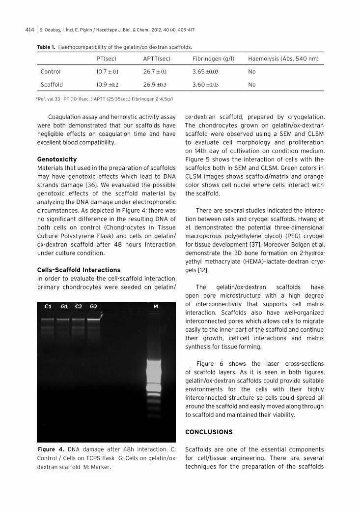
Haemocompatibility and HemolysisFibrinogen adsorption, activated partial thromboplastin time (APTT), prothrombin time (PT) tests were widely used coagulation assays to determine the haemocompatibility of materials [28-29]. Results summarized in Table 1 demonstrate the haemocompatibility of the scaffolds. Note that coagulation times give the information about the haemocompatibility through intrinsic and extrinsic coagulation pathways [30]. As seen in Table 1, scaffold has negligibly higher coagulation times from the control. All values were in the range of reference values. Fibrinogen adsorption onto biomaterials surface is a key factor to determine the haemocompatibility of the interested material because of its role in coagulation, and its ability to promote platelet adhesion [31-32].
The hemolytic activity of biomaterials plays an important role with regards to toxicity and correlates with inhibition of cell growth [34-35]. After 1 hour incubation at 37oC, scaffolds had the absorbance value of 0.044 ± 0.007 while the negative control (blood in PBS) was 0.040±0.005 and positive control (blood in distilled water) was 0.924±0.028. Thus, gelatin/ox-dextran scaffolds had shown no evidence of hemolysis after 1 h of incubation (<0.5% hemolysis).
Figure 2. Scanning Electron Micrograph of gelatin/ox-dextran scaffolds.
Figure 3. Cell Viabilities on gelatin/ox-dextran scaffold both direct and indirect MTT assay within 48h.
S. Odabaş, İ. İnci, E. Pişkin / Hacettepe J. Biol. & Chem., 2012, 40 (4), 409-417414
Coagulation assay and hemolytic activity assay were both demonstrated that our scaffolds have negligible effects on coagulation time and have excellent blood compatibility.
GenotoxicityMaterials that used in the preparation of scaffolds may have genotoxic effects which lead to DNA strands damage [36]. We evaluated the possible genotoxic effects of the scaffold material by analyzing the DNA damage under electrophoretic circumstances. As depicted in Figure 4; there was no significant difference in the resulting DNA of both cells on control (Chondrocytes in Tissue Culture Polystyrene Flask) and cells on gelatin/ox-dextran scaffold after 48 hours interaction under culture condition.
Cells-Scaffold Interactions In order to evaluate the cell-scaffold interaction, primary chondrocytes were seeded on gelatin/
ox-dextran scaffold, prepared by cryogelation. The chondrocytes grown on gelatin/ox-dextran scaffold were observed using a SEM and CLSM to evaluate cell morphology and proliferation on 14th day of cultivation on condition medium. Figure 5 shows the interaction of cells with the scaffolds both in SEM and CLSM. Green colors in CLSM images shows scaffold/matrix and orange color shows cell nuclei where cells interact with the scaffold.
There are several studies indicated the interac-tion between cells and cryogel scaffolds. Hwang et al. demonstrated the potential three-dimensional macroporous poly(ethylene glycol) (PEG) cryogel for tissue development [37]. Moreover Bolgen et al. demonstrate the 3D bone formation on 2-hydrox-yethyl methacrylate (HEMA)–lactate–dextran cryo-gels [12].
The gelatin/ox-dextran scaffolds have open pore microstructure with a high degree of interconnectivity that supports cell matrix interaction. Scaffolds also have well-organized interconnected pores which allows cells to migrate easily to the inner part of the scaffold and continue their growth, cell-cell interactions and matrix synthesis for tissue forming.
Figure 6 shows the laser cross-sections of scaffold layers. As it is seen in both figures, gelatin/ox-dextran scaffolds could provide suitable environments for the cells with their highly interconnected structure so cells could spread all around the scaffold and easily moved along through to scaffold and maintained their viability.
CONCLUSIONS
Scaffolds are one of the essential components for cell/tissue engineering. There are several techniques for the preparation of the scaffolds
Table 1. Haemocompatibility of the gelatin/ox-dextran scaffolds.
PT(sec) APTT(sec) Fibrinogen (g/l) Haemolysis (Abs. 540 nm)
Control 10.7 ± 0.1 26.7 ± 0.1 3.65 ±0.03 No
Scaffold 10.9 ±0.2 26.9 ±0.3 3.60 ±0.05 No
*Ref. val.33 PT (10-11sec. ) APTT (25-35sec.) Fibrinogen 2-4,5g/l
Figure 4. DNA damage after 48h interaction. C:
Control / Cells on TCPS flask G: Cells on gelatin/ox-
dextran scaffold M: Marker.

S. Odabaş, İ. İnci, E. Pişkin / Hacettepe J. Biol. & Chem., 2012, 40 (4), 409-417 415
Figure 5. Cells-Scaffold Interactions on (A-B) Confocal Laser Imaging Microscope (C) Scanning Electron Microscope.
Figure 6. Cells on gelatin/ox-dextran scaffold’s layers.

S. Odabaş, İ. İnci, E. Pişkin / Hacettepe J. Biol. & Chem., 2012, 40 (4), 409-417416
which let scaffolds having various different properties. Beside all these, biocompatibility is one of the vital points that a scaffold should hold.
In this current study we demonstrate a well-constructed cryogels as scaffold using gelatin and oxide dextran. Primary chondrocytes-scaffold interactions were evaluated by means of Scanning Electron Microscope and Confocal Microcopy analysis. Possible cytotoxicity, hemolytic activity and genotoxicity were also evaluated. According to our results, these cryogels shows excellent biocompatibility which can have promising candidate for tissue engineering.
ACKNOWLEDGEMENTS
This study was partially supported by EU:FP6-NoE: EXPERTISSUES Project and Hacettepe University Scientific Research and Development Project / BAP 0801602012.
Sedat Odabas is supported by TUBITAK (The Scientific and Technological Research Council of Turkey) as full PhD scholarship.
Prof. Dr. Erhan Piskin is supported as a full member of Turkish Academy of Sciences.
Authors thank to Miss A.Seda Yar for the genotoxicity assay.
R E F E R E N C E S
1. D.W. Hutmacher, Scaffolds in tissue engineering bone
and cartilage, Biomaterials, 21 (2000) 2529.
2. R.S, Langer, J.P. Vacanti, Tissue engineering: the
challenges ahead, Sci. Am., 280 (1999) 86.
3. R. Lanza, R.S. Langer, J.P. Vacanti, Principles of
Tissue Engineering Elsevier Academic Press, (1997)
ISBN 10: 0-12-370615-7.
4. S. Yang, K.F. Leong, Z. Du, C.K. Chua, The Design
of Scaffolds for Use in Tissue Engineering. Part I.
Traditional Factors, Tissue Eng., 7 (2001) 679.
5. L. Zhou, I. Pomerantseva, E.K. Bassett, C.M. Bowley,
X. Zhao, D.A, Bichara, K.M. Kulig, J.P. Vacanti, M.A.
Randolph, C.A. Sundback, Engineering ear constructs
with a composite scaffold to maintain dimensions,
Tissue Eng., Part A. 11-12 (2011) 1573.
6. Y. Wang, U.J, Kim, J.D. Blasioli, H.J. Kim, D.L. Kaplan,
In vitro cartilage tissue engineering with 3D porous
aqueous-derived silk scaffolds and mesenchymal
stem cells, Biomaterials, 26 (2005), 7082.
7. W. Xia, W. Liu, L. Cui, Y. Liu, W. Zhong, D. Liu, J. Wu, K.
Chua, Y. Cao, Tissue engineering of cartilage with the
use of chitosan-gelatin complex scaffolds, J. Biomed.
Res. Part B, 71B (2004) 373.
8. H.S. Yoo, E.A. Lee, J.J. Yoon, T.G. Park, Hyaluronic
acid modified biodegradable scaffolds for cartilage
tissue engineering, Biomaterials, 26 (2005) 1925.
9. A. Chetty, T. Steynberg, S. Moolman, R. Nilen,
A. Joubert, W. Richter, Hydroxyapatite-coated
polyurethane for auricular cartilage replacement: an
in vitro study, J. Biomed. Mater. Res. A, 84 (2005)
475.
10. N. Toyokawa, H. Fujioka, T. Kokubu, I. Nagura, A.
Inui, R. Sakata, M. Satake, H. Kaneko, M. Kurosaka,
Electrospun synthetic polymer scaffold for cartilage
repair without cultured cells in an animal model,
Arthroscopy, 26 (2010) 375.
11. Y. Liu, L. Zhang, G. Zhou, Q. Li, W. Liu, Z. Yu, X. Luo,
T. Jiang, W. Zhang, Y. Cao, In vitro engineering of
human ear-shaped cartilage assisted with CAD/CAM
technology, Biomaterials, 31 (2010) 2176.
12. N. Bolgen, Y. Yang, P. Korkusuz, E. Guzel, A.J. El Haj,
E. Piskin, Three-dimensional ingrowth of bone cells
within biodegradable cryogel scaffolds in bioreactors
at different regimes, Tissue Eng. Part A, 14 (2008)
1743.
13. A. Kumar, F.M. Plieva, I.Y. Galaev, B. Mattiasson,
Affinity fractionation of lymphocytes using a
monolithic cryogel, J. Immun. Meth., 283 (2003) 185.
14. A. Jungbauer, R. Hahn, Monoliths for fast
bioseparation and bioconversion and their
applications in biotechnology, J. Sep. Sci., 27 (2004)
767.
15. S. Nilsang, K.S. Nandakumar, I.Y. Galaev, S.K. Rakshit,
R. Holmdahl, B. Mattiasson, A. Kumar, Monoclonal
Antibody Production Using a New Supermacroporous
Cryogel Bioreactor, Biotech. Progr., 23 (2007) 932.
16. N. Bolgen, I. Vargel, P. Korkusuz, E. Guzel, F. Plieva,
I. Galaev, B. Matiasson, E. Piskin, Tissue responses
to novel tissue engineering biodegradable cryogel
scaffolds: An animal model, J. Biomed. Mat. Res.,
Part A. 91A (2009), 60.
17. I. Inci, H. Kirsebom, I.G. Galaev, B. Mattiasson, E. Piskin,
Gelatin cryogels cross-linked with oxidized dextran
and containing freshly formed hydroxyapatite as
potential bone tissue engineering scaffolds, J. Tissue
Eng. Regen. Med., (2012). İssue,page

S. Odabaş, İ. İnci, E. Pişkin / Hacettepe J. Biol. & Chem., 2012, 40 (4), 409-417 417
18. M. Jurga, M.B. Dainiak, A. Sarnowska, A. Jablonska,
A. Tripathi, F.M. Plieva, I.N. Savina, L. Strojek, H.
Jungvid, A. Kumar, B. Lukomska, K. Domanska-Janik,
N. Forraz, C.P. McGuckin, C.P. The performance of
laminin-containing cryogel scaffolds in neural tissue
regeneration, Biomaterials, 32 (2011) 3423.
19. S. Odabas, G. Feichtinger, P. Korkusuz, I. Inci, E.
Bilgic,A.S. Yar, T. Cavusoglu, S. Menevse, I. Vargel,
E. Piskin, Bmp-7 Expressing Genetically Modified
Primary Chondrocytes in Cryogel Scaffolds for
Rabbit Auricular Cartilage Repair, J. Tissue Eng.
Regen. Med., (2012). İssue,page
20. B. Garipcan, S. Odabas, G. Demirel, J. Burger, S.
Nonnenmann, T.M. Coster, M.E. Gallo, B. Nabet, J.E.
Spanier, E. Piskin, In vitro Biocompatibility of n-type
and Undoped Silicon Nanowires, Adv. Biomaterials, 13
(2011) B3.
21. H. Zhao, L. Ma, Y. Gong, C. Gao, J. Shen, A polylactide/
fibrin gel composite scaffold for cartilage tissue
engineering: fabrication and an in vitro evaluation, J.
Mater. Sci. Mater. Med., 20 (2009) 135.
22. F.C.R. Manning, L.J. Blankenship, J.R.P. Wise, J.
Xu, L.C. Bridgewater, S.R. Patierno, Induction
of Internucleosomal DNA Fragmentation by
Carcinogenic Chromate: Relationship to DNA Damage,
Genotoxicity, and Inhibition of Macromolecular
Synthesis Environ, Environ. Health Perspect., 102
(1994) 159.
23. H.W. Kang, Y. Tabata, Y. Ikada, Fabrication of porous
gelatin scaffolds for tissue engineering, Biomaterials,
14 (1999) 1339.
24. S.G. Lévesque, M.S. Shoichet, Synthesis of cell-
adhesive dextran hydrogels and macroporous
scaffolds, Biomaterials, 30 (2006) 5277.
25. E. Zeiger, B. Gollapudi, P. Spencer, Genetic toxicity
and carcinogenicity studies of glutaraldehyde--a
review, Mutat. Res., 2 (2005) 136.
26. S.H. Oh, I.K. Park, J.M. Kim, J.H. Lee, In vitro and
in vivo characteristics of PCL scaffolds with pore
size gradient fabricated by a centrifugation method,
Biomaterials, 9 (2007) 1664.
27. B. Yang, Z. Yin, J. Cao, Z. Shi, Z. Zhang, H. Song, F. Liu,
B. Caterson, In vitro cartilage tissue engineering using
cancellous bone matrix gelatin as a biodegradable
scaffold, Biomed. Mater, 4 (2010), 045003.
28. F. Khan, M. Snyder, L. Pechet, The Laboratory
of Coagulation A review of present laboratory
techniques, J. Thromb. Thrombolysis, 5 (1998) 83.
29. S. Murugesan, T.J. Park, H. Yang, S. Mousa, R.J.
Linhardt, Blood compatible carbon nanotubes--nano-
based neoproteoglycans, Langmuir, 22 (2006) 3461.
30. A. Noyan, Physiology in Life and Medicine, (2004)
Meteksan Press, 975774610X, 458.
31. A.K. Bajpai, Fibrinogen adsorption onto macroporous
polymeric surfaces: correlation with biocompatibility
aspects, J. Mater. Sci. Mater. Me, 19 (2008) 343.
32. W.B. Tsai, J.M. Grunkemeier, T.A. Horbett, Variations
in the ability of adsorbed fibrinogen to mediate
platelet adhesion to polystyrene-based materials: a
multivariate statistical analysis of antibody binding
to the platelet binding sites of fibrinogen, J. Biomed.
Mater. Res. A, 67 (2003) 1255.
33. Duzen Laboratory Group. Laboratory Tests Booklet,
(2011) ISBN : 9789944587617.
34. G. Wang, Y. Shen, Y. Y. Cao, Q. Yu, R. Guidoin,
Biocompatibility study of plasma-coated nitinol (NiTi
alloy) stents, IET Nanobiotechnol, 1 (2007) 102.
35. J.K. Unger, C. Haltern, B. Dohmen, A. Gressner,
C. Grosse-Siestrup, D.A. Groneberg, R. Rossaint,
Albumin and hydroxyethyl starch 130 kDa/0.4
improve filter clearance and haemocompatibility in
haemo and plasmafiltration an in vitro study, Nephrol.
Dial. Transplant, 20 (2005) 1922.
36. A.S. Lotza, J.B. Havlaa, E. Richtera, K. Frölichb,
R. Staudenmaierc, R. Hagenb, N.H. Kleinsasserb,
Cytotoxic and genotoxic effects of matrices for
cartilage tissue engineering, Toxic. Lett., 190 (2009)
128.
37. Y. Hwang, C. Zhang, S.Varghese, Poly(ethylene
glycol) cryogels as potential cell scaffolds: effect of
polymerization conditions on cryogel microstructure
and properties, J. Mater. Chem., 20 (2010) 345.