general ent “emergencies” - peripheral … of bone into cranial vault with spread of infection...
TRANSCRIPT
GENERAL ENT “EMERGENCIES”
- ENT for A&E
SUREN KRISHNAN OAM, FRACS, MBBS (Adelaide)
CLINICAL ASSOCIATE PROFESSOR OF SURGERY
UNIVERSITY OF ADELAIDE
CHAIRMAN
DEPARTMENT OF OTOLARYNGOLOGY, HEAD & NECK SURGERY
ROYAL ADELAIDE HOSPITAL
VISITING PROFESSOR OF HEAD & NECK SURGERY
SUN YAT SEN UNIVERSITY, GUANGZHOU, CHINA
Spring Seminar on Emergency Medicine
Barossa Valley SA
Sep 29, 2015 - Oct 2, 2015
DEPARTMENT OF OTOLARYNGOLOGY,
HEAD AND NECK SURGERY
ROYAL ADELAIDE HOSPITAL • THE CONTRIBUTORS
– ANSTEY GILES 1920s
– THE LATE JEFF ROZENBILDS
– THE LATE P.V. RAJAGOPALAN
– RON GRISTWOOD
– DAN HAINS
– DARCY ECONOMOS
– MICHAEL JAY
• THE CURRENT DEPARTMENT
– PROFESSOR P.J. WORMALD
– GUY REES
– MICHAEL SCHULTZ
– SAM ARENA
– STEVE FLOREANI
– MICHAEL SWITAJEWSKI
– ALETHEA GROBLER
– J-C HODGE
– S. CHAWLA
– R. VALENTINE
– A. FOREMAN
THE ROYAL ADELAIDE HOSPITAL
ADELAIDE SOUTH AUSTRALIA

Overview • INFECTIONS- Present with Acute Pain, Fever, Discharge & Dysfunction
• NEOPLASMS- Beware Unilateral Symptoms and Signs, Chronic Pain
• Otologic Disorders – Hearing Loss
– Pain
– Disorder of balance
• Nasal Disorders – Nasal Obstruction
– Facial Pain / Disorder of sense of smell
– Bleeding
• Facial, Oral and Pharyngeal Disorders – Facial muscular dysfunction
– Dysphonia / Dysphagia
– Airway

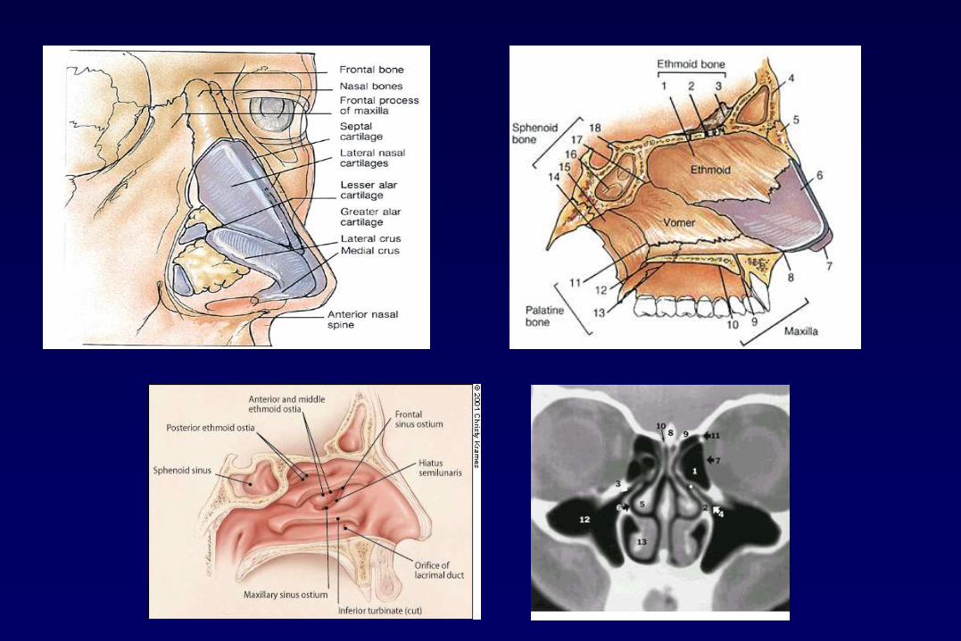
Otologic Disorders Anatomy
• Pinna
• Ear canal
• Tympanic membrane
• Middle ear and mastoid
• Inner Ear
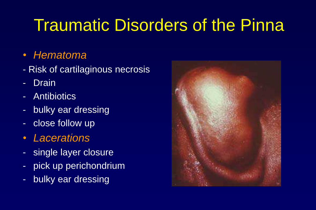
Traumatic Disorders of the Pinna
• Hematoma
- Risk of cartilaginous necrosis
- Drain
- Antibiotics
- bulky ear dressing
- close follow up
• Lacerations
- single layer closure
- pick up perichondrium
- bulky ear dressing
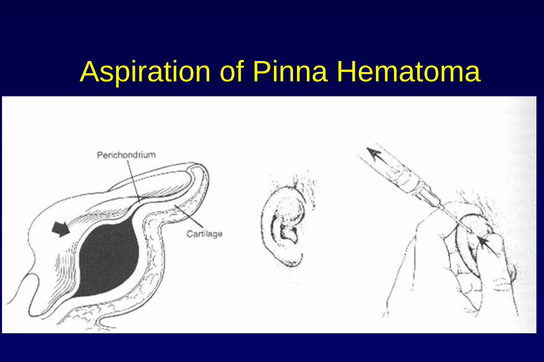
Aspiration of Pinna Hematoma
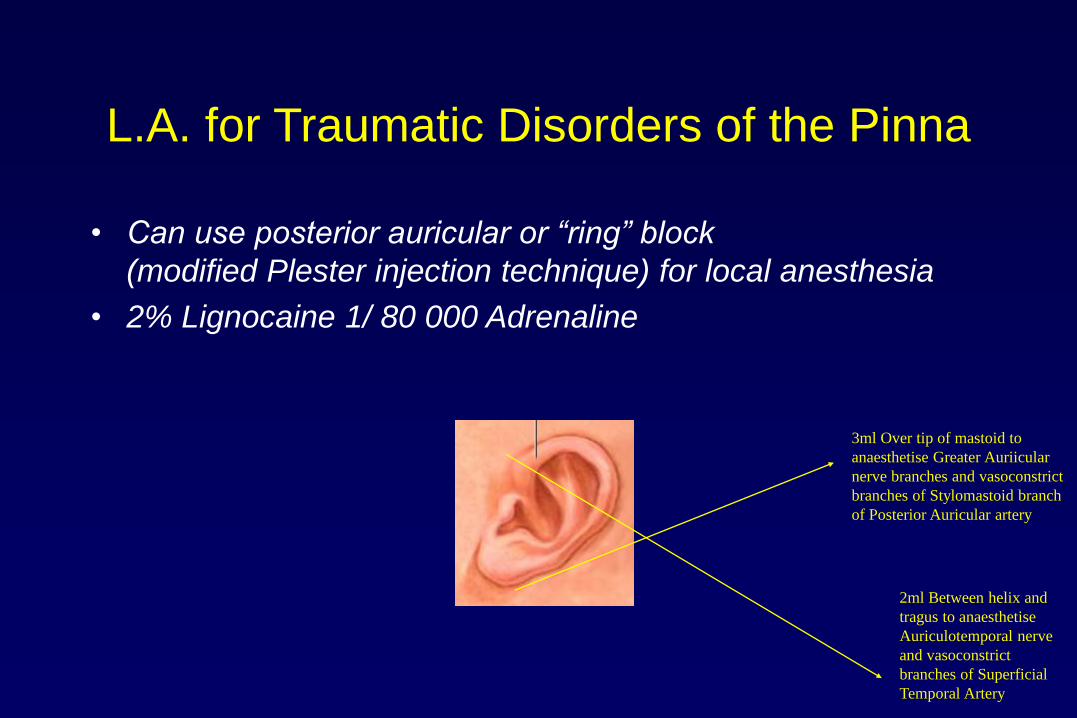
L.A. for Traumatic Disorders of the Pinna
• Can use posterior auricular or “ring” block
(modified Plester injection technique) for local anesthesia
• 2% Lignocaine 1/ 80 000 Adrenaline
3ml Over tip of mastoid to
anaesthetise Greater Auriicular
nerve branches and vasoconstrict
branches of Stylomastoid branch
of Posterior Auricular artery
2ml Between helix and
tragus to anaesthetise
Auriculotemporal nerve
and vasoconstrict
branches of Superficial
Temporal Artery
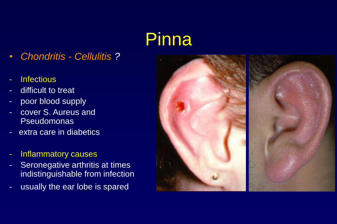
Pinna • Chondritis - Cellulitis ?
- Infectious
- difficult to treat
- poor blood supply
- cover S. Aureus and Pseudomonas
- extra care in diabetics
- Inflammatory causes
- Seronegative arthritis at times indistinguishable from infection
- usually the ear lobe is spared
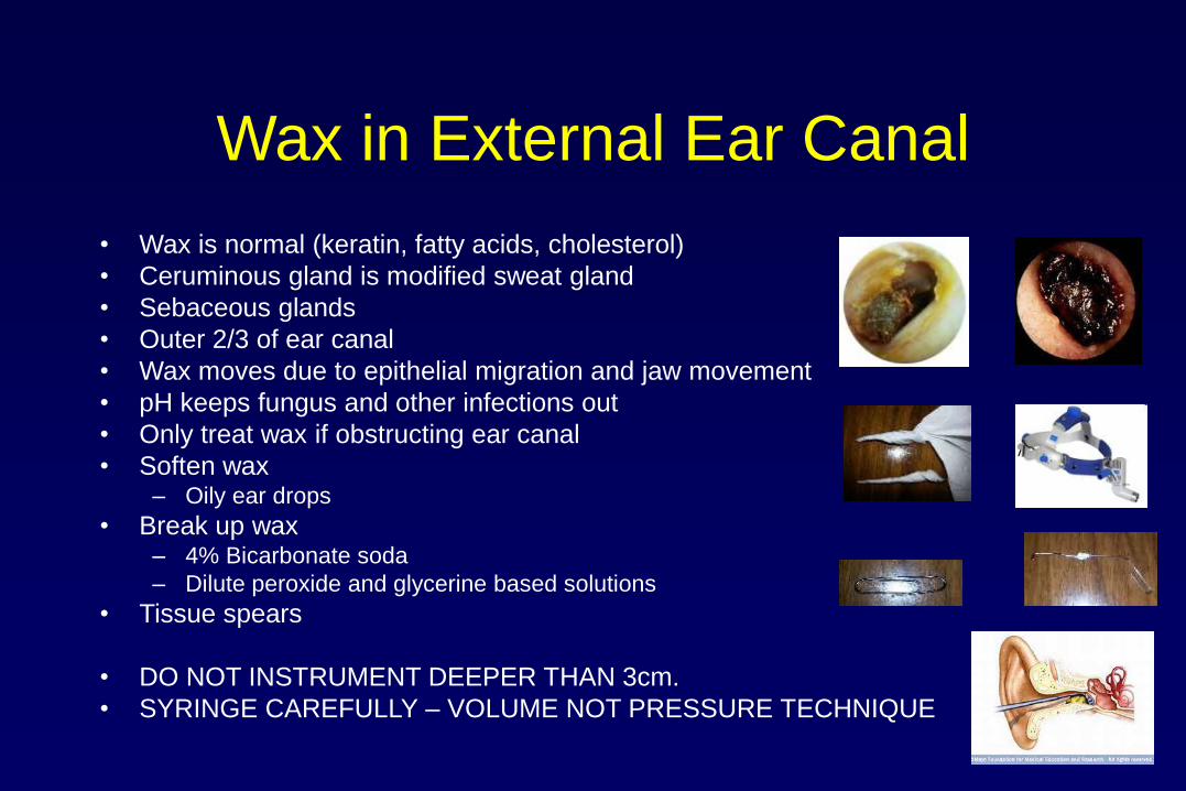
Wax in External Ear Canal
• Wax is normal (keratin, fatty acids, cholesterol)
• Ceruminous gland is modified sweat gland
• Sebaceous glands
• Outer 2/3 of ear canal
• Wax moves due to epithelial migration and jaw movement
• pH keeps fungus and other infections out
• Only treat wax if obstructing ear canal
• Soften wax – Oily ear drops
• Break up wax – 4% Bicarbonate soda
– Dilute peroxide and glycerine based solutions
• Tissue spears
• DO NOT INSTRUMENT DEEPER THAN 3cm.
• SYRINGE CAREFULLY – VOLUME NOT PRESSURE TECHNIQUE

Otitis Externa • Presentation
– Pain / Tenderness
– Scant discharge
• Types
– Viral (vesicles)
– Bacterial
– Fungal
• Treatment
– Ear Toilet
– Powder
– Topical Antibiotic Drops
– Analgaesia
– Water precautions
– ? Systemic Antibiotics
• Malignant O.E.
– Skullbase osteomyelitis
– Immunosuppressed / Diabetics
– Fulminant / lethal
– Pseudomonas
– Intravenous & Long term antibiotics
BACTERIAL O.E.
FUNGAL O.E.

Foreign Bodies in Ear Canal
• Usually put in by patient
• Some bugs fly / crawl in
• Kill bugs with mineral oil, or lignocaine
• remove with micro-forceps, suction or tissue adhesive
• “Lever” / “Slide & Retrieve” technique with right angle probe
• Preferably use microscope
• Beware disc / cell batteries
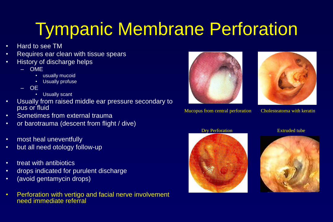
Tympanic Membrane Perforation • Hard to see TM
• Requires ear clean with tissue spears
• History of discharge helps – OME
• usually mucoid
• Usually profuse
– OE • Usually scant
• Usually from raised middle ear pressure secondary to pus or fluid
• Sometimes from external trauma
• or barotrauma (descent from flight / dive)
• most heal uneventfully
• but all need otology follow-up
• treat with antibiotics
• drops indicated for purulent discharge
• (avoid gentamycin drops)
• Perforation with vertigo and facial nerve involvement need immediate referral
Mucopus from central perforation Cholesteatoma with keratin
Dry Perforation Extruded tube
Otitis Media • Acute Otitis Media
– Sequelae • May get better
• May get worse (Chronic Otitis Media)
– Complications • Mastoiditis
• Chronic Otitis Media (Otitis Media with Effusion)
(Chronic Suppurative Otitis Media)
– Sequelae • May get better
• May get worse (Chronic change to ear drum +/- ME contents)
– Complications • Cholesteatoma
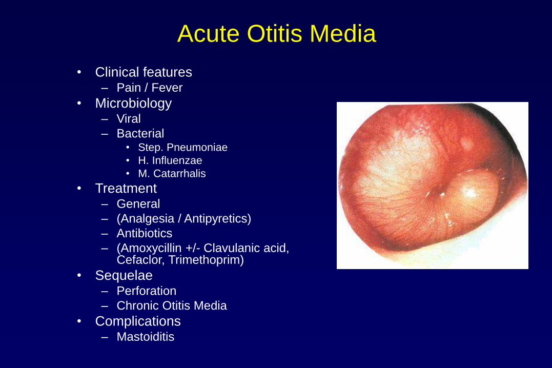
Acute Otitis Media • Clinical features
– Pain / Fever
• Microbiology – Viral
– Bacterial • Step. Pneumoniae
• H. Influenzae
• M. Catarrhalis
• Treatment – General
– (Analgesia / Antipyretics)
– Antibiotics
– (Amoxycillin +/- Clavulanic acid, Cefaclor, Trimethoprim)
• Sequelae – Perforation
– Chronic Otitis Media
• Complications – Mastoiditis
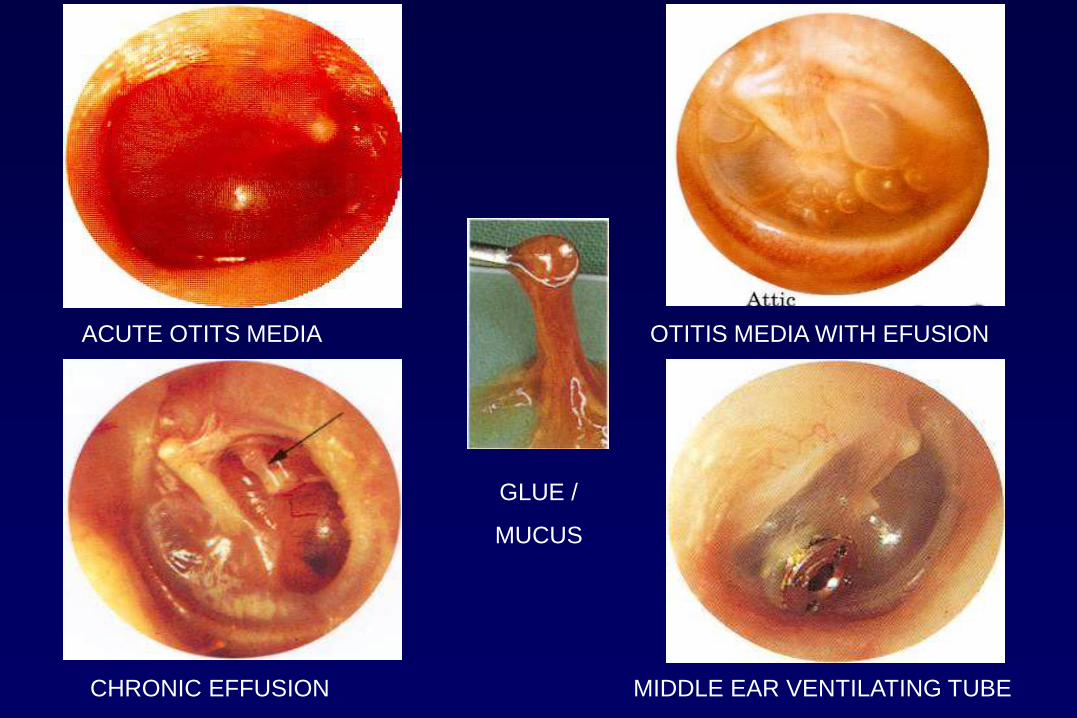
ACUTE OTITS MEDIA OTITIS MEDIA WITH EFUSION
GLUE /
MUCUS
CHRONIC EFFUSION MIDDLE EAR VENTILATING TUBE

Mastoiditis
• Uncommon
• Always has TM signs
• Treatment – Intravenous antibiotics
– Timentin / Chloramphenicol
– Incision & Drainage
– Wide Myringotomy
– Cortical Mastoidectomy
• Complications – Intracranial
• Sigmoid Sinus Thrombosis / thrombophlebitis
• Meningitiis / Cerebritis
• Extradural / Sudural / Cerebral abscess
– Death
Chronic Otitis Media • OTITIS MEDIA WITH EFFUSION
• TYMPANOSCLEROSIS
• CHRONIC TYMPANIC MEMBRANE PERFORATION
• Chronic Suppurative Otitis Media
• Inactive vs Active
• Mucous D/C vs Scant, Malodorous D/C
• Central vs Marginal perforation
• Tubo-tympanic vs Attico-antral
• Safe perforations vs Cholesteatoma
• Worrying symptoms and signs
– Pain
– Facial palsy
– Vertigo
– Sensorineural hearing loss
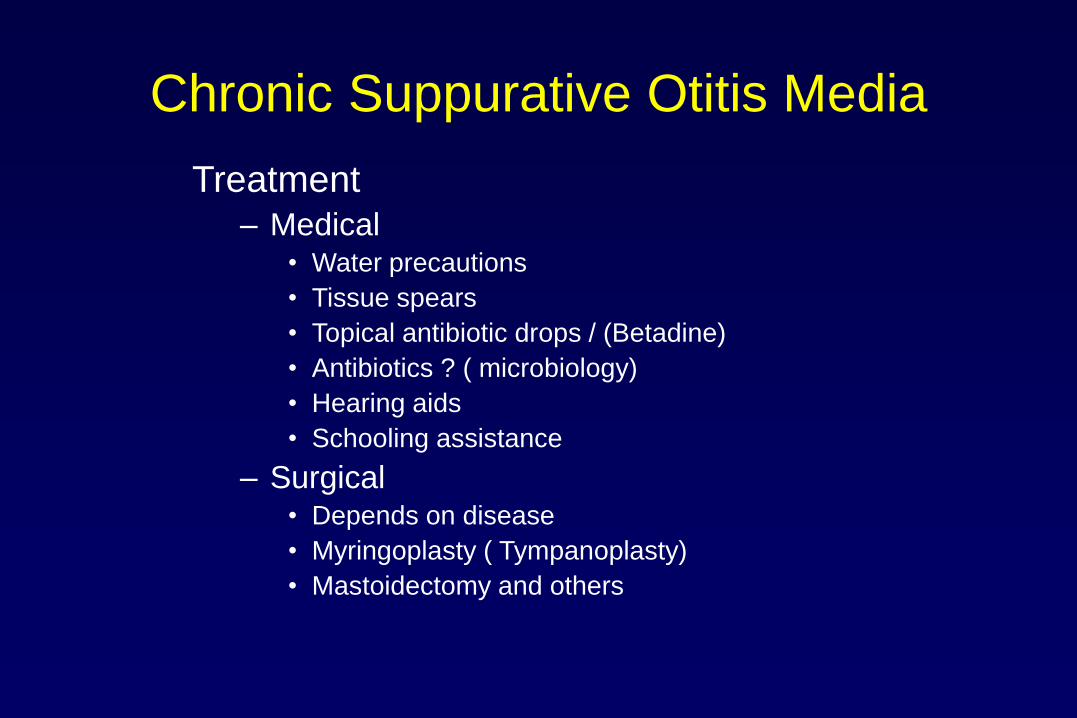
Chronic Suppurative Otitis Media
Treatment – Medical
• Water precautions
• Tissue spears
• Topical antibiotic drops / (Betadine)
• Antibiotics ? ( microbiology)
• Hearing aids
• Schooling assistance
– Surgical • Depends on disease
• Myringoplasty ( Tympanoplasty)
• Mastoidectomy and others
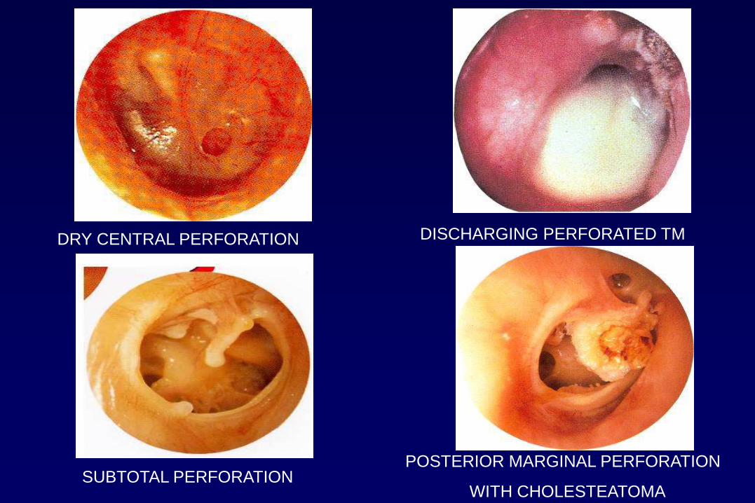
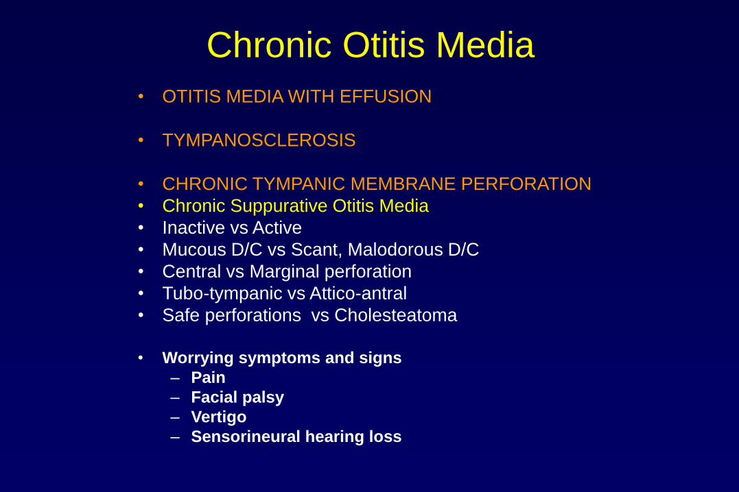
DRY CENTRAL PERFORATION DISCHARGING PERFORATED TM
POSTERIOR MARGINAL PERFORATION
WITH CHOLESTEATOMA SUBTOTAL PERFORATION
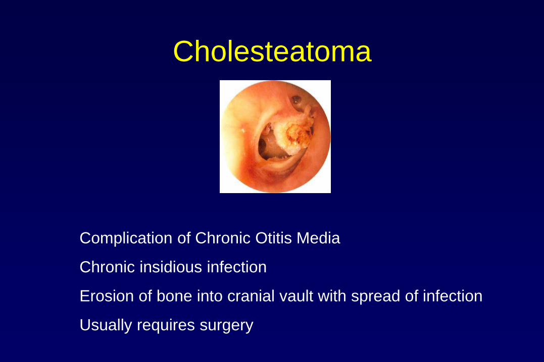
Cholesteatoma
Complication of Chronic Otitis Media
Chronic insidious infection
Erosion of bone into cranial vault with spread of infection
Usually requires surgery
Sudden Sensori-Neural Hearing Loss
• Medical Emergency of the Ear
• Sudden Unilateral SNHL (Tuning Forks)
• +/- Vertigo
• +/- Tinnitus
• Admit – RIB, Steroids, Carbogen, Vasodilators
• Investigate – Audiometry, CBE, ESR, Lipids, TFT, VDRL
– MRI to exclude Acoustic Neuroma and CNS anomalies eg MS
Vertigo • V -Vascular (carotid & v-b)
• E -”Epilepsy” (CNS e.g. TIA, CVA, Migraine, MS)
• R -Drug Therapy
• Aminoglycosides, Anti-malarials (Quinine)
• Beware of topical drops & perforated ear drums
• Chemotherapy
• Diuretic
• T -Trauma (CHI, Skull #, PLF, Baric, ?Acoustic)
• I -Infection (STORCH)
• G -General
• Arthritis of the Neck
• BP – high or low ,
• Cardiac – WPW, Stoke-Adams,
• Diabetes
• O -Otologic
• A - Acoustic neuroma
• B – BPPV (cupulo-lithiasis)
• C – Cholesteatoma
• D – Dehiscence of Semi Canals (Superior)
• E – Endolymphatic Hydrops (Menieres Disease)
• V – Vestibular Neuronitis
Inner Ear – Common causes of Dysequilibrium,
Acute Peripheral vertigo, vestibulopathy
– BPPV (Benign Paroxysmal Positioning Vertigo)
• Treatment = Epley’s Particle Repositioning Manoeuver
– “Labyrinthitis” / “Vestibular Neuronitis”
• acute onset
• no central signs
• usually young
• horizontal nystagmus
• Treatment = Vestibular Sedatives eg SERC, Stemetil, Valium, fluids, rest,
– Meniere’s Disease - vertigo, sensorineural hearing loss, tinnitus, aural fullness
• Treatment = Vestibular Sedatives eg SERC, Stemetil, Valium, fluids, rest,
severe cases may need surgery
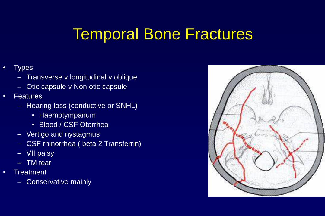
Temporal Bone Fractures
• Types
– Transverse v longitudinal v oblique
– Otic capsule v Non otic capsule
• Features
– Hearing loss (conductive or SNHL)
• Haemotympanum
• Blood / CSF Otorrhea
– Vertigo and nystagmus
– CSF rhinorrhea ( beta 2 Transferrin)
– VII palsy
– TM tear
• Treatment
– Conservative mainly

Referred Otalgia
• Cervical Spondylitis
• Teeth
• Tonsil
• Retro Molar Trigone
• Vallecula
• Pyriform Fossa
• Post Cricoid Hypopharynx
• Larynx

Referred Otalgia
- Acute on Chronic Ear Pain
• TEMPORO MANDIBULAR JOINT DYSFUNCTION
• COSTEN’S SYNDROME
• Pain
• Tinnitus
• Altered hearing
• Altered balance
• Malocclussion
• Tender muscles of mastication
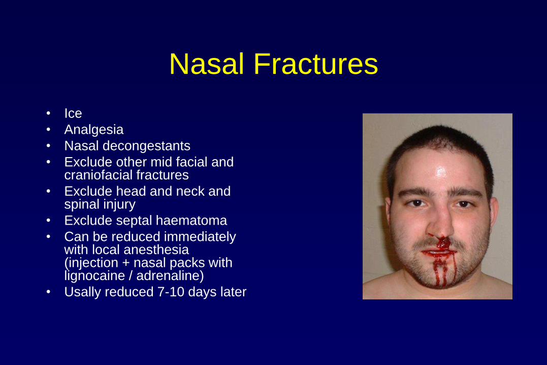
Nasal Fractures
• Ice
• Analgesia
• Nasal decongestants
• Exclude other mid facial and craniofacial fractures
• Exclude head and neck and spinal injury
• Exclude septal haematoma
• Can be reduced immediately with local anesthesia (injection + nasal packs with lignocaine / adrenaline)
• Usally reduced 7-10 days later

EPISTAXES
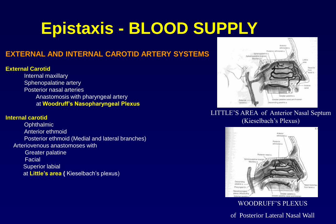
Epistaxis - BLOOD SUPPLY
EXTERNAL AND INTERNAL CAROTID ARTERY SYSTEMS
External Carotid
Internal maxillary
Sphenopalatine artery
Posterior nasal arteries
Anastomosis with pharyngeal artery
at Woodruff’s Nasopharyngeal Plexus
Internal carotid
Ophthalmic
Anterior ethmoid
Posterior ethmoid (Medial and lateral branches)
Arteriovenous anastomoses with
Greater palatine
Facial
Superior labial
at Little’s area ( Kieselbach’s plexus)
LITTLE’S AREA of Anterior Nasal Septum
(Kieselbach’s Plexus)
WOODRUFF’S PLEXUS
of Posterior Lateral Nasal Wall

Epistaxis - EPIDEMIOLOGY
• More common in young and elderly
• The Elderly – A&E attendances more in elderly
– episodes arterial
– last longer due to vessel changes
– More common in colder months, low humidity
• The Young – Local trauma
– More common in warmer months, low humidity
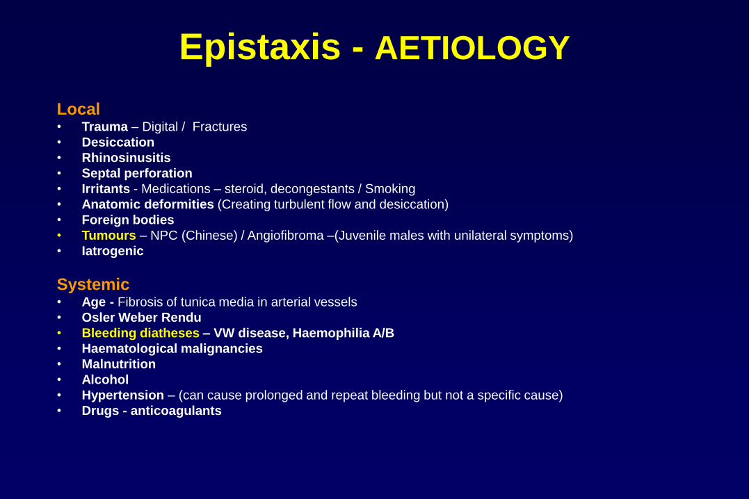
Epistaxis - AETIOLOGY
Local • Trauma – Digital / Fractures
• Desiccation
• Rhinosinusitis
• Septal perforation
• Irritants - Medications – steroid, decongestants / Smoking
• Anatomic deformities (Creating turbulent flow and desiccation)
• Foreign bodies
• Tumours – NPC (Chinese) / Angiofibroma –(Juvenile males with unilateral symptoms)
• Iatrogenic
Systemic • Age - Fibrosis of tunica media in arterial vessels
• Osler Weber Rendu
• Bleeding diatheses – VW disease, Haemophilia A/B
• Haematological malignancies
• Malnutrition
• Alcohol
• Hypertension – (can cause prolonged and repeat bleeding but not a specific cause)
• Drugs - anticoagulants

Epistaxis - Anterior
• 90% of all epistaxis
• Little’s Area (Kisselbach’s plexus)
• usually children, young adults
Etiologies
• Trauma (epistaxis digitorum)
• Summer (Dessication of mucosa)
• Allergies
• Irritants - cocaine, sprays
• Pregnancy / pre-pubertal / elderly

Epistaxis - Posterior
• 10% of all epistaxis
• usually in the elderly
Etiologies
• Winter months and summer from mucosal dryness
• Coagulopathy
• Atherosclerosis
• Neoplasm
• Hypertension (debatable)
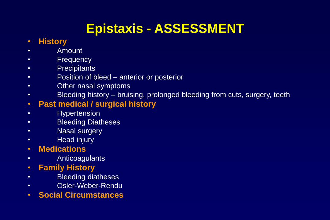
Epistaxis - ASSESSMENT
• History
• Amount
• Frequency
• Precipitants
• Position of bleed – anterior or posterior
• Other nasal symptoms
• Bleeding history – bruising, prolonged bleeding from cuts, surgery, teeth
• Past medical / surgical history • Hypertension
• Bleeding Diatheses
• Nasal surgery
• Head injury
• Medications • Anticoagulants
• Family History • Bleeding diatheses
• Osler-Weber-Rendu
• Social Circumstances
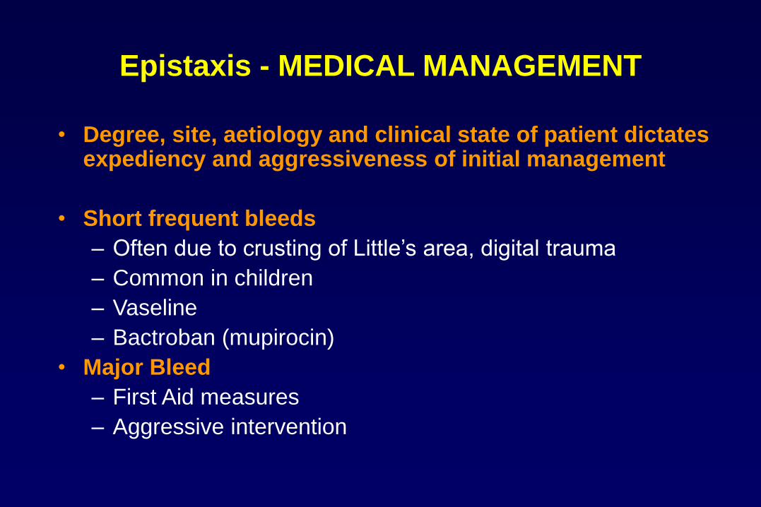
Epistaxis - MEDICAL MANAGEMENT
• Degree, site, aetiology and clinical state of patient dictates expediency and aggressiveness of initial management
• Short frequent bleeds
– Often due to crusting of Little’s area, digital trauma
– Common in children
– Vaseline
– Bactroban (mupirocin)
• Major Bleed
– First Aid measures
– Aggressive intervention
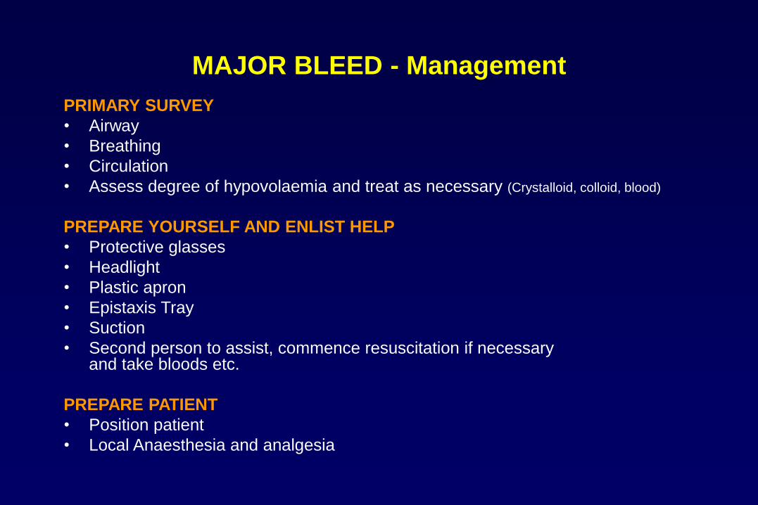
MAJOR BLEED - Management
PRIMARY SURVEY
• Airway
• Breathing
• Circulation
• Assess degree of hypovolaemia and treat as necessary (Crystalloid, colloid, blood)
PREPARE YOURSELF AND ENLIST HELP
• Protective glasses
• Headlight
• Plastic apron
• Epistaxis Tray
• Suction
• Second person to assist, commence resuscitation if necessary and take bloods etc.
PREPARE PATIENT
• Position patient
• Local Anaesthesia and analgesia
MAJOR BLEED - Management MAIN AIM
• to locate offending vessel and cauterise,
• pack if bleeding profuse despite attempts or unable to locate vessel and still bleeding
• An idea of probable location of bleeding important as treatment different
• (Anterior vs Posterior, Above vs Below Middle Turbinate)
LOOK
• Nose –Killian’s or Thudicum’s
• Oropharynx- Tongue depressor
CLEAR NOSE
• Patient blows nose to evacuate clot and to restart bleeding,
• residual clot removed with Frazier sucker
• oropharyngeal clots removed with Yankauer
DECONGEST AND ANAESTHETISE NOSE
• Allows better view into nose
• Vast majority of bleeds occur in Little’s area (anterior septum) and can be very anterior
• Posterior bleeds from posterior septum, posterior lateral nasal wall at Woodruff’s plexus
• Bleeding from above Middle Turbinate suspicious of Anterior Ethmoid Artery Bleed
• Cophenylcaine Forte spray on cotton wool pack
• Cocaine 10 % solution on pledget
• Oxymetazoline 0.05%
• Ephedrine drops 0.5%
• Place pledget directly over bleeder if it can be seen
• If still bleeding infiltrate by injection in submucoperichondrial plane into caudal border of septum or around bleeder with lignocaine 1% and adrenaline 1: 80k with 27G needle
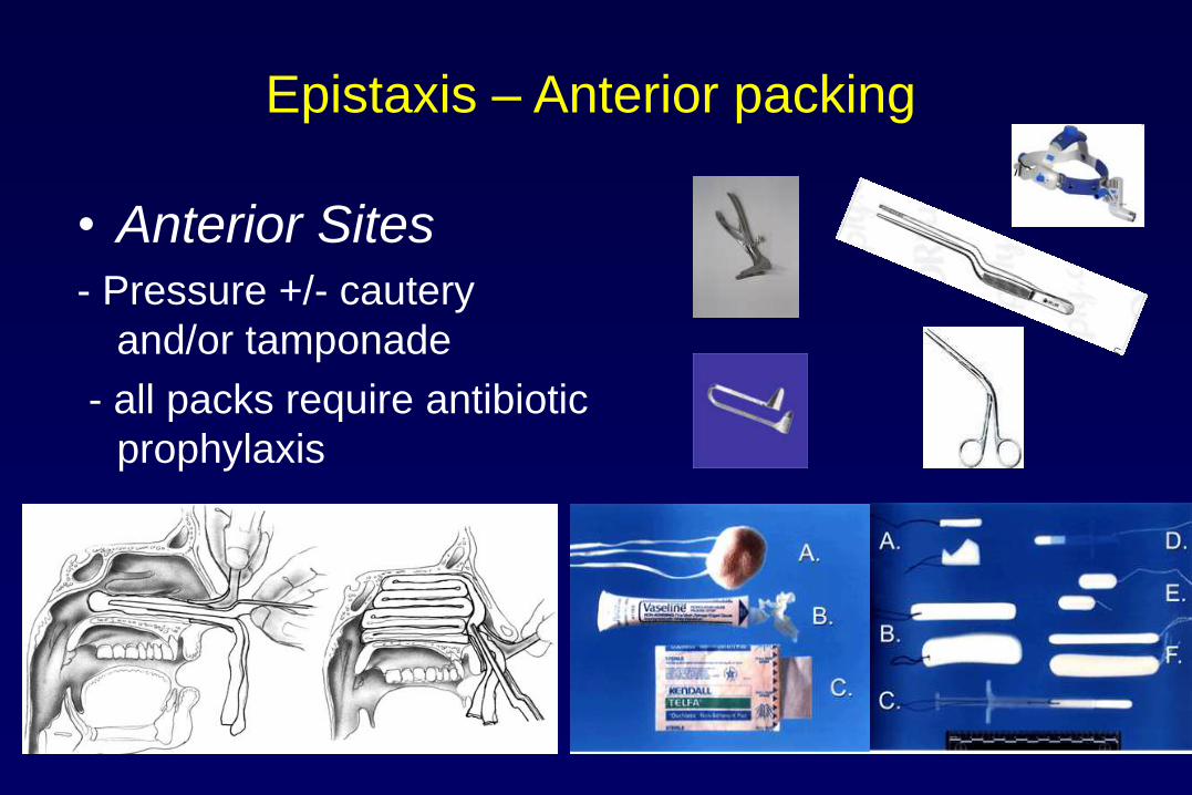
Epistaxis – Anterior packing
• Anterior Sites - Pressure +/- cautery
and/or tamponade
- all packs require antibiotic
prophylaxis
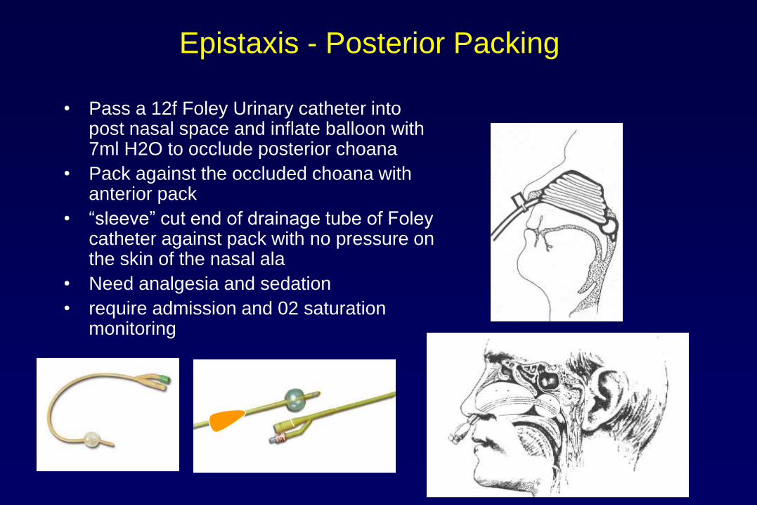
Epistaxis - Posterior Packing
• Pass a 12f Foley Urinary catheter into post nasal space and inflate balloon with 7ml H2O to occlude posterior choana
• Pack against the occluded choana with anterior pack
• “sleeve” cut end of drainage tube of Foley catheter against pack with no pressure on the skin of the nasal ala
• Need analgesia and sedation
• require admission and 02 saturation monitoring
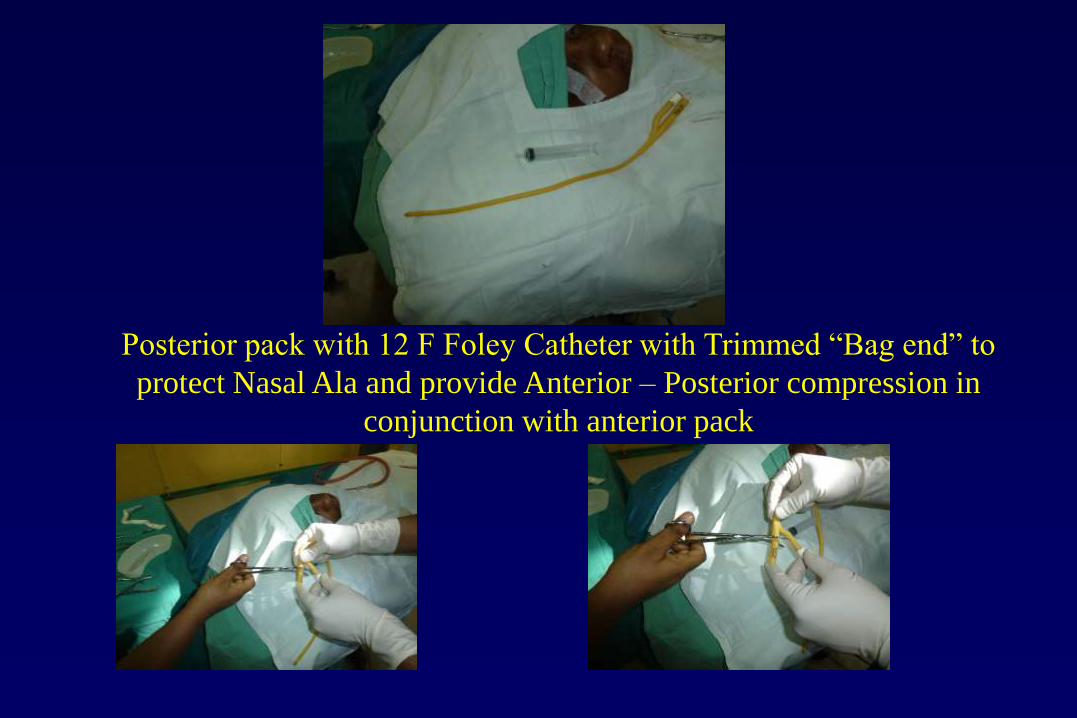
Posterior pack with 12 F Foley Catheter with Trimmed “Bag end” to
protect Nasal Ala and provide Anterior – Posterior compression in
conjunction with anterior pack
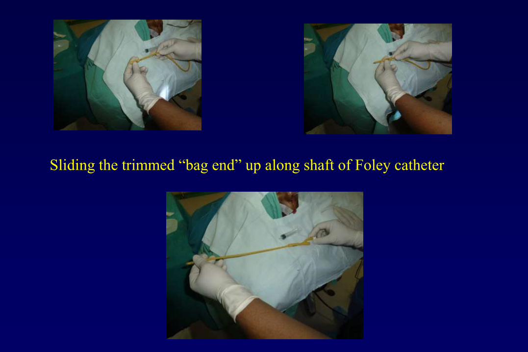
Sliding the trimmed “bag end” up along shaft of Foley catheter

Inserting Foley catheter through posterior choana into post nasal space
Inflating balloon with 7ml of saline
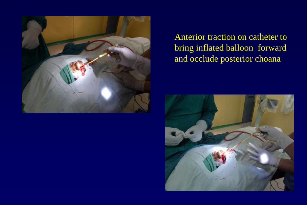
Anterior traction on catheter to
bring inflated balloon forward
and occlude posterior choana
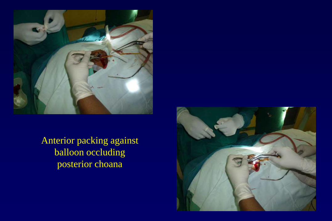
Anterior packing against
balloon occluding
posterior choana
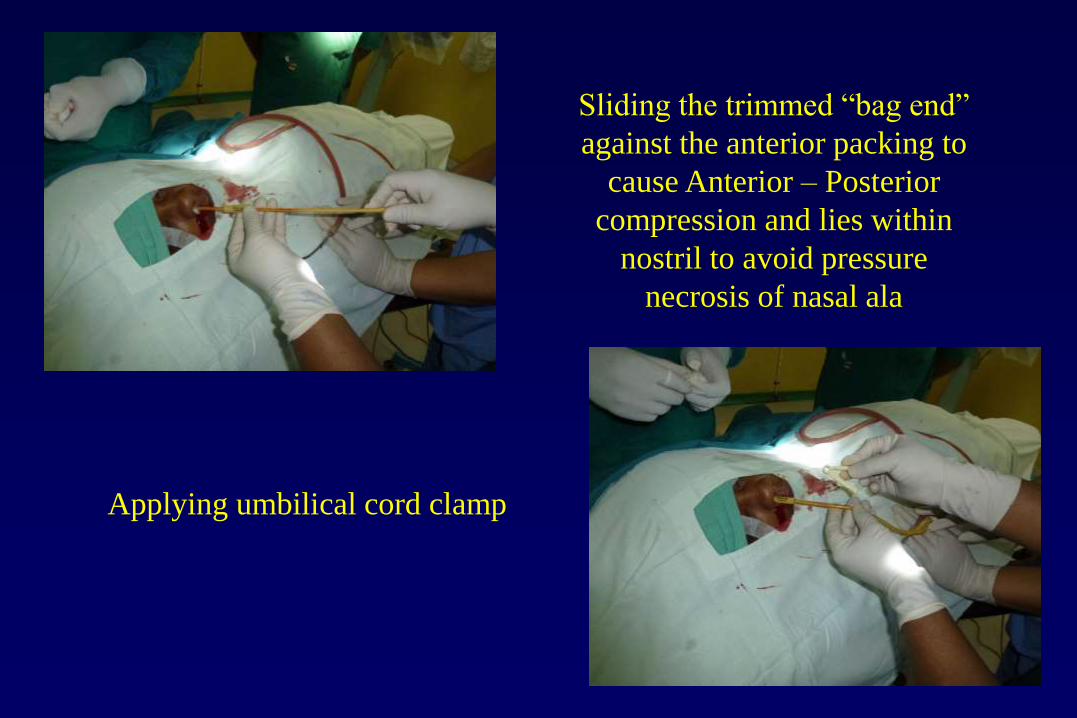
Sliding the trimmed “bag end”
against the anterior packing to
cause Anterior – Posterior
compression and lies within
nostril to avoid pressure
necrosis of nasal ala
Applying umbilical cord clamp

Applied umbilical cord clamp

Epistaxis - Complications
• severe bleeding – life threatening haemorrhage
• hypoxia, hypercarbia
• sinusitis, otitis media
• necrosis of the columella or nasal ala
MAJOR BLEED not settling with packing -
Management
• EMBOLISATION
• LIGATION OF SPHENO-PALATINE ARTERY – ENDOSCOPIC TRANS NASAL APPROACH
• LIGATION OF INTERNAL MAXILLARY ARTERY – TRANS ANTRAL ENDOSCOPE ASSISTED
• LIGATION OF EXTERNAL CAROTID ARTERY – SKIN CREASE INCISION 2 FINGER BREADTHS BELOW MANDIBLE
– RETRACT SUBMANDIBULAR GLAND ANTERIORLY
– RETRACT STERNO-MASTOID MUSCLE POSTERIORLY
– COMMON CAROTID ARTERY AND BIFURCATION
– IDENTIFY EXTERNAL CAROTID ARTERY
– CONFIRM BY IDENTIFYING AT LEAST THREE BRANCHES
– LIGATE ABOVE FACIAL ARTERY TAKE OFF
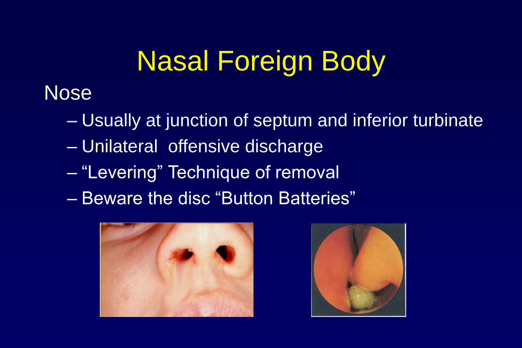
Nasal Foreign Body Nose
– Usually at junction of septum and inferior turbinate
– Unilateral offensive discharge
– “Levering” Technique of removal
– Beware the disc “Button Batteries”
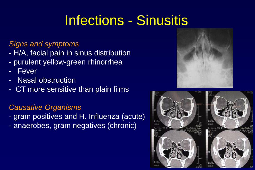
Infections - Sinusitis
Signs and symptoms
- H/A, facial pain in sinus distribution
- purulent yellow-green rhinorrhea
- Fever
- Nasal obstruction
- CT more sensitive than plain films
Causative Organisms
- gram positives and H. Influenza (acute)
- anaerobes, gram negatives (chronic)
Infections - Sinusitis Treatment
acute - amoxil
chronic - amoxil-clavulinic acid, clindamycin, quinolones
Supportive - decongestants, saline sprays, steroid nasal sprays, analgesia, ice and heat packs
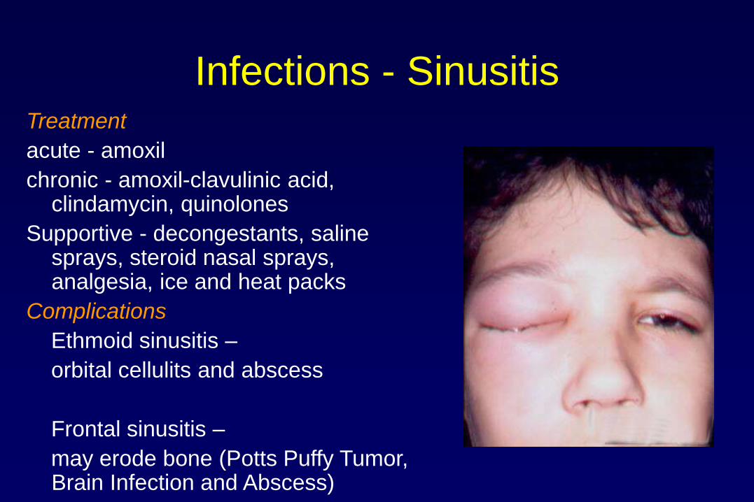
Complications
Ethmoid sinusitis –
orbital cellulits and abscess
Frontal sinusitis –
may erode bone (Potts Puffy Tumor, Brain Infection and Abscess)

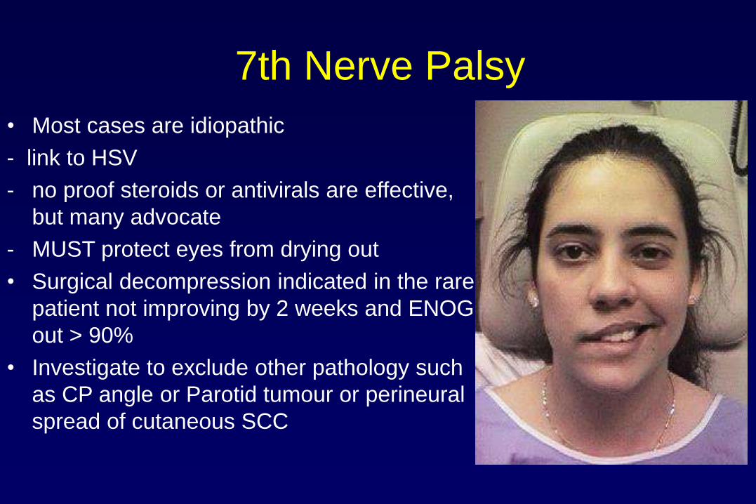
7th Nerve Palsy
• Most cases are idiopathic
- link to HSV
- no proof steroids or antivirals are effective,
but many advocate
- MUST protect eyes from drying out
• Surgical decompression indicated in the rare
patient not improving by 2 weeks and ENOG
out > 90%
• Investigate to exclude other pathology such
as CP angle or Parotid tumour or perineural
spread of cutaneous SCC
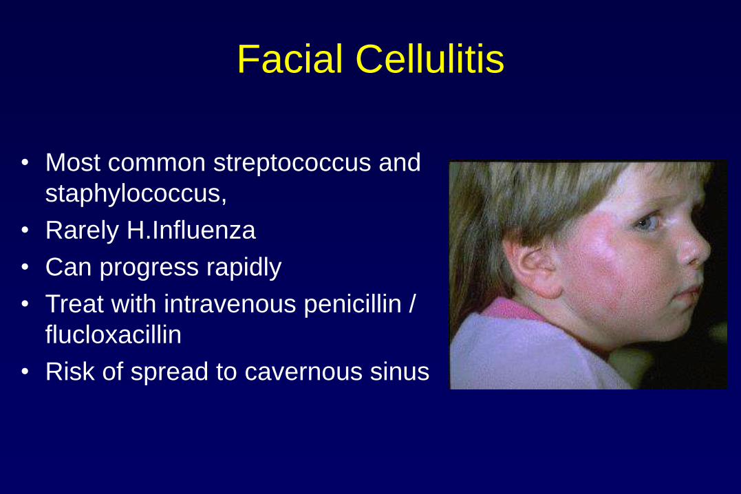
Facial Cellulitis
• Most common streptococcus and
staphylococcus,
• Rarely H.Influenza
• Can progress rapidly
• Treat with intravenous penicillin /
flucloxacillin
• Risk of spread to cavernous sinus
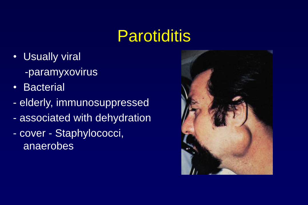
Parotiditis • Usually viral
-paramyxovirus
• Bacterial
- elderly, immunosuppressed
- associated with dehydration
- cover - Staphylococci,
anaerobes
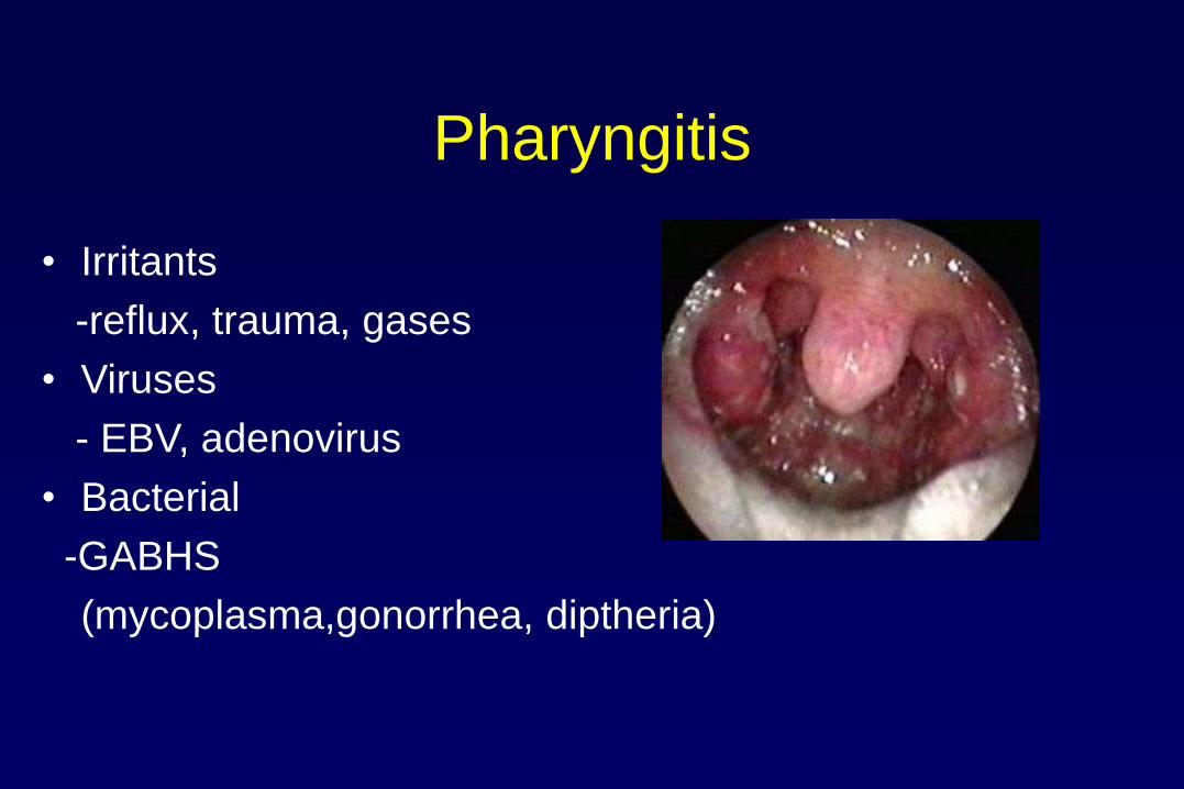
Pharyngitis
• Irritants
-reflux, trauma, gases
• Viruses
- EBV, adenovirus
• Bacterial
-GABHS
(mycoplasma,gonorrhea, diptheria)

Tonsillitis
• Presentation
• Types
• Tests
– Monospot
– FBE if admitting
– Throat swab not
helpful
• Treatment
– Oral Penicillin
– Admit if can’t drink

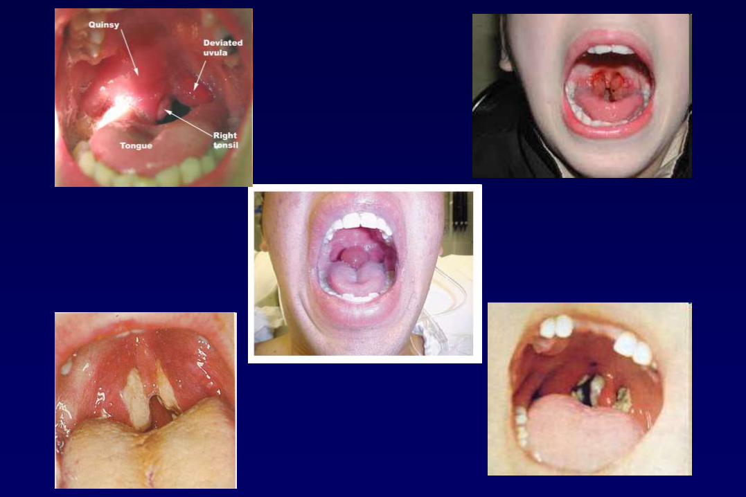
Quinsy (Peritonsillar Abscess)
• Presentation – Unilateral pain and fever
– Usually 4 days after sore throat
– Muffled “Hot potato” voice
– Trismus
– Palatal swelling
– Uvula oedema and deviation
• Aetiology – ? Weber’s Glands
– ? Complication of Tonsilitis
• Treatment – IV Penicillin / Flagyl / Steroids
– Aspiration (diagnostic)
– Incision + Drainage
• Can be life threatening
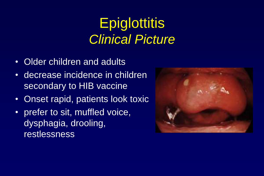
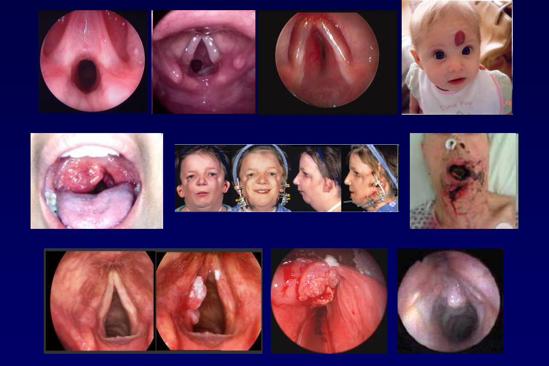
Epiglottitis Clinical Picture
• Older children and adults
• decrease incidence in children
secondary to HIB vaccine
• Onset rapid, patients look toxic
• prefer to sit, muffled voice,
dysphagia, drooling,
restlessness
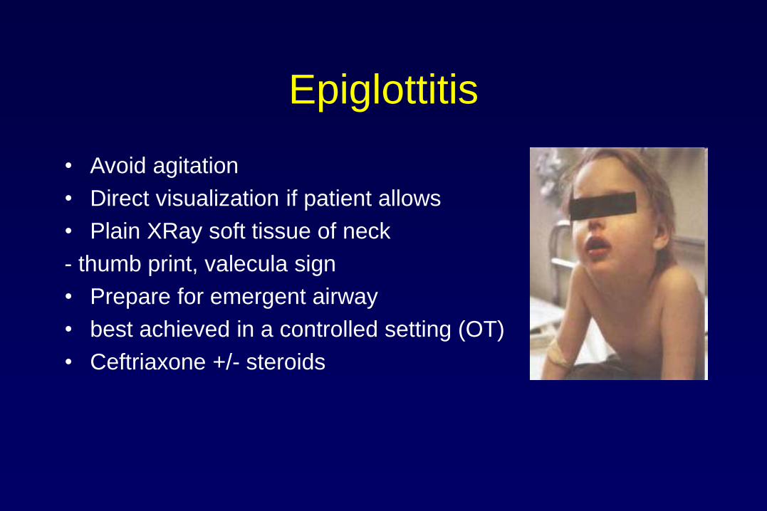
Epiglottitis
• Avoid agitation
• Direct visualization if patient allows
• Plain XRay soft tissue of neck
- thumb print, valecula sign
• Prepare for emergent airway
• best achieved in a controlled setting (OT)
• Ceftriaxone +/- steroids
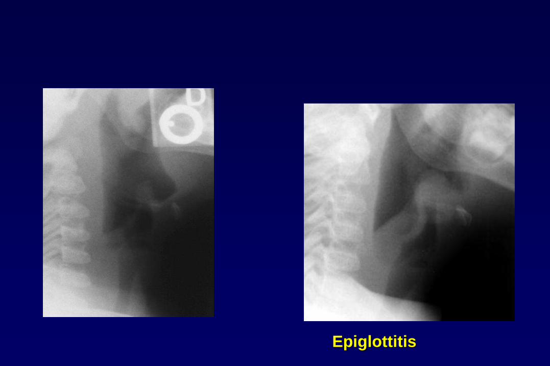
Epiglottitis
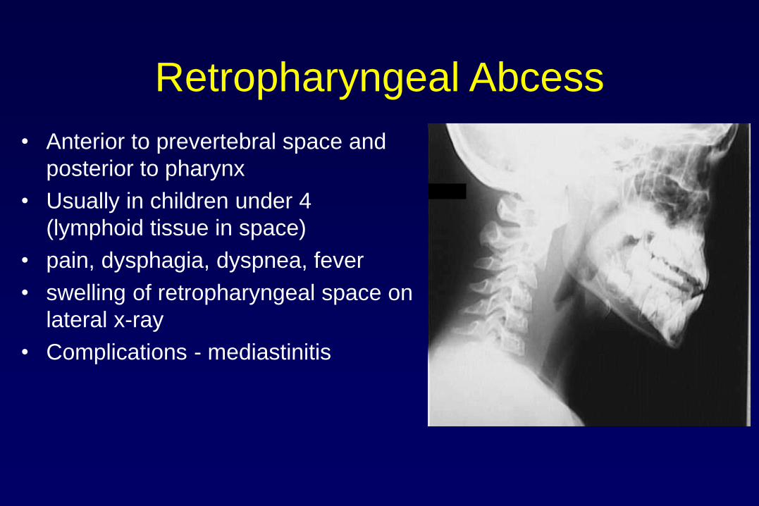
Retropharyngeal Abcess
• Anterior to prevertebral space and
posterior to pharynx
• Usually in children under 4
(lymphoid tissue in space)
• pain, dysphagia, dyspnea, fever
• swelling of retropharyngeal space on
lateral x-ray
• Complications - mediastinitis
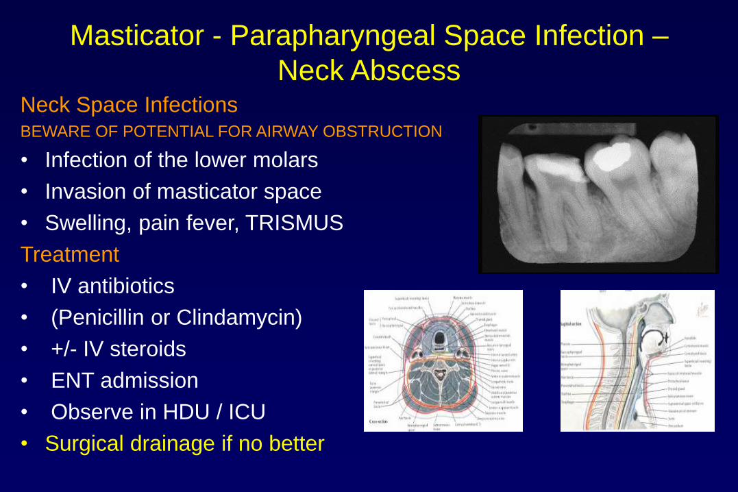
Masticator - Parapharyngeal Space Infection –
Neck Abscess Neck Space Infections BEWARE OF POTENTIAL FOR AIRWAY OBSTRUCTION
• Infection of the lower molars
• Invasion of masticator space
• Swelling, pain fever, TRISMUS
Treatment
• IV antibiotics
• (Penicillin or Clindamycin)
• +/- IV steroids
• ENT admission
• Observe in HDU / ICU
• Surgical drainage if no better
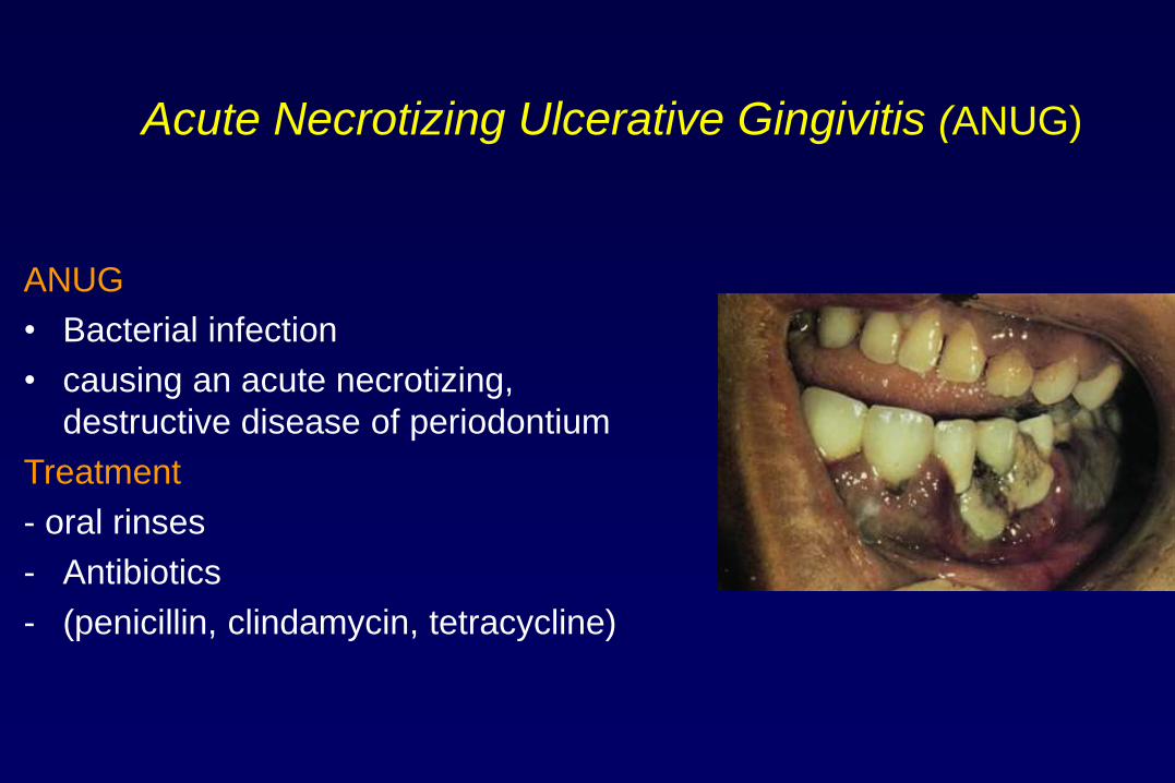
Acute Necrotizing Ulcerative Gingivitis (ANUG)
ANUG
• Bacterial infection
• causing an acute necrotizing,
destructive disease of periodontium
Treatment
- oral rinses
- Antibiotics
- (penicillin, clindamycin, tetracycline)
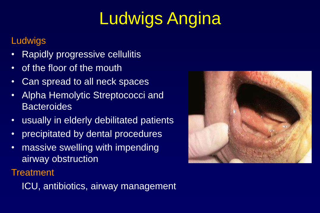
Ludwigs Angina Ludwigs
• Rapidly progressive cellulitis
• of the floor of the mouth
• Can spread to all neck spaces
• Alpha Hemolytic Streptococci and
Bacteroides
• usually in elderly debilitated patients
• precipitated by dental procedures
• massive swelling with impending
airway obstruction
Treatment
ICU, antibiotics, airway management
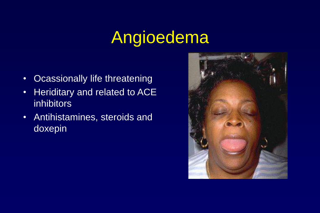
Angioedema
• Ocassionally life threatening
• Heriditary and related to ACE
inhibitors
• Antihistamines, steroids and
doxepin
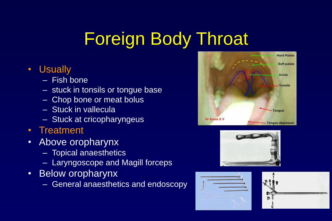
Foreign Body Throat
• Usually – Fish bone
– stuck in tonsils or tongue base
– Chop bone or meat bolus
– Stuck in vallecula
– Stuck at cricopharyngeus
• Treatment
• Above oropharynx – Topical anaesthetics
– Laryngoscope and Magill forceps
• Below oropharynx – General anaesthetics and endoscopy
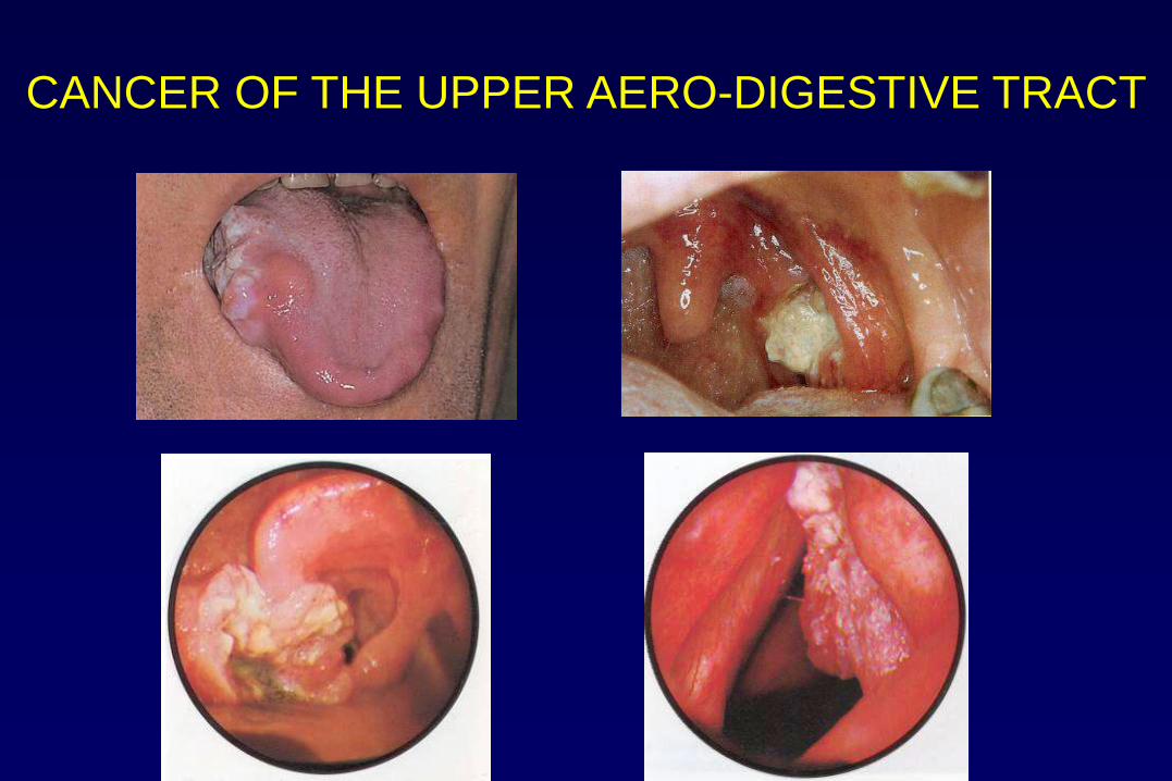
CANCER OF THE UPPER AERO-DIGESTIVE TRACT
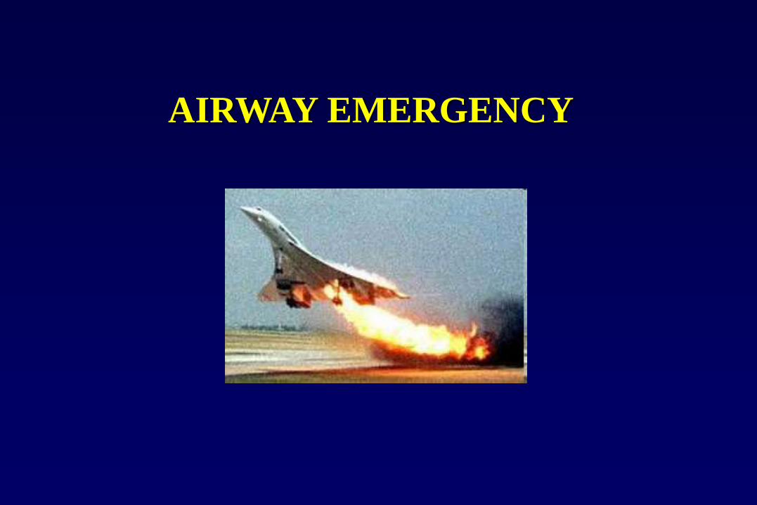
AIRWAY EMERGENCY
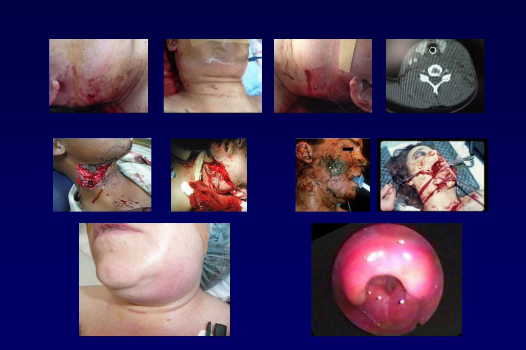
NECK TRAUMA • ORGANS & STRUCTURES
• Vascular ( most common )
• Pharyngoesophageal
• Laryngotracheal
• Others ( cranial nerve, thoracic duct, brachial plexus, spinal cord)
• VASCULAR INJURY
• External hemorrhage
• Extending soft tissue hematoma, distort or obstruct the airway
• Disruption of cerebral perfusion ( CVA )
– Zone 1 ( below cricoid cartilage )
– Zone 2 ( between cricoid cartilage and mandible angle )
– Zone 3 ( above mandible angle )

•Anatomy & Physiology
•Acute Clinical Issues of UAO
its assessment & management
•Best Practice
•Cricothyroidotomy
•Tracheostomy
•indications
•procedure
•complications
•Summary

ANATOMY OF LARYNX

PHYSIOLOGY OF LARYNX
Airway & Voice
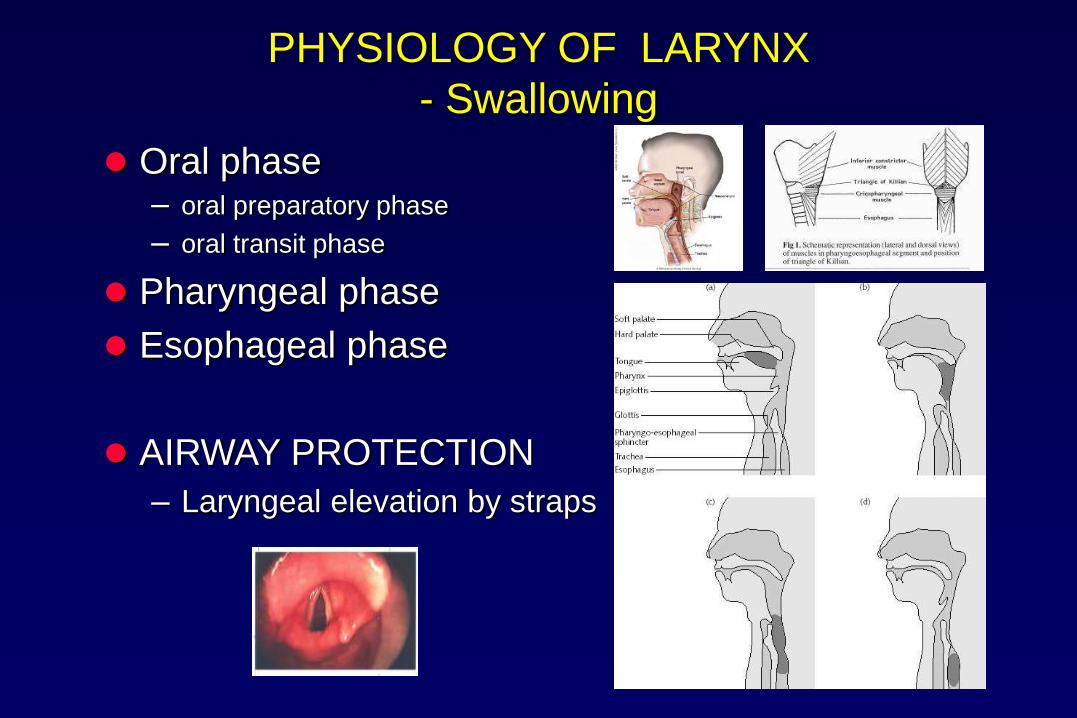
Oral phase – oral preparatory phase
– oral transit phase
Pharyngeal phase
Esophageal phase
AIRWAY PROTECTION
– Laryngeal elevation by straps
PHYSIOLOGY OF LARYNX
- Swallowing

Airway Emergencies Frightening – Patient & Doctor
Stridor vs Stertor
Distressed (+/- Drooling)
Desaturating – agitated
Assess level of obstruction – Listen to stridor
– Examine ENT & Neck
Swelling of face/tongue/neck
Sub-cutaneous emphysema
– Investigate
– ONLY if STABLE
– or ABLE TO STABILISE
– Fibreoptic Laryngoscopy
– Plain XRay Neck
– CT Neck
Treatment
– High flow 02 (Heliox)
– Nasopharyngeal airway
– Oral airway
– Nebulised adrenaline
– Steroids
– Bag and mask
– Intubation -ventilating
bronchoscope if difficult
– Cricothyroidotomy
– Tracheostomy
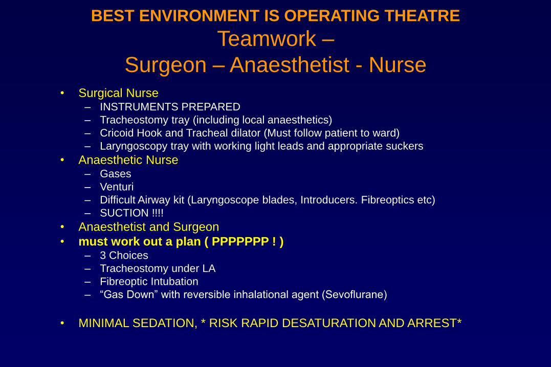
BEST ENVIRONMENT IS OPERATING THEATRE
Teamwork –
Surgeon – Anaesthetist - Nurse • Surgical Nurse
– INSTRUMENTS PREPARED
– Tracheostomy tray (including local anaesthetics)
– Cricoid Hook and Tracheal dilator (Must follow patient to ward)
– Laryngoscopy tray with working light leads and appropriate suckers
• Anaesthetic Nurse – Gases
– Venturi
– Difficult Airway kit (Laryngoscope blades, Introducers. Fibreoptics etc)
– SUCTION !!!!
• Anaesthetist and Surgeon
• must work out a plan ( PPPPPPP ! ) – 3 Choices
– Tracheostomy under LA
– Fibreoptic Intubation
– “Gas Down” with reversible inhalational agent (Sevoflurane)
• MINIMAL SEDATION, * RISK RAPID DESATURATION AND ARREST*

CRICOTHYROIDOTOMY
• IN ABSOLUTE EMERGENCY
• POSITION PATIENT WITH NECK EXTENDED
• MARK OUT MIDLINE
– SURFACE ANATOMY OF LARYNX / HYOID / THYROID / CRICOID
– “FIX” THE LARYNX AGAINST PREVERTEBRAL FASCIA
• PERFORM CRICOTHYROTOMY
• CONVERT TO FORMAL TRACHEOSTOMY
– ( OR OTHER SAFE AIRWAY eg FIBREOPTIC ET TUBE)
– AS SOON AS PATIENT IS STABLE
– BETTER SIZED AIRWAY
– SAFER AIRWAY / EASIER TO SECURE
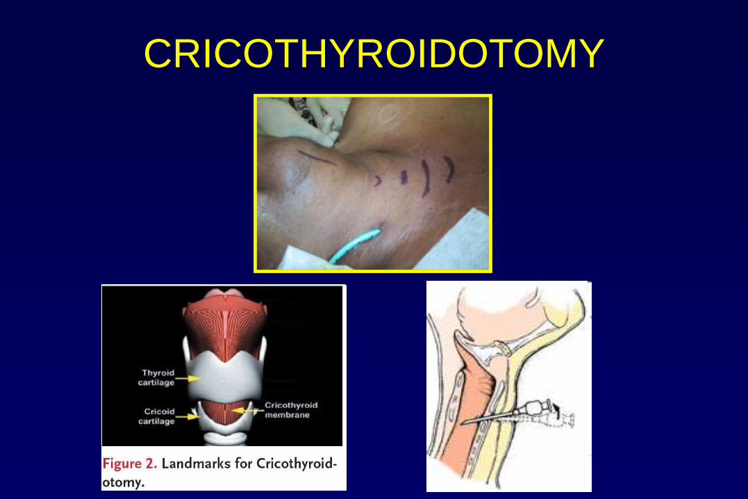
CRICOTHYROIDOTOMY
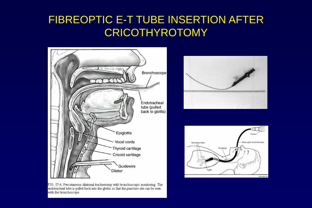
FIBREOPTIC E-T TUBE INSERTION AFTER
CRICOTHYROTOMY
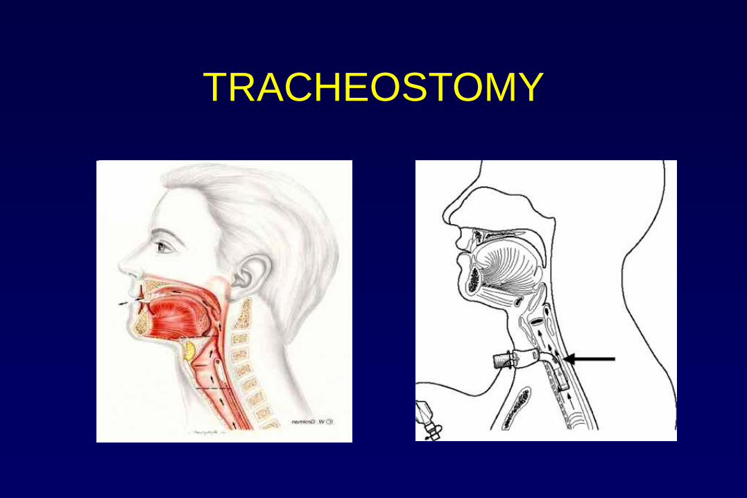
TRACHEOSTOMY

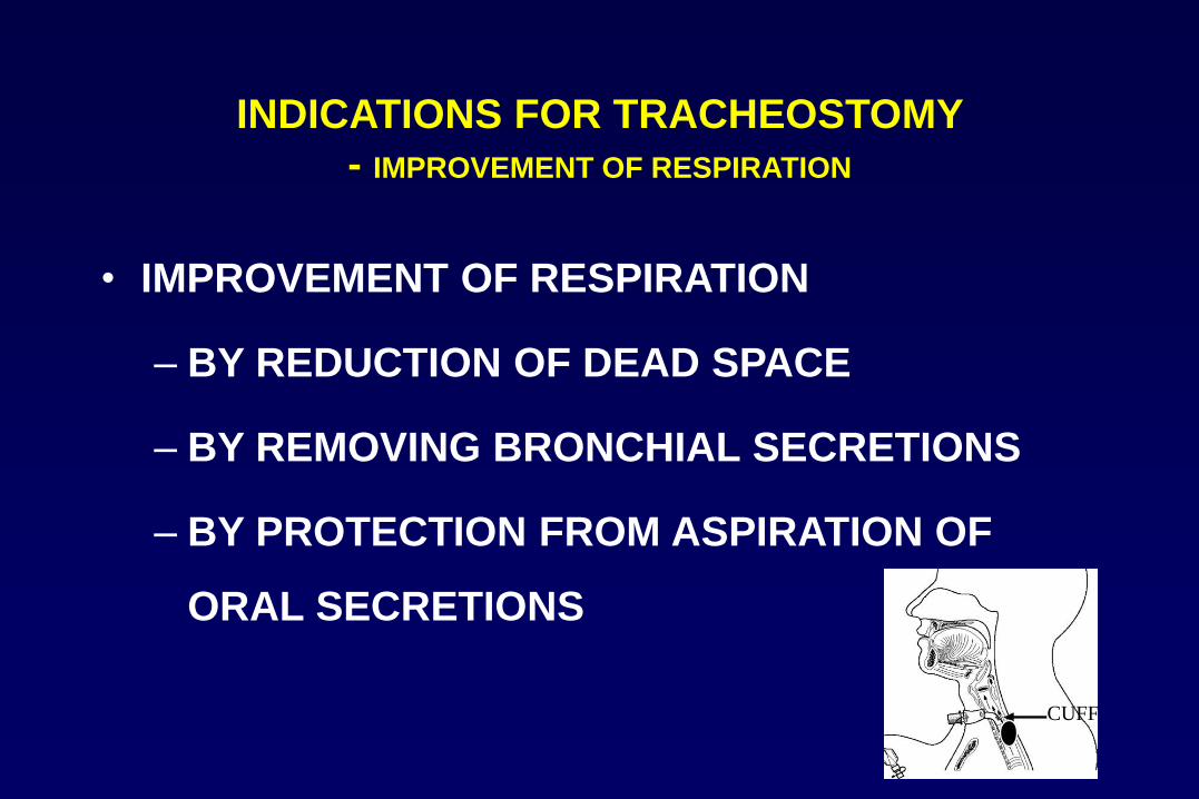
INDICATIONS FOR TRACHEOSTOMY
• UPPER AIRWAY OBSTRUCTION
• IMPROVEMENT OF RESPIRATION
• PROVISION OF VENTILATION

INDICATIONS FOR TRACHEOSTOMY - UPPER AIRWAY OBSTRUCTION
• UPPER AIRWAY OBSTRUCTION
– SITE
• SUPRAGLOTTIS – SUPRAGLOTTIC LARYNX / OROPHARYNX / MOUTH
• GLOTTIS
• SUBLOTTIS
– CONGENITAL
• WEBS
• SUBGLOTTIC HAEMANGIOMA
• BILATERAL VOCAL CORD PALSY
• LARYNGO MALACIA +/- TRACHEO MALACIA
• PIERRE - ROBIN SYNDROME
– ACQUIRED
• TRAUMA (face, jaws, oral cavity, pharynx)
• INFECTION (acute epiglottitis, laryngo-tracheo-bronchitis)
• INFLAMMATION (burns, caustic ingestion)
• IATROGENIC
• TUMOURS
• ANGIOEDEMA
• SLEEP APNOEA
INDICATIONS FOR TRACHEOSTOMY
- IMPROVEMENT OF RESPIRATION
• IMPROVEMENT OF RESPIRATION
– BY REDUCTION OF DEAD SPACE
– BY REMOVING BRONCHIAL SECRETIONS
– BY PROTECTION FROM ASPIRATION OF
ORAL SECRETIONS
CUFF
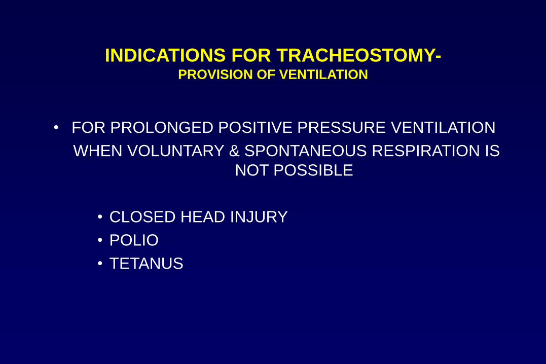
INDICATIONS FOR TRACHEOSTOMY- PROVISION OF VENTILATION
• FOR PROLONGED POSITIVE PRESSURE VENTILATION
WHEN VOLUNTARY & SPONTANEOUS RESPIRATION IS
NOT POSSIBLE
• CLOSED HEAD INJURY
• POLIO
• TETANUS

PERCUTANEOUS DILATION TRACHEOSTOMY
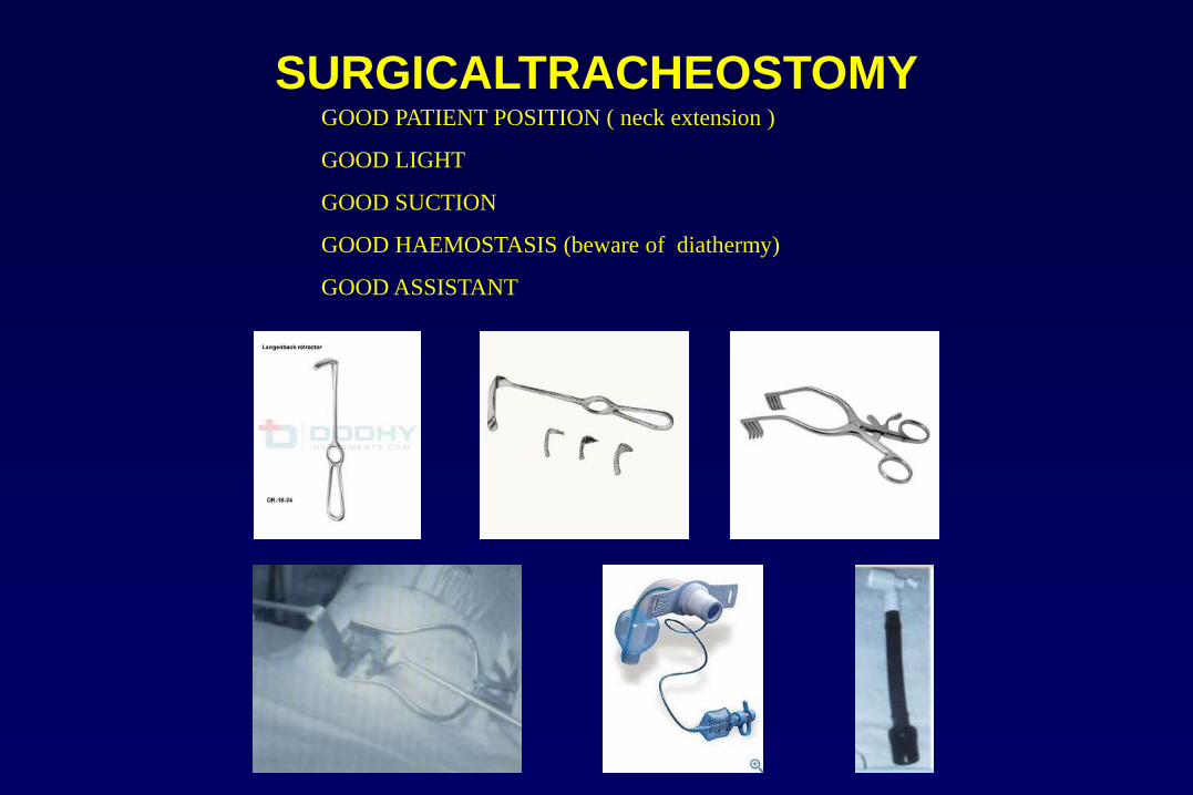
SURGICALTRACHEOSTOMY GOOD PATIENT POSITION ( neck extension )
GOOD LIGHT
GOOD SUCTION
GOOD HAEMOSTASIS (beware of diathermy)
GOOD ASSISTANT
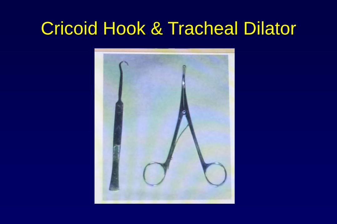
Cricoid Hook & Tracheal Dilator
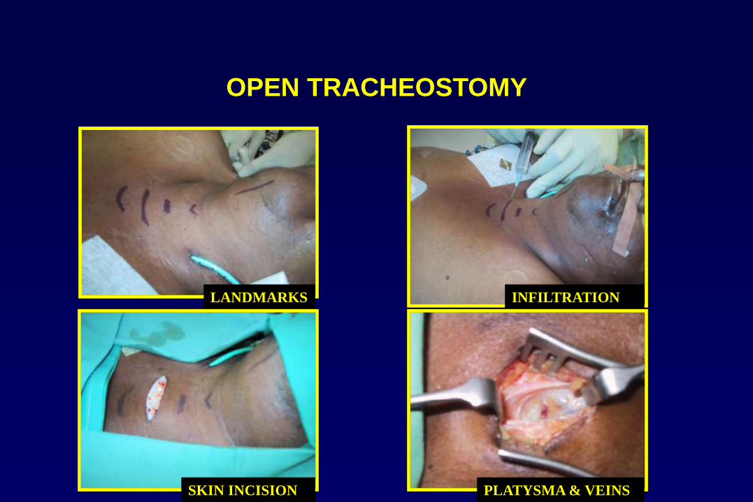
OPEN TRACHEOSTOMY
LANDMARKS INFILTRATION
SKIN INCISION PLATYSMA & VEINS
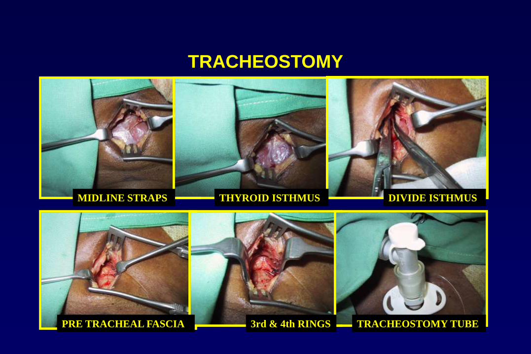
TRACHEOSTOMY
MIDLINE STRAPS THYROID ISTHMUS DIVIDE ISTHMUS
PRE TRACHEAL FASCIA 3rd & 4th RINGS TRACHEOSTOMY TUBE

Cuffed
(allows ventilation &
protects from aspiration)
Uncuffed
Non fenestrated
Fenestrated
(allows speech)
Shiley Portex
Types of Tracheostomy tubes
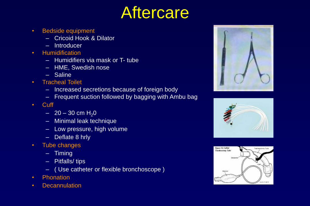
Aftercare
• Bedside equipment
– Cricoid Hook & Dilator
– Introducer
• Humidification
– Humidifiers via mask or T- tube
– HME, Swedish nose
– Saline
• Tracheal Toilet
– Increased secretions because of foreign body
– Frequent suction followed by bagging with Ambu bag
• Cuff
– 20 – 30 cm H20
– Minimal leak technique
– Low pressure, high volume
– Deflate 8 hrly
• Tube changes
– Timing
– Pitfalls/ tips
– ( Use catheter or flexible bronchoscope )
• Phonation
• Decannulation
COMPLICATIONS OF TRACHEOSTOMY
• IMMEDIATE
• INTERMEDIATE
• LATE

COMPLICATIONS OF TRACHEOSTOMY - IMMEDIATE
• IMMEDIATE
– MISPLACED TUBE (FALSE TRACT) • BIG / THICK NECK
• EFFORTFUL COUGHING
– HAEMORRHAGE (VENOUS CONGESTION)
• THYROID, JUGULAR VEINS
– SUBCUTANEOUS EMPHYSEMA
– PNEUMOMEDIASTINUM
– PNEUMOTHORAX • POSITIVE PRESSURE VENTILATION
• COUGHING / AIR SUCTION
• INCISION INTO PLEURA
– AIR EMBOLISM
– APNOEA
– CARDIAC ARREST
– LOCAL TISSUE DAMAGE • CRICOID
• TRACHEAL CARTILAGE
• RECURRENT LARYNGEAL NERVE
COMPLICATIONS OF TRACHEOSTOMY - INTERMEDIATE
• INTERMEDIATE
• ( PPPPPPP ! ) – GOOD INTENSIVE NURSING CARE – REGULAR & CAREFUL ASEPTIC SUCTION / CUFF CHECK etc
– ACCIDENTAL DECANNULATION
– CRUSTING
– INFECTION
– TRACHEAL NECROSIS
– TRACHEO-ARTERIAL FISTULA
– TRACHEO-OESOPHAGEAL FISTULA
– DYSPHAGIA
COMPLICATIONS OF TRACHEOSTOMY – LATE
• LATE
– TRACHEAL STENOSIS
– TRACHEO-CUTANEOUS FISTULA
– SCAR
– DIFFICULT DECANNULATION
• PERSISTENCE OF PROBLEM REQUIRING TRACHEOSTOMY
• ANTERIOR TRACHEAL WALL DISLOCATION
• GRANULATION AROUND STOMA
• SUBGLOTTIC STENOSIS
• TRACHEOMALACIA
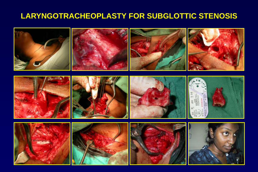
LARYNGOTRACHEOPLASTY FOR SUBGLOTTIC STENOSIS


Summary of structures to consider in UAO
Palatine tonsil
Tongue
Oro pharynx
Retropharyngeal space
Root of tongue
Geniohyoid muscle
Mylohyoid muscle
Submandibular space
Vallecula
Epiglottis
Hypophyarynx
Vocal cord
Thyroid cartilage
Larynx
Cricoid cartilage
Trachea
Sternum
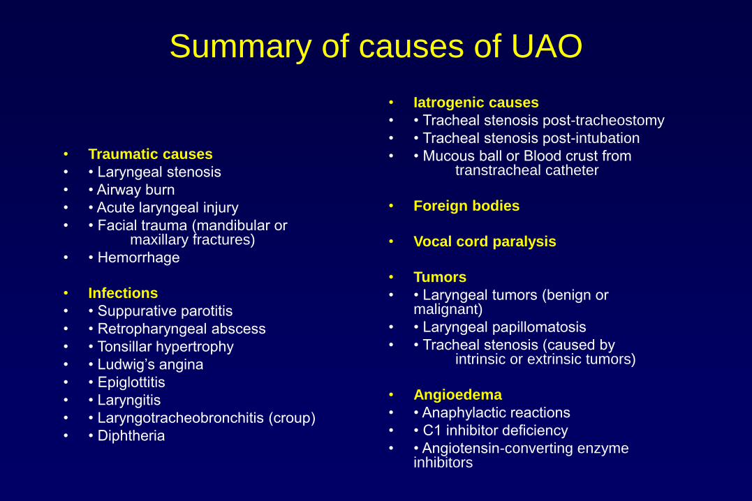
Summary of causes of UAO
• Traumatic causes
• • Laryngeal stenosis
• • Airway burn
• • Acute laryngeal injury
• • Facial trauma (mandibular or maxillary fractures)
• • Hemorrhage
• Infections
• • Suppurative parotitis
• • Retropharyngeal abscess
• • Tonsillar hypertrophy
• • Ludwig’s angina
• • Epiglottitis
• • Laryngitis
• • Laryngotracheobronchitis (croup)
• • Diphtheria
• Iatrogenic causes
• • Tracheal stenosis post-tracheostomy
• • Tracheal stenosis post-intubation
• • Mucous ball or Blood crust from transtracheal catheter
• Foreign bodies
• Vocal cord paralysis
• Tumors
• • Laryngeal tumors (benign or malignant)
• • Laryngeal papillomatosis
• • Tracheal stenosis (caused by intrinsic or extrinsic tumors)
• Angioedema
• • Anaphylactic reactions
• • C1 inhibitor deficiency
• • Angiotensin-converting enzyme inhibitors

Algorithm for management of
Upper Airway Obstruction
Stridor suggestive of UAO
Quick history and physical examination
Impending respiratory failure Gradual onset and mild symptoms
Urgent establishment of patent airway
Direct
or Fibre-optic
intubation
Cricothyroidotomy
vs
Tracheotomy
Is ET intubation possible?
Selection of
appropriate
ancillary studies:
• Laryngo-Bronchoscopy
• CT upper airway
• Spirometry
Yes No
Stanford University
Division of Emergency Medicine
References
Emily L. Senecal, M.D.
Emergency Medicine
Massachusetts General Hospital
Tracheostomy: A Multi-Professional Handbook
edited by Claudia Russell, Basil Matta

The best way to find yourself is to
lose yourself in the service of others.
MEDICAL SOLUTIONS THAT MAKE DREAMS HAPPEN
DREAMIN IS MY CHARITY, MAKE DREAMIN YOURS
dreamin.org.au