general syndesmology - usmf · groove" joint, the name is derived from the greek...
TRANSCRIPT

GENERAL SYNDESMOLOGY
Human Anatomy Department
Dr. Babuci Angela
Copyright©2014 Babuci Angela
State University of Medicine and Pharmacy “Nicolae Testemitanu“Republic of Moldova

Plan of the lecture
Types of bone articulations.
Classification of joints.
Main and auxiliary elements of a joint.
Biomechanics of joints.
Development of joints.
Copyright©2014 Babuci Angela

General syndesmology
Arthrology is the science of bone articulations.
Three types of bone articulations are distinguished:
Synarthroses – contiguous articulations (uninterrupted type of articulation).
Hemiarthroses, or symphyses (half-joints).
Diarthroses, or synovial joints.
Copyright©2014 Babuci Angela
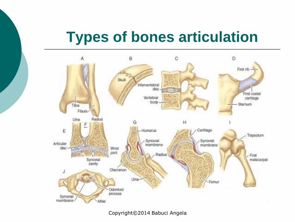
Types of bones articulation
Copyright©2014 Babuci Angela
Membranae interosseae
Ligamenta
Elastica
Sutura serrata Sutura squamosa Sutura plana
Junctura ossea
(synostosis)
Syndesmosis
Gomphosis
Suturae
Synarthrosis Hemiarthrosis
(symphysis)
Junctura fibrosa
Synsarkosis
Temporales Permanentes
Junctura cartilaginea
Fibrosa et hialinica
Copyright©2014 Babuci Angela
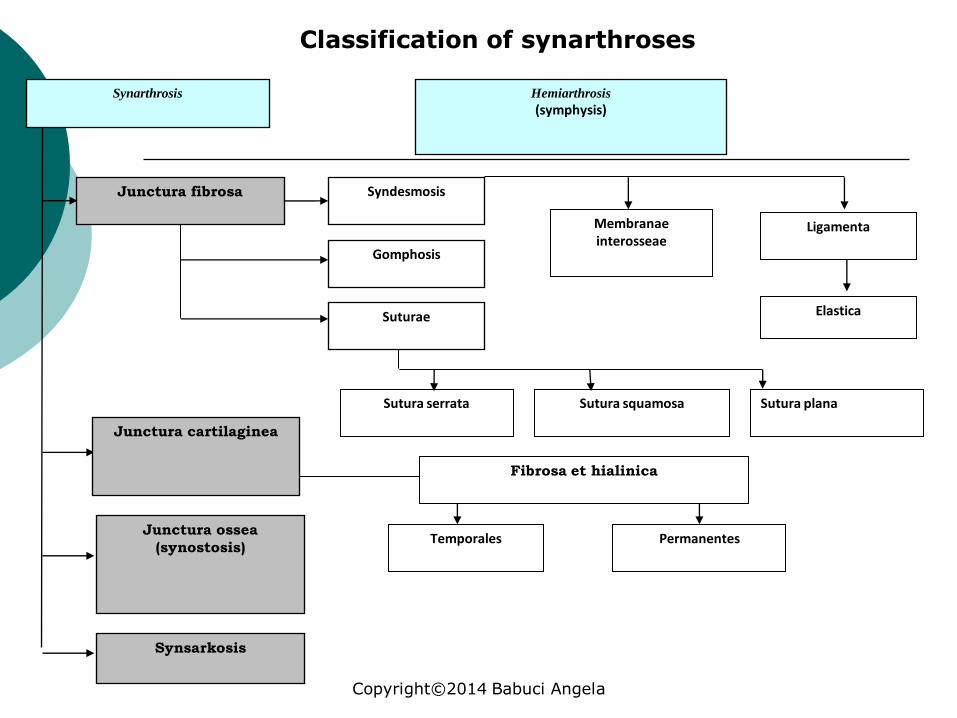
Classification of synarthroses

Classification of synarthroses
Synarthroses are contiguous articulations of bones by means of connective tissue, cartilaginous, bony and muscle tissue.
They are divided into: synfibroses, synchondroses, synostoses,
synsarcoses.
Copyright©2014 Babuci Angela
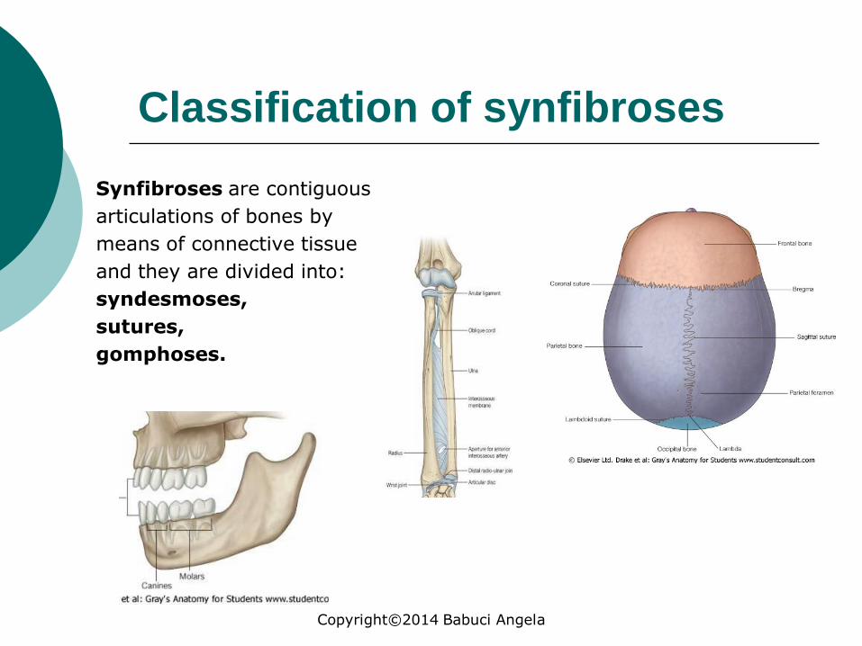
Classification of synfibroses
Synfibroses are contiguous
articulations of bones by
means of connective tissue
and they are divided into:
syndesmoses,
sutures,
gomphoses.
Copyright©2014 Babuci Angela

CLASSIFICATION OF SYNARTHROSES
Syndesmoses Ligaments (ligamenta) consist of
connective tissue that connects bones having a structure of fibrous bundles and they are characteristic for all joints e.g. ligaments of the vertebral column, proper ligaments of the scapula, ligaments between the bones of the pelvis, etc.
Membranes (membranae), when the connective tissue fills a large space between the bones, e.g. (interosseous membrane between the bones of the forearm and between the bones of the leg, membrana obturatoria etc).Remnants of the primary connective tissue that remain between the bones of the skull-cap are called fontanelles (fonticuli).
Synelastoses ligamenta flava (yellow ligaments) between the vertebral arches.
Copyright©2014 Babuci Angela

Sutures
Serrate sutures
(between the bones of the vault of the skull).
Squamous sutures
(between the temporal squama with the parietal bone).
Plane sutures
(between the bones of the facial skull).
Copyright©2014 Babuci Angela

The functional role of sutures
The sutures connect the bones of the skull and due to their elasticity, they assure the tridimensional growth of the skull.
After 25 years the sutures start their ossification and may transform to synostoses.
When the closure of sutures is disturbed by some factors there can appear abnormalities of the cranio-facial skeleton.
Between the sutures of the bones of the neurocranium can appear sutural, or wormian bones.
Copyright©2014 Babuci Angela

Articulation of the tooth with the dental alveoles is named gomphosis.
It is a fibrous joining by means of the dento-alveolar ligament that forms the dental periodontium.
The main fibers of the dento-alveolar ligament are named Sharpey's fibers and they join with cementum and with the alveolar periosteum.
Copyright©2014 Babuci Angela
Gomphosis
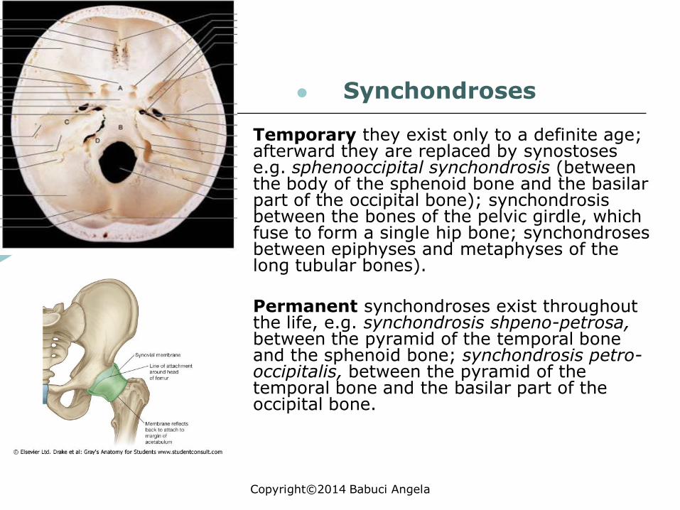
Synchondroses
Temporary they exist only to a definite age; afterward they are replaced by synostosese.g. sphenooccipital synchondrosis (between the body of the sphenoid bone and the basilar part of the occipital bone); synchondrosisbetween the bones of the pelvic girdle, which fuse to form a single hip bone; synchondrosesbetween epiphyses and metaphyses of the long tubular bones).
Permanent synchondroses exist throughout the life, e.g. synchondrosis shpeno-petrosa,between the pyramid of the temporal bone and the sphenoid bone; synchondrosis petro-occipitalis, between the pyramid of the temporal bone and the basilar part of the occipital bone.
Copyright©2014 Babuci Angela

Synostoses
The sacrum
The hip bone (after its ossification)
The long tubular bones (after ossification of their metaphyses)
Copyright©2014 Babuci Angela

Schindylesis
o Schindylesis is an articulation in which two bones are joined by fitting the ridge of one bone into the groove of another.
Also, it is known as a "wedge-and-groove" joint, the name is derived from the Greek 'skhindulesis', meaning "to cleave", as in cutting of a stump with an axe.
This fibrous suture joint can be found between the vomer and the perpendicular plate of the ethmoid bone as well as between the vomer and the gap between the maxilla and palatine bone, articulation of the rostrum of the sphenoid bone and perpendicular plate of the ethmoid bone with the vomer.
Copyright©2014 Babuci Angela

Synsarcosis
Synsarcosis is such a type of bone articulation when the articulating bones are joined by means of muscle tissue, e.g. subscapular muscle joins the scapula with the ribs, the hyoid bone is joined by means of suprahyoid muscle to the mandible and by means of infrahyoid muscles it is joined to the sternum.
Copyright©2014 Babuci Angela

Hemiarthroses, or Symphyses
Symphyses, or half-joints are transitional articulations, the joining tissue of which can be of fibrous or cartilaginous matter.
Inside the cartilage there is a slit-like cavity that is lined with synovial membrane and contains synovial fluid.
Outside the symphysis does not have an articular capsule.
The symphysis may be strengthened by some ligaments.
Slightly movements are possible in a symphysis.
Copyright©2014 Babuci Angela

Symphyses
The most typical half-joint is the pubic symphysis which forms between the two pubic bones.
Symphyses are present between the bodies of the vertebrae, and between the manubrium sterni and its body.
Copyright©2014 Babuci Angela
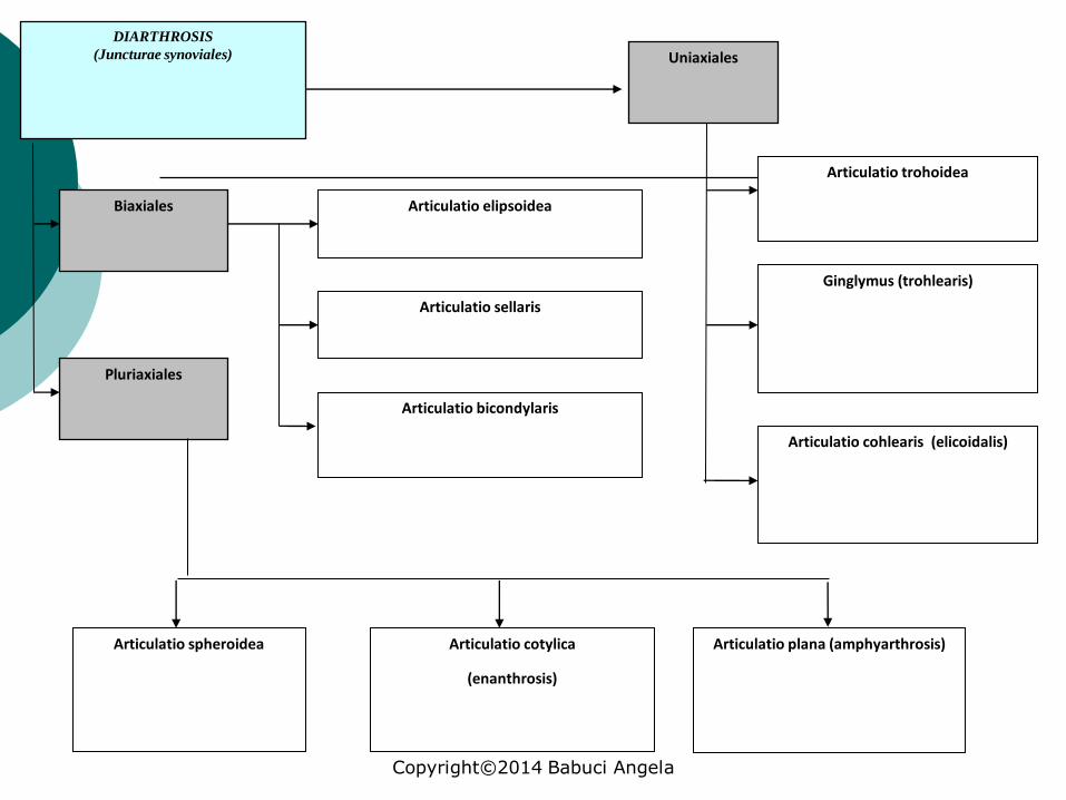
DIARTHROSIS
(Juncturae synoviales)
Biaxiales
Pluriaxiales
Articulatio elipsoidea
Articulatio sellaris
Articulatio bicondylaris
Ginglymus (trohlearis)
Articulatio trohoidea
Uniaxiales
Articulatio cohlearis (elicoidalis)
Articulatio spheroidea Articulatio plana (amphyarthrosis)Articulatio cotylica
(enanthrosis)
Copyright©2014 Babuci Angela

True joint (synovial joint), or diarthrosis
Each synovial joint consists of main and auxiliary elements.
The main elements of a joint: The articular surfaces,
facies articulares. The articular capsule,
capsula articularis. The articular cavity,
cavitas articularis.
Copyright©2014 Babuci Angela
6

Articular surfaces
The articular surfaces are smooth and are covered with cartilage that is adherent to the bone.
The articular cartilage is of white-bluish color, and it contains about 50-60% of water that together with other components assures its elasticity.
The thickness of the articular cartilage varies from 1mm to 12mm.
With ages the thickness of the articular cartilage decreases.
Usually the articular surfaces are congruent.
The articular cartilage in concave articular surfaces is thicker on the periphery and in convex articular surfaces it is thicker in its central part.
It does not contain blood vessels and nerve endings.
The nourishment of the articular surfaces is assured by the synovial fluid and arteries that supply the articular capsule.
Copyright©2014 Babuci Angela

Articular surfaces
The articular surfaces of almost all the joints of the human body are covered with hyaline cartilage, excepting following joints, which articular surfaces are covered by fibrous cartilage:
a) temporo-mandibular joint
b) sternoclavicular joint
c) acromioclavicular joint
Note:
The tubular bones join by the ends.
The flat bones join by their margins.
The irregular bones join either by their margins or by their surfaces.
Copyright©2014 Babuci Angela

Articular capsule The articular capsule encloses the articular
surfaces and keeps them in contact to each other.
The articular capsule, capsula articularis, consists of two layers:
external layer – the fibrous membrane; internal layer – the synovial membrane (it
produces synovial fluid).
The articular capsule is thin and elastic in those joints in which the range of movements is large and it is thick in the joints with a small range of movements.
The articular capsule protects the joint from different pathological processes that may occur in its vicinity.
Copyright©2014 Babuci Angela

Synovial fluid
The synovial fluid is produced by the internal layer of the synovial membrane.
It is transparent, sticky and yellowish in color.
It consists of about 95% of water and 5 % of organic matters such as proteins, glucose, hyaluronidase, etc.
It assures nourishment of the articular surfaces.
The synovial fluid lubricates the articular surfaces to facilitate the movements.
Copyright©2014 Babuci Angela
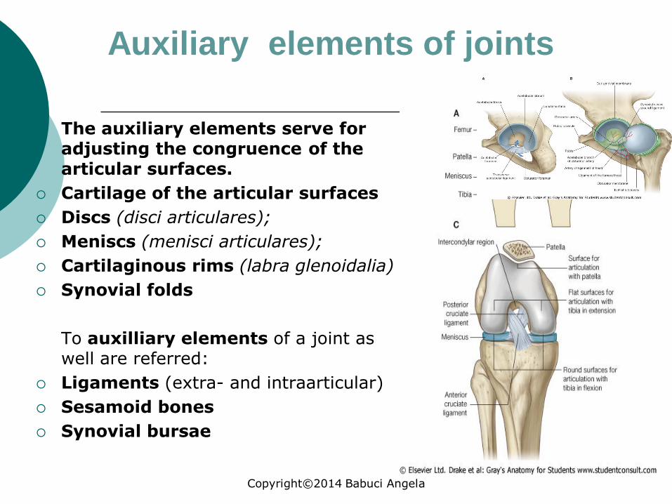
Auxiliary elements of joints
The auxiliary elements serve for adjusting the congruence of the articular surfaces.
Cartilage of the articular surfaces
Discs (disci articulares);
Meniscs (menisci articulares);
Cartilaginous rims (labra glenoidalia)
Synovial folds
To auxilliary elements of a joint as well are referred:
Ligaments (extra- and intraarticular)
Sesamoid bones
Synovial bursae
Copyright©2014 Babuci Angela

Types of diarthroses
Copyright©2014 Babuci Angela

Classification
of joints
Simple joint(articulatio simplex) it is formed only by two articular surfaces.
Copyright©2014 Babuci Angela
Classification of joints
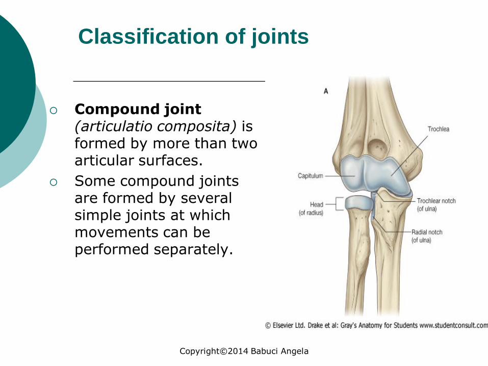
Compound joint (articulatio composita) is formed by more than two articular surfaces.
Some compound joints are formed by several simple joints at which movements can be performed separately.
Copyright©2014 Babuci Angela

Classification
of joints
Complex joint (articulatio complexa) contains intra-articular cartilages.
This cartilage divides the joint completely into two compartments (when the intra-articular cartilage is a disc), or incompletely (when the intra-articular cartilage is a menisc).
Copyright©2014 Babuci Angela

Classification
of joints
Combined joint includes two or more anatomically separated joints, but they function together.
Copyright©2014 Babuci Angela
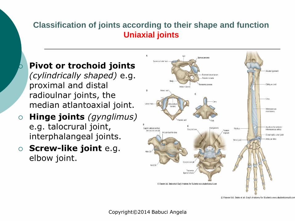
Classification of joints according to their shape and function
Uniaxial joints
Pivot or trochoid joints(cylindrically shaped) e.g. proximal and distal radioulnar joints, the median atlantoaxial joint.
Hinge joints (gynglimus) e.g. talocrural joint, interphalangeal joints.
Screw-like joint e.g. elbow joint.
Copyright©2014 Babuci Angela

Biaxial joints
Ellipsoid joints(articulatio ellipsoidea)e.g. radiocarpal joint.
Condyloid (or bicondylar) joints(articulatio condylaris) e.g. knee joint, atlantooccipital joint.
Saddle joint (articulatio sellaris) carpometacarpal joint of the thumb, calcaneocuboid joint.
Copyright©2014 Babuci Angela
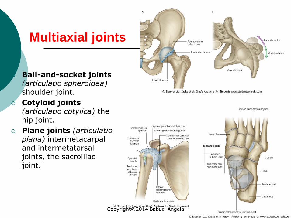
Multiaxial joints
Ball-and-socket joints (articulatio spheroidea)shoulder joint.
Cotyloid joints (articulatio cotylica) the hip joint.
Plane joints (articulatio plana) intermetacarpal and intermetatarsal joints, the sacroiliac joint.
Copyright©2014 Babuci Angela

Biomechanics of joints
Movements in the joints depend on the shape of the joint and they can be performed on the following axises:
Movement on the frontal axis: flexion (flexio) and extension (extensio).
Movement on the sagittal axis: abduction (abductio)and adduction (adductio).
Movement on the vertical axis: rotation (rotation):inward pronation (pronatio) and outward supination(supinatio).
Movement in a circular manner is named circumduction(circumductio).
Copyright©2014 Babuci Angela
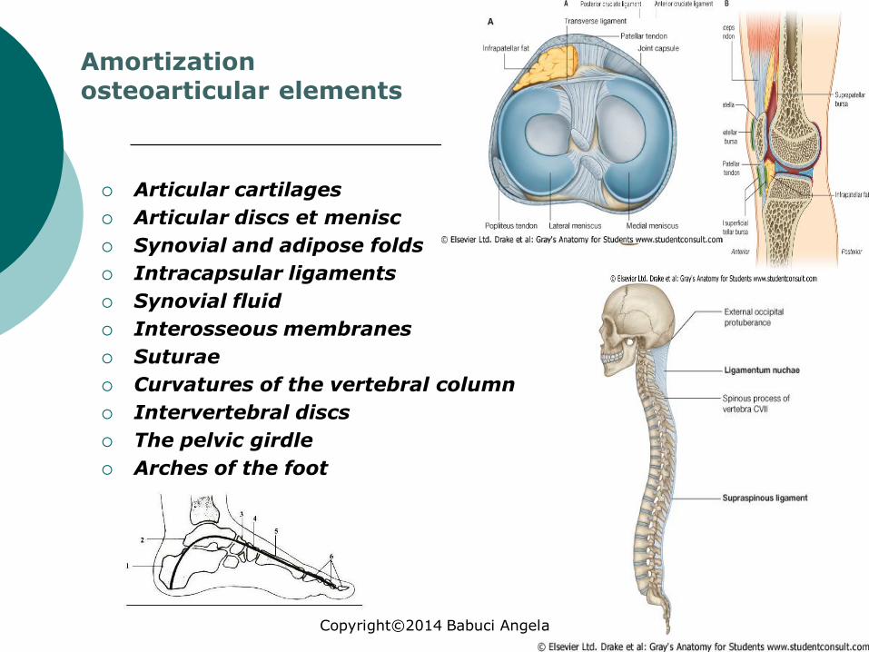
Amortization osteoarticular elements
Articular cartilages
Articular discs et menisc
Synovial and adipose folds
Intracapsular ligaments
Synovial fluid
Interosseous membranes
Suturae
Curvatures of the vertebral column
Intervertebral discs
The pelvic girdle
Arches of the foot
Copyright©2014 Babuci Angela

Biomechanics of joints
Copyright©2014 Babuci Angela

Biomechanics of joints
Copyright©2014 Babuci Angela

Biomechanics of joints
Copyright©2014 Babuci Angela

Development of joints
At the end of the second month of the intrauterine period of development the bones are laid down, as a thickening of the mesenchyme between the cartilaginous ends of the future bones.
The mesenchyme resorbs and a space (the future cavity) appears in it.
Copyright©2014 Babuci Angela

Development of joints
At the site of the future joint the bones come in contact with one another by means of cartilaginous articular surfaces, but from the mesenchyme that surrounds the former joint cavity develops the articular capsule.
In some joints in the mesenchyme located between the articular surfaces appear two spaces and in those cases there a disc forms, which completely separates the articular cavity into two floors, a bilocular joint forms.
When the central part of a disc fails to develop, than a meniscus forms in those joints.
Copyright©2014 Babuci Angela

Age specific features of joints
The development of bones articulations is directly dependent on the formation of the bony and connective tissue structures as well as dependent on muscular tissue.
The most important and active factor that determines the formation of joints after birth is the action of muscles on a given joint.
In a new born almost all the joint elements are encountered, but they continue their development for a while to acquire the final geometrical shapes characteristic for adults.
Copyright©2014 Babuci Angela

Age specific features of the joints elements
1. In a new born the articular rims in the shoulder and hip joints are not well developed.
2. The glenoid cavity of the scapula and the acetabulum are not deep enough.
3. The articular capsule of the joints is relatively thick.
4. The articular disc of the distal radioulnar joint is still not formed, at the same time the disc of the temporomandibular joint closely resembles a disc in an adult.
Copyright©2014 Babuci Angela