generation of four induced pluripotent stem cell lines
TRANSCRIPT
Contents lists available at ScienceDirect
Stem Cell Research
journal homepage: www.elsevier.com/locate/scr
Generation of four induced pluripotent stem cell lines (FHUi003-A,FHUi003-B, FHUi004-A and FHUi004-B) from two affected individuals of afamilial neurohypophyseal diabetes insipidus familySatoru Yoshidaa, Hanayuki Okurab, Hidetaka Sugac, Mika Soenc, Yohei Kawaguchic,Junki Kurimotoc, Takashi Miyatac, Hiroshi Takagic, Hiroshi Arimac, Tatsuya Fujikawad,Fumio Otsukae, Akifumi Matsuyamaa,⁎
a Department of Regenerative Medicine and Stem Cell Biology, Fujita Health University School of Medicine, Toyoake, Japanb Department of Regenerative Medicine Support Promotion Facility, Center for Research Promotion and Support, Fujita Health University, Toyoake, Japanc Department of Endocrinology and Diabetes, Nagoya University Graduate School of Medicine, Nagoya, Japand Department of General Internal Medicine, Mitoyo General Hospital, Kanonji, Japane Department of General Medicine, Okayama University Graduate School of Medicine, Dentistry Pharmaceutical Sciences, Okayama, Japan
A B S T R A C T
Four disease-specific induced pluripotent stem cell (iPSC) lines were respectively derived from peripheral blood mononuclear cells of two affected individuals in afamily affected by familial neurohypophyseal diabetes insipidus carrying the c.314G > C mutation. The expression of pluripotency markers (NANOG, OCT4, andSOX2), maintenance of a normal karyotype, absence of episomal vectors used for iPSC generation, and presence of the original pathogenic mutation were confirmedfor each iPSC line. The ability to differentiate into three germ layers was confirmed by a teratoma formation assay. These iPSC lines can help in disease recapitulationin vitro using organoids and elucidation of disease mechanisms.
Resource Table
Unique stem cell linesidentifier
FHUi003-AFHUi003-BFHUi004-AFHUi004-B
Alternative names of st-em cell lines
FNDI-iPS-A1b, A1b2as1 (FHUi003-A)FNDI-iPS-A11b, A11b (FHUi003-B)FNDI-iPS-B3a, B3a (FHUi004-A)FNDI-iPS-B7a, B7as1 (FHUi004-B)
Institution Fujita Health University School of MedicineContact information of
distributorAkifumi Matsuyama, [email protected]
Type of cell lines iPSCOrigin HumanCell Source Peripheral blood mononuclear cells (PBMCs)Clonality ClonalMethod of reprogram-
mingTransgene free (Episomal vectors)
Multiline rationale Two lines from each of two patients (a male and a female)of the same family, harboring the same disease and geneticmutation
Gene modification YESType of modification HereditaryAssociated disease Familial neurohypophyseal diabetes insipidus (FNDI)
Resource Table (continued)
Gene/locus AVP/20p13Method of modification N/AName of transgene or r-
esistanceN/A
Inducible/constitutivesystem
N/A
Date archived/stock da-te
December 2019
Cell line repository/ba-nk
https://hpscreg.eu/cell-line/FHUi003-Ahttps://hpscreg.eu/cell-line/FHUi003-Bhttps://hpscreg.eu/cell-line/FHUi004-Ahttps://hpscreg.eu/cell-line/FHUi004-B
Ethical approval Ethics Committee of Fujita Health University School ofMedicine (approval number HG19-062).Ethical Committee of Nagoya University Hospital (ap-proval number 2018-006-4).Ethical Committee of Mitoyo General Hospital (approvalnumber 18CR01-067).All animal experiments were approved by the Animal Careand Use Committee of KATAGIRI VMD OFFICE (approvalnumber FHU19-1).
https://doi.org/10.1016/j.scr.2020.101960Received 14 July 2020; Received in revised form 31 July 2020; Accepted 17 August 2020
⁎ Corresponding author at: Department of Regenerative Medicine and Stem Cell Biology, Fujita Health University School of Medicine, Toyoake, Japan.E-mail address: [email protected] (A. Matsuyama).
Stem Cell Research 48 (2020) 101960
Available online 26 August 20201873-5061/ © 2020 The Authors. Published by Elsevier B.V. This is an open access article under the CC BY license (http://creativecommons.org/licenses/BY/4.0/).
T

1. Resource utility
The lack of good in vitro models of familial neurohypophyseal dia-betes insipidus (FNDI) has made pathological analysis difficult. Thegenerated FNDI-iPSCs can be useful for disease recapitulation in vitrousing the hypothalamic-pituitary unit of organoids (Kasai et al., 2020;Suga, 2016), along with elucidation of the pathology and drug dis-covery Table 1.
2. Resource details
FNDI is a progressive form of diabetes insipidus caused by mutationin the AVP gene, which encodes a preproprotein including the anti-diuretic hormone arginine vasopressin (AVP), AVP carrier proteinneurophysin 2, and copeptin. The mutation leads to deficient AVP se-cretion from the pituitary gland, resulting in excessive and thin urineproduction accompanied by a polyuric symptom due to impaired waterreabsorption in the kidneys (Arima and Oiso, 2010; Hagiwara et al.,2019). We generated disease-specific iPSCs from peripheral bloodmononuclear cells (PBMCs) of two donor patients of an FNDI family,both of whom carry a heterozygous mutation (c.314G > C) in theneurophysin 2-coding region of AVP. We here report two iPSC linesderived from each of the two patients for a total of four cell lines.Characterization of the iPSC lines is summarized in Table 2. Eachgenerated iPSC colony was picked and expanded. Each iPSC line showsthe typical iPSC morphology with a high nucleus to cytoplasm ratio(Fig. 1A), and expression of pluripotent markers NANOG, OCT4, andSOX2 was confirmed by immunocytochemical analysis (Fig. 1B). Posi-tive cells for the markers in at least three independent areas were de-tected for each iPSC line with an average positive ratio of > 95%, >92%, and > 95% for NANOG, OCT4, and SOX2, respectively (Fig. 1D;Table 2). The ability of each iPSC line to differentiate into the threegerm layers was confirmed by an in vivo teratoma formation assay;
histological analysis of the developed teratoma revealed formation ofthe neural tissue (ectoderm), cartilage and muscle (mesoderm), andgut-like columnar epithelium (endoderm, Fig. 1C). Differentiation intopigment cells (ectoderm) and the adipose tissue (mesoderm) was alsofound in FNDI-A1b (FHUi003-A) and FNDI-A11b (FHUi003-B)(Fig. 1C). After more than a dozen passage cultures, none of the residualepisomal vectors used for generating the iPSCs was detected by poly-merase chain reaction (PCR) analysis targeting the DNA sequence ofOriP and EBNA1 in the vector (Fig. 1E). The original heterozygousmutation causing the disease was confirmed in the iPSCs derived fromboth patients by sequencing (Fig. 1F). G-banding analysis showed anormal karyotype of each iPSC line as 46XY (FNDI-A1b and A11b) or46XX (FNDI-B3a and B7a; Supplementary Fig. 1A). No sign of Myco-plasma infection was detected by the MycoAlert™ assay (SupplementaryFig. 1B). The identity of each iPSC line was confirmed by STR analysis(submitted in archive with journal) with an evaluation value of 1.
3. Materials and methods
3.1. Generation iPSCs
PBMCs were separated from whole blood of the patients usingdensity-gradient centrifugation with Lympho Spin Medium(pluriSelect) and cultured for 5 days in PBMC medium (StemFit®AK02N medium (Ajinomoto) without C solution, supplemented with50 ng/mL of IL-6, 50 ng/mL of SCF, 10 ng/mL of TPO, 20 ng/mL of Flt-3L, 20 ng/mL of IL-3, and 10 ng/mL of G-CSF). On day 5, to generateiPSCs from PBMCs, episomal vectors of transgenes for reprogramming(Epi5™ Episomal iPSC Reprogramming Kit, Thermo Fisher Scientific)were delivered into the cultured PBMCs by electroporation using 4D-Nucleofector™ System (Lonza) with the P3 Primary Cell 4D-Nucleofector™ X kit and the pre-installed program EO-117 according tothe manufacturer’s instructions. The Cells were plated with PBMC
Table 1Summary of lines.
iPSC line names Abbreviation in figures Gender Age Ethnicity Genotype of locus Disease
FNDI-iPS-A1b, A1b2as1 (FHUi003-A) FNDI_A1b Male 71 Japanese c.314G > C, heterozygous Familial neurohypophyseal diabetes insipidusFNDI-iPS-A11b, A11b (FHUi003-B) FNDI_A11b Male 71 Japanese c.314G > C, heterozygous Familial neurohypophyseal diabetes insipidusFNDI-iPS-B3a, B3a (FHUi004-A) FNDI_B3a Female 45 Japanese c.314G > C, heterozygous Familial neurohypophyseal diabetes insipidusFNDI-iPS-B7a, B7as1 (FHUi004-B) FNDI_B7a Female 45 Japanese c.314G > C, heterozygous Familial neurohypophyseal diabetes insipidus
Table 2Characterization and validation.
Classification Test Result Data
Morphology Photography Normal Fig. 1 panel APhenotype Qualitative analysis (Immunocytochemistry) Positive staining for NANOG, OCT4, and SOX2 Fig. 1 panel B
Quantitative analysis (Immunocytochemistrycounting)
NANOG: > 95%OCT4: > 92%SOX2: > 95%
Fig. 1 panel D
Genotype Karyotype (G-banding) and resolution FNDI-A1b, A11b: 46XYFNDI-B3a, B7a: 46XXResolution 300–500
Supplementary Fig. 1 panel A
Identity Microsatellite PCR (mPCR) ORSTR analysis
Not performed N/A16 STR loci tested;16/16 matched
Submitted in archive withjournal
Mutation analysis (IF APPLICABLE) Sequencing Heterozygous mutation,NM_000490:c.314G > C
Fig. 1 panel F
Southern Blot OR WGS N/A N/AMicrobiology and virology Mycoplasma Mycoplasma testing by luminescence. Negative Supplementary Fig. 1 panel BDifferentiation potential Teratoma formation Ectoderm: neural tissue, pigment cells
Mesoderm: cartilage, muscle, adipose tissueEndoderm: Gut-like columnar epithelium
Fig. 1 panel C
Donor screening (OPTIONAL) HIV 1 + 2 Hepatitis B, Hepatitis C N/A N/AGenotype additional info
(OPTIONAL)Blood group genotyping N/A N/AHLA tissue typing N/A N/A
S. Yoshida, et al. Stem Cell Research 48 (2020) 101960
2

Fig. 1. Characterization of generated FNDI-iPSC lines.
S. Yoshida, et al. Stem Cell Research 48 (2020) 101960
3
medium onto a multi-well plate coated with iMatrix-511 (Nippi) andthe half volume of complete StemFit® medium were added on day 3, 5,and 7. After day 9, medium change with fresh StemFit® medium wasperformed every 2–3 days until the iPSCs colonies grew enough forpicking. Obtained iPSC colonies were individually picked and trans-ferred onto new multi-well plates coated with iMatrix-511, followed byculture in StemFit® for further expansion. For subculture, the culturediPSCs were dissociated using Accutase (Innovative Cell Technologies)and plated onto an iMatrix-511-coated plate once a week. During dis-sociation and plating, 10 µM of the ROCK-inhibitor Y-27632 (CaymanChemical) was added to the media and was removed after culture day 1.Cells were maintained at 37 °C in a humidified 5% CO2 incubator. Toconfirm the absence of the reprograming vectors in the iPSCs, total DNAisolated from the established iPSCs at passage (P14–P17) usingQIAamp® DNA mini kit (QIAGEN) was subjected to PCR analysis usingthe specific primer pairs listed in Table 3. DNA from cells at P1 carryingthe episomal vector was used as a positive control.
3.2. Immunocytochemistry
The expression of pluripotent markers was examined by fluorescentimmunocytochemistry. iPSCs were fixed with 4% paraformaldehyde for10 min. The blocking reaction and dilution of antibodies (Table 3) wereperformed using 5% normal donkey serum (Jackson Immunoresearch)in phosphate-buffered saline with 0.01% Triton-X. The fixed cells wereincubated with the primary antibody overnight at 4 °C, followed byreaction with the secondary antibodies for 1 h at room temperature.The fluorescent signal was visualized using a BX-51 fluorescent mi-croscope and DP71 digital camera (Olympus).
3.3. Teratoma formation assay
A teratoma formation assay was performed to assess the differ-entiation ability of the iPSCs. A total of 5 × 10⁵ iPSCs (P21–P24) weresuspended in Matrigel (Corning) diluted in StemFit® AK02N at a 1:1ratio and injected into the testes of 7-week-old NOD.CB17-Prkdcscid/Jclmice (CLEA Japan). Fourteen weeks after injection, the animals weresacrificed and the developed teratoma was excised. Paraffin-embeddedsections prepared from the excised tissue were stained with hematox-ylin and eosin for histological analysis. All animal experiments wereconducted by KATAGIRI-OFFICE.
3.4. Direct sequencing
Direct sequencing was performed to detect the pathogenic mutationin the established iPSCs. The target region of the isolated genomic DNAwas amplified by PCR using Blend Taq® Plus (TOYOBO) and the primerpair listed in Table 3. PCR products purified using LaboPass™ PCR(COSMO GENETECH) were subjected to cycle sequencing by EurofinGenomics using the primer shown in Table 3.
3.5. Karyotype analysis
The karyotype of 20 cells (P25–P28) in metaphase was examined foreach iPSC line by G-banding at Nihon Gene Research Laboratories.
3.6. STR analysis
The identity of each established iPSC line was confirmed by STRanalysis using the PowerPlex® 16 System (Promega) at BEX Co., Ltd.
3.7. Mycoplasma test
Mycoplasma contamination was tested using MycoAlert™Mycoplasma Detection Kit (Lonza) according to the manufacturer’sinstructions. The test was performed at the same time as the test forMEN1-iPSC lines, which previously we reported (Yoshida et al., 2020),and the same negative/positive control value was shown(Supplementary Fig. 1B).
Declaration of Competing Interest
The authors declare that they have no known competing financialinterests or personal relationships that could have appeared to influ-ence the work reported in this paper.
Acknowledgements
We thank the patients for participating in this work. We also thankTomohiko Nishitomi for technical assistance. This work was supportedby grants from the Acceleration Program for Intractable DiseasesResearch utilizing Disease-specific iPS cells of the Research CenterNetwork for Realization of Regenerative Medicine (RCNRRM) fundedby Japan Agency for Medical Research and Development (AMED,Japan, Grant Number JP19bm0804011). We would like to thankEditage (www.editage.com) for English language editing.
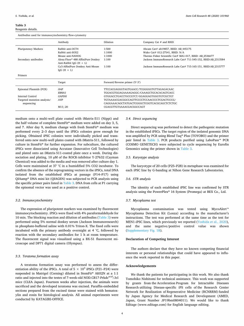
Table 3Reagents details.
Antibodies used for immunocytochemistry/flow-cytometry
Antibody Dilution Company Cat # and RRID
Pluripotency Markers Rabbit anti-OCT4 1:500 Abcam Cat# ab19857, RRID: AB_445175Rabbit anti-SOX2 1:1000 Wako Cat# 012-27541, RRID: N/AMouse anti-NANOG 1:1000 Thermo Fisher Scientific Cat# MA1-017, RRID: AB_2536677
Secondary antibodies Alexa Fluor® 488 AffiniPure DonkeyAnti-Rabbit IgG (H + L)
1:100 Jackson ImmunoResearch Labs Cat# 711-545-152, RRID:AB_2313584
Cy3-AffiniPure Donkey Anti-MouseIgG (H + L)
1:100 Jackson ImmunoResearch Labs Cat# 715-165-151, RRID:AB_2315777
Primers
Target Forward/Reverse primer (5′-3′)
Episomal Plasmids (PCR) OriP TTCCACGAGGGTAGTGAACC/TCGGGGGTGTTAGAGACAACEBNA1 TGGACGTGGAGAAAAGAGGC/CAAAGCTGCACACAGTCACC
Internal Control GAPDH GTGGACCTGACCTGCCGTCT/GGAGGAGTGGGTGTCGCTGTTargeted mutation analysis/
sequencingAVP TGTAAAACGACGGCCAGTTCCCCTCCAACCCCTCGACTCCCG/
CAGGAAACAGCTATGACTCGGGCTCGGTCACGCAGCTCTCTGCM13_-20 CGACGTTGTAAAACGACGGCCAGT
S. Yoshida, et al. Stem Cell Research 48 (2020) 101960
4

Appendix A. Supplementary data
Supplementary data to this article can be found online at https://doi.org/10.1016/j.scr.2020.101960.
References
Arima, H., Oiso, Y., 2010. Mechanisms underlying progressive polyuria in familial neu-rohypophysial diabetes insipidus. J. Neuroendocrinol. 22, 754–757. https://doi.org/10.1111/j.1365-2826.2010.02028.x.
Hagiwara, D., Grinevich, V., Arima, H., 2019. A novel mechanism of autophagy-asso-ciated cell death of vasopressin neurons in familial neurohypophysial diabetes in-sipidus. Cell Tissue Res. 375, 259–266. https://doi.org/10.1007/s00441-018-2872-4.
Kasai, T., Suga, H., Sakakibara, M., Ozone, C., Matsumoto, R., Kano, M., Mitsumoto, K.,Ogawa, K., Kodani, Y., Nagasaki, H., Inoshita, N., Sugiyama, M., Onoue, T.,Tsunekawa, T., Ito, Y., Takagi, H., Hagiwara, D., Iwama, S., Goto, M., Banno, R.,Takahashi, J., Arima, H., 2020. Hypothalamic contribution to pituitary functions isrecapitulated in vitro using 3D-cultured human iPS cells. Cell Rep. 30, 18–24.https://doi.org/10.1016/j.celrep.2019.12.009.
Suga, H., 2016. Making pituitary hormone-producing cells in a dish. Endocr. J. 63,669–680. https://doi.org/10.1507/endocrj.EJ16-0232.
Yoshida, S., Okura, H., Suga, H., Nishitomi, T., Sakurai, A., Arima, H., Matsuyama, A.,2020. Generation of three induced pluripotent stem cell (iPSC) lines from a multipleendocrine neoplasia type 1 (MEN1) patient and three iPSC lines from an unaffectedrelative of the patient. Stem Cell Res. 46. https://doi.org/10.1016/j.scr.2020.101846.
S. Yoshida, et al. Stem Cell Research 48 (2020) 101960
5