genetic evidence that transcription activation by rhas...
TRANSCRIPT

JOURNAL OF BACTERIOLOGY,0021-9193/00/$04.0010
Sept. 2000, p. 4959–4969 Vol. 182, No. 17
Copyright © 2000, American Society for Microbiology. All Rights Reserved.
Genetic Evidence that Transcription Activation by RhaSInvolves Specific Amino Acid Contacts with Sigma 70
PRASANNA M. BHENDE AND SUSAN M. EGAN*
Department of Molecular Biosciences, University of Kansas, Lawrence, Kansas 66045
Received 21 April 2000/Accepted 9 June 2000
RhaS activates transcription of the Escherichia coli rhaBAD and rhaT operons in response to L-rhamnose andis a member of the AraC/XylS family of transcription activators. We wished to determine whether s70 mightbe an activation target for RhaS. We found that s70 K593 and R599 appear to be important for RhaS activationat both rhaBAD and rhaT, but only at truncated promoters lacking the binding site for the second activator,CRP. To determine whether these positively charged s70 residues might contact RhaS, we constructed alaninesubstitutions at negatively charged residues in the C-terminal domain of RhaS. Substitutions at four RhaSresidues, E181A, D182A, D186A, and D241A, were defective at both truncated promoters. Finally, we assayedcombinations of the RhaS and s70 substitutions and found that RhaS D241 and s70 R599 met the criteria forinteracting residues at both promoters. Molecular modeling suggests that s70 R599 is located in very closeproximity to RhaS D241; hence, this work provides the first evidence for a specific residue within an AraC/XylSfamily protein that may contact s70. More than 50% of AraC/XylS family members have Asp or Glu at theposition of RhaS D241, suggesting that this interaction with s70 may be conserved.
The RhaS protein is the L-rhamnose-responsive transcrip-tion activator of the Escherichia coli L-rhamnose catabolic andtransport operons rhaBAD and rhaT, respectively (12, 13, 52,53, 57), and is a member of the AraC/XylS family of transcrip-tion activators (17, 18, 44, 53). Full activation of both therhaBAD and rhaT promoters requires activation by CRP bind-ing immediately upstream of RhaS (13, 57). RhaS alone is ableto activate rhaBAD expression by about 1,000-fold (13). In thepresence of RhaS, CRP activates rhaBAD an additional 30- to50-fold; however, CRP is unable to activate to any significantextent in the absence of RhaS (13).
The AraC/XylS family of transcription activators is namedfor its most well-studied member, AraC. The AraC proteinconsists of two functionally separable domains (7). The N-terminal AraC domain is responsible for both dimerization andL-arabinose binding, while the C-terminal domain is responsi-ble for both DNA binding and transcription activation. InRhaS, the C-terminal domain is also responsible for DNAbinding (4), and it is likely that the N-terminal domain func-tions in dimerization and L-rhamnose binding. The AraC/XylSfamily consists of more than 130 proteins that are identified bya 99-amino-acid region of sequence similarity within the DNA-binding domain of AraC (17, 18, 44, 53). One subset of AraC/XylS family proteins regulates expression of genes involved incarbon metabolism. This group includes AraC, RhaS, RhaR,and MelR from E. coli and XylS from Pseudomonas putida,which are among the most well characterized of the AraC/XylSfamily proteins (4, 5, 8, 12, 13, 16, 19, 29, 30, 38, 39, 53–55).Another large and important subset of AraC/XylS family pro-teins are those that regulate expression of virulence factors inbacterial pathogens (18). A few examples of this large groupinclude CfaD from enterotoxigenic E. coli, SprA from Salmo-nella enterica serovar Typhimurium, and UreR from a varietyof enteric pathogens (9, 14, 28, 48).
While DNA binding has been well characterized in a num-
ber of AraC/XylS family members (4–6, 12, 43, 45, 54), tran-scription activation by AraC/XylS family proteins is less wellunderstood. It has been shown that activation of several pro-moters dependent upon AraC/XylS family activators requiresthe C-terminal domain (CTD) of the a subunit of RNA poly-merase (RNAP). The a-CTD is the most well-characterizedactivation target and is required by a large number of activatorproteins (reviewed in references 11 and 24). Perhaps the mostdirect evidence for an interaction between an AraC/XylS fam-ily activator and a-CTD has been found with the Ada proteinat the alkA promoter. In this case mobility shift assays showedthat a substitution in a-CTD eliminated the ability of purifieda subunit to supershift the DNA-bound form of either Ada ormeAda (34). Strong evidence also exists for an interaction be-tween a-CTD and the MarA, SoxS, and Rob proteins in caseswhere these activators bind to DNA upstream but not over-lapping the 235 region of the promoter (25–27). Finally, at atruncated rhaBAD promoter where RhaS was the only activa-tor, deletion of a-CTD led to a 180-fold defect, and alaninesubstitutions identified eight residues in a-CTD that were can-didates for making contacts with RhaS (23).
There is also evidence that the mechanism of transcriptionactivation by some AraC/XylS family proteins may involveinteractions with the s70 subunit of RNAP, usually in caseswhere the binding site for the activator overlaps the 235 re-gion of the promoter. In fact, the very first substitution isolatedin s70 (originally named alt and with the substitution R596H)involved an interaction with AraC. This substitution increasedthe ability of AraC to activate transcription in the absence ofCRP, such that cya mutant cells regained the ability to usearabinose as the sole carbon source (50, 56). The more recentfinding that other s70 substitutions at positions near R596,especially K593A, significantly reduce activation by AraC inthe absence of CRP supports the hypothesis that wild-typeAraC and s70 make an interaction that contributes to tran-scription activation (36).
Biochemical evidence for an interaction between s70 andAda also exists. Ada differs from many other AraC/XylS familyproteins in that it can activate transcription from either a sitethat overlaps the 235 region (at alkA) or from a site that is 5
* Corresponding author. Mailing address: 8031 Haworth Hall, De-partment of Molecular Biosciences, University of Kansas, Lawrence,KS 66045. Phone: (785) 864-4294. Fax: (785) 864-5294. E-mail: [email protected].
4959
on February 7, 2019 by guest
http://jb.asm.org/
Dow
nloaded from

to 7 bp upstream of the 235 region (at ada and aidB) (1, 15, 35,47). The N-terminal half of Ada, which includes the AraC/XylSfamily domain, is capable of binding to DNA and activatingtranscription at the alkA promoter but is not sufficient fortranscription activation at promoters where Ada binds up-stream of the 235 region (1). At the alkA promoter, a set ofpositively charged amino acids in s70 was important for acti-vation by Ada (33). A heparin-resistant ternary complex couldbe formed between DNA, Ada, and RNAP containing wild-type s70, but not with RNAP containing s70 substitutionsK593A, K597A, or R603A (33), indicating that these s70 res-idues might be directly involved in an interaction with Ada.
The focus of our work has been the mechanism of transcrip-tion activation by the RhaS protein. The binding site for RhaSoverlaps the 235 region of both the rhaBAD and rhaT pro-moters by 4 bp, and hence it seemed likely that a target oftranscription activation by RhaS might be s70. To test thispossibility, we first tested activation by RhaS in strains express-ing a library of s70 derivatives with single alanine substitutionsin region 4.2 and at the very C-terminal end of s70. We foundthat activation by RhaS was defective in the presence of severals70 derivatives, most notably K593A and R599A, but only attruncated promoters that lacked the binding sites for the sec-
ond activator, CRP. In an effort to identify RhaS amino acidsthat might contact these positively charged s70 residues, weconstructed alanine substitutions in nearly all of the negativelycharged residues in the C-terminal domain of RhaS. A numberof the RhaS derivatives were defective for activation in com-bination with wild-type s70. Finally, we combined the RhaSand s70 derivatives and found one combination, RhaS D241Aplus s70 R599A, which showed no greater defect than theindividual derivatives at both the truncated rhaBAD and rhaTpromoters. This phenotype suggests that the two substitutionsmay define an interaction between the RhaS and s70 proteinsthat is important for transcription activation.
MATERIALS AND METHODS
Culture media and growth conditions. Cultures for b-galactosidase assay weregrown in 13 MOPS buffered medium (42); 13 MOPS consisted of 40 mM3-(N-morpholino)propanesulfonic acid (MOPS); 4 mM tricine, 0.01 mM FeSO4,9.5 mM NH4Cl, 0.276 mM K2SO4, 0.5 mM CaCl2, 0.528 mM MgCl2, 50 mMNaCl, 3 3 1029 M Na2Mo4, 4 3 1027 M H3BO3, 3 3 1028 M CoCl2, 1028 MCuSO4, 8 3 1028 M MnCl2, 1028 M ZnSO4, 1.32 mM K2HPO4, 10 mMNaHCO3, 0.2% Casamino Acids, and 0.002% thiamine. For other experiments(cloning, strain construction, Ter test, etc.), cells were grown in tryptone-yeastextract medium (37), with or without antibiotic, or TB maltose (0.8% Bacto-
TABLE 1. Oligos used in this studya
Oligo no. Oligo sequence (59–39) Use
744 CGC GGA TCC CCA CTG GAT GCG CCG AGA TCG Hybridizes within rhaB; used to amplify recombined rhaS allelesfor diagnostic PCR and sequencing
880 CTA ACA TCG TCG GCA TCG Hybridizes within rhaT; used to amplify recombined rhaS allelesfor sequencing
898 TGA GTA AAG CTT TTA TTG CAG AAA GCC ATC CCG Downstream end of rhaS; used to amplify rhaS alleles forchromosomal replacements
1170 CCG GAA TTC TTG TGG TGA TGT GAT GCT CAC Upstream of rhaS; used to amplify rhaS alleles for chromosomalreplacements
2068 ATG ACC GTA TTA CAT AGT GTG GATb rhaS sequencing2069 TTA TTG CAG AAA GCC ATC CCG TCCb rhaS sequencing2074 TGG TTG CAC AGA TGG AAC AGCb rhaS sequencing2075 GTT GAG ACG TGA TGC GCT GTTb rhaS sequencing2083 GTG GGA TCC ATG ACC GTA TTA CAT AGT Upstream for diagnostic PCR on plasmid clones of all rhaS
alleles2096 GCG GGA TCC GCG TTA CTC ATC TTC TTA Downstream F(rhaT-lacZ)D84 and D1332097 CGC GAA TTC AAG GGT ATG GTT TTG CAG Upstream F(rhaT-lacZ)D1332130 GGC CTG GCT GGC AGA CCA TTT TG SDM; RhaS E181Ala2131 CAT CGG CAA AAT GGT CTG Diagnostic PCR; RhaS E181Ala2134 CCA TTT TGC CGC AGA GGT GAA TTG SDM; RhaS E186Ala2135 CAT CCC AAT TCA CCT CTG Diagnostic PCR; RhaS E186Ala2136 TTG CCG ATGCAG TGA ATT GG SDM; RhaS E187Ala2137 CGG CAT CCC AAT TCA CTG Diagnostic PCR; RhaS E187Ala2138 CCG TGG CGGCAC AAT TTT CT SDM; RhaS D195Ala2139 CGC AGT GAA AGA GAA AAT TGT G Diagnostic PCR; RhaS D195Ala2141 TGT CAG TAA CGC TGG CTG Diagnostic PCR; RhaS E236Ala2142 CGT TAC TGC AAT CGC CTA TC SDM; RhaS D241Ala2143 CAC AGC GAT AGG CGA TTG Diagnostic PCR; RhaS D241Ala2146 TCA CCG CGT GCA ATT CGC CA SDM; RhaS D268Ala2147 CCG TCC CTG GCG AAT TG Diagnostic PCR; RhaS D268Ala2148 AGG GAC GGGCAG GCT TTC T SDM; RhaS D274Ala2149 TTA TTG CAG AAA GCC TG Diagnostic PCR; RhaS D274Ala2152 CCG GAA TTC ACT TAA TGC CGT GAT TG Upstream F(rhaT-lacZ)D842154 TGG CTG GAG GCT CAT TTT GCC SDM; RhaS D182Ala2155 ACG CCA CAG CGC AGC CAG CGT TA SDM; RhaS E236Ala2156 CCT CAT CGG CAA AAT GAG Diagnostic PCR; RhaS D182Ala2161 TTT GTT TGC GTT TAC TGG CAG ATA Downstream Plac-bet exo kan2162 ACG GCA ACG GCC TTG AAC TGA AAT Upstream Plac-bet exo kan2185 TTC GCC GAG CAT TTA ACT GGT C SDM; RhaS E261Ala2186 GCG GTG ACC AGT TAA ATG Diagnostic PCR; RhaS E261Ala
a Oligos were used for cloning, diagnostic or regular PCR, and site-directed mutagenesis (SDM). Regions not complementary to wild-type rha genes are underlined.b Oligos IRD41 dye labeled for use in a LI-COR automated sequencer.
4960 BHENDE AND EGAN J. BACTERIOL.
on February 7, 2019 by guest
http://jb.asm.org/
Dow
nloaded from

Tryptone, 0.5% NaCl, 0.2% maltose). Ampicillin was used at 125 or 200 mg/ml,as indicated.
General methods. Standard methods were used for restriction endonucleasedigestion, ligation, transformation, and purification of plasmid DNA. Primers forautomated DNA sequencing were IRD41 dye labeled (Table 1) and customsynthesized by LI-COR, Inc. (Lincoln, Nebr.). DNA sequences were verified byautomated dideoxy sequencing on a LI-COR 4000L sequencer. Sequencing re-actions were performed using the Thermo Sequenase fluorescence-labeled-primer cycle sequencing kit from Amersham Pharmacia Biotech (Piscataway,N.J.). All DNA sequences were confirmed on both strands.
Strains, plasmids, and phage. The E. coli strains, l phage, and plasmids usedin this study are described in Table 2. All assays were performed using culturesof strains derived from ECL116 (2). In all cases, lacZ translational fusions wereassayed as single-copy lysogens integrated into the E. coli chromosome at attl. Alibrary encoding wild-type s70 and alanine substitution derivatives of s70 were agift from C. Gross and were carried on the plasmid pGEX2T (10, 36).
Alanine substitutions of negatively charged amino acids in the DNA-bindingdomain of RhaS were constructed by site-directed mutagenesis of rhaS (PromegaGeneEditor In Vitro Mutagenesis System) with plasmid pSE159 as the template.The recommended protocol was followed, except that we found that lengtheningthe expression period after transformation into the mutS strain from 1 to 2 hgreatly increased the success of the procedure. Single-stranded plasmid templatewas used to construct all substitutions. Oligos, Etc., and Integrated DNA Tech-nologies synthesized oligonucleotide primers for site-directed mutagenesis andidentification of mutants (Table 1). Mutations were initially identified by adiagnostic PCR procedure using oligonucleotide (oligo) 744 and a second diag-nostic oligo for each mutation. In the diagnostic oligos, two nucleotides at the 39end were complementary to the mutant allele and therefore not to the wild-typeallele (Table 1). No PCR product was generated in any case from the wild-typeallele; however, templates carrying the mutant alleles yielded a product in allcases. DNA sequencing of the entire rhaS gene on both strands confirmed allmutations and ensured that there were no additional mutations.
Construction of rhaT-lacZ fusions. The full-length rhaT promoter (includingboth the CRP and RhaS-binding sites) was amplified by PCR using primers 2097and 2096 and whole cells of E. coli ECL116 as the source of template DNA. Thetruncated rhaT promoter (with only the RhaS-binding site) was amplified byPCR using primers 2096 and 2152 and whole cells of E. coli DH5a as the sourceof template DNA. The PCR products were digested at the EcoRI site in 2097 and2152 and the BamHI site in 2096 and cloned between the EcoRI and BamHI sitesof pRS414, yielding plasmids carrying full-length (pSE203) and truncated(pSE204) fusions, respectively. The DNA sequence of the promoter regions andfusion junctions were sequenced on both strands. The translational fusions thusconstructed with full-length and truncated rhaT promoters were transferred tolRS45 and lRS74 (both limm21), respectively, by in vivo recombination (51) togenerate recombinant phages lSME107 and lSME108 (Table 2). StrainsSME2186 and SME2187 carrying a single-copy lysogen of the recombinant lphage were obtained by transducing ECL116 with phage carrying the full-lengthand the truncated fusions, respectively. Lysogens carrying the full-length fusionwere identified as pinpoint blue colonies amid a white lawn on a nutrient agarplate containing X-Gal (5-bromo-4-chloro-3-indolyl-b-D-galactopyranoside) andL-rhamnose. Lysogens of the truncated fusion were selected by spreading thetransduction mixture on a plate carrying a lawn of lgt30 (limm21). In this case,lysogens were differentiated from l-resistant cells by their sensitivity to theheteroimmune phage lCh6 (limm434) when cross-streaked. For both the full-length and the truncated fusions, single lysogens were identified by the Ter test(22) and confirmed by b-galactosidase assay. P1 phage-mediated generalizedtransduction (40) was used to introduce an in-frame deletion of approximatelytwo-thirds of rhaS (13) linked to Tn10 into SME2186 and SME2187 to generateSME2341 and SME2342, respectively. The presence of the rhaS deletion wasconfirmed by PCR analysis.
Recombination of rhaS alleles into the chromosome. Chromosomal replace-ments by mutant rhaS alleles were constructed using an E. coli strain carryingbacteriophage l recombination functions resulting in increased homologousrecombination frequencies (41). SME2394 was constructed by P1 transduction ofthe l bet exo operon under the control of the lac promoter from KM22 into SME2393 with selection for kanamycin resistance (41). The presence of Plac-bet exowas confirmed by PCR with oligos 2161 and 2162. Alleles of rhaS to be recom-bined were amplified by PCR using oligos 898 and 1170 with the correspondingrhaS clone in pALTER-1 (pSE193-196 and pSE199) as a template. Then, 100 mlof CaCl2-treated SME2394 competent cells were transformed with approxi-mately 500 ng of unpurified PCR product. The transformation mixtures wereplated onto nutrient agar plates containing X-Gal, IPTG (isopropyl-b-D-thioga-lactopyranoside; 1 mM), and L-rhamnose and incubated at 37°C for 72 to 96 h.Tiny blue colonies picked from amid the white lawn were patched onto nutrientagar plates containing X-Gal and L-rhamnose with and without ampicillin. Blue,ampicillin-resistant colonies had been transformed with plasmid DNA that hadserved as template in the PCR reaction, while blue, ampicillin-sensitive colonieshad been transformed with the PCR-generated DNA fragments and had under-gone the desired chromosomal replacement. Performing PCR on blue, ampicil-lin-sensitive colonies with oligo 898 downstream and oligo 744 upstream identi-fied replacements of the in-frame rhaS deletion in SME2394 with full-length rhaSalleles. The presence of the mutant rhaS allele was tested by the same diagnostic
TABLE 2. Strains used in this study
Strain, phage,or plasmid Genotype Source or reference
E. coli strainsKM22 D(recC ptr recB recD)::Plac-bet
exo kan41
ECL116 F2 DlacU169 endA hsdR thi 2SME1035 ECL116 recA::cat l SME103 13SME1036 ECL116 recA::cat l SME104 13SME1082 ECL116 DrhaSa Laboratory collectionSME1087 ECL116 DrhaS recA::cat
l SME101Laboratory collection
SME1088 ECL116 DrhaS recA::catl SME104
Laboratory collection
SME1222 SME1082 l SME103 4SME1851 ECL116 l SME104 Laboratory collectionSME2186 ECL116 l SME107 This studySME2187 ECL116 l SME108 This studySME2341 SME2186 DrhaS zih-35::Tn10 This studySME2342 SME2187 DrhaS zih-35::Tn10 This studySME2393 SME1222 DrhaS zih-35::Tn10 This studySME2394 SME2393 Plac-bet exo kan This studySME2603 SME1851 rhaS(E181A)
recA::kanThis study
SME2604 SME1851 rhaS(D182A)recA::kan
This study
SME2605 SME1851 rhaS(D186A)recA::kan
This study
SME2606 SME1851 rhaS(E187A)recA::kan
This study
SME2607 SME1851 rhaS(D241A)recA::kan
This study
SME2608 SME1851 rhaS(wt) recA::kanb This studySME2609 SME2187 rhaS(E181A)
recA::kanThis study
SME2610 SME2187 rhaS(D182A)recA::kan
This study
SME2611 SME2187 rhaS(D186A)recA::kan
This study
SME2612 SME2187 rhaS(E187A)recA::kan
This study
SME2613 SME2187 rhaS(D241A)recA::kan
This study
SME2614 SME2187 rhaS(wt) recA::kan This study
PhagelRS45 bla9-lacZscatt1 imm21 ind1 51lRS74 bla9-placUV5-lacZ1 att1
imm21 ind151
l SME101 l RS45 F(rhaB-lacZ)D226 13l SME103 l RS45 F(rhaB-lacZ)D110 13l SME104 l RS45 F(rhaB-lacZ)D84 13l SME107 l RS45 F(rhaT-lacZ)D133 This studyl SME108 l RS74 F(rhaT-lacZ)D84 This study
PlasmidspALTER-1 Aps Tetr; lacZ, f1 ori Promega Corp.pSE159 Apr pALTER-1 rhaS (wt) 4pSE193 pSE159 (RhaS E181A) This studypSE194 pSE159 (RhaS D182A) This studypSE195 pSE159 (RhaS D186A) This studypSE196 pSE159 (RhaS E187A) This studypSE197 pSE159 (RhaS D195A) This studypSE198 pSE159 (RhaS E236A) This studypSE199 pSE159 (RhaS D241A) This studypSE200 pSE159 (RhaS E261A) This studypSE201 pSE159 (RhaS D268A) This studypSE202 pSE159 (RhaS D274A) This studypRS414 Apr 9lacZ lacY lacA 51pSE203 pRS414 F(rhaT-lacZ)D133 This studypSE204 pRS414 F(rhaT-lacZ)D84 This study
a Construction of this DrhaS allele is described elsewhere (13).b wt, wild type.
VOL. 182, 2000 RhaS INTERACTION WITH s70 4961
on February 7, 2019 by guest
http://jb.asm.org/
Dow
nloaded from

PCR used to identify the mutations after the initial site-directed mutagenesis.For DNA sequencing, the rhaS alleles were amplified by PCR using oligos 880and 744 with whole cells as the source of template DNA. The PCR products werepurified using QIAquick PCR purification kit (Qiagen, Inc.). Both strands of thePCR products were sequenced using the IRD44-labeled oligos listed in Table 1.Once confirmed, the wild-type and mutant rhaS alleles were introduced intoSME1851 and SME2187 by phage P1-mediated generalized transduction (40)using selection for the linked Tn10. Finally, recA::kan was moved into each of thestrains by P1 transduction with selection for kanamycin resistance.
b-Galactosidase assay. Strains to be assayed for b-galactosidase activity weregrown and assayed as previously described (4). Briefly, they were first grown inTY broth containing ampicillin and then transferred to 13 MOPS minimalmedium with ampicillin (200 mg/ml) and limiting carbon source (0.04% glycerol)for overnight growth. The overnight culture was diluted 1:100 into 13 MOPSmedium containing 0.4% glycerol as the carbon source; 0.2% L-rhamnose wasadded as the inducer along with ampicillin (200 mg/ml), and the culture wasgrown to an A600 of approximately 0.4. Assays were performed as described byMiller (40) except that in assays of the truncated rhaT fusion [F(rhaT-lacZ)D84],cultures were concentrated 20-fold (s70 derivatives) or 114-fold (RhaS deriva-tives and RhaS-s70 derivative combinations) upon addition of Z buffer. This ismuch greater than the 2.5-fold concentration in the standard assay. These assayswere allowed to incubate for up to 3 days. Assays of RhaS derivatives andcombinations of RhaS and s70 derivatives at the truncated rhaT fusion wereperformed in a total volume of 0.1 ml rather than in the standard 1 ml so thatvery large culture volumes did not need to be grown. Under these conditions, thevast majority of the optical-density-at-420-nm readings were greater than 0.1,while the very lowest readings were greater than 0.05. Specific activities wereaveraged from at least three independent assays, with two replicates in eachassay.
RESULTS
Sigma70 substitutions at rhaBAD. Lonetto et al. (36) con-structed a library of 17 single alanine substitutions near theC-terminal end of the s70 subunit of RNA polymerase. Theyfound that s70 residues in this region were required for acti-vation of a variety of promoters in which an activator proteinbinds to a site that overlaps the 235 region of the promoter.To determine whether contacts with s70 were important foractivation by RhaS, we first tested the library of alanine sub-stitutions in s70 at the rhaBAD and rhaT promoters. Thestrains that we assayed had a gene encoding wild-type s70 inthe chromosome and carried the gene encoding the s70 deriv-atives on a plasmid. Lonetto et al. (36) showed that in theabsence of IPTG induction the s70 derivatives were producedfrom these plasmids at a level that is only slightly higher thanthat of wild-type s70; hence, only about 50% of the RNAP isexpected to contain non-wild-type s70. The strains also carrieda wild-type rha locus at the normal chromosomal location anda single-copy l specialized transducing phage carrying a trans-lational fusion of the rhaBAD or rhaT promoter with lacZ. Thepromoter fusions were either full length and included the bind-
ing sites for both the CRP and RhaS activators or truncatedand included only the binding site for RhaS (Fig. 1). Deletionof the CRP-binding site from the fusions was preferable todeletion of the crp gene has been shown to decrease rhaBADexpression both due to the direct loss of CRP activation and todecreased rhaS expression from the CRP-dependent rhaSRpromoter.
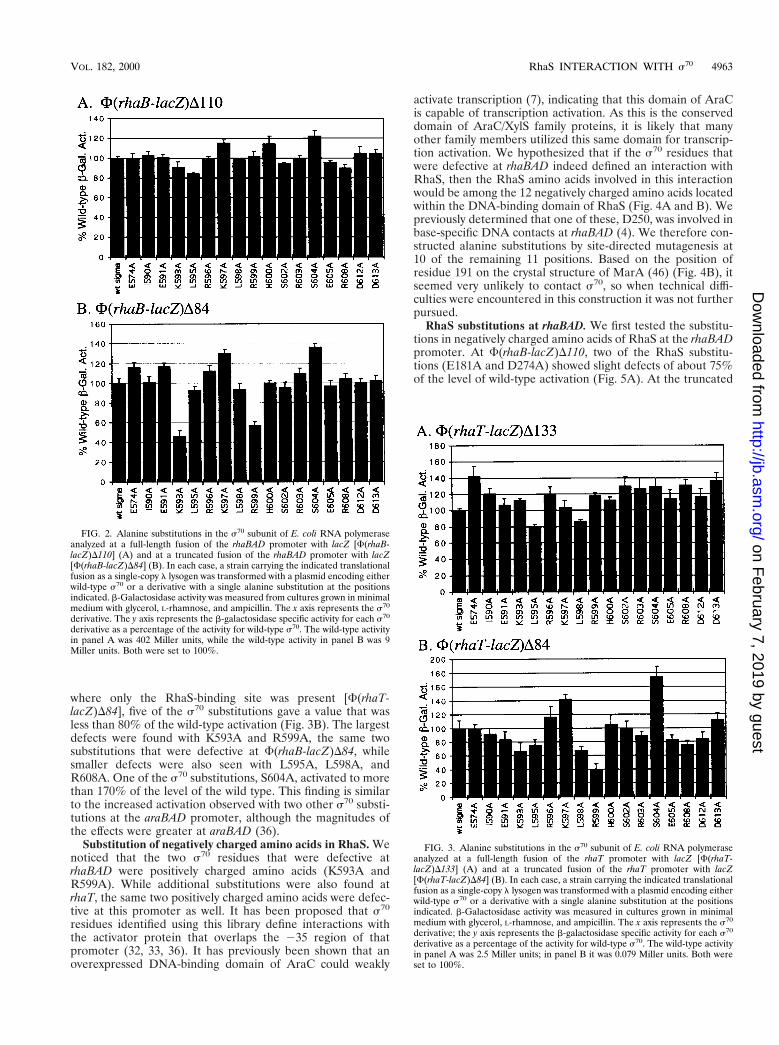
We first tested the s70 substitution library at the full-lengthrhaBAD promoter fusion [F(rhaB-lacZ)D110] and found thatthere were no significant defects with any of the s70 substitu-tions at this promoter (Fig. 2A). We next assayed the library atthe truncated rhaBAD promoter fusion that included only theRhaS-binding site [F(rhaB-lacZ)D84] (Fig. 2B). At this fusion,two of the s70 substitutions, K593A and R599A, allowed acti-vation to only 46 and 58% of the wild type, respectively. Giventhat only 50% of the RNAP was likely to contain the s70
substitution at K593 or R599 in each case, these defects arereasonably large. Residue K593 was also found to be importantfor activation by AraC, but R599 was not (36). Also, similar tothe findings at araBAD, the s70 substitutions only had a signif-icant effect when the CRP-binding site was not present up-stream of rhaBAD.
Although the s70 K593A and R599A derivatives onlyshowed defects at non-native, truncated promoters, we wouldargue that this information is likely to be biologically relevant.It is possible that these residues are also important for RhaSactivation in the full-length promoter, but for reasons de-scribed in the Discussion, such as redundancy, they did notshow any detectable defect in the presence of CRP activation.Further, if these s70 residues are important for activation byRhaS in the absence of CRP, it is possible that other AraC/XylS family proteins that activate transcription without the aidof a second activator, such as CRP, may also require theseresidues.
s70 substitutions at rhaT. To develop a more general pic-ture of the role of the C-terminal end of s70 in activation byRhaS, we tested the s70 library at rhaT promoter fusions. Asshown in Fig. 1, the location of the RhaS and CRP-bindingsites relative to the core promoter at rhaT is the same as thatat rhaBAD. Assays of b-galactosidase activity were modified asdescribed in Materials and Methods to allow accurate mea-surement of the low activities expressed from the truncatedrhaT fusion. At the full-length rhaT fusion that included boththe RhaS and CRP-binding sites [F(rhaT-lacZ)D133], L595Awas slightly defective, but none of the other substitutions weredefective (Fig. 3A). When tested at a truncated rhaT promoter
FIG. 1. (a) Schematic representation of the rhaBAD and rhaT promoter regions. RNA polymerase and the two activator proteins CRP and RhaS are shown boundto DNA in their respective positions. (b) DNA sequences of the rhaBAD and rhaT promoter regions, extending from the 235 regions to the most upstream endpointof the promoter fusions used in this work. The positions of the RhaS-binding sites are shown by everted arrows, and the positions of the CRP-binding sites are shownby inverted arrows. The 235 regions of each promoter are marked, and the upstream endpoints of promoter fusions with lacZ are identified by a “D.”
4962 BHENDE AND EGAN J. BACTERIOL.
on February 7, 2019 by guest
http://jb.asm.org/
Dow
nloaded from
where only the RhaS-binding site was present [F(rhaT-lacZ)D84], five of the s70 substitutions gave a value that wasless than 80% of the wild-type activation (Fig. 3B). The largestdefects were found with K593A and R599A, the same twosubstitutions that were defective at F(rhaB-lacZ)D84, whilesmaller defects were also seen with L595A, L598A, andR608A. One of the s70 substitutions, S604A, activated to morethan 170% of the level of the wild type. This finding is similarto the increased activation observed with two other s70 substi-tutions at the araBAD promoter, although the magnitudes ofthe effects were greater at araBAD (36).
Substitution of negatively charged amino acids in RhaS. Wenoticed that the two s70 residues that were defective atrhaBAD were positively charged amino acids (K593A andR599A). While additional substitutions were also found atrhaT, the same two positively charged amino acids were defec-tive at this promoter as well. It has been proposed that s70
residues identified using this library define interactions withthe activator protein that overlaps the 235 region of thatpromoter (32, 33, 36). It has previously been shown that anoverexpressed DNA-binding domain of AraC could weakly
activate transcription (7), indicating that this domain of AraCis capable of transcription activation. As this is the conserveddomain of AraC/XylS family proteins, it is likely that manyother family members utilized this same domain for transcrip-tion activation. We hypothesized that if the s70 residues thatwere defective at rhaBAD indeed defined an interaction withRhaS, then the RhaS amino acids involved in this interactionwould be among the 12 negatively charged amino acids locatedwithin the DNA-binding domain of RhaS (Fig. 4A and B). Wepreviously determined that one of these, D250, was involved inbase-specific DNA contacts at rhaBAD (4). We therefore con-structed alanine substitutions by site-directed mutagenesis at10 of the remaining 11 positions. Based on the position ofresidue 191 on the crystal structure of MarA (46) (Fig. 4B), itseemed very unlikely to contact s70, so when technical diffi-culties were encountered in this construction it was not furtherpursued.
RhaS substitutions at rhaBAD. We first tested the substitu-tions in negatively charged amino acids of RhaS at the rhaBADpromoter. At F(rhaB-lacZ)D110, two of the RhaS substitu-tions (E181A and D274A) showed slight defects of about 75%of the level of wild-type activation (Fig. 5A). At the truncated
FIG. 2. Alanine substitutions in the s70 subunit of E. coli RNA polymeraseanalyzed at a full-length fusion of the rhaBAD promoter with lacZ [F(rhaB-lacZ)D110] (A) and at a truncated fusion of the rhaBAD promoter with lacZ[F(rhaB-lacZ)D84] (B). In each case, a strain carrying the indicated translationalfusion as a single-copy l lysogen was transformed with a plasmid encoding eitherwild-type s70 or a derivative with a single alanine substitution at the positionsindicated. b-Galactosidase activity was measured from cultures grown in minimalmedium with glycerol, L-rhamnose, and ampicillin. The x axis represents the s70
derivative. The y axis represents the b-galactosidase specific activity for each s70
derivative as a percentage of the activity for wild-type s70. The wild-type activityin panel A was 402 Miller units, while the wild-type activity in panel B was 9Miller units. Both were set to 100%.
FIG. 3. Alanine substitutions in the s70 subunit of E. coli RNA polymeraseanalyzed at a full-length fusion of the rhaT promoter with lacZ [F(rhaT-lacZ)D133] (A) and at a truncated fusion of the rhaT promoter with lacZ[F(rhaT-lacZ)D84] (B). In each case, a strain carrying the indicated translationalfusion as a single-copy l lysogen was transformed with a plasmid encoding eitherwild-type s70 or a derivative with a single alanine substitution at the positionsindicated. b-Galactosidase activity was measured in cultures grown in minimalmedium with glycerol, L-rhamnose, and ampicillin. The x axis represents the s70
derivative; the y axis represents the b-galactosidase specific activity for each s70
derivative as a percentage of the activity for wild-type s70. The wild-type activityin panel A was 2.5 Miller units; in panel B it was 0.079 Miller units. Both wereset to 100%.
VOL. 182, 2000 RhaS INTERACTION WITH s70 4963
on February 7, 2019 by guest
http://jb.asm.org/
Dow
nloaded from

rhaBAD promoter fusion, F(rhaB-lacZ)D84, six of the alaninesubstitutions in RhaS were defective (Fig. 5B). E181A showedthe greatest defect at 28% of the level of wild-type activation,while the other five defective substitutions activated to 56 to68% of the wild-type level. It is also interesting to notice thatthe substitution at E236 resulted in a level of 279% of thewild-type activation at this truncated promoter but was notsignificantly different than wild-type at the full-length F(rhaB-lacZ)D110 fusion. This is similar to the increased activation bytwo of the s70 substitutions (E591A and R596A) when testedat araBAD in the absence of CRP (36). E261A also resulted ingreater than wild-type activation of F(rhaB-lacZ)D84, in thiscase to 166% of the wild-type level.
RhaS substitutions at rhaT. The same substitutions of neg-atively charged residues of RhaS were also tested for activationof rhaT. At the full-length rhaT promoter fusion [F(rhaT-lacZ)D133], four of the substituted RhaS proteins were slightlydefective for activation (Fig. 6A). Each of the substitutions atpositions E181, D182, D241, and D274 activated to 62 to 70%of the wild-type RhaS. Three of those four substitutions (E181,D182, and D241) were also defective at the truncated rhaTfusion that lacked the CRP-binding site [F(rhaT-lacZ)D84]
(Fig. 6B). The defects of these substitutions at the truncatedpromoter were much more severe and resulted in only about10% of the wild-type activation. Interestingly, the substitutionat D274 was not defective at the truncated promoter. Twoadditional substitutions were somewhat defective at the trun-cated promoter but not at the full-length promoter (D186Aand E187A).
Combination of RhaS and s70 substitutions. We nextwished to combine the RhaS and s70 substitutions to test forevidence of interactions between combinations of alleles. Werecombined the defective rhaS alleles into the normal chromo-somal rhaS locus using the gene replacement strategy of Mur-phy (41). In this procedure an E. coli strain carries phage lrecombination genes and, as a result, is capable of high-fre-quency replacement of chromosomal genes with alleles carriedon PCR-generated DNA fragments. In the original descriptionof this method, the recombined alleles could be identified bypositive selection (for example lacZ::kan). Using a rhaB-lacZfusion strain background, we were able to identify replace-ments of an in-frame deletion of rhaS with our partially func-tional rhaS alleles by screening for tiny blue colonies amid a
FIG. 4. Model of the C-terminal domain of RhaS bound to DNA based on the crystal structure of a MarA-DNA complex (44). (A) “Front” view of RhaS C-terminaldomain (white) in a space-filling model with the negatively charged residues highlighted and numbered. DNA is shown in a stick model and is colored cyan. RhaSresidues (in red) were defective at both the rhaBAD and the rhaT promoters, while residues in orange were either not defective, were defective at only one promoter,or were not tested (D250 and D191). In this view the N-terminal subdomain of RhaS is on the left and the C-terminal subdomain is on the right. The approximateposition of the 235 region of the promoter is shown as a gray bar. (B) Same as panel A, except rotated around the vertical axis by approximately 180° to give the “back”view (i.e., the N-terminal subdomain is on the right, and the C-terminal subdomain is on the left). (C) A model of the C-terminal region of s70 (residues 550 to 613,orange, based on the DNA-binding domain of NarL) has been added to the RhaS C-terminal domain model. RhaS is in the same view as in panel A, but only the RhaSresidue 241 is highlighted in red. The s70 residue 599 is highlighted in violet. (D) Same as panel C, but rotated by somewhat less than 90° around the vertical axis. Themodeling of s70 onto the MarA-DNA complex was performed using the program Insight II, and panels A through D were drawn using RasMol version 2.6 for theMacintosh.
4964 BHENDE AND EGAN J. BACTERIOL.
on February 7, 2019 by guest
http://jb.asm.org/
Dow
nloaded from

lawn and so did not require positive selection (see Materialsand Methods).
The goal of our analysis was to determine geneticallywhether any of the combinations of defective substitutions inRhaS and s70 might identify specific amino acid contacts be-tween the two proteins. The logic behind our analysis was thatthe combination of any two substitutions that do not identifyspecific amino acid contacts should result in a greater defectthan either of the individual substitutions. On the other hand,the combination of two substitutions that do identify specificamino acid contacts would be expected to result in a defect thatis no greater than the more defective individual substitution. Inthis case, each of the individual substitutions would have al-ready lost the contact, so a substitution in the second residueinvolved in that contact would be expected to result in no
further defect. We tested the RhaS E181A, D182A, D186A,and D241A substitutions in combination with the s70 K593Aand R599A substitutions at each of the truncated rhaB-lacZand rhaT-lacZ fusions.
The combinations of RhaS and s70 substitutions were firsttested for activation of the truncated F(rhaB-lacZ)D84 fusion.In most cases, the combinations of substitutions gave percentactivation values that were less than the values for either of thesubstitutions alone (Fig. 7). In fact, in all but one case thepercent activation for the combination of two substitutions wasapproximately equal to the product of the values for each ofthe substitutions alone in the same assay. Since each of the twos70 substitutions (K593A and R599A) alone activated to ap-proximately 50%, one can easily see that most of the combi-nations of RhaS and s70 substitutions activated to very nearlyhalf of the percent activation by the RhaS substitution alone.In contrast, the combination of RhaS D241A and s70 R599Aresulted in a percent activation that was no less (and in fact wassomewhat greater) than the percent activation of the RhaS
FIG. 5. Alanine substitutions in RhaS analyzed at a full-length fusion of therhaBAD promoter with lacZ [F(rhaB-lacZ)D110] (A) and at a truncated fusionof the rhaBAD promoter with lacZ [F(rhaB-lacZ)D84] (B). In each case, a straincarrying the indicated translational fusion as a single-copy l lysogen was trans-formed with a plasmid encoding either wild-type RhaS or a derivative with asingle alanine substitution at the positions indicated. b-Galactosidase activity wasmeasured from cultures grown in minimal medium with glycerol, L-rhamnose,and ampicillin. The x axis represents the RhaS derivative. The y axis representsthe b-galactosidase specific activity for each RhaS derivative as a percentage ofthe activity for wild-type RhaS. The wild-type activity in panel A was 453 Millerunits for all of the assays except for E261A, where the wild-type activity was 204Miller units, and in panel B it was 9.4 Miller units for all of the assays except forD241A, where the wild-type activity was 9.3 Miller units, and E261A, wherewild-type activity was 3.9 Miller units. The activity in the case of E236A in panelB (marked with an asterisk) was 279%, but is drawn off the scale to avoidcompression of the other values. The wild-type activity was set to 100%.
FIG. 6. Alanine substitutions in RhaS analyzed at a full-length fusion of therhaT promoter with lacZ [F(rhaT-lacZ)D133] (A) and a truncated fusion of therhaT promoter with lacZ [F(rhaT-lacZ)D84] (B). In each case, a strain carryingthe appropriate translational fusion as a single-copy l lysogen was transformedwith a plasmid encoding either wild-type RhaS or a derivative with a singlealanine substitution at the positions indicated. b-Galactosidase activity was mea-sured from cultures grown in minimal medium with glycerol, L-rhamnose, andampicillin. The x axis represents the RhaS derivative. The y axis represents theb-galactosidase specific activity for each RhaS derivative as a percentage of theactivity for wild-type RhaS. The wild-type activity in panel A was 1.38 Miller unitsfor all of the assays except for with E261A, where the wild-type activity was 0.34Miller units, and in panel B was 0.048 Miller units for all of the assays except forwith E261A, where the wild-type activity was 0.027 Miller units. The wild-typeactivity was set to 100%.
VOL. 182, 2000 RhaS INTERACTION WITH s70 4965
on February 7, 2019 by guest
http://jb.asm.org/
Dow
nloaded from

D241A and s70 R599A substitutions individually. These resultsare consistent with the conclusion that RhaS D241A and s70
R599A may define an interaction between RhaS and s70 andthat none of the other combinations of substitutions testeddefine an interaction at F(rhaB-lacZ)D84.
The RhaS and s70 combinations were also tested for activa-tion at the truncated F(rhaT-lacZ)D84 fusion (Fig. 8). Againthe combination of RhaS D241A and s70 R599A resulted in apercent activation that was no worse than that of the each ofthe two substitutions alone and was somewhat greater than theRhaS substitution alone. This result further strengthens thehypothesis that RhaS D241A and s70 R599 define an interac-tion between the wild-type RhaS and s70 proteins.
One additional combination of RhaS and s70 substitutions,RhaS E181A and s70 R593A, was no worse than the individualsubstitutions at the F(rhaT-lacZ)D84 fusion (Fig. 8). In thiscase, the value for the b-galactosidase expression with RhaS181A alone was extremely low (in the range of backgroundlevels); therefore, we are not confident that we could repro-ducibly measure a lower level from the combination of theRhaS and s70 derivatives. This combined with the fact that thiscombination was only identified at rhaT and not at rhaBADsuggests that this may not represent a real interaction. Thishypothesis is further supported by our molecular modeling(see below) which does not place RhaS 181 and s70 593 inclose proximity (not shown).
Modeling of RhaS interaction with s70. There are currentlystructures available for the DNA-binding domain of two AraC/XylS family proteins, MarA and Rob (31, 46). The C-terminaldomain of RhaS shares 24% identity and 46% similarity with
MarA and 31% identity and 45% similarity with Rob. Accord-ing to Kwon et al., the main chain atoms of the conservedportions of the MarA and Rob structures are extremely simi-lar, with a root mean square deviation of 0.9 Å (31), suggestingthat modeling of RhaS residues onto either structure wouldgive nearly the same result. The only major difference betweenthe MarA and Rob structures is that MarA makes base-specificcontacts with DNA using both of its helix-turn-helix motifs,while Rob only makes base-specific contacts with its N-termi-nal helix-turn-helix motif (31, 46). As we have evidence thatboth helix-turn-helix motifs of RhaS make base-specific con-tacts with DNA (4), RhaS was modeled based on the structureof the MarA-DNA complex (Brookhaven Data Bank file1BLO) (Fig. 4) (46).
We know (based on specific amino-acid–base-pair contacts[4]) that RhaS is oriented with its C-terminal subdomain over-lapping the 235 region of the promoter by 4 bp, therebydefining the position of s70 relative to the RhaS model. Wemodeled s70 residues 550 to 613 based on the DNA-bindingdomain of NarL (Brookhaven Data Bank file 1RNL) as pre-viously proposed by Lonetto et al. (36) and substituted theresidues of s70 for the NarL residues (Fig. 4C and D). Thisregion of NarL was modeled onto DNA exactly as describedearlier (3). The DNAs in the NarL-DNA complex and theMarA-DNA complex were manually superimposed, and s70
residues 584 and 588 were aligned as closely as possible withthe fifth and third positions of the 235 hexamer, respectively,based on previously identified contacts (20, 49). Once the C-terminal region of s70 was modeled onto the MarA-DNAcomplex, the DNA onto which NarL was initially modeled wasFIG. 7. Combinations of RhaS and s70 alanine substitutions at F(rhaB-
lacZ)D84. aRhaS substitutions were tested in combination with either s70 K593A(A) or s70 R599A (B) at the F(rhaB-lacZ)D84 fusion. The b-galactosidasespecific activity for each combination is represented as a percentage of theactivity found for the combination of wild-type RhaS and wild-type s70, whichwas 9.1 Miller units and was set to 100% for both graphs.
FIG. 8. Combinations of RhaS and s70 alanine substitutions at F(rhaT-lacZ)D84. aRhaS substitutions were tested in combination with either s70 K593A(A) or s70 R599A (B) at the F(rhaT-lacZ)D84 fusion. The b-galactosidasespecific activity for each combination is represented as the percentage of theactivity found for the combination of wild-type RhaS and wild-type s70, whichwas 0.18 Miller units and was set to 100% for both graphs.
4966 BHENDE AND EGAN J. BACTERIOL.
on February 7, 2019 by guest
http://jb.asm.org/
Dow
nloaded from

deleted and s70 residue 599 was highlighted. Finally, RhaSresidue 241, which our results indicate interacts with s70 resi-due 599, was also highlighted. As is shown in Fig. 4C and D,RhaS 241 and s70 599 are very near one another on the modeland are therefore in an excellent position to participate in acontact between RhaS and s70.
DISCUSSION
Activation by RhaS requires amino acids near the C-termi-nal end of s70. Two residues near the C-terminal end of s70,K593A and R599A, were found to be important for activationat lacZ fusions with both the truncated rhaBAD and rhaTpromoters (Fig. 2B and Fig. 3B). Other work has shown thatnone of these s70 substitutions are generally defective for tran-scription (32, 33, 36). These truncated promoters have bindingsites for only one activator protein, RhaS. Hence, residuesK593 and R599 in s70 are apparently required for transcriptionactivation by RhaS and might be involved in direct contactswith RhaS.
It is very interesting that none of the s70 substitutions re-sulted in defects worse than 79% of wild-type at full-lengthrhaBAD and rhaT promoter fusions (Fig. 2A and Fig. 3A). Thefull-length fusions include the binding site for CRP in additionto that for RhaS, indicating that in the presence of CRP thecontribution of these residues to rhaBAD and rhaT activationwas either decreased or eliminated. Very similar results werefound at the araBAD promoter where residues in this region ofs70 were only important in a cya mutant strain (36). In the cyamutant strain, CRP would not be bound to its site, and AraCwould be the only activator of araBAD expression. These re-sults may indicate that CRP has an influence on transcriptionactivation of rhaBAD, rhaT, and araBAD that is redundant withthe role of these s70 residues. Alternatively, in the presence ofCRP the total number of interactions at this promoter may belarge enough that the loss of any one interaction does notresult in a significant defect. Finally, it is also possible that theproposed contacts between RhaS and s70 only occur in theabsence of CRP binding. CRP might alter the geometry of thetranscription activation complex such that RhaS and s70 are nolonger in precisely the correct position to interact.
Negatively charged residues in RhaS important for activa-tion. We reasoned that s70 K593A and R599A might defineinteractions with RhaS and, if so, that the partner residues inRhaS would probably be negatively charged. Upon substitutionof most of the Asp and Glu residues in the C-terminal domainof RhaS to Ala, we found that E181A, D182A, D186A, andD241A were defective at both the truncated rhaBAD and rhaTpromoters (Fig. 5B and Fig. 6B). When modeled on the struc-ture of MarA (46), the positions of these residues of RhaSsuggest a possible face of RhaS that could interact with s70
(Fig. 4A). RhaS E181, however, aligns with a residue on MarA,where alanine substitution resulted in a severe defect both atpromoters where MarA binds overlapping the 235 region andat promoters where MarA binds further upstream (21), sug-gesting that this residue may have a role other than interactionwith s70. We cannot rule out that some of these RhaS residuesare defective due to DNA-binding defects. We would argue,however, that the evidence for residue D241, in particularwhen in combination with substitutions in s70 (Fig. 7 and 8 andsee below), argues that the defect caused by at least this sub-stitution is not due to a DNA-binding defect.
Genetic evidence for contacts between RhaS and s70. Ifresidues within two proteins are involved in direct protein-protein contacts with each other, than one would expect thatsubstitution of either one or both of the residues might have
the same phenotype (in this case, the same defect in transcrip-tion activation). It is also possible, however, that one or both ofthe residues will have secondary effects on protein folding orstability and therefore would have a larger overall effect ontranscription activation. In this case, substitution of both of theresidues involved in a contact would be expected to have thesame defect as that of the single residue with the greaterdefect. On the other hand, the combination of two substitu-tions that do not define a direct protein-protein contact wouldbe expected to have a defect that was greater than either ofthe individual substitutions. We have used this reasoning toanalyze the combination of substitutions in s70 and RhaS todetermine whether any of the residues might define a protein-protein contact that might contribute to transcription activa-tion.
The combination of the RhaS D241A and s70 R599A sub-stitutions showed a pattern of defects that was consistent withthe wild-type RhaS and s70 proteins making protein-proteincontacts at these positions at both the truncated rhaBAD andrhaT promoters (Fig. 7 and 8). We do not yet have directbiochemical evidence to support the existence of a contactbetween these residues; however, several arguments can bemade to support the hypothesis that these genetic results mayindicate a real interaction. First, the same combination ofresidues showed genetic evidence for an interaction at both thetruncated rhaBAD and rhaT promoters. Second, consideringthat D241 is located within the first helix of H-T-H 2 of RhaS(helix-5 of the MarA structure) (4) and that H-T-H 2 binds toa major groove that overlaps the 235 region of the promoter(12), D241 appears to be ideally positioned on the surface ofRhaS to make contact with s70 (Fig. 4). Third, we have mod-eled the C-terminal region of s70 onto the model of the RhaS-DNA complex and found that RhaS D241 and s70 R599 lie invery close proximity to one another (Fig. 4C and D). Finally,more than half of the AraC/XylS family proteins aligned byGallegos et al. (18) have an Asp or Glu that aligns with RhaSD241, indicating that this residue is conserved among familymembers, perhaps for a role in transcription activation. Con-sistent with this, neither AraC nor Ada have a negativelycharged residue that aligns with RhaS D241, and in both ofthese cases s70 R599A was not defective for activation (33, 36).Further, RhaR does have an Asp at the position that alignswith RhaS D241, and R599A was found to be defective foractivation by RhaR (V. Rao and S. M. Egan, unpublishedresults). RhaS D241 represents the first residue of an AraC/XylS family protein that has been implicated in a direct role intranscription activation through a contact with s70.
ACKNOWLEDGMENTS
We are very grateful to Carol Gross for providing the s70 alaninesubstitution library, Jeffrey Urbauer for assistance with the modelingof s70 onto the MarA-DNA complex, and Keenan Murphy for provid-ing strain KM22. We thank the members of our laboratory for criticaldiscussions and Carolyn Holcroft for comments on the manuscript. Wealso thank Susan Bear for constructing pSE159; Patrick Angell forconstruction of pSE204 and l SME108; and Jessica Kueker, VydehiRao, and Patrick Angell for technical assistance with strain construc-tion and b-galactosidase assays. We thank an anonymous reviewer forsuggesting the modeling of s70 on the MarA-DNA complex and JamesTherrien and other members of the University of Kansas BiochemicalResearch Service Laboratory for help with automated DNA sequenc-ing.
This work was supported by Public Health Service grant GM55099from the National Institute of General Medical Sciences and theFranklin Murphy Molecular Biology Endowment, both to S.M.E.
VOL. 182, 2000 RhaS INTERACTION WITH s70 4967
on February 7, 2019 by guest
http://jb.asm.org/
Dow
nloaded from

REFERENCES
1. Akimuru, H., K. Sakumi, T. Yoshikai, M. Anai, and M. Sekiguchi. 1990.Positive and negative regulation of transcription by a cleavage product ofAda protein. J. Mol. Biol. 216:261–273.
2. Backman, K., Y.-M. Chen, and B. Magasanik. 1981. Physical and geneticcharacterization of the gln A-glnG region of the Escherichia coli chromo-some. Proc. Natl. Acad. Sci. USA 78:3743–3747.
3. Baikalov, I., I. Schroder, M. Kaczor-Grzeskowiak, K. Grzeskowaik, R. P.Gunsalus, and R. E. Dickerson. 1996. Structure of the Escherichia coliresponse regulator NarL. Biochemistry 35:11053–11061.
4. Bhende, P. M., and S. M. Egan. 1999. Amino acid-DNA contacts by RhaS:an AraC family transcription activator. J. Bacteriol. 181:5185–5192.
5. Bourgerie, S. J., C. M. Michan, M. S. Thomas, S. J. W. Busby, and E. I.Hyde. 1997. DNA binding and DNA bending by the MelR transcriptionactivator protein from Escherichia coli. Nucleic Acids Res. 25:1685–1693.
6. Brunelle, A., and R. Schleif. 1989. Determining residue-base interactionsbetween AraC protein and araI DNA. J. Mol. Biol. 209:607–622.
7. Bustos, S. A., and R. F. Schleif. 1993. Functional domains of the AraCprotein. Proc. Natl. Acad. Sci. USA 90:5638–5642.
8. Caswell, R., J. Williams, A. Lyddiatt, and S. Busby. 1992. Overexpression,purification and characterization of the Escherichia coli MelR transcriptionactivator protein. Biochem. J. 287:493–499.
9. D’Orazio, S. E. F., and C. M. Collins. 1993. The plasmid-encoded ureasegene cluster of the family Enterobacteriaceae is positively regulated by UreR,a member of the AraC family of transcriptional activators. J. Bacteriol.175:3459–3467.
10. Dombroski, A. J., W. A. Walter, M. T. J. Record, D. A. Siegele, and C. A.Gross. 1992. Polypeptides containing highly conserved regions of transcrip-tion initiation factor s70 exhibit specificity of binding to promoter DNA. Cell70:501–512.
11. Ebright, R. H., and S. Busby. 1995. The Escherichia coli RNA polymerase asubunit: structure and function. Curr. Opin. Genet. Dev. 5:197–203.
12. Egan, S. M., and R. F. Schleif. 1994. DNA-dependent renaturation of aninsoluble DNA binding protein. Identification of the RhaS binding site atrhaBAD. J. Mol. Biol. 243:821–829.
13. Egan, S. M., and R. F. Schleif. 1993. A regulatory cascade in the inductionof rhaBAD. J. Mol. Biol. 234:87–98.
14. Eichelberg, K., W.-D. Hardt, and J. E. Galan. 1999. Characterization ofSprA, an AraC-like transcriptional regulator encoded within the Salmonellatyphimurium pathogenicity island 1. Mol. Microbiol. 33:139–152.
15. Furuichi, M., C. G. Yu, M. Anai, L. Sakumi, and M. Sekiguchi. 1992.Regulatory elements for expression of the alkA gene in response to alkylat-ing agents. Mol. Gen. Genet. 236:25–32.
16. Gallegos, M.-T., S. Marques, and J. L. Ramos. 1996. Expression of the TOLplasmid xylS gene in Pseudomonas putida occurs from a s70-dependentpromoter or from s70- and s54-dependent tandem promoters according tothe compound used for growth. J. Bacteriol. 178:2356–2361.
17. Gallegos, M.-T., C. Michan, and J. L. Ramos. 1993. The XylS/AraC family ofregulators. Nucleic Acids Res. 21:807–810.
18. Gallegos, M.-T., R. Schleif, A. Bairoch, K. Hofmann, and J. L. Ramos. 1997.AraC/XylS family of transcriptional regulators. Microbiol. Mol. Biol. Rev.61:393–410.
19. Gallegos, M.-T., S. Marques, and J. L. Ramos. 1996. The TACAN4TGCAmotif upstream from the 235 region in the s70-sS-dependent Pm promoterof the TOL plasmid is the minimum DNA segment required for transcriptionstimulation by XylS regulators. J. Bacteriol. 178:6427–6434.
20. Gardella, T., H. Moyle, and M. M. Susskind. 1989. A mutant Escherichia colis70 subunit of RNA polymerase with altered promoter specificity. J. Mol.Biol. 206:579–590.
21. Gillette, W. K., R. G. Martin, and J. L. Rosner. 2000. Probing the Escherichiacoli transcriptional activator MarA using alanine-scanning mutagenesis: res-idues important for DNA binding and activation. J. Mol. Biol. 299:1245–1255.
22. Gottesman, M. E., and M. B. Yarmolinsky. 1968. The integration and exci-sion of the bacteriophage lambda genome. Cold Spring Harbor Symp.Quant. Biol. 33:735–747.
23. Holcroft, C. C., and S. M. Egan. 2000. Roles of cyclic AMP receptor proteinand the carboxyl-terminal domain of the a subunit in transcription activationof the Escherichia coli rhaBAD operon. J. Bacteriol. 182:3529–3535.
24. Ishihama, A. 1992. Role of the RNA polymerase a subunit in transcriptionactivation. Mol. Microbiol. 6:3283–3288.
25. Jair, K., R. G. Martin, J. L. Rosner, N. Fujita, A. Ishihama, and R. E. J.Wolf. 1995. Purification and regulatory properties of MarA protein, a tran-scriptional activator of Escherichia coli multiple antibiotic and superoxideresistance promoters. J. Bacteriol. 177:7100–7104.
26. Jair, K.-M., X. Yu, K. Skarstad, B. Thony, N. Fujita, A. Ishihama, andR. E. J. Wolf. 1996. Transcriptional activation of promoters of the superoxideand multiple antibiotic resistance regulons by Rob, a binding protein of theEscherichia coli origin of chromosomal replication. J. Bacteriol. 178:2507–2513.
27. Jair, K.-W., W. P. Fawcett, N. Fujita, A. Ishihama, and R. E. Wolf, Jr. 1996.Ambidextrous transcriptional activation by SoxS: requirement for the C-
terminal domain of the RNA polymerase alpha subunit in a subset of Esch-erichia coli superoxide-inducible genes. Mol. Microbiol. 19:307–317.
28. Jordi, B. J. A. M., B. A. M. van der Zeijst, and W. Gaastra. 1994. Regions ofthe CFA/I promoter involved in the activation by the transcriptional activa-tor CfaD and repression by the histone-like protein H-NS. Biochimie 76:1052–1054.
29. Kaldalu, N., T. Mandel, and M. Ustav. 1996. TOL plasmid transcriptionfactor XylS binds specifically to the Pm operator sequence. Mol. Microbiol.20:569–579.
30. Kessler, B., M. Herrero, K. N. Timmis, and V. DE Lorenzo. 1994. Geneticevidence that the XylS regulator of the Pseudomonas TOL meta operoncontrols the Pm promoter through weak DNA-protein interactions. J. Bac-teriol. 176:3171–3176.
31. Kwon, H. J., M. H. J. Bennik, B. Demple, and T. Ellenberger. 2000. Crystalstructure of the Escherichia coli Rob transcription factor in complex withDNA. Nat. Struct. Biol. 7:424–430.
32. Landini, P., J. A. Brown, M. R. Volkert, and S. J. W. Busby. 1998. Adaprotein-RNA polymerase s subunit interaction and a subunit-promoterDNA interactions are necessary at different steps in transcription activationat the Escherichia coli ada and aidB promoters. J. Biol. Chem. 273:13307–13312.
33. Landini, P., and S. J. Busby. 1999. The Escherichia coli Ada protein caninteract with two distinct determinants in the s70 subunit of RNA polymer-ase according to promoter architecture: identification of the target of Adaactivation at the alkA promoter. J. Bacteriol. 181:1524–1529.
34. Landini, P., T. Gaal, W. Ross, and M. R. Volkert. 1997. The RNA polymer-ase a subunit carboxyl-terminal domain is required for both basal and acti-vated transcription from the alkA promoter. J. Biol. Chem. 272:15914–15919.
35. Landini, P., and M. R. Volkert. 1995. Transcriptional activation of theEscherichia coli adaptive response gene aidB is mediated by binding ofmethylated Ada protein. Evidence for a new consensus sequence for Ada-binding sites. J. Biol. Chem. 270:8285–8289.
36. Lonetto, M. A., V. Rhodius, K. Lamberg, P. Kiley, S. Busby, and C. Gross.1998. Identification of a contact site for different transcription activators inregion 4 of the Escherichia coli RNA polymerase s70 subunit. J. Mol. Biol.284:1353–1365.
37. Maloy, S. R., V. J. Stewart, and R. K. Taylor. 1996. Genetic analysis of patho-genic bacteria. Cold Spring Harbor Laboratory Press, Cold Spring Harbor, N.Y.
38. Marques, S., M.-T. Gallegos, M. Manzanera, A. Holtel, K. N. Timmis, andJ. L. Ramos. 1998. Activation and repression of transcription at the doubletandem divergent promoters for the xylR and xylS genes of the TOL plasmidof Pseudomonas putida. J. Bacteriol. 180:2889–2894.
39. Michan, C. M., S. J. W. Busby, and E. I. Hyde. 1995. The Escherichia coliMelR transcription activator: production of a stable fragment containing theDNA-binding domain. Nucleic Acids Res. 23:1518–1523.
40. Miller, J. H. 1972. Experiments in molecular genetics. Cold Spring HarborLaboratory Press, Cold Spring Harbor, N.Y.
41. Murphy, K. C. 1998. Use of bacteriophage l recombination functions topromote gene replacement in Escherichia coli. J. Bacteriol. 180:2063–2071.
42. Neidhardt, F. C., P. L. Bloch, and D. F. Smith. 1974. Culture medium forenterobacteria. J. Bacteriol. 119:736–747.
43. Niland, P., R. Huhne, and B. Muller-Hill. 1996. How AraC interacts specif-ically with its target DNAs. J. Mol. Biol. 254:667–674.
44. Ramos, J. L., F. Rojo, L. Zhou, and K. N. Timmis. 1990. A family of positiveregulators related to the Pseudomonas putida TOL plasmid XylS and theEscherichia coli AraC activators. Nucleic Acids Res. 18:2149–2152.
45. Ramos, J. L., S. Marques, and K. N. Timmis. 1997. Transcriptional controlof the Pseudomonas TOL plasmid catabolic operons is achieved throughinterplay of host factors and plasmid-encoded regulators. Annu. Rev. Mi-crobiol. 51:341–373.
46. Rhee, S., R. G. Martin, J. L. Rosner, and D. R. Davies. 1998. A novelDNA-binding motif in MarA: the first structure for an AraC family tran-scriptional activator. Proc. Natl. Acad. Sci. USA 95:10413–10418.
47. Sakumi, K., and M. Sekiguchi. 1989. Regulation of the expression of the adagene controlling the adaptive response: interactions with the ada promoterand RNA polymerase. J. Mol. Biol. 205:373–385.
48. Savelkoul, P. H. M., G. A. Willshaw, M. M. McConnell, H. R. Smith, A. M.Hamers, B. A. M. van der Zeijst, and W. Gaastra. 1990. Expression of CFA/Ifimbriae is positively regulated. Microb. Pathog. 8:91–99.
49. Siegele, D. A., J. C. Hu, W. A. Walter, and C. A. Gross. 1989. Alteredpromoter recognition by mutant forms of the sigma 70 subunit of Escherichiacoli RNA polymerase. J. Mol. Biol. 206:591–603.
50. Silverstone, A. E., M. Goman, and J. G. Scaife. 1972. ALT: a new factorinvolved in the synthesis of RNA by Escherichia coli. Mol. Gen. Genet.118:223–234.
51. Simons, R. W., F. Houman, and N. Kleckner. 1987. Improved single andmulticopy lac-based cloning vectors for protein and operon fusions. Gene53:85–96.
52. Tate, C. G., J. A. R. Muiry, and P. J. F. Henderson. 1992. Mapping, cloning,expression, and sequencing of the rhaT gene which encodes a novel L-rhamnose-H1 transport protein in Salmonella typhimurium and Escherichiacoli. J. Biol. Chem. 287:6923–6932.
4968 BHENDE AND EGAN J. BACTERIOL.
on February 7, 2019 by guest
http://jb.asm.org/
Dow
nloaded from

53. Tobin, J. F., and R. F. Schleif. 1987. Positive regulation of the Escherichiacoli L-rhamnose operon is mediated by the products of tandemly repeatedregulatory genes. J. Mol. Biol. 196:789–799.
54. Tobin, J. F., and R. F. Schleif. 1990. Purification and properties of RhaR, thepositive regulator of the L-rhamnose operons of Escherichia coli. J. Mol. Biol.211:75–89.
55. Tobin, J. F., and R. F. Schleif. 1990. Transcription from the rha operon psr
promoter. J. Mol. Biol. 211:1–4.56. Travers, A. A., R. Buckland, M. Goman, S. S. G. LeGrice, and J. G. Scaife.
1978. A mutation affecting the s subunit of RNA polymerase changes tran-scriptional specificity. Nature 273:354–358.
57. Via, P., J. Badia, L. Baldoma, N. Obradors, and J. Aguilar. 1996. Transcrip-tional regulation of the Escherichia coli rhaT gene. Microbiology 142:1833–1840.
VOL. 182, 2000 RhaS INTERACTION WITH s70 4969
on February 7, 2019 by guest
http://jb.asm.org/
Dow
nloaded from