activation of transcription and retrotransposition of a ... · activation of transcription and ......
TRANSCRIPT

Activation of transcription and retrotransposition ofa novel retroelement, Steamer, in neoplastichemocytes of the mollusk Mya arenariaGloria Arriagadaa,b,1, Michael J. Metzgera,1, Annette F. Muttrayc, James Sherryc, Carol Reinischc, Craig Streetd,W. Ian Lipkind, and Stephen P. Goffa,2
aDepartment of Biochemistry and Molecular Biophysics, Columbia University, New York, NY 10032; bDepartamento de Ciencias Biologicas, Universidad AndresBello, Viña del Mar 2520000, Chile; cEnvironment Canada, Water Science & Technology Directorate, Burlington, ON, Canada L7R 4A6; and dCenter forInfection and Immunity, Mailman School of Public Health, Columbia University, New York, NY 10032
Contributed by Stephen P. Goff, May 30, 2014 (sent for review January 24, 2014)
Bivalve mollusks of the North Atlantic, most prominently the softshell clam Mya arenaria, are afflicted with an epidemic transmissi-ble disease of the circulatory system closely resembling leukemia.The disease is characterized by a dramatic expansion of blast-likecells in the hemolymph with high mitotic index. Examination ofhemolymph of diseased clams revealed high levels of reverse tran-scriptase activity, the hallmark of retroviruses and retroelements.By deep sequencing of RNAs from hemolymph, we identified tran-scripts of a novel retroelement, here named Steamer. The DNA ofthe element is marked by long terminal repeats and encodes a sin-gle large protein with similarity to mammalian retroviral Gag-Polproteins. Steamer mRNA levels were specifically elevated in dis-eased hemocytes, and high expression was correlated with diseasestatus. DNA copy number per genome was present at enormouslyhigh levels in diseased hemocytes, indicative of extensive reversetranscription and retrotransposition. Steamer activation in M. are-naria is an example of a catastrophic induction of genetic instabilitythat may initiate or advance the course of leukemia.
retrotransposon | mobile genetic element | integration |disseminated neoplasia | hemic neoplasia
The soft shell clam Mya arenaria is one of the most primitivespecies in the animal kingdom to manifest a leukemia-like
disease, variously termed hematopoietic, hemic, or disseminatedneoplasia (reviewed in ref. 1). The disease is characterized by thepresence of abnormal, rounded, rapidly proliferating hemocytescontaining large pleiomorphic nuclei and multiple nucleoli. Thetumor cells are polyploid or aneuploid (2–4), exhibit abnormal levelsand cytoplasmic localization of the p53 tumor suppressor protein(5), and often express a 200-kDa cell surface antigen defined bymonoclonal antibody 1e10 (6–9). The increase in DNA content canbe detected by flow cytometry, and the severity of the disease can beestablished using histological methods. As the disease progresses,normal amitotic hemocytes are replaced by proliferating leukemiacells that invade all tissues, with fatal consequences. A similar dis-ease has been described in several species of bivalves, includingoysters (Crassostrea virginica, Crassostrea gigas, Ostrea edulis), mus-sels (Mytilus edulis,Mytilus galloprovincialis,Mytilus trossulus,Mytiluschilensis), cockles (Cerastoderma edule), and clams (Macoma spp.,M. arenaria, and Mya trunata) over a wide geographic distribution.Despite many reports describing some of its characteristics
(1, 10), little is known about the onset and etiology of the disease.Stressors such as pollution (1, 11), temperature (12), and over-crowding have been implicated in disease development. There isevidence that the disease can be transmitted from infected touninfected individuals (13, 14), indicative of an infectious etiol-ogy. Unfiltered hemolymph, lysed hemocytes (15), and even fil-tered hemolymph isolated from BrdU-treated animals (16) werefound to induce disease in healthy animals, raising the possibilityof a filterable transmissible agent such as a virus. Induction ofdisease by the retroviral inducer BrdU (17) suggested the possible
involvement of an endogenous retrovirus or retrotransposon.Some studies have detected reverse transcriptase (RT) activity inneoplastic clam tissues (14, 18–21), suggesting that a retroele-ment or retrovirus might be involved in the disease process, but todate searches for viruses and retroviral sequences from leukemicclams have not been successful (22).
ResultsIdentification of a Novel Retroelement, Steamer. To test for thepresence of retroviruses or retroelement virus-like particles, weassayed cell-free hemolymph from diseased and healthy clamsfor RT activity, using a synthetic homopolymer substrate (23).Hemolymph from diseased clams frequently exhibited high levelsof RT activity, 2- to 10-fold above the background activity ob-served in hemolymph from healthy clams (Fig. 1A). To confirmthat the RT activity was released by neoplastic hemocytes ratherthan other tissues, we cultured the hemocytes and determinedthe level of RT activity accumulated in the media (postculturedhemolymph). Hemocytes from diseased animals cultured in vitroreleased high levels of RT into the culture medium, 20- to 50-fold greater than culture medium of hemocytes from healthyanimals (Fig. 1B). To identify the potential source of the RTactivity, we cultured cells from a diseased clam with high RTactivity, isolated total RNA from the culture medium, and used454 sequencing of cDNAs to generate a database of ∼200,000sequence reads. Searching these sequences revealed 15 readswith matches to retroviral protease, RT, or integrase sequences.
Significance
The soft shell clam in many areas of the North Atlantic isafflicted with a fatal leukemia-like disease of unknown origin.Leukemic cells from the diseased animals were found to releasereverse transcriptase and to express high RNA levels of a pre-viously unknown member of the gypsy family of retroelements,Steamer. The DNA copy number of the element was increasedto enormously high levels in diseased cells, mediated by reversetranscription and integration into the host genome. The acti-vation of Steamer expression and transposition may initiate oraccelerate the course of leukemia and constitutes a potentialdiagnostic marker of the disease.
Author contributions: G.A., M.J.M., J.S., C.R., and S.P.G. designed research; G.A., M.J.M.,A.F.M., J.S., C.R., and S.P.G. performed research; G.A., M.J.M., C.S., W.I.L., and S.P.G.analyzed data; and G.A., M.J.M., and S.P.G. wrote the paper.
The authors declare no conflict of interest.
Data deposition: The sequence reported in this paper has been deposited in the GenBankdatabase (accession no. KF319019).1G.A. and M.J.M. contributed equally to this work.2To whom correspondence should be addressed. Email: [email protected].
This article contains supporting information online at www.pnas.org/lookup/suppl/doi:10.1073/pnas.1409945111/-/DCSupplemental.
www.pnas.org/cgi/doi/10.1073/pnas.1409945111 PNAS | September 30, 2014 | vol. 111 | no. 39 | 14175–14180
GEN
ETICS

RT-PCR reactions, using different combinations of primers basedon these sequences and RNA preparations from cell-free he-molymph of a leukemic clam, yielded three long overlappingDNA fragments (Fig. 1 C and D). The sequence of a completecopy of the retroelement containing these fragments (4,968 bp)was obtained by genome walking using DNA from a healthyanimal (Fig. S1). This retroelement was named Steamer for thecommon name of the host clam and also, by tradition in thetransposon field, for a mode of transportation.The Steamer element contains a single long ORF with sequence
similarity to retroviral Gag and Pol proteins, flanked by 177-bp di-rect repeats similar to the LTRs of integrated proviral DNAs (Fig.1E). The region of similarity to Gag includes the major homologyregion, the most highly conserved motif of retroviral capsid proteins(24), and a nucleocapsid domain with two putative zinc fingerscontaining CCCC and CCHC motifs. The Pol region includes sim-ilarities to the retroviral protease with diagnostic DSG active sitemotif (25); an RT with a polymerase domain containing an IADD(“YxDD”) box (26) as well as an RNase H domain with a diagnosticDG/AS box (27); and an integrase with a HHCC zinc finger anda characteristic D,D(35)E motif (28). There is no stop codon sep-arating the Gag and Pol ORFs and no ORF similar to an envelopeprotein. The element contains a primer binding site complementaryto the 3′ end of the Leu (CAG codon) tRNA of the purple seaurchin (29) (TGGTGTCAGAAG), suggesting that Leu tRNAlikely functions as the primer for minus strand DNA synthesis, anda polypurine tract sequence serving as primer for plus strand DNAsynthesis (30). A maximum likelihood phylogenetic tree (31), con-structed using representative retrotransposon amino acid sequences(32) and the Gag, protease, RT, and integrase domains of Steamer,indicated that Steamer is a member of the Mag lineage of retro-transposons (33), a subset of the larger family of gypsy/Ty3 elements(32), with closest similarity to the sea urchin retrotransposon SURL(34, 35) (Fig. S2).
Expression of Steamer RNA Is Elevated in Diseased Hemocytes. Totest for expression of SteamerRNA transcripts, soft shell clams werecollected from Prince Edward Island (PEI) in Canada and di-agnosed according to hemocyte morphology. Total RNA was iso-lated from hemocytes of normal (n = 43) and moderately (n = 10)and heavily leukemic (n = 21) individuals, and the levels of SteamerRNA were determined by quantitative RT-PCR (qRT-PCR) andnormalized to a housekeeping RNA. Steamer RNA levels weregenerally low in the normal and moderately leukemic animals, al-though spanning a large range, and occasional examples were foundwith high expression (Fig. 2). A large proportion of the highly leu-kemic samples showed enormously high levels of expression, manyfold above the healthy controls. The average level of expression inthe diseased animals was ∼27-fold above that in the normal, and themean levels of Steamer RNA strongly correlated with disease status(P< 0.0005.) The data are consistent with animals showing sporadicinduction of RNA at times during the progression of disease, withperiods of very high levels of expression occurring with increasingfrequency in more advanced disease.
SteamerDNACopyNumber IsMassively Elevated inDiseasedHemocytes.The high levels of Steamer RNAs in leukemic hemocytes raised thepossibility that retroelement-encoded gene products with RT andintegrase functions might be available to mediate active reversetranscription and transposition of Steamer DNAs. To test for thepresence of reverse-transcribed DNAs, we examined total DNAfrom normal and leukemic clams for Steamer sequences bySouthern blotting. Restriction digests of DNA from hemocytes ofseveral healthy clams with BamHI to produce 5′ junction frag-ments of Steamer (Fig. 3A) revealed a small number of bands (2–4)of uniform intensity and varying sizes, suggestive of a low copynumber of elements per genome present at highly polymorphicsites (Fig. 3B). DNA from hemocytes of a leukemic animalrevealed an intense smear of heterogeneous fragments, indicativeof many new, randomly integrated copies. Digests of normal DNA
Fig. 1. RT activity and isolation of Steamer DNA from hemolymph of leukemic M. arenaria. (A) RT activity in cell-free hemolymph of indicated leukemic ornormal M. arenaria animals was determined by assay for incorporation of {32P}dTTP onto a homopolymer substrate (23). Spot intensity reports the yield oflabeled DNA synthesized in vitro, quantitated by ImageJ and normalized to the average of normal values. (B) Hemocytes from the indicated animals di-agnosed as leukemic or normal (L or N) were cultured in vitro, and RT activity present in the postculture supernatant medium was determined as in A. (C)Alignment of selected sequences obtained by deep sequencing of cDNAs from a leukemic clam with a retroviral pol gene. PCR primers, forward (F) andreverse (R), are indicated. DNAs amplified by various primer pairs are indicated below the element diagram. (D) DNAs amplified in PCR reactions using cDNAobtained from leukemic clams as a template. Major amplified products are indicated by arrows at the right. (E) Schematic of Steamer genome annotated withcharacteristic retroelement features. The 5′ and 3′ LTR and the locations of the coding sequences for CA (capsid), NC (nucleocapsid), PR (protease), RT, RH(RNaseH), and IN (integrase) domains are indicated. Characteristic sequence features of each domain, and predicted primer binding site (PBS) and polypurinetrack (PPT) are indicated.
14176 | www.pnas.org/cgi/doi/10.1073/pnas.1409945111 Arriagada et al.

with DraI predicted to release an internal Steamer fragment yieldeda single major product of the expected size with only a few otherfragments, indicating that most of the copies were intact and ho-mogeneous. Digestion of leukemic DNA yielded an intense bandat the expected size, as well as a number of other fainter fragments,suggesting that most of the newly acquired copies were also intact.Additional digests of DNAs from two normal and three diseasedanimals with KpnI, again predicted to release an internal frag-ment, were examined with similar results (Fig. 3C). The patternsare consistent with the presence of a low copy number of elementsendogenous to the genome of healthy animals, and the appearanceof a large number of newly integrated Steamer DNAs in diseasedcells. Digests performed with additional enzymes confirmed theseconclusions (Fig. S3). DNA fragments expected for unintegratedor episomal DNAs were not detected, although low levels of suchDNAs cannot be ruled out.To quantify the Steamer DNA copy number, we carried out
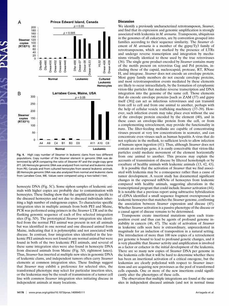
qPCR reactions with genomic DNA, normalizing to a single-copygene, elongation factor 1 (EF1). DNA from hemocytes of sixhealthy clams from PEI gave a signal of ∼3–10 copies per haploidgenome, consistent with the findings from the Southern blots.DNAs from hemocytes of diseased animals, assayed either asprimary cells (n = 4) or after culturing (n = 3), yielded copynumbers ranging from 100 to 200 (Fig. 4A). Additionally, clamsfrom an independent population of M. arenaria from LarrabeeCove, ME were assayed for diseased status, and hemocyte DNAwas analyzed for Steamer copy number. This isolated populationconfirmed the strong association between Steamer DNA copynumber in hemocytes and disease, with 3–10 copies in normalanimals and 150–300 copies in diseased hemocytes (Fig. 4B). Thecombined Southern and qPCR data suggest that Steamer is anextraordinarily active retrotransposon in diseased animals andundergoes massive expansion and integration into the soft shellclam genome in tumor cells.To determine the structure of the Steamer DNAs in more de-
tail, we used inverse PCR to amplify the Steamer integration sites(Fig. 5A). DNA of a healthy clam yielded a single major and someminor PCR products (Fig. 5B). The DNA sequence of the majorproduct revealed integration site junctions corresponding to thepredicted LTR 5′ and 3′ ends, and a 5-bp direct repeat flankingthe integration site (Fig. 5D). This specific element was cloned
using flanking primers, yielding a full-length Steamer retro-transposon (4,968 bp) with an intact Gag-Pol reading frame. Theoriginal cDNA products obtained by RT-PCR were nearly identicalto the corresponding regions of the genomic sequence (2,453identical out of 2,457 bp). We subsequently searched the original454 sequence reads for matches to the genomic sequence andfound 63 fragments spanning 3,409 bp of the genome with 99%identity. This full-length sequence was thus selected as definingthe prototypical Steamer endogenous retrotransposon (GenBankaccession no. KF319019; Fig. S1).Inverse PCR of two diseased animals from PEI amplified a large
number of integration sites (Fig. 5B), and several were cloned andsequenced from each animal (examples shown in Fig. 5D). InversePCR was also carried on DNA from both hemocytes and siphontissue, of both normal and diseased clams from Maine. Normalanimals showed a small number of integration sites in both tissues,and diseased animals showed small numbers in siphon, but a largeincrease in integration sites can be seen specifically in the leukemic
Fig. 2. Elevated expression of Steamer RNA correlates with disease status. RNAwas extracted fromhemocytes obtained fromnormal (n=43),moderate (n= 10),and heavily leukemic (n = 21) individuals collected from different sites in theNorth Atlantic. Steamer RNA levels were measured using qRT-PCR and the rel-ative standard curve method. The results are expressed as relative levels com-pared with EF1mRNA and are shown on y axis log scale. Each circle, square, andtriangle represents RNA from a single individual animal. The geometric meanvalues, indicated by the horizontal line, were compared by two-tailed t test.
Fig. 3. Leukemic hemocytes have high copy numbers of Steamer DNA. Thepresence of Steamer in genomic DNA of four normal (N) and a leukemic (L)soft shell clam was analyzed by Southern blotting. (A) Schematic represen-tation of the Steamer retrotransposon. LTRs at the 5′ and 3′ ends, Gag-PolORF, sites for digestion by the indicated restriction enzymes, and location ofthe 32P-labeled probe are indicated. Nucleotide positions are relative to thefirst nucleotide of the U3 portion of the 5′ LTR. (B) Genomic DNA of fournormal (Nor1-4) and one heavily leukemic animal (Dnear-HL03) weredigested with restriction enzymes BamHI, releasing left-junction fragments,or with DraI, releasing an internal fragment, and analyzed by Southern blot.DNA loadings were equal as judged by ethidium stain (not shown). (C) Ge-nomic DNA from two normal individuals (Nor1-2) and three leukemic indi-viduals (Dnear-HL03, Dnear-07, and Dnear-08) were digested with KpnI,releasing an internal fragment, and analyzed by Southern Blot. The migra-tion of the DNA molecular markers is indicated at the left of the panels, andmajor fragment recognized by the probe is indicated by *.
Arriagada et al. PNAS | September 30, 2014 | vol. 111 | no. 39 | 14177
GEN
ETICS
hemocyte DNA (Fig. 5C). Some siphon samples of leukemic ani-mals with higher copies are probably due to contamination withhemocytes. These finding show that the amplification is specific tothe diseased hemocytes and not due to diseased individuals inher-iting a high number of endogenous copies. To characterize specificintegration sites in multiple animals from both PEI and Maine,PCR was performed using primers in the Steamer LTR and in theflanking genomic sequence of each of five selected integrationsites (Fig. 5D). The prototypical Steamer integration site identi-fied from the normal PEI animal was present in not all animalsbut was identified in one normal and one diseased animal fromMaine, indicating that it is polymorphic and not associated withdisease. In contrast, four integration sites identified in the leu-kemic PEI animals were not found in any normal animal but werefound in both of the two leukemic PEI animals, and several ofthese same integration sites were also found in hemocyte DNAfrom diseased animals from Maine (Fig. 5D, rightmost lanes).Thus, Steamer has inserted at multiple new sites in genomic DNAof leukemic clams, and independent tumors often carry Steamerelements at common integration sites. These findings suggestthat Steamer may exhibit a preference for selected sites, thetransformed phenotype may select for particular insertion sites,or the leukemias may be the result of transmission of a tumor cellline with common Steamer integration sites initiating disease inindependent animals at many locations.
DiscussionWe identify a previously uncharacterized retrotransposon, Steamer,and find that its expression and genomic amplification is stronglyassociated with leukemia inM. arenaria. Transposons, ubiquitousin the genomes of all eukaryotes, are by convention grouped intofamilies according to their sequence similarity. The Steamer el-ement of M. arenaria is a member of the gypsy/Ty3 family ofretrotransposons, which are marked by the presence of LTRsand undergo reverse transcription and integration by mecha-nisms virtually identical to those used by the true retroviruses(36). The single gene product encoded by Steamer contains manyof the motifs present on retrovirus Gag and Pol proteins, in-cluding those of the capsid, nucleocapsid, protease, RT, RNaseH, and integrase. Steamer does not encode an envelope protein.Most gypsy family members do not encode envelope proteins,and most retrotransposition events mediated by these elementsare likely to occur intracellularly, by the formation of cytoplasmicvirion-like particles that mediate reverse transcription and DNAintegration into the genome of the same cell. Those elementsthat do encode envelope proteins [such as ZAM (37) and gypsyitself (38)] can act as infectious retroviruses and can transmitfrom cell to cell and from one animal to another, perhaps withthe help of cellular vesicle trafficking machinery (37–39). How-ever, such infection events may take place even without the useof the envelope protein encoded by the element (40), and inthese cases an envelope-like protein from the cell, or froma complementing retroelement, may provide the functionality intrans. The filter-feeding mollusks are capable of concentratingviruses present at very low concentrations in seawater, and canconcentrate even viruses such as human hepatitis A virus that donot replicate in the mollusk, to sufficient levels to allow infectionof humans upon ingestion (41). Thus, although Steamer does notcontain an envelope gene, it is easily conceivable that virion-likeparticles could mediate movement of the element horizontallyfrom one animal to another. This process may explain theaccounts of transmission of disease by filtered hemolymph or bycoculture of healthy animals with leukemic animals (13, 42, 43).It is possible that the activation of Steamer that we find associ-
ated with leukemia may be a consequence rather than a cause oftumor development. A recent study has documented significantchanges in the expressed mRNAs of hemocytes from leukemiccompared with healthy animals, suggesting alterations in thetranscriptional program that could include Steamer activation (44).It is notable that a previous report using subtractive hybridizationof cDNA identified a small sequence fragment overexpressed inleukemic hemocytes that matches the Steamer genome, confirmingthe association between Steamer expression and disease (45).Whether Steamer activation is a passive phenotype of the disease ora causal agent of disease remains to be determined.Transposons create insertional mutations upon each trans-
position event and thus can be agents of profound genome in-stability in cancers (46, 47). The scale of activation of Steamerin leukemic cells seen here is extraordinary, unprecedented inmagnitude for an induction of transposition in a natural setting.The introduction of more than 100 new copies of a retroelementper genome is bound to lead to profound genetic changes, and itis very plausible that Steamer activity and amplification is involvedas a factor or cofactor in the initial development of the leukemia.There are so many new copies of Steamer DNA per genome inthe leukemia cells that it will be hard to determine whether therehas been an insertional activation of a critical oncogene, but theleukemias are clearly polyclonal with respect to Steamer inser-tions and are acquiring new proviruses as the pool of transformedcells expands. One or more of the new insertions could signifi-cantly alter the phenotypes of these cells.The observation that multiple integrations are found at the same
sites in independent diseased animals (and not in normal tissue
Fig. 4. High copy number of Steamer in leukemic clams from two differentpopulations. Copy number of the Steamer element in genomic DNA was de-termined by qPCR comparing the ratio of Steamer RT and the single-copy geneEF1. (A) Hemocyte genomic DNAwas analyzed from normal and leukemic clamsfrom PEI, Canada and from cultured hemocytes from several leukemic animals.(B) Hemocyte genomic DNAwas also analyzed from normal and leukemic clamsfrom Larrabee Cove, ME. Values were compared using a two-tailed t test.
14178 | www.pnas.org/cgi/doi/10.1073/pnas.1409945111 Arriagada et al.

from the same animals) is intriguing. It is possible that Steamerintegration events may be occurring in a targeted fashion, or thatparticular sites are selected as part of the process of oncogenesis.Another possible explanation is that a primary leukemia with aprimordial repertoire of integration sites is spread between animalsas a transmissible leukemia cell line, similar to a transmissible ca-nine venereal tumor (48), or to a facial tumor disease of the Tas-manian devil (49). Such a mechanism would not explain thetransmission of disease by filtered hemolymph or the induction ofdisease by BrdU.Endogenous retroviruses and retroelements in mammals are
often induced by DNA damaging agents, notably halogenatednucleosides such as BrdU and iododeoxyuridine, and this in-duction can be enhanced by polycyclic hydrocarbons (50). Thus,exposures to environmental toxins may be triggers for the acti-vation of Steamer and disease. An induction of Steamer eitherearly or late in the course of disease would induce rapid geneticinstability and so could accelerate or promote disease progression.This scenario may account for the ability of BrdU to experimen-tally induce disease in clams (17).
Recent studies have shown that some clam populations aremore susceptible than others to induction of disease by DNAdamaging agents (16). If Steamer is responsible for the disease,susceptible populations may harbor a higher copy number ofSteamer or distinctive copies that are more readily induced forexpression. Both inheritance of a high number of endogenouscopies of the element and somatic amplification of the elementwithin individuals could contribute to development of disease.The availability of sequence probes for Steamer will allow surveysof the prevalence of the element in various populations, tests ofexperimental transmission from animal to animal, and furthertests for its functional involvement with disease. Because genomesof M. arenaria are highly polymorphic for the Steamer element,the DNA probes should also allow the development of pop-ulations of M. arenaria that lack the element entirely throughselective breeding, and such element-free populations may beless prone to induction of leukemia by environmental stresses.The identification of Steamer and its dramatic amplification inleukemia provides a potential marker for the disease and a re-markable example of catastrophic genomic instability causedby retroelements.
Fig. 5. Identification of Steamer integration sites in normal and diseased clams. Inverse PCR was used to clone and sequence integration sites of theSteamer retroelement. (A) Schematic of inverse PCR methodology: genomic DNA was digested with MfeI (cleaving only in the flanking DNA), circularized byligation, and redigested with NsiI at internal sites (N), and finally PCR was performed with outward-directed LTR primers. (B) Inverse PCR was performedusing mantle tissue DNA of one normal animal (N), and hemocyte DNA of two heavily leukemic animals (L) from PEI, Canada, and the PCR products wereanalyzed by agarose gel electrophoresis. For WfarNM01, the white arrowhead marks amplification of the internal Steamer sequence (due to incompleteNsiI cleavage) and the black arrowhead marks the junction product of a single Steamer copy. The leukemic samples yielded a large number of hetero-geneous junction products. (C) Inverse PCR was performed using DNA from both siphon tissue and hemocytes for normal and leukemic animals from Maine,USA. (D) Representative DNA sequences of individual cloned integration sites from normal (one site) and leukemic DNAs (two sites from each leukemic PEIanimal) are shown. The genomic DNA flanking sequences, the 5-bp duplicated repeats, and the Steamer termini are shown. The presence of the integrationsites in the source DNAs and in DNA from other animals was determined for each of the sequences shown by a diagnostic PCR using a forward primer in theSteamer LTR and a reverse primer in the flanking genomic DNA (Right; products are ∼150 bp and marked with a filled triangle). Amplification of EF1 isincluded as a control.
Arriagada et al. PNAS | September 30, 2014 | vol. 111 | no. 39 | 14179
GEN
ETICS

Materials and MethodsM. arenaria Collection. M. arenaria were collected from PEI, Canada andLarrabee Cove, ME and diagnosed by microscopic analysis of cell morphol-ogy as described in SI Materials and Methods.
Molecular Analyses. RT activity assays, 454 sequencing, genome walking,Southern blot analysis, qRT-PCR, qPCR, and inverse PCR were conducted bystandard methods using the specific primers and conditions described indetail in SI Materials and Methods.
ACKNOWLEDGMENTS. We thank Dr. Brian Beal (University of Maine atMachias) for obtaining and providing us with clams from Maine, and NiklasThalén for assistance in integration site analysis. This work was supportedby the Howard Hughes Medical Institute (G.A., M.J.M., and S.P.G.), the PewFellows Program (G.A.), Fondo Nacional de Desarrollo Cientifico y Tecnolo-gico Grant 1130852 (to G.A.), National Institutes of Health (NIH) TrainingGrant T32 CA009503 (to M.J.M.), and awards from the National ScienceFoundation (to C.R.), Strategic Applications of Genomics in the Environment(to J.S.), the Pesticide Science Fund (to J.S. and C.R.), and NIH (to C.S.and W.I.L.).
1. Barber B (2004) Neoplastic diseases of commercially important marine bivalves. AquatLiving Resour 17(4):449–466.
2. Cooper K, Brown RS, Chang PW (1982) Accuracy of blood cytologial screening tech-niques for the diagnosis of a possible hematopoetic neoplasm in the bivalve mollusc,Mya arenaria. J Invertebr Pathol 39(3):281–289.
3. Lowe DM, Moore MN (1978) Cytology and quantitative cytochemistry of a poliferativeatypical hemocytic condition in Mytilus edulis (Bivalvia, mollusca). J Natl Cancer Inst60(6):1455–1459.
4. Reno PW, House M, Illingworth A (1994) Flow cytometry and chromosome analysis ofSoftshell clams, Mya arenaria, with disseminated neoplasia. J Invertebr Pathol 64(3):163–172.
5. Walker C, et al. (2011) p53 superfamily proteins in marine bivalve cancer and stressbiology. Adv Marine Biol 59:1–36.
6. Miosky DL, Smolowitz RM, Reinisch CL (1989) Leukemia cell specific protein of thebivalve mollusc Mya arenaria. J Invertebr Pathol 53(1):32–40.
7. Reinisch CL, Charles AM, Troutner J (1983) Unique antigens on neoplastic cells of thesoft shell clam Mya arenaria. Dev Comp Immunol 7(1):33–39.
8. Smolowitz RM, Reinisch CL (1993) A novel adhesion protein expressed by ciliatedepithelium, hemocytes, and leukemia cells in soft-shell clams. Dev Comp Immunol17(6):475–481.
9. White MK, Miosky D, Flessas DA, Reinisch CL (1993) The expression of an adhesion-related protein by clam hemocytes. J Invertebr Pathol 61(3):253–259.
10. Muttray A, et al. (2012) Haemocytic leukemia in Prince Edward Island (PEI) soft shellclam (Mya arenaria): Spatial distribution in agriculturally impacted estuaries. Sci TotalEnviron 424:130–142.
11. Pariseau J, et al. (2009) Potential link between exposure to fungicides chlorothaloniland mancozeb and haemic neoplasia development in the soft-shell clam Mya are-naria: A laboratory experiment. Mar Pollut Bull 58(4):503–514.
12. Schneider KR (2008) Heat stress in the intertidal: Comparing survival and growth of aninvasive and native mussel under a variety of thermal conditions. Biol Bull 215(3):253–264.
13. Collins CM, Mulcahy MF (2003) Cell-free transmission of a haemic neoplasm in thecockle Cerastoderma edule. Dis Aquat Organ 54(1):61–67.
14. Oprandy JJ, et al. (1981) Isolation of a viral agent causing hematopoietic neoplasia inthe soft-shell clam Mya arenaria. J Invertebr Pathol 34(1):45–51.
15. Walker C, et al. (2009) Mass culture and characterization of tumor cells from a natu-rally occurring invertebrate cancer model: Applications for human and animal diseaseand environmental health. Biol Bull 216(1):23–39.
16. Taraska NG, Anne Böttger S (2013) Selective initiation and transmission of dissemi-nated neoplasia in the soft shell clam Mya arenaria dependent on natural diseaseprevalence and animal size. J Invertebr Pathol 112(1):94–101.
17. Oprandy JJ, Chang PW (1983) 5-bromodeoxyuridine induction of hematopoieticneoplasia and retrovirus activation in the soft-shell clam, Mya arenaria. J InvertebrPathol 42(2):196–206.
18. Romalde JL, et al. (2007) Evidence of retroviral etiology for disseminated neoplasia incockles (Cerastoderma edule). J Invertebr Pathol 94(2):95–101.
19. AboElkhair M, et al. (2009) Reverse transcriptase activity associated with haemicneoplasia in the soft-shell clam Mya arenaria. Dis Aquat Organ 84(1):57–63.
20. AboElkhair M, et al. (2009) Reverse transcriptase activity in tissues of the soft shellclamMya arenaria affected with haemic neoplasia. J Invertebr Pathol 102(2):133–140.
21. House ML, Kim CH, Reno PW (1998) Soft shell clams Mya arenaria with disseminatedneoplasia demonstrate reverse transcriptase activity. Dis Aquat Organ 34(3):187–192.
22. AboElkhair M, et al. (2012) Lack of detection of a putative retrovirus associated withhaemic neoplasia in the soft shell clam Mya arenaria. J Invertebr Pathol 109(1):97–104.
23. Goff S, Traktman P, Baltimore D (1981) Isolation and properties of Moloney murineleukemia virus mutants: Use of a rapid assay for release of virion reverse transcriptase.J Virol 38(1):239–248.
24. Craven RC, Leure-duPree AE, Weldon RA, Jr, Wills JW (1995) Genetic analysis of themajor homology region of the Rous sarcoma virus Gag protein. J Virol 69(7):4213–4227.
25. Loeb DD, Hutchison CA, 3rd, Edgell MH, Farmerie WG, Swanstrom R (1989) Muta-tional analysis of human immunodeficiency virus type 1 protease suggests functionalhomology with aspartic proteinases. J Virol 63(1):111–121.
26. Yuki S, Ishimaru S, Inouye S, Saigo K (1986) Identification of genes for reverse tran-scriptase-like enzymes in two Drosophila retrotransposons, 412 and gypsy; a rapid
detection method of reverse transcriptase genes using YXDD box probes. NucleicAcids Res 14(7):3017–3030.
27. Kanaya S, et al. (1990) Identification of the amino acid residues involved in an activesite of Escherichia coli ribonuclease H by site-directed mutagenesis. J Biol Chem265(8):4615–4621.
28. Kulkosky J, Jones KS, Katz RA, Mack JP, Skalka AM (1992) Residues critical forretroviral integrative recombination in a region that is highly conserved amongretroviral/retrotransposon integrases and bacterial insertion sequence trans-posases. Mol Cell Biol 12(5):2331–2338.
29. Chan PP, Lowe TM (2009) GtRNAdb: A database of transfer RNA genes detected ingenomic sequence. Nucleic Acids Res 37(Database issue):D93–D97.
30. Sorge J, Hughes SH (1982) Polypurine tract adjacent to the U3 region of the Roussarcoma virus genome provides a cis-acting function. J Virol 43(2):482–488.
31. Guindon S, et al. (2010) New algorithms and methods to estimate maximum-likelihood phylogenies: Assessing the performance of PhyML 3.0. Syst Biol 59(3):307–321.
32. Llorens C, et al. (2011) The Gypsy Database (GyDB) of mobile genetic elements: Re-lease 2.0. Nucleic Acids Res 39(Database issue):D70–D74.
33. Michaille JJ, Mathavan S, Gaillard J, Garel A (1990) The complete sequence of mag,a new retrotransposon in Bombyx mori. Nucleic Acids Res 18(3):674.
34. Springer MS, Davidson EH, Britten RJ (1991) Retroviral-like element in a marine in-vertebrate. Proc Natl Acad Sci USA 88(19):8401–8404.
35. Gonzalez P, Lessios HA (1999) Evolution of sea urchin retroviral-like (SURL) elements:Evidence from 40 echinoid species. Mol Biol Evol 16(7):938–952.
36. Levin HL (2002) Newly identified retrovtransposons of the Ty3/gypsy class in fungi, plants,and vertebrates. Mobile DNA II, eds Craig NL, Craigie R, Gellert M, Lambowitz AM (ASM,Washington, DC), pp 684–701.
37. Brasset E, et al. (2006) Viral particles of the endogenous retrovirus ZAM from Dro-sophila melanogaster use a pre-existing endosome/exosome pathway for transfer tothe oocyte. Retrovirology 3:25.
38. Song SU, Gerasimova T, Kurkulos M, Boeke JD, Corces VG (1994) An env-like proteinencoded by a Drosophila retroelement: Evidence that gypsy is an infectious retrovirus.Genes Dev 8(17):2046–2057.
39. Kim A, et al. (1994) Retroviruses in invertebrates: The gypsy retrotransposon is ap-parently an infectious retrovirus of Drosophila melanogaster. Proc Natl Acad Sci USA91(4):1285–1289.
40. Chalvet F, et al. (1999) Proviral amplification of the Gypsy endogenous retrovirus ofDrosophila melanogaster involves env-independent invasion of the female germline.EMBO J 18(9):2659–2669.
41. Ruddy SJ, et al. (1969) An epidemic of clam-associated hepatitis. JAMA 208(4):649–655.42. Elston RA, Kent ML, Drum AS (1988) Transmission of hemic neoplasia in the bay
mussel, Mytilus edulis, using whole cells and cell homogenate. Dev Comp Immunol12(4):719–727.
43. McLaughlin SM, Farley CA, Hetrick FM (1992) Transmission studies of sarcoma in thesoft-shell clam, Mya arenaria. In Vivo 6(4):367–370.
44. Siah A, McKenna P, Berthe FC, Afonso LOB, Danger JM (2013) Transcriptome analysisof neoplastic hemocytes in soft-shell clamsMya arenaria: Focus on cell cycle molecularmechanism. Res Immunol 3:95–103.
45. Siah A, McKenna P, Danger JM, Johnson GR, Berthe FC (2011) Induction of trans-posase and polyprotein RNA levels in disseminated neoplastic hemocytes of soft-shellclams: Mya arenaria. Dev Comp Immunol 35(2):151–154.
46. Inaki K, Liu ET (2012) Structural mutations in cancer: mechanistic and functional in-sights. Trends Genet 28(11):550–559.
47. Solyom S, et al. (2012) Extensive somatic L1 retrotransposition in colorectal tumors.Genome Res 22(12):2328–2338.
48. Cohen D (1985) The canine transmissible venereal tumor: A unique result of tumorprogression. Adv Cancer Res 43:75–112.
49. Pearse AM, Swift K (2006) Allograft theory: Transmission of devil facial-tumour dis-ease. Nature 439(7076):549.
50. Yoshikura H, et al. (1977) Enhancement of 5-iododeoxyuridine-induced endogenousC-type virus activation by polycyclic hydrocarbons: Apparent lack of parallelism be-tween enhancement and carcinogenicity. J Natl Cancer Inst 58(4):1035–1040.
14180 | www.pnas.org/cgi/doi/10.1073/pnas.1409945111 Arriagada et al.