genome-wide analysis of multiprotein … analysis of multiprotein complexes and metalloproteins in...
TRANSCRIPT

GENOME-WIDE ANALYSIS OF MULTIPROTEIN COMPLEXES AND
METALLOPROTEINS IN THE HYPERTHERMOPHILIC ARCHAEON PYROCOCCUS
FURIOSUS
by
JOSEPH WALKER SCOTT
(Under the Direction of Michael W. W. Adams)
ABSTRACT
Multiprotein complexes and metalloproteins largely determine the metabolic potential of
an organism. The genes that encode multiprotein complexes and metalloproteins in any
organism are largely unknown. Predicting what these genes are from genome sequence data is
virtually impossible due to great diversity of amino acids sequences that are involved in protein
oligomerization and metal binding. In this dissertation, an experimental method is presented that
circumvents this problem by the direct purification of multiprotein complexes and
metalloproteins from native microbial biomass. The biomass of a model microorganism,
Pyrococcus furiosus, was fractionated by non-denaturing multistep chromatography and
chromatographic fractions were analysed by mass spectrometry to identify proteins that co-
eluted in common fractions. Potential multiprotein complex identification was based on the
cofractionation of proteins encoded by adjacent genes in the P. furiosus genome. A total of 106
potential heteromeric multiprotein complexes were identified, of which only 20 were already
known or predicted. One of them, PC-81 (PF1838/PF1837) was of interest because of its
homology to two previously purified heterotetrameric () enzymes termed ACS1

(PF1540/PF1787) and ACS2 (PF1540/PF1837). Since PC-81 and ACS2 had a common -
subunit (PF1837), we recombinantly expressed in E. coli all ten ACS complexes from the five
subunits and two subunits encoded in the P. furiosus genome. All ten enzymes were active
with varying but overlapping substrate specificities. Metalloproteins were identified in a similar
manner except that fractions were analyzed by ICP-MS in addition to peptide mass spectrometry.
Of the 343 metal peaks identified during biomass fractionation, 158 were not associated with a
known metalloprotein. Two metalloproteins were purified to homogeneity. One of them,
PF0086 or alanyl-tRNA synthetase COOH terminus had been predicted to contain zinc.
However, it co-purified with nickel rather than a zinc peak through three chromatography steps.
The other, PF1972 or anaerobic ribonucleotide reductase activase, is a known iron protein that
very surprisingly co-purified with molybdenum as well as iron through five chromatography
steps. This method therefore identified one new nickel and one new molybdenum-containing
protein from P. furiosus, in addition to approximately 100 new multiprotein complexes, thereby
demonstrating the general applicability of this approach.
INDEX WORDS: Hyperthermophilic archaea, Pyrococcus furiosus, multiprotein complexes,
metalloproteome, nickel, molybdenum, proteomics, genomics, acetyl-CoA
synthetase, alanyl-tRNA synthetase, anaerobic ribonucleotide reductase
activase, glycolysis, gluconeogenesis, GTP-dependent
phophoenolpyurvate carboxykinase, ATP, GTP, biomass fractionation,
mass spectrometry, ICP-MS

GENOME-WIDE ANALYSIS OF MULTIPROTEIN COMPLEXES AND
METALLOPROTEINS IN THE HYPERTHERMOPHILIC ARCHAEON PYROCOCCUS
FURIOSUS
by
JOSEPH WALKER SCOTT
B.S., University of Arkansas, 1998
M.S., University of Florida, 2002
A Dissertation Submitted to the Graduate Faculty of The University of Georgia in Partial
Fulfillment of the Requirements for the Degree
DOCTOR OF PHILOSOPHY
ATHENS, GEORGIA
2014

© 2014
Joseph Walker Scott
All Rights Reserved
GENOME-WIDE ANALYSIS OF MULTIPROTEIN COMPLEXES AND
METALLOPROTEINS IN THE HYPERTHERMOPHILIC ARCHAEON PYROCOCCUS
FURIOSUS
by
JOSEPH WALKER SCOTT
Major Professor: Michael W. W. Adams
Committee: Michael K. Johnson
Robert. A. Scott
William B. Whitman
Electronic Version Approved:
Maureen Grasso
Dean of the Graduate School
The University of Georgia
May 2014

iv
DEDICATION
This dissertation is dedicated to Janie Walker.

v
ACKNOWLEDGEMENTS
I must acknowledge those who encouraged me to continue when I did not think I even
cared anymore. Names that readily come to mind are Dr. Michael W. W. Adams, Dr. Angeli Lal
Menon, and Mr. Farris Poole II.

vi
TABLE OF CONTENTS
Page
ACKNOWLEDGEMENTS .............................................................................................................v
LIST OF TABLES ....................................................................................................................... viii
LIST OF FIGURES ....................................................................................................................... ix
CHAPTER
1 INTRODUCTION AND LITERATURE REVIEW .....................................................1
Genomics .................................................................................................................1
Pyroccous furiosus as a model organism .................................................................7
Multiprotein complexes .........................................................................................12
Metalloproteins ......................................................................................................15
Predictive strategies to identify protein complexes ...............................................18
Methods that detect protein complexes ..................................................................21
Summary and objectives ........................................................................................25
References ..............................................................................................................26
2 IDENTIFYING NOVEL PROTEIN COMPLEXES IN THE PROTEOME OF THE
HYPERTHERMOPHILIC ARCHAEON PYROCOCCUS FURIOSUS .....................39
Introduction ............................................................................................................39 Introduction 41
Materials and Methods ...........................................................................................40
Results and Discussion ..........................................................................................44
References ..............................................................................................................67

vii
3 CHARACTERIZATION OF TEN HETEROTETRAMERIC NDP-DEPENDENT
ACYL-COA SYNTHETASES OF THE HYPERTHERMOPHILIC ARCHAEON
PYROCOCCUS FURIOUSUS ...............................................................................74
Abstract ..................................................................................................................75
Introduction ............................................................................................................75
Materials and Methods ...........................................................................................79
Results and Discussion ..........................................................................................82
References ............................................................................................................101
4 IDENTIFYING NOVEL METALLOPROTEINS IN THE PROTEOME OF THE
HYPERTHERMOPHILIC ARCHAEON PYROCOCCUS FURIOSUS .............107
Introduction ..........................................................................................................107 Introduction 112
Materials and Methods .........................................................................................112
Results and Discussion ........................................................................................116
References ............................................................................................................124
5 IDENTIFICATION OF A NOVEL NICKEL PROTEIN AND A NOVEL
MOLYBDENUM PROTEIN IN THE HYPERTHERMOPHILIC ARCHAEON
PYROCOCCUS FURIOSUS ................................................................................132
Introduction ..........................................................................................................132 Introduction 138
Materials and Methods .........................................................................................134
Results and Discussion ........................................................................................143
References ............................................................................................................168
6 DISCUSSION AND CONCLUSIONS .....................................................................171
References ............................................................................................................184

viii
LIST OF TABLES
Page
Table 1.1: Examples of –omics technologies ..................................................................................6
Table 1.2: Predicted versus measured metal contents of recombinant Pyrococcus furiosus
proteins ...............................................................................................................................17
Table 1.3: ORFs whose gene expression is dramatically up regulated in peptide-grown cells and
their potential operon arrangement ....................................................................................19
Table 1.4: Comparison of methods to detect protein-protein interactions .....................................22
Table 2.1: Known PCs ...................................................................................................................50
Table 2.2: Expected PCs ................................................................................................................51
Table 2.3: Weak and Unlikely PCs ................................................................................................53
Table 2.4: Tentative PCs ................................................................................................................54
Table 2.5: Strong PCs ....................................................................................................................56
Table 3.1: Kinetic parameters of ACSx-A isoforms with CoA derivatives ..................................91
Table 3.2: Nucleotide kinetics of ACS1 isoforms .........................................................................93
Table 4.1: Characterized metalloproteins of P. furiosus..............................................................109
Table 4.2: Number of metal peaks detected from primary and secondary fractionations ...........120
Table 4.3: Metal peak detection in P. furiosus, E. coli, and S. solfataricus ................................123

ix
LIST OF FIGURES
Page
Figure 1.1: Completed genome in genome on-line database by year ..............................................2
Figure 1.2: Universal phylogenetic tree ...........................................................................................9
Figure 2.1: Fractonation schemes of P. furiosus cytoplasmic protein through multiple
chrmomatographic steps ....................................................................................................41
Figure 2.2: Elution profile of the P. furiosus cell free extract fractionation on a DEAE column .46
Figure 2.3: Proposed pathway of peptide metabolism in fermentative hyperthermophilic archaea ..
........................................................................................................................................................62
Figure 3.1: SDS-PAGE analyses of recombinant P. furiosus ACS isoenzymes ...........................84
Figure 3.2: Heat map of activities of ten ACS isoenzymes with eighteen organic acids with ATP
or GTP as cosubstrates .......................................................................................................86
Figure 3.3: Proposed function of ACS-G and ACS -A isoenzymes during growth of
P. furiosus on carbohydrates or peptides ...........................................................................95
Figure 3.4: Phylogenetic tree of ACS subunits ..........................................................................99
Figure 4.1: Fractionation scheme of P. furiosus cytoplasmic proteins through primary and
secondary purification steps .............................................................................................113
Figure 4.2: Metal concentration profiles of P. furiosus extract after DEAE sepharose
fractionation of cytoplasmic extract.................................................................................118
Figure 5.1: Nickel concentration profile and unique peptide of P. furiosus cell free extract after
DEAE fractionation .........................................................................................................144

x
Figure 5.2: Nickel and peptide elution of the DEAE fraction pool after chromatography using a
hydroxyapatite column.....................................................................................................146
Figure 5.3: Purification and analysis of nickel peak C fractions of size exclusion column ........149
Figure 5.4: SDS-gel band MALDI-TOF analysis of the nickel peak fraction .............................151
Figure 5.5: Metal content of recombinant PF0086 purified from E. coli cells grown in rich media
or rich media supplemented with either 200 M Ni, Co, or Zn ......................................154
Figure 5.6: PF0086 has four paralogs in the P. furiosus genome ................................................156
Figure 5.7: Purification of the novel molybdenum protein PF1972 ............................................160
Figure 5.8: Amino acid alignment of anaerobic ribonucleotide reductase activases ...................162
Figure 5.9: Expression of PF1971 and PF1972 in E.coli analyzed by SDS-gel electrophoresis..165
Figure 6.1: Domain organization of NDP dependent acyl-CoA synthetases (ACSs)..................175
Figure 6.2: Amino acid alignments of A and G subunits of Thermococcales ACSs ..............179

1
CHAPTER 1
INTRODUCTION AND LITERATURE REVIEW
Genomics.
The amount of resources in money, people, and time invested into sequencing projects over the
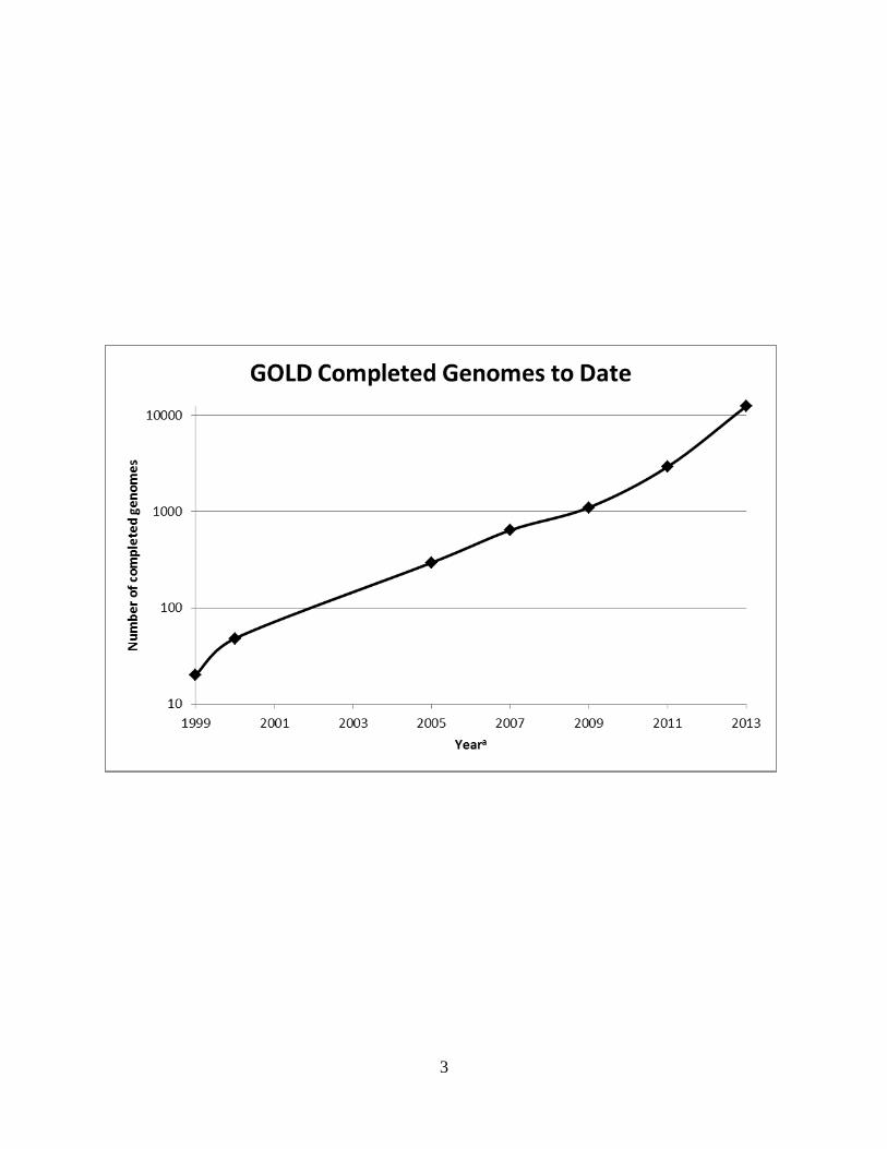
years reflects an expectation of a tremendous return. The number of sequenced genomes
increases exponentially each year (Figure 1.1). Over 12,000 genomes of individual organisms
have been sequenced with numerous, additional, sequencing projects underway (Genomes
Online Database). Sequencing of the human genome was predicted to take 15 years and to cost
$3 billion [1]. It involved an international effort from 20 research institutions in the United
States, United Kingdom, France, Germany, Japan, and China. The expected return from
sequencing the human genome was knowledge of the chromosomal location of every human
gene, the nucleotide sequence of those genes, and the relationship of the genes to disease.
Ideally, this knowledge would be helpful in assessing an individual’s susceptibility to diseases
while also predicting a patient’s response to medical treatments. A question more fundamental
than those concerning the relationship between human health and genetic make-up is the
underlying motivation that propels not only genomic studies, but all studies in the life sciences.
That question is: How does life maintain itself? The genome has been called “the blueprint of
life” [2]. This lofty designation suggests that DNA contains the information to build all the parts
necessary for the maintenance of life. Therefore, in theory a study of an organism’s genome
alone would facilitate prediction of the life processes in a biological system based upon the
principle of genes. However, since the completion of the map of the human genome in 2003,
some have suggested that many statements concerning the promise of genomics have been

2
Figure 1.1
Completed genomes in Genome On-line Database by year.
aReferences for given years. 1999 [3]; 2000 [4]; 2005 [5]; 2007 [6]; 2009 [7]; 2011 [8]
2013 Number taken from Genomes On-line Database on 12/27/2013.
3

4
premature. Eric Lander of the Whitehead institute has said: We’ve called the genome the
blueprint, the holy grail, all sorts of things. But it’s only a parts list. If I gave you the parts list
for the Boeing 777 and it has 100,000 parts, I don’t think you could screw it together, and you
certainly wouldn’t understand why it flew [9]. There are several reasons why the promised
benefits of genomics have been delayed. Some of which are 1) the functions of roughly one-half
of the genes in E. coli and S. cerevisiae (arguably the best studied organisms whose genomes
have been sequenced) have not been experimentally verified making it difficult to draw accurate
conclusions concerning the complex nature of biological systems [10-12]. 2) As of yet it is
difficult to discern sequence information concerning interaction between individual gene
products that form functional units. 3) Related to the previous point is the difficulty in
discerning post translational modifications such as phosphorylation, glycosylation, acetylation,
cleavage etc. that affects function. 4) Life is more than DNA. Life exists as an interplay
between DNA, its gene products, and the environment. Gene products may require certain
temperatures, proton concentrations, or small inorganic molecules such as salts or metal ions to
carry out biological functions which are needed to sustain life. The increasing number of
sequenced genomes has prompted a trend toward using genomic structure as a basis in
formulating and answering questions about cellular processes. This shift is a defining feature of
what has been termed the post-genomic era [13].
An obvious advantage to having the sequences of whole genomes available is that it
precludes the necessity to purify a protein and determine its amino acid sequence before cloning
the gene that expresses it. A simple search of a sequence database allows one to bypass those
steps and begin a cloning project by designing oligonuclotide primers based on the DNA
sequence of the gene of interest. Genome sequencing has also allowed for the development of

5
sub-disciplines with the common suffix of –omics which denotes their relationship to genomics.
Two common examples of -omics studies are transcriptomics and proteomics. Transcriptomics
is a discipline which involves the analysis of the RNAs expressed by a specific cell type or
organism at specific times or under a specific set of conditions. Likewise, proteomics studies the
complete set or subset of proteins that are expressed temporally and spatially. The –omics suffix
can be appended to any root that describes a collection of any biologically relevant molecules
(Table 1.1). Each of these disciplines complements genomics by helping to tease out higher
biological meaning from sequence data. A chief characteristic of -omics technologies is the
generation of a lot of data in a relatively short amount of time. For example whole-genome
transcriptomics experiments in the form of microarrays can be used to analyze thousands of
genes spotted onto chips which are used to disclose the expression patterns of all genes in a
particular genome. Likewise, in proteomics experiments, mass spectrometry can detect a
multitude of proteins expressed in a tissue, cell type, organelle, bacterium etc. Again, these
proteomics experiments are dependent on the completed genomic sequences of the organism
under study since the peptide mass fingerprint is searched against a data base of virtually
digested proteins that have been deduced from the genome sequence of the host organisms.
The high-throughput nature of the so called ‘-omics revolution’ has produced a study of biology
that increasingly has the salient feature of being data rather than hypothesis driven. Some in the
scientific community have expressed sentiments that data driven science has no merit [14].
Nobel Prize winning scientist, Arthur Kornberg does not go as far as suggesting genomics is
useless, however he bemoans what he sees as biochemistry ‘becoming less fashionable’ in the
post genomic era [15]. He states that the purification and characterization of enzymes from a
cell-free system “does something that proteomic and other ‘omics’ cannot yet do.” Although

6
Table 1.1 Example of –omics technologies
Discipline Subject of study
Genomics- DNA sequences, Gene organization
Epigenomics- DNA modifications, Modification of DNA binding proteins
Transcriptomics- Expression profiles of coding RNA. Also includes the study of non-coding RNA
Proteomics- Entire complement of proteins produced by an organism
Glycomics- Entire complement of sugar chains produced by an organism
Lipidomics- Entire complement of cellular lipids
Metabolomics- Entire complement of metabolites produced by an organism.
Metalloproteomics- Entire complement of metalloproteins produced by an organism

7
transcriptomic and proteomic experiments provide clues as to the functions of genes, the data
generated is similar to genome sequence data in that there is plenty of it and it does not provide
the information needed to discern the specific functions of the genes expressed.
On the other hand, it could be argued that a focus on the characterization of individual
proteins is too narrow to be able to represent how that protein functions in a network thereby
giving an incomplete picture of the role the protein plays in a cell [16]. The studies which are
the subject of this work reflect a view that old-fashioned biochemistry and the methods used in
the era of post genomics can be used in concert to achieve a comprehensive view of the
relationship between the genome, its gene products and the environment in which they function.
Pyrococcus furiosus as a model organism.
The haploid human genome is composed of 23 chromosomes, three billion pairs of nucleotides
and 20,000-30,000 protein encoding genes. Many of the genes are transcribed into mRNA that is
subject to splicing events and produce proteins which are different than would be predicted from
the DNA sequence. In addition, there are 210 distinct human cell types all having the same
DNA (with a few exceptions), but expressing genes differentially. Furthermore, 98 % of the
Human genome sequence is repeat elements, transposons and non-coding DNA and has been
referred to as ‘junk-DNA’. However, there have been developments that suggest possible
functions for non-coding DNA [17]. It is clear from the complexity and mere size of the human
genome that an effort to decipher it presents an almost insurmountable challenge for those
researchers who dare to study it.
Pyrococcus furious, a much simpler species than Homo sapiens, is a single celled
organism from the archaea domain of life. Interestingly the universal tree of life indicates that
the archaea and eucarya domains share a common line of evolution from the last universal
8
common ancestor (LUCA), while the bacteria domain follows its own line (Figure 1.2). Thus
although members of the bacteria and archaea domains are single celled prokaryotes, archaea are
more evolutionarily related to the eucarya than to bacteria. Pyrococcus furiosus was originally
isolated in 1986 from a shallow marine vent, but is commonly found in deep-sea vents [19]. P.
furiosus is a hyperthermophile with an optimal growth temperature of 100°C and is known as the
‘E. coli of life at 100°C’ because it is by far the best studied hyperthermophile. It has been the
subject of studies regarding metabolism, gene organization, genetics, physiology, and enzyme
characterization [20-24]. In fact, the experiments presented in this paper were performed in a
laboratory that has used P. furiosus as a model organism for over 20 years. In this lab, P.
furiosus has previously been the subject of a genomics project that aimed to determine the
structures of all the proteins expressed in its genome [25-27]. The premise of this structural
genomics project was based on the belief that proteins would provide structural information that
could be correlated to function so that rather than depending solely on sequence to decipher the
genome, protein structure could be used as well. One of the major hindrances in this effort was
that only about 25 % of the open-reading-frames in a given prokaryotic genome will yield stable,
soluble protein when expressed alone. This percentage does not include membrane proteins.
One explanation for this attrition rate is that these proteins are subunits that need their partners
for stability or that lack a post translational modification that is not performed in E. coli. The
genome of P. furiosus was sequenced in 2001 as part of a DOE funded project and was found to
have a size of 1.9 million base pairs which make up around 2100 open reading frames (ORF)
[28]. 59 % or 1233 of those ORFs are members of over 400 basic transcriptional units called
operons [29], a claim supported by ORFs either being within or overlapping by 16 nucleotides on
the same strand [30; 31] and by their co-regulation under differing growth conditions as

9
Figure 1.2
Universal Phylogenetic Tree
The bold lines represent the hyperthermophilic genera. This phylogenetic tree is adapted from
[18].

10
Green non-Sulfur
Bacteria
Gram positives
Proteobacteria
Cyanobacteri
Flavobacteria
Aquifex
Thermotog
Eukarya
Bacteria
Archaea
3- thermolithotrophicus;
2- igneus;
4- vanniellii
1- jannaschii;
Methanopyrus
Halobacterium
Methanoplanus
Methanospirillum
Methanosarcina
Thermo-
coccus
Sulfolobus
Desulfurococcus
Thermoproteus
Thermofilum
Methanobacterium
Pyrobaculum
Halococcus
Archaeoglobus
Pyrodictium
Methano-
thermus
Methanococcus
1
4
3 2
Pyro-
coccus

11
demonstrated from whole-genome transcriptomic analysis. In P. furiosus, most of the
characterized protein complexes are encoded by adjacent genes. Thus, the small, well-defined,
highly regulated genome of P. furiosus will aid in predicting the protein partners that are
required to be co-expressed to produce stable soluble protein. However, a well-defined genome
is not the same thing as a perfectly defined genome. That being said, there are some errors in the
P. furiosus sequence that add complications. The original genome of P. furiosus was
independently annotated by The Institute for Genomic Research (TIGR) and the National Center
for Biotechnology Information (NCBI). More than 500 of the annotated genes differed
significantly in size in the two databases [22]. Therefore, more direct experimental confirmation
on a broad scale would be useful not only in defining ORFs but also in defining the operons that
encode the proteins which make up multi-subunit complexes and modified proteins.
Purification of proteins from native biomass is the most direct method to determine which
protein complexes and protein modifications occur in the cell. P. furiosus can be routinely
grown in kilogram quantities in readily accessible fermentors, a capability which provides a
virtually limitless amount of biomass for protein purification and characterization [32].
The ability of P. furiosus to convert peptides and complex carbohydrates into hydrogen
makes it attractive from a biomass conversion standpoint and its ability to live at boiling
temperatures lends itself to the understanding of cellular processes at high temperatures. In
addition, some believe that hyperthermophiles, such as P. furious are the most slowly evolving
of all organisms and thus are representative of the most ancient organisms. It may be the case
that the first life forms originated from deep sea vent environments which are at temperature at
which CO2 fixation takes place abiotically [33-35]. Thus a greater understanding of the P.
furiosus genome could lend itself to a greater understanding of evolution.

12
Multiprotein complexes.
As our knowledge of protein structure increases, it is becoming more apparent that the
majority of proteins function in complexes with other proteins, while only a small fraction
function in isolation [36]. A lack of a protein partner during overexpression is a convincing
argument as to why the Structural Genomics effort had a high attrition rate. It has been
suggested that when a protein is in a complex, the possibility of denaturation is minimized due to
a reduced surface area exposed to solvent [37].
On the other hand, multiple subunits also make it possible for complexes to replace parts
of the complex if any subunit is damaged for a reason. If the enzyme was rather composed of a
single polypeptide any damage might destroy the function of the whole protein. Multi-subunit
complexes can form large molecular assemblies that function in cellular processes such as
protein synthesis [38; 39], cell motility [40], and extracelluar degradation of plant biomass [41].
Some even larger or supramolecular assemblies function in what are called metabolons. These
are temporary complexes of enzymes held together by noncovalent interactions and are involved
in a common pathway which allows for the efficient channeling of substrates from the beginning
to the end of the pathway [42-44]. Some molecular complexes are highly stable such as
ribosomes, while others are less so and possibly participate in transient interactions that are
important for signaling or regulatory networks. With this in mind, it becomes even more
apparent why a greater knowledge of the nature of protein complexes in P. furiosus would be
very helpful in learning more about the biology of this organism.
Protein complexes have been categorized in our lab into three types based upon the
organization of the genes that express their component subunits [45]. The complexes may be
either transient or permanent.

13
Type 1 complexes. These are heteromeric protein complexes encoded by at least two ORFs that
are adjacent in the genome. The 2-keto acid oxidoreductases (KOR) of P. furious are examples
of this type of complex [46-49]. These enzymes are involved in the oxidative decarboxylation of
2-keto acids which results in a coenzyme A derivative. The genome sequence of P. furiosus
indicates that the KORs are organized into operons. Four have been purified and characterized
[49]. Pyruvate ferredoxin oxidoreductase (POR), 2-ketoisovalerate ferredoxin oxidoreductase
(VOR), and 2-ketoglutarate ferredoxin oxidoreductase (KGOR) have 2222 quaternary
structures. The subunit of POR and VOR is shared by those two complexes. Indole pyruvate
ferredoxin oxidoreductase (IOR) has an 22 quaternary structure. However, the subunits appear
to be a fused mosaic of the homologous subunits of the other KORs. The P. furiosus genome
also encodes a fifth KOR homolog with genes immediately downstream of KGOR. Microarray
data show that this putative 2-keto acid ferredoxin oxidoreductase (XOR) is co-regulated with
KGOR and shares its subunit with it much like POR shares its subunit with VOR. [50] An
additional putative KOR homolog is encoded by the adjacent ORFs PF0753 and PF0754 [45].
This KOR has been partially purified from native P. furiosus biomass [45], however additional
experimental data about this complex is unavailable. With only two genes encoding the
complex, it may have a quaternary structure similar to IOR. It is evident that the genome
sequence can be a useful aid in predicting protein complexes when working with an organism
such as P. furiosus which has a small, well-defined, and highly regulated genome.
Type 2 complexes. The second type of complex that we have categorized is composed of
subunits that are encoded by genes which are not adjacent in the genome and therefore not co-
regulated in an operon [45]. Therefore, the genome sequence is not helpful in predicting these
types of complexes. Examples of this type of complex are acetyl-CoA synthetases (ACS) 1 and

14
2 [51]. ACS1 catalyzes the last step in sugar fermentation and fermentation of some amino
acids. ACS2 also catalyzes the last step in fermentation of some amino acids. These enzymes
were purified by following enzyme activity. They were found to be composed of two different
types of subunits in an 22 quaternary organization. N-terminal sequencing was used to
determine the amino acid sequences of the enzyme subunits. All subunits that made up the two
enzymes had different amino acid sequences. After the genome of P. furiosus was sequenced, it
was determined that ACS1 was composed of subunits encoded by PF1540 and PF1787 and
ACS2 was made of subunits encoded by PF0532 and PF1837. An observation of the gene
numbers makes it apparent that none of genes of either complex are adjacent in the genome.
Type 3 complexes. The third type of complex is composed of identical subunits. As opposed to
heteromers, type 3 complexes are homomers which are composed of subunits encoded by a
single gene. Given a polypeptide sequence, it is not clear what oligomerization state will be
formed if any. There are several techniques that are frequently used which can help determine
the oligomeric state. These include: analytical gel filtration chromatography, mass spectrometry,
analytical ultracentrifugation, and dynamic light scattering among others [52].
Homooligomerization allows cells to make larger protein complexes without requiring an
increase in genome size [36]. Most homooligomers are dimers although much larger complexes
do exist. An intriguing example of a very large complex produced in P. furiosus is the sulfur
induced protein A (Sip A) [53; 54]. PF2025 or sipA is upregulated by sulfide and elemental
sulfur. It encodes a 19 kDa protein that aggregates into a homooligomer of up to approximately
100 MDa and is composed of about 5,000 identical subunits and up to 45,000 iron sulfur
clusters. It has a unique, repetitive C-terminal tail that is required for polymerization [55].

15
However, the sequence in of itself provided no insight into its function. Of all three types of
complexes described, genome structure has the most bearing in predicting type 1 complexes.
Metalloproteins.
A study of the nutritional requirements of every life form would suggest that metal ions
are required for metabolic function. Bulk metals such as sodium, potassium, calcium, and
magnesium are present in organisms in relatively large amounts [56]. Bulk metals can be used to
maintain osmotic balance, carry charges across membranes, among other processes. The bulk
metals are not necessarily bound to organic molecules when performing their functions although
they often are. On the other hand, iron, zinc, copper, cobalt, nickel and other metals are present
at trace levels in cells, tissues and biological fluid [57]. The trace amounts of these metals infer a
function with proteins that are also present in ‘trace’ amounts [58]. The catalytic nature of
enzyme proteins allows them to function at low concentrations. Proteins that incorporate metal
ions into their structures are called metalloproteins. The assimilation and incorporation of metals
into proteins is the basis of many life functions including electron transfer, catalysis, and protein
structure stabilization. Some estimates suggest metalloproteins comprise up to 40 % of all
proteins [59]. The designation of metalloprotein has typically been conferred to a protein after
purification [60-62]. There are several informatics approaches to identify metalloproteins.
However, these methods are limited in that they depend heavily on homology to characterized
metalloproteins [60; 62-65]. One-third or more of genes in a typical cell encode proteins that
have not been experimentally verified to exist [66] so it is not known whether or not these genes
encode metalloproteins. The metal binding sites of most metalloproteins contribute only a small
number of ligands to coordinate the metal [67]. Because of the three dimensional folding of
proteins, placement of the metal amino acid ligands in the primary sequence can be varied to a
16
great degree. Therefore, it is very difficult to predict with available informatic methods which of
the hypothetical proteins are metalloproteins.
Another impediment to more robust predictive capabilities may be biased sampling of the
metalloproteins that have been structurally characterized [67]. During crystallization, crystals
are often soaked in heavy metals to aid in solving the protein structure. Therefore, those proteins
with binding sites that readily displace and exchange metals have an increased probability of
being solved than those proteins that do not release their metal. Those binding sites that do
readily exchange their metals may represent only a small proportion of the possible folds that do
bind metals.
Because of the abundance of protein needed for crystallization, recombinant
overexpression and production of proteins is often done heterologously in E. coli. Recombinant
metalloproteins are known to be often produced with misincorporated metals or without a metal
at all [68-71]. An illustration of this can be seen in a study that measured metal content in
recombinant P. furiosus proteins expressed in E. coli (Table 1.2). Of 45 putative P. furiosus
ironproteins expressed in E. coli only five were shown by ICP-MS to contain iron. Nine of the
45 contained zinc. On the other hand, of the 27 putative P. furiosus zinc proteins expressed in E.
coli, only 6 contained zinc and 6 contained iron. In addition, of 58 P. furiosus proteins predicted
to contain metals other than iron or zinc, four of those heterologously expressed in E. coli
contained iron and eight contained zinc. There are 840 hypothetically conserved ORFs in the
genome with no prediction for metal content. Of 37 of these heterologously expressed in E. coli,

17
Table 1.2. Predicted versus measured metal contents of recombinant Pyrococcus furiousus proteins.
Predicted metal content # ORFs in Pf Genome predicted from InterPro #Pf ORFs
expressed in E. coli
# proteins containing
Fe Zn
Fe-containing: 306/1352 (23 %) 45 5/45 9/45
Zn-containing: 245/1352 (18 %) 27 6/27 6/27
Not Fe or Zn: 801/1352 (59 %) 58 4/58 8/58
‘Unknown’ ORFs 840 37 3/37 5/37
18
three contained iron and five contained zinc. The lack of reliability in metalloprotein
predictions shows that certainty of specific metal association with native proteins is most likely
determined by analyzing natively purified proteins. Unfortunately, that method is not conducive
to genome wide studies since it is labor intensive, time consuming and requires a method of
detection required for each enzyme. Moreover, since some metals serve only structural roles,
enzyme assays would not aid in the purification of proteins of this type.
Predictive strategies to identify protein complexes.
As mentioned above, a method that is often used to predict protein complexes is based on
homology to characterized protein complexes. In addition, a signature sequence such as Cys-X-
X-Cys is predictive of metal binding, although it may not indicate what specific metal binds.
Metal binding motifs are found in only a small percentage of known metalloproteins and thus are
unable to be used to predict metalloproteins lacking those motifs [63; 72-74].
Other strategies that depend on the genome can be helpful in predicting interacting proteins.
Operon prediction is useful since cells (especially prokaryotic cells) often maximize efficiency in
their response to stimuli by co-expressing proteins that work together in the cell. Because
multiple ORFS in an operon are expressed on a polycistronic RNA molecule they are required to
be in close proximity to each other in the genome. In fact, algorithms that use the distance
between two adjacent genes on the same DNA strand have been used to predict the number of
operons in a genome and ultimately the number of protein-protein complexes in prokaryotic
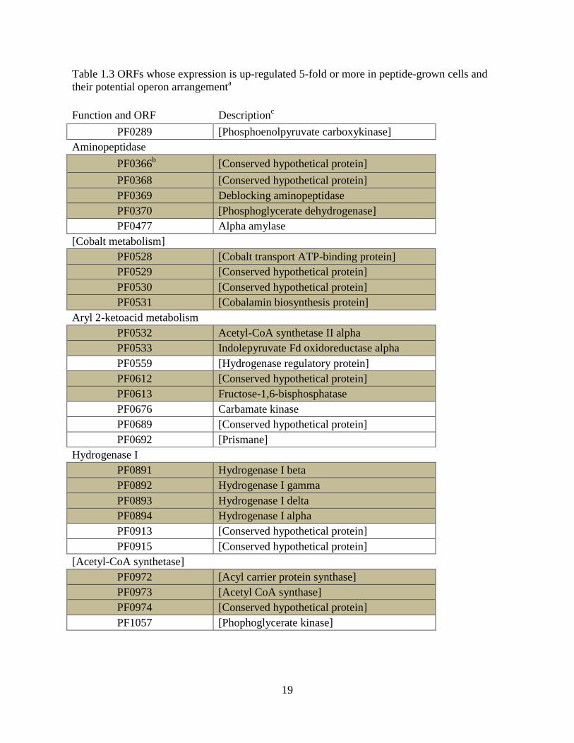
genomes [75]. Similarly, transcriptomic analysis can be used to indirectly predict protein-protein
complexes by observing which ORFS are co-regulated in response to some environmental
challenge. Table 1.3 list ORFs that are dramatically up-regulated in peptide grown cells [50].
Many of those ORFs are adjacent and predicted to form operons. A total of 1460 ORFs are
19
Table 1.3 ORFs whose expression is up-regulated 5-fold or more in peptide-grown cells and
their potential operon arrangementa
Function and ORF Descriptionc
PF0289 [Phosphoenolpyruvate carboxykinase]
Aminopeptidase
PF0366b [Conserved hypothetical protein]
PF0368 [Conserved hypothetical protein]
PF0369 Deblocking aminopeptidase
PF0370 [Phosphoglycerate dehydrogenase]
PF0477 Alpha amylase
[Cobalt metabolism]
PF0528 [Cobalt transport ATP-binding protein]
PF0529 [Conserved hypothetical protein]
PF0530 [Conserved hypothetical protein]
PF0531 [Cobalamin biosynthesis protein]
Aryl 2-ketoacid metabolism
PF0532 Acetyl-CoA synthetase II alpha
PF0533 Indolepyruvate Fd oxidoreductase alpha
PF0559 [Hydrogenase regulatory protein]
PF0612 [Conserved hypothetical protein]
PF0613 Fructose-1,6-bisphosphatase
PF0676 Carbamate kinase
PF0689 [Conserved hypothetical protein]
PF0692 [Prismane]
Hydrogenase I
PF0891 Hydrogenase I beta
PF0892 Hydrogenase I gamma
PF0893 Hydrogenase I delta
PF0894 Hydrogenase I alpha
PF0913 [Conserved hypothetical protein]
PF0915 [Conserved hypothetical protein]
[Acetyl-CoA synthetase]
PF0972 [Acyl carrier protein synthase]
PF0973 [Acetyl CoA synthase]
PF0974 [Conserved hypothetical protein]
PF1057 [Phophoglycerate kinase]

20
Table 1.3 continued.
[Amino acid metabolism]
PF1245 [d-Nopaline dehydrogenase]
PF1246 [Sarcosine oxidase, beta]
PF1253 [Aspartate transaminase]
PF1341 [Aminomethyltransferase]
2-Keto acid ferredoxin oxidoreductases
PF1767 2-Ketoglutarate Fd oxidoreductase, delta
PF1768 2-Ketoglutarate Fd oxidoreductase alpha
PF1769 2-Ketoglutarate Fd oxidoreductase beta
PF1770 2-Ketoglutarate Fd oxidoreductase gamma
PF1771 [2-Ketoacid Fd oxidoreductase, alpha]
PF1772 [2-Ketoacid Fd oxidoreductase, beta]
PF1773 [2-Ketoacid Fd oxidoreductase, gamma]
PF1874 [Glyceraldehyde-3-P dehydrogenase]
[Ferredoxin:NADPH oxidoreductase II]
PF1910 [Fd NADPH oxidoreductase II]
PF1911 [Fd NADPH oxidoreductase II]
[Unknown]
PF2001 [Conserved hypothetical protein]
PF2002 [Sucrose transport protein]
PF2047 [1-Asparaginase] aModified from [50]
bShaded ORFs show genes adjacent in genome.
cThe ORF description is derived from the genemate annotation (given within brackets) or from
experimental data (no brackets).

21
predicted to be within 470 operons in the P. furiosus genome. Of those predicted operons, 349
have been validated by DNA microarray data. A tally of how many of those validated operons
encode proteins that form complexes is incomplete. Nonetheless, knowledge of the function of
those proteins in the predicted operons is invaluable in helping to focus efforts to identify novel
protein-protein interactions.
Methods that detect Protein Complexes.
Genomics has not developed to a point where protein-protein interactions can be
predicted from sequence analysis. Protein complexes have historically been identified by
purifying an enzyme activity of interest through multiple liquid chromatographic steps followed
by analysis of the enzymatically active fractions. Co-elution of two or more polypeptides with
enzymatic activity through multiple chromatographic columns is evidence for an enzyme
composed of multiple subunits. Furthermore, metal analysis of purified fractions could reveal
that an enzyme forms a complex with metal ions.
Protein purification alone is an insufficient strategy to resolve every complex in a cell.
Protein purification often requires that function be known before a protein is purified since
enzyme activity is routinely used to detect the presence of the protein during purification.
Methods have been developed that do not require an explicit knowledge of protein function to
make queries regarding protein complex formation [76-78] (Table 1.4). The yeast two hybrid
system detects protein-protein interactions by exploiting the modular structure of transcriptional
activators [79-81]. Transcriptional activators have a DNA binding domain and a transcriptional
activating domain. Hybrid proteins composed of the DNA binding domain and a “bait” protein
and the transcriptional activation domain and a “prey” are produced in a cell. If the bait and prey
proteins interact they will bring the DNA binding and transcriptional activation domains together

22
Table 1.4 Comparison of methods to detect protein-protein interactions
Method Advantages Disadvantages
Y2Ha Detects transient interactions Limited to binary interactions; needs genetically tractable
host
FRETb Detects transient interactions Limited to binary interactions; needs genetically tractable
host
Phage display Highest Throughput Not suitable for identifying transient interactions;
Limited to binary interactions
TAPc
Not limited to binary interactions Not suitable for identifying transient interactions;
needs genetically tractable host
a Y2H Yeast two hybrid.
b Fluorescence Resonance Energy Transfer
c Tandem Affinity Purification

23
enabling them to express a reporter gene such as -galactosidase. Yeast two hybrid is generally
done in yeast cells which may affect the conformation of the fused bait and prey proteins if they
are derived from organisms other than yeast. Furthermore, the fusion itself may affect the
behavior of the proteins. Fluorescence resonance energy transfer (FRET) detects the interaction
of proteins by tagging interacting proteins with fluorophores [81; 82]. This may be done
chemically or genetically. When the donor flouorophore is excited it emits light at a wavelength
that is absorbed by the acceptor fluorophore provided the donor and acceptor fluorophores are in
close proximity and thus interacting. After the acceptor fluorophore is excited, it emits light at a
wavelength which is detected by a fluorometer or with microscopy.
In phage display, cDNAs or segments of genomes are fused with genes that encode phage
surface coat proteins [82]. When fusion proteins are expressed on the surface of phages, entire
libraries can be screened for interaction between the displayed protein and a protein of interest
that is fixed to a surface. After several enrichment steps that wash away viruses that do not bind
to the protein of interest, the DNA sequence responsible for the interacting protein is identified
by infecting E. coli with the phage that displays a protein that specifically interacts with the
protein of interest. Infected E. coli will produce enough DNA for sequencing and identification
of the gene that encodes the interacting protein. As a rule billions of clones can be screened in a
week using the phage display technique compared to millions in 2 to 4 weeks with Y2H [83].
Tandem Affinity Purification (TAP) is used to purify stable complexes from whole cells
by the use of a tag fused to a protein suspected of having binding partners [84-88]. The tag
consists of two affinity domains in tandem. The extreme domain is protein A, followed by a
tobacco etch virus (TEV) cleavage site, a calmodulin binding peptide and finally the bait protein.
This construct is expressed in the cell where the bait protein is able to interact with its natural

24
binding partners to form a protein-protein complex. The first purification step involves
incubation with IgG beads to pull down the protein complex. This step is followed by cleavage
of the TEV cleavage site with TEV protease which exposes the calmodulin binding domain. The
second purification step uses calmodulin coated beads in the presence of calcium to pull down
the complex from solution. The complex is eluted from the beads with EDTA and the
components of the protein complex are identified with mass spectrometry. The advantage of
TAP over the yeast two hybrid system, FRET and Phage Display is that it is not limited to binary
interactions. The absence of a third protein partner in the binary systems might preclude the
interaction of the proteins being tested. The advantage of yeast two hybrid and FRET systems
over TAP and Phage Display is that they are able to detect transient interactions whereas in the
case of TAP and Phage Display those interactions may be lost during multiple purification steps
[89]. In all cases fusion proteins are necessary to carry out the experiments, a feature that may
destabilize interactions between protein partners. In addition, execution of these methods are
limited to biological systems that have well developed genetic systems.
Recent efforts to decipher the metalloproteome (the set of proteins that have metal-
binding capacity) of a given tissue or organism analyzed native cell extracts and side stepped the
need for a genetic system [90]. One group used immobilized metal affinity chromatography
(IMAC) to analyze the copper and zinc metalloproteomes of human liver cells. The soluble
fraction of these cells were loaded onto IMAC columns with either bound copper or zinc.
Presumably, proteins that bind copper or zinc would be retained onto the column until they were
eluted. Mass spectrometry revealed that different proteins eluted depending on whether the
column had bound copper or zinc. Down sides of this method is that it detects mostly abundant
metalloproteins, the process only analyzes one metalloproteome at a time and, lastly the method

25
may not detect metalloproteins that have a metal buried deep within the protein structure.
Another methalloproteomic method was used with Ferroplasma acidifilum [91]. F. acidifilum is
an archaeon that lives in heavy metal environments containing high concentrations of iron and
sulfur. Its native metalloproteome was analyzed by resolving its proteins on a 2-D native PAGE
gel and staining the gel with the chemiluminescent substrate luminal. The protein spots which
luminesced because of the presence of a metal were cored and extracted from the gel. Analysis
by mass spectrometry and ICPMS identified the protein and its associated metal, respectively.
Most proteins found were associated with iron as a structural component as opposed to a
functional component due to adaptation of F. acidifilum to high concentrations of iron. It is
questionable whether this method would work in a proteome that is not adapted to high
concentrations of metal since proteins that are not adapted to high metal concentrations may bind
less metal and not be as readily detected by this procedure.
Summary and Objectives.
Although the accessibility to whole genome sequences is an essential element for a
comprehensive understanding of how organisms function, more information is needed than can
be provided by genome sequences alone to meet this aim. Transcriptomics and proteomics can
reveal which genes are expressed both temporally and spatially in different cell types and protein
structural genomics studies can provide data concerning the structures and folds available which
affect the biological function of proteins. These and other available ‘-omics’ technologies
complement genomics which in and of itself cannot be used to bridge the gap between the
information found in DNA sequences and the cumulative effects of that information on the state
of the cell.

26
The proteome of a cell encompasses all of the proteins that are expressed by a particular
genome. The function of many of these proteins has not been discovered. Since the presence of
protein complexes are vital to major cellular processes, [92] determining what protein
interactions occur in the cell is invaluable in our understanding of the role these proteins fulfill.
Initiatives have been put forth by government agencies to facilitate research that would make
linkages from the abundance of information found in genomes to the protein interactions that
manifest themselves in cells. The objectives of the research discussed in this thesis are:
1. To find and characterize novel protein-protein assemblies in P furiosus by analyzing
natively fractionated P. furiosus biomass.
2. To find novel P. furiosus metalloproteins by analyzing natively fractionated P. furiosus
biomass.
It is understood that the narrow scope of this thesis will not be able to address all the
challenges presented by the post-genomic era. However, it is my hope to only to add my small
contribution to those of many others with the expectation that more information increases the
rate at which the complexities of life can be fashioned into understandable patterns and
ultimately be used to manipulate life to the advantage of the human race.
References
1. Alberts, B.M., Botstein, D., Brenner, S., Cantor, C. R., Doolittle, R. F., Hood, L.,
McKusick, V. A., Nathans, D., Olson, M. V., Orkin, S., Rosenberg, L. E., Ruddle, F. H.,
Tilghman, S., Tooze, and J. Watson, J. D. 1988. Report of the committee on mapping and
sequencing the human genome. National Academy Press, Washington, D.C.

27
2. Kanehisa, M. 2000. Post-genome Informatics. Oxford University Press, Oxford. 1-23 p.
3. Kyrpides, N.C., "Genomes OnLine Database (GOLD 1.0): a monitor of complete and
ongoing genome projects world-wide," Bioinformatics, vol. 15, no. 9, pp. 773-4, 1999.
4. Bernal, A., U. Ear and N. Kyrpides, "Genomes OnLine Database (GOLD): a monitor of
genome projects world-wide," Nucleic Acids Res, vol. 29, no. 1, pp. 126-7, 2001.
5. Liolios, K., N. Tavernarakis, P. Hugenholtz and N.C. Kyrpides, "The Genomes On Line
Database (GOLD) v.2: a monitor of genome projects worldwide," Nucleic Acids Res, vol. 34, no.
Database issue, pp. D332-4, 2006.
6. Liolios, K., K. Mavromatis, N. Tavernarakis and N.C. Kyrpides, "The Genomes On Line
Database (GOLD) in 2007: status of genomic and metagenomic projects and their associated
metadata," Nucleic Acids Res, vol. 36, no. Database issue, pp. D475-9, 2008.
7. Liolios, K., I.M. Chen, K. Mavromatis, N. Tavernarakis, P. Hugenholtz, V.M. Markowitz
and N.C. Kyrpides, "The Genomes On Line Database (GOLD) in 2009: status of genomic and
metagenomic projects and their associated metadata," Nucleic Acids Res, vol. 38, no. Database
issue, pp. D346-54, 2010.
8. Pagani, I., K. Liolios, J. Jansson, I.M. Chen, T. Smirnova, B. Nosrat, V.M. Markowitz
and N.C. Kyrpides, "The Genomes OnLine Database (GOLD) v.4: status of genomic and
metagenomic projects and their associated metadata," Nucleic Acids Res, vol. 40, no. Database
issue, pp. D571-9, 2012.
9. Wright, S.H. 2001. Lander celebrates genome milestone in heavily attended talk. In MIT
Tech Talk. MIT News Office, Cambridge, MA.
10. Riley, M., T. Abe, M.B. Arnaud, M.K. Berlyn, F.R. Blattner, R.R. Chaudhuri, J.D.
Glasner, T. Horiuchi, I.M. Keseler, T. Kosuge, H. Mori, N.T. Perna, G. Plunkett, 3rd, K.E. Rudd,

28
M.H. Serres, G.H. Thomas, N.R. Thomson, D. Wishart and B.L. Wanner, "Escherichia coli K-
12: a cooperatively developed annotation snapshot--2005," Nucleic Acids Res, vol. 34, no. 1, pp.
1-9, 2006.
11. Keseler, I.M., C. Bonavides-Martinez, J. Collado-Vides, S. Gama-Castro, R.P. Gunsalus,
D.A. Johnson, M. Krummenacker, L.M. Nolan, S. Paley, I.T. Paulsen, M. Peralta-Gil, A. Santos-
Zavaleta, A.G. Shearer and P.D. Karp, "EcoCyc: a comprehensive view of Escherichia coli
biology," Nucleic Acids Res, vol. 37, no. Database issue, pp. D464-70, 2009.
12. Christie, K.R., E.L. Hong and J.M. Cherry, "Functional annotations for the
Saccharomyces cerevisiae genome: the knowns and the known unknowns," Trends Microbiol,
vol. 17, no. 7, pp. 286-94, 2009.
13. Griffiths, P.E. and K. Stotz, "Genes in the postgenomic era," Theor Med Bioeth, vol. 27,
no. 6, pp. 499-521, 2006.
14. Kell, D.B. and S.G. Oliver, "Here is the evidence, now what is the hypothesis? The
complementary roles of inductive and hypothesis-driven science in the post-genomic era,"
Bioessays, vol. 26, no. 1, pp. 99-105, 2004.
15. Kornberg, A., "Biochemistry matters," Nat Struct Mol Biol, vol. 11, no. 6, pp. 493, 2004.
16. Van Regenmortel, M.H., "Reductionism and complexity in molecular biology. Scientists
now have the tools to unravel biological and overcome the limitations of reductionism," EMBO
Rep, vol. 5, no. 11, pp. 1016-20, 2004.
17. Elgar, G. and T. Vavouri, "Tuning in to the signals: noncoding sequence conservation in
vertebrate genomes," Trends Genet, vol. 24, no. 7, pp. 344-52, 2008.
18. Stetter, K.O., "Hyperthermophilic procaryotes," FEMS Microbiol Rev, vol. 18, no. 2-3,
pp. 149-158, 1996.

29
19. Fiala, G. and K.O. Stetter, "Pyrococcus-Furiosus Sp-Nov Represents a Novel Genus of
Marine Heterotrophic Archaebacteria Growing Optimally at 100-Degrees C," Arch Microbiol,
vol. 145, no. 1, pp. 56-61, 1986.
20. Driskill, L.E., K. Kusy, M.W. Bauer and R.M. Kelly, "Relationship between glycosyl
hydrolase inventory and growth physiology of the hyperthermophile Pyrococcus furiosus on
carbohydrate-based media," Appl Environ Microb, vol. 65, no. 3, pp. 893-897, 1999.
21. Schut, G.J., S.D. Brehm, S. Datta and M.W.W. Adams, "Whole-genome DNA
microarray analysis of a hyperthermophile and an archaeon: Pyrococcus furiosus grown on
carbohydrates or peptides," Journal of Bacteriology, vol. 185, no. 13, pp. 3935-3947, 2003.
22. Poole, F.L., B.A. Gerwe, R.C. Hopkins, G.J. Schut, M.V. Weinberg, F.E. Jenney and
M.W.W. Adams, "Defining genes in the genome of the hyperthermophilic archaeon Pyrococcus
furiosus: Implications for all microbial genomes," J Bacteriol, vol. 187, no. 21, pp. 7325-7332,
2005.
23. Lipscomb, G.L., K. Stirrett, G.J. Schut, F. Yang, F.E. Jenney, R.A. Scott, M.W.W.
Adams and J. Westpheling, "Natural Competence in the Hyperthermophilic Archaeon
Pyrococcus furiosus Facilitates Genetic Manipulation: Construction of Markerless Deletions of
Genes Encoding the Two Cytoplasmic Hydrogenases," Appl Environ Microbio, vol. 77, no. 7,
pp. 2232-2238, 2011.
24. Thorgersen, M.P., K. Stirrett, R.A. Scott and M.W.W. Adams, "Mechanism of oxygen
detoxification by the surprisingly oxygen-tolerant hyperthermophilic archaeon, Pyrococcus
furiosus," P Natl Acad Sci USA, vol. 109, no. 45, pp. 18547-18552, 2012.
25. Wang, B.C., M.W. Adams, H. Dailey, L. DeLucas, M. Luo, J. Rose, R. Bunzel, T.
Dailey, J. Habel, P. Horanyi, F.E. Jenney, Jr., I. Kataeva, H.S. Lee, S. Li, T. Li, D. Lin, Z.J. Liu,

30
C.H. Luan, M. Mayer, L. Nagy, M.G. Newton, J. Ng, F.L. Poole, 2nd, A. Shah, C. Shah, F.J.
Sugar and H. Xu, "Protein production and crystallization at SECSG -- an overview," J Struct
Funct Genomics, vol. 6, no. 2-3, pp. 233-43, 2005.
26. Sugar, F.J., F.E. Jenney, Jr., F.L. Poole, 2nd, P.S. Brereton, M. Izumi, C. Shah and M.W.
Adams, "Comparison of small- and large-scale expression of selected Pyrococcus furiosus genes
as an aid to high-throughput protein production," J Struct Funct Genomics, vol. 6, no. 2-3, pp.
149-58, 2005.
27. Jenney, F.E., Jr., P.S. Brereton, M. Izumi, F.L. Poole, 2nd, C. Shah, F.J. Sugar, H.S. Lee
and M.W. Adams, "High-throughput production of Pyrococcus furiosus proteins: considerations
for metalloproteins," J Synchrotron Radiat, vol. 12, no. Pt 1, pp. 8-12, 2005.
28. Robb, F.T., D.L. Maeder, J.R. Brown, J. DiRuggiero, M.D. Stump, R.K. Yeh, R.B. Weiss
and D.M. Dunn, "Genomic sequence of hyperthermophile, Pyrococcus furiosus: implications for
physiology and enzymology," Methods Enzymol, vol. 330, pp. 134-57, 2001.
29. Bergman, N.H., K.D. Passalacqua, P.C. Hanna and Z.S. Qin, "Operon prediction for
sequenced bacterial genomes without experimental information," Appl Environ Microbiol, vol.
73, no. 3, pp. 846-54, 2007.
30. Moreno-Hagelsieb, G. and J. Collado-Vides, "A powerful non-homology method for the
prediction of operons in prokaryotes," Bioinformatics, vol. 18 Suppl 1, pp. S329-36, 2002.
31. Salgado, H., G. Moreno-Hagelsieb, T.F. Smith and J. Collado-Vides, "Operons in
Escherichia coli: Genomic analyses and predictions," P Natl Acad Sci USA, vol. 97, no. 12, pp.
6652-6657, 2000.
32. Verhagen, M.F., A.L. Menon, G.J. Schut and M.W. Adams, "Pyrococcus furiosus: large-
scale cultivation and enzyme purification," Methods Enzymol, vol. 330, pp. 25-30, 2001.
31
33. Cody, G.D., N.Z. Boctor, T.R. Filley, R.M. Hazen, J.H. Scott, A. Sharma and H.S.
Yoder, Jr., "Primordial carbonylated iron-sulfur compounds and the synthesis of pyruvate,"
Science, vol. 289, no. 5483, pp. 1337-40, 2000.
34. Corliss, D.A., J. A. Baross; and S. E. Hoffmann, "An hypothesis concerning the
relationship between submarine hot springs and the origin of life on Earth.," Oceanlogica Acta
SP, pp. 59-69., 1981.
35. Huber, C. and G. Wachtershauser, "Activated acetic acid by carbon fixation on (Fe,Ni)S
under primordial conditions," Science, vol. 276, no. 5310, pp. 245-7, 1997.
36. Hashimoto, K., H. Nishi, S. Bryant and A.R. Panchenko, "Caught in self-interaction:
evolutionary and functional mechanisms of protein homooligomerization," Phys Biol, vol. 8, no.
3, pp. 035007, 2011.
37. Miller, S., A.M. Lesk, J. Janin and C. Chothia, "The accessible surface area and stability
of oligomeric proteins," Nature, vol. 328, no. 6133, pp. 834-6, 1987.
38. Bamford, D.H., R.J. Gilbert, J.M. Grimes and D.I. Stuart, "Macromolecular assemblies:
greater than their parts," Curr Opin Struct Biol, vol. 11, no. 1, pp. 107-13, 2001.
39. Wilson, D.N. and K.H. Nierhaus, "Ribosomal proteins in the spotlight," Crit Rev
Biochem Mol Biol, vol. 40, no. 5, pp. 243-67, 2005.
40. Chiu, W., M.L. Baker and S.C. Almo, "Structural biology of cellular machines," Trends
Cell Biol, vol. 16, no. 3, pp. 144-50, 2006.
41. Gilbert, H.J., "Cellulosomes: microbial nanomachines that display plasticity in quaternary
structure," Mol Microbiol, vol. 63, no. 6, pp. 1568-76, 2007.
42. Srere, P.A., "Complexes of sequential metabolic enzymes," Annu Rev Biochem, vol. 56,
pp. 89-124, 1987.

32
43. Ovadi, J. and P.A. Srere, "Macromolecular compartmentation and channeling," Int Rev
Cytol, vol. 192, pp. 255-80, 2000.
44. Ovadi, J. and V. Saks, "On the origin of intracellular compartmentation and organized
metabolic systems," Mol Cell Biochem, vol. 256-257, no. 1-2, pp. 5-12, 2004.
45. Menon, A.L., F.L. Poole, 2nd, A. Cvetkovic, S.A. Trauger, E. Kalisiak, J.W. Scott, S.
Shanmukh, J. Praissman, F.E. Jenney, Jr., W.R. Wikoff, J.V. Apon, G. Siuzdak and M.W.
Adams, "Novel multiprotein complexes identified in the hyperthermophilic archaeon Pyrococcus
furiosus by non-denaturing fractionation of the native proteome," Mol Cell Proteomics, vol. 8,
no. 4, pp. 735-751, 2009.
46. Blamey, J.M. and M.W. Adams, "Purification and characterization of pyruvate ferredoxin
oxidoreductase from the hyperthermophilic archaeon Pyrococcus furiosus," Biochim Biophy
Acta, vol. 1161, no. 1, pp. 19-27, 1993.
47. Adams, M.W. and A. Kletzin, "Oxidoreductase-type enzymes and redox proteins
involved in fermentative metabolisms of hyperthermophilic Archaea," Adv Protein Chem, vol.
48, pp. 101-180, 1996.
48. Smith, E.T., J.M. Blamey and M.W. Adams, "Pyruvate ferredoxin oxidoreductases of the
hyperthermophilic archaeon, Pyrococcus furiosus, and the hyperthermophilic bacterium,
Thermotoga maritima, have different catalytic mechanisms," Biochemistry, vol. 33, no. 4, pp.
1008-1016, 1994.
49. Schut, G.J., A.L. Menon and M.W. Adams, "2-keto acid oxidoreductases from
Pyrococcus furiosus and Thermococcus litoralis," Method Enzymol, vol. 331, pp. 144-158, 2001.
33
50. Schut, G.J., S.D. Brehm, S. Datta and M.W. Adams, "Whole-genome DNA microarray
analysis of a hyperthermophile and an archaeon: Pyrococcus furiosus grown on carbohydrates or
peptides," J Bacteriol, vol. 185, no. 13, pp. 3935-3947, 2003.
51. Mai, X. and M.W. Adams, "Purification and characterization of two reversible and ADP-
dependent acetyl coenzyme A synthetases from the hyperthermophilic archaeon Pyrococcus
furiosus," J Bacteriol, vol. 178, no. 20, pp. 5897-5903, 1996.
52. Dafforn, T.R., "So how do you know you have a macromolecular complex?," Acta
Crystallogr D Biol Crystallogr, vol. 63, no. Pt 1, pp. 17-25, 2007.
53. Clarkson, S.M., E.C. Newcomer, E.G. Young and M.W. Adams, "The elemental sulfur-
responsive protein (SipA) from the hyperthermophilic archaeon Pyrococcus furiosus is regulated
by sulfide in an iron-dependent manner," J Bacteriol, vol. 192, no. 21, pp. 5841-3, 2010.
54. Schut, G.J., J. Zhou and M.W. Adams, "DNA microarray analysis of the
hyperthermophilic archaeon Pyrococcus furiosus: evidence for anNew type of sulfur-reducing
enzyme complex," J Bacteriol, vol. 183, no. 24, pp. 7027-36, 2001.
55. Clarkson, S.M., and M.W. Adams, "The hyperthermophilic archaeon Pyrococcus furiosus
utilizes environmental iron(II) monosulfide cluster complexes as an iron source.," Manuscript in
preparation., 2013.
56. Lehninger, A.L., "Role of Metal Ions in Enzyme Systems," Physiol Rev, vol. 30, no. 3,
pp. 393-429, 1950.
57. Bowen, H.J.M. 1966. Trace elements in biochemistry. Academic Press, London.
58. Riordan, J.F., "The role of metals in enzyme activity," Ann Clin Lab Sci, vol. 7, no. 2, pp.
119-29, 1977.

34
59. Seravalli, J. and S.W. Ragsdale, "Expanding the biological periodic table," Chem Biol,
vol. 17, no. 8, pp. 793-4, 2010.
60. Bertini, I.A.S.H.S.H. 2001. Handbook of metalloproteins. Marcel Dekker, New York.
61. Lippard, S.J.B. 1994. Principles of bioorganic chemistry. University Science Books, Mill
Valley, CA, pp 3-41.
62. Castagnetto, J.M., S.W. Hennessy, V.A. Roberts, E.D. Getzoff, J.A. Tainer and M.E.
Pique, "MDB: the Metalloprotein Database and Browser at The Scripps Research Institute,"
Nucleic Acids Res, vol. 30, no. 1, pp. 379-82, 2002.
63. Kasampalidis, I.N., I. Pitas and K. Lyroudia, "Conservation of metal-coordinating
residues," Proteins, vol. 68, no. 1, pp. 123-30, 2007.
64. Andreini, C., I. Bertini, G. Cavallaro, G.L. Holliday and J.M. Thornton, "Metal-MACiE:
a database of metals involved in biological catalysis," Bioinformatics, vol. 25, no. 16, pp. 2088-
9, 2009.
65. Waldron, K.J., J.C. Rutherford, D. Ford and N.J. Robinson, "Metalloproteins and metal
sensing," Nature, vol. 460, no. 7257, pp. 823-30, 2009.
66. Bork, P., "Powers and pitfalls in sequence analysis: the 70 % hurdle," Genome Res, vol.
10, no. 4, pp. 398-400, 2000.
67. Yannone, S.M., S. Hartung, A.L. Menon, M.W. Adams and J.A. Tainer, "Metals in
biology: defining metalloproteomes," Curr Opin Biotechnol, vol. 23, no. 1, pp. 89-95, 2012.
68. Eidsness, M.K., S.E. O'Dell, D.M. Kurtz, Jr., R.L. Robson and R.A. Scott, "Expression of
a synthetic gene coding for the amino acid sequence of Clostridium pasteurianum rubredoxin,"
Protein Eng, vol. 5, no. 4, pp. 367-71, 1992.
35
69. Meng, L., S. Ruebush, M. D'Souza V, A.J. Copik, S. Tsunasawa and R.C. Holz,
"Overexpression and divalent metal binding properties of the methionyl aminopeptidase from
Pyrococcus furiosus," Biochemistry, vol. 41, no. 23, pp. 7199-208, 2002.
70. Dai, Y., P.C. Wensink and R.H. Abeles, "One protein, two enzymes," J Biol Chem, vol.
274, no. 3, pp. 1193-5, 1999.
71. Mori, S., S. Sumino and T. Kasumi, "Substrate specificity of a tripeptidase as a
metalloenzyme purified from Lactococcus lactis subsp. lactis biovar. diacetylactis ATCC
13675," J Biosci Bioeng, vol. 93, no. 4, pp. 360-6, 2002.
72. Shu, N., T. Zhou and S. Hovmoller, "Prediction of zinc-binding sites in proteins from
sequence," Bioinformatics, vol. 24, no. 6, pp. 775-82, 2008.
73. Passerini, A., M. Punta, A. Ceroni, B. Rost and P. Frasconi, "Identifying cysteines and
histidines in transition-metal-binding sites using support vector machines and neural networks,"
Proteins, vol. 65, no. 2, pp. 305-16, 2006.
74. Andreini, C., I. Bertini and A. Rosato, "A hint to search for metalloproteins in gene
banks," Bioinformatics, vol. 20, no. 9, pp. 1373-80, 2004.
75. Tran, T.T., P. Dam, Z. Su, F.L. Poole, 2nd, M.W. Adams, G.T. Zhou and Y. Xu, "Operon
prediction in Pyrococcus furiosus," Nucleic Acids Res, vol. 35, no. 1, pp. 11-20, 2007.
76. Berggard, T., S. Linse and P. James, "Methods for the detection and analysis of protein-
protein interactions," Proteomics, vol. 7, no. 16, pp. 2833-42, 2007.
77. Piehler, J., "New methodologies for measuring protein interactions in vivo and in vitro,"
Curr Opin Struct Biol, vol. 15, no. 1, pp. 4-14, 2005.
36
78. Azarkan, M., J. Huet, D. Baeyens-Volant, Y. Looze and G. Vandenbussche, "Affinity
chromatography: a useful tool in proteomics studies," J Chromatogr B Analyt Technol Biomed
Life Sci, vol. 849, no. 1-2, pp. 81-90, 2007.
79. Fields, S. and O. Song, "A novel genetic system to detect protein-protein interactions,"
Nature, vol. 340, no. 6230, pp. 245-6, 1989.
80. Uetz, P. and R.E. Hughes, "Systematic and large-scale two-hybrid screens," Curr Opin
Microbiol, vol. 3, no. 3, pp. 303-8, 2000.
81. Koegl, M. and P. Uetz, "Improving yeast two-hybrid screening systems," Brief Funct
Genomic Proteomic, vol. 6, no. 4, pp. 302-12, 2007.
82. Zozulya, S., M. Lioubin, R.J. Hill, C. Abram and M.L. Gishizky, "Mapping signal
transduction pathways by phage display," Nat Biotechnol, vol. 17, no. 12, pp. 1193-8, 1999.
83. Willats, W.G., "Phage display: practicalities and prospects," Plant Mol Biol, vol. 50, no.
6, pp. 837-54, 2002.
84. Bauer, A. and B. Kuster, "Affinity purification-mass spectrometry. Powerful tools for the
characterization of protein complexes," Eur J Biochem, vol. 270, no. 4, pp. 570-8, 2003.
85. Ho, Y., A. Gruhler, A. Heilbut, G.D. Bader, L. Moore, S.L. Adams, A. Millar, P. Taylor,
K. Bennett, K. Boutilier, L. Yang, C. Wolting, I. Donaldson, S. Schandorff, J. Shewnarane, M.
Vo, J. Taggart, M. Goudreault, B. Muskat, C. Alfarano, D. Dewar, Z. Lin, K. Michalickova, A.R.
Willems, H. Sassi, P.A. Nielsen, K.J. Rasmussen, J.R. Andersen, L.E. Johansen, L.H. Hansen, H.
Jespersen, A. Podtelejnikov, E. Nielsen, J. Crawford, V. Poulsen, B.D. Sorensen, J. Matthiesen,
R.C. Hendrickson, F. Gleeson, T. Pawson, M.F. Moran, D. Durocher, M. Mann, C.W. Hogue, D.
Figeys and M. Tyers, "Systematic identification of protein complexes in Saccharomyces
cerevisiae by mass spectrometry," Nature, vol. 415, no. 6868, pp. 180-3, 2002.

37
86. Rigaut, G., A. Shevchenko, B. Rutz, M. Wilm, M. Mann and B. Seraphin, "A generic
protein purification method for protein complex characterization and proteome exploration," Nat
Biotechnol, vol. 17, no. 10, pp. 1030-2, 1999.
87. Gavin, A.C., M. Bosche, R. Krause, P. Grandi, M. Marzioch, A. Bauer, J. Schultz, J.M.
Rick, A.M. Michon, C.M. Cruciat, M. Remor, C. Hofert, M. Schelder, M. Brajenovic, H.
Ruffner, A. Merino, K. Klein, M. Hudak, D. Dickson, T. Rudi, V. Gnau, A. Bauch, S. Bastuck,
B. Huhse, C. Leutwein, M.A. Heurtier, R.R. Copley, A. Edelmann, E. Querfurth, V. Rybin, G.
Drewes, M. Raida, T. Bouwmeester, P. Bork, B. Seraphin, B. Kuster, G. Neubauer and G.
Superti-Furga, "Functional organization of the yeast proteome by systematic analysis of protein
complexes," Nature, vol. 415, no. 6868, pp. 141-7, 2002.
88. Puig, O., F. Caspary, G. Rigaut, B. Rutz, E. Bouveret, E. Bragado-Nilsson, M. Wilm and
B. Seraphin, "The tandem affinity purification (TAP) method: a general procedure of protein
complex purification," Methods, vol. 24, no. 3, pp. 218-29, 2001.
89. Collins, M.O. and J.S. Choudhary, "Mapping multiprotein complexes by affinity
purification and mass spectrometry," Curr Opin Biotechnol, vol. 19, no. 4, pp. 324-30, 2008.
90. She, Y.M., S. Narindrasorasak, S. Yang, N. Spitale, E.A. Roberts and B. Sarkar,
"Identification of metal-binding proteins in human hepatoma lines by immobilized metal affinity
chromatography and mass spectrometry," Mol Cell Proteomics, vol. 2, no. 12, pp. 1306-18,
2003.
91. Ferrer, M., O.V. Golyshina, A. Beloqui, P.N. Golyshin and K.N. Timmis, "The cellular
machinery of Ferroplasma acidiphilum is iron-protein-dominated," Nature, vol. 445, no. 7123,
pp. 91-4, 2007.

38
92. Alberts, B., "The cell as a collection of protein machines: preparing the next generation
of molecular biologists," Cell, vol. 92, no. 3, pp. 291-4, 1998.

39
CHAPTER 2
IDENTIFYING NOVEL PROTEIN COMPLEXES IN THE PROTEOME OF THE
HYPERTHERMOPHILIC ARCHAEON PYROCOCCUS FURIOSUS
Introduction
Molecular machines like other types of machines are composed of multiple components.
All parts are necessary for the machine to function. Furthermore like a blueprint, the genome has
the information for the parts of the molecular machines which can be replaced with new parts
when old ones are worn out or damaged. Without these molecular machines the processes that
govern life would come to a grinding halt. Therefore, it is of immeasurable value to determine
the components of these machines and how they work together to carry out their specific
functions. A number of methods that determine which components of molecular machines work
together have been developed [1-3]. However, limitations exist in their utility due to
manipulations at the molecular level that may affect the native conformation of the proteins
which are the component parts of the molecular machines. Furthermore, the available methods
do not allow for comprehensive analysis, often focusing on one protein-protein interaction at a
time.
Stable protein complexes have been historically identified by purification of enzyme
activity through multiple chromatographic steps [4; 5]. While this method preserves the native
conformation of proteins, it is impractical in broad proteomics analysis of protein complexes
because it cannot be used to identify complexes that are not detected by enzymatic assays.
Furthermore, identifying protein complexes at a proteomic scale by purifying enzymatic activity

40
would require various assays to detect various enzymes, a process that could not be done in a
timely manner. A procedure developed in the lab of Dr. Michael Adams draws from the well
established, non-denaturing method of protein purification and uses a single method of detection,
namely mass spectrometry rather than enzyme activity to detect protein-protein interactions.
Of particular interest was the interaction between PF1837 and PF1838 a novel potential
complex designated PC-81. This complex is a homolog of two acetyl-CoA synthetases (ACS)
that have been purified from P. furiosus native biomass. Those complexes form 22
heterotetrameric structures with subunits encoded by unlinked genes. Additional ACS subunit
homologs are encoded in the genome and all ACS homologs were detected during fractionation
by mass spectrometry. This observation served as the basis of recombinant expression
experiments performed to examine complex formation of the ACSs. The findings of that study
are the subject of chapter 3 of this thesis.
Materials and Methods.
Preparation of anaerobic cell-free extract. P. furiosus (DSM 3638) was grown under anaerobic,
reducing conditions at 90°C in a 600-liter fermenter on maltose and peptides and harvested in
late log phase [6]. The procedures for preparing anaerobic cell-free extract and for running the
first chromatography step have been described previously [6] Briefly, 300 g of frozen cells were
gently lysed by osmotic shock anaerobically under a continuous flow of argon in 900 ml of
50mM Tris-HCl (pH 8.0) containing 2mM sodium dithionite as a reductant (Buffer A) and 0.5
g/ml DNAse I to reduce viscosity. The cell lysate was centrifuged at 100,000 x g for 2 h at
18°C, and the clear supernatant, representing the cytoplasmic fraction was immediately loaded
onto the first column. This and all subsequent columns were run under anaerobic, reducing

41
Figure 2.1.
Fractionation schemes of P. furiosus cytoplasmic proteins through multiple
chromatographic steps.
Cytoplasmic proteins were separated on a DEAE anion exchange column which was the first
fractionation. Fractions from the DEAE column were combined into 15 pools that were
fractionated through subsequent columns. Some of the fractions of secondary purification were
pooled and fractionated further. The chromatography steps included DEAE (diethylaminoethyl
cellulose), HAP (hydroxyapatite), PS (phenyl sepharose), BS (blue sepharose), and SX200
(superdex 200) which are indicated in the figure. The fractions generated were analyzed by
either CV or HT or both.

42

43
conditions where all buffers were degassed and maintained under a positive pressure of argon,
and all liquid transfers were made using needles and syringes.
Large scale anaerobic fractionation of cytoplasmic proteins. . First column (c1) fractionation. A
column tree representing the complete fractionation is shown in Figure 2.1. All columns were
run using an ÄKTATM
basic automated LC system (GE Healthcare). The P. furiosus
cytoplasmic fraction was diluted 4-fold in Buffer A to reduce the ionic strength and was loaded
onto a 10 X 20-cm (1.5-liter) column of DEAE-Sepharose Fast Flow (GE Healthcare) that had
been equilibrated previously with the same buffer until the effluent was anaerobic and reducing.
Unbound protein was washed off the column with Buffer A. Bound proteins were eluted using a
linear gradient (15 liters) of 0-500 mM NaCl in Buffer A, and 125-ml anaerobic fractions were
collected (120 fractions). Any remaining tightly bound proteins were eluted from the column
using 1-liter step gradients of 1, 1.5, and 2 M NaCl respectively, in Buffer A (high salt washes).
Secondary (c2) chromatographic steps. A subset of 37 gradient fractions from the first column
separation were selected using a combination of protein profiles and enzyme activities to create 6
fraction pools, c1-1 through c1-6. The control proteins and enzyme activities were as follows:
c1-1, ferredoxin-NADPH oxidoreductase (FNOR) [7] and rubrerythrin [8]; c1-2, 2-ketisovalerate
ferredoxin oxidoreductase (VOR) and pyruvate ferredoxin reductase (POR) [9]; c1-3,
aldehyde:ferredoxin oxidoreductase [10]; c1-4, soluble hydrogenase 1 (SH1) [11]; c1-5,
rubrdoxin [12]; and c1-6 ferredoxin (Fd)[13]. These pools, c1-1 through c1-6, were fractionated
through six subsequent c2 columns. The remaining 83 gradient fractions were combined to
generate nine additional pools, in this case based solely on SDS-PAGE and protein profiles.
These pools c1-7 through c1-15, were fractionated through nine subsequent c2 columns to give a
total of 15 columns at the c2 level (Figure 2.1). SDS-PAGE and native PAGE were carried out

44
using precast 4-20 % gradient gels (Criterion gel system, Bio-Rad). Protein concentrations were
estimated by the method of Bradford [14] using bovine serum albumin as the standard.
Third (c3) and fourth (c4) column steps. Six c3 and four c4 level columns were run using
fraction pools that were created for the further purification of the control proteins by the
published procedures (Figure 2.1).
Potential protein complex identification using mass spectrometry. Protein digestion, peptide
separations, and mass spectrometry were all done in the lab of Gary Siuzdak at the Scripps
Research Institute. Protein complex identification was performed computationally using a
customized function developed by Farris Poole at the University of Georgia to assess the strength
of potential type I complexes based on known type I complexes. Annotation of the proteins are
based on the public National Center for Biotechnology Information (NCBI) annotation of the
genome [15] except when a protein has been purified from P. furiosus or closely related
organism or was hypothetical, but contained a domain described in the InterPro database [16].
Details of these methods are given in the original paper.
Results and Discussion
This method was highly dependent upon the sequenced genome of P. furiosus. Analysis
of the genome of P. furiosus which was sequenced in 2001 [17] showed it to be small (1.9 Mb)
and well organized with 1,460 of a total of 2,125 total open reading frames (ORFs) predicted to
be organized into 470 operons [18]. Genes within in the same operon often have a functional
relationship. This relationship may encompass the steps of a biochemical pathway or may
represent polypeptides that assemble into a multimeric protein complex. Heteromeric protein
complexes of this type are designated type I. Type II complexes are composed of subunits
encoded by unlinked genes, and type III complexes are homomeric proteins. (For a discussion of
45
the different types of multimeric protein complexes see chapter 1). Soluble hydrogenase 1 [11],
pyruvate oxidoreductase (POR), 2-ketoisovalerate oxidoreductase (VOR) [9] and ferredoxin
NADP+ oxidoreductase (FNOR) [7] are examples of type I complexes since the genes encoding
the subunits of these complexes are adjacent in the P. furiosus genome. Although, heteromeric
proteins encoded by unlinked genes are present in P. furiosus [19], we focus here on the
purification of complexes encoded by linked genes because access to the genome sequence aids
in the systematic prediction of this particular population of novel protein-protein interactions in
P. furiosus.
Fractionation of native P. furiosus biomass was carried out through sequential
chromatographic steps which included anion exchange, hydroxyapatite, hydrophobic interaction,
and size exclusion chromatography. Since P. furiosus grows anaerobically, the fractionation of
its biomass was carried out anaerobically to preserve any protein-protein interactions that might
be disrupted in the presence of oxygen. 126 column fractions were collected as P. furiosus cell
free extract was purified over an anion exchange column in the first fractionation. Each fraction
was analyzed by MS/MS. Figure 2.2 shows the number of proteins identified in each fraction
along with its protein concentrations. Protein identification was determined using conventional
(CV) or high through-put (HT) separation techniques followed by tandem mass spectrometry and
Mascot searches of the NCBI annotation of the Pyrococcus furiosus genome. The CV method
requires more time, due to longer HPLC peptide elution gradients, and is not conducive to high-
throughput experimentation. However, the CV method has a greater capacity to resolve peptides
which increases their detectability by mass spectrometry. Thus, the CV method was used to
detect proteins in fractions with greater numbers of species which include those fractions that
have been purified by only one chromatographic step.

46
Figure 2.2
Elution profile of the P. furiosus cell free extract fractionation on a DEAE column.
Elution profile show number of proteins detected in each fraction (bars), protein concentrations
of each fraction (line), and the pools of the DEAE fraction that were further purified.
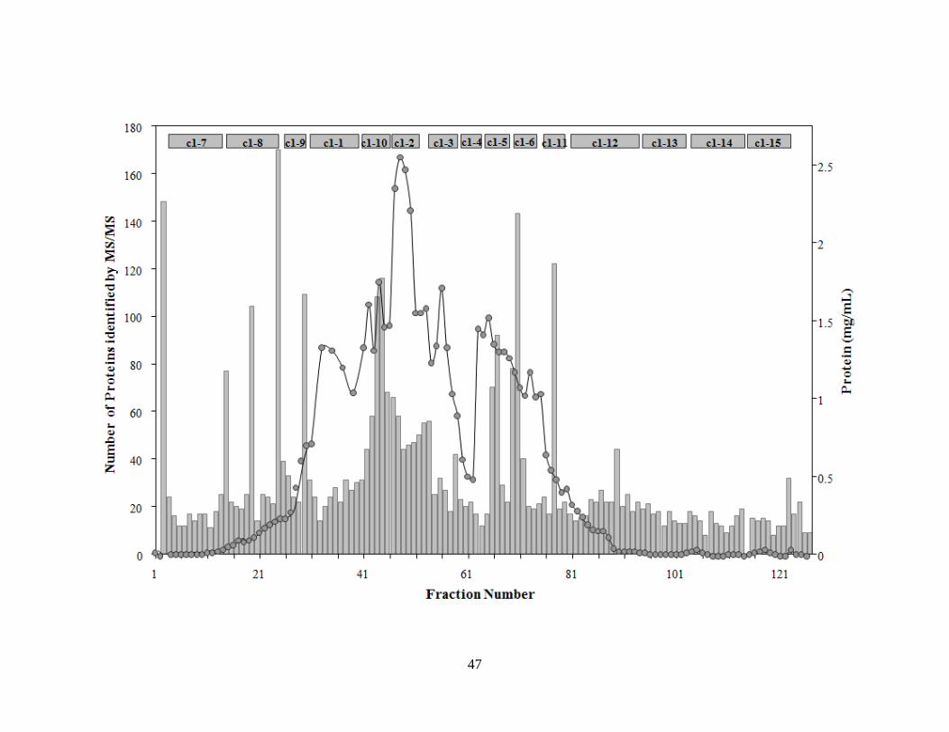
47

48
The HT method was typically applied to those fractions that were less complex, since less
resolving power is needed when separating a smaller number of peptides. In addition to a shorter
time requirement during peptide separations, the HT method further saves time by its adaptation
to an automated 96 well format. To test the differences in sensitivity of the CV and HT methods,
a set of ten fractions were analyzed by both methods. A total of 403 proteins were detected
with387 being identified by the CV method and 111 by the HT method (with 95 detected by both
methods). Of those fractions containing more than 60 proteins, the HT method detected less
than25 % of those detected by the CV methods. Conversely, the number of proteins detected in
fractions that had roughly 20 proteins or less was similar using both approaches.
Identification of type 1 PCs was accomplished by cross tabulation of the identified
peptides and the fractions from which they were identified using an Excel pivot table. The
identities of the proteins present were listed in the rows and the fractions from which they were
detected were listed in the columns. An customized function was written with the pivot data and
used to assign an association score based on (i) the co-elution of proteins in common fractions
and (ii) if the proteins in common fractions were expressed from adjacent genes. The association
scores range from 0.0 defining no association to 1.0 defining the strongest association.
As fractions are purified through sequential orthologous steps, they become less complex
and theoretically more enriched with proteins that co-elute due to protein-protein interactions.
Those proteins that co-elute through multiple steps of chromatography and were found to be
encoded by adjacent genes were considered to form functional interactions. 243 or roughly 25 %
of the 967 proteins identified by mass spectrometry were encoded by adjacent genes. These 243
proteins are predicted to form 106 potential type I protein complexes (labeled PC1-PC106).
Four previously characterized type I heteromeric enzymes were used as positive controls. They

49
all had well established purification schemes which were followed and were detectable by
enzymatic assays. These included SH1, POR, VOR, and FNOR. Likewise five known
homomeric proteins were used as negative controls. Rubredoxin [12] and NADPH-rebredoxin
oxidoreductase [20] are monomers, while aldehyde ferredoxin oxidoreductase [10], rubrerythrin
[8], and ferredoxin [13] are type III complexes composed of more than one identical subunit.
Known PCs. 61 of the 243 proteins that make up the 106 potential PCs have been previously
purified and characterized from P. furiosus biomass or in recombinant form. Of the 61, 45 of
them are components of 16 potential PCs identified using the method employed in this study
(Table 2.1). The proteins that constitute the positive controls are included in this number.
Thirteen of the 16 PCs have been purified from P. furious native biomass, while three had been
characterized using recombinant proteins. Identification of these known PCs validates the
criteria used to find novel PCs.
Expected PCs. Five PCs (PC-37 [21], PC-58 [22; 23], PC-63 [24], PC-74 [25], and PC-76 [26])
were categorized as expected because their subunit proteins were homologous to proteins that
formed complexes in a closely related organism (Table 2.2). These PCs were involved in protein
biosynthesis, RNA processing, central carbon metabolism and amino acid metabolism. With the
exception of PC-58, they all have association values between 0.2 and 0.3, and all but PC-58 is
predicted to be expressed from an operon by a method used for operon prediction in P. furiosus
[18].
Novel potential PCs. Eighty-six PCs were deemed novel potential PCs. These were type I PCs
that do not fit the definitions of a known or expected PC. In other words, they had not been
characterized previously in P. furiosus and are neither anticipated due to characterization of
homologous complexes in other organisms. The potential PCs have were divided into three

50
Table 2.1. Known PCs. Dark gray boxes indicate where two or more components of a complex
co-elute from a column into the same fraction. Light gray boxes indicate the presence of a
subunit that did not co-elute with another component of its complex. Blank squares indicate that
the protein was not identified in any fraction. PC-34 represents two known PCs, making the total
16. A.V. is the association value. The protein annotation differs from NCBI data base
annotation when the component ‘known’ as indicated by a reference, is homologous to a known
complex in a closely related organism, or has a known InterPro domain (indicated by brackets).
PC ORF Annotation MW pI c1
c2
-7
c2
-8
c2
-9
c2
-1
c2
-10
c2
-2
c2
-3
c2
-4
c2
-5
c2
-6
c2
-11
c2
-12
c2
-13
c2
-14
c2
-15
c3
-1
c3
-2
c3
-3
c3
-4
c3
-5
c3
-6
c4
-1
c4
-2
c4
-3
c4
-4
A.V.
CONTROL PCs
PC-33 PF0891 hydrogenase I beta 43.4 6.3 0.40
PC-33 PF0892 hydrogenase I gamma 33.1 9.0 0.40
PC-33 PF0893 hydrogenase I delta 29.2 5.2 0.40
PC-33 PF0894 hydrogenase I alpha 48.3 5.7 0.40
PC-34 PF0965 pyruvate ferredoxin oxidoreductase beta 36.3 9.1 0.42
PC-34 PF0966 pyruvate ferredoxin oxidoreductase alpha 44.2 5.0 0.42
PC-34 PF0967 pyruvate ferredoxin oxidoreductase delta 12.0 5.2 0.42
PC-34 PF0968 2-ketoisovalerate ferredoxin oxidoreductase beta 34.8 9.1 0.42
PC-34 PF0969 2-ketovalerate ferredoxin oxidoreductase alpha 44.0 5.3 0.42
PC-34 PF0970 2-ketisovalerate ferredoxin oxidoreductase delta 11.9 5.5 0.42
PC-34 PF0971 pyruvate/2-ketovalerate ferredoxin oxidoreductase gamma 20.0 5.4 0.42
PC-49 PF1327 ferredoxin:NADPH oxidoreductase (FNOR I alpha) 52.7 7.1 0.46
PC-49 PF1328 ferredoxin:NADPH oxidoreductase (FNOR I beta) 30.8 7.2 0.46
KNOWN PCs
PC-2 PF0018 DNA polymerase II small subunit 69.2 5.0 0.20
PC-2 PF0019 DNA polymerase II large subunit 143.2 6.2 0.20
PC-2 PF0020 <beta-lactamase-like (lactamase_B)> 23.8 5.5 0.20
PC-5 PF0092 replication factor C, large subunit 55.3 5.8 0.26
PC-5 PF0093 replication factor C, small subunit 97.8 6.7 0.26
PC-5 PF0094 protein disulfide oxidoreductase 25.8 4.9 0.26
PC-10 PF0181 [ATPase subunit F] 11.7 5.2 0.11
PC-10 PF0182 ATPase subunit A 113.5 6.1 0.11
PC-10 PF0183 ATPase subunit B 51.9 5.3 0.11
PC-10 PF0184 ATPase subunit D 25.0 9.6 0.11
PC-22 PF0533 indolepyruvate oxidoreductase alpha 71.3 6.8 0.80
PC-22 PF0534 indolepyruvate ferredoxin oxidoreductase beta 23.5 6.4 0.80
PC-26 PF0598 aspartate carbamoyltransferase, regulatory subunit 16.9 7.8 0.39
PC-26 PF0599 aspartate carbamoyltransferase, catalytic subunit 34.7 6.1 0.39
PC-46 PF1245 proline dehydrogenase alpha 54.9 8.1 0.46
PC-46 PF1246 proline dehydrogenase beta 42.5 7.2 0.46
PC-50 PF1331 hydrogenase II delta 26.3 5.9 0.44
PC-50 PF1332 hydrogenase II alpha 46.2 5.0 0.44
PC-56 PF1433 mbh11 membrane bound hydrogenase beta 20.2 5.1 0.31
PC-56 PF1434 mbh12 membrane bound hydrogenase alpha 47.9 6.6 0.31
PC-62 PF1562 DNA-directed RNA polymerase subunit a 44.4 5.7 0.21
PC-62 PF1563 DNA-directed RNA polymerase subunit a' 103.1 6.4 0.21
PC-62 PF1564 DNA-directed RNA polymerase subunit b 127.0 6.3 0.21
PC-62 PF1565 DNA-directed RNA polymerase subunit h 9.2 8.8 0.21
PC-65 PF1577 hypothetical protein PF1577 48.2 9.3 0.23
PC-65 PF1578 DNA topoisomerase VI subunit A 44.1 8.4 0.23
PC-65 PF1579 DNA topoisomerase VI subunit B 64.4 8.8 0.23
PC-68 PF1642 DNA-directed RNA polymerase subunit k 6.2 9.5 0.23
PC-68 PF1643 DNA-directed RNA polymerase subunit n 8.2 6.1 0.23
PC-106 PF2018 replication protein A32 31.2 5.1 0.25
PC-106 PF2019 replication protein A14 14.2 4.9 0.25
PC-106 PF2020 replication protein A41 41.0 4.9 0.25

51
Table 2.2. Expected PCs. For details, see the legend to Table 2.1
PC ORF Annotation MW pI c1
c2
-7
c2
-8
c2
-9
c2
-1
c2
-10
c2
-2
c2
-3
c2
-4
c2
-5
c2
-6
c2
-11
c2
-12
c2
-13
c2
-14
c2
-15
c3
-1
c3
-2
c3
-3
c3
-4
c3
-5
c3
-6
c4
-1
c4
-2
c4
-3
c4
-4
PC-37 PF0989 phenylalanyl-tRNA synthetase alpha subunit 57.8 9.2
PC-37 PF0990 phenylalanyl-tRNA synthetase beta subunit 64.0 4.9
PC-58 PF1461 glutamyl-tRNA(Gln) amidotransferase subunit D 48.6 5.7
PC-58 PF1462 glutamyl-tRNA(Gln) amidotransferase subunit E 71.2 5.6
PC-63 PF1568 [ribonuclease ph (exosome complex)] 27.8 8.9
PC-63 PF1569 [RNA binding S1 (exosome complex)] 29.5 8.9
PC-74 PF1767 [2-ketoglutarate:ferredoxin oxidoreductase delta] 9.7 6.9
PC-74 PF1768 [2-ketoglutarate:ferredoxin oxidoreductase alpha] 44.0 6.1
PC-76 PF1795 [sarcosine oxidase, alpha subunit] 51.4 9.1
PC-76 PF1796 [polyferredoxin] 18.9 6.9
PC-76 PF1797 [L-proline dehydrogenase alpha subunit] 9.8 7.9
PC-76 PF1798 [L-proline dehydrogenase beta subunit] 42.4 6.1

52
categories pertaining to the likelihood of their associations. The three categories are weak or
unlikely novel PCs, tentative novel PCs, and strong novel potential PCs.
Weak or unlikely novel PCs. Predicted complexes which have one or more of their subunits
represented in many fractions, but which are not typically found in the same fraction of its
proposed partner are assigned to this category. Thirteen of the potential type I complexes made
from a total of 27 proteins are weak or unlikely novel PCs (Table 2.3). An example is PC-4
(PF0042/PF0043), a proposed complex made up of phosphoenolpyruvate synthase [27] and a
seemingly unrelated metalloprotease. While phosphoenolpyruvate synthase was found in
over150 fractions, its predicted partner was found only in six fractions. Therefore, the co-elution
of the proteins encoded by PF0042 and PF0043 of PC-4 is most likely due to coincidence rather
than a functional interaction.
Tentative novel PCs. Thirty PCs containing 61 proteins were designated as tentative because
they were only identified in fractions of the first purification step (Table 2.4). The validity of 11
of the tentative novel PCs is supported by evidence of them belonging to an operon. These
proteins may not have been detected in the later steps of purification because the later steps were
analyzed using HT mass spectrometry methodology only, whereas, the first column was
analyzed by the CV and HT methods. Some of the complexes detected in the first
chromatographic step are likely to be real complexes that are at such a low abundance as not be
detected by the HT method in the later columns. However, some potential PCs from the first
purification step may have been identified mainly due to the high complexity of early fractions
which may have resulted in subunits that were encoded by adjacent genes not being resolved into
separate fractions. Strong novel. Forty-three of the potential complexes are characterized as
being strongly associated (Table 2.5). Thirty-nine of them are predicted to be encoded by

53
Table 2.3 Weak or unlikely PCs. For details see legend to table 2.1.
PC ORF Annotation MW pI c1
c2
-7
c2
-8
c2
-9
c2
-1
c2
-10
c2
-2
c2
-3
c2
-4
c2
-5
c2
-6
c2
-11
c2
-12
c2
-13
c2
-14
c2
-15
c3
-1
c3
-2
c3
-3
c3
-4
c3
-5
c3
-6
c4
-1
c4
-2
c4
-3
c4
-4
A.V.
PC-4 PF0042 metal-dependent hydrolase 28.3 6.0 0.00
PC-4 PF0043 phosphoenolpyruvate synthetase 90.5 5.2 0.00
PC-13 PF0207 argininosuccinate synthase 46.0 5.3 0.14
PC-13 PF0208 argininosuccinate lyase 51.3 5.6 0.14
PC-21 PF0495 reverse gyrase 140.0 7.5 0.18
PC-21 PF0496 <transcriptional regulator winged helix (Wing_hlx_tran_reg)> 30.6 5.6 0.18
PC-25 PF0588 [phosphoglucomutase] 49.6 5.7 0.14
PC-25 PF0589 [phosphomannose isomerase] 52.6 5.8 0.14
PC-31 PF0868 NDP-sugar synthase 47.2 5.1 0.08
PC-31 PF0869 aspartyl-tRNA synthetase 51.0 5.3 0.08
PC-44 PF1203 formaldehyde:ferredoxin oxidoreductase 69.7 6.0 0.11
PC-44 PF1204 seryl-tRNA synthetase 53.2 6.7 0.11
PC-44 PF1205 <nucleotide binding protein, PINc (PINc)> 17.3 8.9 0.11
PC-67 PF1640 30S ribosomal protein S2 23.0 9.3 0.07
PC-67 PF1641 <enolase (Enolase)> 37.7 4.4 0.07
PC-87 PF1931 <circadian clock protein KaiC (KAIC)> 27.7 8.6 0.13
PC-87 PF1932 <transcriptional regulator winged helix (Wing_hlx_tran_reg)> 31.1 5.2 0.13
PC-90 PF1993 50S ribosomal protein L10 37.1 4.9 0.16
PC-90 PF1994 50S ribosomal protein L12 11.2 4.3 0.16
PC-96 PF0220 hexulose-6-phosphate synthase (d-arabino 3-hexulose 6-phosphate formaldehyde lyase) 47.6 6.3 0.15
PC-96 PF0221 hypothetical protein PF0221 26.7 5.0 0.15
PC-97 PF0463 hydrolase related to 2-haloalkanoic acid dehalogenase 27.0 5.1 0.07
PC-97 PF0464 glyceraldehyde-3-phosphate ferredoxin oxidoreductase 73.9 6.3 0.07
PC-99 PF1499 30S ribosomal protein S19e 17.4 9.7 0.18
PC-99 PF1500 <PRC-barrel-like (PRCH_cytoplasmic)> 10.4 4.6 0.18
PC-105 PF1956 fructose-1,6-bisphosphate aldolase class I (Fba1) 31.1 6.6 0.17
PC-105 PF1957 <putative agmatinase (Agmatinase)> 36.0 5.0 0.17

54
Table 2.4. Tentative PCs. Tenative complexes were detected only in the DEAE fraction of the
first purification step. See Table 1 legend for details.
PC ORF Annotation MW pI A.V.
PC-3 PF0037 <armadillo-like helical (ARM-like)> 65.5 8.5 0.19
PC-3 PF0038 <beta-lactamase-like (lactamase_B)> 22.3 6.0 0.19
PC-14 PF0232 transcription regulatory protein, arsR family 21.5 6.2 0.15
PC-14 PF0233 [acetyl-CoA synthetase Q2, alpha subunit] 50.4 6.0 0.15
PC-15 PF0378 50S ribosomal protein L31 11.1 10.1 0.19
PC-15 PF0379 50S ribosomal protein L39e 6.3 12.4 0.19
PC-16 PF0382 prefoldin, beta subunit (pfdA) 19.7 9.5 0.18
PC-16 PF0383 hypothetical protein PF0383 12.8 4.6 0.18
PC-18 PF0431 <phosphoribosylformylglycinamidine cyclo-ligase (purM)> 36.8 5.3 0.23
PC-18 PF0432 <HAD-superfamily hydrolase, subfamily IIA (HAD-SF-IIA)> 30.6 5.2 0.23
PC-19 PF0440 ribonucleotide reductase 200.9 6.1 0.15
PC-19 PF0441 galactose-1-phosphate uridylyltransferase 38.5 8.6 0.15
PC-20 PF0491 <transcription factor TFIIE, alpha subunit (TFIIE_alpha)> 22.9 8.8 0.2
PC-20 PF0492 hypothetical protein PF0492 11.0 5.6 0.2
PC-23 PF0540 <thymidylate synthase complementing protein ThyX (thyX)> 29.0 8.9 0.23
PC-23 PF0541 <Peptidase M24, catalytic core (Peptidase_M24_cat_core)> 32.8 6.7 0.23
PC-27 PF0637 <CRISPR-associated protein, MJ0385 (cas_Csa4)> 39.6 6.3 0.23
PC-27 PF0638 <CRISPR-associated protein, MJ0385 (cas_Csa4)> 44.3 6.8 0.23
PC-36 PF0982 hypothetical protein PF0982 21.6 4.8 0.05
PC-36 PF0983 pcna sliding clamp (proliferating-cell nuclearantigen) 28.0 4.5 0.05
PC-39 PF1115 <PUA (PUA)> 19.5 8.0 0.2
PC-39 PF1116 glycerol-3-phosphate cytidyltransferase 16.9 8.6 0.2
PC-43 PF1189 hypothetical protein PF1189 12.4 5.5 0.14
PC-43 PF1190 <rubrerythrin (Rubrerythrin)> 20.7 5.2 0.14
PC-45 PF1242 dehydrogenase subunit alpha 73.8 8.5 0.23
PC-45 PF1243 hypothetical protein PF1243 11.2 5.0 0.23
PC-53 PF1375 <translation elongation factor EF1A, eukaryotic and archaeal (EF-1_alpha)> 47.6 8.7 0.39
PC-53 PF1376 30S ribosomal protein S10 11.7 10.2 0.39
PC-54 PF1379 cell division protein pelota 40.5 7.2 0.22
PC-54 PF1380 arginyl-tRNA synthetase 72.3 6.2 0.22
PC-55 PF1385 DNA polymerase, bacteriophage-type 22.5 9.0 0.25
PC-55 PF1386 <helix-turn-helix type 3 (HTH_CROC1)> 26.4 9.3 0.25
PC-57 PF1454 <radical SAM (Radical_SAM)> 68.7 6.3 0.2
PC-57 PF1455 <von Willebrand factor, type A (VWFADOMAIN)> 10.3 4.9 0.2
PC-59 PF1482 <beta-grasp fold, ferredoxin-type (Ferredoxin_fold)> 8.2 8.2 0.21
PC-59 PF1483 <DNA-binding SAP (SAP)> 28.3 5.3 0.21
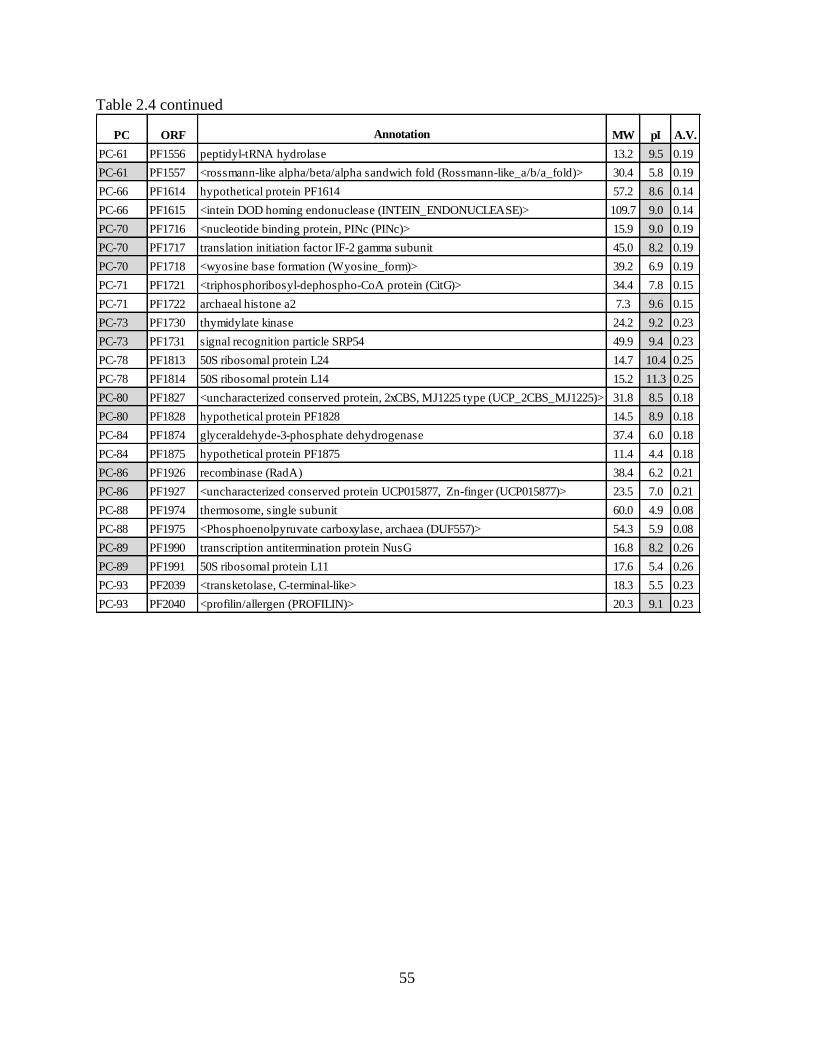
55
Table 2.4 continued
PC ORF Annotation MW pI A.V.
PC-61 PF1556 peptidyl-tRNA hydrolase 13.2 9.5 0.19
PC-61 PF1557 <rossmann-like alpha/beta/alpha sandwich fold (Rossmann-like_a/b/a_fold)> 30.4 5.8 0.19
PC-66 PF1614 hypothetical protein PF1614 57.2 8.6 0.14
PC-66 PF1615 <intein DOD homing endonuclease (INTEIN_ENDONUCLEASE)> 109.7 9.0 0.14
PC-70 PF1716 <nucleotide binding protein, PINc (PINc)> 15.9 9.0 0.19
PC-70 PF1717 translation initiation factor IF-2 gamma subunit 45.0 8.2 0.19
PC-70 PF1718 <wyosine base formation (Wyosine_form)> 39.2 6.9 0.19
PC-71 PF1721 <triphosphoribosyl-dephospho-CoA protein (CitG)> 34.4 7.8 0.15
PC-71 PF1722 archaeal histone a2 7.3 9.6 0.15
PC-73 PF1730 thymidylate kinase 24.2 9.2 0.23
PC-73 PF1731 signal recognition particle SRP54 49.9 9.4 0.23
PC-78 PF1813 50S ribosomal protein L24 14.7 10.4 0.25
PC-78 PF1814 50S ribosomal protein L14 15.2 11.3 0.25
PC-80 PF1827 <uncharacterized conserved protein, 2xCBS, MJ1225 type (UCP_2CBS_MJ1225)> 31.8 8.5 0.18
PC-80 PF1828 hypothetical protein PF1828 14.5 8.9 0.18
PC-84 PF1874 glyceraldehyde-3-phosphate dehydrogenase 37.4 6.0 0.18
PC-84 PF1875 hypothetical protein PF1875 11.4 4.4 0.18
PC-86 PF1926 recombinase (RadA) 38.4 6.2 0.21
PC-86 PF1927 <uncharacterized conserved protein UCP015877, Zn-finger (UCP015877)> 23.5 7.0 0.21
PC-88 PF1974 thermosome, single subunit 60.0 4.9 0.08
PC-88 PF1975 <Phosphoenolpyruvate carboxylase, archaea (DUF557)> 54.3 5.9 0.08
PC-89 PF1990 transcription antitermination protein NusG 16.8 8.2 0.26
PC-89 PF1991 50S ribosomal protein L11 17.6 5.4 0.26
PC-93 PF2039 <transketolase, C-terminal-like> 18.3 5.5 0.23
PC-93 PF2040 <profilin/allergen (PROFILIN)> 20.3 9.1 0.23

56
Table 2.5. Strong PCs. For details, see the legend in Table 2.1.
PC ORF Annotation MW pI c1
c2
-7
c2
-8
c2
-9
c2
-1
c2
-10
c2
-2
c2
-3
c2
-4
c2
-5
c2
-6
c2
-11
c2
-12
c2
-13
c2
-14
c2
-15
c3
-1
c3
-2
c3
-3
c3
-4
c3
-5
c3
-6
c4
-1
c4
-2
c4
-3
c4
-4
A.V.
PC-1 PF0014 hypothetical protein PF0014 14.8 6.4 0.25
PC-1 PF0015 hypothetical protein PF0015 27.1 5.0 0.25
PC-6 PF0096 <4Fe-4S ferredoxin, iron-sulfur binding (4FE4S_FERREDOXIN)> 8.9 8.2 0.24
PC-6 PF0097 42 kDa subunit bacteriochlorophyll synthase-like protein 44.1 6.1 0.24
PC-7 PF0138 <ferritin/ribonucleotide reductase-like (Ferritin/RR_like)> 37.4 5.4 0.26
PC-7 PF0139 [2-keto acid ferredoxin oxidoreductase 3 gamma] 22.0 8.4 0.26
PC-8 PF0146 potassium channel, putative 22.1 4.7 0.31
PC-8 PF0147 potassium channel related protein 23.0 4.8 0.31
PC-9 PF0160 <peptidase U62, modulator of DNA gyrase (PmbA_TldD)> 50.8 5.1 0.27
PC-9 PF0161 <peptidase U62, modulator of DNA gyrase (PmbA_TldD)> 46.7 6.7 0.27
PC-11 PF0198 phosphoribosylformylglycinamidine synthase subunit I 25.4 5.9 0.24
PC-11 PF0199 phosphoribosylformylglycinamidine synthase subunit II 78.2 5.2 0.24
PC-12 PF0202 Isocitrate dehydrogenase 44.3 6.5 0.36
PC-12 PF0203 citrate synthase 43.0 7.2 0.36
PC-17 PF0390 hypothetical protein PF0390 42.2 5.3 1.00
PC-17 PF0391 hypothetical protein PF0391 13.8 8.9 1.00
PC-24 PF0548 <hydrogenase expression/formation protein (HYPC)> 8.3 4.9 0.21
PC-24 PF0549 <hydrogenase formation HypD protein (Hydrgn_mat_hypD)> 41.2 6.8 0.21
PC-28 PF0639 <CRISPR-associated HD (cas3_HD)/CH> 27.1 8.3 0.25
PC-28 PF0640 <helicase superfamily 1 and 2 ATP-binding (HELICASE_ATP_BIND_1)> 75.3 8.9 0.25
PC-28 PF0641 <CRISPR-associated protein, MJ0382 (cas_cas5a)> 29.8 9.4 0.25
PC-29 PF0642 <CRISPR-associated regulatory Csa2 (DUF73)> 37.3 6.4 0.39
PC-29 PF0643 hypothetical protein PF0643 13.4 6.6 0.39
PC-30 PF0753 ferrodoxin oxidoreductase beta subunit 32.4 8.8 0.73
PC-30 PF0754 2-keto acid:ferredoxin oxidoreductase subunit alpha 63.1 5.5 0.73
PC-32 PF0872 <kaiA binding (KaiA-binding)> 14.7 6.2 0.23
PC-32 PF0873 hypothetical protein PF0873 5.8 8.1 0.23
PC-35 PF0972 <hydroxymethylglutaryl-coenzyme A synthase (HMG_coA_syn)> 38.2 7.9 0.31
PC-35 PF0973 acetyl-CoA acetyltransferase 42.6 4.8 0.31
PC-35 PF0974 hypothetical protein PF0974 15.9 8.5 0.31
PC-38 PF1033 <thioredoxin-like fold (IPR012336)> 24.7 5.5 0.20
PC-38 PF1034 <orotate phosphoribosyl transferase (pyrE)> 20.1 5.5 0.20
PC-40 PF1121 <CRISPR-associated protein Cas5, Tneap type (cas_Cas5t)> 23.9 9.3 0.21
PC-40 PF1122 <CRISPR-associated regulatory DevR (cas_Cst2_DevR)> 39.9 6.1 0.21
PC-40 PF1123 <CRISPR-associated protein, CXXC_CXXC region (cas_CXXC_CXXC)> 55.6 6.7 0.21
PC-41 PF1124 <CRISPR-associated protein, TM1791 (cas_TM1791_cmr6)> 38.7 8.9 0.23
PC-41 PF1125 <CRISPR-associated protein, TM1791.1 (cas_Cmr5)> 19.7 8.6 0.23
PC-41 PF1126 <CRISPR-associated RAMP Cmr4 (cas_RAMP_Cmr4)> 32.6 5.6 0.23

57
Table 2.5 continued.
PC ORF Annotation MW pI c1
c2
-7
c2
-8
c2
-9
c2
-1
c2
-10
c2
-2
c2
-3
c2
-4
c2
-5
c2
-6
c2
-11
c2
-12
c2
-13
c2
-14
c2
-15
c3
-1
c3
-2
c3
-3
c3
-4
c3
-5
c3
-6
c4
-1
c4
-2
c4
-3
c4
-4
A.V.
PC-42 PF1186 <FAD-dependent pyridine nucleotide-disulphide oxidoreductase (Pyr_redox_2)> 48.7 5.7 0.21
PC-42 PF1187 hypothetical protein PF1187 39.0 5.1 0.21
PC-47 PF1268 <Cobalamin (vitamin B12)-independent methionine synthase MetE, N-terminal (Meth_synt_1)> 35.5 5.2 0.23
PC-47 PF1269 methionine synthase 39.3 5.9 0.23
PC-48 PF1283 rubrerythrin 19.5 5.7 0.16
PC-48 PF1284 <histone-fold (Histone-fold)> 6.8 9.1 0.16
PC-48 PF1285 <FeS assembly protein SufD (sufD)> 40.2 7.1 0.16
PC-48 PF1286 <FeS assembly protein SufB (sufB)> 53.5 6.0 0.16
PC-48 PF1287 <FeS assembly ATPase SufC (SufC)> 27.2 6.1 0.16
PC-51 PF1339 hypothetical protein PF1339 25.8 5.3 0.48
PC-51 PF1340 <von Willebrand factor, type A (VWFADOMAIN)> 25.4 8.5 0.48
PC-52 PF1356 glucose-1-phosphate uridylyltransferase 32.3 5.3 0.21
PC-52 PF1357 UDP- or dTTP-glucose 4-epimerase or 4-6-dehydratase 38.1 5.8 0.21
PC-60 PF1529 <vitamin B6 biosynthesis protein (PDXS_SNZ_2)> 36.9 5.8 0.20
PC-60 PF1530 <thiamine biosynthesis Thi4 protein (Thi4)> 27.2 5.3 0.20
PC-64 PF1573 <peptidase U62, modulator of DNA gyrase (PmbA_TldD)> 51.1 5.6 0.25
PC-64 PF1574 <peptidase U62, modulator of DNA gyrase (PmbA_TldD)> 48.8 5.3 0.25
PC-69 PF1713 carbamoyl-phosphate synthase small subunit 41.7 5.9 0.34
PC-69 PF1714 carbamoyl-phosphate synthase large subunit 118.1 5.7 0.34
PC-72 PF1728 NDP-sugar synthase 40.1 5.4 0.24
PC-72 PF1729 phospho-sugar mutase 26.5 5.6 0.24
PC-75 PF1771 [2-keto acid:ferredoxin oxidoreductase alpha] 43.1 5.3 0.40
PC-75 PF1772 [2-keto acid:ferrredoxin oxidoreductase beta] 31.1 9.2 0.40
PC-75 PF1773 [2-keto acid:ferredoxin oxidoreductase gamma] 19.6 6.0 0.40
PC-77 PF1807 50S ribosomal protein L32e 15.5 11.2 0.22
PC-77 PF1808 50S ribosomal protein L6 20.9 8.7 0.22
PC-77 PF1809 30S ribosomal protein S8 14.7 9.6 0.22
PC-79 PF1824 50S ribosomal protein L4 28.7 10.6 0.25
PC-79 PF1825 50S ribosomal protein L3 41.4 10.1 0.25
PC-81 PF1837 acetyl-CoA synthetase II beta 26.4 5.6 0.25
PC-81 PF1838 [acetyl-CoA synthetase NTB alpha] 51.8 5.6 0.25
PC-82 PF1842 <prokaryotic chromosome segregation and condensation protein ScpA (ScpA_ScpB)> 25.1 4.7 0.23
PC-82 PF1843 chromosome segregation protein smc 147.8 8.7 0.23
PC-83 PF1857 cobalt transport ATP-binding protein cbio 24.7 6.4 0.24
PC-83 PF1858 cysteine synthase 31.6 8.8 0.24

58
Table 2.5 continued.
PC ORF Annotation MW pI c1
c2
-7
c2
-8
c2
-9
c2
-1
c2
-10
c2
-2
c2
-3
c2
-4
c2
-5
c2
-6
c2
-11
c2
-12
c2
-13
c2
-14
c2
-15
c3
-1
c3
-2
c3
-3
c3
-4
c3
-5
c3
-6
c4
-1
c4
-2
c4
-3
c4
-4
A.V.
PC-85 PF1909 ferredoxin 7.3 3.9 0.10
PC-85 PF1910 [sulfide dehydrogenase alpha SudX] 52.5 8.0 0.10
PC-85 PF1911 [sulfide dehydrogenase SudY] 32.7 5.7 0.10
PC-91 PF1999 glycine dehydrogenase subunit 1 50.2 5.5 0.25
PC-91 PF2000 glycine dehydrogenase subunit 2 56.1 6.0 0.25
PC-92 PF2005 glycerol-3-phosphate dehydrogenase 54.9 5.9 0.25
PC-92 PF2006 NADH oxidase 45.5 6.3 0.25
PC-92 PF2007 hypothetical protein PF2007 12.8 9.2 0.25
PC-94 PF0008 <histidine triad (HIT) protein (HIT_2)> 18.9 6.2 0.20
PC-94 PF0009 <molybdenum cofactor biosynthesis (MoeB)> 25.5 5.6 0.20
PC-95 PF0070 <prefoldin (Prefoldin)> 36.9 9.3 0.22
PC-95 PF0071 <RNA-binding protein, containing Pseudouridine synthase and Archaeosine transglycosylase (PUA) domain> 66.0 8.2 0.22
PC-98 PF0838 hypothetical protein PF0838 10.0 9.6 0.22
PC-98 PF0839 <PilT protein, N-terminal (PIN)> 14.7 4.7 0.22
PC-100 PF1558 30S ribosomal protein S7 24.7 10.0 0.28
PC-100 PF1559 30S ribosomal protein S12 16.4 10.6 0.28
PC-101 PF1589 <PfkB (PfkB)> 28.3 6.9 0.22
PC-101 PF1590 <NUDIX (NUDIX_hydrolase)> 18.9 6.0 0.22
PC-102 PF1648 30S ribosomal protein S11 14.7 10.4 0.25
PC-102 PF1649 30S ribosomal protein S4 21.3 10.0 0.25
PC-102 PF1650 30S ribosomal protein S13 16.9 10.9 0.25
PC-103 PF1679 <3-isopropylmalate dehydratase large subunit (LeuC)> 41.2 5.5 0.26
PC-103 PF1680 [3-isopropylmalate dehydratase small subunit] 18.0 6.6 0.26
PC-104 PF1803 50S ribosomal protein L30 17.7 10.2 0.21
PC-104 PF1804 30S ribosomal protein S5P 26.6 9.6 0.21

59
operons. The components of individual PCs were found to have similar elution profiles, were
expressed by genes that appear to be in an operon, were co-regulated at the rRNA level, and
were expressed by genes that had similar gene structure to homologous genes found in related
organisms. A discussion of several of the strong novel potential PCs found illustrates the
potential of this method. A function could not be ascribed to many of the strong PCs because
one or more of the components of the complex were describe as “hypothetical” or were poorly
annotated or both. PC-17 is an example of a hypothetical complex composed of two
hypothetical subunits which are found together in at least 47 fractions indicating that PC-17 is a
very stable complex. PC-42 is an example of a hypothetical complex in which only one of its
proposed subunits has a known general function. Although it is known that one of the subunits is
a flavoprotein, this is not enough information to help discern the function of the whole complex.
Similarly, PC-51 has some components that are annotated as homologs of the von Willebrand
factor which is found in higher eukaryotes.
Twelve ribosomal proteins form five strong novel PCs (PC-77, PC-79, PC-100, PC-102,
and PC-104). These PCs appear to be subcomplexes of complete ribosomes. It may be that the
HT mass spectrometry method that detected the complexes did not detect some subunits which
were in reality associated with detected subunits. If this is in fact the case, the ribosomal
complexes detected are not in reality novel.
Proteins of the clustered regularly interspaced short palindromic repeats
(CRISPR)/CRISPR-associated (cas) system were also detected in the native biomass of P.
furiosus. CRISPRs are known to be involved in resistance to bacteriophages and constitute a
prokaryotic immune system [28]. Eleven of the subunits form four strong novel potential PCs
(PC-28, PC-29, PC-40 and PC-41). The genes for PC-28 and PC-29 are predicted to form a

60
single operon [18] which is down regulated during cold-shock experiments [29]. Indirect
evidence from the closely related T. kodakarensis using a plasmid challenge assay suggested that
PC-28, PC-29, and PC-40 could be involved in CRISPR-Cas mediated defense against foreign
DNA in P. furiosus [30]. PC-41 contains three of six proteins Cmr [Cas module RAMP (repeat-
associated mysterious proteins)] proteins which are important for RNA cleavage [31]. The
remaining three which are encoded by PF1128-PF1130 were not identified as a Type I complex.
It may be that they do not form a complex or mass spectrometry was unable to detect them.
Based on the NCBI annotation of type I complexes, several strong, novel potential PCs
have a role in nucleotide and polynucleotide metabolism. PC-11 is composed of phosphoribosyl-
formylglyicinamide synthase subunits I and II. This heterodimeric complex is involved in purine
biosynthesis. PC-9 and PC-64 are paralogous complexes and appear to be involved in
modulating the activity of DNA gyrase. These two complexes probably account for the unusual
DNA topoisomerase and reverse gyrase activites that are unique to hyperthermophilic organisms
[32]. The annotations of PC-82 subunits suggest that this complex may be involved in
chromosome segregation [33]. This complex was only found in four fractions suggesting it to be
in low abundance in the cell. PC-95 forms a complex composed of one subunit that might be
involved in RNA metabolism and another one involved in protein folding. The possible
relationship between these two functions remains unclear.
Four PCs likely participate in some aspect of carbohydrate metabolism. PC-12 [34; 35]
appears to be made of subunits that participate in the tricarboxylic acid cycle, while the others
(PC-52, PC-72 and PC-101) are concerned with monosaccharide phosphate derivatives. With
the exception of PC-101, each of the carbohydrate metabolism related PCs are predicted to be
expressed from an operon [18]. PC-101 is proposed to be composed of a subunit with a

61
carbohydrate kinase activity and a subunit that is a NUDIX protein. The latter subunit is a type
of NDP hydrolase. The crystal structure of a recombinantly produced NUDIX ortholog from
Pyrobaculum aerophilum has been solved as a homodimeric protein [36]. However, with limited
information about the function of these proteins, a physiological heteromeric protein remains a
possibility, especially in light of the fact that PC-101 was natively purified.
The annotations of proteins in four PCs implicate their involvement in amino acid
biosynthesis: PC-47 (methionine), PC-91 (glycine), PC-69 (arginine), and PC-103 (leucine).
Experimental evidence for the function of PC-69, which is annotated as a carbamoyl synthetase,
shows an up-regulation of the genes that encode these subunits when growing on maltose, an
observation which is consistent with a role of this complex in arginine biosynthesis [37]. The
annotations of PC-103 (PF1679/PF1680) identify it as a putative 3-isopropylmalate dehydratase.
However, PF1679 and PF1680 are paralogs of PF0938 and PF0939 which are in the leucine
biosynthesis operon and more likely encode the 3-isopropylmalate dehydratase. A study was
done in the closely related Pyrococcus horikoshii which suggests that PC-103 is most likely
involved lysine rather than leucine biosynthesis [38].
Pyrococcus furiosus can use peptides as a carbon and energy source [39]. Figure 2.3
shows the proposed peptide fermentation pathway. In this pathway, peptides are digested by
proteases to individual amino acids. Various 2-ketoacids are produced by the oxidative
deamination of the amino acids. This step is carried out by aminotransferases. The resulting 2-
ketoacids are converted to acyl-CoA derivatives by ferredoxin (Fd) linked 2-keto acid
oxidoreductases (KORs). There are four KORs that have been characterized, each of which has
been identified in this study as either a known or expected PC. As mentioned earlier, PC-34
actually represents two type-I complexes, pyruvate ferredoxin: oxidoreductase (POR) [40] and 2-

62
Figure 2.3.
Proposed pathway of peptide metabolism in fermentative hyperthermophilic archaea.
Fdox, oxidized ferredoxin; Fdred, reduced ferredoxin; POR, pyruvate oxidoreductase; VOR, 2-
ketoisovalerate oxidoreductase; IOR, indolepyruvate oxidoreductase; KGOR, 2-ketoglutarate
oxidoreductase; CoA, coenzyme A; ACS acyl-CoA synthetase. ACS1 uses acetyl-CoA and CoA
derivatives derived from branched chain amino acids (represented by isobutyric acid in the
figure). ACS2 utilizes the substrates of ACS1 in addition to CoA derivatives derived from
aromatic amino acids (represented by phenylacetic acid in the figure). ACS3 by homology to
succinyl-CoA synthetase of T. kodakarensis succinyl-CoA, the CoA derivative derived from
Glu/Gln fermentation. POR oxodatively decarboxylates pyruvate. VOR oxidatively
decarboxylates branched chain 2-keto acids, IOR oxidatively decarboxylates aromatic 2-keto
acids, and KGOR oxidatively decarboxylates 2-ketoglutarate.
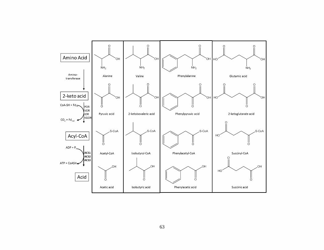
63
64
ketoisovalerate ferredoxin: oxidoreductase (VOR) [41], whose genes are adjacent in the genome
(PF0965-PF0971). PC-22 is indolepyruvate ferredoxin: oxidoreductase (IOR) [42] and PC-74 is
an ortholog of the 2-ketoglutarate: ferredoxin: oxidoreductase (KGOR) that was characterized in
Thermococcus litoralis [25]. PC-30 and PC-75 are two additional KOR paralogs that were
detected as type-I complexes. Transcriptomic analyses of the genes which encode PC-75
(PF1771-PF1773) show that it is up- regulated in peptide grown cultures while PC-30
(PF0753/PF0754) is not significantly affected [43]. Presumably these KORs encompass the
metabolism of additional amino acid derivatives not used by the other KORs since the KORs that
have so far been characterized are shown to be involved in the metabolism of at most nine of the
20 amino acids [21; 37; 38].
The reductant formed from the oxidation of 2-ketoacids reduces ferredoxin which in turn
reduces NADP+ via ferredoxin: NADP
+: oxidoreductase (FNOR I), a known PC (PC-49). PC-85
is categorized as a strong novel type I PC that is a paralog to FNOR I. Called FNOR II, PC-85 is
inversely regulated in relation to FNOR1 when grown on peptides and maltose [37]. Although
the FNOR homologs catalyze the same reaction, there is likely a physiological significance to
their differential regulation. It may be that they affect the regulation of the different KGORs at
an enzymatic level during growth on either peptides or carbohydrates.
The acyl-CoA products generated by the KORs are used in ATP synthesis by
heterotetrameric (22) NDP- dependent acyl-CoA synthetases. There are a total of five
subunit (PF1540/PF0532/PF0233/PF1085/PF1838) paralogs and two subunit
(PF1787/PF1837) paralogs found in the P. furiosus genome. ACS1 (PF1540/PF1787) and ACS2
(PF0532/PF1837) have been purified from native P. furiosus biomass [19] and are classified as
type II protein complexes because the genes that encode their subunits are unlinked in the

65
genome. Interestingly, an NDP dependent ACS that was purified and characterized from
Thermococcus kodakarensis uses succinate as a substrate, indicating its role in
glutamate/glutamine metabolism [44]. The T. kodakarensis enzyme has the same subunit
stoichiometry as the characterized P. furiosus ACSs. Its subunit is an ortholog to PF0233 and
its subunit is an ortholog to the P. furiosus ACS2 (PF1837) subunit. PC-81;
(PF1838/PF1837) a strong potential novel type I PC detected by the method used in this study
appears to be an additional ACS. Therefore, three ACS enzymes have identical subunit, but in
association with three differing subunits. The presence of genes for only two subunits in the
genome along with evidence for the association of ACS2 association with three different ACS
subunits suggest that a variety of combinations of and subunits is possible. All five
subunits and both subunits were detected in the fractions by mass spectrometry although it is
difficult to decipher what specific complexes are formed because multiple ACS and subunit
paralogs co-elute in common fractions most likely due to similar pIs and physical sizes. Similar
to the uncharacterized KOR paralogs, characterization of the remaining ACS paralogs will
provide a more complete picture of which amino acids participate in the peptide fermentation
pathway.
Related methods to the one discussed here have been developed in that they use mass
spectrometry to identify intact complexes by co-migration of their components. One such
method uses gradient centrifugation to sort high molecular weight complexes in organelles which
are then analyzed by mass spectrometry to detect their component parts [45]. A method very
similar to the method described here used non-denaturing aerobic fractionation of E.coli extract
and identifies complexes by co-elution of proteins from chromatographic columns as well [46].
However, they use isobaric tags (iTRAQ) to produce elution profiles for each protein based on

66
relative abundances to determine which proteins co-elute. While this method is very
sophisticated, the investigators only used two levels of purification and only identified abundant
and well characterized protein complexes.
Synopsis. A total of 106 potential complexes were identified employing the method described
here of identifying co-eluting proteins by mass spectrometry. Eighty-six of the 106 were
considered as novel potential and the confidence in their validity as complexes were described as
either strong novel, tentative novel, or weak/unlikely novel. Thirty potential PCs were identified
in fractions from the first purification step. These complexes were labeled as “tentative” since
they were not identified in fractions of downstream columns. Although deemed as such, some
“tentative” complexes are likely real and may not have been identified in later fractions because
those fractions were analyzed by the HT method rather than the more sensitive CV method. The
advantage of the HT method is that it generates data faster than CV method. However, because
of the complexity of some fractions, the HT method is unable to detect some proteins that may
actually be present in fractions. As a pilot fractionation, a main purpose of this experiment was
to see if it was a workable method, therefore, the fractionation was not as extensive as it could
have been. Only 5 % of fractions derived from two purification steps were fractionated on
additional columns, and only 12 % of those fractions were fractionated further. Clearly, a lot of
data is to be had from the fractionation and analysis of every fraction. Furthermore, additional
fractionation would reduce the complexity of the fractions and thus make it possible for the HT
method to identify a higher percentage of proteins. The added resolution would increase the
power of this method by making it possible to not only identify type 1 complexes, but also type 2
complexes as well as low abundant complexes. The low abundance of some complexes, preclude
their native characterization. However, once identified, they may be produced recombinantly to

67
provide enough material for further study. In addition, the data recorded during the execution of
this method could be later used as a resource which provides a road map that would be useful in
the development of purification schemes for protein complexes of interest.
References
1. Berggard, T., S. Linse and P. James, "Methods for the detection and analysis of protein-
protein interactions," Proteomics, vol. 7, no. 16, pp. 2833-42, 2007.
2. Piehler, J., "New methodologies for measuring protein interactions in vivo and in vitro,"
Curr Opin Struct Biol, vol. 15, no. 1, pp. 4-14, 2005.
3. Azarkan, M., J. Huet, D. Baeyens-Volant, Y. Looze and G. Vandenbussche, "Affinity
chromatography: a useful tool in proteomics studies," J Chromatogr B Analyt Technol
Biomed Life Sci, vol. 849, no. 1-2, pp. 81-90, 2007.
4. Srere, P.A. and C.K. Mathews, "Purification of Multienzyme Complexes," Methods in
Enzymology, vol. 182, pp. 539-551, 1990.
5. McHenry, C.S. and W. Crow, "DNA polymerase III of Escherichia coli. Purification and
identification of subunits," J Biol Chem, vol. 254, no. 5, pp. 1748-53, 1979.
6. Verhagen, M.F., A.L. Menon, G.J. Schut and M.W. Adams, "Pyrococcus furiosus: large-
scale cultivation and enzyme purification," Methods Enzymol, vol. 330, pp. 25-30, 2001.
7. Ma, K. and M.W. Adams, "Sulfide dehydrogenase from the hyperthermophilic archaeon
Pyrococcus furiosus: a new multifunctional enzyme involved in the reduction of
elemental sulfur," J Bacteriol, vol. 176, no. 21, pp. 6509-17, 1994.
8. Weinberg, M.V., F.E. Jenney, Jr., X. Cui and M.W. Adams, "Rubrerythrin from the
hyperthermophilic archaeon Pyrococcus furiosus is a rubredoxin-dependent, iron-
containing peroxidase," J Bacteriol, vol. 186, no. 23, pp. 7888-95, 2004.

68
9. Schut, G.J., A.L. Menon and M.W. Adams, "2-keto acid oxidoreductases from
Pyrococcus furiosus and Thermococcus litoralis," Methods in Enzymology, vol. 331, pp.
144-158, 2001.
10. Mukund, S. and M.W. Adams, "The novel tungsten-iron-sulfur protein of the
hyperthermophilic archaebacterium, Pyrococcus furiosus, is an aldehyde ferredoxin
oxidoreductase. Evidence for its participation in a unique glycolytic pathway," J Biol
Chem, vol. 266, no. 22, pp. 14208-16, 1991.
11. Bryant, F.O. and M.W. Adams, "Characterization of hydrogenase from the
hyperthermophilic archaebacterium, Pyrococcus furiosus," J Biol Chem, vol. 264, no. 9,
pp. 5070-9, 1989.
12. Blake, P.R., J.B. Park, F.O. Bryant, S. Aono, J.K. Magnuson, E. Eccleston, J.B. Howard,
M.F. Summers and M.W. Adams, "Determinants of protein hyperthermostability:
purification and amino acid sequence of rubredoxin from the hyperthermophilic
archaebacterium Pyrococcus furiosus and secondary structure of the zinc adduct by
NMR," Biochemistry, vol. 30, no. 45, pp. 10885-95, 1991.
13. Aono, S., F.O. Bryant and M.W. Adams, "A novel and remarkably thermostable
ferredoxin from the hyperthermophilic archaebacterium Pyrococcus furiosus," J
Bacteriol, vol. 171, no. 6, pp. 3433-9, 1989.
14. Bradford, M.M., "Rapid and sensitive method for quantitation of microgram quantities of
protein utilizing principle of protein-dye binding," Analytical Biochemistry, vol. 72, no.
1-2, pp. 248-254, 1976.

69
15. Pruitt, K.D., T. Tatusova and D.R. Maglott, "NCBI reference sequences (RefSeq): a
curated non-redundant sequence database of genomes, transcripts and proteins," Nucleic
Acids Res, vol. 35, no. Database issue, pp. D61-5, 2007.
16. Quevillon, E., V. Silventoinen, S. Pillai, N. Harte, N. Mulder, R. Apweiler and R. Lopez,
"InterProScan: protein domains identifier," Nucleic Acids Res, vol. 33, no. Web Server
issue, pp. W116-20, 2005.
17. Robb, F.T., D.L. Maeder, J.R. Brown, J. DiRuggiero, M.D. Stump, R.K. Yeh, R.B. Weiss
and D.M. Dunn, "Genomic sequence of hyperthermophile, Pyrococcus furiosus:
implications for physiology and enzymology," Methods in Enzymology, vol. 330, pp.
134-157, 2001.
18. Tran, T.T., P. Dam, Z. Su, F.L. Poole, 2nd, M.W. Adams, G.T. Zhou and Y. Xu, "Operon
prediction in Pyrococcus furiosus," Nucleic Acids Res, vol. 35, no. 1, pp. 11-20, 2007.
19. Mai, X. and M.W. Adams, "Purification and characterization of two reversible and ADP-
dependent acetyl coenzyme A synthetases from the hyperthermophilic archaeon
Pyrococcus furiosus," Journal of Bacteriology, vol. 178, no. 20, pp. 5897-5903, 1996.
20. Ma, K. and M.W. Adams, "A hyperactive NAD(P)H:Rubredoxin oxidoreductase from
the hyperthermophilic archaeon Pyrococcus furiosus," J Bacteriol, vol. 181, no. 17, pp.
5530-3, 1999.
21. Shiraki, K., M. Tsuji, Y. Hashimoto, K. Fujimoto, S. Fujiwara, M. Takagi and T.
Imanaka, "Genetic, enzymatic, and structural analyses of phenylalanyl-tRNA synthetase
from Thermococcus kodakaraensis KOD1," J Biochem, vol. 134, no. 4, pp. 567-74, 2003.
22. Oshikane, H., K. Sheppard, S. Fukai, Y. Nakamura, R. Ishitani, T. Numata, R.L. Sherrer,
L. Feng, E. Schmitt, M. Panvert, S. Blanquet, Y. Mechulam, D. Soll and O. Nureki,

70
"Structural basis of RNA-dependent recruitment of glutamine to the genetic code,"
Science, vol. 312, no. 5782, pp. 1950-4, 2006.
23. Schmitt, E., M. Panvert, S. Blanquet and Y. Mechulam, "Structural basis for tRNA-
dependent amidotransferase function," Structure, vol. 13, no. 10, pp. 1421-33, 2005.
24. Ramos, C.R., C.L. Oliveira, I.L. Torriani and C.C. Oliveira, "The Pyrococcus exosome
complex: structural and functional characterization," J Biol Chem, vol. 281, no. 10, pp.
6751-9, 2006.
25. Mai, X. and M.W. Adams, "Characterization of a fourth type of 2-keto acid-oxidizing
enzyme from a hyperthermophilic archaeon: 2-ketoglutarate ferredoxin oxidoreductase
from Thermococcus litoralis," Journal of Bacteriology, vol. 178, no. 20, pp. 5890-5896,
1996.
26. Kawakami, R., H. Sakuraba, H. Tsuge, S. Goda, N. Katunuma and T. Ohshima, "A
second novel dye-linked L-proline dehydrogenase complex is present in the
hyperthermophilic archaeon Pyrococcus horikoshii OT-3," FEBS J, vol. 272, no. 16, pp.
4044-54, 2005.
27. Hutchins, A.M., J.F. Holden and M.W. Adams, "Phosphoenolpyruvate synthetase from
the hyperthermophilic archaeon Pyrococcus furiosus," J Bacteriol, vol. 183, no. 2, pp.
709-15, 2001.
28. Deveau, H., J.E. Garneau and S. Moineau, "CRISPR/Cas system and its role in phage-
bacteria interactions," Annu Rev Microbiol, vol. 64, pp. 475-93, 2010.
29. Weinberg, M.V., G.J. Schut, S. Brehm, S. Datta and M.W. Adams, "Cold shock of a
hyperthermophilic archaeon: Pyrococcus furiosus exhibits multiple responses to a

71
suboptimal growth temperature with a key role for membrane-bound glycoproteins," J
Bacteriol, vol. 187, no. 1, pp. 336-48, 2005.
30. Elmore, J.R., Y. Yokooji, T. Sato, S. Olson, C.V. Glover, 3rd, B.R. Graveley, H. Atomi,
R.M. Terns and M.P. Terns, "Programmable plasmid interference by the CRISPR-Cas
system in Thermococcus kodakarensis," RNA Biol, vol. 10, no. 5, pp. 828-40, 2013.
31. Terns, R.M. and M.P. Terns, "The RNA- and DNA-targeting CRISPR-Cas immune
systems of Pyrococcus furiosus," Biochem Soc Trans, vol. 41, no. 6, pp. 1416-21, 2013.
32. Forterre, P., "A hot story from comparative genomics: reverse gyrase is the only
hyperthermophile-specific protein," Trends Genet, vol. 18, no. 5, pp. 236-7, 2002.
33. Tadesse, S. and P.L. Graumann, "Differential and dynamic localization of topoisomerases
in Bacillus subtilis," J Bacteriol, vol. 188, no. 8, pp. 3002-11, 2006.
34. Steen, I.H., D. Madern, M. Karlstrom, T. Lien, R. Ladenstein and N.K. Birkeland,
"Comparison of isocitrate dehydrogenase from three hyperthermophiles reveals
differences in thermostability, cofactor specificity, oligomeric state, and phylogenetic
affiliation," J Biol Chem, vol. 276, no. 47, pp. 43924-31, 2001.
35. Muir, J.M., R.J. Russell, D.W. Hough and M.J. Danson, "Citrate synthase from the
hyperthermophilic Archaeon, Pyrococcus furiosus," Protein Eng, vol. 8, no. 6, pp. 583-
92, 1995.
36. Wang, S., C. Mura, M.R. Sawaya, D. Cascio and D. Eisenberg, "Structure of a Nudix
protein from Pyrobaculum aerophilum reveals a dimer with two intersubunit beta-sheets,"
Acta Crystallogr D Biol Crystallogr, vol. 58, no. Pt 4, pp. 571-8, 2002.
37. Schut, G.J., S.D. Brehm, S. Datta and M.W. Adams, "Whole-genome DNA microarray
analysis of a hyperthermophile and an archaeon: Pyrococcus furiosus grown on

72
carbohydrates or peptides," Journal of Bacteriology, vol. 185, no. 13, pp. 3935-3947,
2003.
38. Lombo, T., N. Takaya, J. Miyazaki, K. Gotoh, M. Nishiyama, T. Kosuge, A. Nakamura
and T. Hoshino, "Functional analysis of the small subunit of the putative homoaconitase
from Pyrococcus horikoshii in the Thermus lysine biosynthetic pathway," FEMS
Microbiol Lett, vol. 233, no. 2, pp. 315-24, 2004.
39. Fiala, G. and K.O. Stetter, "Pyrococcus furiosus sp. nov. represents a novel genus of
marine heterotrophic archaebacteria growing optimally at 100-degrees C," Archives of
Microbiology, vol. 145, no. 1, pp. 56-61, 1986.
40. Blamey, J.M. and M.W. Adams, "Purification and characterization of pyruvate ferredoxin
oxidoreductase from the hyperthermophilic archaeon Pyrococcus furiosus," Biochimica
et Biophysica Acta, vol. 1161, no. 1, pp. 19-27, 1993.
41. Heider, J., X. Mai and M.W. Adams, "Characterization of 2-ketoisovalerate ferredoxin
oxidoreductase, a new and reversible coenzyme A dependent enzyme involved in peptide
fermentation by hyperthermophilic archaea," Journal of Bacteriology, vol. 178, no. 3, pp.
780-787, 1996.
42. Mai, X. and M.W. Adams, "Indolepyruvate ferredoxin oxidoreductase from the
hyperthermophilic archaeon Pyrococcus furiosus. A new enzyme involved in peptide
fermentation," Journal of Biological Chemistry, vol. 269, no. 24, pp. 16726-16732, 1994.
43. Schut, G.J., J. Zhou and M.W. Adams, "DNA microarray analysis of the
hyperthermophilic archaeon Pyrococcus furiosus: evidence for anNew type of sulfur-
reducing enzyme complex," J Bacteriol, vol. 183, no. 24, pp. 7027-36, 2001.

73
44. Shikata, K., T. Fukui, H. Atomi and T. Imanaka, "A novel ADP-forming succinyl-CoA
synthetase in Thermococcus kodakaraensis structurally related to the archaeal nucleoside
diphosphate-forming acetyl-CoA synthetases," Journal of Biological Chemistry, vol. 282,
no. 37, pp. 26963-26970, 2007.
45. Balbo, A., K.H. Minor, C.A. Velikovsky, R.A. Mariuzza, C.B. Peterson and P. Schuck,
"Studying multiprotein complexes by multisignal sedimentation velocity analytical
ultracentrifugation," Proc Natl Acad Sci U S A, vol. 102, no. 1, pp. 81-6, 2005.
46. Dong, M., L.L. Yang, K. Williams, S.J. Fisher, S.C. Hall, M.D. Biggin, J. Jin and H.E.
Witkowska, "A "tagless" strategy for identification of stable protein complexes genome-
wide by multidimensional orthogonal chromatographic separation and iTRAQ reagent
tracking," J Proteome Res, vol. 7, no. 5, pp. 1836-49, 2008.

74
CHAPTER 3
CHARACTERIZATION OF TEN HETEROTETRAMERIC NDP-DEPENDENT ACYL-COA
SYNTHETASES OF THE HYPERTHERMOPHILIC ARCHAEON
PYROCOCCUS FURIOSUS1
1Scott, J.W., F.L. Poole, and M.W.W. Adams. 2014. Archaea. 2014:Article ID 176863.
Reprinted here with permission of the publisher.

75
Abstract
The hyperthermophilic archaeon Pyrococcus furiosus grows by fermenting peptides and
carbohydrates to organic acids. In the terminal step, acyl-CoA synthetase (ACS) isoenzymes
convert acyl-CoA derivatives to the corresponding acid and conserve energy in the form of ATP.
ACS1 and ACS2 were previously purified from P. furiosus and have 22 structures but the
genome contains genes encoding three additional -subunits. The ten possible combinations of
and genes were expressed in E. coli and each resulted in stable and active 22 isoenzymes.
The -subunit of each isoenzyme determined CoA-based substrate specificity and between them
they accounted for the CoA derivatives of fourteen amino acids. The -subunit determined
preference for adenine or guanine nucleotides. The GTP-generating isoenzymes are proposed to
play a role in gluconeogenesis by producing GTP for GTP-dependent phosphoenolpyruvate
carboxykinase and for other GTP-dependent processes. Transcriptional and proteomic data
showed that all ten isoenzymes are constitutively expressed indicating that both ATP and GTP
are generated from the metabolism of most of the amino acids. A phylogenetic analysis showed
that the ACSs of P. furiosus and other members of the Thermococcales are evolutionarily
distinct from those found throughout the rest of biology, including those of other
hyperthermophilic archaea.
Introduction
Members of the Thermococcales order of the Archaea are anaerobic, heterotrophic
microorganisms that grow optimally at or above 80°C [1]. They include species of Pyrococcus
and Thermococcus, all of which utilize peptides as carbon and energy sources and many also
ferment carbohydrates [2; 3]. Glucose is metabolized via a modified Emden-Meyerhof pathway
that involves ADP- rather than ATP-dependent hexo- and phosphofructokinases [4] and a

76
ferredoxin-linked glyceraldehyde-3-phosphate (GAP) oxidoreductase (GAPOR) rather than an
NAD-linked GAP dehydrogenase [5; 6]. The pyruvate that is generated by glycolysis is oxidized
by pyruvate ferredoxin oxidoreductase (POR) [7] to yield CO2 and acetyl-CoA, and energy is
conserved in the form of ATP by an ADP-dependent acetyl-CoA synthetase that generates
acetate [8]. The general acyl CoA synthetase reaction is shown in (1). This reaction is in
contrast to the two-step conversion in anaerobic bacteria whereby acetyl CoA is transformed to
acetate via phosphotransacetylase and acetate kinase:
R. CO. SCoA+ADP+Pi⟶RCOOH+ATP+CoASH (1)
The proposed pathway for peptide fermentation involves the hydrolysis of peptides into
individual amino acids followed by their oxidative deamination to 2-keto acids [9]. Further
metabolism of the 2-keto acids is analogous to pyruvate oxidation by POR, in which they are
oxidatively decarboxylated to the acyl-CoA derivatives that then serve as substrates for ADP-
dependent CoA synthetases. This involves three other ferredoxin-dependent keto acid
oxidoreductases. Indolepyruvate ferredoxin oxidoreductase (IOR) and 2-ketoisovalerate
ferredoxin oxidoreductase (VOR) oxidize the transaminated products of aromatic amino acids
[10] and of branched chain amino acids [11], respectively, while 2-ketoglutarate ferredoxin
oxidoreductase (KGOR) oxidizes 2-ketoglutarate, which is derived from glutamate and
glutamine [12]. Hence POR is involved in the fermentation of both sugars and peptides
(pyruvate is the product of alanine transamination), while IOR, KGOR, and VOR function only
in peptide fermentation. The genome of the hyperthermophile P. furiosus contains two
additional ferredoxin-dependent oxidoreductases. XOR is in an operon with KGOR and is
77
coregulated with KGOR at the transcriptional level [13] but the substrate specificity of this fifth
2-ketoacid oxidoreductase is not known. In addition, a homolog composed of the gene products
of PF0753 and PF0754 was identified as a protein complex during the fractionation of P.
furiosus biomass [14]. The substrate specificity of this sixth 2-ketoacid oxidoreductase is also
unknown.
ACS therefore catalyzes the terminal step of peptide fermentation, the conversion of acyl
CoA derivatives to the corresponding acid with concomitant synthesis of ATP [1]. Two forms of
ACS, ACS1, and ACS2, have been characterized from P. furiosus both in native [8; 15] and
recombinant forms [16]. Each consists of two subunits with estimated molecular weights of 45
() and 25 kDa () with molecular masses for the holoenzymes of approximately 140 kDa,
indicative of structures. In addition to acetyl CoA, ACS1 used isobutyryl-CoA as a
substrate, while ACS2 used phenylacetyl-CoA and indoleacetyl-CoA. It was therefore proposed
that ACS1 was involved in the metabolism of alanine and branched chain amino acids and
utilized the products of POR and VOR, while ACS2 played a role in the metabolism of aromatic
amino acids via IOR [8]. The two enzymes also differed in their specificity for purine
nucleotides as ACS1 preferred GDP/GTP to ADP/ATP while ACS2 preferred ADP/ATP to
GDP/GTP [8]. From their N-terminal sequences it was evident that ACS1 and ACS2 did not
share any subunit [8] and from the P. furiosus genome sequence [17] it was evident that ASC1
(PF1540), ACS1 (PF1787), ACS2 (PF0532), and ACS2 (PF1837) are encoded by unlinked
genes.
The product of the KGOR reaction is succinyl CoA but no ACS-type activity was
detected in extracts of P. furiosus cells using this as a substrate [8]. However, an ADP-
dependent succinyl-CoA synthetase activity was purified from Thermococcus kodakarensis, a

78
member of the Thermococcales and a close relative of P. furiosus [18]. This enzyme, which will
be referred to here as ACS3, also had an apparent 22 subunit stoichiometry. The gene
encoding the subunit of T. kodkarensis ACS3 has a close homolog in the P. furiosus genome
(PF0233) while that encoding the -subunit corresponded to PF1837, which encodes the -
subunit of P. furiosus ACS2. Hence this -subunit is associated with two -subunits to give
either P. furiosus ACS2 or ACS3. Further inspection of the P. furiosus genome reveals that,
while there are only two genes that encode ACS -subunits (PF1837 and PF1787), two
additional -subunits are present encoded by PF1085 and PF1838. These will be designated as
ACS4 and ACS5, respectively. Recently, homologs of these two types of ACSs in P. furiosus
were characterized from T. kodakarensis [19]. It was reported that one of them (TK2127) was
specific for 2-(imidazole-4-yl)-acetate, a degradation product of histidine fermentation, while the
other (TK0944) had a broad substrate range similar to that of P. furiosus ACS2. However, in the
T. kodakarensis study, TK2127 and TK0944 were characterized with only one of the two
possible -subunits (TK0943) and only using adenine nucleotides.
P. furiosus and other members of the Thermococcales therefore have the genetic potential
to synthesize up to ten ACS isoenzymes, depending upon which of the five -subunits associates
with which of the two -subunits. The corresponding enzymes will be designated herein as
ACSx-A or ACSx-G, where A (PF1837) and G (PF1787) represent the two -subunits encoded
by the indicated genes, while x (x = 1–5) represents the -subunits encoded by PF1540 (1),
PF0532 (2), PF0233 (3), PF1085 (4), and PF1838 (5), respectively. The two -subunits are 59 %
identical at the amino acid sequence level. Hence the two ACS isoforms that have been purified
from P. furiosus biomass correspond to ACS1-G and ACS2-A, while a third form, ACS3-A, is
evident by analogy with the T. kodakarensis enzyme. Hence this analysis begs the questions of

79
whether the -subunits in these three forms are able to form catalytically active complexes with
the alternate -subunit, which -subunits are associated with ACS4 and ACS5, and what are the
substrate specificities of all of these complexes in terms of both acyl-CoA derivatives and
adenine and guanine nucleotides. Herein we have addressed these questions by generating the
recombinant forms of the ten possible ACS complexes in P. furiosus and assessing their
preferences for substrates corresponding to all twenty amino acids.
Materials and methods.
Production of recombinant ACSs. The genes encoding the five -subunits were amplified by
PCR from P. furiosus genomic DNA with the following primer sets: PF0233 (ACS3), 5′-
AATTTGACATATGACAGTTAACCTAGACTTTC-3' (+) and 5′-
CCGCTCGAGTTAGAGCTCAGCTAAATACTTTC-3' (−). PF0532 (ACS2), 5′-
AATTTGACATATGCTTGACTACTTCTTTAATCCAAG-3' (+) and 5′-
CCGCTCGAGTTAACCATTTACCCCACCTCCAACATTC-3' (−). PF1085 (ACS4), 5′-
AATTTGACATATGAGGTACTTCTTTTACCCAAATAG-3' (+) and 5′-
CCGCTCGAGTCATTCGCTACTAGACCTTAGGCTGAG-3' (−). PF1540 (ACS1), 5′-
AATTTGACATATGAGTTTGGAGGCTCTTTTTAATC-3' (+) and 5′-
CCGCTCGAGTTACTTTTCTTTGTGTTTTGCTTTC-3' (−). PF1838 (ACS5), 5′-
AATTTGACATATGATTAACAACTTGGACATTAAAG-3' (+) and 5′-
CCGCTCGAGTCATTCCCCCATCTTCCTCAAATATTC-3' (−). NdeI and XhoI restriction
sites are underlined for the + and − strands respectively. The resulting DNA fragments encoding
the subunits were each inserted into the pET24a(+) Kanr expression vector (Novagen,
Madison, WI). The genes encoding the two β subunit homologs were also amplified from the P.
furiosus genomic DNA with the following primers: PF1787 (-G), 5′-

80
GATGCCATGGACAGGGTTGCTAAGGCTAGGG-3' (+) and 5′-
TAATTTGAGCGGCCGCCTAAAGAATCATCCTAGC-3' (−). PF1837 (-A), 5′-
AATTTGACATATGATTAACAACTTGGACATTAAAG-3' (−). and 5′-
CCGCTCGAGTCATTCCCCCATCTTCCTCAAATATTC-3' (−). NcoI and NotI restriction
sites are underlined for the + and − strands respectively, for the PF1787 DNA and NdeI and
XhoI restriction sites underlined for the + and − strands respectively, for the PF1837 DNA. The
amplified PF1787 DNA fragment was inserted into pET21d Ampr expression vector and the
amplified PF1837 DNA fragment was inserted into pET21b Ampr expression vector. The
sequence of each inserted cassette was confirmed by the Sanger method. The subunit
expression plasmids (pET24a(+):PF0233, pET24a(+):PF0532, pET24a(+):PF1085,
pET24a(+):PF1540, and pET24a(+):PF1838) were cotransformed with each -subunit
expression plasmid (pET21d:PF1787 and pET21b:PF1837) into E. coli BL21(DE3) pRIL.
For recombinant protein production, cultures were grown with the appropriate antibiotics
at 37°C in 250 mL 2xYT media to an optical density between 0.8 and 1.0 at 600 nm. Expression
of the recombinant genes was induced by the addition of 0.4 mM IPTG (isopropyl-β-D
thiogalactopyranoside) at 37°C. Following 3 hours of induction, the cultures were harvested by
centrifugation. The pelleted cells were resuspended in lysis buffer (20 mM bis-Tris, pH 7.0,
containing 150 mM NaCl, 10 mM MgCl, 1 mM PMSF (phenylmethanesulfonylfluoride),
0.27 mg/mL lysozyme and 0.007 mg/mL DNAse I) and incubated at room temperature for 30
minutes. After centrifugation at 13,000 ×g for 20 min, the supernatant was heated at 80°C for
15 min and centrifuged to remove precipitated heat-labile E. coli proteins. The supernatant was
applied to a 10 mL HiTrap desalting column (made by connecting two 5 mL columns in tandem)
(GE Healthcare) that had been equilibrated with desalting buffer (20 mM bis-Tris, pH 7.0

81
containing 150 mM NaCl and 10 mM MgCl). The resulting enzyme preparations were used for
substrate specificity analyses. Protein concentrations were determined by the Bradford method
[20].
To prepare sample for molecular weight determination, the supernatant from the heat
treatment step described above (500 L, ~1.5 mg protein) was diluted 10-fold in buffer A
(20 mM bis-Tris, pH 7.0 containing 2 mM dithiothreitol (DTT) and 5 % glycerol) and applied to
a 1 mL Mono Q column (GE Healthcare) that had been equilibrated with buffer A. The ACS
was eluted with a 20 mL linear gradient from 0 to 0.3 M NaCl in buffer A. Fractions (1 mL)
were collected and those containing the ACS as determined by SDS electrophoresis were
combined and concentrated to a volume of 200 L in an Amicon Ultra centrifugation device
having a cut-off of 30 kDa. This was applied to a SX200 10/30 (24 mL) gel filtration column
that had been previously equilibrated with 50 mM potassium phosphate containing 200 mM KCl,
5 % glycerol, 2 mM DTT, and pH 7.0 and calibrated with standard proteins β-amylase (200 kDa),
alcohol dehydrogenase (150 kDa), BSA (66 kDa), carbonic anhydrase (29 kDa), and cytochrome
c (12.5 kDa).
High-throughput enzyme assays. Since the CoA-forms of the 2-keto acids from all 18 amino
acids (assuming that glutamate/glutamine and aspartate/asparagine represent two amino acids)
are not commercially available, substrate specificities of each ACS isoenzyme was measured
using the reverse reaction of (1) using 18 different acid substrates. A discontinuous assay in
which phosphate release from the NTP was quantitated was used in a 96-well format. The ACS
sample was that obtained after the heat treatment step described above. The 50 L reaction
mixture contained 20 mM bis-Tris buffer, pH 7.0, 0.5 g ACS, 20 mM acid substrate, 1 mM ATP
or GTP, 2.0 mM MgCl2, and 0.5 mM coenzyme-A. It was heated at 80°C for 1 minute in a 96-

82
well heating block. The reaction was stopped by adding 1.0 N H2SO4. The reaction was found
to be linear over at least a 2 min assay period. Free phosphate produced by the hydrolysis of
NTP was measured using the BioVision colorimetric assay kit (Mountain View, CA). All liquid
handling was carried out robotically (Beckman Coulter Biomek FX, Brea, CA). Kinetic
parameters were determined by the same method using NTP substrate concentrations over the
range from 0.0625 mM to 4.0 mM. The 96-well plate format was also used to assay activity in
the forward direction (1) with the appropriate acyl-CoA derivative. The reaction mixture (50 L)
contained acyl-CoA (0.031 mM to 2.0 mM), K2HPO4 (10 mM), MgCl2 (2 mM), ADP (2 mM),
and ACS1 (0.5 g) in 20 mM bis-Tris buffer (pH 7.0). After heating to 80°C for 90 sec, the free
CoASH released was measured by the addition of 200 L of 5′5′-dithiobis(2-nitrobenzoic acid)
(DTNB) (0.4 mM) in 100 mM K2PO4, pH 7.2.
Results and Discussion
Previously ACS1-G of P. furiosus was purified from native biomass [8] and the
recombinant forms of the two subunits (ACS1- and -G) generated separately in E. coli were
combined in vitro to give a heterotetrameric enzyme that was indistinguishable from that purified
from P. furiosus [16]. It was unclear, however, if production and combination of individual
subunits would be successful for all possible ACS isoenzymes, particularly if cotranslation was
required for efficient heteromeric complex formation. Since the T. kodakarensis homologs of
the P. furiosus ACS2-A and ACS3-A isoenzymes (which contain the same subunit) were
produced in E. coli using a single plasmid encoding both subunits [18], a similar coexpression
approach was used herein. Five plasmids conferring kanamycin resistance were constructed
which also encoded each of the five -subunits, and two plasmids were constructed conferring
ampicillin resistance which also encoded each of the -subunits. Each plasmid encoding a -

83
plasmid was then transformed into E. coli with a plasmid encoding one of the -subunits to
create all ten possible combinations. Heat treatment of the cell-free extracts of each E. coli strain
gave rise to two major proteins as determined by SDS PAGE with sizes corresponding to those
predicted from the gene sequences of the larger (, 47.8–51.8 kDa) and smaller (, 25.8–
26.4 kDa) ACS subunits, as shown in Figure 3.1. To determine which of the - and -subunit
combinations assembled into heteromeric structures, each was analyzed by gel filtration
chromatography. All ten eluted with estimated molecular weights of approximately 140 kDa and
there was no evidence of individual monomers indicating that all formed stable 22 structures
when their respective genes were coexpressed in E. coli.
The substrate specificities of ten recombinant ACS isoenzymes were determined at 80°C
using a 96-well plate approach and the eighteen organic acid substrates that correspond to the
twenty amino acids (the same acids are generated by glutamate and glutamine, and by aspartate
and asparagine) with either ATP or GTP as the co-substrate. A heat map representing the
relative activity of each isoform is shown in Figure 3.2, where 100 % activity represents the
highest measured value with either ATP or GTP. A lower limit was arbitrarily set at 30 % of the
specific activity produced by the most active substrate and this was used to establish the
substrate specificity of each enzyme. As anticipated, ACS1 was most active with acids derived
from alanine and the branched chain amino acids, but surprisingly, it also had significant activity
with thioglycolate, the end product of cysteine fermentation. In agreement with previous results
[8], ACS2 had overlapping specificity with ACS1 activity but, unlike ACS1, it was also highly
active with substrates representing the aromatic amino acids. Surprisingly, ACS2 was also
active with 3-methylthiopropionate, the end product of methionine fermentation. Hence ACS1
and ACS2 in combination show reasonable activity with acids derived from 9 of the 20 amino

84
Figure 3.1
SDS-PAGE analyses of recombinant P. furiosus ACS isoenzymes.
The lanes correspond to the following: 1: ACS1-G; 2: ACS1-A; 3: ACS2-G; 4: ACS2-A; 5:
ACS3-G; 6: ACS3-A, 7: ACS4-G; 8: ACS4-A; 9: ACS5-G; and 10: ACS5-A. Each lane
contains approximately 2 g of the protein obtained after the heat-treatment step. Lanes M
indicate molecular weight markers (given in kDa).

85
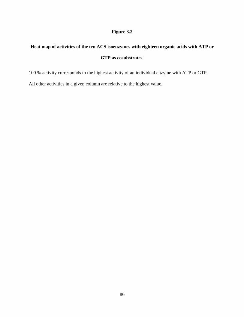
86
Figure 3.2
Heat map of activities of the ten ACS isoenzymes with eighteen organic acids with ATP or
GTP as cosubstrates.
100 % activity corresponds to the highest activity of an individual enzyme with ATP or GTP.
All other activities in a given column are relative to the highest value.

87

88
acids. One of the exceptions is succinate, derived from glutamate and glutamine via 2-
ketoglutarate. The only isoenzyme that showed activity with succinate was ACS3 (Figure 3.2).
However, ACS3 had similar activity with the methionine derivative, comparable to that of
ACS2.
ACS4 was the only isoenzyme that shows significant activity with imidazole 4-acetate,
which is derived from histidine. It is also the only ACS that shows detectable activity with the
organic acid derived from arginine (Figure 3.2). On the other hand, like ACS2, the substrate
specificity of ACS4 indicates that this isoenzyme is involved in the degradation of alanine and
the aromatic amino acids. ACS5 also shows a broad substrate range similar to that of ACS2, and
similarly does not utilize derivatives of methionine or lysine. However, ACS5 is the only
enzyme that has significant activity with lactate, which is the end product of the threonine
pathway. Considering all ten isoenzymes, there are six amino acids whose acid derivatives
werenot utilized by any of them and these were glycine, serine, proline, lysine, aspartate, and
asparagine. Of the fourteen acids utilized by the recombinant ACSs in the work presented
herein, nine of them (acetate, 3-methylthiopropionate, isobutyrate, isovalerate, 2-methylbutyrate,
phenylacetic acid, 4-hydroxyphenylacetic acid, indole-3-acetic acid, and succinate) result from
the four KORs that have been characterized [10-12]. However, the genome of P. furiosus
harbors two additional KORs. The genes for one (XOR, PF1771-PF1773) are expressed and are
regulated by carbon source [13] while the genes for the other (PF0753, PF0754) are also
expressed and the corresponding proteins have been identified in P. furiosus biomass [14].
Furthermore, it is not known if the KORs that have been characterized can use as substrates the
harbors two additional KORs. The genes for one (XOR, PF1771-PF1773) are expressed and are
regulated by carbon source [13] while the genes for the other (PF0753, PF0754) are also
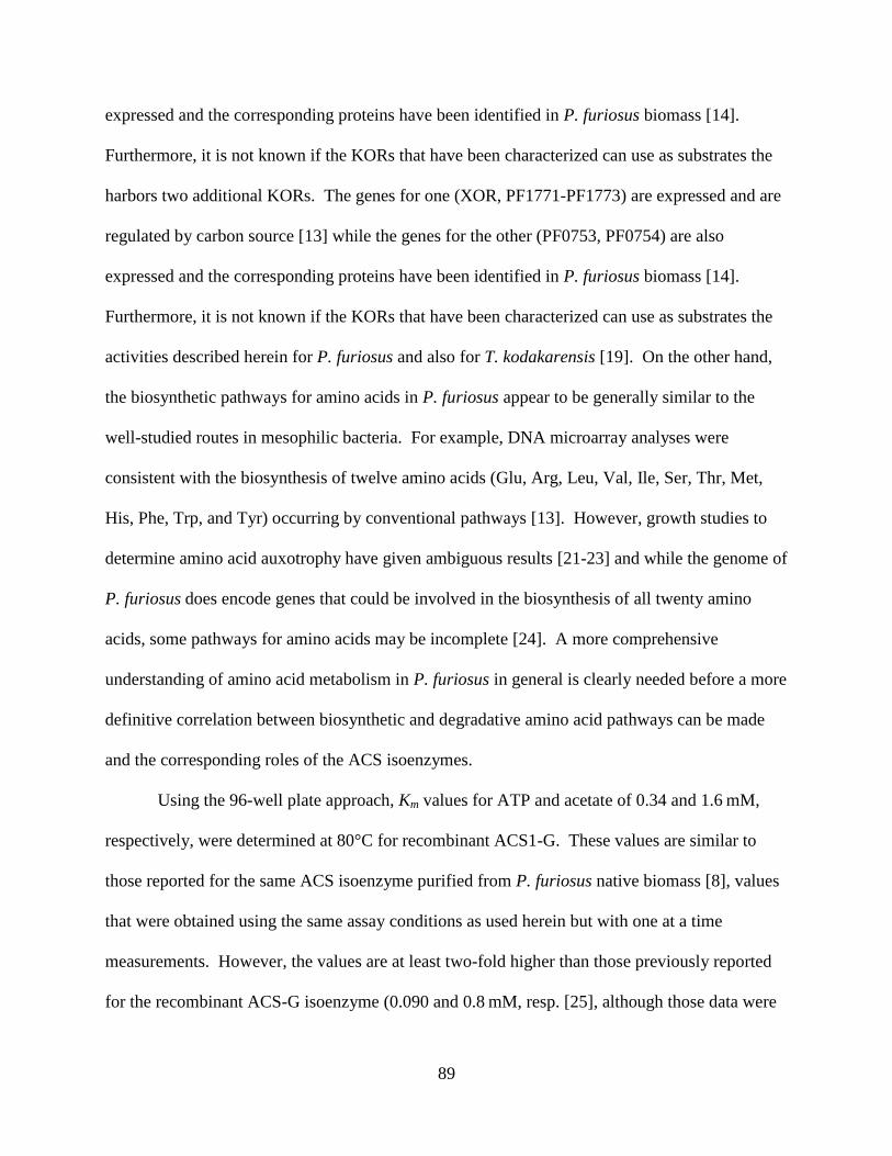
89
expressed and the corresponding proteins have been identified in P. furiosus biomass [14].
Furthermore, it is not known if the KORs that have been characterized can use as substrates the
harbors two additional KORs. The genes for one (XOR, PF1771-PF1773) are expressed and are
regulated by carbon source [13] while the genes for the other (PF0753, PF0754) are also
expressed and the corresponding proteins have been identified in P. furiosus biomass [14].
Furthermore, it is not known if the KORs that have been characterized can use as substrates the
activities described herein for P. furiosus and also for T. kodakarensis [19]. On the other hand,
the biosynthetic pathways for amino acids in P. furiosus appear to be generally similar to the
well-studied routes in mesophilic bacteria. For example, DNA microarray analyses were
consistent with the biosynthesis of twelve amino acids (Glu, Arg, Leu, Val, Ile, Ser, Thr, Met,
His, Phe, Trp, and Tyr) occurring by conventional pathways [13]. However, growth studies to
determine amino acid auxotrophy have given ambiguous results [21-23] and while the genome of
P. furiosus does encode genes that could be involved in the biosynthesis of all twenty amino
acids, some pathways for amino acids may be incomplete [24]. A more comprehensive
understanding of amino acid metabolism in P. furiosus in general is clearly needed before a more
definitive correlation between biosynthetic and degradative amino acid pathways can be made
and the corresponding roles of the ACS isoenzymes.
Using the 96-well plate approach, Km values for ATP and acetate of 0.34 and 1.6 mM,
respectively, were determined at 80°C for recombinant ACS1-G. These values are similar to
those reported for the same ACS isoenzyme purified from P. furiosus native biomass [8], values
that were obtained using the same assay conditions as used herein but with one at a time
measurements. However, the values are at least two-fold higher than those previously reported
for the recombinant ACS-G isoenzyme (0.090 and 0.8 mM, resp. [25], although those data were

90
based on a coupled assay at 55°C, which might account for the differences. From the data
presented in Figure 3.2 it is evident that all five ACS -subunits utilize acetyl CoA (forward
reaction of (1)) and the kinetic constants for each ACS-A enzyme is given in Table 3.1. The Km
and the kcat/Km values are quite similar for ACS1, ACS2, ACS4 and ACS5, with ACS3 having
comparable affinity but much lower activity. Hence all four isoenzymes likely utilize acetyl
CoA in vivo. Similarly, the results indicate that ACS2 and ACS5 are comparably active with
CoA derivatives derived from branched chain amino acids (Table 3.1). Unfortunately, the
primary substrate for ACS3, succinyl-CoA, could not be used at 80°C as it is extremely unstable
at the assay temperature and direct comparisons were not possible. Similarly, the primary CoA
substrates for ACS4 and ACS5, derived from histidine and threonine, are not commercially
available. It is interesting to compare the results described herein with P. furiosus to those
recently reported with T. kodakaraensis by Shikata et al. [18] and Awano et al. [19]. For
example, while the substrate specificities for the ACS3 homolog from each organism were very
similar, P. furiosus ACS4 utilized acids derived from histidine, methionine, phenylalanine,
tryptophan and arginine, while its counterpart in T. kodakaraensis was only reactive with the
histidine derivative. Similarly, in P. furiosus the substrate range of ACS5 is extended compared
to that of the T. kodakaraensis homolog as it was also active with lactate, a derivative of
threonine fermentation. While kinetic comparisons are limited since the T. kodakaraensis
homologs were studied mainly using acids as the substrates, in both studies ACS3 exhibited
Michaelis-Menten kinetics when acetyl CoA and ADP were used as substrates. However, the T.
kodakaraensis enzyme had a much lower Km value (41 μM) and higher catalytic efficiency
(0.460 μM−1
·s−1
) than P. furiosus ACS3 (474 M and 0.012 M−1
·s−1
). It is not clear to what
extent this reflects physiological differences between the two organisms. The specificities for

91
Table 3.1. Kinetic parameters of ACSx-A isoforms with CoA derivatives.
Enzyme Substrate Km (µM)
kcat
/Km
(µM-1
· s-1
)
ACS1-A acetyl-CoA 137 ± 91 0.140
ACS2-A acetyl-CoA 350 ± 2 0.114
ACS2-A isovaleryl-CoA 466 ± 119 0.109
ACS2-A phenylacetyl-CoA 3320 ± 1320 0.150
ACS3-A acetyl-CoA 474 ± 165 0.012
ACS4-A acetyl-CoA 242 ± 46 0.119
ACS5-A acetyl-CoA 620 ± 1.0 0.112
ACS5-A isobutyryl-CoA 2730 ± 1080 0.083
ACS5-A isovaleryl-CoA 820 ± 407 0.120

92
the organic substrates of the ACS isoenzymes of P. furiosus containing each of the five -
subunits was independent of whether they were associated with the -A or -G subunits (Figure
3.2). However, the type of subunit did determine the nucleotide specificity. With all ten
isoenzymes, those containing the -A subunit were more active with ATP than GTP. In contrast,
enzymes containing the -G subunit were generally more active with GTP than ATP or at least
showed comparable activity with ATP and GTP (Figure 3.2). This was further demonstrated by
the kinetic parameters of ACS1-G and ACS1-A with GTP and ATP as substrates.
The catalytic efficiency of ACS1-A was approximately 14-fold higher with ATP than
with GTP, while the opposite was true with ACS1-G (Table 3.2). Hence the nucleotide
specificity of the ACS isoenzymes is determined by the -subunit. This is consistent with the
presence of two ATP-grasp fold subdomains in the -subunits (residues 23–139 in A and 25–
139 in G), one of which includes a conserved histidinyl residue (His71) at the active site [26].
P. furiosus ACSs presumably transfer the phosphate group from the -subunit to this conserved
histidine of the G- subunit, which then transfers the phosphate group to the nucleoside
diphosphate [26].
Hence, the ten ACS isoenzymes in P. furiosus can account for the metabolism of the
majority (14 of 20) of amino acids, using both adenine and guanine nucleotides. The ACS
reaction is the only means of conserving energy during peptide degradation, and the same is true
for sugar fermentation. This is due to the unusual modification of the glycolytic pathway in P.
furiosus, where GAP oxidation is coupled to the reduction of ferredoxin instead of NAD,
resulting in the conservation of energy at the ferredoxin oxidation step via a respiratory

93
Table 3.2 Nucleotide kinetics of ACS1 isoforms.
Isoform Nucleotide Km (µM)
kcat /Km
(µM-1
· s-1
)
ACS1-A ATP 132 ± 19 0.14
ACS1-A GTP 223 ± 58 0.01
ACS1-G ATP 342 ± 63 0.011
ACS1-G GTP 87 ± 7 0.144

94
membrane-bound hydrogenase [27; 28] rather than by substrate level phosphorylation (SLP) via
1,3-bisphosphoglycerate [3; 29]. Hence the ACS reaction generating acetate is the only source
of ATP via SLP. The results presented herein show that all five ACS-A isoenzymes appear to be
efficient in catalyzing this reaction, as well as being involved in peptide degradation. Since ATP
is the principle energy carrier within the cell, it would be expected that all ACS-A isoenzymes
(containing the -A subunit) would catalyze these reactions. It is therefore not obvious what
advantage is gained by using the -G subunit to produce GTP. It would seem futile for an NDP
kinase to transphosphorylate ADP with the GTP since ATP is made via ACS isoenzymes with
the -A subunit.
Insight into the function of ACS isoenzymes containing the -G subunit is provided by
Fukuda and coworkers [30] who described a GTP-dependent phosphoenolpyruvate
carboxykinase (PCK) from T. kodakarensis. PCKs have been historically classified into GTP-
and ATP-dependent enzymes, with ATP-dependent enzymes described in bacteria, yeast, plants,
and some eukaryotic parasites while the GTP-dependent enzyme being found in mammals and
some other eukaryotes [31]. Interestingly, transcription of the gene encoding PCK was
upregulated when T. kodakarensis cells were grown under gluconeogenic (with pyruvate and
peptides as the carbon sources) rather than under glycolytic conditions (using starch as the
carbon source). DNA microarray analysis showed that the same is true in P. furiosus when cells
are grown on peptides and a sugar (maltose) [13]. As shown in Figure 3.3, PCK catalyzes the
first step of gluconeogenesis, the decarboxylation of oxaloacetate to yield phosphoenolpyruvate
and CO2. Hence we propose that during growth of P. furiosus by peptide fermentation, the
function of the five ACS-G isoenzymes containing the -G subunit is to provide GTP for the

95
Figure 3.3
Proposed function of ACS -G and ACS -A isoenzymes during growth of P. furiosus on
carbohydrates or peptides.
The abbreviations are: POR, pyruvate ferredoxin oxidoreductase; KAOR, 2-keto acid
oxidoreductases (IOR, VOR, KGOR and POR, see text for details).
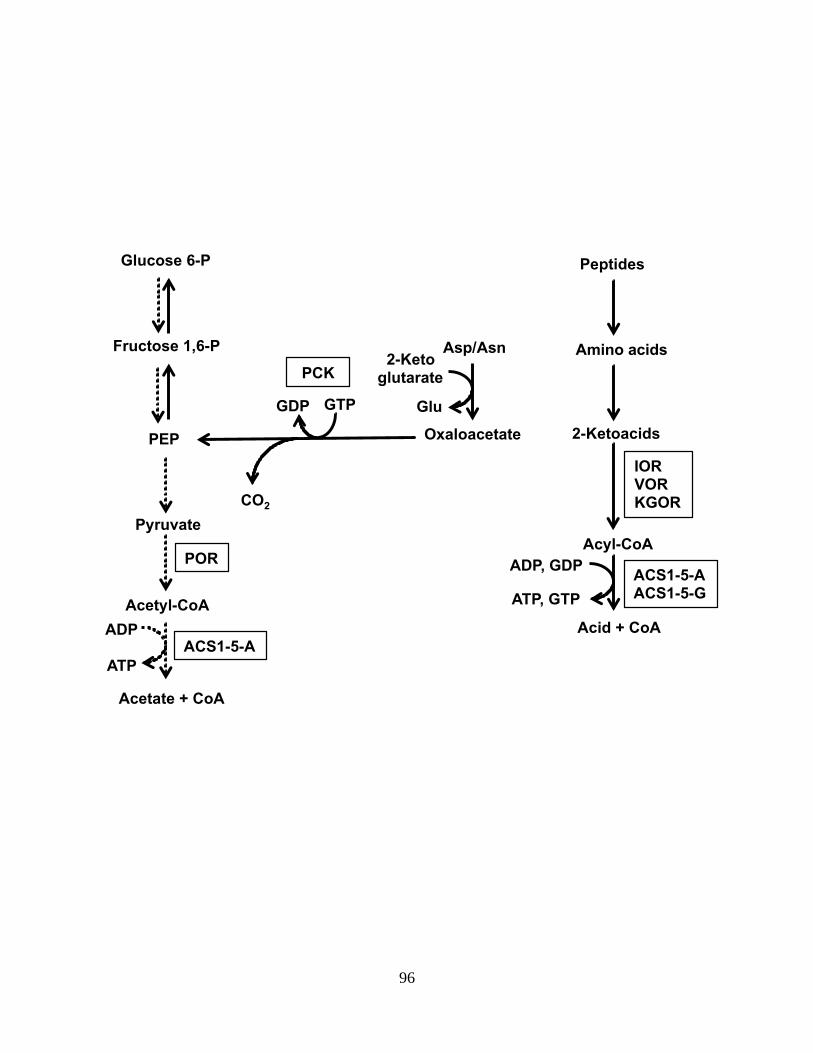
96
Glucose 6-P
Fructose 1,6-P
PEP
Pyruvate
POR
Acetyl-CoA
Acetate + CoA
ADP
ATP
ACS1-5-A
Peptides
Amino acids
2-Ketoacids
Acyl-CoA
ADP, GDP
ATP, GTP
ACS1-5-A ACS1-5-G
Oxaloacetate
Asp/Asn 2-Keto
glutarate
Glu GTP GDP
PCK
CO2
Acid + CoA
IOR VOR KGOR

97
gluconeogenic-related PCK reaction. The ATP-independent generation of GTP by the ACS-G
isoenzymes could also provide GTP for the archaeal translational machinery [32].
These data therefore suggest that all ten isoenzymes have physiological relevance in P.
furiosus. That they all are functional is supported by DNA microarray data, which show that all
ten genes are expressed under both gluconeogenic and glycolytic growth conditions and there
appears to be little if any differential expression between the two growth modes for any of these
genes [13]. The one exception is that the gene encoding the -subunit of ACS2 (PF0532), which
forms an operon with the genes encoding IOR, is up-regulated during growth on peptides.
Proteomic analyses of P. furiosus grown on maltose and peptides also showed that all ten
subunits of the ACS isoenyzmes are detected during fractionation of a cytoplasmic extract [14].
Moreover, immunoprecipitation analyses of T. kodakarensis showed that the -G and the -A
subunits associated with multiple -subunits [18]. Taken together, these data show that the ten
ACS isoenzymes are constitutively produced and are able to generate both ATP and GTP from
the wide variety of coenzyme-A derivatives produced during peptide fermentation. The same
appears to be true for all members of the Thermococcales since, like P. furiosus, their genomes
also contain genes encoding two -subunits and five -subunits of ACS.
Homologs of ACS, known as succinyl-CoA synthetases (SCS), have been characterized
from eukaryotes as well as bacteria [33; 34]. As with the P. furiosus enzymes, their nucleotide
specificity is determined by their subunits [35], although these differ from those in the ACSs of
the Thermococcales as they contain an additional ligase domain [26; 36]. A phylogenetic tree of
the -subunits of the ACSs and SCSs is shown in Figure 3.4. It is striking that those of the
Thermococcales form one clade while all other -subunits fall into a second widely divergent
clade. Each clade contains subunits that have a preference for one or the other purine nucleotide,

98
indicating that they each arose in each clade by independent gene duplications. Interestingly, the
ACSs that are found in Archaea that are not members of the Thermococcales, such as those in
some methanogens and in the hyperthermophilic sulfate-reducer Archaeoglobus fulgidus, fall
within a distinct branch within the eukaryote/bacterial clade of the tree rather than in the
Thermococcales branch (Figure 3.4). Clearly, the -subunits found in the ACSs of the
Thermococcales are very distinct from those in all other ACSs/SCSs found throughout biology,
including those in other hyperthermophilic archaea. Why and how this occurred are beyond the
scope of this paper but are interesting subjects worthy of additional study.

99
Figure 3.4.
Phylogenetic tree of ACS subunits.
The unweighted pair group method of arithmetic mean (UPGMA) tree is shown. A neighbor-
joining tree (data not shown) also shows similar relationships for the vertebrate and
Thermococcales subunits with a few changes for the remaining species. Archaea are highlighted
red, Bacteria are in black and Eukarya in green. Tree and Bootstrap values were generated using
CLC Main Work Bench 6 with 100 resamples.

100

101
References
1. Stetter, K.O., "Hyperthermophilic procaryotes," FEMS Microbiol Rev, vol. 18, no. 2-3,
pp. 149-158, 1996.
2. de Vos, W.M., S.W.M. Kengen, W.G.B. Voorhorst and J. van der Oost, "Sugar
utilization and its control in hyperthermophiles," Extremophiles, vol. 2, no. 3, pp. 201-205, 1998.
3. Verhees, C.H., S.W. Kengen, J.E. Tuininga, G.J. Schut, M.W. Adams, W.M. De Vos and
J. Van Der Oost, "The unique features of glycolytic pathways in Archaea," Biochem J, vol. 375,
pp. 231-246, 2003.
4. Kengen, S.W., F.A. de Bok, N.D. van Loo, C. Dijkema, A.J. Stams and W.M. de Vos,
"Evidence for the operation of a novel Embden-Meyerhof pathway that involves ADP-dependent
kinases during sugar fermentation by Pyrococcus furiosus," J Biol Chem, vol. 269, no. 26, pp.
17537-17541, 1994.
5. Mukund, S. and M.W. Adams, "Glyceraldehyde-3-phosphate ferredoxin oxidoreductase,
a novel tungsten-containing enzyme with a potential glycolytic role in the hyperthermophilic
archaeon Pyrococcus furiosus," J Biol Chem, vol. 270, no. 15, pp. 8389-92, 1995.
6. Siebers, B. and P. Schönheit, "Unusual pathways and enzymes of central carbohydrate
metabolism in Archaea," Curr Opin Microbiol, vol. 8, no. 6, pp. 695-705, 2005.
7. Blamey, J.M. and M.W. Adams, "Purification and characterization of pyruvate ferredoxin
oxidoreductase from the hyperthermophilic archaeon Pyrococcus furiosus," Biochim Biophy
Acta, vol. 1161, no. 1, pp. 19-27, 1993.
8. Mai, X. and M.W. Adams, "Purification and characterization of two reversible and ADP-
dependent acetyl coenzyme A synthetases from the hyperthermophilic archaeon Pyrococcus
furiosus," J Bacteriol, vol. 178, no. 20, pp. 5897-5903, 1996.

102
9. Adams, M.W. and A. Kletzin, "Oxidoreductase-type enzymes and redox proteins
involved in fermentative metabolisms of hyperthermophilic Archaea," Adv Protein Chem, vol.
48, pp. 101-180, 1996.
10. Mai, X. and M.W. Adams, "Indolepyruvate ferredoxin oxidoreductase from the
hyperthermophilic archaeon Pyrococcus furiosus. A new enzyme involved in peptide
fermentation," J Biol Chem, vol. 269, no. 24, pp. 16726-16732, 1994.
11. Heider, J., X. Mai and M.W. Adams, "Characterization of 2-ketoisovalerate ferredoxin
oxidoreductase, a new and reversible coenzyme A dependent enzyme involved in peptide
fermentation by hyperthermophilic archaea," J Bacteriol, vol. 178, no. 3, pp. 780-787, 1996.
12. Mai, X. and M.W. Adams, "Characterization of a fourth type of 2-keto acid-oxidizing
enzyme from a hyperthermophilic archaeon: 2-ketoglutarate ferredoxin oxidoreductase from
Thermococcus litoralis," J Bacteriol, vol. 178, no. 20, pp. 5890-5896, 1996.
13. Schut, G.J., S.D. Brehm, S. Datta and M.W. Adams, "Whole-genome DNA microarray
analysis of a hyperthermophile and an archaeon: Pyrococcus furiosus grown on carbohydrates or
peptides," J Bacteriol, vol. 185, no. 13, pp. 3935-3947, 2003.
14. Menon, A.L., F.L. Poole, 2nd, A. Cvetkovic, S.A. Trauger, E. Kalisiak, J.W. Scott, S.
Shanmukh, J. Praissman, F.E. Jenney, Jr., W.R. Wikoff, J.V. Apon, G. Siuzdak and M.W.
Adams, "Novel multiprotein complexes identified in the hyperthermophilic archaeon Pyrococcus
furiosus by non-denaturing fractionation of the native proteome," Mol Cell Proteomics, vol. 8,
no. 4, pp. 735-751, 2009.
15. Glasemacher, J., A.K. Bock, R. Schmid and P. Schønheit, "Purification and properties of
acetyl-CoA synthetase (ADP-forming), an archaeal enzyme of acetate formation and ATP

103
synthesis, from the hyperthermophile Pyrococcus furiosus," Eur J Biochem, vol. 244, no. 2, pp.
561-567, 1997.
16. Musfeldt, M., M. Selig and P. Schönheit, "Acetyl coenzyme A synthetase (ADP forming)
from the hyperthermophilic archaeon Pyrococcus furiosus: identification, cloning, separate
expression of the encoding genes, acdAI and acdBI, in Escherichia coli, and in vitro
reconstitution of the active heterotetrameric enzyme from its recombinant subunits," J Bacteriol,
vol. 181, no. 18, pp. 5885-5888, 1999.
17. Robb, F.T., D.L. Maeder, J.R. Brown, J. DiRuggiero, M.D. Stump, R.K. Yeh, R.B. Weiss
and D.M. Dunn, "Genomic sequence of hyperthermophile, Pyrococcus furiosus: implications for
physiology and enzymology," Methods Enzymol, vol. 330, pp. 134-157, 2001.
18. Shikata, K., T. Fukui, H. Atomi and T. Imanaka, "A novel ADP-forming succinyl-CoA
synthetase in Thermococcus kodakaraensis structurally related to the archaeal nucleoside
diphosphate-forming acetyl-CoA synthetases," J Biol Chem, vol. 282, no. 37, pp. 26963-26970,
2007.
19. Awano, T., A. Wilming, H. Tomita, Y. Yokooji, T. Fukui, T. Imanaka and H. Atomi,
"Characterization of Two Members among the Five ADP-Forming Acyl Coenzyme A (Acyl-
CoA) Synthetases Reveals the Presence of a 2-(Imidazol-4-yl)Acetyl-CoA Synthetase in
Thermococcus kodakarensis," J Bacteriol, vol. 196, no. 1, pp. 140-7, 2014.
20. Bradford, M.M., "Rapid and sensitive method for quantitation of microgram quantities of
protein utilizing principle of protein-dye binding," Anal Biochem, vol. 72, no. 1-2, pp. 248-254,
1976.

104
21. Raven, N. and R.J. Sharp, "Development of defined and minimal media for the growth of
the hyperthermophilic archaeon Pyrococcus furiosus Vc1.," FEMS Microbiol Lett, vol. 146, no.
1, pp. 135-141, 1997.
22. Blumentals, II, S.H. Brown, R.N. Schicho, A.K. Skaja, H.R. Costantino and R.M. Kelly,
"The hyperthermophilic archaebacterium, Pyrococcus furiosus. Development of culturing
protocols, perspectives on scaleup, and potential applications," Ann NY Acad Sci, vol. 589, pp.
301-14, 1990.
23. Hoaki, T., M. Nishijima, M. Kato, K. Adachi, S. Mizobuchi, N. Hanzawa and T.
Maruyama, "Growth requirements of hyperthermophilic sulfur-dependent heterotrophic archaea
isolated from a shallow submarine geothermal system with reference to their essential amino
acids," App Environ Microb, vol. 60, no. 8, pp. 2898-904, 1994.
24. Ward, D.E., S.W. Kengen, J. van Der Oost and W.M. de Vos, "Purification and
characterization of the alanine aminotransferase from the hyperthermophilic Archaeon
pyrococcus furiosus and its role in alanine production," J Bacteriol, vol. 182, no. 9, pp. 2559-66,
2000.
25. Musfeldt, M. and P. Schönheit, "Novel type of ADP-forming acetyl coenzyme A
synthetase in hyperthermophilic archaea: heterologous expression and characterization of
isoenzymes from the sulfate reducer Archaeoglobus fulgidus and the methanogen
Methanococcus jannaschii," J Bacteriol, vol. 184, no. 3, pp. 636-644, 2002.
26. Bräsen, C., M. Schmidt, J. Grötzinger and P. Schönheit, "Reaction mechanism and
structural model of ADP-forming Acetyl-CoA synthetase from the hyperthermophilic archaeon
Pyrococcus furiosus: evidence for a second active site histidine residue," J Biol Chem, vol. 283,
no. 22, pp. 15409-15418, 2008.

105
27. Sapra, R., K. Bagramyan and M.W. Adams, "A simple energy-conserving system: proton
reduction coupled to proton translocation," P Natl Acad Sci USA, vol. 100, no. 13, pp. 7545-
7550, 2003.
28. Verhaart, M.R., A.A. Bielen, J. van der Oost, A.J. Stams and S.W. Kengen, "Hydrogen
production by hyperthermophilic and extremely thermophilic bacteria and archaea: mechanisms
for reductant disposal," Environ Technol, vol. 31, no. 8-9, pp. 993-1003, 2010.
29. Mukund, S. and M.W. Adams, "Glyceraldehyde-3-phosphate ferredoxin oxidoreductase,
a novel tungsten-containing enzyme with a potential glycolytic role in the hyperthermophilic
archaeon Pyrococcus furiosus," J Biol Chem, vol. 270, no. 15, pp. 8389-8392, 1995.
30. Fukuda, W., T. Fukui, H. Atomi and T. Imanaka, "First characterization of an archaeal
GTP-dependent phosphoenolpyruvate carboxykinase from the hyperthermophilic archaeon
Thermococcus kodakaraensis KOD1," J Bacteriol, vol. 186, no. 14, pp. 4620-4627, 2004.
31. Matte, A., L.W. Tari, H. Goldie and L.T. Delbaere, "Structure and mechanism of
phosphoenolpyruvate carboxykinase," J Biol Chem, vol. 272, no. 13, pp. 8105-8108, 1997.
32. Garrett, R.A. and H. Klenk. 2007. Translational mechanisms and protein synthesis. In
Archaea: evolution, physiology, and molecular biology. Blackwell Publishing, Malden, MA, pp
217-230.
33. Lambeth, D.O., K.N. Tews, S. Adkins, D. Frohlich and B.I. Milavetz, "Expression of two
succinyl-CoA synthetases with different nucleotide specificities in mammalian tissues," J Biol
Chem, vol. 279, no. 35, pp. 36621-36624, 2004.
34. Johnson, J.D., W.W. Muhonen and D.O. Lambeth, "Characterization of the ATP- and
GTP-specific succinyl-CoA synthetases in pigeon. The enzymes incorporate the same alpha-
subunit," J Biol Chem, vol. 273, no. 42, pp. 27573-27579, 1998.

106
35. Johnson, J.D., J.G. Mehus, K. Tews, B.I. Milavetz and D.O. Lambeth, "Genetic evidence
for the expression of ATP- and GTP-specific succinyl-CoA synthetases in multicellular
eucaryotes," J Biol Chem, vol. 273, no. 42, pp. 27580-27586, 1998.
36. Sànchez, L.B., M.Y. Galperin and M. Müller, "Acetyl-CoA synthetase from the
amitochondriate eukaryote Giardia lamblia belongs to the newly recognized superfamily of acyl-
CoA synthetases (Nucleoside diphosphate-forming)," J Biol Chem, vol. 275, no. 8, pp. 5794-
5803, 2000.

107
CHAPTER 4
IDENTIFYING NOVEL METALLOPROTEINS IN THE PROTEOME OF THE
HYPERTHERMOPHILIC ARCHAEON PYROCOCCUS FURIOSUS
Introduction
Metalloproteins are very significant in biology. Some estimates suggest that metals are an
integral part of up to 40 % of all proteins [1]. The incorporation of metals into proteins
contributes to protein stability and confers enzymes with virtually unlimited catalytic potential.
Examples of major processes in which metalloproteins participate are, respiration (e.g.
cytochrome oxidase [Fe/Cu]), photosynthesis (e.g. photosystem II [Mn]), and nitrogen
assimilation (e.g. nitrate reductase [Mo], and nitrogenase [Fe/Mo]). The knowledge that a protein
contains a metal ion or metal associated prosthetic group greatly advances the understanding of
the protein’s structure and function. Consequently, knowing what metals bind to what proteins
advances our knowledge of biochemical pathways, our understanding of human disease and is
essential to progress in bioremediation efforts. Hence, metalloproteomes are great stores of vital
information. However, only a limited number of proteins that have been characterized are
metalloproteins. Due to the diversity of metalloproteins, the nature of the sites that bind metals,
and the small sample size of characterized metalloproteins, it is not often presently possible to
meaningfully predict the number and types of metalloproteins in any organism from its genome
sequence. Even if a putative metal binding site is determined, it is not obvious which metal

108
binds to it. For example, both zinc and iron sulfur clusters are bound by motifs containing
multiple cysteinyl residues that may not be easily differentiated [2].
Historically the identification of metalloproteins has tended to be of secondary concern,
and the presence of a metal ion in a particular protein may not even be suspected until the protein
is purified and subsequently analyzed. Given that only a small fraction of proteins have been
characterized biochemically and that the metal contents of proteins are not routinely analyzed,
coupled with the limitations of metalloprotein informatics, the true extent of the metalloproteome
of any organism is clearly under defined. Some studies have focused on the metal contents of
either purified proteins or of biological material such as blood or urine in response to
environmental impact or disease [3], rather than on genome-wide approaches. Other studies
have used high throughput colorimetric methods for metalloproteome analysis but involved a
limited number of samples [4]. Still other studies included analyses of recombinant biological
samples such as in structural genomics initiatives [5]. Analyses of recombinant proteins
produced in structural genomics are prone to misinterpretation as the host organism used to
produce the recombinant protein by heterologous expression may insert the wrong metal [6] or
no metal at all. It is therefore essential that native biomass be analyzed for potential
metalloproteins. Thus far, a genome wide analysis of the native metalloproteome of an organism
has yet to be performed. Therefore, the native metalloproteome of any organism has never been
characterized.
Herein we investigated the metalloproteome of the model organism P. furiosus. Thirty-
four metalloproteins have thus far been purified from P. furiosus native biomass with most
containing iron (Fe), while others contain nickel (Ni), tungsten (W), cobalt (Co) and zinc (Zn)
Table 4.1. For the most part, these proteins were purified in the typical way whereby, the

109
Table 4.1. Characterized metalloproteins of P. furiosus.
No. Gene
name Protein annotation
Protein
expression
form
Metal Reference
1 PF0080 tungsten transport protein A
WtpA recombinant W [8]
2 PF0094 disulfide oxidoreductases recombinant Zn [9]
3 PF0196 glucose-6-phosphate isomerase native Fe [10]
4 PF0346 aldehyde:ferredoxin
oxidoreductase native W, Fe [11]
5 PF0440 ribonucleotide reductase native Co [12]
6 PF0456 cobalt-activated
carboxypeptidase native Co [13]
7 PF0464 glyceraldehyde-3-phosphate
ferredoxin oxidoreductase native W, Fe [14]
8 PF0477 alpha-amylase recombinant Zn [15]
9 PF0533
PF0534
indolepyruvate ferredoxin
oxidoreductase native Fe [16]
10 PF0541 methionine aminopeptidase recombinant Co, Mn,
Fe [17-19]
11 PF0549 hydrogenase maturation protein
HypD recombinant Fe [20]
12 PF0597 Aminoacylase native Co, Zn [21]
13 PF0610 winged helix-turn-helix variant recombinant Zn, Fe [22]
14 PF0742 Ferritin native Fe, Zn [23]
15
PF0891
PF0892
PF0893
PF0894
hydrogenase I native Ni,Fe [24]
16
PF0965
PF0966
PF0967
PF0971
pyruvate ferredoxin
oxidoreductase native Fe [25]
17
PF0968
PF0969
PF0970
PF0971
2-ketoisovalerate ferredoxin
oxidoreductase native Fe [26]
18 PF0991 threonine dehydrogenase recombinant Zn [27]

110
Table 4.1. continued.
19 PF1167 Rad50 recombinant Zn [28]
20 PF1193 Dps-like protein recombinant Fe [29]
21 PF1203 formaldehyde:ferredoxin
oxidoreductase Native W, Fe [30]
22 PF1281 superoxide reductase Native Fe [31]
23 PF1282 Rubredoxin Native Fe [32]
24 PF1283 Rubrerythrin Native Fe [33]
25 PF1327
PF1328
ferredoxin NADPH
oxidoreductase (FNOR I ) Native Fe [34]
26
PF1329
PF1330
PF1331
PF1332
hydrogenase II Native Fe [35]
27 PF1343 proline dipeptidase Native Co [36]
28 PF1433
PF1434 mbh12 hydrogenase Native Ni, Fe [37]
29 PF1480 formaldehyde:ferredoxin
oxidoreductase wor5 Native W, Fe [38]
30 PF1861 lysyl aminopeptidase Native Zn, Co [39]
31 PF1909 Ferredoxin Native Fe [40]
32 PF1910
PF1911 sulfide dehydrogenase Native Fe [41]
33 PF1953 N-terminal cystathionine beta
synthase recombinant Fe (Zn) [42]
34 PF1961
tungsten-containing
formaldehyde ferredoxin
oxidoreductase wor4
Native W, Fe [43]

111
presence of a metal was determined after purification by following enzyme activity. One
exception is aldehyde ferredoxin oxidoreductatse (PF0346) once deemed RTP (red tungsten
protein) [7]. This enzyme was first purified by following a red color during column
chromatography due to the presence of iron and tungsten chromophores in the enzyme. Here we
present an approach that is also independent of enzyme activity, however, this method detects,
identifies and purifies metalloproteins on a genome-wide scale. It is based on the native
purification of metalloproteins and detects them by following metals through a series of
chromatographic steps while using protein mass spectrometry to identify the proteins that co-
elute with the metals. The metals are assayed by inductively coupled mass spectrometry (ICP-
MS). ICP-MS is highly sensitive, has high isotope specificity, has a large dynamic range, detects
almost all elements [44; 45], and because it can perform simultaneous multi-elemental analysis is
conducive to high throughput analysis. Furthermore, because this method does not discriminate
on the basis of whether an organism can be genetically manipulated, it can be applied to any
biological system. Like the method used to identify novel protein-protein interactions [46], the
method described here also uses peptide mass spectrometry to assign proteins to the metal peaks
delineated by ICP-MS and thus predicts which proteins bind which metals.
The power of this method is demonstrated in the detection of possibly hundreds of
uncharacterized metalloproteins in P. furiosus. Many of the uncharacterized metalloproteins
had metals already known to be present in P. furiosus proteins, such as Fe, Ni, W, Co, and Zn.
However, there is also evidence for metalloproteins containing (V) vanadium, (Mo)
molybdenum, (Mn) manganese, and Pb (lead) that have not previously been known to be present
in P. furiosus proteins. For example, the only nickel enzymes described in P. furiosus are
hydrogenases [37; 47], however multiple potential nickel binding proteins were detected in its

112
biomass which could not be accounted for. The purification of one of these novel nickel proteins
and a novel molybdenum binding protein will be the subject of the following chapter of this
thesis. However, this chapter focuses on the application of this method with respect to a global
view of the metalloproteome of P. furiosus.
Materials and Methods.
Preparation of anaerobic cell-free extract. P. furiosus (DSM 3638) was grown under anaerobic,
reducing conditions at 90°C in a 600-liter fermenter on maltose and peptides and harvested in
late log phase [46]. The procedures for preparing anaerobic cell-free extract and for running the
first chromatography step have been described previously [46]. Briefly, 300 g of frozen cells
were gently lysed by osmotic shock anaerobically under a continuous flow of argon in 900 ml of
50mM Tris-HCl (pH 8.0) containing 2mM sodium dithionite as a reductant (Buffer A) and 0.5
g/ml DNAse I to reduce viscosity. The cell lysate was centrifuged at 100,000 x g for 2 h at
18°C, and the clear supernatant, representing the cytoplasmic fraction was immediately loaded
onto the first column. This and all subsequent columns were run under anaerobic, reducing
conditions where all buffers were degassed and maintained under a positive pressure of argon,
and all liquid transfers were made using needles and syringes.
Large scale anaerobic fractionation of cytoplasmic proteins. First column (c1) fractionation. A
column tree representing the complete fractionation is shown in Figure 4.1. All columns were
run using an ÄKTATM
basic automated LC system (GE Healthcare). The P. furiosus
cytoplasmic fraction was diluted 4-fold in Buffer A to reduce the ionic strength and was loaded
onto a 10 X 20-cm (1.5-liter) column of DEAE-Sepharose Fast Flow (GE Healthcare) that had
been equilibrated previously with the same buffer until the effluent was anaerobic and reducing.
Unbound protein was washed off the column with Buffer A. Bound proteins were eluted using a

113
Figure 4.1
Fractionation scheme of P. furiosus cytoplasmic proteins through primary and secondary
purification steps.
Cytoplasmic proteins were separated on a DEAE anion exchange column which was the first
fractionation. Fractions from the DEAE column were combined into 15 pools that were
fractionated through subsequent columns. The chromatography steps included DEAE
(diethylaminoethyl cellulose), HAP (hydroxyapatite), PS (phenyl sepharose), BS (blue
sepharose), and SX200 (superdex 200) which are indicated in the figure. Details of secondary
fractionations are given in reference 46. The fractions generated were analyzed by either CV or
HT or both. Fractions were analyzed by both ICP-MS and peptide mass spectrometry.

114

115
linear gradient (15 liters) of 0-500 mM NaCl in Buffer A, and 125-ml anaerobic fractions were
collected (120 fractions). Any remaining tightly bound proteins were eluted from the column
using 1-liter step gradients of 1, 1.5, and 2 M NaCl respectively, in Buffer A (high salt washes).
Secondary (c2) chromatographic steps. A subset of 37 gradient fractions from the first column
separation were selected using a combination of protein profiles and enzyme activities to create 6
fraction pools, c1-1 through c1-6. The control proteins and enzyme activities were as follows:
c1-1, ferredoxin-NADPH oxidoreductase (FNOR) [34] and rubrerythrin [33]; c1-2, 2-
ketisovalerate ferredoxin oxidoreductase (VOR) and pyruvate ferredoxin reductase (POR) [48];
c1-3, aldehyde:ferredoxin oxidoreductase [49]; c1-4, soluble hydrogenase 1 (SH1) [24]; c1-5,
rubredoxin [32]; and c1-6 ferredoxin (Fd)[40]. These pools, c1-1 through c1-6, were
fractionated through six subsequent c2 columns. The remaining 83 gradient fractions were
combined to generate nine additional pools, in this case based solely on SDS-PAGE and protein
profiles. These pools c1-7 through c1-15, were fractionated through nine subsequent c2 columns
to give a total of 15 columns at the c2 level (Figure 4.1). SDS-PAGE and native PAGE were
carried out using precast 4-20 % gradient gels (Criterion gel system, Bio-Rad). Protein
concentrations were estimated by the method of Bradford [50] using bovine serum albumin as
the standard.
Protein identification. The procedures for protein identification in solution using high-
throughput tandem mass spectrometry (HT-MS/MS) were described previously [46]. Mass list
generation and data base searches are described in reference 51.
Metal analyses. Metal analysis was done by Aleks Cvetokic and Saratchandra Shanmukh using
ICP-MS. A detailed description of the method in ref 51. Identification of metalloproteins in
P. furiosus. Farris Poole, William Lancaster, and Jeremy Praissman carried out metalloprotein
116
bioinformatics analysis. A detailed description of the method is given [51].
Fractionation of S solfataricus and E. coli. Angeli Menon of the University of Georgia and a
collaborator, Dr.Steve Yanonne, of Lawrence Berkeley National Laboratory grew and
fractionated S. solfataricus. Angeli Menon and Michael Thorgersen of the University of Georgia
grew and fractionated E. coli. A detailed description of the growth is in the reference [51].
Results and Discussion
Analysis of P. furiosus cell free extract indicates that twenty-one elements of the forty-
four found in the growth medium are assimilated by P. furiosus into its cytoplasmic fraction.
Ultrafiltration experiments using a pore size of 5kDa indicated that eighteen of the metals bind to
proteins while (Cr) chromium, (Sr) strontium, and (Ru) ruthenium do not. In order to identify
proteins that tightly bind metals, the cytoplasmic extract was loaded onto an anion exchange
column and fractions were collected and analyzed for metal content along with protein content.
Of the 54 elements assayed in the fractions by ICP-MS, distinct peaks were detected in the
fractions for 10 metals (Co, Fe, Mn, Mo, Ni, Pb, U, V, W, and Zn). These metals remained
bound to the column through extensive washing in buffer of low ionic strength and eluted only
after the ionic strength was increased. It is assumed that the metals are bound to P. furiosus
proteins either covalently or by strong ionic interactions. Co, Fe, Ni, W, and Zn-proteins have
been previously identified in P. furiosus, however, Mo, Mn, and V containing proteins have not,
and with the exception of detoxification proteins, Pb and U proteins have not been identified in
any organism. Interestingly, V, Pb, and Mo were not specifically added to the growth medium
[46], but had peaks detected for each metal and were at concentrations of 0.602 ± 0.0209 M,
0.031 ± 0.001 M, and 0.118 ± 0.006 M, respectively. It is assumed these metals are impurities
that were derived from organic components (e.g. tryptone and yeast extract) in the media. The

117
observation of uptake and assimilation of these metals suggests that they have biological roles in
P. furiosus.
The concentration profiles of V, Pb, Co, Ni, Mn, Mo, U, and W are shown in Figure 4.2
(Fe and Zn are not shown). Peptide mass spectrometry of the DEAE fractions identified 696
distinct proteins. A total of 76 metal peaks were also detected in those fractions, but twenty-
three of the metal peaks could not be assigned to known metalloproteins. The DEAE fractions
were combined into 15 separate pools and each was subjected to further purification in order to
further analyze the metal peaks. Secondary purifications resulted in 790 new fractions. Peptide
mass spectrometry identified 770 proteins in these fractions. The secondary purifications
resolved the 76 metal peaks of the DEAE fractionation into 343 metal peaks. The total number of
peaks for each metal from the primary and secondary columns is summarized in Table 4.2. Of
these, only 185 could be assigned to known metalloproteins or proteins that have a predicted
metal binding domain. The remaining 158 peaks with no assignment thus, represent novel
metalloproteins having uncharacterized metal binding domains. Eight of the unassigned metal
peaks were further purified in an attempt to determine what protein they were bound to. After
three chromatography steps PF0742, annotated as ferritin, was purified as a homogeneous
fraction with a 1.20 ± 0.11 Fe atoms mol-1
protein, but with only 0.010 ± 0.001 U atoms mol-1
protein. PF0215, annotated as an enolase, was purified through six steps with another uranium
peak. The metal stoichiometry for this protein was 0.00010 ± 0.00004 U atoms mol-1
protein.
PF1343, a known metalloprotease contained 0.65 ± 0.09 Zn atoms mol-1
, however it contained
substoichiometric amounts of lead (0.01 ± 0.002 Pb atoms mol-1
protein). Similarly, purification

118
Figure 4.2.
Metal concentration profiles of P. furiosus extract after DEAE-sepharose fractionation of
cytoplasmic extract.
Cytoplasmic extract was separated on a DEAE anion exchange column. The fractions were
analyzed by ICP-MS to determine the concentrations of (a) vanadium (V) and lead (Pb); (b)
nickel (Ni) and cobalt (Co); (c) manganese (Mn) and molybdenum (Mo); and (d) uranium (U)
and tungsten (W). The arrows indicate metal peaks detected.

119

120
Table 4.2. Number of metal peaks detected from primary and secondary fractionations. The number of peaks for a given metal is
indicated for each column. No value indicates the absence of metal peak. Numbers in parentheses indicates peaks for which a protein
can be assigned. Uranium peaks were not purified past the first column step. Adapted from
(ref)
Met
al
C1 tota
ls
C2-1
HA
P
C2-2
PS
C2-3
HA
P
C2-4
HA
P
C2-5
HA
P
C2-6
HA
P
C2-7
HA
P
C2-8
HA
P
C2-9
HA
P
C2-1
0 H
AP
C2-1
1 H
AP
C2-1
2 H
AP
C2-1
3 H
AP
C2-1
4 H
AP
C2-1
5 H
AP
C2 tota
ls
Fe 14(14) 7(7) 2(1) 5(5) 7(7) 6(6) 4(4) 5(5) 4(4) 3(3) 6(6) 3(3) 1(1) 4(3) 1(1) 58(56)
Co 13(11) 7(5) 6(1) 3(2) 5(2) 8(3) 2(1) 3(0) 6(0) 3(1) 3(3) 3(1) 4(3) 3(0) 4(3) 2(1) 62(26)
W 13(12) 5(5) 3(1) 5(5) 4(3) 4(2) 4(2) 2(1) 7(6) 4(4) 2(2) 5(4) 5(2) 1(1) 1(1) 52(39)
Ni 7(2) 5(0) 1(0) 5(1) 3(2) 5(4) 2(0) 2(1) 4(0) 4(0) 3(1) 4(4) 3(0) 1(0) 42(13)
Mo 8(6) 5(5) 2(1) 4(0) 6(0) 1(0) 2(2) 2(1) 5(3) 2(1) 4(2) 2(1) 2(2) 37(19)
Zn 6(6) 4(4) 4(4) 4(2) 2(2) 2(2) 3(3) 4(4) 4(3) 1(1) 28(25)
Pb 5(0) 7(0) 7(0) 1(0) 1(0) 4(0) 1(0) 2(0) 2(0) 6(0) 1(0) 2(0) 34(0)
Mn 4(2) 4(2) 2(0) 2(0) 2(1) 3(2) 2(1) 4(0) 2(1) 21(7)
V 3(0) 3(0) 2(0) 1(0) 6(0)
U 3(0) 3(0)
Total
metal
peaks in
column
76(53) 40(24) 16(4) 33(17) 30(18) 24(16) 10(7) 14(3) 31(15) 25(14) 22(13) 26(17) 34(18) 11(4) 15(10) 9(5) 343(185)

121
of an additional Pb peak through five columns yielded PF0257, a pyrophosphatase. While this
protein had an Fe stoichiometry of 1.40 ± 0.02, its Pb stoichiometry was relatively low at 0.007 ±
0.001. Thus, PF0257 is apparently a new iron protein. Nickel and molybdenum binding proteins
were also purified and identified. Unlike the lead and uranium proteins these contained higher
amounts of metals. PF0056 is a cupin/putative sugar-binding protein that tightly binds nickel
through five chromatography steps. Although it has no predicted nickel binding domain, it
contained 0.47 ± 0.05 Ni atoms mol-1
protein and 0.50 ± 0.08 Zn atoms mol-1
. The nickel atom
is predicted to be coordinated by three His residues and one Glu residue as suggested from a
structure of a homologous protein. PF1587 purified with a molybdenum and known
tungstoprotein (PF0464) through 6 chromatography steps. PF0464 was found to lack
molybdenum, therefore it was concluded that PF1587 was the protein that binds molybdenum.
The discovery of new nickel and molybdenum binding proteins is significant because in P.
furiosus, nickel enzymes have thus far been only known to be involved in hydrogen metabolism
and in P. furiosus tungsten as a rule is used rather than molybdenum in enzymes. An additional
nickel binding protein (PF0086) and molybdenum binding protein (PF1972) were extensively
examined and the results of that study are the subject of chapter 5.
The method used herein to characterize metalloproteomes was further validated in
experiments with extracts of Escherichia coli and Sulfolobus solfataricus. E. coli is a facultative
anaerobic mesophile. Its environment is very different from that of P. furiosus in that it is
common in the human gut rather than marine vents whereas, S. solfataricus thrives in a fresh
water environment that is both aerobic and acidic. E. coli and S. solfataricus biomass was
produced and the cell free extracts of each was fractionated off a DEAE-Sepharose column.
Although, the growth media of P. furiosus, E. coli, and S. solfataricus all include the same 44

122
metals, fractionation revealed unique concentration profiles for each extract. Table 4.3 makes a
tally of the metal peaks detected in each of the three microorganisms. Peaks for Co, Fe, Mo,
Mn, V, Zn, and Pb were found in in the extracts of all species, but differed in their elution with
respect to ionic strength. In addition, while E. coli uniquely demonstrated the presence of
cadmium (Cd) and arsenic (As), S. solfataricus fractionation uniquely revealed tin (Sn) and
antimony (Sb), barium (Ba), and thallium (Tl) peaks. Furthermore, like P. furiosus, E. coli
fractions contained U and Ni peaks while those of S. solfataricus did not. P. furiosus was the
only organism that had tungsten (W) peaks. Differing metal concentration profiles not only
suggests that proteins in the three species interact differently with the metals present in the
media, but that they also have differing preferences as to which metals they assimilate. This
observation may reflect an adaptation to metal availability in their respective natural
environments. Thus, an additional benefit of this method is that it may aid in the development of
culture conditions that may be needed to grow fastidious microbes. In other words, it may be
that some cultures do not grow in the lab because their proteins cannot function due to a lack of
proper metals.
Improper metal insertion may affect a function of a protein in two ways. 1) It may disrupt
its reaction mechanism or 2) it may render the protein structure unstable. Therefore, knowing
what metals interact with a protein in its native form may prove beneficial in recombinant
expression of proteins since the heterologous host may insert the wrong metal or no metal at all.
Since uranium and lead have not been shown to have any metabolic function, their assimilation
into cells and apparent capacity to bind to proteins may indicate that higher concentrations of
these metals could contribute to metal toxicity.

123
Table 4.3 Metal peak detection in P. furiosus, E. coli, and S. solfataricus. indicate that a peak
was detected and indicates a peak was not detected.
Metal P. furiosus E. coli S. solfataricus
V
Mn
Fe
Co
Ni
Cu
Zn
As
Mo
Cd
Sn
Ba
W
Tl
Pb
U
Sb

124
The approach described herein is generally applicable to a survey of the metalloproteome
of any cell type. A comparison of the metal concentration profiles P. furiosus, S. solfataricus
and E. coli, illustrates the differences in the metalloproteomes of these organisms. The use of
peptide mass spectrometry along with column chromatrography, and ICP-MS, was used to assign
proteins to metal peaks in P. furiosus. The fact that about half of the peaks could not be assigned
to a protein demonstrates that metalloproteomes of organisms are poorly characterized.
References
1. Seravalli, J. and S.W. Ragsdale, "Expanding the biological periodic table," Chem Biol,
vol. 17, no. 8, pp. 793-4, 2010.
2. Passerini, A., C. Andreini, S. Menchetti, A. Rosato and P. Frasconi, "Predicting zinc
binding at the proteome level," BMC Bioinformatics, vol. 8, pp. 39, 2007.
3. Lobinski, R., D. Schaumloffel and J. Szpunar, "Mass spectrometry in bioinorganic
analytical chemistry," Mass Spectrom Rev, vol. 25, no. 2, pp. 255-89, 2006.
4. Atanassova, A., M. Hogbom and D.B. Zamble, "High throughput methods for analyzing
transition metals in proteins on a microgram scale," Methods Mol Biol, vol. 426, pp. 319-30,
2008.
5. Shi, W., C. Zhan, A. Ignatov, B.A. Manjasetty, N. Marinkovic, M. Sullivan, R. Huang
and M.R. Chance, "Metalloproteomics: high-throughput structural and functional annotation of
proteins in structural genomics," Structure, vol. 13, no. 10, pp. 1473-86, 2005.
6. Eidsness, M.K., S.E. O'Dell, D.M. Kurtz, Jr., R.L. Robson and R.A. Scott, "Expression of
a synthetic gene coding for the amino acid sequence of Clostridium pasteurianum rubredoxin,"
Protein Eng, vol. 5, no. 4, pp. 367-71, 1992.

125
7. Mukund, S. and M.W.W. Adams, "Characterization of a Tungsten-Iron-Sulfur Protein
Exhibiting Novel Spectroscopic and Redox Properties from the Hyperthermophilic
Archaebacterium Pyrococcus-Furiosus," Journal of Biological Chemistry, vol. 265, no. 20, pp.
11508-11516, 1990.
8. Bevers, L.E., P.L. Hagedoorn, G.C. Krijger and W.R. Hagen, "Tungsten transport protein
A (WtpA) in Pyrococcus furiosus: the first member of a new class of tungstate and molybdate
transporters," J Bacteriol, vol. 188, no. 18, pp. 6498-505, 2006.
9. Ren, B., G. Tibbelin, D. de Pascale, M. Rossi, S. Bartolucci and R. Ladenstein, "A
protein disulfide oxidoreductase from the archaeon Pyrococcus furiosus contains two thioredoxin
fold units," Nat Struct Biol, vol. 5, no. 7, pp. 602-11, 1998.
10. Hansen, T., M. Oehlmann and P. Schonheit, "Novel type of glucose-6-phosphate
isomerase in the hyperthermophilic archaeon Pyrococcus furiosus," J Bacteriol, vol. 183, no. 11,
pp. 3428-35, 2001.
11. Mukund, S. and M.W. Adams, "Glyceraldehyde-3-phosphate ferredoxin oxidoreductase,
a novel tungsten-containing enzyme with a potential glycolytic role in the hyperthermophilic
archaeon Pyrococcus furiosus," Journal of Biological Chemistry, vol. 270, no. 15, pp. 8389-
8392, 1995.
12. Riera, J., F.T. Robb, R. Weiss and M. Fontecave, "Ribonucleotide reductase in the
archaeon Pyrococcus furiosus: a critical enzyme in the evolution of DNA genomes?," Proc Natl
Acad Sci U S A, vol. 94, no. 2, pp. 475-8, 1997.
13. Cheng, T.C., V. Ramakrishnan and S.I. Chan, "Purification and characterization of a
cobalt-activated carboxypeptidase from the hyperthermophilic archaeon Pyrococcus furiosus,"
Protein Sci, vol. 8, no. 11, pp. 2474-86, 1999.

126
14. Mukund, S. and M.W. Adams, "Glyceraldehyde-3-phosphate ferredoxin oxidoreductase,
a novel tungsten-containing enzyme with a potential glycolytic role in the hyperthermophilic
archaeon Pyrococcus furiosus," J Biol Chem, vol. 270, no. 15, pp. 8389-92, 1995.
15. Savchenko, A., C. Vieille, S. Kang and J.G. Zeikus, "Pyrococcus furiosus alpha-amylase
is stabilized by calcium and zinc," Biochemistry, vol. 41, no. 19, pp. 6193-201, 2002.
16. Mai, X. and M.W. Adams, "Indolepyruvate ferredoxin oxidoreductase from the
hyperthermophilic archaeon Pyrococcus furiosus. A new enzyme involved in peptide
fermentation," Journal of Biological Chemistry, vol. 269, no. 24, pp. 16726-16732, 1994.
17. Copik, A.J., S. Waterson, S.I. Swierczek, B. Bennett and R.C. Holz, "Both nucleophile
and substrate bind to the catalytic Fe(II)-center in the type-II methionyl aminopeptidase from
Pyrococcus furiosus," Inorg Chem, vol. 44, no. 5, pp. 1160-2, 2005.
18. Tahirov, T.H., H. Oki, T. Tsukihara, K. Ogasahara, K. Yutani, K. Ogata, Y. Izu, S.
Tsunasawa and I. Kato, "Crystal structure of methionine aminopeptidase from hyperthermophile,
Pyrococcus furiosus," J Mol Biol, vol. 284, no. 1, pp. 101-24, 1998.
19. Copik, A.J., B.P. Nocek, S.I. Swierczek, S. Ruebush, S.B. Jang, L. Meng, M. D'Souza V,
J.W. Peters, B. Bennett and R.C. Holz, "EPR and X-ray crystallographic characterization of the
product-bound form of the MnII-loaded methionyl aminopeptidase from Pyrococcus furiosus,"
Biochemistry, vol. 44, no. 1, pp. 121-9, 2005.
20. Watanabe, S., R. Matsumi, T. Arai, H. Atomi, T. Imanaka and K. Miki, "Crystal
structures of [NiFe] hydrogenase maturation proteins HypC, HypD, and HypE: insights into
cyanation reaction by thiol redox signaling," Mol Cell, vol. 27, no. 1, pp. 29-40, 2007.

127
21. Story, S.V., A.M. Grunden and M.W. Adams, "Characterization of an aminoacylase from
the hyperthermophilic archaeon Pyrococcus furiosus," J Bacteriol, vol. 183, no. 14, pp. 4259-68,
2001.
22. Wang, X., H.S. Lee, F.J. Sugar, F.E. Jenney, Jr., M.W. Adams and J.H. Prestegard,
"PF0610, a novel winged helix-turn-helix variant possessing a rubredoxin-like Zn ribbon motif
from the hyperthermophilic archaeon, Pyrococcus furiosus," Biochemistry, vol. 46, no. 3, pp.
752-61, 2007.
23. Tatur, J., P.L. Hagedoorn, M.L. Overeijnder and W.R. Hagen, "A highly thermostable
ferritin from the hyperthermophilic archaeal anaerobe Pyrococcus furiosus," Extremophiles, vol.
10, no. 2, pp. 139-48, 2006.
24. Bryant, F.O. and M.W. Adams, "Characterization of hydrogenase from the
hyperthermophilic archaebacterium, Pyrococcus furiosus," J Biol Chem, vol. 264, no. 9, pp.
5070-9, 1989.
25. Blamey, J.M. and M.W. Adams, "Purification and characterization of pyruvate ferredoxin
oxidoreductase from the hyperthermophilic archaeon Pyrococcus furiosus," Biochimica et
Biophysica Acta, vol. 1161, no. 1, pp. 19-27, 1993.
26. Heider, J., X. Mai and M.W. Adams, "Characterization of 2-ketoisovalerate ferredoxin
oxidoreductase, a new and reversible coenzyme A dependent enzyme involved in peptide
fermentation by hyperthermophilic archaea," Journal of Bacteriology, vol. 178, no. 3, pp. 780-
787, 1996.
27. Machielsen, R. and J. van der Oost, "Production and characterization of a thermostable L-
threonine dehydrogenase from the hyperthermophilic archaeon Pyrococcus furiosus," FEBS J,
vol. 273, no. 12, pp. 2722-9, 2006.

128
28. Hopfner, K.P., L. Craig, G. Moncalian, R.A. Zinkel, T. Usui, B.A. Owen, A. Karcher, B.
Henderson, J.L. Bodmer, C.T. McMurray, J.P. Carney, J.H. Petrini and J.A. Tainer, "The Rad50
zinc-hook is a structure joining Mre11 complexes in DNA recombination and repair," Nature,
vol. 418, no. 6897, pp. 562-6, 2002.
29. Ramsay, B., B. Wiedenheft, M. Allen, G.H. Gauss, C.M. Lawrence, M. Young and T.
Douglas, "Dps-like protein from the hyperthermophilic archaeon Pyrococcus furiosus," J Inorg
Biochem, vol. 100, no. 5-6, pp. 1061-8, 2006.
30. Roy, R., S. Mukund, G.J. Schut, D.M. Dunn, R. Weiss and M.W. Adams, "Purification
and molecular characterization of the tungsten-containing formaldehyde ferredoxin
oxidoreductase from the hyperthermophilic archaeon Pyrococcus furiosus: the third of a putative
five-member tungstoenzyme family," J Bacteriol, vol. 181, no. 4, pp. 1171-80, 1999.
31. Jenney, F.E., Jr., M.F. Verhagen, X. Cui and M.W. Adams, "Anaerobic microbes: oxygen
detoxification without superoxide dismutase," Science, vol. 286, no. 5438, pp. 306-9, 1999.
32. Blake, P.R., J.B. Park, F.O. Bryant, S. Aono, J.K. Magnuson, E. Eccleston, J.B. Howard,
M.F. Summers and M.W. Adams, "Determinants of protein hyperthermostability: purification
and amino acid sequence of rubredoxin from the hyperthermophilic archaebacterium Pyrococcus
furiosus and secondary structure of the zinc adduct by NMR," Biochemistry, vol. 30, no. 45, pp.
10885-95, 1991.
33. Weinberg, M.V., F.E. Jenney, Jr., X. Cui and M.W. Adams, "Rubrerythrin from the
hyperthermophilic archaeon Pyrococcus furiosus is a rubredoxin-dependent, iron-containing
peroxidase," J Bacteriol, vol. 186, no. 23, pp. 7888-95, 2004.

129
34. Ma, K. and M.W. Adams, "Sulfide dehydrogenase from the hyperthermophilic archaeon
Pyrococcus furiosus: a new multifunctional enzyme involved in the reduction of elemental
sulfur," J Bacteriol, vol. 176, no. 21, pp. 6509-17, 1994.
35. Ma, K., R. Weiss and M.W. Adams, "Characterization of hydrogenase II from the
hyperthermophilic archaeon Pyrococcus furiosus and assessment of its role in sulfur reduction,"
J Bacteriol, vol. 182, no. 7, pp. 1864-71, 2000.
36. Ghosh, M., A.M. Grunden, D.M. Dunn, R. Weiss and M.W. Adams, "Characterization of
native and recombinant forms of an unusual cobalt-dependent proline dipeptidase (prolidase)
from the hyperthermophilic archaeon Pyrococcus furiosus," J Bacteriol, vol. 180, no. 18, pp.
4781-9, 1998.
37. Sapra, R., M.F. Verhagen and M.W. Adams, "Purification and characterization of a
membrane-bound hydrogenase from the hyperthermophilic archaeon Pyrococcus furiosus," J
Bacteriol, vol. 182, no. 12, pp. 3423-8, 2000.
38. Bevers, L.E., E. Bol, P.L. Hagedoorn and W.R. Hagen, "WOR5, a novel tungsten-
containing aldehyde oxidoreductase from Pyrococcus furiosus with a broad substrate
Specificity," J Bacteriol, vol. 187, no. 20, pp. 7056-61, 2005.
39. Story, S.V., C. Shah, F.E. Jenney, Jr. and M.W. Adams, "Characterization of a novel
zinc-containing, lysine-specific aminopeptidase from the hyperthermophilic archaeon
Pyrococcus furiosus," J Bacteriol, vol. 187, no. 6, pp. 2077-83, 2005.
40. Aono, S., F.O. Bryant and M.W. Adams, "A novel and remarkably thermostable
ferredoxin from the hyperthermophilic archaebacterium Pyrococcus furiosus," J Bacteriol, vol.
171, no. 6, pp. 3433-9, 1989.
130
41. Hagen, W.R., P.J. Silva, M.A. Amorim, P.L. Hagedoorn, H. Wassink, H. Haaker and F.T.
Robb, "Novel structure and redox chemistry of the prosthetic groups of the iron-sulfur
flavoprotein sulfide dehydrogenase from Pyrococcus furiosus; evidence for a [2Fe-2S] cluster
with Asp(Cys)3 ligands," J Biol Inorg Chem, vol. 5, no. 4, pp. 527-34, 2000.
42. Proudfoot, M., S.A. Sanders, A. Singer, R. Zhang, G. Brown, A. Binkowski, L. Xu, J.A.
Lukin, A.G. Murzin, A. Joachimiak, C.H. Arrowsmith, A.M. Edwards, A.V. Savchenko and A.F.
Yakunin, "Biochemical and structural characterization of a novel family of cystathionine beta-
synthase domain proteins fused to a Zn ribbon-like domain," J Mol Biol, vol. 375, no. 1, pp. 301-
15, 2008.
43. Roy, R. and M.W. Adams, "Characterization of a fourth tungsten-containing enzyme
from the hyperthermophilic archaeon Pyrococcus furiosus," J Bacteriol, vol. 184, no. 24, pp.
6952-6, 2002.
44. Sanz-Medel, A., M. Montes-Bayon and M.L.F. Saanchez, "Trace element speciation by
ICP-MS in large biomolecules and its potential for proteomics," Analytical and Bioanalytical
Chemistry, vol. 377, no. 2, pp. 236-247, 2003.
45. Sanz-Medel, A., M. Montes-Bayon, M.D.R.F. de la Campa, J.R. Encinar and J. Bettmer,
"Elemental mass spectrometry for quantitative proteomics," Analytical and Bioanalytical
Chemistry, vol. 390, no. 1, pp. 3-16, 2008.
46. Menon, A.L., F.L. Poole, 2nd, A. Cvetkovic, S.A. Trauger, E. Kalisiak, J.W. Scott, S.
Shanmukh, J. Praissman, F.E. Jenney, Jr., W.R. Wikoff, J.V. Apon, G. Siuzdak and M.W.
Adams, "Novel multiprotein complexes identified in the hyperthermophilic archaeon Pyrococcus
furiosus by non-denaturing fractionation of the native proteome," Molecular and Cellular
Proteomics, vol. 8, no. 4, pp. 735-751, 2009.
131
47. Ma, K. and M.W. Adams, "Hydrogenases I and II from Pyrococcus furiosus," Methods
Enzymol, vol. 331, pp. 208-16, 2001.
48. Schut, G.J., A.L. Menon and M.W. Adams, "2-keto acid oxidoreductases from
Pyrococcus furiosus and Thermococcus litoralis," Methods in Enzymology, vol. 331, pp. 144-
158, 2001.
49. Mukund, S. and M.W. Adams, "The novel tungsten-iron-sulfur protein of the
hyperthermophilic archaebacterium, Pyrococcus furiosus, is an aldehyde ferredoxin
oxidoreductase. Evidence for its participation in a unique glycolytic pathway," J Biol Chem, vol.
266, no. 22, pp. 14208-16, 1991.
50. Bradford, M.M., "Rapid and sensitive method for quantitation of microgram quantities of
protein utilizing principle of protein-dye binding," Analytical Biochemistry, vol. 72, no. 1-2, pp.
248-254, 1976.
51. Cvetkovic, A., A.L. Menon, M.P. Thorgersen, J.W. Scott, F.L. Poole, 2nd, F.E. Jenney,
Jr., W.A. Lancaster, J.L. Praissman, S. Shanmukh, B.J. Vaccaro, S.A. Trauger, E. Kalisiak, J.V.
Apon, G. Siuzdak, S.M. Yannone, J.A. Tainer and M.W. Adams, "Microbial metalloproteomes
are largely uncharacterized," Nature, vol. 466, no. 7307, pp. 779-782, 2010.
132
CHAPTER 5
IDENTIFICATION OF A NOVEL NICKEL PROTEIN AND A NOVEL MOLYBDENUM
PROTEIN IN THE HYPERTHERMOPHILIC ARCHAEON PYROCOCCUS FURIOSUS
Introduction.
A method for identifying novel metalloproteins based on the detection of metals rather
than enzyme activity during fraction by column chromatography was recently developed by
Cvetkovic et al. [1]. Fractions were also analyzed by peptide mass spectrometry to identify
proteins that co-elute with various metals. There are several transition metals that are generally
found in metalloproteins. These include V, Mn, Fe, Co, Ni Cu, Zn, Mo and W. The model
organism used in the study, P. furiosus, has known proteins that associate with a subset of these
metals, including Fe, Ni, Co, W, and Zn, but V-, Mn-, and Mo-containing proteins have not been
found in P. furiosus. However, the method used by Cvetkovic et al. [1] identified several
proteins that bind these metals. Interestingly, potential metalloproteins were also identified that
bound U and Pb. U and Pb are amongst the most toxic elements to humans and only one
naturally-occurring Pb-containing protein, which is involved in detoxification, has been
characterized [1]. Purified Pb and U proteins from P. furiosus were found to contain their
respective metals at substoichiometric levels, and thus these metals are unlikely to be
physiologically relevant. Proteins were identified that bound metals at stoichiometric levels,
which not only expanded the known number of metalloproteins found in P. furiosus, but also the
number of metals that are used by proteins in this organism [1].

133
There were 76 distinct metal-containing peaks that eluted from the chromatography
column in the first purification step. These were resolved into 343 peaks when the fractions
were purified by a second chromatography step. Nearly half of these (158) could not be
identified as known metalloproteins. Those metals that could be assigned to metalloproteins
coeluted with proteins that had either been previously characterized as metalloproteins or with
proteins that had predicted metal-binding domains. Such a high percentage of proteins that could
not be identified as metalloproteins shows that bioinfomatic approaches are insufficient to
predict every metalloprotein and that identifying metalloproteins directly from biomass is an
effective alternative. Furthermore, identification of additional metalloproteins in this way would
be useful in enriching databases with metalloproteins that could consequently help them to
become more efficacious in their predictive capabilities.
Herein, we focused efforts on the purification of two metal peaks that could not be
assigned to known metalloproteins after the initial fractionation. One peak contained nickel and
the other contained molybdenum. Hydrogenases are the only known enzymes in P. furiosus that
use nickel. They catalyze reactions either involved in the evolution or uptake of hydrogen gas.
Therefore, it was interesting that 42 nickel peaks were detected during the fractionation of P.
furiosus biomass, a number far too great to account for the known nickel enzymes not only in P.
furiosus, but for all known nickel enzyme characterized from any organism. Even more
intriguing is the number of molybdenum peaks identified during fractionation. There were 37
molybdenum peaks identified during the fractionation of P. furious biomass. However, a
molydoprotein had never been previously characterized or identified in this organism. Thus the
identification of both nickel- and molybdenum-containing proteins add new dimensions to
understanding of the biochemistry and metabolism of P. furiosus.
134
Materials and methods.
Preparation of anaerobic cell-free extract. P. furiosus (DSM 3638) was grown under anaerobic,
reducing conditions at 90°C in a 600-liter fermenter on maltose and peptides and harvested in
late log phase [2]. The procedures for preparing anaerobic cell-free extract and for running the
first chromatography step have been described previously [2]. Briefly, 300 g of frozen cells
were gently lysed by osmotic shock anaerobically under a continuous flow of argon in 900 ml of
50mM Tris-HCl (pH 8.0), containing 2 mM sodium dithionite as a reductant (DEAE Buffer A)
and 0.5 g/ml DNAse I to reduce viscosity. The cell lysate was centrifuged at 100,000 x g for 2
h at 18 °C, and the clear supernatant, representing the cytoplasmic fraction was immediately
loaded onto the first column. This and all subsequent columns were run under anaerobic,
reducing conditions where all buffers were degassed and maintained under a positive pressure of
argon, and all liquid transfers were made using needles and syringes.
Large scale anaerobic fractionation of cytoplasmic proteins. First column (c1) fractionation. All
columns were run using an ÄKTATM
basic automated LC system (GE Healthcare). The P.
furiosus cytoplasmic fraction was diluted 4-fold in Buffer A to reduce the ionic strength and was
loaded onto a 10 X 20-cm (1.5-liter) column of DEAE-Sepharose Fast Flow (GE Healthcare)
that had been equilibrated previously with the same buffer until the effluent was anaerobic and
reducing. Unbound protein was washed off the column with DEAE Buffer A. Bound proteins
were eluted using a linear gradient (15 liters) of 0-500 mM NaCl in DEAE Buffer A, and 125-ml
anaerobic fractions were collected (120 fractions). Any remaining tightly bound proteins were
eluted from the column using 1-liter step gradients of 1, 1.5, and 2 M NaCl respectively, in
Buffer A (high salt washes).

135
Metal analyses. Metal analysis was done by Aleks Cvetkovic and Saratchandra Shanmukh using
ICP-MS. A detailed description of the method is can be found elsewhere [3]
Purification of a nickel peak. The nickel profile of the DEAE Sepharose FF column, which is
the first purification step, is found in Figure 4.2 of chapter 4. Fractions 28-37 were chosen for
further purification. This nickel peak eluted relatively early in the column at an NaCl
concentration between 113 mM and 154 mM.
Hydroxyapatite purification of nickel peak. Fractions 28-37 from the previous column
were pooled and diluted 20-fold to reduce ionic strength and then loaded onto a 13 X 5-cm (255
mL) column of ceramic hydroxyapatite (BioRad) that had previously been equilibrated with
5mM K-phosphate containing 1mM dithionite (HAP-buffer A) until the column was anaerobic
and reducing as determined by the reduction of a solution of methyl viologen. A phosphate
gradient from 5 to 500 mM KPO4 was used in the elution and 50 mL fractions were collected.
Fractions that eluted from the hydroxyapatite column between a phosphate concentration of 215
and 235mM were chosen for further purification.
Gel filtration purification of nickel peak. Fractions 31-40 from the previous column were
combined and concentrated by 10 kDa MWCO ultrafiltration device (Amicon) to decrease the
volume from 200 mL to 5 mL and was loaded onto a 60 X 3.5-cm (577 mL) Superdex 75 (GE
Healthcare) gel filtration column that had previously been equilibrated in 50mM Tris HCl,
200mM KCl containing 1mM DT (dithionite) (SX75-buffer A). The fractions (8.5 mL) were
collected and the Ni to protein ratios were determined. Protein concentrations were estimated by
the method of Bradford [4] using bovine serum albumin as the standard. The SX75 column
was calibrated with the standard proteins -amylase (200 kDa), alcohol dehydrogenase (150

136
kDa), bovine serum albumin (66 kDa), carbonic andydrase (29 kDa), and cytochrome c
(12.5kDa).
SDS-PAGE analysis of SX75 fractions. The fractions were analyzed on a 4-20 % SDS
PAGE Criterion®
gel (BioRad). The gel was stained by fixing the gel in 25 % (v/v) 2-propanol,
10 % (v/v) acetic acid followed by staining in 0.006 % (w/v) Coomassie Blue R-250, 10 % ( v/v)
acetic acid. The gel was destained in a mixture of 20 % (v/v)ethanol and 10 %(v/v) acetic acid.
Identification of a novel nickel protein in P. furiosus. The procedure for protein identification in
solution using high-throughput tandem mass spectrometry (HT-MS/MS) was described
previously [2]. The nickel protein was also identified by MALDI of in-gel trypsin digested
peptides. SX75 fractions were first separated by SDS-PAGE as described in the previous
section. The bands that correspond to fractions which represented the peak with the highest
nickel to protein ratio were cut from the gel. The gel bands were processed and digested for 16
h at 37°C according to the manufacturer’s protocol. Recombinant porcine trypsin provided with
the protocol was used for the in gel digestion (Roche Applied Science). The peptides were
purified with C-18 reversed-phase NuTip cartridges according to the manufacturer’s instructions
(Glygen). The peptides were eluted with 1 L of a saturated solution of -cyano-4-
hydroxycinnamic acid (Sigma-Aldrich) dissolved in 50 % (v/v) acetonitrile containing 0.1 %
(v/v) trifluoroacetic acid (TFA) and spotted onto an MTP 384 Massive MALDI target (Bruker
Daltonics) along with 1 L of Proteoass Pepide & Protein MALDI-MS Calibration Kit
standard (Sigma-Aldrich). The target was analysed using a Bruker Daltonics Autoflex MALDI
time-of-flight mass spectrometer in reflectron mode using positive ion detection.
The mass list was generated by the SNAP peak detection algorithm using a signal-to-
noise threshold of four following baseline correction of the spectra. Proteins were identified by

137
searching the mass list against the National Center for Biotechnology Information (NCBI)
annotation of the P. furiosus genome (NC_003413) using Mascot’s Peptide Mass Fingerprint
tool (version 2.1, Matrix science). The searches were conducted using a peptide mass tolerance
of 1.0, variable modifications of carbamidomethylation (C) and oxidation (M), and a maximum
of one missed cleavage. Proteins with a P< 0.05 (corresponding to a Mascot protein score greater
than 46) were considered significant. Farris Poole, William Lancaster, and Jeremy Praissman
carried out metalloprotein bioinformatics analysis. A detailed description of the method can be
found elsewhere [3].
Recombinant Protein Expression of PF0086. Heterologous expression of PF0086 was carried out
in E. coli with the addition of either no metal, NiCl2, CoCl2 or ZnCl2 (each 200 µM) to cells
growing in NZYCM rich media [3]. The PF0086 open reading frame was amplified from P.
furiosus genomic DNA using the forward primer 5’
GGGAGCTCCATATGACCAGATTGCTATACTATGAAGACGC 3’ containing an NdeI
restriction site and the reverse primer 5’
AAGCTCGAGCGGCCGCCTAATCTTCCAGCCATATCTCCAATC 3’ containing an XhoI
restriction site (restriction sites are underlined). The PCR product was digested with the
restriction enzymes, Nde1 and Xho1, and inserted into the pET24a(+) vector (Novagen). The
sequence of the resulting plasmid, pET24a(+):PF0086, was verified by Sanger sequencing of
both strands at the Integrated Biotech Laboratories facility at the University of Georgia. The
plasmid was transformed into E. coli BL21(DE3) pRIPL, and 1 liter cultures were grown at 37
°C to an OD600 of 0.6-0.7. Isopropyl β-D-1-thiogalactopyranoside (IPTG) was added to a final
concentration of 0.4 mM and either no metal, NiCl2, CoCl2, or ZnCl2 was added to final a
concentration of 200 µM. After a 16 h incubation at 16 °C, cells were harvested by
138
centrifugation, resuspended in 50 mM Tris, pH 8.0, and lysed with lysozyme. Cell free extracts
were prepared by centrifuging the lysed cells at 48,000 x g for 20 min. SDS PAGE analysis of
the cell free extracts demonstrated the production of a ~25 kDa protein not seen in control cells
lacking the recombinant plasmid. To purify PF0086, the cell-extract was heat treated (80 °C for
15 min) and centrifuged at 48,000 x g for 20 minutes. The supernatant was applied to a 5 mL
QHP column (GE Healthcare, Piscataway, NJ) equilibrated with 50 mM Tris-HCl, pH 8.0, and
the bound proteins were eluted using a linear gradient from 0 to 0.5 M NaCl over 20 column
volumes (CVs). The 25 kDa protein eluted from the QHP column at salt concentrations between
0.3 M and 0.4 M NaCl. Those fractions containing the ~25 kDa protein were combined,
concentrated, and applied onto a Superdex 75 16/60 column equilibrated with 50 mM Tris-HCl,
pH 8.0, containing 200 mM KCl. SDS PAGE analysis was used to identify the fractions
containing the ~25 kDa recombinant protein. MALDI-MS analysis confirmed that the major gel
band was PF0086, and ICP-MS was used to determine the metal content of the recombinant
protein.
Cloning and expression of C717A mutated AlaRS. PF0270 was amplified from a plasmid that
had been made during a structural genomics project [5]. The Stratagene QuikChange Lightening
Site Directed Mutagenesis kit was used to change cysteine 717 to alanine. The sense primer was
5'-CATTGAAGACTGGGATGTTCAAGCTGCTGGTGGGACTCAC-3’ and the antisense
primer was 5’-GTGAGTCCCACCAGCAGCTTGAACATCCCAGTCTTCAATG-3'. The
underlined bases cause the changes in the amino acid. The sequence of the mutated gene was
verified by Sanger sequencing of both strands at the Integrated Biotech Laboratories facility at
the University of Georgia. The new plasmid was transformed into E. coli BL21(DE3) pRIPL
and grown in 1L of 2xYT medium supplemented with kanamycin and chloramphenicol, and
139
expression was induced when the OD600 reached between 0.6 and 0.8 with 0.4mM IPTG. Cells
were then grown overnight at 18°C. After harvesting, the cells were lysed and heat treated (15
min. @ 80°C) as a purification step. The SG PCR template caused the new mutated plasmid to
have a his tag attached therefore protein was purified over a His column. The heat treated extract
was applied to a 5mL Ni-Sepharose 6 Fast flow column (GE Healthcare) column pre-
equilibrated with 20mM phosphate, pH7.4, containing 500 mM NaCl and 30mM imidazole
(Buffer A). The sample was then eluted from the column using a step gradient. (10 mL step
increases of 100mM, 150mM, 200mM, 250mM, 300mM, 400mM, 450mM, and 500 mM
imidazole.) The his-tagged protein eluted at 150mM imidazole in a 10 mL volume. C717A
AlaRS was then concentrated to 1 mL in an ultrafiltration device having a MWCO of 30kDa.
The concentrated C717A AlaRS was loaded onto a 320 mL Superdex 75 that had been
equilibrated in buffer (50 mM MOPS, pH 7.0, 200mM KCl, 2 mM DTT, 5 % glycerol).
Purfication of a molybdenum peak. The molybenum profile of the DEAE Sepharose FF column,
which is the first purification step, is found in Figure 4.2. Fractions 82-85 were chosen for
further purification based on molybdenum and protein content. This molybdenum peak eluted
relatively late in the gradient at an NaCl concentration between 325 and 342 mM.
Superdex75 purification of the molybdenum peak. Fractions 82-85 (500 mL) from the previous
column were combined and concentrated by ultrafiltration in a device (Amicon) with a MWCO
of 10 kDa to decrease the volume to 3 mL. The concentrated pool was loaded onto a (60 X
2.6-cm) 320 mL SX 75 gel filtration column (GE healthcare) that had been equilibrated in buffer
A to give the column and anaerobic and reducing environment. Fractions of 5 mL volumes were
collected. Fractions eluting between 85 mL and 100 mL were collected and combined. The 320
mL SX75 column was calibrated with the standard proteins -amylase (200 kDa), alcohol

140
dehydrogenase (150 kDa), bovine serum album (66 kDa), carbonic andydrase (29 kDa), and
cytochrome c (12.5kDa).
Hydroxyapatite purification of the molybdenum peak. Fractions 20-22 from the previous
column were pooled and diluted 20-fold to reduce the ionic strength and then loaded onto a (11
X 1.0-cm) 8.6 mL column of ceramic hydroxyapatite (BioRad) that had been previously
equilibrated with HAP-buffer A until the column was made anaerobic and reducing. A
phosphate gradient from 5 to 500 mM KPO4 was used in the elution and 2 mL fractions were
collected. Fractions that eluted between a phosphate concentration of 147 mM and 178 mM
collected for further purification.
Phenyl sepharose purification of the molybdenum peak. Fractions 25-29 (10 mL) from
the previous column were pooled and mixed with an equal volume (10 mL) of buffer B [2 M
(NH4)2SO4, 50 mM Tris-HCl, 1mM DT, pH 7.0]. The mixture was loaded onto a (5 X 0.7-cm) 2
mL HiTrap HP phenyl sepharose column (GE Healthcare) that had been equilibrated in 1M
(NH4)2SO4 + 50mM Tris-HCl, 1 mM DT, pH 7.0. Proteins were eluted from the column using a
gradient from 1M (NH4)2SO4 to 0 M (NH4)2SO4 and a 10 mL 0 mM (NH4)2SO4 wash fraction
was collected for further purification.
MonoQ purification of the molybdenum peak. The molybdenum peak eluted in the10
mL low salt wash of the previous column. This fraction was loaded at a 5-fold dilution onto a
MonoQ column that had been equilibrated with DEAE-buffer A until the column had an
anaerobic and reducing environment. Protein was eluted using an NaCl gradient from 200mM to
400mM over 41 mL. Fractions of 1mL were collected.
Protein identification of purified molybdenum protein. MonoQ fractions were run on a 4-20 %
Criterion® gel (BioRad) in SDS buffer. The gel was silver stained according to the protocol for

141
BioRad Silver Stain PlusTM
. Protein concentrations were determined using the NanoOrange®
Protein Quantitation Kit (Invitrogen) and it was determined that 30ng of fraction 36 was loaded
into a protein well. Because the protein concentration was too low for the purified protein to be
seen by the staining method used to detect the nickel protein and because silver staining was not
compatible with MALDI analysis, the fractions were concentrated in a speed vac (Jouan) so that
250 ng of protein could be loaded into a well. The concentrated protein was run on an SDS-
PAGE as before except the gel was stained with a higher concentration of Coomassie R-250
(2.5g/L Coomassie Brilliant R-250, 50 % methanol,10 % acetic acid) and destained in 10 % (v/v)
acetic acid. MALDI mass spectrometry on the protein extracted from the gel was performed as
described above.
Cloning and Expression of PF1972, PF1971, and PF1971-PF1972 dORF. The PF1972 open
reading frame was amplified from P. furiosus genomic DNA using the forward primer 5’-
GATAGGTTCCATATGCTTATCGGAGGATGGAAGGC-3’ containing an NdeI restriction
site and the reverse primer 5’- CCGCTCGAGTTAAGAATACTTCCTGACGATAACTTT-3’
containing an XhoI restriction site (restriction sites are underlined). The PCR product was
digested with the restriction enzyme, NdeI and XhoI and inserted into the pET21b vector
(Novagen). The resulting plasmid was pET21b:PF1972. The PF1971 open reading frame was
amplified from P. furiosus genomic DNA using the forward primer 5’-
GATAGGTTCCATATGGAAAAGGTGGACATCATCCAC-3’ containing an NdeI restriction
site and the reverse primer 5’-CCGGAGCTCTCAGGCTAAGTAGTGTTTCCTTCTC-3’
containing a SacI site (restriction sites are underlined). The PCR product was digested with the
restriction enzymes, NdeI and SacI, and inserted into the pET24a(+) vector (Novagen). The
sequence of the resulting plasmid was pET24a(+):PF1971. The PF1971/PF1972 double open
142
reading frame (dORF) was amplified from P. furiosus genomic DNA using the forward primer
from the PF1971 amplification and the reverse primer for the PF1972 amplification. The PCR
product was digested with the enzymes NdeI and SacI and inserted into the pET24a(+) vector
resulting in pET24a(+):PF1971/PF1972. The sequences of all resulting plasmids were verified
by Sanger sequencing of both strands at the Integrated Biotech Laboratories facility at the
University of Georgia.
Overexpression of recombinant enzymes. The dORF plasmid
[pET24a(+):PF1971/PF1972] was transformed into E. coli BL21(DE3) pRIPL and grown to an
A600 of 0.6-0.7 in a rich medium containing kanamycin and chloramphenicol. Isopropyl -D-1-
thiogalactopyranoside (IPTG) was added to a final concentration of 0.4mM. After a 16 h
incubation at 16°C cells were collected by centrifugation, resuspended in 50mM Tris, 200mM
KCl, pH 8.0, and lysed with lysozyme. Cell free extracts were prepared by centrifuging the
lysed cells at 48,000g for 20 min. SDS-PAGE analysis of the cell-free extracts were compared
with extracts from cells that did not contain the plasmid. Heat treatment at 80°C for 15 minutes
of the extracts denatured the heat labile E. coli proteins. Centrifugation of the heated extracts at
48,000g for 20 minutes resulted in extract enriched for the recombinant heat stable P. furiosus
proteins. pET21b:PF1972 was transformed into E. coli BL21(DE3) pRIPL and induced in the
same manner as above except ampicillin rather than kanamycin was used as the antibiotic.
pET24b:PF1971 and pET24a(+):PF1972 were co-transformed into E. coli BL21(DE3) pRIPL
and grown in rich media supplemented with ampicillin, kanamycin and chloramphenicol. The
culture was induced in the same manner as described above. SDS-PAGE gels (4-20 %) were
used to analyze the gel for protein induction. The gel was stained with Coomassie Brilliant Blue
R-250 dye.

143
Results and Discussion.
While 343 metal peaks were detected during the fractionation of P. furiosus native
biomass, only 7 nickel peaks were detected after the DEAE-FF first purification step, and only
one of them could be assigned to hydrogenase, the only known nickel protein in P. furiosus. To
date, eight nickel dependent enzymes have been described in the scientific literature. These
enzymes are urease, Ni-Fe hydrogenase, carbon monoxide dehydrogenase, acetyl-CoA synthase,
methyl coenzyme M reductase, glyoxylase, aci-reductone dioxygenase, and methylenediurease
[6]. In addition, there are nickel binding proteins that are involved in gene regulation, nickel
uptake and efflux, and nickel insertion into enzymes [7]. After further fractionation of the nickel
peaks from the first chromatography column, the total number of nickel peaks increased to 42.
Of these, 29 could not be assigned to known nickel proteins. Thus, the number of potential
nickel proteins detected exceeds the number of nickel proteins known to be present in P.
furiosus, but also exceeds the number of all known nickel proteins. This observation suggests
that there are many unidentified nickel proteins in P. furiosus.
The nickel profile of the DEAE fractions is shown in Figure 5.1 along with the profile of
the number of unique proteins identified by MS/MS. One major and one minor peak contain
subunits of Ni-Fe hydrogenases. However, known or predicted nickel proteins were not detected
in the remaining nickel peaks. The most prominent nickel peak was chosen for further
purification. It encompassed fractions 31-40 and possessed no detectable hydrogenase subunits
by MS/MS analysis. The fractions were pooled and fractionated over a hydroxyapatite column.
When this peak was purified using a hydroxyapatite column, it resolved into four nickel peaks
designated A, B, C, and D (Figure 5.2). Peak C was chosen for further purification because of its
relatively high nickel to protein ratio. Although peak D had a higher ratio it was not chosen

144
Figure 5.1
Nickel concentration profile and unique peptide profile of P. furiosus cell free extract after
DEAE fractionation.
ICP-MS and MS/MS data were aligned to indicate the number of protiens that co-elute with
nickel peaks during DEAE fractionation. Black arrows indicate nickel peaks in which no known
or predicted nickel binding co-elutes. Open arrows indicate nickel peaks that co-elute with a Ni-
Fe hydrogenase subunit. The PF numbers designate which Ni-Fe hydrogenase subunits were
detected by MS/MS. The black box surrounding the 3rd
and 4th
arrow from the left indicates the
fractions (31-40) that were pooled and fractionated further. PF0894, hydrogenase I alpha
subunit; PF1332, hydrogenase II alpha subunit; PF1434 membrane bound hydrogenase alpha
subunit.

145
146
Figure 5.2.
Nickel and peptide elution profile of the DEAE fraction pool after chromatography using a
hydroxyapatite column.
ICP-MS and MS/MS data were aligned to indicate the number of proteins that co-elute with
nickel peaks during HAP fractionation. The nickel concentration profile show four peaks. The
numbers in parentheses indicate the nickel to protein ratio (nmol/mg). Peak D has the highest
ratio, the protein concentration was very low (data not shown). Therefore, Peak C was chosen
for further purification.

147

148
because the protein concentrations of its fractions were too low. In addition, peak D included a
fraction in which no protein were detected by MS/MS (the reason for this is not known). When
peak C was loaded onto a SX75 size exclusion column, it eluted as one major peak (Figure 5.3)
that corresponded to a protein with a molecular weight of approximately 25 kDa. In addition to
the metal concentration profile obtained with ICP-MS, the protein abundance profile was
obtained by plotting the number of unique peptides detected by MS/MS for a particular protein
according to the fraction where it is detected. After three chromatography steps, PF0086 was
the only protein that had a peptide abundance profile that matched the nickel concentration
profile. When the fractions corresponding to the nickel peak were analyzed by SDS-PAGE, they
appeared to contain three different protein bands (Figure 5.4). However, after each was
extracted from the gel and analysed by MALDI- TOF, it became clear that each band represented
the same protein, PF0086 (Figure 5.4). Multiple bands may be the result of degradation or
incomplete denaturation. PF0086 is annotated as “alanyl-tRNA synthetase COOH terminus”.
The homolog from P. horikoshii was crystallized (PDB 2E1B) using the recombinant form of the
enzyme from E.coli and was shown to be monomeric by Fukunga et al. [8]. This agrees with our
observation that the predicted size of the P. furiosus protein was about 25 kDa, which is about
the same size predicted from size exclusion chromatography. The crystal structure showed that
the P. horikoshii homolog bound one zinc atom. In addition, the primary amino acid sequence of
the P. horikoshii homolog and the P. furiosus proteins indicate that they contain three histidine
residues and one cysteine residue that serve as ligands to the zinc atom. Fukunaga et al.
analyzed the enzyme activity of the zinc-bound form of the enzyme, so it is not known if the
enzyme is active with nickel in the enzyme active site. Furthermore, during purification, the
recombinant protein was dialyzed against a buffer containing 0.2mM zinc acetate to ensure that

149
Figure 5.3.
Purification and analysis of nickel peak C fractions of size exclusion column.
Peak C from the hydroxyapatite column was further fractionated on a Superdex 75 size exclusion
column. The nickel concentration profile and MS/MS peptide data were aligned. The peptide
profile for PF0086 corresponds to the the nickel concentration profile.
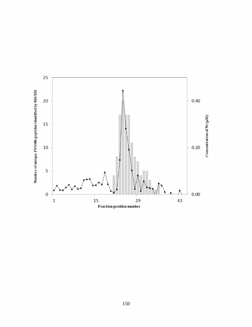
150

151
Figure 5.4
SDS-gel band MALDI-TOF analysis of the nickel peak fraction.
A. SDS-gel of fractions 23-26. The bands (GB-247 to GB-250) were excised from the gel and
analyzed by MALDI-TOF. B. MS/MS data of the SDS gel bands. PF0086 is the only peptide
that is common to all bands. This result agrees with the data from the solution MS/MS of the
nickel peak fraction.

152
A B
GB-247
GB-248
GB-250
GB-249
23 24 25 26

153
zinc was bound to the enzyme. Hence it is not known what metal the native form of the P.
horikoshii enzyme contains.
The report of a recombinant PF0086 homolog binding zinc is not consistent with our
observation that PF0086, when purified under native conditions from native biomass, is a nickel-
containing protein. There are many examples of recombinant proteins that were crystallized with
its non-physiological metal [9] and we expect that the PF0086 homolog is one of those cases. To
further confirm that PF0086 can bind nickel, the recombinant form of the protein was expressed
in E. coli in the presence of either 0.2 mM nickel, cobalt, zinc, or no additional metal. The
recombinant forms of the protein were purified by heat treatment, anion exchange
chromatography and finally size exclusion chromatography. ICP-MS analysis of the purified
forms of each recombinant protein preparation showed that PF0086 mostly binds the metal that
was added to the growth medium (Figure 5.5). When nickel was added to the medium, PF0086
contained 0.86 ± 0.2 Ni atoms mol-1
. It is remarkable that while it appears that E. coli inserts
whatever metal is available, that P. furiosus specifically inserts nickel despite growing in a
medium in which zinc is at a ~50- fold higher concentration than nickel. Taken together with the
fact that zinc was not detected in the PF0086-containing fractions purified from native biomass,
these results are consistent with PF0086 being a nickel-containing protein.
PF0086 is annotated as an alanyl-tRNA synthetase COOH terminus and has four paralogs
encoded in the P. furiosus genome (Figure 5.6). Each paralog has a homolog encoded in the P.
horikoshii genome [8]. They are all thought to be involved in editing mischarged tRNAs.
PF0086 is a truncated from of alanyl-tRNA synthetase termed Ala X-M where M indicates
medium. There are two additional paralogs name AlaX-L (PF0185) and AlaX-S (PF0428),
where L stands for large and S for small. The size designation refers to the length of the
154
Figure 5.5
Metal content of recombinant PF0086 purified from E. coli cells grown in rich media or
rich media supplemented with either 200 M Ni, Co, or Zn.
PF0086 was overexpressed in rich media containing either no added metal or added nickel,
cobalt, or zinc (each 200 M). The proteins were purified by heat treatment, anion exchange
chromatography and size exclusion chromatography. The fractions from the last purification
step were analyzed by ICP-MS. PF0086 bound most to the metal that was added to the media.

155

156
Figure 5.6
PF0086 has four paralogs in the P. furiosus genome.
PF0086 has four paralogs in the genome and all are predicted to be involved in tRNA editing.
They differ in size and function, but all have a conserved metal binding motif that is assumed to
bind zinc. Results presented herein suggest that the metal binding domain actually binds nickel.
Adapted from [8].
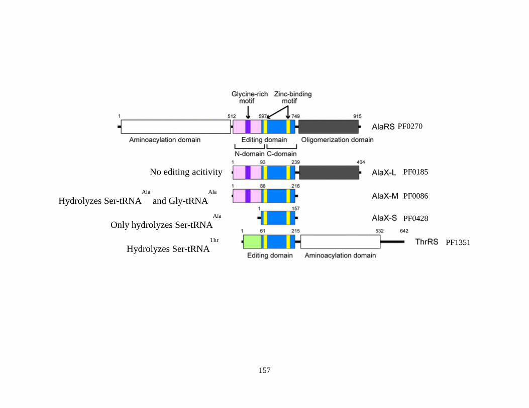
157
PF0185
PF0086
PF0428
No editing acitivity
Hydrolyzes Ser-tRNAAla
and Gly-tRNAAla
Only hydrolyzes Ser-tRNAAla
Hydrolyzes Ser-tRNAThr
PF0270
PF1351

158
proteins. AlaRS (PF0270) is the full length enzyme and contains domains for charging tRNA
with alanine, removing mischarged tRNAs and it also has an oligomerization domain. AlaX- L
(PF0185) lacks the aminoacylation and, although it has an editing domain, editing activity has
yet to be detected with this enzyme. AlaX-M (PF0086), the enzyme that was purified in this
study, lacks both an aminoacylation domain and an oligomerization domain.
AlaX-M (PF0086) can hydrolyze serine and glycine amino acids from mischarged
tRNAAla
, but cannot add the correct alanine amino acid [8]. AlaX-S (PF0428) is the shortest of
the paralogs and does not retain the glycine rich motif although it still has the conserved zinc
binding motif. This enzyme can only hydrolyze mischarged serines from tRNAAla
. Threonine
tRNA synthetase (ThrRS) can remove mischarged serines from tRNAThr
and charge it with the
correct amino acid. It also does not have the glycine rich motif but shares the conserved zinc
binding domain. The metal binding domain is a common structural component of all of these
paralogs and the fractionation data suggests that all of the other paralogs also bind nickel.
However, the PF0086 paralogs were not detected (in significant amounts) by MS/MS during the
fractionation of P. furiosus biomass and so their metal contents remain uncharacterized.
To determine if nickel has any effect on the function of PF0086, it would need to be
assayed using a radioactive assay where (14
C)-Ser-tRNAAla
or (14
C)-Gly-tRNAAla
are the
substrates. After the enzyme reaction is spotted on a filter, it is washed with ethanol allowing
the free amino acid to wash through the filter, while those which are still bonded to the tRNAAla
are retained on the filter. The amount of radioactivity remaining on the filter indicates the
activity level of the enzyme. The substrates are normally made by making a mutated form of
AlaRS (C717A AlaRS) which has no editing ability and can only add amino acids. Although we
expressed and purified the mutant form of the enzyme unfortunately, our collaborator, Dr. Dieter

159
Söll (Yale Univ.) was unable to synthesize the substrate after repeated attempts. Hence, even
though we had prepared different forms of recombinant PF0086 containing different amounts of
nickel, zinc, and/or cobalt, the effect of the metal on activity could not be determined.
The approach used to identify PF1972 as a molybdenum protein was modified from that
used previously for PF0086 in that MS/MS was not used during fractionation. Instead the protein
was identified solely by MALDI-MS after in-gel protein digestion and extraction of the protein
band. In other words, the molybdenum peak was purified on successive chromatography
columns until a single protein band was evident on an SDS-gel. The protein was then identified
by MS/MS.
PF1972 was one of 18 molybdenum peaks that co-eluted with proteins predicted to bind
molybdenum (Figure 5.7). It contained 0.77 ± 0.43 atoms mol-1
Mo protein. PF1972 is
annotated as an anaerobic ribonucleotide activase while PF1971 is annotated as anaerobic
ribonucleotide reductase and is in an operon with PF1972. In both eukaryotes and prokaryotes
ribonucleotide reductase synthesize deoxyribonucleotides by the reduction of the 2’ hydroxyl
group of ribonucleotides. PF1971 is a member of the class of ribonucleotide reductases found in
strict and facultative anaerobes which use a catalytically essential glycyl radical in the active site.
[10; 11]. PF1972 is the activator responsible for producing the glycyl radical in active site of
the reductase.
Anaerobic ribonucleotide reductase activases are “radical-SAM” enzymes and are
characterized by a (4Fe-4S) center that is coordinated by three conserved cysteines. However, in
hyperthermophiles like P. furiosus, there are four conserved cysteine residues in addition to the
three found in all anaerobic ribonucleotide reductase activases (Figure 5.8). ICP-MS analysis of
the purified P. furiosus protein did detect iron in the fraction as well as molybdenum and showed

160
Figure 5.7
Purification of the novel molybdenum protein PF1972.
The graphs show the elution profile of protein (gray) and molybdenum (blue). The boxed in
fractions were pooled and purified through an additional chromatography step. C1, DEAE-Fast
Flow anion exchange column; C2, Superdex 75 size exclusion; C3, hydroxyapatite; C4, phenyl
sepharose hyrdrophobic interaction; C5, the last step was on a mono Q anion exchange. The
purified protein was identified as PF1972 by MALDI-MS. The fractions were analyzed on a
denaturing gel and silver stained [3].

161
0
0.04
0.08
0.12
0.16
0
0.05
0.1
0.15
0.2
1 11 21 31 41
Mo
(µM
)
Pro
tein
(m
g/m
L)
Fraction
0
0.03
0.06
0.09
0
0.05
0.1
0.15
0.2
1 11 21 31 41
Mo
(µM
)
Pro
tein
(m
g/m
L)
Fraction
0
0.07
0.14
0.21
0.28
0.35
0
0.5
1
1.5
2
1 11 21 31 41
Mo
(µM
)
Pro
tein
(m
g/m
L)
Fraction
0
0.01
0.02
0
0.05
0.1
0.15
0.2
1 11 21
Mo
(µM
)
Pro
tein
(m
g/m
L)
Fraction
0
0.03
0.06
0.09
0.12
0.15
0
1
2
3
1 21 41 61 81 101 121
Mo
(µM
)
Pro
tein
(m
g/m
L)
Fraction
C1 DEAE-FF
C4 PS C2 SX75
C5 MonoQ C3 HAP
50
7060
40
30
2520
15
10 35 36
SDS-PAGE
0
0.03
0.06
0.09
0
0.2
0.4
0.6
33 34 35 36 37 38
Mo
(µM
)Fe
(µ
M)
Fraction
Fe
Mo
C5 MonoQ

162
Figure 5.8.
Amino acid alignment of anaerobic ribonucleotide reductase activases.
The alignment shows the three cysteines that are conserved in all RNRs activases. They are
indicated by asterisks (*). Four additional cysteines are conserved only in the hyperthermophiles
(boxed in red).

163
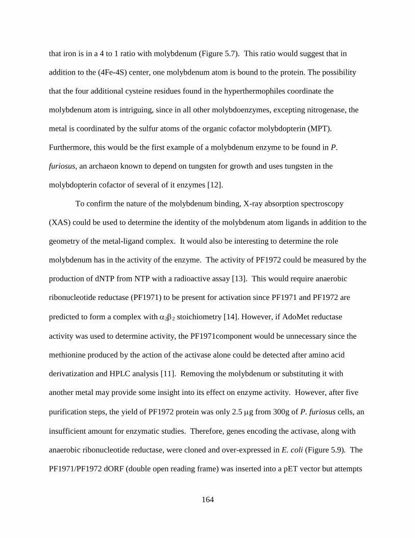
164
that iron is in a 4 to 1 ratio with molybdenum (Figure 5.7). This ratio would suggest that in
addition to the (4Fe-4S) center, one molybdenum atom is bound to the protein. The possibility
that the four additional cysteine residues found in the hyperthermophiles coordinate the
molybdenum atom is intriguing, since in all other molybdoenzymes, excepting nitrogenase, the
metal is coordinated by the sulfur atoms of the organic cofactor molybdopterin (MPT).
Furthermore, this would be the first example of a molybdenum enzyme to be found in P.
furiosus, an archaeon known to depend on tungsten for growth and uses tungsten in the
molybdopterin cofactor of several of it enzymes [12].
To confirm the nature of the molybdenum binding, X-ray absorption spectroscopy
(XAS) could be used to determine the identity of the molybdenum atom ligands in addition to the
geometry of the metal-ligand complex. It would also be interesting to determine the role
molybdenum has in the activity of the enzyme. The activity of PF1972 could be measured by the
production of dNTP from NTP with a radioactive assay [13]. This would require anaerobic
ribonucleotide reductase (PF1971) to be present for activation since PF1971 and PF1972 are
predicted to form a complex with 22 stoichiometry [14]. However, if AdoMet reductase
activity was used to determine activity, the PF1971component would be unnecessary since the
methionine produced by the action of the activase alone could be detected after amino acid
derivatization and HPLC analysis [11]. Removing the molybdenum or substituting it with
another metal may provide some insight into its effect on enzyme activity. However, after five
purification steps, the yield of PF1972 protein was only 2.5 g from 300g of P. furiosus cells, an
insufficient amount for enzymatic studies. Therefore, genes encoding the activase, along with
anaerobic ribonucleotide reductase, were cloned and over-expressed in E. coli (Figure 5.9). The
PF1971/PF1972 dORF (double open reading frame) was inserted into a pET vector but attempts

165
Figure 5.9.
Expression of PF1971 and PF1972 in E. coli analyzed by SDS-gel electrophoresis.
The subunits of anaerobic ribonucleotide reductase (PF1971 and PF1972) were expressed either
separately or together. Shown is the whole cell extracts of the induced cells. The SDS-gel
shows extracts of whole E. coli cells. Lane 1, PF1971 uninduced; Lane 2, PF1971 induced;
Lane 3, PF1972 uninduced; Lane 4, PF1972 induced. Lane 5, PF1971/PF1972 dORF uninduced;
Lane 6, PF1971/PF1972 dORF induced; Lanes 7, molecular weight standard. PF1971 has a
predicted molecular weight of 70.1 kDa and the predicted molecular weight of PF1972 is 27.6
kDa. Boxes highlight protein bands with sizes corresponding to PF1971 and PF1972.

166
90 80
70
60
50 30
25
20
10
1 2 3 4 5 6 7
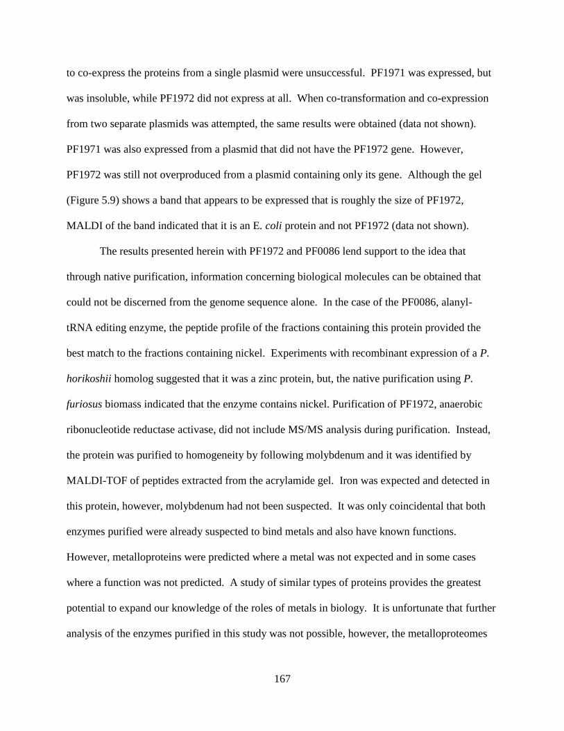
167
to co-express the proteins from a single plasmid were unsuccessful. PF1971 was expressed, but
was insoluble, while PF1972 did not express at all. When co-transformation and co-expression
from two separate plasmids was attempted, the same results were obtained (data not shown).
PF1971 was also expressed from a plasmid that did not have the PF1972 gene. However,
PF1972 was still not overproduced from a plasmid containing only its gene. Although the gel
(Figure 5.9) shows a band that appears to be expressed that is roughly the size of PF1972,
MALDI of the band indicated that it is an E. coli protein and not PF1972 (data not shown).
The results presented herein with PF1972 and PF0086 lend support to the idea that
through native purification, information concerning biological molecules can be obtained that
could not be discerned from the genome sequence alone. In the case of the PF0086, alanyl-
tRNA editing enzyme, the peptide profile of the fractions containing this protein provided the
best match to the fractions containing nickel. Experiments with recombinant expression of a P.
horikoshii homolog suggested that it was a zinc protein, but, the native purification using P.
furiosus biomass indicated that the enzyme contains nickel. Purification of PF1972, anaerobic
ribonucleotide reductase activase, did not include MS/MS analysis during purification. Instead,
the protein was purified to homogeneity by following molybdenum and it was identified by
MALDI-TOF of peptides extracted from the acrylamide gel. Iron was expected and detected in
this protein, however, molybdenum had not been suspected. It was only coincidental that both
enzymes purified were already suspected to bind metals and also have known functions.
However, metalloproteins were predicted where a metal was not expected and in some cases
where a function was not predicted. A study of similar types of proteins provides the greatest
potential to expand our knowledge of the roles of metals in biology. It is unfortunate that further
analysis of the enzymes purified in this study was not possible, however, the metalloproteomes

168
of P. furiosus and all other organisms provide an almost immeasurable amount of potential
targets that can be investigated by similar means.
References
1. Chen, P.R., E.C. Wasinger, J. Zhao, D. van der Lelie, L.X. Chen and C. He,
"Spectroscopic insights into lead(II) coordination by the selective lead(II)-binding protein
PbrR691," J Am Chem Soc, vol. 129, no. 41, pp. 12350-1, 2007.
2. Menon, A.L., F.L. Poole, 2nd, A. Cvetkovic, S.A. Trauger, E. Kalisiak, J.W. Scott, S.
Shanmukh, J. Praissman, F.E. Jenney, Jr., W.R. Wikoff, J.V. Apon, G. Siuzdak and M.W.
Adams, "Novel multiprotein complexes identified in the hyperthermophilic archaeon Pyrococcus
furiosus by non-denaturing fractionation of the native proteome," Mol Cell Proteomics, vol. 8,
no. 4, pp. 735-751, 2009.
3. Cvetkovic, A., A.L. Menon, M.P. Thorgersen, J.W. Scott, F.L. Poole, 2nd, F.E. Jenney,
Jr., W.A. Lancaster, J.L. Praissman, S. Shanmukh, B.J. Vaccaro, S.A. Trauger, E. Kalisiak, J.V.
Apon, G. Siuzdak, S.M. Yannone, J.A. Tainer and M.W. Adams, "Microbial metalloproteomes
are largely uncharacterized," Nature, vol. 466, no. 7307, pp. 779-782, 2010.
4. Bradford, M.M., "Rapid and sensitive method for quantitation of microgram quantities of
protein utilizing principle of protein-dye binding," Anal Biochem, vol. 72, no. 1-2, pp. 248-254,
1976.
5. Wang, B.C., M.W. Adams, H. Dailey, L. DeLucas, M. Luo, J. Rose, R. Bunzel, T.
Dailey, J. Habel, P. Horanyi, F.E. Jenney, Jr., I. Kataeva, H.S. Lee, S. Li, T. Li, D. Lin, Z.J. Liu,
C.H. Luan, M. Mayer, L. Nagy, M.G. Newton, J. Ng, F.L. Poole, 2nd, A. Shah, C. Shah, F.J.
Sugar and H. Xu, "Protein production and crystallization at SECSG -- an overview," J Struct
Funct Genomics, vol. 6, no. 2-3, pp. 233-43, 2005.

169
6. Ragsdale, S.W., "Nickel-based Enzyme Systems," J Biol Chem, vol. 284, no. 28, pp.
18571-5, 2009.
7. Mulrooney, S.B. and R.P. Hausinger, "Nickel uptake and utilization by microorganisms,"
FEMS Microbiol Rev, vol. 27, no. 2-3, pp. 239-61, 2003.
8. Fukunaga, R. and S. Yokoyama, "Structure of the AlaX-M trans-editing enzyme from
Pyrococcus horikoshii," Acta Crystallogr D Biol Crystallogr, vol. 63, no. Pt 3, pp. 390-400,
2007.
9. Eidsness, M.K., S.E. O'Dell, D.M. Kurtz, Jr., R.L. Robson and R.A. Scott, "Expression of
a synthetic gene coding for the amino acid sequence of Clostridium pasteurianum rubredoxin,"
Protein Eng, vol. 5, no. 4, pp. 367-71, 1992.
10. Ollagnier, S., E. Mulliez, J. Gaillard, R. Eliasson, M. Fontecave and P. Reichard, "The
anaerobic Escherichia coli ribonucleotide reductase. Subunit structure and iron sulfur center," J
Biol Chem, vol. 271, no. 16, pp. 9410-6, 1996.
11. Mulliez, E., D. Padovani, M. Atta, C. Alcouffe and M. Fontecave, "Activation of class III
ribonucleotide reductase by flavodoxin: a protein radical-driven electron transfer to the iron-
sulfur center," Biochemistry, vol. 40, no. 12, pp. 3730-6, 2001.
12. Johnson, J.L., K.V. Rajagopalan, S. Mukund and M.W. Adams, "Identification of
molybdopterin as the organic component of the tungsten cofactor in four enzymes from
hyperthermophilic Archaea," J Biol Chem, vol. 268, no. 7, pp. 4848-52, 1993.
13. Eliasson, R., E. Pontis, M. Fontecave, C. Gerez, J. Harder, H. Jornvall, M. Krook and P.
Reichard, "Characterization of components of the anaerobic ribonucleotide reductase system
from Escherichia coli," J Biol Chem, vol. 267, no. 35, pp. 25541-7, 1992.

170
14. Ollagnier, S., C. Meier, E. Mulliez, J. Gaillard, V. Schuenemann, A. Trautwein, T.
Mattioli, M. Lutz and M. Fontecave, "Assembly of 2Fe-2S and 4Fe-4S clusters in the anaerobic
ribonucleotide reductase from Escherichia coli," J Am Chem Soc, vol. 121, no. 27, pp. 6344-
6350, 1999.

171
CHAPTER 6
DISCUSSION AND CONCLUSIONS
One may argue that an underlying premise of this dissertation is that employment of the
stereotypical linear scientific method found in high school textbooks would severely limit our
progress in science. Many textbooks describe the scientific method as beginning with an
observation, followed by a hypothesis or explanation of the observation, experiments to test the
hypotheses, experimental results, and finally a conclusion that is drawn from the data collected.
Sometimes the data is inconclusive but other times it either supports or contradicts the
hypotheses. In the latter case, the hypothesis is rejected and a new hypothesis formulated and
tested. Therefore, to perform an experiment without first starting with a seemingly objective
observation or with a hypothesis might seem backwards to the novice. However, we were able
to make discoveries of new protein-protein and metalloprotein complexes in P. furiosus by doing
just that.
The main question that was asked when starting this project was, “can we identify novel
protein complexes?” In contrast to the textbook version of the scientific method, the study did
not begin with an observation per se. The observations were generated after experimental
analyses of the biomass with MS/MS and ICP-MS. As such, data was collected without being
guided by observations or a particular hypothesis in mind. This approach is characteristic of the
post-genomics era because now there are many methods that can be used to generate large
amounts of data in a short amount of time. It is then the scientists’ job to extrapolate workable
hypothesis from the data. We used native fractionation and MS/MS analysis of the fractions to

172
identify 106 potential protein-protein complexes based on co-fractionation of proteins encoded
by adjacent genes. The criteria used to identify the complexes left out those complexes that were
not encoded by adjacent genes, however the criteria greatly simplified the data so that we could
identify those proteins with a high likelihood of forming protein-protein complexes. Of the 106
potential protein complexes that were identified, only 20 were known or expected. Likewise,
MS/MS and ICP-MS were used to identify 343 metal peaks resolved during the native
fractionation of P. furiosus biomass. Of these, 158 co-eluted with proteins that were not
predicted to bind a metal. It was from this large data set that the hypotheses that guided the
research of this dissertation were generated.
One of the potential 106 novel potential protein-protein complexes was purified through
three column chromatography steps as PC-81. The co-eluting proteins of PC-81were encoded by
PF1837 and PF1837 ORFs. The PF1837 protein had previously co-purified with PF0532 as
ACS2, an 22 heterotetrameric enzyme from native biomass. ACS2 and its homolog ACS1
both catalyze terminal steps in the peptide fermentation pathway of P. furiosus. ACS1 also is a
heterotetramer, but is encoded by PF1540 () andPF1787 () which are unlinked in the genome.
Three additional subunit homologs are encoded in the genome, but have never been
characterized in P. furiosus. No additional homologs of the subunits are encoded in the P.
furiosus genome. Because the ACS2 subunit, PF1837, also co-purified with PF1838 as the
novel PC-81 complex, we hypothesized that each subunits was able to form a heterotetrameric
enzyme with more than one subunit. With two subunits and five subunits encoded in the
genome, we predicted that a total of 10 ACS complexes are used by P. furiosus and other
members of the Thermococcales which have the same set of genes. The genes of all and
subunits were cloned and each subunit combination was recombinantly co-expressed in E. coli.

173
Size exclusion chromatography estimated molecular weight of roughly140 kDa for each
combination of and subunits. This size estimation is in agreement with the 22
heterotetrameric stoichiometry found for the native enzymes. Enzyme assays were performed on
each recombinant complex to assess their functionality. Acids were used as substrates in the
CoA derivative formation direction since acyl-CoAs that account for the degradative pathways of
each amino acids were not commercially available. Every combination of subunit formed active
enzymes. However, six substrates were not used by any of the recombinant enzymes, which may
indicate that those amino acids participate in alternative pathways. Interestingly, acid substrate
specificity was dependent only on thesubunit of the complex and was independent of the
subunit. The observation begs the question of the purpose of two subunit paralogs. Previous
work by Mai and Adams [1] using the native enzymes had shown that ACS1 had a greater
affinity for guanine nucleotides than adenine nucleotides and ACS2 had a greater affinity for
adenine nucleotides over guanine nucleotides, and this hinted at a possible explanation. ACS1
used PF1787 as its subunit and ACS2 used PF1837 as its subunit. Bioinformatic analysis of
both subunits designates each as ATP grasp 5 domains which infers that the subunit contain
the ATP binding site. Therefore, we hypothesized that the subunits confer different nucleotide
specificities to the complexes. This hypothesis was tested and supported by our data.
Fukuda et al. [2] described a GTP dependent phosphoenolpyruvate carboxykinase
(PEPCK) from Thermococcus kodakarensis that was up-regulated when grown in gluconeogenic
conditions (pyruvate) and down-regulated when grown in glycolytic conditions (starch).
Similarly, the homolog in P. furiosus was up-regulated when grown on amino acids and down-
regulated when grown on maltose. Therefore, we concluded that when grown under
gluconeogenic conditions, the GTP formed by the PF1787 (G) complexes is used by the GTP

174
dependent PEPCK to drive gluconeogenesis from amino acids. This hypothesis could be tested
by varying growth condition of a PF1787 deletion mutant. An observation of good growth under
glycolytic conditions, but poor growth under gluconeogenic conditions compared to wild type,
would support the hypothesis. Of course, the caveat may be that G ACSs are the only sources
of GTP in the cell and other processes such as protein translation may depend on it. However, if
there are other sources of GTP that can support GTP dependent processes, a deletion of PF1787
may help to identify what those are.
Alternate subunits that confer nucleotide specificity to succinyl-CoA synthetase (SCS)
have been described in a number of eukaryotes. Similar to the ACSs, a single SCS subunit can
form complexes with two different subunits which confer specificity to either ATP or GTP. A
phylogenetic tree of the subunits of SCSs found in vertebrates, nematodes, yeast and bacteria
suggested that ATP- and GTP- specific subunits evolved after yeast, but before C. elegans [3].
The ACSs in lower life forms appeared to possess only one SCS homolog that was thought to be
ATP specific, but were later shown to have broad nucleotide specificity [4].
The SCSs and the ACSs of the Thermococcales share the same domains, however, these
domains are arranged in different order (Figure 6.1). For example, there is an additional ligase
domain in the subunit and a lack of a ligase domain in the subunit of Thermococcus ACS
compared to the SCS. Analysis of the SCS sequences does not reveal any significant matches
with Thermococcus ACS sequences. Domain rearrangement might be responsible for this result.
This may be the reason ACSs from Thermococcales were not included in previous SCS
phylogenetic trees [3] In chapter 3, we have included a phylogenetic tree of vertebrate,
invertebrate, bacteria, and archaea SCS and ACS subunits. It is interesting that there appears

175
Figure 6.1
Domain organization of NDP dependent acyl-CoA synthetases (ACSs).
A. Thermococcales ACSs. B. Classical SCS found in bacteria, eukaryotes, and some archaea.
Numbers indicate domain order and homologous domains between the ACSs and the classical
SCSs. Adapted from [5].

176

177
to be two independent duplications that gave rise to the ATP- and GTP- specific subunits. This
observation suggests that there was selective pressure to compartmentalize ATP and GTP
function independently in distantly-related organisms. One possible explanation for this
compartmentalization may be due to the presence of an ATP- or GTP- specific enzyme that is
dependent on the nucleotide triphosphates (NTP) produced by an NDP-dependent acyl-CoA
synthetase. If the sole acyl-CoA synthetase in a cell had broad specificity, there may have been
insufficient amounts of a particular NTP produced that was needed by an NTP-dependent
enzyme, specific to that particular NTP, to work efficiently.
As a result, a paralog of the subunit evolved that made more of the NTP dependent
enzyme’s preferred nucleotide. The acquired specificity of the additional subunit may have
negated the necessity of the original synthetase enzyme to have broad specificity and as a result,
it evolved to specifically complement the function of the new ATP- or GTP- specific synthetase.
PEPCK is a candidate enzyme that may have contributed to the selective pressure. Since the
PEPCK in the Thermococcales, is a GTP dependent enzyme, it would work more efficiently in
an environment with an enzyme that specifically makes GTP. It is interesting that in the
eukaryote, Giardia lamblia, the domains of the homologous enzyme are arranged in the same
order as those of the Thermococcales, however, they are fused into one polypeptide.
Furthermore, the C-terminal domain of the G. lamblia enzyme is homologous to the two
subunit of the Thermococcales proteins. While both Giardia and Thermoccocus orders use GTP-
dependent PEPCKs, only the Thermococcales use the peptide fermentation pathway to generate
ATP. Furthermore, the single ACS in G. lamblia prefers ATP as a substrate [6]. As such,
another method of GTP synthesis occurs in Giardia and this method appears to be through the
use of GDP as a substrate of PEPCK in an alternative shunt from PEP to pyruvate. In this

178
pathway, GTP is produced when PEP is carboxylated by PEPCK to make oxaloacetate which
then is reduced to malate. Malate is then decarboxylated to pyruvate [7]. G. lamblia is an
intestinal parasite that uses glucose as its primary carbon source and does not appear to use
PEPCK in gluconeogenesis. Thus Giardia does not need GTP to drive gluconeogenesis as the
Themococcales do, and as such the Thermococcales may have evolved two subunits to
accommodate a need in gluconeogenesis while Giardia did not.
The amino acid alignment of the subunits of some members of the Thermococcales is
shown in Figure. 6.2. Residues that are conserved in both the A and G proteins may provide
information concerning amino acids that contribute to the overall shape of the subunits, and also
their interactions with the subunits There are also amino acids that are only conserved within
the respective A and G subunits. These residues may be involved in determining nucleotide
specificity. In particular those residues on the subunit which are in close spatial proximity to
the conserved His257
of the subunit when the enzyme is fully assembled are reasonable
candidates. The proposed reaction mechanism of this enzyme shows a phosphorylated conserved
His257 which transiently phosphorylates conserved His
71 His
71then phosphorylates ADP to
make ATP. A three dimensional model of ACS1 was made by Bräsen et al. [5] using the dimeric
crystal structure of the ACS1 homolog in P. horikoshii and the crystal structure of the
monomeric P. horikoshii ACS1 homolog as templates. The model shows that a conserved
Asp212 is in close proximity to His
257in the fully assembled enzyme, however, this residue is
conserved in both A and Gthus cannot represent a residue that confers nucleotide specificity.
Mutational studied may be necessary to determine which amino acids contribute to nucleotide
specificity. It would be interesting if residues which are spatially distal to His257contribute to
nucleotide specificity.

179
Figure 6.2.
Amino acid alignments A and G subunits of Thermococcales ACSs.
Alignment shows residues that are completely conserved and those that are conserved with in A
or G. Those residues that are conserved in all subunits may be important in determining the
shape of the protein or may be involved oligomerization while those that are conserved within
A or G may determine nucleotide specificity.

180

181
The structural model made by Bräsen included coenzyme A, ADP, and Mg2+
as
substrates, however it did not include a carboxylic acid [5]. It is interesting that when assaying
ACS3 at pH 8.0 the activity towards succinate is only 20 % of that of 3-methylthiopropionate,
the substrate representing methionine fermentation (data not shown). However, when the assay
was done at a pH of 7.0, SCSs activity toward succinate increased and exceeded its activity with
3-methylthiopropionate. A pH change from 8 to 7 did not significantly alter the activities of any
other ACS complex toward any of the other substrates. Succinate was the only dicarboxylic acid
substrate used. Although the pKa values of succinate are 4.21 and 5.41, a lowering of the pH
may increase the population of the molecule that is protonated at one of the carboxyl groups.
Since other substrates have only one negatively charged functional group, the change in pH may
cause the succinate to resemble the remainder of the substrates at a pH of 7. This reasoning may
also explain why Mai et al. [1] could not detect succinyl CoA synthetase activity when they
assayed the fractions at pH8.0. Imanaka et al. [8] were able to detect activity of the native T.
thermokodakarensis homolog of this enzyme, however they performed their assay at a pH of 6.5
instead of 8. This observation may add to our understanding of substrate binding of the acid and
the enzyme reaction mechanism itself.
It is unfortunate that enzyme activity experiments could not be performed with PF0086
and PF1972. We have demonstrated that PF0086 co-purifies with nickel. We also showed that
the recombinant enzyme can bind nickel, cobalt or zinc ions when grown in the presence of these
metals. Enzyme assays could not be done with PF0086 because the substrate was not available.
The mutated full length tRNAAla
synthetase (PF0270 C717A) was made and should have been
able to add mischarged serine and glycine amino acid residues while being unable to remove
them from tRNAAla
. The mutated protein was expressed, purified and sent to our collaborators

182
who tried using it to synthesize a radioactive substrate. However they were unable to make
substrate with this enzyme and attempted to make the substrate with E. coli tRNA and a mutated
E coli tRNAAla
synthetase homolog. Because tRNAs are highly conserved, we thought E. coli
tRNA could be used as substrate. However, a substrate could not be synthesized with E. coli
either. It is unclear why this substrate was unable to be generated by our collaborators, who have
a great deal of experience with this type of experiment.
Further study of PF1972 was also hindered because it could not be recombinantly
expressed in E. coli. PF1972 was purified from P. furiosus biomass and contained molybdenum
and iron. Four conserved cysteine residues exclusively conserved in the hyperthermophilic
homolog of PF1972 are the candidate ligands for molybdenum binding. This hypothesis could
have been tested if there were sufficient amounts of material to test. Coincidentally, the
laboratory of Dr. William Lanzilotta at the University of Georgia was concurrently attempting to
express this enzyme and was also unsuccessful in its attempts. Finally, Brian Vaccaro, another
graduate student in the Adams’ laboratory, attempted to homologously express the protein in P.
furiosus under the control of a constitutive promoter. Unfortunately his efforts did not meet with
success either. It is possible that PF1972 could be over-produced if other promoters are used.
For example, our laboratory routinely uses the promoter for the highly-expressed S-layer protein
[9]. Therefore, other options are available for the homologous expression of PF1972 in P.
furiosus. A His-tag was also engineered into PF1972 for homologous expression in P. furiosus.
It might be worthwhile to see if the absence of this tag enhances expression of the protein.
Without the ability to make recombinant forms of the P. furiosus enzymes and/or assay their
activity the ability of characterizing them is greatly limited. Enzyme activity assays of PF0086
and PF1972 would have been able to demonstrate the effects of various metals on enzyme

183
activity. Studies have reported that different metals significantly affect the rate of a reaction or
even the type of products generated by an enzyme [10; 11]. Such data make it critical to
understand which metals bind to enzymes and proteins in their native physiological state.
One may question if the method used in the studies reported in this thesis are the most
efficient way to characterize novel protein-protein interactions and the nature of metalloproteins.
After all, we were not able to fully characterize PF0086 and PF1972. This might be rephrased
as, “is it better to start an experimental project with a hypothesis or with data?” However, this
question may be hard to answer since both scenarios have their upsides and downsides. Starting
with data may seem less focused and more risky since there is no concrete question being asked.
Starting with a question helps one to anticipate future plans and possible caveats. However,
because genome-related technology has allowed so much data to be generated, there are more
opportunities to discover something novel and interesting. Furthermore, since the data in our
studies were gathered from native fractionation, hypotheses can be made that take into account
information that is not available from sequence alone.
The use of native biomass fractionation to discover novel metalloproteins and novel
protein-protein interactions is not a new concept. However, it is a concept that has increasingly
become overshadowed by attempts to use genomics to make predictions concerning the functions
of enzymes before they have been expressed and purified. Here we have combined native
fractionation with MS/MS and ICP/MS to identify novel protein-protein interactions and novel
metalloproteins. It is expected that the information acquired during our experiments can fill
some gaps in information about protein-protein complexes and metalloproteins in P. furiosus. In
many cases this information may be applied to homologous proteins in other organisms. In such

184
cases, others will be able to make their own hypotheses based on the data gathered from the
results of my studies presented herein.
References
1. Mai, X. and M.W. Adams, "Purification and characterization of two reversible and ADP-
dependent acetyl coenzyme A synthetases from the hyperthermophilic archaeon
Pyrococcus furiosus," J Bacteriol, vol. 178, no. 20, pp. 5897-5903, 1996.
2. Fukuda, W., T. Fukui, H. Atomi and T. Imanaka, "First characterization of an archaeal
GTP-dependent phosphoenolpyruvate carboxykinase from the hyperthermophilic
archaeon Thermococcus kodakaraensis KOD1," J Bacteriol, vol. 186, no. 14, pp. 4620-
4627, 2004.
3. Johnson, J.D., J.G. Mehus, K. Tews, B.I. Milavetz and D.O. Lambeth, "Genetic evidence
for the expression of ATP- and GTP-specific succinyl-CoA synthetases in multicellular
eucaryotes," J Biol Chem, vol. 273, no. 42, pp. 27580-27586, 1998.
4. Weitzman, P.D.J. and H. Jaskowskahodges, "Patterns of Nucleotide Utilization in
Bacterial Succinate Thiokinases," Febs Lett, vol. 143, no. 2, pp. 237-240, 1982.
5. Bräsen, C., M. Schmidt, J. Grötzinger and P. Schönheit, "Reaction mechanism and
structural model of ADP-forming Acetyl-CoA synthetase from the hyperthermophilic
archaeon Pyrococcus furiosus: evidence for a second active site histidine residue," J Biol
Chem, vol. 283, no. 22, pp. 15409-15418, 2008.
6. Sànchez, L.B., M.Y. Galperin and M. Müller, "Acetyl-CoA synthetase from the
amitochondriate eukaryote Giardia lamblia belongs to the newly recognized superfamily
of acyl-CoA synthetases (Nucleoside diphosphate-forming)," J Biol Chem, vol. 275, no.
8, pp. 5794-5803, 2000.

185
7. Muller, M., M. Mentel, J.J. van Hellemond, K. Henze, C. Woehle, S.B. Gould, R.Y. Yu,
M. van der Giezen, A.G. Tielens and W.F. Martin, "Biochemistry and evolution of
anaerobic energy metabolism in eukaryotes," Microbiol Mol Biol Rev, vol. 76, no. 2, pp.
444-95, 2012.
8. Shikata, K., T. Fukui, H. Atomi and T. Imanaka, "A novel ADP-forming succinyl-CoA
synthetase in Thermococcus kodakaraensis structurally related to the archaeal nucleoside
diphosphate-forming acetyl-CoA synthetases," J Biol Chem, vol. 282, no. 37, pp. 26963-
26970, 2007.
9. Hopkins, R.C., J. Sun, F.E. Jenney, Jr., S.K. Chandrayan, P.M. McTernan and M.W.
Adams, "Homologous expression of a subcomplex of Pyrococcus furiosus hydrogenase
that interacts with pyruvate ferredoxin oxidoreductase," PLoS One, vol. 6, no. 10, pp.
e26569, 2011.
10. Holmquist, B. and B.L. Vallee, "Metal substitutions and inhibition of thermolysin:
spectra of the cobalt enzyme," J Biol Chem, vol. 249, no. 14, pp. 4601-7, 1974.
11. Dai, Y., P.C. Wensink and R.H. Abeles, "One protein, two enzymes," J Biol Chem, vol.
274, no. 3, pp. 1193-5, 1999.