germinal center exclusion of autoreactive b cells is...
TRANSCRIPT

Research article
TheJournalofClinicalInvestigation http://www.jci.org Volume 115 Number 11 November 2005 3205
Germinal center exclusion of autoreactive B cells is defective in human systemic
lupus erythematosusAmedeo Cappione III,1 Jennifer H. Anolik,1 Aimee Pugh-Bernard,1 Jennifer Barnard,1
Paul Dutcher,1 Gregg Silverman,2 and Iñaki Sanz1
1Department of Medicine, University of Rochester School of Medicine, Rochester, New York, USA. 2UCSD, La Jolla, California, USA.
BreachofBcelltoleranceiscentraltothepathogenesisofsystemiclupuserythematosus(SLE).However,howBcelltoleranceissubvertedinhumanSLEispoorlyunderstoodduetodifficultiesinidentifyingrelevantauto-reactiveBcellsandinobtaininglymphoidtissue.Wehavecircumventedtheselimitationsbyusingtonsilbiop-siestostudyautoreactiveBcells(9G4Bcells),whoseregulationisabnormalinSLE.Hereweshowthat9G4BcellsarephysiologicallyexcludedduringtheearlystagesoftheGCreactionbeforeacquiringacentroblastphenotype.Furthermore,weprovideevidencetoindicatethatananergicresponsetoBcellreceptorstimula-tionmayberesponsibleforsuchbehavior.Incontrast,inSLE,9G4Bcellsprogressedunimpededthroughthischeckpoint,successfullyparticipatedinGCreactions,andexpandedwithinthepost-GCIgGmemoryandplasmacellcompartments.Thefaultyregulationof9G4BcellswasnotsharedbyRApatients.Toourknowl-edge,thisworkrepresentsthefirstcomparativeanalysisofthefateofaspecificautoreactivehumanBcellpopulation.TheresultsidentifyadefectivetolerancecheckpointthatappearstobespecificforhumanSLE.
IntroductionSystemic lupus erythematosus (SLE) is a prototypical autoimmune disease characterized by the production of antinuclear autoanti-bodies (1). SLE patients also frequently produce antilymphocyte antibodies of pathogenic potential that preferentially target CD45 (2, 3). These autoantibodies reflects a critical breakdown of B cell tolerance and the ensuing expansion of autoreactive B cells capable of inducing disease through both antibody-dependent and anti-body-independent mechanisms (4). Regrettably, understanding how tolerance is abrogated in SLE has been hampered by experi-mental limitations in identifying relevant autoreactive B cells.
We have proposed that B cells bearing the 9G4 idiotype (9G4 B cells) represent a useful system to study B cell tolerance in SLE, since the expression of this idiotype (which identifies antibodies encoded by the VH4.34 heavy chain) is synonymous with autoreac-tivity against N-acetyllactosamine (NAL) determinants expressed by the iI blood group antigen and other self glycoproteins includ-ing CD45 (3, 5, 6). Moreover, the 9G4-determined autoreactivity is censored in normal subjects but greatly and specifically expanded in patients with active SLE. Thus, while 9G4 B cells represent 5–10% of normal naive B cells, serum 9G4 antibodies are scarce in nor-mal sera (3, 7). In contrast, these autoantibodies are abundant in patients with active SLE, representing 10–45% of total serum IgG (3, 8, 9). Importantly, 9G4 antibodies represent the vast major-ity of anti–B cell CD45 antibodies and a significant fraction of anti–native double-stranded DNA (anti-dsDNA) antibodies, carry a disease specificity of 95%, and correlate with disease activity and organ involvement (3, 8–10). These observations support the con-
tention that 9G4 antibodies are of clinical relevance in SLE and that physiological tolerogenic mechanisms acting upon 9G4 B cells are defective in this disease.
Considering our demonstration that 9G4 B cells fail to form mature GCs in healthy subjects, we hypothesized that physio-logical mechanisms preventing the participation of these B cells in productive GC reactions were faulty in SLE (5). Alternatively, as shown for rheumatoid factor–transgenic B cells in autoim-mune-prone mice, production of isotype-switched, somatically mutated autoantibodies can also originate outside the GC (11). Discriminating between these 2 possibilities requires the analy-sis of secondary lymphoid tissue, which is seldom available from SLE patients. We have overcome this obstacle by using tonsil biopsy samples from patients with SLE and RA controls. The results indicate that in SLE, but not in RA, 9G4 B cells escape normal censoring and actively participate in productive GC reac-tions, leading to the generation of substantially increased levels of IgG memory and plasma cells. The specific peripheral toler-ance checkpoint that is overcome occurs at an early point in the GC reaction during the transition from the pre-GC to the centro-blast stage, thus implicating faulty GC exclusion of autoreactive B cells in the pathogenesis of SLE.
Results9G4 B cells are expanded in the GCs of SLE patients. Our previous stud-ies of healthy individuals indicated that the vast majority of mature 9G4 B cells have a naive phenotype, fail to form GCs, and do not differentiate into mature bone marrow plasma cells. On that basis, we hypothesized that healthy autoreactive 9G4 B cells are barred from participating in productive GC reactions (5). In the present study, flow cytometry was used to compare the fate of 9G4 B cells in SLE tonsils (n = 6) and healthy tonsils (n = 15). As shown in Figure 1, most healthy 9G4 B cells (80–90%) consistently expressed a naive (Bm1/Bm2) phenotype, failed to acquire a GC phenotype, and did not contribute significantly to post-GC IgG memory
Nonstandardabbreviationsused: APC, allophycocyanin; BAFF, B cell–activating factor of the TNF family; BCR, B cell receptor; dsDNA, native double-stranded DNA; FDC, follicular DC; MZ, marginal zone; NAL, N-acetyllactosamine; SA, streptavidin; SLE, systemic lupus erythematosus.
Conflictofinterest: The authors have declared that no conflict of interest exists.
Citationforthisarticle: J. Clin. Invest. 115:3205–3216 (2005). doi:10.1172/JCI24179.
research article
3206 TheJournalofClinicalInvestigation http://www.jci.org Volume 115 Number 11 November 2005
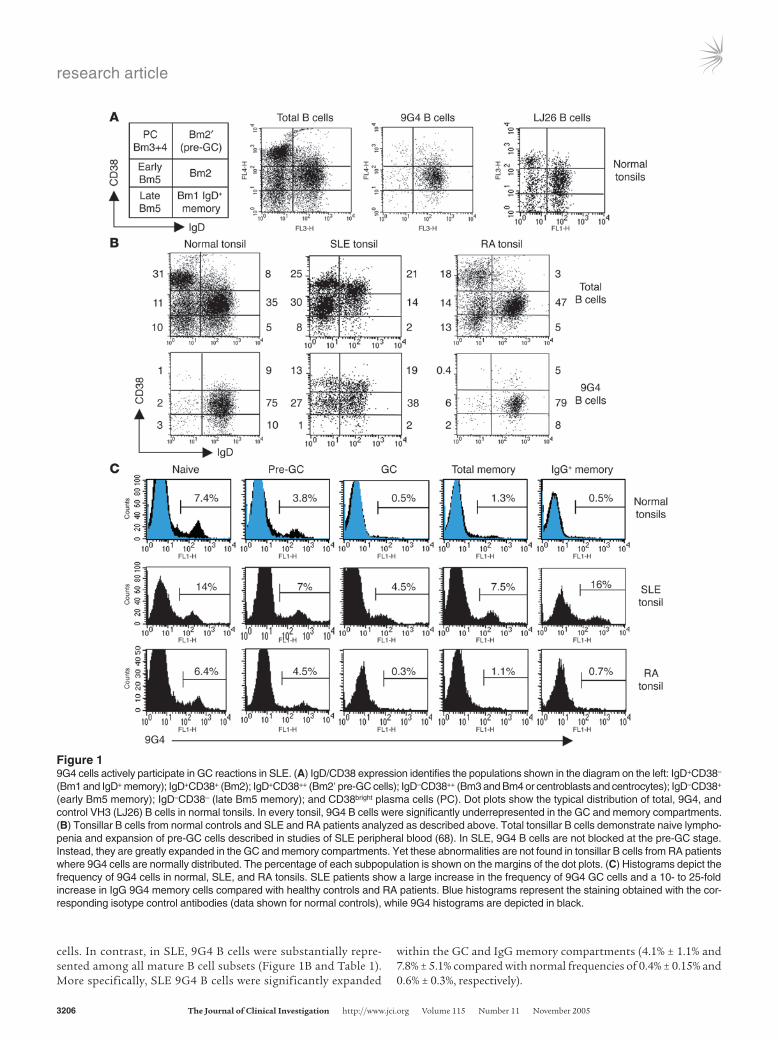
cells. In contrast, in SLE, 9G4 B cells were substantially repre-sented among all mature B cell subsets (Figure 1B and Table 1). More specifically, SLE 9G4 B cells were significantly expanded
within the GC and IgG memory compartments (4.1% ± 1.1% and 7.8% ± 5.1% compared with normal frequencies of 0.4% ± 0.15% and 0.6% ± 0.3%, respectively).
Figure 19G4 cells actively participate in GC reactions in SLE. (A) IgD/CD38 expression identifies the populations shown in the diagram on the left: IgD+CD38– (Bm1 and IgD+ memory); IgD+CD38+ (Bm2); IgD+CD38++ (Bm2′ pre-GC cells); IgD–CD38++ (Bm3 and Bm4 or centroblasts and centrocytes); IgD–CD38+ (early Bm5 memory); IgD–CD38– (late Bm5 memory); and CD38bright plasma cells (PC). Dot plots show the typical distribution of total, 9G4, and control VH3 (LJ26) B cells in normal tonsils. In every tonsil, 9G4 B cells were significantly underrepresented in the GC and memory compartments. (B) Tonsillar B cells from normal controls and SLE and RA patients analyzed as described above. Total tonsillar B cells demonstrate naive lympho-penia and expansion of pre-GC cells described in studies of SLE peripheral blood (68). In SLE, 9G4 B cells are not blocked at the pre-GC stage. Instead, they are greatly expanded in the GC and memory compartments. Yet these abnormalities are not found in tonsillar B cells from RA patients where 9G4 cells are normally distributed. The percentage of each subpopulation is shown on the margins of the dot plots. (C) Histograms depict the frequency of 9G4 cells in normal, SLE, and RA tonsils. SLE patients show a large increase in the frequency of 9G4 GC cells and a 10- to 25-fold increase in IgG 9G4 memory cells compared with healthy controls and RA patients. Blue histograms represent the staining obtained with the cor-responding isotype control antibodies (data shown for normal controls), while 9G4 histograms are depicted in black.
research article
TheJournalofClinicalInvestigation http://www.jci.org Volume 115 Number 11 November 2005 3207
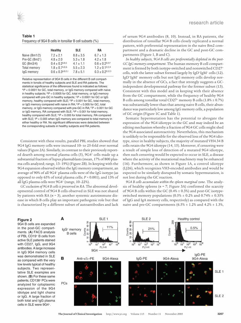
Consistent with these results, parallel PBL studies showed that 9G4 IgG memory cells were increased 10- to 25-fold over normal values (Figure 2A). Similarly, in contrast to their previously report-ed dearth among normal plasma cells (5), 9G4+ cells made up a substantial fraction of lupus plasmablasts (mean, 17% of 800 plas-ma cells analyzed; range, 13–19%) (Figure 2B). In keeping with the 9G4 expansion observed within the IgG memory compartment, an average of 90% of all 9G4+ plasma cells were of the IgG isotype (as opposed to only 65% of total plasma cells; P < 0.001), and 13% of all IgG plasma cells were 9G4+ (range, 10–22%).
GC exclusion of 9G4 B cells is preserved in RA. The abnormal devel-opmental control of 9G4 B cells observed in SLE was not shared by patients with RA (n = 3), another systemic autoimmune dis-ease in which B cells play an important pathogenic role but that is characterized by a different subset of autoantibodies and lack
of serum 9G4 antibodies (8, 10). Instead, in RA patients, the distribution of tonsillar 9G4 B cells closely replicated a normal pattern, with preferential representation in the naive Bm2 com-partment and a dramatic decline in the GC and post-GC com-partments (Figure 1, B and C).
In healthy subjects, 9G4 B cells are preferentially depleted in the post-GC IgG memory compartment. The human memory B cell compart-ment is formed by both isotype-switched and nonswitched CD27+ cells, with the latter subset formed largely by IgD+IgM+ cells (12). IgD+IgM+ memory cells but not IgG memory cells develop nor-mally in the absence of GCs, a fact that strongly suggests a GC-independent developmental pathway for the former subset (13). Consistent with this model and in keeping with their absence from the GC compartment, while the frequency of healthy 9G4 B cells among tonsillar total CD27+ memory B cells (1.8% ± 0.7%) was substantially lower than that among naive B cells, their abun-dance was particularly low among IgG memory cells, a population of GC origin (Figure 1C and Table 1).
Somatic hypermutation has the potential to abrogate the expression of the 9G4 idiotype in the GC and may indeed be an editing mechanism whereby a fraction of 9G4 GC cells might shed the 9G4-associated autoreactivity. Nevertheless, this mechanism is unlikely to be responsible for the observed loss of the 9G4 idio-type, since in healthy subjects, the majority of mutated VH4.34 B cells retain the 9G4 idiotype (14, 15). Moreover, if censoring were a result of simple loss of detection of a mutated 9G4 idiotype, then such censoring would be expected to occur in SLE, a disease where the activity of the mutational machinery may be enhanced (16). Furthermore, as shown in Figure 1A, a control idiotype (LJ26), which recognizes VH3-encoded antibodies and would be expected to be similarly disrupted by somatic hypermutation, is not lost during the GC reaction.
9G4 B cells accumulate within the spleen marginal zone. The analy-sis of healthy spleens (n = 7; Figure 3A) confirmed the scarcity of 9G4 B cells within the GC (0.4% ± 0.3%) and post-GC isotype-switched memory populations (0.5% ± 0.2% and 0.74% ± 0.35% of IgG and IgA memory cells, respectively) as compared with the naive and pre-GC compartments (6.5% ± 1.2% and 4.2% ± 1.3%,
Table 1Frequency of 9G4 B cells in tonsillar B cell subsets (%)
Healthy SLE RANaive (Bm1/2) 7.2 ± 2.1 8.8 ± 3.5 6.7 ± 1.3Pre-GC (Bm2′) 4.8 ± 2.0 5.3 ± 1.8 4.2 ± 1.8GC (Bm3/4) 0.4 ± 0.2A,B,C 4.1 ± 1.1 0.6 ± 0.2D,E,F
Total memory 1.8 ± 0.7A,B,G 5.5 ± 2.3 1.2 ± 0.1D,E,H
IgG memory 0.6 ± 0.3A,B,C,I 7.8 ± 5.1 0.3 ± 0.2D,E,F,I
Relative representation of 9G4 B cells in the different B cell compart-ments in tonsils of healthy subjects and SLE and RA patients. The statistical significance of the differences found is indicated as follows: AP < 0.0001 for GC, total memory, or IgG memory compared with naive in healthy subjects; BP < 0.0005 for GC, total memory, or IgG memory compared with pre-GC in healthy subjects; CP < 0.0001 for GC or IgG memory, healthy compared with SLE; DP < 0.001 for GC, total memory, or IgG memory compared with naive in RA; EP < 0.005 for GC, total memory, or IgG memory compared with pre-GC in RA; FP < 0.001 for GC or IgG memory, RA compared with SLE; GP < 0.001 for total memory, healthy compared with SLE; HP < 0.005 for total memory, RA compared with SLE; IP < 0.005 when IgG memory are compared to total memory in either healthy or RA. No significant differences were detected between the corresponding subsets in healthy subjects and RA patients.
Figure 29G4 B cells are expanded in the post-GC compart-ments. (A) FACS analysis of PBL CD19+ B cells from active SLE patients stained with CD27, IgG, and 9G4 antibodies. A large increase in IgG 9G4 memory cells was demonstrated in SLE as compared with the very low levels typical of healthy subjects. Two represen-tative SLE examples are shown. (B) For these same patients, CD138+ PCs were analyzed for cytoplasmic expression of the 9G4 idiotype and light chains or IgG. A large fraction of both total and IgG plasma cells in SLE were 9G4+.

research article
3208 TheJournalofClinicalInvestigation http://www.jci.org Volume 115 Number 11 November 2005
respectively). Interestingly however, 9G4 cells were substantially represented among marginal zone (MZ) B cells (4.8% ± 1.4%), a population formed to a large extent by memory B cells including a subset bearing surface IgM and IgD with the ability to recirculate in the peripheral blood (Figure 3, B–F) (17–19). Furthermore, MZ 9G4 B cells predominantly expressed CD27, a marker of human memory B cells (Figure 3D). Yet, even within the MZ, 9G4 B cells were scarce among isotype-switched B cells (Figure 3, A, E, and F). Instead, 9G4 B cells almost universally expressed an IgM+IgD+ sur-
face phenotype whether in the tonsil, peripheral blood, or spleen (data shown for the spleen in Figure 3F).
Of note, this difference in 9G4 expression between IgM and IgG memory cells is consistent with the relative abundance of VH4-34 expression detected by PCR analysis of normal tonsillar B cell subsets (14). Together, both studies indicate that while naive 9G4 B cells may differentiate into MZ IgM memory cells, presumably in a T cell–independent fashion, they do not par-ticipate in productive GC reactions required for the generation
Figure 3Analysis of 9G4 cells in healthy spleens. (A) CD19+ spleen B cells were analyzed with IgD, CD38, CD27, and 9G4 antibodies as described above (n = 7 spleens). 9G4 B cells are very scarce within the GC and post-GC compartments (IgG and IgA memory). Representative results are shown as histograms. (B) Staining for IgM, IgD, CD21, and CD23 expression identified transitional (T1 and T2), follicular (FO), and MZ populations with a distribution similar to mouse B cells (12, 69). We identified an additional fraction composed of significant numbers of IgD+ cells, which represents a distinct subset of IgD+ MZ B cells (MZ*) (19). (C) Total spleen B cells were fractionated into MZ and follicular subsets as described above and further analyzed for the frequency of 9G4 B cells. The frequency of spleen follicular 9G4 cells was similar to the tonsil, and a lower but significant frequency was observed in the MZ fraction. (D) The majority of total and 9G4 MZ B cells express CD27. (E) The dearth of IgG and IgA 9G4 B cells was consistently documented in the spleen whether using total B cells or fractionated MZ B cells. (F) Dot plot analysis of total and 9G4 spleen B cells demonstrated that the vast majority of 9G4 B cells express an IgM+IgD+ phenotype. (G) Within the naive compartment, 9G4 B cells express significantly lower levels of surface IgM. MFI, mean fluorescence intensity.
research article
TheJournalofClinicalInvestigation http://www.jci.org Volume 115 Number 11 November 2005 3209
Figure 49G4 cells are normally censored at the GC founder stage. (A) The left dot plot is representative of normal tonsils, demonstrating a prominent GC founder population (fraction e). As shown in the dot plot on the right, even in these tonsils, 9G4 B cells fail to progress past the pre-GC compartment and are scarce among GC founders. (B) Tonsils were analyzed for the expression of developmental markers CD10, CD44, and CD27 on conventional Bm1–Bm5 subsets (fractions a, b, f, and g). Pre-GC/Bm2′ cells were further divided into 3 fractions (c–e), with e containing the putative GC founders. As shown in the corresponding histograms, CD10 (a GC marker) was progressively acquired in fractions c–f, while CD44 (a marker downregulated in GC) was progressively lost. CD27 also experienced progressive upregulation in frac-tions c–f. Strikingly, the highest expression of the nuclear proliferation protein Ki67 was observed in fraction e. These results are consistent with fraction e representing GC founders undergoing the initial phases of clonal expansion. Importantly, the majority of 9G4 B cells was lost during pre-GC progression, greatly underrepresented among GC founders, and failed to expand within the GC, where their frequency continued to decline. (C) The scarce 9G4 B cells present within the GC founder and GC compartments were further analyzed for intracellular Ki67 expression. Consistent with their inability to form productive GC reactions, and in contrast to total B cells within these fractions, 9G4 B cells expressed very low levels of Ki67.

research article
3210 TheJournalofClinicalInvestigation http://www.jci.org Volume 115 Number 11 November 2005
of IgG memory cells. Indeed, our results lend further support for the existence of a GC-independent developmental pathway leading to the generation of IgM+IgD+ memory cells but not IgG memory cells (13, 19).
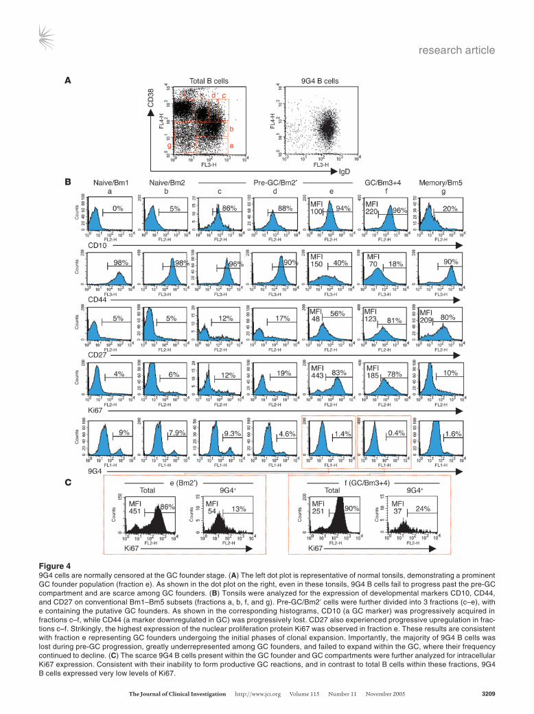
Development of healthy 9G4 B cells is blocked at the GC founder stage. In order to specifically pinpoint the checkpoint(s) responsible for the GC exclusion of 9G4 B cells in healthy subjects, we conducted detailed tracking studies of these cells throughout the pre-GC compartment. As illustrated in Figure 4, the pre-GC/Bm2′ popu-
lation can be further divided into several fractions that gradually lose surface IgD expression while acquiring GC markers including CD10. Analysis of these fractions allowed us to define a subset that expresses low levels of surface IgD (intermediate between pre-GC and centroblast levels) and the highest levels of Ki67 among all tonsillar B cells, including centroblasts.
Our results suggest that cells of this fraction, which can be visu-alized as a distinct population in about 25% of normal tonsils (shown in Figure 4A), are the earliest identifiable GC cells and are
Figure 59G4 GCs are present in SLE patients at high frequency. (A) SLE tonsil biopsies stained with anti-IgD (left). Mature GCs formed by expansions of 9G4 cells were frequently identified in SLE. The upper row of the enlarged images depicts a typical mature 9G4+ proliferative GC with a well-formed FDC network (CD23/FDC) and positive Ki67 staining. The lower row shows similar findings by immunofluorescence: follicular mantle (IgD-PE, red), GC (CD38–7-aminomethylcoumarin, blue), and 9G4 (Alexa 488, green). (B) Representative example of a 9G4+ GC in an SLE spleen shown by 3-color immunofluorescence. (C) SLE GC showing the expansion of both 9G4 and LC1 B cells. (D) Healthy tonsils stained by immunofluorescence: IgD (red), Ki67 (blue), and either 9G4 or LC1 (both green). In contrast to SLE, healthy tonsils lack 9G4+ GC. Yet proliferat-ing LC1 GCs were readily demonstrated. (E) A representative field from healthy spleens demonstrating the absence of 9G4 staining in the GCs is shown on the left. These results were routinely corroborated by immunofluorescence (middle panels). The photograph on the right illustrates the absence of 9G4 B cells from the GC and their accumulation within the FO and the MZ. (F) Enzymatic staining of serial sections obtained from tonsil biopsies of patients with RA failed to demonstrate 9G4+ GCs. Instead, as in healthy subjects, 9G4 B cells were restricted to the follicular mantle. In contrast, LC1+ GCs were readily identified in these patients.
research article
TheJournalofClinicalInvestigation http://www.jci.org Volume 115 Number 11 November 2005 3211
likely to represent GC founders. Of note, approximately 90% of all 9G4 B cells fail to acquire this pre-centroblast, GC founder pheno-type (Figure 4B). Strikingly, while the majority of all GC founders and GC cells express high levels of Ki67, the residual 9G4 B cells found within these compartments expressed very low levels of this proliferation nuclear protein (Figure 4C). Together, these data indicate that the main censoring of autoreactive 9G4 B cells occurs during the early phases of the GC reaction, with relatively few 9G4 B cells differentiating into GC founders. Moreover, residual 9G4 B cells with a GC founder or a centroblast phenotype appear unable to proliferate and do not develop into more mature centrocytes.
Histological studies of 9G4 B cells in SLE patients and normal subjects. The participation of 9G4 B cells in mature GC reactions in SLE was confirmed histologically. Proliferating Ki67+ 9G4 B cells con-
tributed to 18% of 160 mature GC from 6 SLE tonsils (Figure 5, A and C). Similar findings were also obtained with 2 SLE spleens (Figure 5B). The interpretation of these results may be confound-ed by retention of circulating 9G4 antibodies by follicular DCs (FDCs). However, we believe that the actual presence of 9G4 B cells in the GC, as indicated by the FACS results, is strongly supported by costaining with B cell GC markers such as CD38. Furthermore, FDC-bound 9G4 antibody would be expected to produce staining of most GC and comparable staining of neighboring GC of similar age as indicated by well-developed FDC networks. Such findings, however, were not observed.
This abundance of 9G4+ GC in SLE is in stark contrast to the scarcity of similar structures in healthy subjects. Thus far, we have analyzed 705 secondary follicles (tonsils: 500 follicles, n = 15;
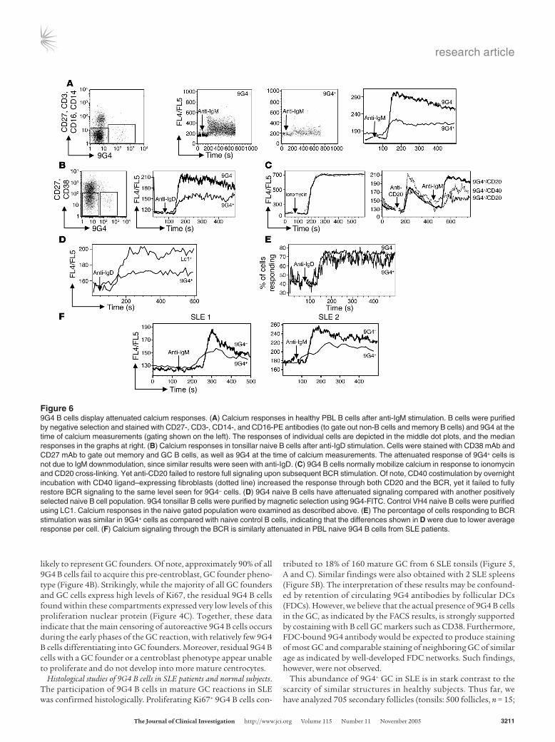
Figure 69G4 B cells display attenuated calcium responses. (A) Calcium responses in healthy PBL B cells after anti-IgM stimulation. B cells were purified by negative selection and stained with CD27-, CD3-, CD14-, and CD16-PE antibodies (to gate out non-B cells and memory B cells) and 9G4 at the time of calcium measurements (gating shown on the left). The responses of individual cells are depicted in the middle dot plots, and the median responses in the graphs at right. (B) Calcium responses in tonsillar naive B cells after anti-IgD stimulation. Cells were stained with CD38 mAb and CD27 mAb to gate out memory and GC B cells, as well as 9G4 at the time of calcium measurements. The attenuated response of 9G4+ cells is not due to IgM downmodulation, since similar results were seen with anti-IgD. (C) 9G4 B cells normally mobilize calcium in response to ionomycin and CD20 cross-linking. Yet anti-CD20 failed to restore full signaling upon subsequent BCR stimulation. Of note, CD40 costimulation by overnight incubation with CD40 ligand–expressing fibroblasts (dotted line) increased the response through both CD20 and the BCR, yet it failed to fully restore BCR signaling to the same level seen for 9G4– cells. (D) 9G4 naive B cells have attenuated signaling compared with another positively selected naive B cell population. 9G4 tonsillar B cells were purified by magnetic selection using 9G4-FITC. Control VH4 naive B cells were purified using LC1. Calcium responses in the naive gated population were examined as described above. (E) The percentage of cells responding to BCR stimulation was similar in 9G4+ cells as compared with naive control B cells, indicating that the differences shown in D were due to lower average response per cell. (F) Calcium signaling through the BCR is similarly attenuated in PBL naive 9G4 B cells from SLE patients.

research article
3212 TheJournalofClinicalInvestigation http://www.jci.org Volume 115 Number 11 November 2005
adult spleens: 120 follicles, n = 7; infant spleen: 85 follicles, n = 1) and found only 1 instance of a GC formed by expansion of 9G4 B cells in the infant spleen (data not shown). Instead, as shown by triple immunofluorescence, in healthy subjects, 9G4 B cells were predominantly localized both in tonsils and spleens within the follicular mantle (Figure 5, D and E). In addition, scrutiny of the well-developed spleen MZ confirmed the FACS data by showing that 9G4 B cells are substantially represented within this anatomi-cal compartment (Figure 5E).
In contrast to 9G4 B cells, control B cells expressing antibodies encoded by other VH4 gene family members (as recognized by the antiidiotypic mAb LC1) did form mature proliferating GCs with the same frequency in normal subjects and SLE patients (38% and 40%, respectively) (Figure 5, C and D, and data not shown). The high frequency of LC1+ GCs would be expected in the absence of significant counterselection because LC1+ VH4 cells represent approximately 20% of all naive B cells, and GCs usually contain the progeny of 3–5 founder cells (20, 21). The abundance of LC1+ GCs provides a meaningful counterpoint to the scarcity of 9G4+ GCs and represents a useful estimate of the magnitude of negative selection experienced by 9G4 B cells. Considering that VH4.34 B cells represent 30–50% of all naive VH4 cells (20, 22), in the absence of negative selection, 12–25% of all GCs would be expected to be 9G4+, a figure that is consis-tent with our findings in SLE patients. Instead, our analysis of large numbers of GCs from multiple donors indicates that the frequency of such events must be well below 1%. This striking scarcity of mature 9G4+ GCs is in keeping with the experience of other investigators (J. Spencer and D. Dunn-Walters, Guy’s, King’s and St Thomas’ Medical School, London, United King-dom; personal communication), who have failed to observe any such structures in the course of extensive studies of human GCs (17). Consistent with the oligoclonal composition of mature GCs, approximately 30% of all SLE LC1+ GCs also contained expansions of 9G4 cells, an event never identified in healthy subjects (Figure 5C).
Finally, histological analysis of tonsil specimens obtained from RA patients (n = 3) demonstrated that, much like in healthy con-trols, 9G4 cells were anatomically localized to the follicular mantle and absent from the GC proper (Figure 5F).
9G4 B cells display an anergic phenotype. As a first step to explain the failure of naive 9G4 B cells to proliferate and form produc-tive GCs in healthy subjects, their response to B cell receptor (BCR) stimulation was analyzed using intracellular calcium flux as a global readout of proximal signaling. Both tonsillar and peripheral blood B cells were used for these experiments and consistently showed markedly decreased Ca2+ f lux as compared with control naive B cells (Figure 6, A–E). Despite their diminished response to BCR stimulation, 9G4 B cells responded normally to stimulation with either calcium iono-phores or through CD20, a putative calcium channel. Yet initial stimulation through either CD20 or CD40 failed to restore full response upon subsequent BCR stimulation (Figure 6C). Inter-estingly, naive 9G4 B cells obtained from the same SLE patients in whom positive 9G4 GC had been identified through tonsil biopsy also displayed attenuated Ca2+ responses after BCR stim-ulation, as shown in Figure 6F.
These attenuated responses are reminiscent of anti-HEL and anti-dsDNA transgenic B cells which are rendered anergic by chronic stimulation by the corresponding self antigens (23, 24).
As in these models, an anergic phenotype is also supported by the expression of low levels of surface IgM in 9G4 as compared with control naive B cells (shown in Figure 3G).
DiscussionTo the best of our knowledge, our results represent the first analy-sis of the comparative fate of a discreet population of mature autoreactive B cells of pathogenic potential in healthy and auto-immune human subjects. Our work also provides some of the first insights into the mechanisms of human peripheral tolerance. In its totality, it demonstrates that physiological mechanisms that prevent autoreactive naive follicular B cells of pathogenic potential from participating in a productive GC reaction are faulty in SLE. This failure is not shared by patients with RA, and therefore, at least for this particular subset of autoreactive B cells, the underly-ing tolerance defect appears to be specific for SLE.
The human mature B cell repertoire is highly populated by autoreactive B cells of pathogenic potential as illustrated by 9G4 B cells (5). More recently, it has been shown that despite the presence of earlier checkpoints acting upon immature and transitional B cells, up to 20% of all human naive B cells produce autoantibodies that prominently include antinuclear antibodies (25). In addition, 2 articles published during the preparation of this manuscript suggest that patients with SLE and RA are defective in censoring transitional autoreactive B cell (26, 27). In spite of the fact that the mature autoimmune repertoire is quite different between SLE and RA, no significant differences were detected in the antigenic autoreactivity of the mature B cells analyzed in these patients. These results suggest that, as illustrated by the specificity of 9G4 B cells for SLE, subsequent events are critical in selecting the autoimmune repertoire char-acteristic of these diseases. Hence, the available evidence strong-ly indicates a need for effective censoring of mature B cells in order to maintain peripheral tolerance. The work presented here supplies evidence in that regard.
How are 9G4 B cells censored in healthy subjects? In order to prevent autoimmunity, censoring mechanisms, including anergy and sequestration into the MZ, ultimately forbid the participation of mature autoreactive B cells in productive GC reactions, thereby precluding their expansion into the long-lived IgG memory and plasma cell compartments. However, some autoreactive B cells may still filter through these pre-GC checkpoints and initiate GC reactions. Moreover, autoreactivity can arise de novo in the GC through somatic hypermutation (28, 29). Both these pos-sibilities create the need for specific GC reaction checkpoints and, indeed, GC censoring has been demonstrated in different mouse models (29–34).
The failure of healthy 9G4 B cells to generate productive GC reactions could be due either to inability to initiate a GC reaction (GC exclusion) or to active censoring during ongoing GC reactions (GC censoring). Our current results favor GC exclusion, since naive 9G4 B cells readily acquire a pre-GC phenotype, yet the majority of them fail to develop into GC founders (Figure 4). Yet a significant frequency of 9G4 B cells is present in the GC founder population. Importantly, neither 9G4 B cells with a founder phenotype nor the rare 9G4 cells that acquire a GC phenotype are able to proliferate and form mature GC reactions. It is therefore plausible that a frac-tion of 9G4 B cells could escape GC exclusion at the main pre-GC checkpoint and experience active GC censoring, as recently shown for murine anti-DNA B cells (33).

research article
TheJournalofClinicalInvestigation http://www.jci.org Volume 115 Number 11 November 2005 3213
Our results indicate that anergy plays a major role in the cen-soring of 9G4 B cells, and it may provide a unifying mechanism to explain the inability of these cells to generate either antibody-forming cells or GCs. In several transgenic models, anergy is asso-ciated with developmental arrest, follicular exclusion, and prema-ture death (24, 35). In other transgenic systems, autoreactive B cells avoid anergy and develop into mature follicular B cells but are ulti-mately excluded from the GC pathway either by clonal ignorance or by learned ignorance, in the latter case due to progressive devel-opmental downregulation of both surface IgM and IgD (36, 37). The behavior of 9G4 B cells departs from these models in several respects since they display an anergic phenotype characterized by low IgM levels (but normal IgD) and greatly attenuated calcium flux in response to BCR stimulation, yet they develop into mature B cells both in the follicular and the long-lived MZ compartments (18). Interestingly, similar uncoupling of anergy and developmen-tal arrest has been described for anti-insulin transgenic B cells, which can also accumulate within the MZ (38). Moreover, as it is the case for 9G4 cells in SLE, the attenuated BCR response of anti-insulin B cells is maintained in NOD mice (38). Yet a critical differ-ence is that anti-insulin B cells do not participate in GC reactions, even in an autoimmune background where they contribute to the induction of autoimmune diabetes.
It is important to bear in mind that while anergy could be induced by exposure to self antigen within the follicle (such as CD45 and other NAL-bearing glycoproteins), it could also be imprinted in 9G4 B cells at earlier developmental stages, as shown for anti–p-azophenylarsonate B cells in transgenic mice (39). In these mice, low-intensity chronic stimulation received at the immature stage in the bone marrow is sufficient to induce full maturation in the spleen while maintaining a refractory response to antigenic stimulation (39). In addition to NAL-bearing glyco-proteins expressed by different cell lineages, red blood cells would also provide an abundance of antigen capable of engaging imma-ture 9G4 B cells in the bone marrow.
Importantly, an anergic response to antigen engagement could facilitate GC exclusion by impairing the ability of 9G4 B cells to appropriately respond to cognate T cell help, by rendering these cells unable to effectively compete for B cell–activating factor of the TNF family (BAFF) or by increasing sensitivity to Fas-medi-ated deletion (40–43). These mechanisms need not be mutually exclusive and may well work synergistically. Interestingly, both absence of T cell help and defective BAFF-mediated signaling may also result in unsuccessful GC reactions (44–47). Therefore, these mechanisms could also contribute to GC censoring of 9G4 B cells that escape GC exclusion.
The abundance of MZ 9G4 B cells suggests that their exclusion from the GC could also result from being selected into a GC-inde-pendent, T cell–independent developmental pathway. While it is formally possible that such commitment could take place after 9G4 B cells acquire a pre-GC phenotype, most of the available evidence indicates that MZ B cells originate from transitional precursors (48). Therefore, our results can be reconciled with cur-rent models of B cell development by proposing the existence of separate subsets of transitional 9G4 B cells with the potential to differentiate into either follicular or MZ B cells on the basis of distinct antigenic reactivity and different BCR signaling strength (49). Under this model, transitional 9G4 B cells endowed with attenuated or abolished autoreactivity, possibly through recep-tor editing, would be selected into the MZ (50). A similar binary
model of autoreactivity and selection could also be operative in follicular B cells, which may also have the potential to differenti-ate into MZ B cells (18). These models are currently being investi-gated in our laboratory by determining the antigenic reactivity of 9G4 B cells in the corresponding compartments.
How is tolerance subverted in SLE? It is apparent that the strength and quality of signaling through the BCR inform most of the decisions made by B cells (41, 49, 51). Consequently, the toler-ance breakdown of 9G4 B cells in SLE could have been simply explained by breach of anergic responses to BCR stimulation. Theoretically, this could occur even at early developmental stag-es, and indeed, such a mechanism could be invoked to explain the observation that censoring of autoreactive transitional B cells may be defective in SLE (27). However, in contrast to the persistence of 9G4 B cells observed in mature, long-lived com-partments, censoring of transitional B cells seems to be mediat-ed by deletion. Furthermore, our data show that, like in healthy subjects, lupus naive 9G4 B cells capable of generating mature GC reactions maintain attenuated responses to BCR stimula-tion. This observation suggests that the breakdown of tolerance observed in SLE is due either to abnormal extrinsic costimu-lation or abnormal intrinsic response of 9G4 B cells to T cell–dependent or –independent stimuli or to survival factors. Thus, it is possible that, as demonstrated in different animal mod-els, abnormal T cell help and/or defective T regulatory activity might contribute to the successful participation of 9G4 B cells in productive GC reactions in SLE patients (52, 53).
T cell–independent costimulation of B cells through TLRs has the potential to overcome B cell tolerance and could partici-pate in the deregulation of 9G4 cells in SLE (54). Significantly, at least a fraction of 9G4 B cells recognize antigens capable of engaging 1 or more members of the expanding family of TLRs. Such antigens include LPS (for TLR4 and RP105) and DNA (for TLR9) (55). Furthermore, our preliminary results indicate that 9G4 antibodies may bind to apoptotic cells, which could provide costimulation through either TLR4 or TLR9 (56–58). Apoptotic bodies, which may not be readily available in healthy subjects due to highly efficient clearance mechanisms, have been shown to accumulate in SLE GC due to deficient clearance and could sup-ply the necessary antigenic drive for 9G4 B cells (59, 60). Such reactivity could both explain the participation of lupus 9G4 B cells in GC reactions and define a pathogenic role for 9G4 anti-bodies, since lupus IgG complexed to apoptotic cells stimulate the production of IFN-α, and 9G4 antibodies constitute a sub-stantial fraction of lupus serum IgG (3, 61).
As previously discussed, insufficient BAFF signaling may con-tribute to the censoring of anergic B cells, and interestingly, the behavior of 9G4 B cells is reminiscent of mouse GC B cells defec-tive in BAFF-receptor signaling, which are characterized by an autonomous proliferative defect with reduced Ki67 expression and inability to sustain productive GC reactions (45, 46). Conversely, excessive BAFF stimulation may overcome B cell anergy and induce systemic autoimmunity (43, 62, 63). Given that human SLE is characterized by increased BAFF levels, it is tempting to speculate that this abnormality might contribute to the breakdown of toler-ance observed in lupus 9G4 B cells (64).
In summary, our work demonstrates the existence of a defective checkpoint in the maintenance of peripheral B cell tolerance that appears to be specific to patients with SLE. It also shows that the anergic phenotype of healthy 9G4 B cells is not overcome
research article
3214 TheJournalofClinicalInvestigation http://www.jci.org Volume 115 Number 11 November 2005
simply by intrinsic BCR hyperresponse of SLE B cells but that additional extrinsic costimulation must be at play. Additional cellular, biochemical, and genetic analysis of 9G4 B cells, cur-rently underway in our laboratory, and a better understanding of the antigen-binding properties of 9G4 antibodies should shed considerable light on the mechanisms responsible for the breach of tolerance observed in SLE.
MethodsHuman samples. Samples were obtained with informed consent using proto-cols approved by the University of Rochester Medical Center Institutional Review Board. Peripheral blood and tonsils were obtained as previously described (5). SLE patients were selected if they had a diagnosis of SLE with at least 4 American College of Rheumatology (ACR) criteria, had a SLE disease activity index of at least 10, and were only treated with antimalari-als and/or low-dose prednisone (<10 mg/d). RA patients were recruited if they had a clinical diagnosis of RA and fulfilled ACR criteria (65). Tonsil samples were acquired from consenting SLE and RA patients by triangu-lar adenoid forceps biopsy. Spleens were obtained from healthy individu-als with traumatic spleen rupture and from 2 SLE patients at autopsy. A spleen from a 2-year-old girl who died from an accidental fall was obtained from the National Disease Research Interchange.
B cell isolation. PBMCs were isolated by gradient centrifugation at 4°C using Ficoll-Paque (Amersham Biosciences). PBL B cells were obtained through magnetic positive selection using CD19 microbeads (MACS; Miltenyi Biotec) with a final purity of greater than 98%. Splenic and tonsillar B cell suspensions were generated, and T cells depleted using 2-aminoethylisothiouronium bromide–sheep red blood cells (2-AET-SRBC; Colorado Serum Co.) (5). The resulting cells (>97–99% CD19+) were directly analyzed by flow cytometry.
FACS analysis. Single-cell suspensions (106/sample) were labeled at 4°C with predetermined optimal concentrations of fluorophore-conjugated mAbs and pair-matched isotype controls. The following antibodies were used: anti-CD19–allophycocyanin (anti-CD19–APC) (SJ25C1), anti-CD27–PE (L128), streptavidin-peridinin chlorophyll protein, rat IgG2a-FITC (isotype control for 9G4) (BD); biotinylated anti-IgD and anti-IgD–FITC (IA6-2), anti-IgM–PE (G20-127), biotinylated anti-IgG (G18-145), anti-CD10–PE (HI10a), anti-CD21–PE or –APC (B-1Y4), anti-CD23–PE (M-L233) or –APC (EBV-CS-5), anti-CD38–APC (H1T2), biotinylated anti-CD44 (G44-26), anti-CD77–FITC (5B5), and anti-Ki67–PE (B56; BD Biosciences — Pharmingen). The 9G4 mAb was kindly provided by F.K. Ste-venson (Tenovus Research Laboratories, Birmingham, United Kingdom) and recognizes a framework 1 region–encoded idiotype that is expressed by all unmutated and close to 90% of mutated VH4.34 B cells present in the normal repertoire (14, 15). Control VH3 B cells were detected with the anti-idiotypic mAb LJ26 (66). All samples were analyzed via FACSCalibur using CellQuest software version 3.3 (BD Biosciences). In total, 50,000–100,000 events, gated for live B lymphocytes, were collected for each sample. Statis-tical significance was assessed using nonparametric Mann-Whitney U test with GraphPad Prism software version 3.
Plasma cell analysis. CD138+ peripheral blood plasma cells were ana-lyzed by intracellular staining as previously described (5). Briefly, 105 cells were cytospun, fixed with 2% paraformaldehyde, and labeled with biotinylated 9G4/SA–Alexa 488 (SA, streptavidin) (Invitrogen Corp.) and either anti–κ/λ F(ab′)2–PE (SouthernBiotech) or anti-IgG-PE (BD Biosciences — Pharmingen). Slides were analyzed using an Olympus BX40 microscope with a BX-FLA Mercury source reflected light fluo-rescence attachment. Acquired images were overlaid using Image-Pro software (version 4.1; MediaCybernetics). Plasma cell morphology was also assessed by Giemsa staining.
Immunohistochemistry studies. Serial tonsil sections were stained using a Dako LSAB2 System (DakoCytomation). Tonsil tissue was flash frozen in OCT, and 4-µm-thick acetone-fixed cryostat sections were incubated at room temperature for 60 minutes with primary antibodies (anti-CD20 antibody [Dako N1502], anti-CD3 antibody [Dako M0835], 9G4 or LC1, anti-IgD, anti-CD23 [Dako M4M6], Ki67 [Dako Ki-S5], anti-FDC [Dako CNA-42]). The mouse mAb LC1 recognizes a subset of VH4 genes that does not include VH4.34, in a mutually exclusive fashion with 9G4 (67). After rinsing with ×1 PBS, sections were incubated for 30 minutes at room temperature with biotinylated anti-mouse or anti-rat Ig secondary antibodies, rinsed again, incubated with Dako strepta-vidin-peroxidase reagent for an additional 30 minutes, and developed using a 3-amino-9-ethylcarbazole chromogen solution for 10 minutes at room temperature. The slides were subsequently counterstained with hematoxylin for 2–5 minutes.
Immunofluorescence studies. Frozen tissue sections were fixed in ice-cold acetone, blocked with normal mouse, rat, and goat serum (5%), washed with ×1 PBS, then stained with the following antibodies in the appro-priate normal serum (1%): 9G4 (rat, IgG2aK) or LC1 (mouse, IgG1) fol-lowed by biotinylated goat anti-rat Ig or anti-mouse Ig (BD Biosciences — Pharmingen), respectively, and developed with a third layer of SA–Alexa 488. Sections were then blocked with Streptavidin/Biotin Blocking Kit (Vector Laboratories), washed, then stained with a mixture of anti-IgD-PE (mouse, IA6-2; BD Biosciences — Pharmingen) and biotinylated anti-CD38 (mouse, HIT2; CALTAG Laboratories), or anti-Ki67 (B56; BD Biosciences — Pharmingen) followed by SA–7-aminomethylcoumarin. Sections were washed, mounted, and analyzed by fluorescence micros-copy as previously described (5).
Intracellular calcium measurements. B cells were purified by negative selection (indirect B Cell Isolation Kit; Miltenyi Biotec), stained as described below, resuspended in HBSS containing 1 mM Ca and Mg and 1% FCS at a concentration of 2 × 10⁶ cells/ml, and loaded with Indo-1 AM (Invitrogen Corp.) (2 µM final) for 30 minutes at 37°C in the presence of Pluronic F127 (Molecular Probes Inc.). To identify naive B cell populations of interest, the cells were stained with anti-CD38 and anti-CD27 to gate out memory and GC B cells (spleen and tonsil) or CD3/14/16/27 to gate out remaining non-B cells and memory B cells (peripheral blood) at the time of the calcium measurements. Calcium responses were measured on a BD FACS Vantage SE with UV excita-tion. Data was collected and displayed as the relative ratio of intensities of Indo fluorescence (Ca-bound Indo violet emission 405 nm/free Indo blue emission 485 nm) for each cell over time and analyzed with FlowJo software version 6.2.1 (Tree Star Inc.). Samples were analyzed for a 30- to 60-second baseline in the respective gated naive B cell populations (9G4+, 9G4–, LC1+) at 37°C followed by the addition of 20 µg/ml F(ab′)2 goat anti-human IgM or anti-IgD.
AcknowledgmentsWe are indebted to E.-H. Lee (Pulmonary Unit, University of Rochester) for invaluable help in the procurement of spleen speci-mens and to E.C.B Milner (Department of Medicine, University of Rochester) for helpful suggestions and critical reading of the manuscript. We are also indebted to F.K. Stevenson and K. Pot-ter (Tenovus Research Laboratories) for the kind gift of the 9G4 hybridoma and to J. Spencer and D. Dunn-Walters (Guy’s, King’s and St. Thomas’ Medical School) for sharing unpublished infor-mation. This work was supported in part by grants to J.H. Ano-lik (National Institute of Arthritis and Musculoskeletal and Skin Diseases K08AR048303 and the Lupus Foundation of America), I. Sanz (NIH National Institute of Allergy and Infectious Disease

research article
TheJournalofClinicalInvestigation http://www.jci.org Volume 115 Number 11 November 2005 3215
RO1 AI049660-01A1 and U19-Rochester Autoimmunity Center of Excellence AI56390), and G. Silverman (UCSD Rheumatic Dis-eases Core Center P30AR47360).
Received for publication December 13, 2004, and accepted in revised form July 26, 2005.
Address correspondence to: Iñaki Sanz, University of Rochester School of Medicine, 601 Elmwood Avenue, Box 695, Rochester,
New York 14642, USA. Phone (585) 275-2891; Fax: (585) 442-3214; E-mail: [email protected].
Amedeo Cappione III’s present address is: Guava Technologies, Hayward, California, USA.
Aimee Pugh-Bernard’s present address is: Department of Immu-nology, National Jewish Medical and Research Center, Denver, Colorado, USA.
1. Hochberg, M.C. 1997. Updating the American Col-lege of Rheumatology revised criteria for the clas-sification of systemic lupus erythematosus [letter]. Arthritis Rheum. 40:1725.
2. Winfield, J.B., Winchester, R.J., and Kunkel, H.G. 1975. Association of cold-reactive antilymphocyte antibodies with lymphopenia in systemic lupus erythematosus. Arthritis Rheum. 18:587–594.
3. Cappione, A.J., Pugh-Bernard, A.E., Anolik, J.H., and Sanz, I. 2004. Lupus IgG VH4.34 antibodies bind to a 220-kDa glycoform of CD45/B220 on the surface of human B lymphocytes. J. Immunol. 172:4298–4307.
4. Chan, O.T., Madaio, M.P., and Shlomchik, M.J. 1999. The central and multiple roles of B cells in lupus pathogenesis. Immunol. Rev. 169:107–121.
5. Pugh-Bernard, A.E., et al. 2001. Regulation of inherently autoreactive VH4-34 B cells in the main-tenance of human B cell tolerance. J. Clin. Invest. 108:1061–1070.
6. Silberstein, L.E., et al. 1991. Variable region gene analysis of pathologic human autoantibodies to the related i and I red blood cell antigens. Blood. 78:2372–2386.
7. Stevenson, F.K., Smith, G.J., North, J., Hamblin, T.J., and Glennie, M.J. 1989. Identification of normal B-cell counterparts of neoplastic cells which secrete cold agglutinins of anti-I and anti-i specificity. Br. J. Haematol. 72:9–15.
8. Isenberg, D., Spellerberg, M., Williams, W., Griffiths, M., and Stevenson, F. 1993. Identifica-tion of the 9G4 idiotope in systemic lupus erythe-matosus. Br. J. Rheumatol. 32:876–882.
9. Isenberg, D.A., et al. 1998. Correlation of 9G4 idio-tope with disease activity in patients with systemic lupus erythematosus. Ann. Rheum. Dis. 57:566–570.
10. van Vollenhoven, R.F., et al. 1999. VH4-34 encoded antibodies in systemic lupus erythematosus: a spe-cific diagnostic marker that correlates with clinical disease characteristics. J. Rheumatol. 26:1727–1733.
11. William, J., Euler, C., Christensen, S., and Shlom-chik, M.J. 2002. Evolution of autoantibody responses via somatic hypermutation outside of germinal centers. Science. 297:2066–2070.
12. Carsetti, R., Rosado, M.M., and Wardmann, H. 2004. Peripheral development of B cells in mouse and man. Immunol. Rev. 197:179–191.
13. Weller, S., et al. 2001. CD40-CD40L independent Ig gene hypermutation suggests a second B cell diver-sification pathway in humans. Proc. Natl. Acad. Sci. U. S. A. 98:1166–1170.
14. Zheng, N.-Y., et al. 2004. Human immunoglobu-lin selection associated with class switch and pos-sible tolerogenic origins for Cdelta class-switched B cells. J. Clin. Invest. 113:1188–1201. doi:10.1172/JCI200420255.
15. Mockridge, C.I., et al. 2004. Common patterns of B cell perturbation and expanded V4-34 immuno-globulin gene usage in autoimmunity and infection. Autoimmunity. 37:9–15.
16. Dorner, T., Heimbacher, C., Farner, N.L., and Lip-sky, P.E. 1999. Enhanced mutational activity of Vkappa gene rearrangements in systemic lupus erythematosus. Clin. Immunol. 92:188–196.
17. Dunn-Walters, D.K., Isaacson, P.G., and Spencer, J.
1995. Analysis of mutations in immunoglobulin heavy chain variable region genes of microdissected marginal zone (MGZ) B cells suggests that the MGZ of human spleen is a reservoir of memory B cells. J. Exp. Med. 182:559–566.
18. Lopes-Carvalho, T., and Kearney, J.F. 2004. Devel-opment and selection of marginal zone B cells. Immunol. Rev. 197:192–205.
19. Weller, S., et al. 2004. Human blood IgM “memory” B cells are circulating splenic marginal zone B cells har-boring a pre-diversified immunoglobulin repertoire. Blood. 104:3647–3654.
20. Suzuki, I., Pfister, L., Glas, A., Nottenburg, C., and Milner, E.C. 1995. Representation of rearranged VH gene segments in the human adult antibody repertoire. J. Immunol. 154:3902–3911.
21. MacLennan, I.C. 1994. Germinal centers. Annu. Rev. Immunol. 12:117–139.
22. Pascual, V., Wilson, P., Liu, Y.J., Banchereau, J., and Capra, J.D. 1997. Biased VH4 gene segment repertoire in the human tonsil. Chem. Immunol. 67:45–57.
23. Dolmetsch, R.E., Lewis, R.S., Goodnow, C.C., and Healy, J.I. 1997. Differential activation of transcrip-tion factors induced by Ca2+ response amplitude and duration. Nature. 386:855–858.
24. Mandik-Nayak, L., Bui, A., Noorchashm, H., Eaton, A., and Erikson, J. 1997. Regulation of anti-double-stranded DNA B cells in nonautoimmune mice: localization to the T-B interface of the splenic follicle. J. Exp. Med. 186:1257–1267.
25. Wardemann, H., et al. 2003. Predominant autoanti-body production by early human B cell precursors. Science. 301:1374–1377.
26. Samuels, J., Ng, Y.-S., Coupillaud, C., Paget, D., and Meffre, E. 2005. Impaired early B cell tolerance in patients with rheumatoid arthritis. J. Exp. Med. 201:1659–1667.
27. Yurasov, S., et al. 2005. Defective B cell tolerance checkpoints in systemic lupus erythematosus. J. Exp. Med. 201:703–711.
28. Rathmell, J.C., et al. 1995. CD95 (Fas)-dependent elimination of self-reactive B cells upon interaction with CD4+ T cells. Nature. 376:181–184.
29. Linton, P., Rudie, A., and Klinman, N. 1991. Tolerance susceptibility of newly generating memory B cells. J. Immunol. 146:4099–4104.
30. Han, S., et al. 1995. Cellular interaction in germinal centers. Roles of CD40 ligand and B7-2 in estab-lished germinal centers. J. Immunol. 155:556–567.
31. Pulendran, B., Smith, K.G., and Nossal, G.J. 1995. Soluble antigen can impede affinity maturation and the germinal center reaction but enhance extrafollicular immunoglobulin production. J. Immunol. 155:1141–1150.
32. Shokat, K.M., and Goodnow, C.C. 1995. Anti-gen-induced B-cell death and elimination dur-ing germinal-centre immune responses. Nature. 375:334–338.
33. Paul, E., Lutz, J., Erikson, J., and Carroll, M.C. 2004. Germinal center checkpoints in B cell tolerance in 3H9 transgenic mice. Int. Immunol. 16:377–384.
34. Notidis, E., Heltemes, L., and Manser, T. 2002. Dominant, hierarchical induction of peripheral tolerance during foreign antigen-driven B cell
development. Immunity. 17:317–327. 35. Cyster, J.G., Hartley, S.B., and Goodnow, C.C.
1994. Competition for follicular niches excludes self-reactive cells from the recirculating B-cell rep-ertoire. Nature. 371:389–395.
36. Hannum, L.G., Ni, D., Haberman, A.M., Weigert, M.G., and Shlomchik, M.J. 1996. A disease-related rheumatoid factor autoantibody is not tolerized in a normal mouse: implications for the origins of autoantibodies in autoimmune disease. J. Exp. Med. 184:1269–1278.
37. Liu, X., and Manser, T. 2005. Antinuclear antigen B cells that down-regulate surface B cell receptor during development to mature, follicular phe-notype do not display features of anergy in vitro. J. Immunol. 174:4505–4515.
38. Acevedo-Suarez, C.A., Hulbert, C., Woodward, E.J., and Thomas, J.W. 2005. Uncoupling of anergy from developmental arrest in anti-insulin B cells sup-ports the development of autoimmune diabetes. J. Immunol. 174:827–833.
39. Benschop, R.J., et al. 2001. Activation and anergy in bone marrow B cells of a novel immunoglobulin transgenic mouse that is both hapten specific and autoreactive. Immunity. 14:34–43.
40. Foote, L.C., Marshak-Rothstein, A., and Rothstein, T.L. 1998. Tolerant B lymphocytes acquire resis-tance to Fas-mediated apoptosis after treatment with interleukin 4 but not after treatment with specific antigen unless a surface immunoglobulin threshold is exceeded. J. Exp. Med. 187:847–853.
41. Cooke, M.P., et al. 1994. Immunoglobulin signal transduction guides the specificity of B cell-T cell interactions and is blocked in tolerant self-reactive B cells. J. Exp. Med. 179:425–438.
42. Roark, J.H., Bui, A., Nguyen, K.A., Mandik, L., and Erikson, J. 1997. Persistence of functionally com-promised anti-double-stranded DNA B cells in the periphery of non-autoimmune mice. Int. Immunol. 9:1615–1626.
43. Lesley, R., et al. 2004. Reduced competitiveness of autoantigen-engaged B cells due to increased dependence on BAFF. Immunity. 20:441–453.
44. de Vinuesa, C.G., et al. 2000. Germinal centers without T cells. J. Exp. Med. 191:485–494.
45. Vora, K.A., et al. 2003. Cutting edge: germinal centers formed in the absence of B cell-activat-ing factor belonging to the TNF family exhibit impaired maturation and function. J. Immunol. 171:547–551.
46. Rahman, Z.S., Rao, S.P., Kalled, S.L., and Manser, T. 2003. Normal induction but attenuated pro-gression of germinal center responses in BAFF and BAFF-R signaling-deficient mice. J. Exp. Med. 198:1157–1169.
47. Lentz, V.M., and Manser, T. 2001. Cutting edge: germinal centers can be induced in the absence of T cells. J. Immunol. 167:15–20.
48. Pillai, S., Cariappa, A., and Moran, S.T. 2005. Mar-ginal zone B cells. Annu. Rev. Immunol. 23:161–196.
49. Grimaldi, C.M., Hicks, R., and Diamond, B. 2005. B cell selection and susceptibility to autoimmunity. J. Immunol. 174:1775–1781.
50. Li, Y., Li, H., and Weigert, M. 2002. Autoreactive B cells in the marginal zone that express dual receptors.
research article
3216 TheJournalofClinicalInvestigation http://www.jci.org Volume 115 Number 11 November 2005
J. Exp. Med. 195:181–188. 51. Rui, L., Vinuesa, C.G., Blasioli, J., and Goodnow,
C.C. 2003. Resistance to CpG DNA-induced auto-immunity through tolerogenic B cell antigen recep-tor ERK signaling. Nat. Immunol. 4:594–600.
52. Seo, S.J., et al. 2002. The impact of T helper and T regulatory cells on the regulation of anti-double-stranded DNA B cells. Immunity. 16:535–546.
53. Vinuesa, C.G., et al. 2005. A RING-type ubiqui-tin ligase family member required to repress fol-licular helper T cells and autoimmunity. Nature. 435:452–458.
54. Leadbetter, E.A., et al. 2002. Chromatin-IgG com-plexes activate B cells by dual engagement of IgM and Toll-like receptors. Nature. 416:603–607.
55. Spellerberg, M.B., Chapman, C.J., Mockridge, C.I., Isenberg, D.A., and Stevenson, F.K. 1995. Dual recognition of lipid A and DNA by human anti-bodies encoded by the VH4-21 gene: a possible link between infection and lupus. Hum. Antibodies Hybridomas. 6:52–56.
56. Pugh-Bernard, A., Hocknell, K., Cappione, A., Anolik, J., and Sanz, I. 2002. VH4-34 anti-I/i autoantibodies recognize apoptotic cells. Arthritis Rheum. 46:S126.
57. Devitt, A., et al. 1998. Human CD14 mediates
recognition and phagocytosis of apoptotic cells. Nature. 392:505–509.
58. Viglianti, G.A., et al. 2003. Activation of autoreac-tive B cells by CpG dsDNA. Immunity. 19:837–847.
59. Baumann, I., et al. 2002. Impaired uptake of apop-totic cells into tingible body macrophages in ger-minal centers of patients with systemic lupus ery-thematosus. Arthritis Rheum. 46:191–201.
60. Cohen, P.L., et al. 2002. Delayed apoptotic cell clear-ance and lupus-like autoimmunity in mice lacking the c-mer membrane tyrosine kinase. J. Exp. Med. 196:135–140.
61. Bave, U., Alm, G.V., and Ronnblom, L. 2000. The combination of apoptotic U937 cells and lupus IgG is a potent IFN-alpha inducer. J. Immunol. 165:3519–3526.
62. Thien, M., et al. 2004. Excess BAFF rescues self-reactive B cells from peripheral deletion and allows them to enter forbidden follicular and marginal zone niches. Immunity. 20:785–798.
63. Gross, J.A., et al. 2000. TACI and BCMA are recep-tors for a TNF homologue implicated in B-cell autoimmune disease. Nature. 404:995–999.
64. Zhang, J., et al. 2001. Cutting edge: a role for B lym-phocyte stimulator in systemic lupus erythematosus.
J. Immunol. 166:6–10. 65. Arnett, F.C., et al. 1988. The American Rheuma-
tism Association 1987 revised criteria for the clas-sification of rheumatoid arthritis. Arthritis Rheum. 31:315–324.
66. Cary, S.P., Lee, J., Wagenknecht, R., and Silver-man, G.J. 2000. Characterization of superanti-gen-induced clonal deletion with a novel clan III-restricted avian monoclonal antibody: exploiting evolutionary distance to create antibodies specific for a conserved V-H region surface. J. Immunol. 164:4730–4741.
67. Potter, K.N., Li, Y.C., and Capra, J.D. 1994. The cross-reactive idiotopes recognized by the mono-clonal antibodies 9G4 and LC1 are located in framework region 1 of two non-overlapping sub-sets of human VH4 family encoded antibodies. Scand. J. Immunol. 40:43–49.
68. Odendahl, M., et al. 2000. Disturbed peripheral B lymphocyte homeostasis in systemic lupus erythe-matosus. J. Immunol. 165:5970–5979.
69. Carsetti, R., Kohler, G., and Lamers, M.C. 1995. Transitional B cells are the target of negative selection in the B cell compartment. J. Exp. Med. 181:2129–2140.