giant piloleiomyoma of the forehead - koreamed synapse · a 77-year-old man presented with a 5.5...
TRANSCRIPT
GW Kim, et al
S144 Ann Dermatol
Received July 28, 2010, Revised September 17, 2010, Accepted for publication September 17, 2010
Corresponding author: Moon-Bum Kim, Medical Research Institute, Pusan National University, 1-10 Ami-dong, Seo-gu, Busan 602-739, Korea. Tel: 82-51-240-7338, Fax: 82-51-245-9467, E-mail: drkmp@ hanmail.net
This is an Open Access article distributed under the terms of the Creative Commons Attribution Non-Commercial License (http:// creativecommons.org/licenses/by-nc/3.0) which permits unrestrictednon-commercial use, distribution, and reproduction in any medium, provided the original work is properly cited.
Ann Dermatol Vol. 23, Suppl. 2, 2011 http://dx.doi.org/10.5021/ad.2011.23.S2.S144
CASE REPORT
Giant Piloleiomyoma of the Forehead
Gun-Wook Kim, Hyun-Je Park, Hoon-Soo Kim, Su-Han Kim, Hyun-Chang Ko1, Byung-Soo Kim1, Moon-Bum Kim1
Department of Dermatology, School of Medicine, Pusan National University, 1Medical Research Institute, Pusan National University, Busan, Korea
Cutaneous piloleiomyomas are benign smooth muscle tumors arising from the arrector pili muscles. Piloleio-myomas appear as firm dermal papules of skin color or with a reddish to brown surface, and are commonly located on the extremities. Histologically, these lesions are composed of interlacing bundles of smooth muscle cells in the reticular dermis. Our case presented with an unusually large nodule on the forehead that was accompanied by intermittent pain. Histological analysis was compatible with piloleiomyoma and the lesion showed haphazardly arranged bundles of smooth muscle in the dermis. We describe herein an interesting case of a giant piloleiomyoma occurring on the forehead. (Ann Dermatol 23(S2) S144∼S146, 2011)
-Keyword-Piloleiomyoma
INTRODUCTION
Leiomyomas are benign dermal tumors arising from the arrector pili muscles, the dartoic, vulvar, or mammary smooth muscles, or the muscles enveloping dermal blood vessels1,2. They are subclassified into three groups accor-ding to the origin of the tumor1,2: piloleiomyomas, genital leiomyomas, and angioleiomyomas. Among these, the
most common type is multiple piloleiomyomas1,3,4. They vary in number and size and are often spontaneously painful or sensitive to touch and cold. The lesions can occur as grouped, linear, or dermatomal arrangements of firm, red to brown intradermal nodules3.
CASE REPORT
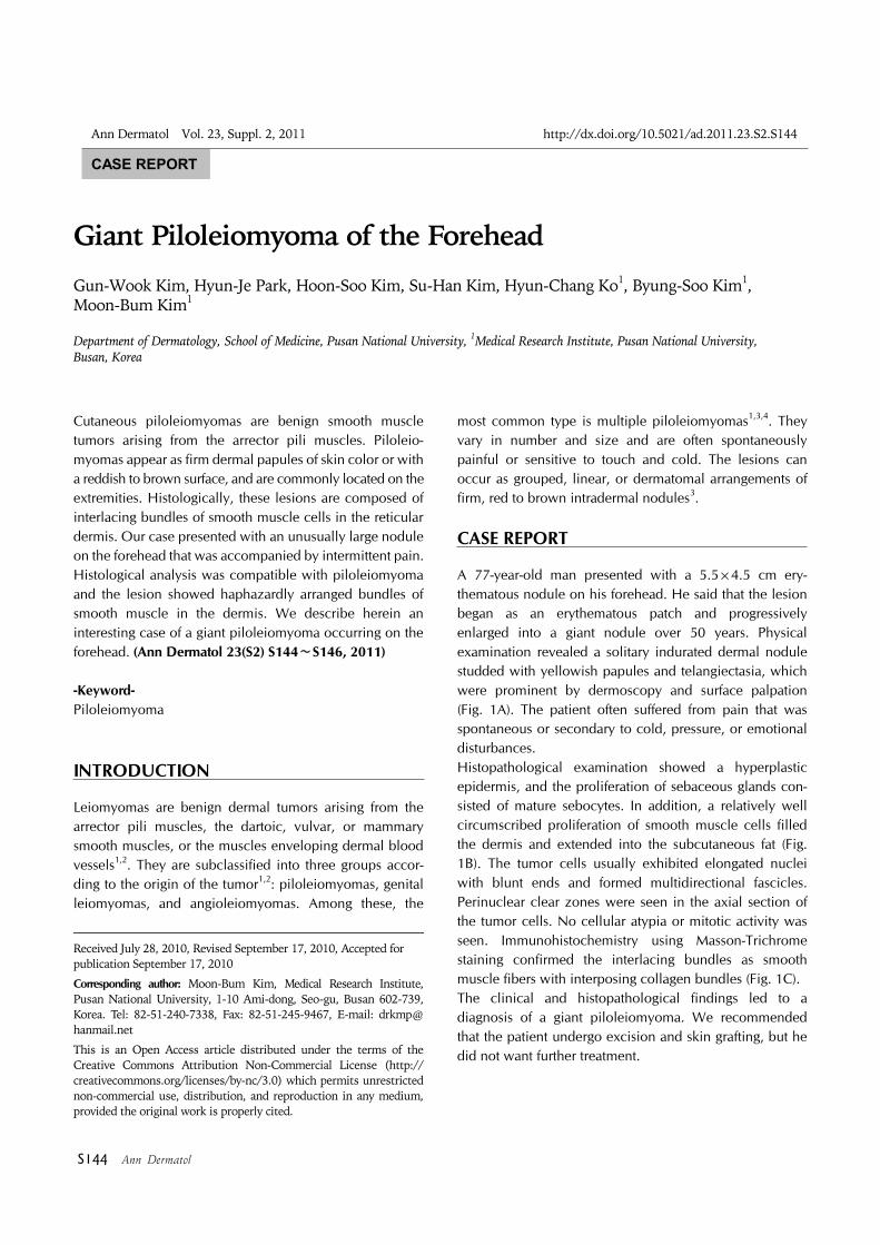
A 77-year-old man presented with a 5.5×4.5 cm ery-thematous nodule on his forehead. He said that the lesion began as an erythematous patch and progressively enlarged into a giant nodule over 50 years. Physical examination revealed a solitary indurated dermal nodule studded with yellowish papules and telangiectasia, which were prominent by dermoscopy and surface palpation (Fig. 1A). The patient often suffered from pain that was spontaneous or secondary to cold, pressure, or emotional disturbances. Histopathological examination showed a hyperplastic epidermis, and the proliferation of sebaceous glands con-sisted of mature sebocytes. In addition, a relatively well circumscribed proliferation of smooth muscle cells filled the dermis and extended into the subcutaneous fat (Fig. 1B). The tumor cells usually exhibited elongated nuclei with blunt ends and formed multidirectional fascicles. Perinuclear clear zones were seen in the axial section of the tumor cells. No cellular atypia or mitotic activity was seen. Immunohistochemistry using Masson-Trichrome staining confirmed the interlacing bundles as smooth muscle fibers with interposing collagen bundles (Fig. 1C).The clinical and histopathological findings led to a diagnosis of a giant piloleiomyoma. We recommended that the patient undergo excision and skin grafting, but he did not want further treatment.

Giant Piloleiomyoma of the Forehead
Vol. 23, Suppl. 2, 2011 S145
DISCUSSION
Cutaneous leiomyomas are uncommon benign smooth muscle neoplasms of the skin that are frequently unrecognized by clinicians3. The precise etiology of this disease is still unknown, but the involvement of a genetic predisposition has been suggested in reports on the familial occurrence of piloleiomyomas1.Piloleiomyomas are the most common type of cutaneous leiomyoma. They present as dermal, pink- or skin-colored papules or nodules, which are fixed to the skin. Piloleiomyomas are usually distributed in multiple lesions with each of them ranging from several millimeters to 1 cm in size. Their distribution is most commonly clustered, linear, or along Blaschko's line. The extensor extremities, trunk, and sides of the face and neck are the most common locations1,2.According to a study by Raj et al.4, which analyzed 53 cases of cutaneous piloleiomyomas in 45 patients, multiple piloleiomyomas are slightly more common than solitary lesions (ratio 1.2:1). There is an equal distri-bution between both sexes, and most patients are adults with a wide age distribution from 17 to 74 years. The limbs are a common site for both multiple and solitary
piloleiomyomas with solitary lesions favoring the limbs and multiple lesions the trunk. However, the forehead is a relatively uncommon site for a solitary piloleiomyoma, occurring in only 2 of 18 cases. In our case, the lesion was confined to the forehead as a solitary nodule. In addition, our case is interesting because the tumor was excep-tionally large, at least 5 times larger than the largest previously documented piloleiomyoma. Raj et al.4 show-ed that the clinical size of multiple piloleiomyomas varied from a few millimeters to 1.5 cm with an average size of 0.7 cm. Moreover, the largest solitary lesion was 1.5 cm in diameter. Although Mitra et al.5 reported a case of a diffuse piloleiomyoma 15 cm in diameter on the right knee, there has been persistent controversy whether that case was a true piloleiomyoma because it is difficult to differentiate between smooth muscle hamartoma and leiomyoma. Other than this controversial case, to our knowledge, there are no reports of a cutaneous pilo-leiomyoma larger than the case we have presented. In general, the tumors are painless initially but may become painful with time. The diagnosis may be facilitated by the characteristic provocation of pain on touching or chilling the lesion, although the cause for this remains unclear1. Several authors believe the pain or
Fig. 1. (A) A solitary erythematous nodule studded with yellowishpapules and telangiectasia that are prominent on dermoscopy.(B) The lesion is composed of interlacing bundles of smooth muscle cells (H&E, ×40). (C) Immunostaining with Masson- Trichrome highlights the smooth muscle fibers (red) with collagenbundles (blue) interspersed (Masson-Trichrome stain, ×40).

GW Kim, et al
S146 Ann Dermatol
tenderness may be secondary to pressure on nerve fibers within the tumor, whereas others suggest it may be solely due to the contraction of muscle fibers3. Subcutaneous nodules may be difficult to diagnose clinically because they do not have a characteristic surface change. However, it is possible to make a reasonable differential diagnosis if the nodule is painful. The acronym "ENGLAND" is often used to refer to these painful tumors: eccrine spiradenoma, neuroma, glomus tumor, leiom-yoma, angiolipoma, neurilemmoma, and dermato-fibroma6. With regard to the diagnosis of piloleiomyomas, the association with pain or a pseudo-Darier’s sign due to muscle fiber contraction may be suggestive, and other painful tumors lack muscle on Masson trichrome stains1,3.Histologically, piloleiomyomas are poorly circumscribed lesions consisting of interlacing smooth muscle fiber bundles with varying degrees of intermingled collagen3. They are located in the dermis and can infiltrate the surrounding tissue with extension into the subcutis1. The smooth muscle fibers are fusiform and are composed of eosinophilic cytoplasm with elongated blunt-ended nuclei and perinuclear halos in cross-sections1. In problematic cases, Masson-trichrome staining facilitates the differen-tiation of red smooth muscle fibers from the blue-green collagen. If necessary, desmin, smooth muscle actin, and muscle-specific actin may help to accentuate the smooth muscle origin of the tumor3.Treatment of cutaneous leiomyomas depends on the degree of severity of symptoms and the extent of lesions. If only a few lesions are present, simple excision may be indicated1,3. For those tumors with multiple lesions, medi-cations such as nitroglycerin, nifedipine, and phenoxy-benzamine have been used to treat symptomatic leiomyomas, but with limited success7,8. In addition,
cryotherapy and electrocoagulation have shown little benefit9.Our patient was unusual, especially in that he had a giant piloleiomyoma occurring on the forehead. A giant piloleiomyoma, although rare, should be considered in the differential diagnosis of an erythematous nodule with paroxysmal pain, especially on the face.
REFERENCES
1. White LE, Levy RM, Alam M. Leiomyoma. In: Wolff K, Goldsmith LA, Katz SI, Gilchrest BA, Paller AS, Leffell DJ, editors. Fitzpatrick's dermatology in general medicine. 7th ed. New York: McGraw-Hill, 2008:1172-1173.
2. Kohler S. Tumors of smooth muscle. In: Bolognia J, Rapini R, Jorizzo J. Dermatology. 2nd ed. St Louis: Mosby Elsevier, 2008:1831-1835.
3. Holst VA, Junkins-Hopkins JM, Elenitsas R. Cutaneous smooth muscle neoplasms: clinical features, histologic findings, and treatment options. J Am Acad Dermatol 2002;46:477-490.
4. Raj S, Calonje E, Kraus M, Kavanagh G, Newman PL, Fletcher CD. Cutaneous pilar leiomyoma: clinicopathologic analysis of 53 lesions in 45 patients. Am J Dermatopathol 1997;19:2-9.
5. Mitra A, Gudgeon PW, Merchant W, Shah M. A case of diffuse pilar leiomyoma or acquired smooth muscle hamartoma? Clin Exp Dermatol 2009;34:e145-e147.
6. Naversen DN, Trask DM, Watson FH, Burket JM. Painful tumors of the skin: "LEND AN EGG". J Am Acad Dermatol 1993;28:298-300.
7. Thompson JA Jr. Therapy for painful cutaneous leiomyomas. J Am Acad Dermatol 1985;13:865-867.
8. Thyresson HN, Su WP. Familial cutaneous leiomyomatosis. J Am Acad Dermatol 1981;4:430-434.
9. Montgomery H, Winkelmann RK. Smooth-muscle tumors of the skin. AMA Arch Derm 1959;79:32-40.