glottic and skull indices in canine brachycephalic airway
TRANSCRIPT
Caccamo et al. BMC Veterinary Research 2014, 10:12http://www.biomedcentral.com/1746-6148/10/12
RESEARCH ARTICLE Open Access
Glottic and skull indices in canine brachycephalicairway obstructive syndromeRoberta Caccamo1*†, Paolo Buracco1†, Giuseppe La Rosa2, Matteo Cantatore3 and Stefano Romussi4†
Abstract
Background: Forty dogs presented for brachycephalic airway obstructive syndrome with laryngeal collapse notover 1st degree (saccule eversion) underwent glottis endoscopic and radiographic skull measurements beforesurgery. Fifteen Pugs, fifteen French and ten English Bulldogs were included. The goals were prospectively tocompare three common brachycephalic breeds for anatomical differences regarding glottis and skullmeasurements, and to assess if any correlation between glottis and skull measurements was present. Linearmeasurements were used to obtain glottis and skull indices. Correlations between glottis and skull indices andglottic measurements were evaluated. Finally, glottis indices were compared among the three breeds.
Results: No correlation was found for glottis and skull indices. The glottic index differed among the three breeds(smaller in Pugs and higher in English Bulldogs), ultimately representing a morphologic indicator of the differentlarynx shape in the three breeds (more rounded in English Bulldogs, more elliptical in Pugs and in-between inFrench Bulldogs).
Conclusions: The lack of correlation between skull/glottic indices does not support skull morphology as predictorof glottic morphology. As Pugs had the lowest glottic index, it may be speculated that Pugs’ original narrow glotticwidth may predispose to further progressive respiratory deterioration more easily than in the other two breeds.
Keywords: Canine brachycephalic airway obstructive syndrome, Skull measurements, Glottic measurements,Glottic index, Laryngeal collapse
BackgroundBrachycephalic airway obstruction syndrome (BAOS) is awell-known combination of several upper airway abnor-malities [1-3]. These abnormalities may be defined as pri-mary (stenotic nares, elongated and thick soft palate,excessive nasopharyngeal turbinates, and hypoplastictrachea) [4-6] or secondary (mainly laryngeal collapse) [7].Redundant and hypertrophied pharyngeal folds, macro-glossia, laryngeal edema, enlarged tonsils, and bronchialcollapse may also be present [8-10]. Respiratory signs(snoring, coughing, stertor, stridor, dyspnea, tachypnea,exercise intolerance, and cyanosis/syncope) are usuallyprogressive, and their severity depends on the degree ofairway obstruction [3]. Gagging, retching, regurgitationand vomiting, along with other lower gastrointestinal
* Correspondence: [email protected]†Equal contributors1Department for Veterinary Science, Faculty of Veterinary Medicine, Universityof Turin, Grugliasco, ItalyFull list of author information is available at the end of the article
© 2014 Caccamo et al.; licensee BioMed CentrCommons Attribution License (http://creativecreproduction in any medium, provided the or
signs (such as dilated stomach and flatulence caused byaerophagia) may be associated [11-13].To our knowledge, there are no published studies that
evaluate inter-breed variations of endoscopic larynx exa-mination and, more specifically, of glottic anatomy inbrachycephalic dogs. Furthermore, no study has investi-gated endoscopic glottic measurements in brachycephalicbreeds, or assessed laryngeal and skull morphology aspotential predictors for BAOS progression. As laryngealcollapse appears as one of the most threatening secondaryeffects of airway obstruction during the progression ofBAOS, authors’ goal was to look for potential differencesof the laryngeal shape among three common brachyce-phalic canine breeds presented for BAOS surgery. For thispurpose only symptomatic dogs for BAOS with not over afirst degree laryngeal collapse at endoscopy were chosen.The study was divided in two prospective phases. Phase I:laryngeal morphometric study comparing English andFrench bulldogs and Pugs. Phase II: evaluation of the cor-relation between endoscopic glottic and radiographic skull
al Ltd. This is an open access article distributed under the terms of the Creativeommons.org/licenses/by/2.0), which permits unrestricted use, distribution, andiginal work is properly cited.

Caccamo et al. BMC Veterinary Research 2014, 10:12 Page 2 of 7http://www.biomedcentral.com/1746-6148/10/12
measurements. Besides, glottis indices were comparedamong the three breeds. Attention was also driven toascertain if the larynx shape (mainly a narrow glottisopening) could be predicted based on the morphometricstudy of the skull.
MethodsDogs of three brachycephalic breeds (Pug, French Bulldogand English Bulldog) diagnosed with BAOS at the Schoolof Veterinary Medicine of Milan and Turin betweenJanuary 2006-December 2009 were consecutively enrolledin the study. Patients were excluded from the study ifairway endoscopy identified the presence of laryngealcollapse more severe than grade I [7].This prospective study complied with institutional
guidelines for research on animals, and signed consentwas obtained from all owners, and all procedures werepart of standard diagnostic work-up and treatment.Historical and physical examination data were collected
for each animal. In all dogs, work-up prior to anaesthesiaincluded: a complete blood count, an extensive bioche-mical serum profile and a cardiac ultrasonographic exa-mination. In order to proceed with the laryngoscopic andtracheobronchoscopic exam (see later, Phase I), intramus-cular premedication included 0.2 mg/kg methadone alone,or in combination with 10 μg/kg acepromazine. Lateraland dorso ventral radiographs of the chest were also takenat this time, also in order to check for tracheal hypoplasia,as established by the tracheal diameter/thoracic inlet ratio(TI) [8]. Pre-oxygenation was provided for 5 min prior toendoscopy with 100% oxygen (approximately 2 L/min)via a face-mask. General anaesthesia was induced with2-4 mg/kg intravenous propofol; intravenous methylpred-nisolone sodium succinate (1 mg/kg) was given to controllaryngeal edema; intravenous cefazolin (20 mg/kg) wasalso administered. Light plane of anesthesia (with spon-taneous breathing and laryngeal function preserved) wasmaintained via propofol boluses, as required. Oxygen wasdelivered via a catheter alongside the endoscope. Aftercompletion of the endoscopic evaluation, dogs were intu-bated, and anesthesia was maintained with isoflurane inoxygen. Surgical correction of BAOS, performed by twodifferent surgeons (SR and PB), consisted of verticalwedge nostril plasty and partial staphylectomy [14]. For
Table 1 Mean values obtained from glottic measurements
Glottic height
n Mean ± SD Median
Total population 40 209.67 ± 46.41 223.12
Pugs 15 211.80 ± 28.16 223.12
FB 15 221.56 ±43.72 230.67
EB 10 186.31 ± 67.90 178.71
The only significant difference is between Pugs vs English Bulldogs (Wilcoxon Rank
soft palate resection the midpoint of the tonsils was usedas the guide [15]. Laryngeal saccule resection was per-formed at the surgeons’ discretion. Prior to recovery fromanesthesia, a dorso-ventral skull radiograph was taken.
Phase ILaryngoscopy was performed (SR and RC) with dogs insternal recumbency, either using a 5.3-mm × 60-cm fle-xible videobronchoscope (Fujinon EB 250S, Fujinon Inc.,Wayne, New Jersey) or a 5.0-mm X 55-cm flexible fiber-bronchoscope (Olympus BF-P40, Olympus Medical Sys-tems Europe GmbH, Hamburg, Germany). Images andmovies of laryngeal motions were acquired using a videorecording device (Sony GV-D1000E, Sony Corporation,Tokyo, Japan). Further images were obtained from moviesusing the computer software: Matrox Pc-Vrc Remote 2.0.The degree of laryngeal collapse was endoscopicallyassessed based on Leonard’s classification [7]. In particu-lar, according to this classification, saccule eversion is con-sidered as 1st degree laryngeal collapse. Progression of thedisease leads to collapse of the cuneiform processes of ary-tenoids (2nd degree collapse) and can proceed to collapseof the corniculate processes of arytenoids (3rd degreecollapse).Glottic linear measurements, expressed in pixels, were
taken of arytenoids at their most adducted position (endof expiration) using the UTHSCSA Image Tool 3.00 forWindows software. Measurements were: a) linear glotticheight [dorso-ventral diameter (dorsal to ventral com-missure)], b) linear glottic width [transversal diameter(distance between the vocal processes of the two arythe-noid cartilages)], and c) glottic index as a result of glotticwidth × 100/glottic height. Glottic index was calculatedaccording to skull index formula (see Phase II). Tominimize bias, the arithmetic average of three consecu-tive measurements was calculated.
Phase IIDigital dorso-ventral radiographs of the skull were taken(Agfa ADC SOLO CR System, Agfa HealthCare NV,Mortsel, Belgium). The head of the dog was kept parallelto the radiological table using a cushion under the chin.Images were analyzed with the UTHSCSA Image Tool3.00 for Windows software. Linear measurements,
Glottic width Glottic index
Mean ± SD Median Mean ± SD Median
48.70 ± 17.57 42.99 24.44 ± 10.81 20.71
39.97 ± 14.88 36.20 19.35 ± 8.06 17.80
51.03 ± 16.61 45.30 23.58 ± 7.60 20.45
58.32 ± 18.04 53.07 33.35 ± 13.53 31.39
Sum test P = 0.008).

Figure 1 Boxplot of the glottic index in each of the three breeds. Pugs (Median 17.80, 1st Quartile 12.32, 3rd Quartile 21.79); FB (Median 20.45,1st Quartile 17.29, 3rd Quartile 26.23); EB (Median 31.39, 1st Quartile 22.02, 3rd Quartile 41.88).
Figure 2 Endoscopic images of the larynx of a Pug, with linearmeasurements (transversal diameter blue color, longitudinaldiameter red color).
Caccamo et al. BMC Veterinary Research 2014, 10:12 Page 3 of 7http://www.biomedcentral.com/1746-6148/10/12
expressed in pixels, were: a) skull length [inion-prosthion(central surface point on external occipital protuberance-anterior end of interincisive suture, between the roots ofthe upper central incisor teeth)], b) maximum zygomaticwidth [zygion-zygion (the most lateral point of the zygo-matic arch)], and c) skull index (maximum zygomaticwidth × 100/skull length) [16,17]. To minimize bias, thearithmetic average of three different measurements wascalculated.
Statistical evaluationData were expressed as mean ± SD (standard deviation),median and range. Descriptive statistical analysis wasused for age, body weight, and glottic and skull measure-ments. Data concerning everted saccules, hypoplastictrachea, and bronchial collapse were expressed as per-centages. For all variables, the Shapiro-Wilk test of nor-mality was applied. Correlation among age, body weight,and glottic and skull measurements was evaluated;Spearman’s rank correlation test and Pearson’s productmoment correlation were applied to non-normally andnormally distributed variables. The Wilcoxon rank sumtest was used to compare age, body weight and glottic andskull measurements. A P value of <0.05 was consideredstatistically significant for all tests. To evaluate the repea-tability of glottic measurements, the British StandardsInstitution repeatability coefficient was applied [18]. All

Figure 3 Endoscopic images of the larynx of an EB, with linearmeasurements (transversal diameter blue color, longitudinaldiameter red color).
Caccamo et al. BMC Veterinary Research 2014, 10:12 Page 4 of 7http://www.biomedcentral.com/1746-6148/10/12
statistical analyses were performed using the public do-main program R 2.3.0 (R Development Core Team).
ResultsForty dogs affected with BAOS were included in the study,subdivided as follows: 15 Pugs (37.5%), 15 French Bulldogs(FB, 37.5%), and 10 English Bulldogs (EB, 25%). The meanage was 2.64 ± 1.59 SD (median 2.5 years, and range9 months to 6 years). In Pugs the mean age was 2.55 ± 1.2SD (median 2.5 years, range 11 months to 5 years), inFB 2.85 ± 1.95 SD (median 2.5 years, range 9 months to6 years), and in EB 2.44 ± 1.63 SD (median 2.5 years, range9 months to 5 years). There was no significant age dif-ference between the three breeds (P >0.05). Twenty-five ofthe dogs were male (62.5%), whereas fifteen were females(37.5%). The mean body weight was 14.02 ± 8.29 SD(median 10 kg, range 6–40 kg). In Pugs the mean bodyweight was 8.51 ± 1.34 SD (median 8.7 kg, range 6–11 kg),in FB 10.7 ± 2.17 SD (median 11 kg, range 7–12 kg) in FB,and in EB 26.6 ± 6.33 SD (median 24.5 kg, range21–40 kg) in EB. Significant differences in body weight
Table 2 Mean values obtained from skull measurements
Skull length M
n Mean ± SD Median M
Total population 40 989.49 ± 220.32 917.08 1
Pugs 15 794.84 ± 68.82 793.5 8
FB 15 987.57 ± 130.88 936.36 1
EB 10 1284.36 ± 128.71 1279.02 1
Among the three breeds no correlation was found with linear measurements. No si
(Wilcoxon Rank Sum test) were found among Pug vsFB (P = 0.048), Pug vs EB (P = 0.002), and FB vs EB(P = 0.005).Upon thoracic radiography, 6 dogs (15%) had tracheal
hypoplasia (Pugs 0%; 1 FB, 16.7%; 5 EB, 83.3%). All dogshad both an elongated soft palate and stenotic nares;both conditions were surgically corrected. Twenty-seven dogs (67.5%) had everted saccules (10 Pugs,37.0%; 9 FB, 33.3%; 8 EB, 29.6%) and 21 (77.8%) of theseunderwent laryngeal saccule resection (9 Pugs, 8 FB and4 EB). Bronchoscopy also revealed that 4 dogs (10%)had bronchial collapse (2 Pugs, 50%; 0 FB, 0%; 2 EB,50%).
Phase ILaryngeal linear measurement and glottic index-re-lated means, both expressed in pixels, are reported inTable 1. Analysis of direct measurements of the glottisdid not show a significant difference among the threebreeds. The average difference between repeated mea-surements of the glottis (d) was 2.75% (SD = 1.02%) forthe first operator and 1.05% (SD = 0.6%) for the secondone; the British Standards Institution repeatability co-efficient (1.96 SD) was 1.99% and 1.18%, respectively.Glottic index showed an increasing trend, beingsmaller in Pugs and progressively bigger in French andEnglish Bulldogs. In particular, a significant diffe-rence was clearly observed between Pugs and EnglishBulldogs (P = 0.008), with Pugs having the glotticindex significantly smaller (i.e. narrower shape of theglottis) when compared to English Bulldogs (Figure 1);Figures 2 and 3 show laryngoscopic images in 2 dogs(1 Pug and 1 EB). Besides, no significant correlationsbetween age and glottic index were found. Finally, theglottis index and body weight were not significantlycorrelated.
Phase IISkull linear measurement and skull index-related means,both expressed in pixels, are reported in Table 2. Nostatistical correlation, calculated according to Pearson’sparametric correlation test and Spearman’s Rank sum test,between glottis and skull linear measurements and glottisand skull indices, were found, both in the entire
aximum zygomatic width Skull index
ean ± SD Median Mean ± SD Median
065.15 ± 239.29 1091.51 108.04 ± 11.99 108.55
03.94 ± 86.09 802.10 101.39 ± 9.33 101.83
153.55 ± 105.07 1166.00 117.63 ± 9.12 118.07
324.39 ± 134.21 1315.45 103.65 ± 10.50 104.29
gnificant difference was found with skull index.
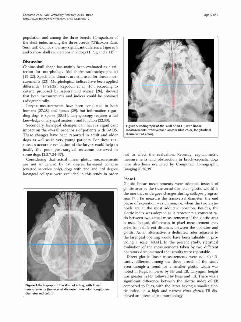
Figure 5 Radiograph of the skull of an EB, with linearmeasurements (transversal diameter blue color, longitudinaldiameter red color).
Caccamo et al. BMC Veterinary Research 2014, 10:12 Page 5 of 7http://www.biomedcentral.com/1746-6148/10/12
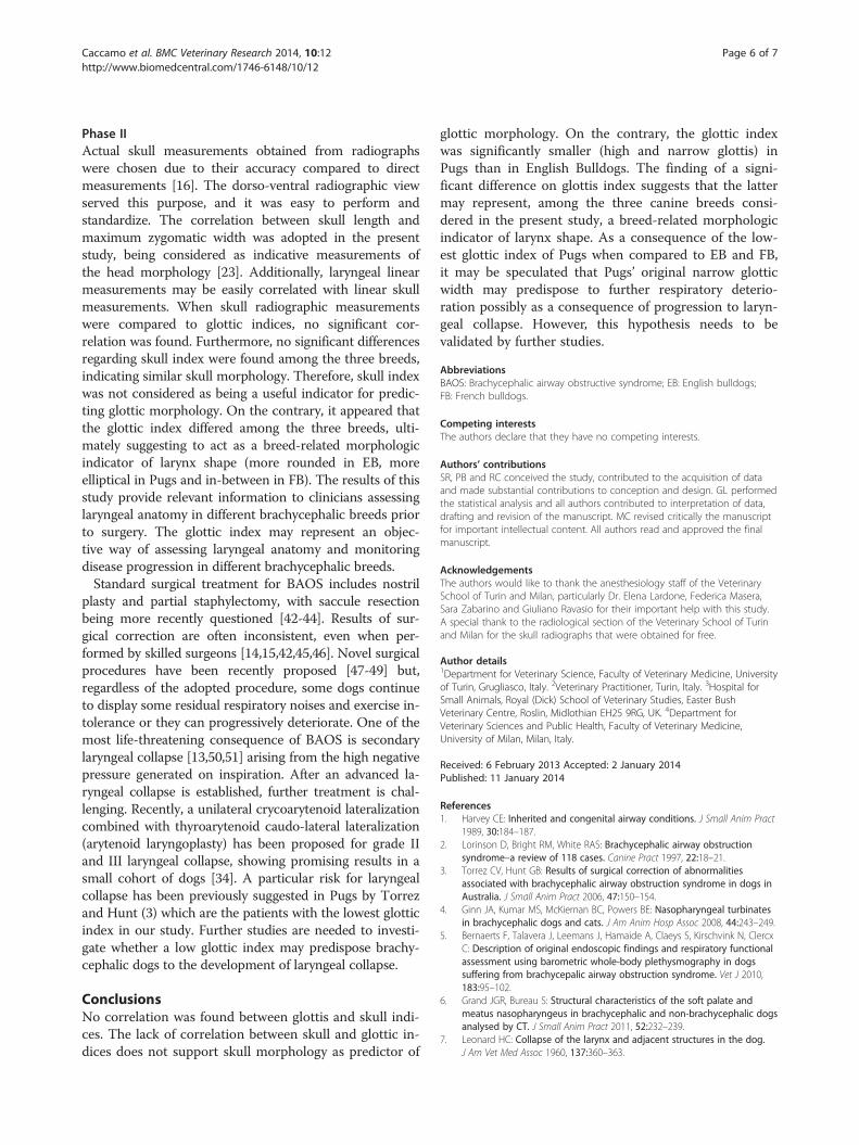
population and among the three breeds. Comparison ofthe skull index among the three breeds (Wilcoxon RankSum test) did not show any significant difference. Figures 4and 5 show skull radiographs in 2 dogs (1 Pug and 1 EB).
DiscussionCanine skull shape has mainly been evaluated as a cri-terion for morphology (dolicho/meso/brachycephalic)[19-22]. Specific landmarks are still used for linear mea-surements [23]. Morphological indices have been applieddifferently [17,24,25]. Regodon et al. [16], according tocriteria proposed by Aguera and Massa [26], showedthat both measurements and indices could be obtainedradiographically.Larynx measurements have been conducted in both
humans [27,28] and horses [29], but information regar-ding dogs is sparse [30,31]. Laryngoscopy requires a fullknowledge of laryngeal anatomy and function [32,33].Secondary laryngeal changes can have a significant
impact on the overall prognosis of patients with BAOS.These changes have been reported in adult and olderdogs as well as in very young patients. For these rea-sons an accurate evaluation of the larynx could help tojustify the poor post-surgical outcome observed insome dogs [2,3,7,34-37].Considering that actual linear glottic measurements
are not influenced by 1st degree laryngeal collapse(everted saccules only), dogs with 2nd and 3rd degreelaryngeal collapse were excluded in this study in order
Figure 4 Radiograph of the skull of a Pug, with linearmeasurements (transversal diameter blue color, longitudinaldiameter red color).
not to affect the evaluation. Recently, cephalometricmeasurements and obstruction in brachycephalic dogshave also been evaluated by Computed TomographicImaging [6,38,39].
Phase IGlottic linear measurements were adopted instead ofglottic area as the transversal diameter (glottic width) isthe one that undergoes changes during collapse progres-sion [7]. To measure the transversal diameter, the endphase of expiration was chosen, i.e. when the two aryte-noids are at the most adducted position. Besides, theglottic index was adopted as it represents a constant ra-tio between two actual measurements; if the glottic areais used instead, differences in pixel measurement mayarise from different distances between the operator andglottis. As an alternative, a dedicated ruler adjacent tothe laryngeal opening would have been valuable in pro-viding a scale [40,41]. In the present study, statisticalevaluation of the measurements taken by two differentoperators demonstrated that results were repeatable.Direct glottic linear measurements were not signifi-
cantly different among the three breeds of the studyeven though a trend for a smaller glottic width wasnoted in Pugs, followed by FB and EB. Laryngeal heightwas greater in FB, followed by Pugs and EB. There was asignificant difference between the glottic index of EBcompared to Pugs, with the latter having a smaller glot-tic index, i.e. a high and narrow rima glottis; FB dis-played an intermediate morphology.
Caccamo et al. BMC Veterinary Research 2014, 10:12 Page 6 of 7http://www.biomedcentral.com/1746-6148/10/12
Phase IIActual skull measurements obtained from radiographswere chosen due to their accuracy compared to directmeasurements [16]. The dorso-ventral radiographic viewserved this purpose, and it was easy to perform andstandardize. The correlation between skull length andmaximum zygomatic width was adopted in the presentstudy, being considered as indicative measurements ofthe head morphology [23]. Additionally, laryngeal linearmeasurements may be easily correlated with linear skullmeasurements. When skull radiographic measurementswere compared to glottic indices, no significant cor-relation was found. Furthermore, no significant differencesregarding skull index were found among the three breeds,indicating similar skull morphology. Therefore, skull indexwas not considered as being a useful indicator for predic-ting glottic morphology. On the contrary, it appeared thatthe glottic index differed among the three breeds, ulti-mately suggesting to act as a breed-related morphologicindicator of larynx shape (more rounded in EB, moreelliptical in Pugs and in-between in FB). The results of thisstudy provide relevant information to clinicians assessinglaryngeal anatomy in different brachycephalic breeds priorto surgery. The glottic index may represent an objec-tive way of assessing laryngeal anatomy and monitoringdisease progression in different brachycephalic breeds.Standard surgical treatment for BAOS includes nostril
plasty and partial staphylectomy, with saccule resectionbeing more recently questioned [42-44]. Results of sur-gical correction are often inconsistent, even when per-formed by skilled surgeons [14,15,42,45,46]. Novel surgicalprocedures have been recently proposed [47-49] but,regardless of the adopted procedure, some dogs continueto display some residual respiratory noises and exercise in-tolerance or they can progressively deteriorate. One of themost life-threatening consequence of BAOS is secondarylaryngeal collapse [13,50,51] arising from the high negativepressure generated on inspiration. After an advanced la-ryngeal collapse is established, further treatment is chal-lenging. Recently, a unilateral crycoarytenoid lateralizationcombined with thyroarytenoid caudo-lateral lateralization(arytenoid laryngoplasty) has been proposed for grade IIand III laryngeal collapse, showing promising results in asmall cohort of dogs [34]. A particular risk for laryngealcollapse has been previously suggested in Pugs by Torrezand Hunt (3) which are the patients with the lowest glotticindex in our study. Further studies are needed to investi-gate whether a low glottic index may predispose brachy-cephalic dogs to the development of laryngeal collapse.
ConclusionsNo correlation was found between glottis and skull indi-ces. The lack of correlation between skull and glottic in-dices does not support skull morphology as predictor of
glottic morphology. On the contrary, the glottic indexwas significantly smaller (high and narrow glottis) inPugs than in English Bulldogs. The finding of a signi-ficant difference on glottis index suggests that the lattermay represent, among the three canine breeds consi-dered in the present study, a breed-related morphologicindicator of larynx shape. As a consequence of the low-est glottic index of Pugs when compared to EB and FB,it may be speculated that Pugs’ original narrow glotticwidth may predispose to further respiratory deterio-ration possibly as a consequence of progression to laryn-geal collapse. However, this hypothesis needs to bevalidated by further studies.
AbbreviationsBAOS: Brachycephalic airway obstructive syndrome; EB: English bulldogs;FB: French bulldogs.
Competing interestsThe authors declare that they have no competing interests.
Authors’ contributionsSR, PB and RC conceived the study, contributed to the acquisition of dataand made substantial contributions to conception and design. GL performedthe statistical analysis and all authors contributed to interpretation of data,drafting and revision of the manuscript. MC revised critically the manuscriptfor important intellectual content. All authors read and approved the finalmanuscript.
AcknowledgementsThe authors would like to thank the anesthesiology staff of the VeterinarySchool of Turin and Milan, particularly Dr. Elena Lardone, Federica Masera,Sara Zabarino and Giuliano Ravasio for their important help with this study.A special thank to the radiological section of the Veterinary School of Turinand Milan for the skull radiographs that were obtained for free.
Author details1Department for Veterinary Science, Faculty of Veterinary Medicine, Universityof Turin, Grugliasco, Italy. 2Veterinary Practitioner, Turin, Italy. 3Hospital forSmall Animals, Royal (Dick) School of Veterinary Studies, Easter BushVeterinary Centre, Roslin, Midlothian EH25 9RG, UK. 4Department forVeterinary Sciences and Public Health, Faculty of Veterinary Medicine,University of Milan, Milan, Italy.
Received: 6 February 2013 Accepted: 2 January 2014Published: 11 January 2014
References1. Harvey CE: Inherited and congenital airway conditions. J Small Anim Pract
1989, 30:184–187.2. Lorinson D, Bright RM, White RAS: Brachycephalic airway obstruction
syndrome–a review of 118 cases. Canine Pract 1997, 22:18–21.3. Torrez CV, Hunt GB: Results of surgical correction of abnormalities
associated with brachycephalic airway obstruction syndrome in dogs inAustralia. J Small Anim Pract 2006, 47:150–154.
4. Ginn JA, Kumar MS, McKiernan BC, Powers BE: Nasopharyngeal turbinatesin brachycephalic dogs and cats. J Am Anim Hosp Assoc 2008, 44:243–249.
5. Bernaerts F, Talavera J, Leemans J, Hamaide A, Claeys S, Kirschvink N, ClercxC: Description of original endoscopic findings and respiratory functionalassessment using barometric whole-body plethysmography in dogssuffering from brachycepalic airway obstruction syndrome. Vet J 2010,183:95–102.
6. Grand JGR, Bureau S: Structural characteristics of the soft palate andmeatus nasopharyngeus in brachycephalic and non-brachycephalic dogsanalysed by CT. J Small Anim Pract 2011, 52:232–239.
7. Leonard HC: Collapse of the larynx and adjacent structures in the dog.J Am Vet Med Assoc 1960, 137:360–363.

Caccamo et al. BMC Veterinary Research 2014, 10:12 Page 7 of 7http://www.biomedcentral.com/1746-6148/10/12
8. Coyne BE, Fingland RB: Hypoplasia of the trachea in dogs: 103 cases(1974–1990). J Am Vet Med Assoc 1992, 201:768–772.
9. Fasanella FJ, Shivley J, Wardlaw JL, Givaruangsawat S: Brachycephalicairway obstructive syndrome in dogs: 90 cases (1991–2008). J Am VetMed Assoc 2010, 237:1048–1051.
10. De Lorenzi D, Bertoncello D, Drigo M: Bronchial abnormalities found in aconsecutive series of 40 brachycephalic dogs. J Am Vet Med Assoc 2009,235:835–840.
11. Lecoindre P, Richard S: Digestive disorders associated with the chronicobstructive respiratory syndrome of brachycephalic dogs: 30 cases(1999–2001). Revue Med Vet 2004, 155:141–146.
12. Poncet CM, Dupré GP, Freiche VG, Estrada MM, Poubanne YA, Bouvy BM:Prevalence of gastrointestinal tract lesions in 73 brachycephalic dogswith upper respiratory syndrome. J Small Anim Pract 2005, 46:273–279.
13. Wykes PM: Brachycephalic airway obstructive syndrome. Probl Vet Med1991, 3:188–197.
14. Hedlund CS: Surgery of the upper respiratory system. In Small AnimalSurgery. 3rd edition. Edited by Fossum TW. St. Louis: Mosby Elsevier;2007:817–866.
15. Harvey CE: Upper airway obstruction surgery. II. Soft palate resection inbrachycephalic dogs. J Am Animal Hosp Assoc 1982, 18:538–544.
16. Regodon S, Robina A, Franco A, Vivo JM, Lignereux Y: Déterminationradiologique et statistique des types morphologiques craniens chez lechien: dolichocéphalie, mésocephalie et brachycéphalie. Anat HistolEmbryol 1991, 20:129–138.
17. Evans HE: Axial skeleton, the skull. In Miller’s Anatomy of the dog. Edited byEvans HE. Philadelphia: Saunders; 1993:128–133.
18. Petrie A, Watson P: Diagnostic test. In Statistic for Veterinary and AnimalScience. Edited by Petrie A, Watson P. London: Blackwell Science Ltd;1999:168–181.
19. Onar V: A morphometric study on the skull of the German Shepherd dog(Alsatian). Anat Histol Embryol 1999, 28:253–256.
20. Alpak H, Mutus R, Onar V: Correlation analysis of the skull and long bonemeasurements of the dog. Ann Anat 2004, 186:323–330.
21. Fox MW: Developmental abnormalities of the canine skull. Can J CompMed Vet Sci 1963, 27:219–222.
22. Koch DA, Arnold S, Hubler M, Montavon PM: Brachycephalic syndrome indogs. Compend Contin Educ Pract Vet 2003, 25:48–55.
23. Stockard CR: The genetic and endocrine basis for differences in form andbehaviour as elucidated by studies of contrasted pure-line dog breedsand their hybrids. Am Anat Mem 1941, 19:1–753.
24. Brehm H, Loeffler K, Komeyli H: The shapes of the canine skull. Anat HistolEmbryol 1985, 14:324–331.
25. Nickel R, Schummer A, Seiferle E: Apparato locomotore. In Trattato diAnatomia degli Animali Domestici. Edited by Nickel R, Schummer A,Seiferle E. Milano: Casa Editrice Ambrosiana; 1991:11–229.
26. Aguera E, Massa R: Introducciòn a la topografia craneo-encefalica en elperro basada en metodos radiograficos. Arch Zootec 1977, 26:9–21.
27. Eckel HE, Sittel C: Morphometry of the larynx in horizontal sections.Am J Otolaryngol 1995, 16:40–48.
28. Dailey SH, Kobler JB, Hillman RE, Tangrom K, Thananart E, Mauri M,Zeitels SM: Endoscopic measurement of vocal fold movement duringadduction and abduction. Laryngoscope 2005, 115:178–183.
29. Lafortuna C, Albertini M, Ferrucci F, Zucca E, Braghieri M, Clement MG,Saibene F: Laryngeal movements during the respiratory cycle measuredwith an endoscopic imaging technique in the conscious horse at rest.Exp Physiol 1999, 84:739–746.
30. Nasri S, Sercarz JA, Berke GS: Non invasive measurement of travellingwave velocity in the canine larynx. Ann Otol Rhinol Laryngol 1994,103:758–766.
31. Romussi S, Stefanello D, Franchini D, Cuciti D: Aritenoidectomia transoralemodificata nel trattamento del collasso laringeo di secondo grado nelcane: valutazioni preliminari. In Proceedings of the VIII Congresso Nazionaledella Società Italiana di Chirurgia Veterinaria: 20–23 June 2001; Olbia.Edited by SICV. 2001:143–149.
32. Padrid PA, McKiernan BC: Endoscopy of the upper respiratory tract of thedog and cat. In Small Animal Endoscopy. 2nd edition. Edited by Tams TR.St. Louis: Mosby; 1999:357–376.
33. Holt DE: Laryngoscopy and Pharyngoscopy. In Textbook of RespiratoryDisease in Dogs and Cats. Edited by King LG. St. Louis: Elsevier;2004:109–112.
34. White RN: Surgical management of laryngeal collapse associated withbrachycephalic airway obstruction syndrome in dogs. J Small Anim Pract2012, 53:44–50.
35. Aron DN, Crowe DT: Upper airway obstruction. General principles andselected conditions in the dog and cat. Vet Clin North Am Small AnimPract 1985, 15:891–917.
36. Pink JJ, Doyle RS, Hughes JML, Tobin E, Bellenger CR: Laryngeal collapse inseven brachycephalic puppies. J Small Anim Pract 2006, 47:131–135.
37. Harvey CE: Review of results of airway obstruction surgery in the dog.J Small Anim Pract 1983, 24:555–559.
38. Schmidt MJ, Neumann AC, Amort KH, Failing K, Kramer M: Cephalometricmeasurements and determination of general skull type of cavalier kingcharles spaniels. Vet Radiol Ultrasound 2011, 4:436–440.
39. Stadler K, Hartman S, Matheson J, O’Brien R: Computed tomographicimaging of dogs with primary laryngeal or tracheal airway obstruction.Vet Radiol Ultrasound 2011, 4:377–384.
40. Miller CJ, McKiernan BC, Pace J, Fettman MJ: The effects of doxapramhydrochloride (dopram-v) on laryngeal function in healthy dogs.J Vet Intern Med 2002, 16:524–528.
41. Demetriou JL, Kirby BM: The effect of two modifications of unilateralarytenoid lateralization on rima glottidis area in dogs. Vet Surg 2003,32:62–68.
42. Riecks TW, Birchard SJ, Stephens JA: Surgical correction of brachycephalicsyndrome in dogs: 62 cases (1991–2004). J Am Vet Med Assoc 2007,230:1324–1328.
43. Poncet CM, Dupré GP, Freiche VG, Bouvy BM: Long-term results of upperrespiratory syndrome surgery and gastrointestinal tract medicaltreatment in 51 brachycephalic dogs. J Small Anim Pract 2006,47:137–142.
44. Cantatore M, Gobbetti M, Romussi S, Brambilla G, Giudice C, Grieco V,Stefanello D: Medium term endoscopic assessment of the surgicaloutcome following laryngeal saccule resection in brachycephalic dogs.Vet Rec 2012, 170:518.
45. Harvey CE: Upper airway obstruction surgery. I. Stenotic nares surgery inbrachycephalic dogs. J Am Animal Hosp Assoc 1982, 18:535–537.
46. Harvey CE: Upper airway obstruction surgery. III. Everted laryngealsaccule surgery in brachycephalic dogs. J Am Animal Hosp Assoc 1982,18:545–547.
47. Findji L, Dupré GP: Folded flap palatoplasty for treatment of elongatedsoft palates in 55 dogs. Vet Med Austria/Wien Tierärztl Mschr 2008,95:56–63.
48. Oechtering GU, Hueber JP, Noeller C: A multi-level, multi-modal approach tosevere brachycephalic airway syndrome. In Proceedings of the 18th AnnualScientific Meeting of the European College Veterinary Surgery: 2–4 July 2009;Nantes. Edited by Tremaine H, Wilderjans H, Ness M. Zurich: European Collegeof Veterinary Surgeons; 2009:478–481.
49. Dunié-Mérigot A, Bouvy B, Poncet C: Comparative use of CO2 laser, diodelaser and monopolar electrocautery for resection of the soft palate indogs with brachycephalic airway obstructive syndrome. Vet Rec 2010,167:700–704.
50. Venker-van Haagen AJ: The larynx. In Ear, nose, throat, and trachebronchialdiseases in dogs and cats. Edited by Venker-van Haagen AJ. Hannover:Schlutersche; 2005:121–165.
51. Robinson NE: Airway physiology. Vet Clin North Am Small Anim Pract 1992,22:1043–1064.
doi:10.1186/1746-6148-10-12Cite this article as: Caccamo et al.: Glottic and skull indices in caninebrachycephalic airway obstructive syndrome. BMC Veterinary Research2014 10:12.