glutamate-like immunoreactivity revealed in rat olfactory ... · kins and evans 1981 ... pool such...
TRANSCRIPT

Histochemistry (1989) 90:427-445 Histochemistry �9 Springer-Verlag 1989
Glutamate-like immunoreactivity revealed in rat olfactory bulb, hippocampus and cerebellum by monoclonal antibody and sensitive staining method C.-j. Liu*, P. Graudes, C. Matute**, M. Cu6nod, and P. Streit*** Brain Research Institute, University of Zfirich, August-Forel-Strasse 1, CH-8029 Zfirich, Switzerland
Accepted May 19, 1988
Summary. Although there is good evidence favoring L-glu- tamate as a major excitatory amino acid transmitter, rela- tively little is known about the distribution of nerve termi- nals using this substance. A method visualizing glutamate- like immunoreactivity at the light microscopic level by means of a monoclonal antibody, mAb 2D7, is described. - The antigen used for immunization was a glutaraldehyde- linked glutamate-BSA conjugate, and hybridomas were dif- ferentially screened by ELISA for production of antibodies recognizing glutamate- but not aspartate-BSA. The cross- reactivity of 'ant i -g lu tamate ' mAb 2D7 as estimated in ab- sorption tests was low even with conjugates closely related to glutamate-BSA, - Semithin sections from rapidly perfu- sion-fixed, plastic-embedded rat brain tissues were etched and stained by a combination of the peroxidase-antiperoxi- dase method and silver enhancement of the diaminobenzi- dine reaction product. Only this amongst several other im- munohistochemical methods tried produced labeling pat- terns which showed terminal-like elements in brain regions such as olfactory bulb, hippocampus and cerebellum, and which were mostly consistent with already available infor- mation on systems using glutamate as neurotransmitter. Particularly striking was the staining of elements reminis- cent of mossy fiber terminals in hippocampus and cerebel- lum as well as of cerebellar parallel fiber terminals.
Introduction
Since the pioneering immunohistochemical work of Stein- busch and colleagues (1978) on serotonin and of Storm- Mathisen and collaborators (1983) on GABA and gluta- mate, immunoreactivities for a variety of low molecular weight compounds including several established or putative neurotransmitters were localized successfully in neural and other tissues (for review: Geffard et al. 1985; Ottersen and Storm-Mathisen 1984, 1987; Steinbusch et al. 1986; Storm- Mathisen and Ottersen 1986). The goal to describe the dis- tribution of glutamate-like immunoreactivity (Olu-LI) in the central nervous system was of particular importance
Present addresses : * Department of Physiology, Tongji Medical University Wu Han, Hubei/People's Republic of China ** Department of Anatomy, Faculty of Medicine, Uuiversidad del Pals Vasco, E-6990 Lejona, Vizcaya/Spain *** To whom offprint requests should be sent
because L-glutamate had become a candidate to play a ma- jor role in excitatory neurotransmission (for review: Wat- kins and Evans 1981; Fonnum 1984; Cotman et al. 1981; Fagg and Foster 1983; Ottersen and Storm-Mathisen 1984, 1986). Despite the methodological limitation that the local- ized material could only be defined as 'glutamate-like im- munoreactivity', antibodies with a high preference for glu- tamate over aspartate and other neuroactive amino acids in combination with a suitable immunohistochemical meth- od seemed to be the most appropriate tools to investigate the localization of glutamate at a high degree of specificity and at a high level of resolution. On the other hand, meth- ods relying on uptake (for review: Ottersen and Storm- Mathisen 1984, 1986) or on transmitter-related retrograde labeling (for review: Cu6nod and Streit 1983) with tritiated D-aspartate as tracer substance identified but did not distin- guish between neuronal elements which seemed to use gluta- mate or aspartate as transmitter. Glutaminase had been suggested to play an important role and glutaminase-like immunoreactivity had, indeed, been demonstrated in struc- tural elements which seemed to be mossy fiber terminals in cerebellum and hippocampus (Wenthold et al. 1986; Wenthold and Altschuler 1986). Nevertheless, key enzymes specifically responsible for the synthesis of the transmitter pool such as glutamic acid decarboxylase in the case of GABA had not been identified in the case of glutamate. The immunohistochemical localization of enzymes involved in the metabolism of excitatory amino acids (for review: Wenthold and Altschuler 1986), thus, had to be interpreted cautiously with respect to the transmitter specificity of la- beled tissue components such as neuronal perikarya. How- ever, heavy Glu-LI in neuronal cell bodies rather than in terminal-like elements had also been the staining pattern usually described at the light microscopic level in perfusion- fixed tissue with antisera to fixative-linked (Ottersen and Storm-Mathisen 1984; Hepler et al. 1986, 1988; Yoshida et al. 1987) or colloidal gold adsorbed (Shiosaka et al. 1986) glutamate as well as with a monoclonal antibody to gluta- mate containing compounds (Madl et al. 1986). Only inves- tigations by means of a light microscopic method on rinsed and immersion-fixed brain slices (Storm-Mathisen et al. 1983, 1986; Ottersen and Storm-Mathisen 1985) or by means of the electron microscopic postembedding immuno- gold method on perfusion-fixed material (Somogyi et al. 1986; Ottersen 1987) had revealed glutamate immunoreac- tive (Glu-ir) terminals. On the other hand, a mapping of Glu-ir terminals seemed to be important since this type

428
of immunoreactivity would have been expected to represent most closely the transmitter pool of glutamate.
The following points initiated the work described in the present paper: To perform mapping studies it would be advantageous to use light rather than electron microscopy. To be closer to the ' i n vivo ' situation it would be better to use rapidly perfusion-fixed tissue than rinsed and immer- sion-fixed material. The former kind of material might also be used for combined light and electron microscopic stu- dies. Monoclonal antibodies to fixative-linked GABA had proved to be useful tools in a previous investigation (Ma- tute and Streit 1986). We, therefore, tried to find monoclon- al antibodies specific for fixative-linked glutamate by means of a differential screening procedure. These defined reagents were then used in combinat ion with a highly sensitive post- embedding staining procedure to describe mostly at a light microscopic level the pattern of immunoreactivi ty com- posed of perikarya and terminal-like elements in olfactory bulb, hippocampal formation and cerebellar cortex of the rat. Some of the findings have been published in abstract form (Matute et al. 1987).
Materials and methods
Preparation of conjugates. Bovine serum albumin (BSA) was used as carrier protein for glutaraldehyde(GA)-linked conjugates, with the following amino acids and peptides: L-glutamate (Glu), L-as- partate (Asp), L-homocysteic acid (HCA), L-cysteic acid (CA), L- cysteinsulfinic acid (CSA), L-glutamine (Gln), 7-L-glutamylgluta- mate (?-GG), glutathione (GSH), fl-L-aspartylglycine (BLAG), y- aminobutyric acid (GABA), fl-alanine ~-Ala), glycine (Gly), and taurine (Tau). The coupling procedure described by Campistron and collaborators 0986a) yielded better results in terms of effi- ciency for the last four compounds in this series. In contrast, the procedure of Storm-Mathisen and collaborators (1983) allowed more reliably to produce conjugate preparations containing no pre- cipitates with the rest of the substances. The reaction mixtures were dialyzed against large volumes of phosphate buffer (PB) or phosphate buffered saline (PBS) over a period of 2-3 days. Ali- quots of the dialyzed solutions were stored frozen at - 2 0 ~ C. In order to monitor the efficiency of the conjugation procedure at least for Glu, Asp, GABA, fl-Ala, Gly and Tau, these amino acids in a radiolabeled form (1 gCi; The Radiochemical Centre, Amer- sham, UK) were included in the reaction, and the radioactivity was determined in small samples before and after dialysis of the reaction mixture. The radioactivity measured after dialysis reached about 5% of the amount added in the case of Glu and Asp and approximately 10% for GABA, fl-.Ala, Gly and Tau. The molar ratio of amino acid conjugated to BSA, therefore, amounted to about 10:1 in the former and 20:1 in the latter group of com- pounds. The series of conjugates mentioned above as well as GA treated BSA were used to test the range of specificity of mAb. These antibodies were further assayed for reactivity on GA-linked conjugates with rat brain material as previously described (Matute and Streit 1986).
Immunizations. Glu-BSA conjugate (0.6 mg protein in 0.1 ml PBS) was emulsified with an equal volume of Freund's adjuvant (FA) and injected into 6-week-old female mice. The different schedules and procedures of immunization followed were: (i') Immunization of brown mice (Ft hybrids of a cross BALB/c x C57BL/6): the animals received an intraperitoneal injection of Glu-BSA conjugate in complete FA. Two weeks later, the intraperitoneal injection was repeated using incomplete FA. Another 2 weeks later and three days before harvesting the spleen cells for fusion with myeloma cells, conjugate (0.3 mg protein, 0.1 ml) was injected intravenously. (ii) 'Short' immunization: BALB/c mice were subcutaneously in- jected with Glu-BSA conjugate emulsified in complete (day 0) and
incomplete FA (day 7), and the cell fusion was performed on day 11. (iii) 'Partial immunosuppression' (modified from Matthew and Patterson 1983): Two days after injecting BSA (0.6mg in 0.1 ml subcutaneously) emulsified I : 1 in complete FA, the cytostat- ic drug cyclophosphamide (Endoxan, Asta; 1 g/kg body weight) was administered intraperitoneally to suppress the immune re- sponse to the carrier protein. Two weeks later, the procedure was repeated (BSA in water; 2 days later, cyclophosphamide at 1 g/kg body weight). After 2 more weeks the BALB/c mice were immu- nized with Glu-BSA conjugate, boosted 1 week later, and their spleen cells were used for the fusion procedure after 4 days.
Cell fusion and cloning. The procedures were performed as pre- viously described (Matute and Streit 1986).
Selection of hybridoma cell lines. Hybridomas which secreted anti- bodies recognizing Glu- but not Asp-BSA conjugates were detected by the differential screening test previously described (Mature and Streit 1986). For these tests, however, ELISA plates were incubated with conjugate at 0.6 gg per well. Only hybridomas yielding strong immunoreactivity on Glu-BSA were selected for cloning and fur- ther testing.
Characterization of antibodies, mAb secreted by the selected cell lines were partially purified from ascitic fluids by ammonium sul- phate precipitation. The precipitates were dissolved in PBS, dia- lyzed, and the volume was adjusted to that of the original ascitic fluid. This solution, called 'purified ascitic fluid' in subsequent sections, was used for further characterization. - Antibody dilution experiments: Purified ascitic fluid (3.2 mg protein per ml) was ser- ially diluted and tested by an ELISA on BSA, GA-treated BSA and a series of amino-acid- or peptide-BSA conjugates (Glu-, HCA-, Asp-, CA-, CSA-, Gln-, GABA-, fl-Ala-, Tau-, Gly-, GSH-, ?-GG-, BLAG). These conjugates were used to coat ELISA plates over a concentration range between 0.1 and 0.6 gg per 50 [xl and well. The absorbance values of the enzymatic reaction product obtained under standard conditions were determined and plotted versus antibody dilution factors. An estimate of the cross-reactivity of an antibody with antigen other than a Glu-BSA conjugate was obtained from normalized plots of three experiments: The dilution factors corresponding to 50% of the maximal absorbance reached in the test were determined for Glu as well as for a given other antigen. The ratio of these dilution factors was calculated and was taken as a measure of cross-reactivity. A cross-reactivity of an 'anti-Glu' antibody with HCA-BSA of less than 1:1000 would, thus, indicate that a half-maximal immunoreactivity obtained on HCA-BSA would be reached on Glu-BSA even at a more than thousand fold lower concentration of the antibody. - Absorption experiments: The inhibition of mAb reactivity for Glu-BSA by peptide- and amino-acid-BSA conjugates was quantified by an EL- ISA procedure similar to that described by Matute and Streit (1986) but with Glu-BSA at 0.6 gg per well. Various amounts (4 ng to 4 mg protein per ml) of peptide- and amino-acid-BSA conjugates were added to a 1:15000 dilution of purified ascitic fluid in 10% (v/v) bovine serum(BS)/PBS. These solutions were allowed to mix for 3 h at room temperature (RT) before being applied to ELISA plates. The absorbance values measured in three independent ab- sorption experiments for each conjugate were normalized and aver- aged. The conjugate concentrations resulting in a 50% reduction of immunoreactivity were determined and provided the basis for another estimate of the levels of cross-reactivity. Thus, a cross- reactivity of an 'anti-Glu' antibody for HCA of less than 1:300 would mean that in comparison to Glu-BSA and at a certain anti- body dilution a more than 300 times higher concentration of HCA- BSA was needed for a 50% reduction of immunoreactivity. - Monoclonal antibodies in purified ascitic fluids were also charac- terized with respect to their isotype and subisotype using a mouse antibody typing kit (Bio-Rad) according to a standard protocol.
Immunohistochemicalprocedures. Adult male albino rats (Sprague- Dawley, SIVZ; 220-250 g) were sedated with ketamine hydrochlo-
429
ride (25 mg/kg i.m.; Ketalar, Parke Davis), deeply anesthetized with pentobarbital (60 mg/kg body weight, i.p.) and perfused through the heart with a rinse solution followed by a fixative. The rinse solution was a HEPES (N-2-hydroxyethylpiperazine-N'- 2-ethanesulfonic acid)-buffered saline (HBS) containing 10 m M HEPES, 140 raM-sodium chloride, I m M cobalt chloride and 6 m M D-glucose (pH 7.4). The aldehyde fixatives were either a 5% phosphate buffered (0.1 M, pH 7.4) GA solution or a mixture of 2.5% GA and 1% formaldehyde (freshly generated from parafor- maldehyde; Robertson et al. 1963) in sodium phosphate buffer (0.1 M, pH 7.4). In most experiments, the first part of the HBS rinse solution was at 37 ~ C, the rest was at room temperature and was followed by 300 ml of ice cold aldehyde mixture at a fast flow rate (about 100-150 ml/min) and another 500-600 ml of the same fixative at a relatively slow rate (approximately 5-15 ml/min). In some cases, the rinse solution and the fixative were both perfused at RT. Efforts were made to fix the brain as quickly as possible. Tissue was only processed if the neck of the animal was stiffened within less than 2 rain after opening the thorax, and if the brain was judged to be hard enough after removal from the skull. The brains were cut into 3 to 5-ram-thick slabs and postfixed in the same fixative at RT for 1 2 days. The slabs were thoroughly rinsed in PBS, enclosed in gelatine and sectioned at 350 gm on a Vibra- tome (Oxford Laboratories, San Mateo, California) in PBS. The slices were postfixed in 1% phosphate buffered (0.1 M, pH 7.4) osmium tetroxide for 2 h at RT, rinsed three times in 50% ethanol, btockstained in acidified ethanolic uranyl acetate at RT (Locke and Krishnan 1971), dehydrated and embedded flat (Holl/inder 1970) in Epon 812. Postembedding staining was performed at the light microscopic level on semithin sections (1 gin; glass knives) or at the electron microscopic level on ultrathin sections (golden interference colour; ultramicrotome Ultracut E, Reichert-Jung, equipped with diamond knives).
Semithin sections were mounted on gelatinized slides, etched for 6-7 min in a mixture of potassium hydroxide (40 g, pellets), methanol (200 ml) and propylene oxide (100 ml) as described by Maxwell (1978) and washed (5 rain each) in methanol, methanol/ PBS (1:1) and PBS. Osmium was removed by treatment for 5 to 7 min with 1% sodium metaperiodate freshly dissolved in water. After two 5-rain washes in PBS, the slides were preincubated in 0.5% ovalbumin (Sigma) in PBS for 20 rain. In the case of sections processed for GABA immunohistochemistry, the incubation in so- dium meta-periodate solution lasted only 1-4 min. They were sub- jected, however, to a l-rain treatment with 0.1 M sodium boro- hydride dissolved in PBS immediately before use, two rinses and two 5-min washes in PBS.
The following immunoperoxidase staining procedures were used
(0 unlabeled antibody method ( 'PAP method ' ; Sternberger et al. 1970; Sternberger 1979). The sections were incubated: overnight at 4 ~ C with monoclonal anti-Glu antibody at 1 : 1000 to 1 : 120000 dilution of purified ascitic fluid or in monoclonal anti-GABA anti- body (Mature and Streit 1986) at 1:500 to 1:4000 dilution (approx- imately 50-100 gl per section); 1-3 h at RT in affinity purified goat anti-mouse IgG (H + L; Kirkegaard and Perry, Gaithersburg, Maryland) diluted 1:20 to 1:50; 1-2 h at RT in a monoelonal mouse peroxidase-antiperoxidase (PAP) complex produced in our laboratory according to Semenenko and collaborators (1985). Three 10-rain washes in PBS were done after incubation with anti- bodies. A 20-rain preincubation in 10% BS/PBS preceded the appli- cation of secondary and tertiary antibodies. All antibody solutions were prepared in 10% BS/PBS. The peroxidase reaction was run for 2-10 min in 0.05% 3,3'-diaminobenzidine (Sigma) and 0.01% H2Oz in 0.05 M Tris buffer (pH 7.4). The sections were dehydrated and coverslipped.
(ii) Silver enhanced PAP staining (' PAP-S procedure'). Solutions and incubation steps were the same as above but the peroxidase reaction lasted only 2-5 rain until a light brown reaction product
could be detected. Subsequently, the sections were rinsed in Tris buffer, thoroughly washed in PBS and kept overnight in 0.2 M phosphate buffer at 4 ~ C. The procedure for silver intensification was essentially that described for Vibratome sections by Liposits and collaborators (1984) and more in detail by van den Pol and Gorcs (1986). Following a treatment in 10% aqueous thioglycolic acid (Sigma) for 4-6 h and two 10-rain washes in a 2% aqueous sodium acetate solution (pH 8.5), the peroxidase reaction product was intensified in developer solution (Gallyas et al. 1982) for 2-6 rain at RT. This developer was freshly prepared from equal amounts of stock solutions A (anhydrous sodium carbonate dis- solved in water at 5%) and B (2 g ammonium nitrate, 2 g silver nitrate, 10 g tungstosilicic acid, Merck, and 3.5 5 ml 35% formal- dehyde dissolved in i000 ml water in this order). In checking the intensity of silver deposition by microscopic examination, care was taken to avoid long illumination which had been found to promote an excessive reaction. The slides were transferred into a stop bath (1% aqueous acetic acid) for 1-5 rain and washed twice for 10 min in 2% acetate solution. After gold toning (0.05% HAuC14"4HzO, Merck, in water) at 4~ for 10rain and a l-rain rinse in 2% sodium acetate, the sections were immersed for 5 rain at RT in 3% aqueous thiosulfate, washed again in 2% acetate solution, de- hydrated and coverslipped. Only water from a Milli Q system (Mil- lipore) was used to prepare the aqueous solutions for the silver intensification and gold toning procedures.
For comparison of the results obtained with the two immunoperoxi- dase procedures described above, silver enhanced immunogold staining ( 'IG-S method') at the level of light microscopy (LM) was performed by a modification of the procedure described by Somogyi and Soltesz (1986) for electron microscopy (EM). The LM immunogold method involved the following steps: After etching and osmium removal, 5-rain washes twice in water and twice in Tris(lO mM)-phosphate(10 mM)-buffered isotonic saline (TPBS; pH 7.4) preceded a 20-rain incubation in 5% (v/v) BS/ TPBS at RT and a rinse (1-2 min) in 1% BS/TPBS. Monoclonal anti-Glu antibodies were mostly used at 1 : 1000 to 1:2000 dilutions of purified ascitic fluid (in 1% BS/TPBS) overnight at 4 ~ C. Three 15-min washes in 1% BS/TPBS and an incubation in 0.5% Tris- buffered (50 raM) polyethyleneglycol (tool wt 20000, Merck) solu- tion were followed by the incubation for 2 h a~t RT with goat antbmouse IgG coated colloidal gold (5 nm, LM grade, Janssen Life Sci. Prod.) diluted 1:10 in the polyethylenCglycol solution. Three l-min rinses and a 10-min wash in water were done before silver enhancement with IntenSE (Janssen) according to a standard protocol provided by the supplier.
Experiments were run on rat brain tissue to test for the specific- ity of the immunostaining. (1) Ascitic fluid from mice injected with P3U1 myeloma cells or BS/PBS were substituted for the monoclonal anti-Glu antibodies, or the secondary antibody was omitted. (2) Absorption experiments: The primary antibody at the same levels of dilution used also for normal immunohistochemical staining was incubated with the different amino-acid- and peptide- conjugates mentioned previously at concentration~ between 0.4 ~tg and 4 mg protein per ml as well as with free gtutamic acid for 3 h at RT before being applied to sections. The re~t of the staining procedure was carried out in parallel with normal immunohisto- chemical experiments. (3) Brain material was fixed by transcardial perfusion with 5% formaldehyde freshly generate d from parafor- maldehyde. Tissue processing and immunohistochemical staining were as described in the preceding paragraphs. Tt~e results of this control experiment were used to decide whether the primary anti- bodies reacted with antigenic sites on molecules fixed by formalde- hyde.
EM immunogold method. The procedure described by Somogyi and Soltesz (1986) was adopted with the following modifications: After osmium removal and three washes, the sections were treated on droplets of 1% aqueous sodium borohydride for 10 rain. Bovine serum at 7.5% (v/v) was substituted for normal goat serum in the blocking step and at 1% in the subsequent two short incuba-

430
| 100-
.m
t~
< or) -~ 50- I.IJ
E to E
x ~ x Glu-BSA
x--.... * HCA- BSA ~ X v GIn-BSA
�9 ~-GG-BSA �9 GSH-BSA
X x o CA-BSA �9 Asp-BSA
I \
X X ~ x
i i i i n I i
10 -1 10-2 10-3 10-4 10-S 10-6 10 -7 D
antibody dilution
| 100-
50-
I I I I I i i
4x10-S 4xi0"5 4xi0-4 ~xlO-3 4xi0-2 ~x10 -i
concentration of inhibit ing conjugate (mg BSA/ml)
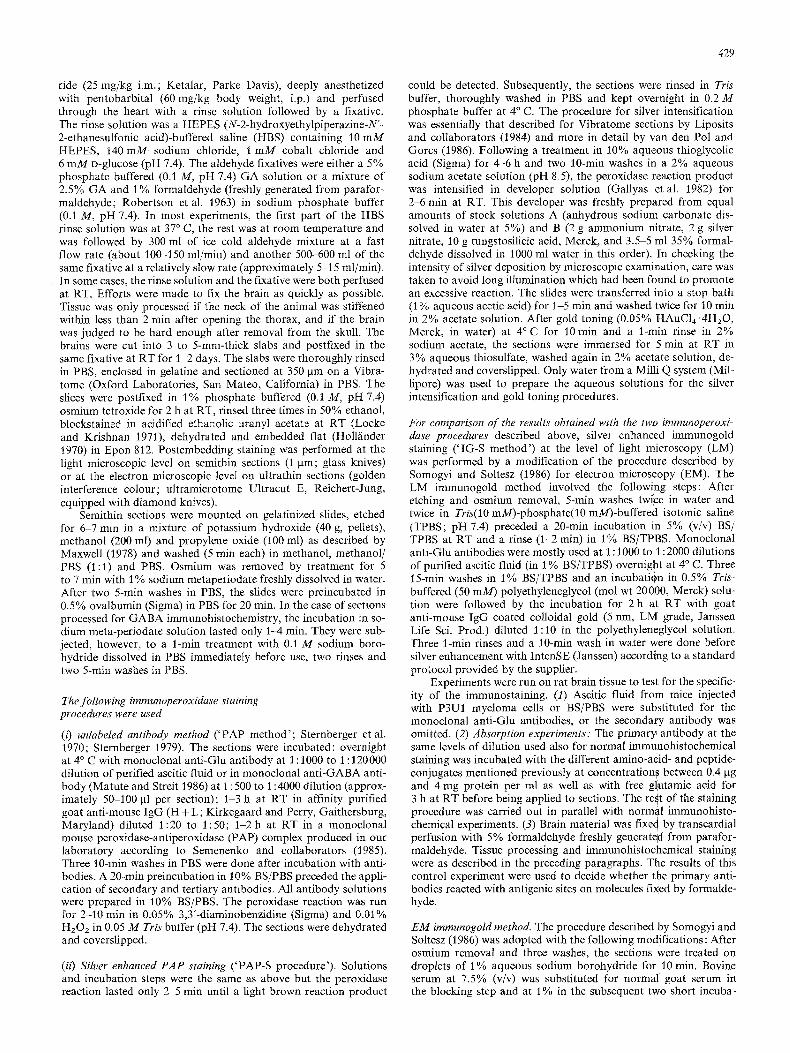
Fig. 1 A and B. Immunoreactivity of monoclonal 'anti-Glu' antibody 2D7 with BSA conjugates in ELISA plates. A Serial dilutions of 'purified ascitic fluid' containing mAb 2D7. Antigens applied to ELISA plates at concentration of 0.1 pg BSA per well. Protein concentration of 'purified ascitic fluid': 3.2 mg/ml. D: factor of antibody dilution. B Immunoreactivity of mAb 2D7 ('purified ascitic fluid' diluted at l : t 5000) with glutamate-BSA conjugate (0.6 Ixg BSA/well) following preincubation with BSA-conjugates listed. Estimates of cross-reactivity calculated from values at 50% level appear in text. - Each point in A and B represents an average derived from three independent experiments each performed in triplicate
tions as well as in the treatment with the primary antibody. The monoclonal anti-Glu antibody was used as a 1:200 to 1 : 1000 dilu- tion of purified ascitic fluid at 4 ~ C overnight. The goat anti-mouse IgG coated colloidal gold (15 nm; Janssen) diluted usually to 1:10 was incubated for 2 h. - After uranyl and lead contrasting, the sections were examined in a Philips EM 201 electron microscope.
R e s u l t s
Characterization of antibodies
The differential screening procedure used in the present s tudy al lowed to select four cell lines since the pr imary hybr idomas , the pr imary and the secondary clones secreted ant ibodies which showed a s trong immunoreact iv i ty on Glu-BSA coated E L I S A plates but only background values on Asp-BSA. One of them, 2D7, had been obtained follow- ing the immuniza t ion procedure described for brown mice, another one, 2G4, by that involving ' pa r t i a l immunosup- press ion ' and the rest, 6C9 and 2C9, by the so-called ' s ho r t immuniza t ion ' procedure. Monoc lona l an t ibody 2D7 was found to be an IgG1, while the other three hybr idomas produced IgM antibodies. The immunoreact iv i ty for GIu- BSA was most intense with m A b 2D7 in E L I S A tests. This ant ibody, therefore, was chosen to be characterized further and appl ied for immunohis tochemical experiments.
In ant ibody di lut ion experiments, more than a thousand times and more than several thousand times higher concen- t ra t ions of m A b 2D7 had to be used on Tau-BSA and on the other amino-acid- and pept ide-BSA conjugates, re- spectively, in order to reach similar levels of immunoreac- tivity as on Glu -BSA (Fig. 1 A). At the di lut ion of purified ascitic fluid which yielded the highest level on Glu-BSA, i.e. at 1 : 10, the reactivity for the other conjugates was still less than half of this maximum. More accurate values than those given above, therefore, could not be obtained in this type of cross-reactivity test per formed as ELISA. Similar
results were obta ined on GA- l inked conjugates of amino acids and peptides with rat bra in mater ia l but a somewhat higher level of cross-reactivity was found (data not shown). Estimates derived from absorpt ion experiments showed that the maximal immunoreact iv i ty on Glu-BSA coated E L I S A plates could be reduced to 50% by preincubat ion of m A b 2D7 (1:15000 dilut ion of purified ascitic fluid) with Glu-BSA at about 0.8 gg protein per ml, but with other conjugates only at concentrat ions increased by the following factors: > 300 for HCA-BSA, >650 for 7-GG- BSA, >1400 for Gln-BSA, >1750 for GSH-BSA, >2100 for CA-BSA, > 2800 for Tau-BSA, > 3500 for G A B A - B S A and > 4400 for Asp- and B L A G - B S A (Fig. 1 B). Wi th the other conjugates tested, CSA, fl-Ala- and Gly-BSA, a reduc- t ion of 50% could not even be reached at a concentrat ion
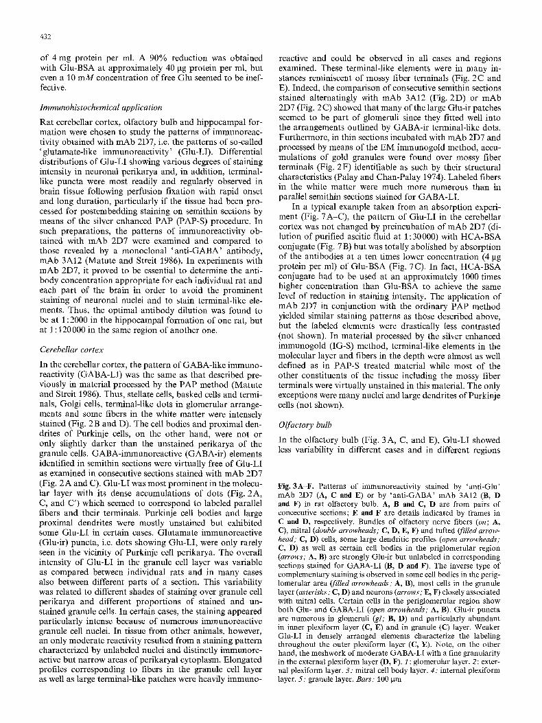
Fig. 2A-F. Immunostaining in rat cerebellar cortex produced by 'anti-Glu' mAb 2D7 (A, C, C', E and F) or by 'anti-GABA' mAb 3A12 (B and D). A, B (overview) and C, D (details) are from a pair of consecutive semithin sections. C and C' are enlargements of areas indicated in A and C, respectively. A and B Note drastic difference in labeling patterns obtained with the two antibodies. C: frame indicating part of area shown enlarged in C. gr: granule cell layer. P. Purkinje cell layer, mol: molecular layer. C and D Complementary labeling in stellate cells (asterisks), Golgi cell (ar- rows), pinceau formed by basket cell terminals (double arrowheads). Mossy fiber terminal-like structures in C (arrowhead) fit into glo- merular arrangements outlined by dots in D (arrowhead). C' Dense- ly packed pucta probably represent parallel fiber terminals in mo- lecular layer. E Numerous strongly stained patches (arrowheads) and some fibers (arrow) are reminiscent of mossy fiber terminals. Granule cells with unlabeled nuclei appear less immunoreactive than those in C. F Large mossy fiber terminal (m/t) with several synaptic contacts (arrows) shows higher surface density of gold granules (EM immunogold procedure) than another terminal nearby (asterisk) or granule cell body (gre). Bars: 100 lain in B, D and E; 1 ~tm in F

Fra
vail
du
labo
rato
ire
de m
icro
scop
ie ~
lect
roni
que
du P
rofe
sseu
r R
AO
VL
KO
Vl~
ILSK
Y,
I-[o
toita
l Sai
nt A
ntoi
ne, 1
)ari
s X
II~
Les
tum
eurs
car
cino
ides
bro
nchi
ques
et
dige
stiv
es d
e l'h
omm
e
432
of 4 mg protein per ml. A 90% reduction was obtained with Glu-BSA at approximately 40 lag protein per ml, but even a 10 m M concentration of free Glu seemed to be inef- fective.
Immunohistochemical application
Rat cerebellar cortex, olfactory bulb and hippocampal for- mation were chosen to study the patterns of immunoreac- tivity obtained with mAb 2D7, i.e. the patterns of so-called 'glutamate-like immunoreactivity' (Glu-LI). Differential distributions of Glu-LI showing various degrees of staining intensity in neuronal perikarya and, in addition, terminal- like puncta were most readily and regularly observed in brain tissue following perfusion fixation with rapid onset and long duration, particularly if the tissue had been pro- cessed for postembedding staining on semithin sections by means of the silver enhanced PAP (PAP-S) procedure. In such preparations, the patterns of immunoreactivity ob- tained with mAb 2D7 were examined and compared to those revealed by a monoclonal ' an t i -GABA' antibody, mAb 3A12 (Matute and Streit 1986). In experiments with mAb 2D7, it proved to be essential to determine the anti- body concentration appropriate for each individual rat and each part of the brain in order to avoid the prominent staining of neuronal nuclei and to stain terminal-like ele- ments. Thus, the optimal antibody dilution was found to be at 1 : 2000 in the hippocampal formation of one rat, but at 1 : 120000 in the same region of another one.
Cerebellar cortex
In the cerebellar cortex, the pattern of GABA-like immuno- reactivity (GABA-LI) was the same as that described pre- viously in material processed by the PAP method (Matute and Streit 1986). Thus, stellate cells, basked cells and termi- nals, Golgi cells, terminal-like dots in glomerular arrange- ments and some fibers in the white matter were intensely stained (Fig. 2 B and D). The cell bodies and proximal den- drites of Purkinje cells, on the other hand, were not or only slightly darker than the unstained perikarya of the granule cells. GABA-immunoreactive (GABA-ir) elements identified in semithin sections were virtually free of Glu-LI as examined in consecutive sections stained with mAb 2D7 (Fig. 2A and C). Glu-LI was most prominent in the molecu- lar layer with its dense accumulations of dots (Fig. 2A, C, and C') which seemed to correspond to labeled parallel fibers and their terminals. Purkinje cell bodies and large proximal dendrites were mostly unstained but exhibited some Glu-LI in certain cases. Glutamate immunoreactive (Glu-ir) puncta, i.e. dots showing Glu-LI, were only rarely seen in the vicinity of Purkinje cell perikarya. The overall intensity of Glu-LI in the granule cell layer was variable as compared between individual rats and in many cases also between different parts of a section. This variability was related to different shades of staining over granule cell perikarya and different proportions of stained and un- stained granule cells. In certain cases, the staining appeared particularly intense because of numerous immunoreactive granule cell nuclei. In tissue from other animals, however, an only moderate reactivity resulted from a staining pattern characterized by unlabeled nuclei and distinctly immunore- active but narrow areas of perikaryal cytoplasm. Elongated profiles corresponding to fibers in the granule cell layer as well as large terminal-like patches were heavily immuno-
reactive and could be observed in all cases and regions examined. These terminal-like elements were in many in- stances reminiscent of mossy fiber terminals (Fig. 2C and E). Indeed, the comparison of consecutive semithin sections stained alternatingly with mAb 3A12 (Fig. 2D) or mAb 2D7 (Fig. 2C) showed that many of the large Glu-ir patches seemed to be part of glomeruli since they fitted well into the arrangements outlined by GABA-ir terminal-like dots. Furthermore, in thin sections incubated with mAb 2D7 and processed by means of the EM immunogold method, accu- mulations of gold granules were found over mossy fiber terminals (Fig. 2 F) identifiable as such by their structural characteristics (Palay and Chan-Palay 1974). Labeled fibers in the white matter were much more numerous than in parallel semithin sections stained for GABA-LI.
In a typical example taken from an absorption experi- ment (Fig. 7A-C), the pattern of Glu-LI in the cerebellar cortex was not changed by preincubation of mAb 2D7 (di- lution of purified ascitic fluid at 1:30000) with HCA-BSA conjugate (Fig. 7 B) but was totally abolished by absorption of the antibodies at a ten times lower concentration (4 lag protein per ml) of Glu-BSA (Fig. 7 C). In fact, HCA-BSA conjugate had to be used at an approximately 1000 times higher concentration than Glu-BSA to achieve the same level of reduction in staining intensity. The application of mAb 2D7 in conjunction with the ordinary PAP method yielded similar staining patterns as those described above, but the labeled elements were drastically less contrasted (not shown). In material processed by the silver enhanced immunogold (IG-S) method, terminal-like elements in the molecular layer and fibers in the depth were almost as well defined as in PAP-S treated material while most of the other constituents of the tissue including the mossy fiber terminals were virtually unstained in this material. The only exceptions were many nuclei and large dendrites of Purkinje cells (not shown).
Olfactory bulb
In the olfactory bulb (Fig. 3 A, C, and E), Glu-LI showed less variability in different cases and in different regions
Fig. 3A-F. Patterns of immunoreactivity stained by 'anti-Glu' mAb 2D7 (A, C and E) or by 'anti-GABA' mAb 3A12 (B, D and F) in rat olfactory bulb. A, B and C, D are from pairs of concecutive sections; E and F are details indicated by frames in C and D, respectively. Bundles of olfactory nerve fibers (on; A, C), mitral (double arrowheads; C, D, E, F) and tufted (filled arrow- head; C, D) cells, some large dendritic profiles (open arrowheads; C, D) as well as certain cell bodies in the priglomerular region (arrows; A, B) are strongly Glu-ir but unlabeled in corresponding sections stained for GABA-LI (B, D and F). The inverse type of complementary staining is observed in some cell bodies in the perig- lomerular area (filled arrowheads," A, B), most cells in the granule layer (asterisks; C, D) and neurons (arrows; E, F) closely associated with mitral cells. Certain cells in the periglomerular region show both Glu- and GABA-LI (open arrowheads'; A, B). Glu-ir puncta are numerous in glomeruli (gl; B, D) and particularly abundant in inner plexiform layer (C, E) and in granule (C) layer. Weaker Glu-LI in densely arranged elements characterize the labeling throughout the outer plexiform layer (C, E). Note, on the other hand, the meshwork of moderate GABA-LI with a fine granularity in the external plexiform layer (D, F). 1 : glomerular layer. 2: exter- nal plexiform layer. 3: mitral cell body layer. 4: internal plexiform layer. 5: granule layer. Bars: 100 gm

433

434
of the same section than that noted in the cerebellar cortex and particularly in the hippocampus. However, the staining in bundles of olfactory nerve fibers and what seemed to be their terminal ramifications within the glomeruli was variable in our material (Fig. 3A and C). On the other hand, there were many small, moderately immunoreactive profiles in all glomeruli (Fig. 3 C). Some distinctly labeled cell bodies in the periglomerular areas and strongly Glu-ir terminal-like elements in these regions were regularly ob- served (Fig. 3 A and C). In the external plexiform layer with its considerable reactivity composed of densely arranged elements, a fine granularity could be observed in the outer half while in the inner part some large dendritic profiles were found (Fig. 3 C). This labeling pattern seemed to corre- spond to staining in certain mitral cell dendrites. Immuno- reactive perikarya of tufted cells were to be detected in various locations of this layer (Fig. 3 C). Glu-LI in the mi- tral cell layer was composed of large well-stained perikarya and strongly labeled dots in the spaces between them (Fig. 3E). The population of closely associated small cell bodies showed no immunoreactivity. Glu-ir dots (Fig. 3 E) and fibrous profiles were seen in the internal plexiform layer. Labeled dots were abundant in the granule layer, whereas only few perikarya were stained (Fig. 3 C). The pattern of GABA-LI (Fig. 3 B, D, and F) contrasted drasti- cally from that described for Glu-LI. However, some excep- tions were to be noted since certain GABA-ir cell bodies in the periglomerular region were also stained by mAb 2D7 (Fig. 3A and B). While olfactory nerve fibers were un- stained, GABA-LI was observed as terminal-like dots in the glomeruli and as many small perikarya closely asso- ciated with these structures (Fig. 3 B and D). A moderately immunoreactive meshwork with an interspersed fine granu- larity and a few stained small cell bodies characterized the labeling pattern in the external plexiform layer (Fig. 3D and F). The most prominent GABA-ir elements in the inter- nal plexiform layer were radial processes (Fig. 3 F) which often could be followed into the granule layer. There, perik- aryal staining was mostly moderate, but many strongly im- munoreactive cell bodies and puncta could be found too (Fig. 3 D). Perikarya showing little reaction product were rare.
Hippocampal formation
Several aspects of the pattern of Glu-LI in the hippocampal formation was strinkingly variable in the different cases examined. Thus, there were differences in terms of staining intensity, location and proportion of labeled perikarya of pyramidal neurons in the CA3 region of the hippocampus proper (Figs. 4A-D, 5A, B, and 7D). In addition, large immunoreactive dots in this area were more prominent in some (Figs 4H, and 5A, B) as compared to other cases (Fig. 4F). These dots probably represented labeled mossy fiber terminals since such structures with a suprapyramidal location could be shown to contain Glu-LI by means of the EM immunogold procedure (Fig. 5 C). Another struc- ture exhibiting a variable staining pattern was the area den- tata (Figs. 4A-D, 6A, B, D, and 7D). The staining intensity and the proportion of labeled perikarya were variable in the granule cell layer, but at its inner margin there were unlabeled cells in all cases. Variability between different rats was also noticed with respect to the density and labeling intensity of small Glu-ir puncta which were arranged rough-
ly in three bands within the molecular layer and which possibly represented labeled terminals of associational and entorhinal afferents. In the hippocampal CA1 region, on the other hand, the abundant granular immunoreactivity in the stratum radiatum, which seemed to be distributed over the termination fields of associational and/or com- missural pathways, and the less numerous but distinct dots in the stratum lacunosum moleculare made up a comparati- vely constant pattern of Glu-LI in our material (Figs. 4 A - D, 6A, B, and 7D). Glu-ir puncta were also relatively fre- quent in the stratum oriens of the CA1 region (Fig. 6B and C). Perikarya and large dendritic profiles of CA1 pyra- midal cells mostly showed little or no staining (Figs. 4A D, 5A-C, and 7D). The overall pattern of Glu-L! was not changed by preincubation of mAb 2D7 (at a 1:16000 dilu- tion of purified ascitic fluid) with HCA-BSA conjugate (Fig. 7 E), but was completely abolished by absorption of the antibodies with a 10 times lower concentration of Glu- BSA (4 gg protein/ml; Fig. 7 F). As in the experiment on material from the cerebellar cortex, absorption tests on tis- sue from the hippocampal formation showed that HCA- BSA conjugate had to be used at an approximately 1000 times higher concentration than Glu-BSA to reach a similar level of reduction in staining intensity. The compari- son of the labeling patterns obtained with the 'ant i -Glu ' mAb 2D7 (Fig. 4 D, F, and H) and the ' an t i -GABA' mAb 3A12 (Fig. 4E, G, and I) demonstrated that the labeling of perikarya and terminal-like dots, in general, was drasti- cally different. However, the level of resolution reached in semithin (1 gm) sections did not allow to exclude complete- ly that both immunoreactivities were present in certain mossy fiber terminals. In several cases, furthermore, a rela- tively high level of GABA-LI was observed in fiber bundles within the stratum lucidum but it was not possible to decide whether they corresponded to bundles of mossy fibers. GABA-ir terminal-like elements, on the other hand, were relatively rare in the stratum lucidum but highly concen- trated around the perikarya of CA3 pyramidal cells (Fig. 4 G and I). Other areas with high densities of labeled dots in the surroundings of cell bodies were the CA1 pyra- midal cell layer and the granule cell layer of the fascia den- tata (Fig. 4 E). Terminal-like puncta containing GABA-LI were less frequent but still prominent in the molecular layer of the fascia dentata as well as the stratum lacunosum mole- culare. Immunoreactive perikarya were most dense in the innermost layer of the granule cells, while a relatively large population was intermingled with the perikarya of pyrami- dal neurons in the hippocampal CA1 and CA3 regions.
Fig. 4A-I. Distribution ofimmunoreactivity observed in rat hippo- campal formation with 'anti-Glu' mAb 2D7 (A-D, F and H) or with 'anti-GABA' mAb 3A12 (E, G and I). Note different staining patterns in different rats (A-D, 7D) particularly in molecular (m) and granule cell (g) layers of area dentata as well as in pyramidal cell layer (between white dashes) of hippocampal CA3 region r: stratum radiatum of CAt. Drastic differences of labeling patterns in D and E (asterisks on blood vessels as landmarks) as well as in F, G and H, I from pairs of consecutive sections. Large Glu-ir dots (circles in F and H) mostly in suprapyramidal area of CA3 region and more numerous but smaller GABA-ir puncta (filled arrowheads; F-I) especially around pyramidal cell perikarya. La- beled cell bodies in G are unstained in F and G (open arrowheads). Same magnifications in A-D and in F-I. Bars: 100 gm in C; 25 gm in I

435

436

437
GABA-ir perikarya could readily be found also in the hilar region, in the stratum lucidum and in the stratum radiatum of CAl. Not all labeled cell bodies showed the same staining intensity, and these variations were observed in all areas of the hippocampal formation examined.
In formaldehyde fixed material as well as in experiments in which the primary or secondary antibodies had been omitted or in which the primary antibody had been replaced by ascitic fluid from mice injected with myeloma cells, the levels of immunoreactivity were as low (not shown) as in cases in which mAb 2D7 had been preabsorbed with Glu- BSA.
Discussion
In the course of the present study, a monoclonal antibody, mAb 2D7, which was selected by a differential screening procedure, was found to exhibit a preference for fixative- linked glutamate and, in combination with a sensitive stain- ing method, was used in three regions of the rat brain to describe the various levels of immunoreactivity in tissue components such as neuronal perikarya and terminal-like elements. The two main issues to be discussed were those regarding the specificity of the antibody for glutamate and the transmitter specificity of the staining patterns observed. In addition, certain technical points and the variability of staining patterns were to be addressed.
A treatment of neural tissue with fixatives containing glutaraldehyde was known to result in the retention not only of glutamate but also of several established or putative amino acid neurotransmitters as well as of other chemically similar compounds. Transmitter related substances were ex- pected to be present at particularly high concentrations in certain locations such as synaptic terminals. It was, thus, essential to develop antibodies with a high specificity for fixative-linked glutamate. Since aspartate and glutamate were both known to occur in brain tissue in millimolar concentrations it was especially important to find anti- bodies which would distinguish well between these structur- ally similar acidic amino acids. This feature, therefore, was used as the main criterion in the screening procedure for the primary hybridomas. The antibodies, furthermore, had to show a strong difference between their reactivity for Glu- and for homocysteate-BSA, since L-HCA had recently be- come a neurotransmitter candidate (Do et al. 1986a, b) and since this sulphur-containing acidic amino acid was closely related to glutamate in its chemical structure. Only few anti- bodies satisfied these combined selection criteria. Amongst these, mAb 2D7 seemed to be the most potent one because it showed a strong immunoreactivity for Glu-BSA even at a high dilution of the ' purified ascitic fluid'.
Was the cross-reactivity of mAb 2D7 with the amino acid- and dipeptide-BSA conjugates used in our tests low enough to assure the specificity of the antibody for fixative-
Fig. 5A-C. Distribution of Glu-LI in hippocampal CA3 region. A and B Different staining intensity of pyramidal neurons from two different rats but strong labeling of large dots probably repre- senting mossy fiber terminals mostly in stratum lucidum (lu). Mod- erate immunoreactivity in bundles of mossy fibers (arrowheads; A). C Glu-ir (EM immunogold method) mossy fiber terminals (raft) with several synaptic contacts (arrows). Same magnification in A, B. Bars: 100 gm in B, t gm in C
linked Glu in immunohistochemical investigations? Since no real differences between mAb 2D7's reactivity with the conjugates other than Glu-BSA could be detected, and since levels of cross-reactivity could only be derived from extra- polated values, the results obtained in antibody dilution tests did not seem to be reliable. With respect to the prob- lem of cross-reactivity, the following information had pre- viously been obtained in a study on monoclonal 'anti- GABA' antibodies (Mature and Streit 1986). Already in that case, absorption experiments performed as ELISA tests had turned out to be more critical, i.e. the levels of cross~ reactivity obtained in absorption experiments were much higher than those determined in antibody dilution experi- ments. Calculations according to the presently used proce- dure set the levels of cross-reactivity of the monoclonal 'ant i -GABA' antibody 3A12 at <1:100 for fl-Ala, at < 1:275 for Glu and at < 1:700 for Gly. As the patterns of immunoreactivity observed in the present study with the monoclonal ' anti-GABA' antibody or with mAb 2D7 were clearly diflbrent and in several cases even complementary, it seemed that the cross-reactivity of mAb 3A12 with Glu of < 1:275 was low enough to avoid a generally mixed staining pattern. If a level of cross-reactivity at this order of magnitude was sufficiently low, and if conclusions about tolerable levels could be generalized to other antibodies and to other conjugates, it could be argued that the levels of cross-reactivity with HCA (< 1:300) as well as with the other amino acids and the dipeptides tested should have been low enough. It was interesting to note that in absorp- tion experiments performed on sections the level of cross- reactivity of mAb 2D7 for HCA was estimated to be even lower, namely at approximately 1 : 1000. More accurate and definitive values for the level of cross-reactivity with HCA, however, could not be obtained since this compound was not available in a radioactively labeled form and, thus, the molar ratio in the conjugates could not be determined. The second highest level of cross-reactivity of mAb 2D7 had been measured with gamma-glutamylglutamate (7-GG; < 1 : 650). The distribution of a potential synthesizing en- zyme for this dipeptide, gamma-glutamyl transpeptidase, had been described to be contained more in non-neuronal elements (Albert et al. 1966; Shine and Haber 1981) and, thus, was clearly different from the distribution of immuno- reactivity observed in our material. Since in investigations with a monoclonal antibody to y-GG (Madl et al. 1986) different histochemical methods had been used, more direct information on the distribution of the dipeptide was not available to decide about a potential contribution by cross- reactivity to the present patterns of immunoreactivity. It was unlikely, on the other hand, that a factor of < 1 : 2800 for taurine was too high, since Purkinje cell perikarya and dendrites had been shown to stain strongly with antisera to taurine (Madsen et al. 1985; Campistron et al. 1986b) while they were mostly unstained with mAb 2D7 in our material. A value of < 1:3500 for GABA was judged to be sufficiently low since, as stated above, the patterns of immunoreactivity observed with mAb 2D7 o r ' anti-GABA' antibody 3A12 were complementary in most locations ex- amined. From a comparison of the present labeling patterns with those described in investigations on the distribution of aspartate-like immunoreactivity (Ottersen and Storm- Mathisen 1985 ; Campistron et al. 1986 a; Saito et al. 1986; Madl et al. 1987; Yoshida et al. 1987; Aoki et al. 1987) one could have been tempted to conclude that at a level

438

439
Fig. 7A-F. Comparison of GIu-LI in consecutive sections (blood vessels with asterisks as landmarks) of cerebellar cortex (A-C) and hippocampal formation (D-F) using mAb 2D7 alone (A and D), this antibody preincubated with homocysteate-BSA conjugate (B and E), or with a ten times lower concentration of Glu-BSA conjugate (C and E). Note similar patterns of immunoreactivity in A and B or in D and E but compiete abolition of staining in C and F. Dilution of 'purified ascitic fluid' at 1:30000 in A-C and 1:16000 in D-F. Same magnification in A-F. Bar in F: 100 pm
Fig. 6A-E. Patters of Glu-LI in parts of hippocampal CA1 region and/or area dentata. A and B Montages of photomicrographs from area including parts of CAI and fascia dentata from two different cases with little labeling in CA1 pyramidal cells (p) and relatively weakly stained perikarya in granule cell layer (g). A few CA1 pyramidal neurons (C) and numerous granule cells (D) are strongly labeled in another animal. Fine terminal-like dots are densely packed in stratum oriens (o; C) and stratum radiatum (r; A, B and C) of CA1, numerous also in dentate molecular layer (m; A and B) and less impressive in stratum lacunosum moleculare (lm; A and B). E Large dots in area of CA3 pyramidal cells and smaller ones in infragranular region (between white dashes) probably represent Glu-LI in different types of mossy fiber terminals. Same magnification in A-E. Bar in E: 100 gm

440
of cross-reactivity of < 1 : 4400 for aspartate this substance was not detected in our immunohistochemical preparations. However, more definitive answers to the question of tolera- ble levels of cross-reactivity for the various compounds used in the present tests would have to be derived from studies involving consecutive semithin sections and a whole series of antibodies with different specificities. As in all other im- munohistochemical investigations, on the other hand, the interpretation with respect to the chemical nature of the material recognized in the tissue by the antibody was mostly limited by the possibility that the antibody cross-reacted with an unknown substance or with a substance which had not been included in the tests. The dipeptide N-acetylaspar- tylglutamate (NAAG) had been found in relatively high concentrations in the central nervous system (Curatolo et al. 1965; Reichelt and Fonnum 1969; Miyake et al. 1981 ; Zaczek et al. 1983; Koller et al. 1984). This compound al- though not used in our tests should, however, not have contributed to the patterns of immunoreactivity in the pres- ent work since the N-acetylated neurotransmitter candidate should not have been retained in the tissue by the fixatives applied. In fact, we had been unable to couple tritiated NAAG to BSA with glutaraldehyde as linker. Furthermore, the multitude of unknown compounds which should have been retained by formaldehyde fixation were not recognized either by mAb 2D7 as demonstrated by the total absence of staining in such material with the immunohistochemical protocol used. The fact that free Glu even at a concentra- tion of 10 m M had not been effective in absorption experi- ments seemed to indicate that the epitope recognized by mAb 2D7 did not only consist of Glu residues but possibly of the glutaraldehyde-glutamate complex as such. Although a high degree of specificity for glutaraldebyde-linked Glu was determined in absorption experiments and was sup- ported by the arguments presented above, the material de- tected by mAb 2D7 could only be defined a s ' glutamate-like immunoreactivity' (Glu-LI). It, nevertheless, seemed to be justified to call mAb 2D7 an 'ant i -Glu ' antibody.
How do the patterns of immunoreactivity observed in cerebellar cortex, olfactory bulb and hippocampal forma- tion correspond to the transmitter specificity of the immun- ostained tissue components?
In the cerebellar cortex, the staining patterns obtained with the ' an t i -GABA' mAb 3A12 and the silver enhanced PAP method were the same as those described in previous studies (Ottersen and Storm-Mathisen 1984; Seguela et al. 1984; Somogyi et al. 1985; Matute and Streit 1986) for the inhibitory elements in the cerebellum which had also been reported to be immunoreactive for the GABA-synthesizing enzyme glutamic acid decarboxylase (for review: Mugnaini and Oertel 1985). The cerebellar constituents showing the most impressive Glu-LI were the densely packed dots inter- preted as stained parallel fiber terminals and the large patches probably representing labeled mossy fiber termi- nals, while granule cell perikarya were only moderately im- munoreactive, cell bodies and dendrites of Purkinje cells were almost unlabeled, and the strongly GABA-ir cell types as well as glial cells were practically unstained. This order of staining intensity seemed to agree well with that deter- mined by Somogyi and collaborators (Somogyi et al. 1986) in a quantitative study at the electron microscopic level on components of the cerebellar cortex in the cat. From the analysis of consecutive ultrathin sections stained alter- natingly with ' an t i -GABA' or ' ant i -Glu ' antisera it had
become clear that GABA-ir terminals contained outstand- ingly low levels of Glu-LI and, thus, a low metabolic gluta- mate pool. The high level of Glu-LI in parallel and mossy fiber terminals, furthermore, was consistent with results previously obtained in biochemical investigations (Young etal. 1974; Valcana etal. 1972; Hudson etal. 1976; McBride et al. 1976; Sandoval and Cotman 1978; Rohde et al. 1979; Roffler-Tarlov and Turey 1982) and with a sug- gested role of glutamate as neurotransmitter at parallel (Chujo et al. 1975; Crepel etal. 1982; Gallo etal . 1982; Levi et al. 1982; Sandoval and Cotman 1978; Stone 1979) and mossy (Garthwaite 1986; Holm and Logan 1986) fiber synapses. Whether some of the immunoreactivity in the mo- lecular layer was related to labeling of climbing fibers could not be decided at the level of resolution in the present inves- tigation. On the other hand, most available evidence pointed to a transmitter role of aspartate at least in part of these cerebellar afferents while the situation in the case of glutamate appeared less clear (for review: Cu6nod et al. 1988).
As noted also by Ottersen and Storm-Mathisen (1984), the patterns of Glu- and GABA-LI in the olfactory bulb were to a large extent complementary. Indeed, mitral and tufted cells as well as a certain subpopulation of perikarya in the periglomerular region were Glu- but not GABA-ir, while another portion of cells in the periglomerular area and the granule cells showed the opposite reactivity. We were unable to distinguish between periglomerular neurons and superficial tufted cells in our material consisting of semithin sections. It was interesting to notice, on the other hand, that a third population of perikarya in the periglo- merular region showed staining with both antibodies in con- secutive semithin sections since GABA-ir cell types had usu- ally been found to exhibit very low levels of Glu-LI (see discussion in Ottersen and Storm-Mathisen 1984). Strong Glu-LI in GABA-ir perikarya, however, could not easily be interpreted in terms of a marker for transmitter specifici- ty. In fact, concern had previously been expressed about the use of high perikaryal Glu-LI as marker for glutama- tergic neurons, since cholinergic and dopaminergic neurons in the brain stem stained rather strongly with 'ant i -Glu ' antisera (Ottersen and Storm-Mathisen 1984, 1985; Storm- Mathisen and Ottersen 1986). Furthermore, in recent stain- ing experiments with mAb 2D7, rat spinal motoneurons showed relatively high Glu-LI (unpublished observation) although these neurons were known to use acetylcholine rather than Glu as transmitter. Several indications had, nev- ertheless, been obtained for the use of aspartate or gluta- mate as transmitter(s) by mitral cells (Harvey et al. 1975; Bradford and Richards 1976; Yamamoto and Matsui 1976; Scholfield etal. 1983; for review: Halasz and Shepherd 1983; Macrides and Davis 1983). While the results of cer- tain neurochemical (Collins 1979a, b; Collins and Probett 1981) and immunohistochemical (Saito et al. 1986) investi- gations seemed to stress a transmitter role for aspartate rather than for glutamate at least in some of the fibers originating from mitral cells and running in the lateral ol- factory tract, such a role had been questioned for both acidic amino acids by data acquired in pharmacological studies (Hori et al. 1981, 1982). Recently, N A A G became a transmitter candidate of the lateral olfactory tract (ffrench-Mullen et al. 1985) and mitral cells were shown to contain NAAG-LI (Anderson et al. 1986; Blakely et al. 1987). However, a potentially existing cross-reactivity of

44:1
mAb 2D7 should not have induced false positive staining in our material since, as pointed out in a previous section, this N-acetylated neuroactive dipeptide should not have been retained in the tissue by the fixatives used.
The dipeptide carnosine (fl-alanyl-L-histidine) seemed to be associated with the peripheral afferents to the olfactory bulb (Margolis 1974; for review: Macrides and Davis 1983). A potential cross-reactivity of an 'anti-GABA' antiserum with carnosine had been suggested to be responsible for immunoreactivity observed in the olfactory nerve layer (Ot- tersen and Storm-Mathisen 1984). No GABA-LI was dis- covered within this region with the 'anti-GABA' mAb 3A12 in the present investigation. There was no ready ex- planation for the fact that only an interindividually variable fraction of olfactory nerve bundles and their terminal ar- borizations in glomeruli contained heavy Glu-LI. This type of immunoreactivity in olfactory nerve fibers was interest- ing to find since Ottersen and Storm-Mathisen (1984) had shown heavy autoradiographic labeling for glomeruli in up- take experiments 'in vitro' with tritiated D-aspartate. Equally strong labeling had also been observed over ele- ments in the spaces between patches of granule cells (Otter- sen and Storm-Mathisen 1984) and, thus, in regions where abundant, strongly Glu-ir but unidentified puncta were lo- cated in the present material. Watanabe and Kawana (1984) had described selective retrograde labeling with tritiated D- aspartate from the olfactory bulb to the anterior olfactory nucleus, pyriform cortex and nucleus of the lateral olfactory tract. As a selective retrograde tracer, this compound had proved to be a rather reliable marker for pathways using an excitatory amino acid as transmitter (for review: Cu6nod and Streit 1983). On the other hand, the labeling patterns obtained in anterograde tracing experiments had shown that the granule layer was an important termination site for the three pathways mentioned above (for review: Mac- rides and Davis 1983). It, thus, seemed likely that at least a certain fraction of the Glu-ir dots in the granule layer represented terminals of such centrifugal afferents and that, in turn, the distinct Glu-LI in such terminals corresponded to a high glutamate pool involved in neurotransmission.
The findings in pharmacological and electrophysiologi- cal (for review: Halasz and Shepherd 1983) as well as in histochemical (Halasz et al. 1981; Ribak et al. 1977; Otter- sen and Storm-Mathisen 1984; Mugnaini and Oertel 1985) studies left little doubt that the granule cells used GABA as neurotransmitter at their inhibitory synapses with the mitral cells. Immunohistochemical data, furthermore, seemed to indicate the GABAergic nature of certain cells in the periglomerular region (Mugnaini and Oertel 1985; Ottersen and Storm-Mathisen 1984; Kosaka et al. 1985, 1987; Gall et al. 1987). The observations in the present in- vestigation on GABA-ir perikarya in the periglomerular area and on GABA-LI in granule cells and, in addition, in a dense meshwork within the outer plexiform layer seemed to be in agreement with these previous results.
The GABA-LI described in the present material for the hippocampalformation did not vary much from animal to animal and was mostly consistent with available knowledge on the GABAergic systems in this part of the brain (for review: Mugnaini and Oertel 1985; Ottersen and Storm- Mathisen 1984). On the other hand, an even more striking interindividual variability than that described in olfactory nerve bundles and their glomerular terminations was no- ticed for the distribution of Glu-LI in certain parts of the
hippocampal formation. The factors causing this variability could not be determined but might have to be attributed to interindividual or local differences in the efficacy of per- fusion-fixation or, more interestingly, to differences in pre- mortal neural activity, which seemed to involve relatively large groups of neurons or certain pathways. The perikarya of hippocampal CA3 pyramidal neurons and of granule cells in the area dentata were such particularly conspicuous groups. However, as discussed in a previous section, the perikaryal level of Glu-LI could not easily be interpreted with respect to transmitter specificity. A relationship be- tween variable Glu-LI and certain pathways was suggested by the staining patterns in the molecular layer of the fascia dentata. The variable staining intensity was determined by the different densities of Glu-ir terminal-like dots which were located in the inner, intervening and outer zones. The inner zone appeared to correspond to the termination area of associational and commissural inputs (Fricke and Cowan 1978; Hjorth-Simonsen 1973; Hjort-Simonsen and Laur- berg 1977; Laurberg 1979; Laurberg and Sorensen 1981; Swanson and Cowan 1977; Swanson et al. 1978; Zimmer 1971) while the intervening and the outer zones seemed to represent projection areas of the medial and the lateral perforant paths (Hjorth-Simonsen 1972; Hjorth-Simonsen and Jeune 1972; Steward 1976).
Particularly the results obtained in combined lesion and release experiments indicated that transmission by terminals at least of comrnissural fibers in the inner zone of the molec- ular layer was mediated by aspartate rather than by gluta- mate (Nadler et al. 1976, 1978). However, i twas not possi- ble to decide at present about the nature of the numerous Glu-ir dots in this region. The entorhinal perforant path axons, on the other hand, were probably the best document- ed example in the mammalian brain of a pathway using glutamate as neurotransmitter (for review: Cotman et al. 1981; Storm-Mathisen 1977a; Walaas 1983; Ottersen and Storm-Mathisen 1984, 1986). This conclusion had been de- rived mostly from observations in autoradiographic (Storm-Mathisen 1977b, 1981; Taxt and Storm-Mathisen 1984) and biochemical (Nadler et al. 1976, 1978; Storm- Mathisen 1977b, 1981) uptake, release (Nadler et al. 1976, 1978; Hamberger et al. 1978, 1979; Dolphin et al. 1982) and transmitter related retrograde labeling(Fischer et al. 1986) studies as well as in measurements of amino acid content in crude synaptosomal preparations following axo- tomy (Nadler and Smith 1981). In autoradiographic uptake experiments with tritiated L-aspartate, the labeling had been described to decrease from the inner to the intervening and to the outer zone of the dentate molecular layer (Taxt and Storm-Mathisen 1984). Glu-LI in slices of the hippocampal formation, which had been briefly soaked in Krebs' solution and immersion-fixed, appeared to be almost as intense in the outer as in the inner zone, whereas the intervening one was more weakly labeled (Storm-Mathisen et al. 1983, 1986; Ottersen and Storm-Mathisen 1985). The same pat- tern had been noticed in tissue from rats perfusion-fixed following a prolonged rinsing period (Yoshida et al. 1987). However, this pattern could also be observed in the present study in some but not all animals which had been flushed briefly and rapidly perfusion-fixed. It was difficult to know whether different labeling intensities observed in the termi- nation fields of medial and lateral perforant paths were related to physiological (McNaughton 1980) and pharma- cological (Harris et al. 1981 ; Lanthorn and Cotman 1981)

442
differences between the two perforant paths. As mentioned before, on the other hand, the pattern of Glu-LI in the dentate molecular layer was variable, and this variability might have reflected different functional states.
As in the case of puncta in the outer and intervening zones of the molecular layer in the dentate area, the relation between Glu-ir dots in the stratum radiatum of the hippo- campal CA1 region and CA3-derived associational and commissural axons would have to be determined by appro- priate lesion experiments. However, the labeling pattern in combination with an extensive literature on a role of gluta- mate as neurotransmitter in terminals of CA3 pyramidal neurons (for review: Walaas 1983; Ottersen and Storm- Mathisen 1984, 1986; Storm-Mathisen 1981; Cotman et al. 1981) suggested that e.g. at least part of the small but abun- dant Glu-ir dots in the CA1 stratum radiatum represented the transmitter pool of glutamate in terminals of Schaffer collaterals and of commissural CA3 axons. Strong Glu-LI in the stratum radiatum of CA1 had also been described by others in tissue subjected to certain rinsing procedures before fixation (Storm-Mathisen et al. 1983, 1986; Ottersen and Storm-Mathisen 1985; Yoshida et al. 1987).
Although the case for glutamate mediated neurotrans- mission at synapses of mossy fibers did not seem to be as strong as in the systems discussed above (for references see: Walaas 1983; Storm-Mathisen 1981; Cotman etal. 1981 ; Ottersen and Storm-Mathisen 1984, 1986), no label- ing of mossy fiber terminals with an 'ant i -Asp' antiserum (Ottersen and Storm-Mathisen 1985; Storm-Mathisen et al. 1986) but heavy labeling with an 'ant i -Glu ' antiserum had been described in rinsed and immersion-fixed slices (Storm- Mathisen et al. 1983, 1986; Ottersen and Storm-Mathisen 1985). While the staining obtained in our perfusion-fixed material was consistent with these previous findings, the stratum lucidum had been shown to be free of Glu-LI by others in tissue following a prolonged rinsing period and perfusion-fixation (Yoshida et al. 1987). This fixation pro- cedure was, thus, much slower than the one used in the present investigation which, in turn, should have retained the glutamate content of brain tissue at levels more closely resembling those of the ' in vivo' situation. However, even with the present procedure, fixation may have varied in different regions of the brain and in different individuals. As mentioned above, this variability of fixation may have been another reason besides differences of premortal activi- ty for the regional and interindividual variability of staining patterns.
Whether and at which proportion Glu-LI in mossy fiber terminals corresponded to a transmitter pool of glutamate could not easily be determined in the present study. How- ever, since the 'ant i -Glu ' mAb 2D7 stained mossy fiber terminals while the ' an t i -GABA' mAb 3A12 labeled what seemed to be a different set of terminal-like elements it became clear that at least not all terminals contained Glu-LI despite the fact that Glu-ir dots were numerous or abundant in most regions of the hippocampal formation. As men- tioned in the discussion on the findings in the cerebellar cortex, on the other hand, GABA-ir terminals had been shown to exhibit a very low Glu-LI (Somogyi et al. 1986) and, thus, to have a very low metabolic pool of glutamate. Only future comparative investigations with antibodies to various compounds might help to find out about the levels of Glu-LI representing the metabolic pool in nerve termi- nals which did not release glutamate but rather another
neuroactive substance. In view of the fact that Glu-ir termi- nal-like elements were numerous in termination fields of pathways allegedly using glutamate as transmitter, and that the transmitter pool of glutamate was expected to be high in glutamatergic nerve terminals, it was particularly inter- esting to note that such terminal-like dots, indeed, could be stained in perfusion-fixed tissue by mAb 2D7. The meth- od applied in the present study, therefore, might allow in the future and in combination with lesion experiments to map at the level of light microscopy pathways with gluta- mate-mediated neurotransmission.
The successful staining of terminal-like puncta seemed mostly to depend on the use of postembedding immunohis- tochemistry since, for unknown reasons, only labeling of perikarya and dendrites but not of terminal-like dots was observed with mAb 2D7 in immunostained Vibratome slices (unpublished data). Careful titration of the 'ant i -Glu ' antibody appeared to be another important point. Thus, the optimal antibody concentration varied amongst the dif- ferent cases possibly as a result of differences in the efficacy of perfusion-fixation. Furthermore, at inadequately high antibody concentrations, the labeling of certain perikarya and especially of terminal-like dots was suppressed possibly as a consequence of the so-called 'Bigbee effect' (Bigbee et al. 1977) which had been determined as one of the dis- turbing factors in the staining of ~ antigen-rich" tissue com- ponents by the peroxidase anti-peroxidase technique. Silver enhancement of the histochemical peroxidase reaction product obtained after a relatively short reaction time, fur- thermore, yielded a better contrast perhaps because staining was more directly proportional to the content of immunore- active material in different tissue elements.
In conclusion, the results obtained in the present study showed that the newly developed 'ant i -Glu ' mAb 2D7 in combination with a peroxidase anti-peroxidase procedure and silver enhancement revealed terminal-like elements at the light microscopic level in appropriately fixed rat brain material. In addition, the patterns of Glu-LI were mostly compatible with available data on systems using glutamate as neurotransmitter in the three brain regions examined.
Acknowledgements. This work was supported by grants 3.389.86 and 3.390.86 of the Swiss National Science Foundation and by the Dr. Eric Slaek-Gyr Foundation. Fellowships were received by Chang-jin Liu from the Eidgen6ssische Stipendienkommission ffir Auslfindische Studierende, by Pedro Grandes from the European Training Program for Brain and Behaviour Research grant STF/ 86/3812) and by Carlos Mature from IBRO (Swiss National Science Foundation, grant 83.161).
We wish to thank Marika Born, Henry J. Waldvogel, Hans Kiinzli, Ruth Emch and Birgit Hofer for their excellent assistance.
References
Albert Z, Orlowski M, Rzucidlo Z, Orlowska J (1966) Studies on gamma-glutamyl transpeptidase activity and its histochemi- cal localization in the central nervous system of man and differ- ent animal species. Acta Histochem 25 : 312-320
Anderson KJ, Monaghan DT, Cangro CB, Namboodiri MAA, Neale JH, Cotman CW (1986) Localization of N-Acetylaspar- tyl-glutamate-like immunoreactivity in selected areas of the rat brain. Neurosci Lett 72:14-20
Aoki E, Semba R, Kato K, Kashiwamata S (1987) Purification of specific antibody against aspartate and immunocytochemical localization of aspartergic neurons in the rat brain. Neuros- cience 21:755 765

443
Bigbee JW, Kosek JC, Eng LF (1977) Effects of primary antiserum dilution on staining of "antigen-rich" tissues with the peroxi- dase anti-peroxidase technique. J Histochem Cytochem 25:443-447
Blakely RD, Ory-Lavall6e L, Grzanna R, Koller K J, Coyle JT (1987) Selective immunocytochemical staining of mitral cells in rat olfactory bulb with affinity purified antibodies against N-acetayl-aspartyl-glutamate. Brain Res 402:373-378
Bradford HF, Richards CD (1976) Specific release of endogenous glutamate from piriform cortex stimulated in vitro. Brain Res 105:168-172
Campistron G, Buijs RM, Geffard M (1986a) Specific antibodies against aspartate and their immunocytochemical application in the rat brain. Brain Res 365:179-184
Campistron G, Geffard M, Buijs RM (1986b) Immunological ap- proach to the detection of taurine and immunocytochemical results. J Neurochem 46:862-868
Chujo T, Yamada Y, Yamamoto C (1975) Sensitivity of Purkinje cell dendrites to glutamic acid. Exp Brain Res 23 : 293-300
Collins GGS (1979 a) Evidence of a neurotransmitter role for aspar- tate and ~-aminobutyric acid in the rat olfactory cortex. J Phys- iol (London) 291 : 51-60
Collins GGS (1979b) Effect of chronic bulbectomy on the depth distribution of amino acid transmitter candidates in rat olfacto- ry cortex. Brain Res 171:55~555
Collins GGS, Probett GA (1981) Aspartate and not glutamate is the likely transmitter of the rat lateral olfactory tract fibres. Brain Res 209:231 234
Cotman CW, Foster A, Lanthorn T (1981) An overview of gluta- mate as a neurotransmitter. Adv Biochem Psychopharmacol 2:1-27
Crepel F, Dhanjal SS, Sears TA (1982) Effect of glutamate, aspar- tate and related derivatives on cerebellar Purkinje cell dendrites in the rat: an in vitro study. J Physiol (London) 329:297-317
Cu6nod M, Streit P (1983) Neuronal tracing using retrograde mi- gration of labeled transmitter-related compounds. In: Bj6rk- fund A, H6kfelt T (eds) Handbook of chemical neuroanatomy, vol 1. Elsevier, Amsterdam, pp 365-397
Cu6nod M, Do KQ, Vollenweider F, Klein A, Streit P (1988) The puzzle of the transmitters in the climbing fibers. In: Experimen- tal Brain Research Series (Symposium on "The olivo-cerebellar system in motor control", Turin). Springer, Berlin Heidelberg New York
Curatoio A, d'Archangelo P, Lino A (1965) Distribution of N- acetyl-aspartic and N-acetyi-aspartyl-glutamic acids in nervous tissue. J Neurochem 12:339-342
Do KQ, Herrling PL, Streit P, Turski WA, Cu6nod M (1986a) In vitro release and electrophysiological effects in situ of homo- cysteic acid, an endogenous N-methyl-(D)-aspartic acid agonist, in the mammalian striatum. J Neurosci 6:2226-2234
Do KQ, Mattenberger M, Streit P, Cu6nod M (1986b) In vitro release of endogenous excitatory sulfur-containing amino acids from various rat brain regions. J Neurochem 46:779-786
Dolphin AC, Errington MC, Bliss TVP (1982) Long-term potentia- tion of the perforant path in vivo is associated with increased glutamate release. Nature 297:496~98
Fagg GE, Foster AC (1983) Amino acid neurotransmitters and their pathways in the mammalian central nervous system. Neu- roscience 9:711-719
ffrench-Mullen JMH, Koller K, Zaczek R, Coyle JT, Hori N, Car- penter DO (1985) N-Acetylaspartylglutamate: possible role as the neurotransmitter of the lateral olfactory tract. Proc Natl Acad Sci USA 82:3897-3900
Fischer BO, Storm-Mathisen J, Ottersen OP (1986) Hippocampal excitatory neurons: anterograde and retrograde axonal trans- port of D-[3H]aspartate. In: Roberts PJ, Storm-Mathisen J, Bradford HF (eds) Excitatory amino acids. Macmillan, Lon- don, pp 442~443
Fonnum F (1984) Glutamate: a neurotransmitter in mammalian brain. J Neurochem 42:1-11
Fricke R, Cowan WM (1978) An autoradiographic study of the
commissural and ipsilateral hippocampo-dentate projections in the adult rat. J Comp Neurol 181:253-270
Gall CM, Hendry SHC, Seroogy KB, Jones EG, Haycock JW (1987) Evidence for coexistence of GABA and dopamine in neurons of the rat olfactory bulb. J Comp Neurol 266:307-318
Gallo V, Ciotti MT, Coletti A, Aloisi F, Levi G (1982) Selective release of glutamate from cerebellar granule cells differentiating in culture. Proc Natl Acad Sci USA 79 : 7919-7923
Gallyas F, G6rcs T, Merchenthaler I (1982) High-grade intensifica- tion of the end-product of diaminobenzidine reaction demon- strating peroxidase activity. J Histochem Cytochem 30:183-184
Garthwaite J (1986) Parallel-fibre- and mossy-fibre-mediated syn- aptic transmission in rat cerebellar slices monitored using a 'grease-gap' method. J Physiol (London) 371:16P
Geffard M, Henrich-Rock AM, Dulluc J, Seguela P (1985) Antisera against small neurotransmitter-like molecules. Neurochem Int 42:1 11
Halfisz N, Shepherd GM (1983) Neurochemistry of the vertebrate olfactory bulb. Neuroscience 10:579-619
Hal/lsz N, Parry DM, Blackett NM, Ljungdahl A, H6kfelt T (1981) [3H]7-Aminobutyrate autoradiography of the rat olfactory bulb. Hypothetical grain analysis of the distribution of silver grains. Neuroscience 6:473-479
Hamberger AC, Chiang GH, Nyl6n ES, Scheff SW, Cotman CW (1978) Stimulus evoked increase in the biosynthesis of the puta- tive neurotransmitter glutamate in the hippocampus. Brain Res 143:549 555
Hamberger AC, Chiang GH, Nyl6n ES, Scheff SW, Cotman CW (1979) Glutamate as a CNS transmitter. I. Evaluation of glu- cose and glutamine as precursors for the synthesis of preferen- tially released glutamate. Brain Res 168:513-530
Harris EW, Koerner JF, Cotman CW (1981) Effects of acidic ami- no acid derivatives on perforant path evoked responses. Soc Neurosci Abstr 7 : 807
Harvey AJ, Scholfield CN, Graham LT, Aprison MH (1975) Puta- tive transmitters in denervated olfactory cortex. J Neurochem 24: 445-449
Hepler JR, Petrusz P, Rustioni A (1986) Antisera to GABA, gluta- mate and aspartate: characterization by immunoabsorption and immunocytochemistry. J Histochem Cytochem 34:110
Hepler JR, Toomin C, McCarthy KD, Conti F, Battaglia G, Rus- tioni A, Petrusz P (1988) Characterization of antisera to gluta- mate and aspartate. J Histochem Cytochem 36:13-22
Hjorth-Simonsen A (1972) Projection of the lateral part of the entorhinal area to the hippocampus and fascia dentata. J Comp Neurol 146:219-231
Hjorth-Simonsen A (1973) Some intrinsic connections of the hippo- campus in the rat: an experimental analysis. J Comp Neurol 147:145-162
Hjorth-Simonsen A, Jeune B (1972) Origin and termination of the perforant path in the rat studied by silver impregnation. J Comp Neurol 144:215-232
Hjorth-Simonsen A, Laurberg S (1977) Commissural connections of the dentate area in the rat. J Comp Neurol 174:591-606
Hollfinder H (1970) The section embedding (SE) technique. A new method for the combined light microscopic and electron micro- scopic examination of central nervous tissue. Brain Res 20: 39-47
Holm JA, Logan SD (1986) Release of amino acids and a novel peptide from isolated fragments of rat cerebr glomerulus. J Physiol (London)373 : 18P
Hori N, Auker CR, Braitman DJ, Carpenter DO (1981) Lateral olfactory tract transmitter: glutamate, aspartate or neither? Cell Mol Neurobiol 1:115-120
Hori N, Auker CR, Braitman D J, Carpenter DO (1982) Pharmaco- logic sensitivity of amino acid responses and synaptic activation of in vitro prepyriform neurons. J Neurophysi01 48:128~1301
Hudson DB, Valcana T, Bean G, Timiras PS (1976) Glutamic acid: a strong candidate as the neurotransmitter of the cerebel- lar granule cell. Neurochem Res 1:73-81
Koller K J, Zaczek R, Coyle JT (1984) N-acetyl-aspartyl-glutamate:

444
regional levels in rat brain and the effects of brain lesions as determined by a new HPLC method. J Neurochem 43 : 1136-1142
Kosaka T, Hataguchi Y, Hama K, Nagatsu I, Wu J-Y (1985) Coexistence of immunoreactivities for glutamate decarboxylase and tyrosine hydroxylase in some neurons in the periglomerular region of the rat main olfactory bulb: possible coexistence of gamma-aminobutyric acid (GABA) and dopamine. Brain Res 343:166-171
Kosaka K, Hama K, Nagatsu I, Wu J-Y, Ottersen OP, Storm- Mathisen J, Kosaka T (1987) Postnatal development of neurons containing both catecholaminergic and GABAergic traits in the rat main olfactory bulb. Brain Res 403 : 355-360
Lanthorn TH, Cotman CW (1981) Baclofen selectively inhibits ex- citatory synaptic transmission in the hippocampus. Brain Res 225:171 178
Laurberg S (1979) Commissural and intrinsic connections of the rat hippocampus. J Comp Neurol 184:685-708
Laurberg S, Sorensen KE (1981) Associational and commissural collaterals of neurons in the hippocampal formation (hilus fas- ciae dentatae and subfield CA3). Brain Res 212:287-300
Levi G, Gordon RD, Gallo V, Wilkin GP, Balazs R (1982) Putative acidic amino acid transmitters in the cerebellum. I. Depolariza- tion-induced release. Brain Res 239:425-445
Liposits Zs, S~tfil6 Gy, Flerk6 B (1984) Application of the silver- gold intensified 3,3'-diaminobenzidine chromogen to the light and electron microscopic detection of the luteinizing hormone- releasing hormone system of the rat brain. Neuroscience 13:513-525
Locke M, Krishnan N (197t) Hot alcoholic phosphotungstic acid and uranyl acetate as routine stains for thick and thin sections. J Cell Biol 50:550-557
Macrides F, Davis BJ (1983) The olfactory bulb. In: Emson PC (ed) Chemcial neuroanatomy. Raven Press, New York, pp 391 426
Madl JE, Larson AA, Beitz AJ (1986) Monoclonal antibody specif- ic for carbodiimide-fixed glutamate: immunocytochemical lo- calization in the rat CNS. J Histochem Cytochem 34:312326
Madl JE, Beitz AJ, Johnson RL, Larson AA (1987) Monoclonal antibodies specific for fixative-modified aspartate: immunocy- tochemical localization in the rat CNS. J Neurosci 7:2639- 2650
Madsen S, Ottersen OP, Storm-Mathisen J (1985) Immunocyto- chemical visualization of taurine: neuronal localization in the rat cerebellum. Neurosci Lett 60:255-260
Margolis FL (1974) Carnosine in the primary olfactory pathway. Science 184: 909-911
Matthew WD, Patterson PH (1983) The production of a monoclon- al antibody that blocks the action of a neurite outgrowth- promoting factor. Cold Spring Harbor Symp Quant Biol 48: 625-63 t
Matute C, Streit P (1986) Monoclonal antibodies demonstrating GABA-like immunoreactivity. Histochemistry 86 : 147-157
Matute C, Liu CY, Grandes P, Cu6nod M, Streit P (1987) Gluta- mate-like immunoreactivity revealed in rat brain by monoclonal antibody and sensitive staining method. Soc Neurosci Abstr 13:1562
Maxwell MH (1978) Two rapid and simple methods used for the removal of resins from 1.0 gm thick epoxy sections. J Microsc (London) 112 : 253-255
McBride WJ, Nadi NS, Altman J, Aprison MH (1976) Effects of selective doses of X-irradiation on the levels of several amino acids in the cerebellum of the rat. Neurochem Res 1:141 152
McNaughton BL (1980) Evidence for two physiologically distinct perforant pathways to the fascia dentata. Brain Res 199:1-19
Miyake M, Kakimoto Y, Sorimachi M (1981) A gas chromato- graphic method for the determination of N-acetyl-L-aspartic acid, N-acetyl-c~-aspartylglutamic acid and fl-citryl-L-glutamic acid and their distributions in brain and other organs of various species of animals. J Nenrochem 36:804-810
Mugnaini E, Oertel WH (1985) An atlas of the distribution of
GABAergic neurons and terminals in the rat CNS as revelaed by GAD immunohistochemistry. In: Bj6rklund A, H6kfelt T (eds) Handbook of chemical neuroanatomy: GABA and neu- ropeptides in the CNS. Elsevier, Amsterdam, pp 436.608
Nadler JV, Smith EM (1981) Perforant path lesion depletes gluta- mate content of fascia dentata synaptosomes. Neurosci Lett 25 : 275-280
Nadler JV, Vaca KW, White WF, Lynch GS, Cotman CW (1976) Aspartate and glutamate as possible transmitters of excitatory hippocampal afferents. Nature 260:538-540
Nadler JV, White WF, Vaca KW, Perry BW, Cotman CW (1978) Biochemical correlates of transmission mediated by glutamate and aspartate. J Neurochem 31:147-155
Ottersen OP (1987) Postembedding light- and electron microscopic immunocytochemistry of amino acids: description of a new model system allowing identical conditions for specificity test- ing and tissue processing. Exp Brain Res 69:167-174
Ottersen OP, Storm-Mathisen J (1984) Neurons containing or ac- cumulating transmitter amino acids. In: Bj6rklund A, H6kfelt T, Kuhar MJ (eds) Handbook of chemical neuroanatomy, vol 3, Classical transmitters and transmitter receptors in the CNS, part II. Elsevier, Amsterdam, pp 141-246
Ottersen OP, Storm-Mathisen J (1985) Different neuronal localiza- tion of aspartate-like and glutamate-like immunoreactivities in the hippocampus of rat, guinea pig and Senegalese baboon (Papio papio), with a note on the distribution of ?-aminobutyr- ate. Neuroscience 16:589-606
Ottersen OP, Storm-Mathisen J (1986) Excitatory amino acid path- ways in the brain. In: Ben Ari Y, Schwarcz R (eds) Excitatory amino acids and epilepsy. Plenum Press, New York, pp 263- 284
Ottersen OP, Storm-Mathisen J (1987) Localization of amino acid neurotransmitters by immunocytochemistry. TINS 10:250- 255
Palay SL, Chan-Palay V (1974) Cerebellar cortex. Cytology and organization. Springer, Berlin Heidelberg New York
Reichelt KL, Fonnum F (1969) Subcellular localization of N-ace- tyl-aspartyl-glutamate, N-acetyl-glutamate and glutathione in brain. J Neurochem 16:1409-1416
Ribak CE, Vaughn JE, Saito K, Barber R, Roberts E (1977) Gluta- mate decarboxylase localization in neurons of the olfactory bulb. Brain Res 126:1-18
Robertson JD, Bodenheimer TS, Stage DE (1963) The ultrastruc- ture of Mauthner celt synapses and nodes in goldfish brains. J Cell Biol 19:159-199
Roffler-Tarlov S, Turey M (1982) The content of amino acids in the developing cerebellar cortex and deep cerebellar nuclei of granule cell deficient mutant mice. Brain Res 247 : 65-73
Rohde BH, Rea MA, Simon JR, McBride WJ (1979) Effects of X-irradiation induced loss of cerebellar granule cells on synap- tosomal levels and the high affinity uptake of amino acids. J Neurochem 32:1431-1435
Saito N, Kumai K, Tanaka C (1986) Aspartate-like immunoreac- tivity in mitral cells of rat olfactory bulb. Neurosci Lett 65 : 89-93
Sandoval ME, Cotman CW (1978) Evaluation of glutamate as a neurotransmitter of cerebellar parallel fibers. Neuroscience 3 : 199-206
Scholfield CN, Moroni F, Corradetti R, Pepeu G (1983) Levels and synthesis of glutamate and aspartate in the olfactory cortex following bulbectomy. J Neurochem 41 : 135 138
Seguela P, Geffard M, Buijs RM, LeMoal M (1984) Antibodies against gamma-aminobutyric acid: specificity studies and im- munocytochemical results. Proc Natl Acad Sci USA 81 : 3888-3892
Semenenko FM, Bramwell S, Sidebottom E, Cuello AC (1985) Development of mouse antiperoxidase secreting hybridoma for use in the production of mouse PAP complex for immunocyto- chemistry and as a parent cell line in the development of hybrid hybridomas. Histochemistry 83 : 405-408
Shine HD, Haber B (1981) Immunocytochemical localization of

gamma-glutamyl transpeptidase in the rat CNS. Brain Res 217 : 339-349
Shiosaka S, Kiyama H, Wanaka A, Tohyama M (1986) A new method for producing a specific and high titre antibody against glutamate using colloidal gold as carrier. Brain Res 382:399-403 -
Somogyi P, Solt6sz I (1986) Immunogold demonstration of GABA in synaptic terminals of intracellularly recorded, horseradish peroxidase-filled basket cells and clutch cells in the cat's visual cortex. Neuroscience 19:1051 1065
Somogyi P, Hodgson AJ, Chubb IW, Penke B, Erdei A (1985) Antisera to gamma-aminobutyric acid. II. Immunocytochemi- cal application to the central nervous system. J Histochem Cy- tochem 33 : 24~248
Somogyi P, Halasy K, Somogyi J, Storm-Mathisen J, Ottersen OP (1986) Quantification of-immunogold labelling reveals en- richment of glutamate in mossy and parallel fibre terminals in cat cerebellum. Neuroscience 19 : 1045-1050
Steinbusch HWM, Verhofstad AA, Joosten HWJ (1978) Localiza- tion of serotonin in the central nervous system by immunohisto- chemistry: description of a specific and sensitive technique and some applications. Neuroscience 3 : 811-819
Steinbusch HWM, De Vente J, Schipper J (1986) Antibodies against small neuroactive substances : immunohistochemistry of monamines in the central nervous system. In: Panula P, Pfiiv/ir- inta H, Soinila S (eds) Neurology and neurobiology, vol 16, Neurohistochemistry: modern methods and applications. Alan R Liss, New York, pp 75 105
Sternberger LA (1979) Immunocytochemistry. John Wiley, New York
Sternberger LA, Hardy Jr PH, Cuculis JJ, Meyer HG (1970) The unlabeled antibody enzyme method of immunohistochemistry. Preparation and properties of soluble antigen-antibody com- plex (horseradish peroxidase-antihorseradish peroxidase) and its use in identification of spirochetes. J Histochem Cytochem 18 : 315-333
Steward O (1976) Topographic organization of the projections from the entorhinal area to the hippocampal formation in the rat. J Comp Neurol 167:285-314
Stone TW (1979) Glutamate as the neurotransmitter of cerebellar granule cells in the rat: electrophysiological evidence. Br J Phar- macol 66 : 291-296
Storm-Mathisen J (1977a) Localization of transmitter candidates in the brain: The hippocampal formation as a model. Prog Neurobiol 8 : 119-181
Storm-Mathisen J (1977 b) Glutamic acid and excitatory nerve end- ings: Reduction of glutamic acid uptake after axotomy. Brain Res 120:379-386
Storm-Mathisen J (1981) Autoradiographic and microchemical lo- calization of high affinity glutamate uptake. In: Roberts PJ, Storm-Mathisen J, Johnston GAR (eds) Glutamate: transmit- ter in the central nervous system. John Wiley, Chichester, pp 89-115
Storm-Mathisen J, Ottersen OP (1986) Antibodies against amino acid neurotransmitters. In: Panula P, Pfiiv/irinta H, Soinila S (eds) Neurology and neurobiology, vol 16, Neurohistochem- istry: modern methods and applications. Alan R Liss, New York, pp 107-136
Storm-Mathisen J, Leknes AK, Bore AT, Vaaland JL, Edminson P, Haug FMS, Ottersen OP (1983) First visualization of gluta-
445
mate and GABA in neurons by immunocytochemistry. Nature 301:517-520
Storm-Mathisen J, Ottersen OP, Fu-Long T (1986) Antibodies for the localization of excitatory amino acids. In: Roberts P J, Storm-Mathisen J, Bradford HF (eds) Excitatory amino acids. Macmillan, London, pp 101-116
Swanson LW, Cowan WM (1977) An autoradiographic study of the organization of the efferent connections of the hippocampal formation in the rat. J Comp Neurol 172:49-84
Swanson LW, Wyss JM, Cowan WM (1978) An autoradiographic study of the organization of intrahippocampal association path- ways in the rat. J Comp Neurol 181:681-716
Taxt T, Storm-Mathisen J (1984) Uptake of D-aspartate and L- glutamate in excitatory axon terminals in hippocampus : autora- diographic and biochemical comparison with 7-aminobutyrate and other amino acids in normal rats and in rats with lesions. Neuroscience 11 : 79-100
Valcana T, Hudson D, Timiras PS (1972) Effects of X-irradiation on the content of amino acids in the developing rat cerebellum. J Neurochem 19:2229-2232
Van den Pol AN, Gorcs T (1986) Synaptic relationships between neurons containing vasopressin, gastrin-releasing peptide, va- soactive intestinal polypeptide, and glutamate decarboxylase immunoreactivity in the suprachiasmatic nucleus: dual ultra- structural immunocytochemistry with gold-substituted silver peroxidase. J Comp Neurol 252 : 507-521
Walaas I (1983) The hippocampus. In: Emson PC (ed) Chemical neuroanatomy. Raven Press, New York, pp 337-358
Watanabe K, Kawana E (1984) Selective retrograde transport of tritiated D-aspartate from the olfactory bulb to the anterior olfactory nucleus, pyriform cortex and nucleus of the lateral olfactory tract in the rat. Brain Res 296:148 151
Watkins JC, Evans RH (1981) Excitatory amino acid transmitters. Annu Rev Pharmacol Toxicol 21:165-204
Wenthold R J, Altschuler RA (1986) Immunocytochemical localiza- tion of enzymes involved in the metabolism of excitatory amino acids. In: Roberts P J, Storm-Mathisen J, Bradford HF (eds) Excitatory amino acids. Macmillan, London, pp 85-101
Wenthold RJ, Skaggs KK, Altschuler RA (1986) Immunocyto- chemical localization of aspartate aminotransferase and gluta- minase immunoreactivities in the cerebellum. Brain Res 363: 371-375
Yamamoto C, Matsui S (1976) Effect of stimulation of excitatory nerve tract on release of glutamic acid from olfactory cortex slices in vitro. J Neurochem 26:487491
Yoshida M, Teramura M, Sakai M, Karasawa N, Nagatsu T, Nagatsu I (1987) Immunohistochemical visualization of gluta- mate and aspartate containing nerve terminal pools in the rat limbic structures. Brain Res 410:169-173
Young AB, Oster-Granite ML, Herndon RM, Snyder SH (1974) Glutamic acid: selective depletion by viral induced granule cell loss in hamster cerebellum. Brain Res 73:1-13
Zaczek R, Koller K, Cotter R, Heller D, Coyle JT (1983) N-acetyl- aspartyl-glutamate: an endogenous peptide with high affinity for a brain 'glutamate' receptor. Proc Natl Acad Sci USA 80:1116-1119
Zimmer J (1971) Ipsilateral afferents to the commissural zone of the fascia dentata, demonstrated in decommissurated rats by silver impregnation. J Comp Neurol 142:393416