gold nanoparticle aggregation-based colorimetric assay for...
TRANSCRIPT
Food Chemistry 162 (2014) 22–26
Contents lists available at ScienceDirect
Food Chemistry
journal homepage: www.elsevier .com/locate / foodchem
Analytical Methods
Gold nanoparticle aggregation-based colorimetric assay for b-caseindetection in bovine milk samples
http://dx.doi.org/10.1016/j.foodchem.2014.04.0490308-8146/� 2014 Elsevier Ltd. All rights reserved.
⇑ Corresponding author. Tel.: +86 0431 87835734; fax: +86 13634318992.E-mail address: [email protected] (Y. Zhou).
Y.S. Li, Y. Zhou ⇑, X.Y. Meng, Y.Y. Zhang, F. Song, S.Y. Lu, H.L. Ren, P. Hu, Z.S. Liu, J.H. ZhangKey Laboratory of Zoonosis, Ministry of Education, Institute of Zoonosis/College of Veterinary Medicine, Jilin University, Changchun 130062, PR China
a r t i c l e i n f o
Article history:Received 5 November 2013Received in revised form 9 February 2014Accepted 12 April 2014Available online 24 April 2014
Keywords:b-CaseinDetectionMilkGold nanoparticle
a b s t r a c t
Traditional Kjeldahl method, used for quality evaluation of bovine milk, has intrinsic defects of time-con-suming sample preparation and two analyses to determine the difference between non-protein nitrogencontent and total protein nitrogen content. Herein, based upon antibody functionalized gold nanoparti-cles (AuNPs), we described a colorimetric method for b-casein (b-CN) detection in bovine milk samples.The linear dynamic range and the LOD were 0.08–250 lg mL�1, and 0.03 lg mL�1 respectively. In addi-tion, the real content of b-CN in bovine milk was measured by using the developed assay. The resultsare closely correlated with those from Kjeldahl method. The advantages of b-CN triggered AuNP aggrega-tion-based colorimetric assay are simple signal generation, the high sensitivity and specificity as well asno need of complicated sample preparation, which make it for on-site detection of b-CN in bovine milksamples.
� 2014 Elsevier Ltd. All rights reserved.
1. Introduction
There are four major caseins (CNs) in bovine milk, namely as1-,as2-, b-, and j-CN. b-CN makes up 37% of the casein content and issteady (Johansson et al., 2009). Therefore, the quantity of b-CNcould be used as an index to evaluate the quality of bovine milk.Kjeldahl method is the officially recognized standard method forquality evaluation of bovine milk (Kamizake, Gonçalves, Zaia, &Dimas, 2003). However, it has two main problems: the relativelylong testing time and the necessity to carry out two analyses todetermine the difference between non-protein nitrogen contentand total protein nitrogen content. Other reported methods, suchas optical immunosensor based assay (Muller-Renaud, Dupont, &Dulieu, 2004), electrospray-ionisation mass spectrometric detec-tion (Gaucheron, Mollé, Léonil, & Maubois, 1995), and chromatog-raphy method (Bramanti, Sortino, Onor, Beni, & Raspi, 2003;Bramanti, Sortino, & Raspi, 2002) require complicated handlingprocedures or technical expertise.
Mono-dispersed colloidal gold nanoparticles (AuNPs) solutionappears red, which has high extinction coefficients and differentcolour (red to blue) in the visible region of the spectrum whenthe AuNPs are well-spaced in comparison with when they areaggregated (Vilela, González, & Escarpa, 2012). Therefore, thechemical reactions between the analyte and AuNPs surroundings
can lead to a change of colour, which allows the colorimetric assayof the target. Recently, various AuNPs aggregation-based strategieshave been widely employed in colorimetric analysis, such as thiol-functionalized cyanuric acid derivative stabilized AuNPs for mela-mine (Ai, Liu, & Lu, 2009), peptide-capped AuNPs for lysozyme andcancer diagnosis (Huang, Zhang, Luo, & Zhao, 2012; Kang et al.,2010), thioctic acid functionalized AuNPs for fumarate (Youk,Kim, Chatterjee, & Ahn, 2008), N-benzyl-4-(pyridin-4-ylmethyl)aniline ligand functionalized AuNPs for Cr(III) (Zhao, Jin, Yan, Liu,& Zhu, 2012), 40-(4-mercaptophenyl)-2,20:60,200-ter-pyridine zinc(II)complex functionalized AuNPs for PO4
3+ (He, Zhao, Chen, Liu, & Zhu,2013), sialic acid stabilized AuNPs for viral detection (Lee, Gaston,Weiss, & Zhang, 2013), aptamer modified AuNPs for bisphenol A(Mei et al., 2013), and ethylenediamine-capped AuNPs for trinitro-toluene (Lin et al., 2012).
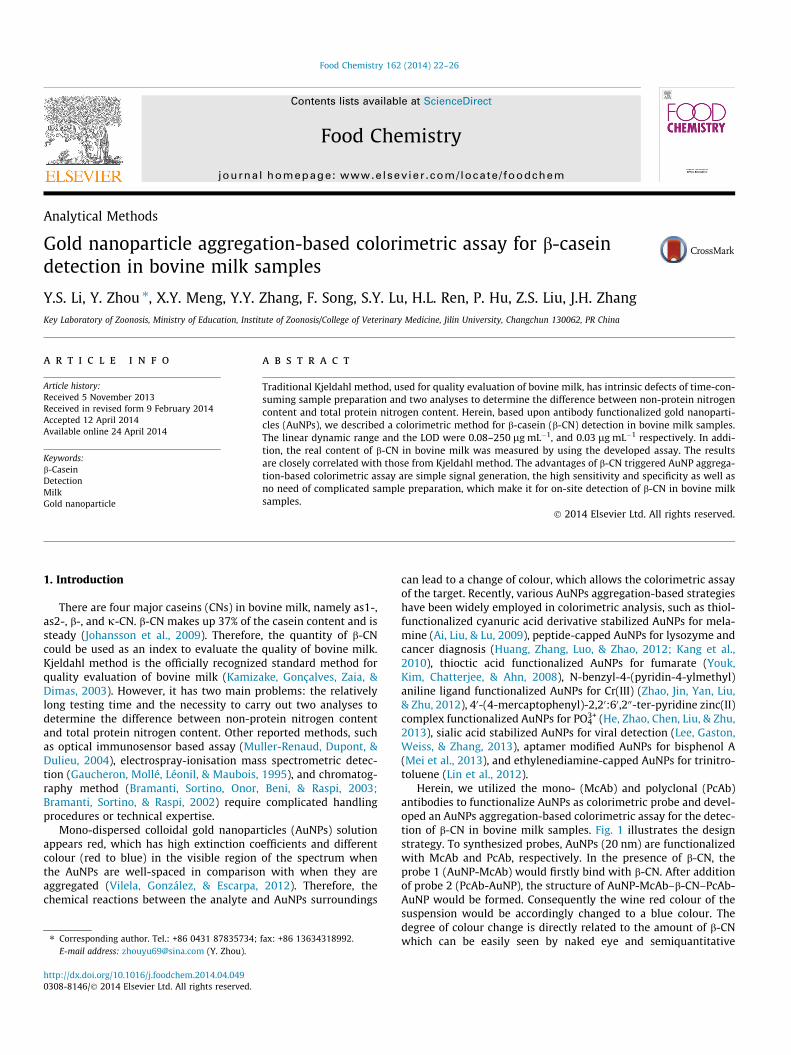
Herein, we utilized the mono- (McAb) and polyclonal (PcAb)antibodies to functionalize AuNPs as colorimetric probe and devel-oped an AuNPs aggregation-based colorimetric assay for the detec-tion of b-CN in bovine milk samples. Fig. 1 illustrates the designstrategy. To synthesized probes, AuNPs (20 nm) are functionalizedwith McAb and PcAb, respectively. In the presence of b-CN, theprobe 1 (AuNP-McAb) would firstly bind with b-CN. After additionof probe 2 (PcAb-AuNP), the structure of AuNP-McAb–b-CN–PcAb-AuNP would be formed. Consequently the wine red colour of thesuspension would be accordingly changed to a blue colour. Thedegree of colour change is directly related to the amount of b-CNwhich can be easily seen by naked eye and semiquantitative
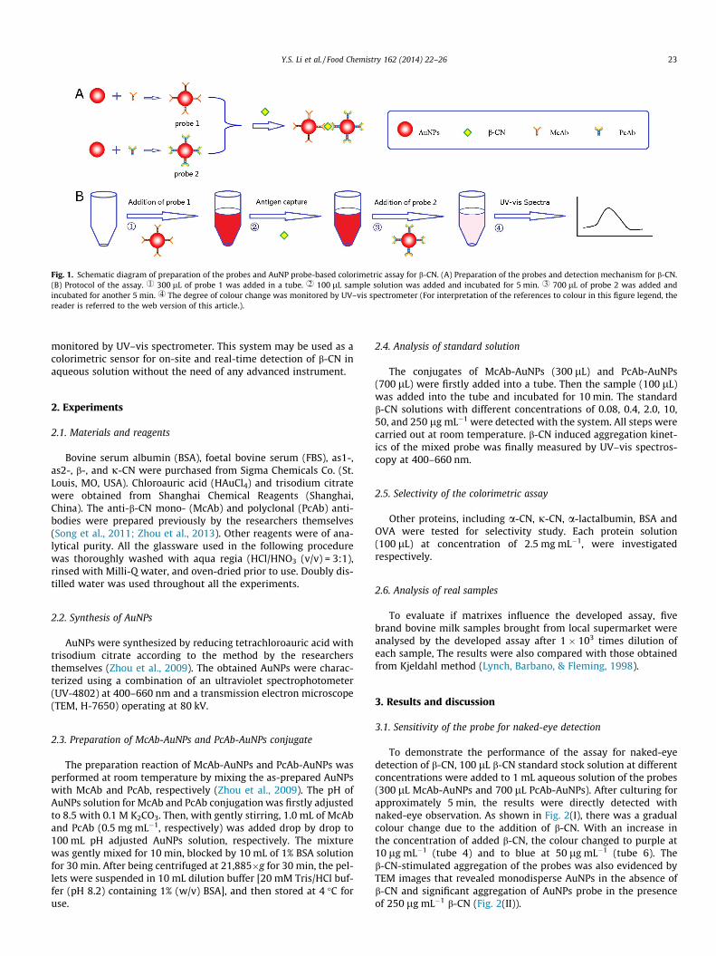
Fig. 1. Schematic diagram of preparation of the probes and AuNP probe-based colorimetric assay for b-CN. (A) Preparation of the probes and detection mechanism for b-CN.(B) Protocol of the assay. r 300 lL of probe 1 was added in a tube. s 100 lL sample solution was added and incubated for 5 min. t 700 lL of probe 2 was added andincubated for another 5 min. u The degree of colour change was monitored by UV–vis spectrometer (For interpretation of the references to colour in this figure legend, thereader is referred to the web version of this article.).
Y.S. Li et al. / Food Chemistry 162 (2014) 22–26 23
monitored by UV–vis spectrometer. This system may be used as acolorimetric sensor for on-site and real-time detection of b-CN inaqueous solution without the need of any advanced instrument.
2. Experiments
2.1. Materials and reagents
Bovine serum albumin (BSA), foetal bovine serum (FBS), as1-,as2-, b-, and j-CN were purchased from Sigma Chemicals Co. (St.Louis, MO, USA). Chloroauric acid (HAuCl4) and trisodium citratewere obtained from Shanghai Chemical Reagents (Shanghai,China). The anti-b-CN mono- (McAb) and polyclonal (PcAb) anti-bodies were prepared previously by the researchers themselves(Song et al., 2011; Zhou et al., 2013). Other reagents were of ana-lytical purity. All the glassware used in the following procedurewas thoroughly washed with aqua regia (HCl/HNO3 (v/v) = 3:1),rinsed with Milli-Q water, and oven-dried prior to use. Doubly dis-tilled water was used throughout all the experiments.
2.2. Synthesis of AuNPs
AuNPs were synthesized by reducing tetrachloroauric acid withtrisodium citrate according to the method by the researchersthemselves (Zhou et al., 2009). The obtained AuNPs were charac-terized using a combination of an ultraviolet spectrophotometer(UV-4802) at 400–660 nm and a transmission electron microscope(TEM, H-7650) operating at 80 kV.
2.3. Preparation of McAb-AuNPs and PcAb-AuNPs conjugate
The preparation reaction of McAb-AuNPs and PcAb-AuNPs wasperformed at room temperature by mixing the as-prepared AuNPswith McAb and PcAb, respectively (Zhou et al., 2009). The pH ofAuNPs solution for McAb and PcAb conjugation was firstly adjustedto 8.5 with 0.1 M K2CO3. Then, with gently stirring, 1.0 mL of McAband PcAb (0.5 mg mL�1, respectively) was added drop by drop to100 mL pH adjusted AuNPs solution, respectively. The mixturewas gently mixed for 10 min, blocked by 10 mL of 1% BSA solutionfor 30 min. After being centrifuged at 21,885�g for 30 min, the pel-lets were suspended in 10 mL dilution buffer [20 mM Tris/HCl buf-fer (pH 8.2) containing 1% (w/v) BSA], and then stored at 4 �C foruse.
2.4. Analysis of standard solution
The conjugates of McAb-AuNPs (300 lL) and PcAb-AuNPs(700 lL) were firstly added into a tube. Then the sample (100 lL)was added into the tube and incubated for 10 min. The standardb-CN solutions with different concentrations of 0.08, 0.4, 2.0, 10,50, and 250 lg mL�1 were detected with the system. All steps werecarried out at room temperature. b-CN induced aggregation kinet-ics of the mixed probe was finally measured by UV–vis spectros-copy at 400–660 nm.
2.5. Selectivity of the colorimetric assay
Other proteins, including a-CN, j-CN, a-lactalbumin, BSA andOVA were tested for selectivity study. Each protein solution(100 lL) at concentration of 2.5 mg mL�1, were investigatedrespectively.
2.6. Analysis of real samples
To evaluate if matrixes influence the developed assay, fivebrand bovine milk samples brought from local supermarket wereanalysed by the developed assay after 1 � 103 times dilution ofeach sample, The results were also compared with those obtainedfrom Kjeldahl method (Lynch, Barbano, & Fleming, 1998).
3. Results and discussion
3.1. Sensitivity of the probe for naked-eye detection
To demonstrate the performance of the assay for naked-eyedetection of b-CN, 100 lL b-CN standard stock solution at differentconcentrations were added to 1 mL aqueous solution of the probes(300 lL McAb-AuNPs and 700 lL PcAb-AuNPs). After culturing forapproximately 5 min, the results were directly detected withnaked-eye observation. As shown in Fig. 2(I), there was a gradualcolour change due to the addition of b-CN. With an increase inthe concentration of added b-CN, the colour changed to purple at10 lg mL�1 (tube 4) and to blue at 50 lg mL�1 (tube 6). Theb-CN-stimulated aggregation of the probes was also evidenced byTEM images that revealed monodisperse AuNPs in the absence ofb-CN and significant aggregation of AuNPs probe in the presenceof 250 lg mL�1 b-CN (Fig. 2(II)).
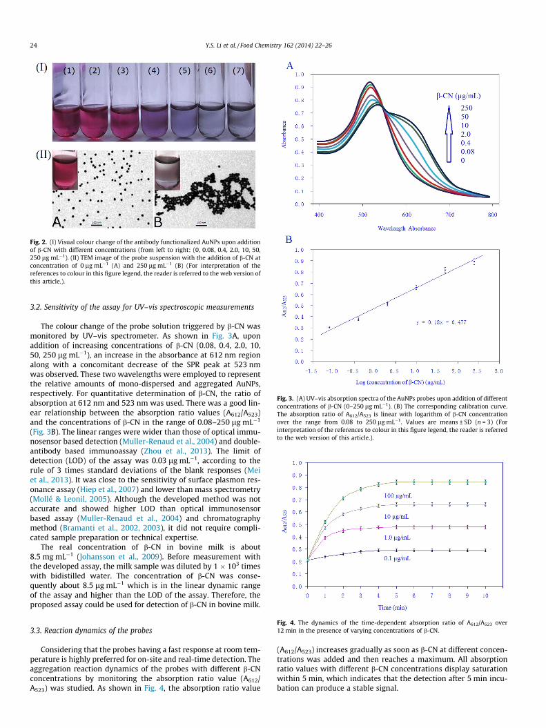
Fig. 2. (I) Visual colour change of the antibody functionalized AuNPs upon additionof b-CN with different concentrations (from left to right: (0, 0.08, 0.4, 2.0, 10, 50,250 lg mL�1). (II) TEM image of the probe suspension with the addition of b-CN atconcentration of 0 lg mL�1 (A) and 250 lg mL�1 (B) (For interpretation of thereferences to colour in this figure legend, the reader is referred to the web version ofthis article.).
Fig. 3. (A) UV–vis absorption spectra of the AuNPs probes upon addition of differentconcentrations of b-CN (0–250 lg mL�1). (B) The corresponding calibration curve.The absorption ratio of A612/A523 is linear with logarithm of b-CN concentrationover the range from 0.08 to 250 lg mL�1. Values are means ± SD (n = 3) (Forinterpretation of the references to colour in this figure legend, the reader is referredto the web version of this article.).
24 Y.S. Li et al. / Food Chemistry 162 (2014) 22–26
3.2. Sensitivity of the assay for UV–vis spectroscopic measurements
The colour change of the probe solution triggered by b-CN wasmonitored by UV–vis spectrometer. As shown in Fig. 3A, uponaddition of increasing concentrations of b-CN (0.08, 0.4, 2.0, 10,50, 250 lg mL�1), an increase in the absorbance at 612 nm regionalong with a concomitant decrease of the SPR peak at 523 nmwas observed. These two wavelengths were employed to representthe relative amounts of mono-dispersed and aggregated AuNPs,respectively. For quantitative determination of b-CN, the ratio ofabsorption at 612 nm and 523 nm was used. There was a good lin-ear relationship between the absorption ratio values (A612/A523)and the concentrations of b-CN in the range of 0.08–250 lg mL�1
(Fig. 3B). The linear ranges were wider than those of optical immu-nosensor based detection (Muller-Renaud et al., 2004) and double-antibody based immunoassay (Zhou et al., 2013). The limit ofdetection (LOD) of the assay was 0.03 lg mL�1, according to therule of 3 times standard deviations of the blank responses (Meiet al., 2013). It was close to the sensitivity of surface plasmon res-onance assay (Hiep et al., 2007) and lower than mass spectrometry(Mollé & Leonil, 2005). Although the developed method was notaccurate and showed higher LOD than optical immunosensorbased assay (Muller-Renaud et al., 2004) and chromatographymethod (Bramanti et al., 2002, 2003), it did not require compli-cated sample preparation or technical expertise.
The real concentration of b-CN in bovine milk is about8.5 mg mL�1 (Johansson et al., 2009). Before measurement withthe developed assay, the milk sample was diluted by 1 � 103 timeswith bidistilled water. The concentration of b-CN was conse-quently about 8.5 lg mL�1 which is in the linear dynamic rangeof the assay and higher than the LOD of the assay. Therefore, theproposed assay could be used for detection of b-CN in bovine milk.
Fig. 4. The dynamics of the time-dependent absorption ratio of A612/A523 over12 min in the presence of varying concentrations of b-CN.
3.3. Reaction dynamics of the probesConsidering that the probes having a fast response at room tem-perature is highly preferred for on-site and real-time detection. Theaggregation reaction dynamics of the probes with different b-CNconcentrations by monitoring the absorption ratio value (A612/A523) was studied. As shown in Fig. 4, the absorption ratio value
(A612/A523) increases gradually as soon as b-CN at different concen-trations was added and then reaches a maximum. All absorptionratio values with different b-CN concentrations display saturationwithin 5 min, which indicates that the detection after 5 min incu-bation can produce a stable signal.
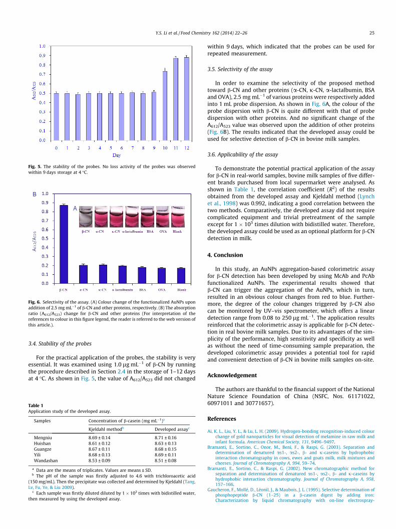
Fig. 5. The stability of the probes. No loss activity of the probes was observedwithin 9 days storage at 4 �C.
Fig. 6. Selectivity of the assay. (A) Colour change of the functionalized AuNPs uponaddition of 2.5 mg mL�1 of b-CN and other proteins, respectively. (B) The absorptionratio (A612/A523) change for b-CN and other proteins (For interpretation of thereferences to colour in this figure legend, the reader is referred to the web version ofthis article.).
Y.S. Li et al. / Food Chemistry 162 (2014) 22–26 25
3.4. Stability of the probes
For the practical application of the probes, the stability is veryessential. It was examined using 1.0 lg mL�1 of b-CN by runningthe procedure described in Section 2.4 in the storage of 1–12 daysat 4 �C. As shown in Fig. 5, the value of A612/A523 did not changed
Table 1Application study of the developed assay.
Samples Concentration of b-casein (mg mL�1)a
Kjeldahl methodb Developed assayc
Mengniu 8.69 ± 0.14 8.71 ± 0.16Huishan 8.61 ± 0.12 8.63 ± 0.13Guangze 8.67 ± 0.11 8.68 ± 0.15Yili 8.68 ± 0.13 8.69 ± 0.11Wandashan 8.53 ± 0.09 8.51 ± 0.08
a Data are the means of triplicates. Values are means ± SD.b The pH of the sample was firstly adjusted to 4.6 with trichloroacetic acid
(150 mg/mL). Then the precipitate was collected and determined by Kjeldahl (Tang,Lv, Fu, Ye, & Liu 2009).
c Each sample was firstly diluted diluted by 1 � 103 times with bidistilled water,then measured by using the developed assay.
within 9 days, which indicated that the probes can be used forrepeated measurement.
3.5. Selectivity of the assay
In order to examine the selectivity of the proposed methodtoward b-CN and other proteins (a-CN, j-CN, a-lactalbumin, BSAand OVA), 2.5 mg mL�1 of various proteins were respectively addedinto 1 mL probe dispersion. As shown in Fig. 6A, the colour of theprobe dispersion with b-CN is quite different with that of probedispersion with other proteins. And no significant change of theA612/A523 value was observed upon the addition of other proteins(Fig. 6B). The results indicated that the developed assay could beused for selective detection of b-CN in bovine milk samples.
3.6. Applicability of the assay
To demonstrate the potential practical application of the assayfor b-CN in real-world samples, bovine milk samples of five differ-ent brands purchased from local supermarket were analysed. Asshown in Table 1, the correlation coefficient (R2) of the resultsobtained from the developed assay and Kjeldahl method (Lynchet al., 1998) was 0.992, indicating a good correlation between thetwo methods. Comparatively, the developed assay did not requirecomplicated equipment and trivial pretreatment of the sampleexcept for 1 � 103 times dilution with bidistilled water. Therefore,the developed assay could be used as an optional platform for b-CNdetection in milk.
4. Conclusion
In this study, an AuNPs aggregation-based colorimetric assayfor b-CN detection has been developed by using McAb and PcAbfunctionalized AuNPs. The experimental results showed thatb-CN can trigger the aggregation of the AuNPs, which in turn,resulted in an obvious colour changes from red to blue. Further-more, the degree of the colour changes triggered by b-CN alsocan be monitored by UV–vis spectrometer, which offers a lineardetection range from 0.08 to 250 lg mL�1. The application resultsreinforced that the colorimetric assay is applicable for b-CN detec-tion in real bovine milk samples. Due to its advantages of the sim-plicity of the performance, high sensitivity and specificity as wellas without the need of time-consuming sample preparation, thedeveloped colorimetric assay provides a potential tool for rapidand convenient detection of b-CN in bovine milk samples on-site.
Acknowledgement
The authors are thankful to the financial support of the NationalNature Science Foundation of China (NSFC, Nos. 61171022,60971011 and 30771657).
References
Ai, K. L., Liu, Y. L., & Lu, L. H. (2009). Hydrogen-bonding recognition-induced colourchange of gold nanoparticles for visual detection of melamine in raw milk andinfant formula. American Chemical Society, 131, 9496–9497.
Bramanti, E., Sortino, C., Onor, M., Beni, F., & Raspi, G. (2003). Separation anddetermination of denatured as1-, as2-, b- and j-caseins by hydrophobicinteraction chromatography in cows, ewes and goats milk, milk mixtures andcheeses. Journal of Chromatography A, 994, 59–74.
Bramanti, E., Sortino, C., & Raspi, G. (2002). New chromatographic method forseparation and determination of denatured as1-, as2-, b- and j-caseins byhydrophobic interaction chromatography. Journal of Chromatography A, 958,157–166.
Gaucheron, F., Mollé, D., Léonil, J., & Maubois, J. L. (1995). Selective determination ofphosphopeptide b-CN (1–25) in a b-casein digest by adding iron:Characterization by liquid chromatography with on-line electrospray-
26 Y.S. Li et al. / Food Chemistry 162 (2014) 22–26
ionization mass spectrometric detection. Journal of Chromatography B:Biomedical Sciences and Applications, 664, 193–200.
He, G. K., Zhao, L., Chen, K., Liu, Y. Y., & Zhu, H. J. (2013). Highly selective andsensitive gold nanoparticle-based colorimetric assay for PO4
3+ in aqueoussolution. Talanta, 106, 73–78.
Hiep, H. M., Endo, T., Kerman, K., Chikae, M., Kim, D. K., Yamamura, S., et al. (2007). Alocalized surface plasmon resonance based immunosensor for the detection ofcasein in milk. Science and Technology of Advanced Materials, 8, 331–338.
Huang, H., Zhang, Q. W., Luo, J. X., & Zhao, Y. (2012). Sensitive colorimetric detectionof lysozyme in human serum using peptide-capped gold nanoparticles.Analytical Methods, 4, 3874–3878.
Johansson, A., Lugand, D., Rolet-Répécaud, O., Mollé, D., Delage, M. M., Peltre, G.,et al. (2009). Epitope characterization of a supramolecular protein assemblywith a collection of monoclonal antibodies: The case of casein micelle.Molecular Immunology, 46, 1058–1066.
Kamizake, N. K. K., Gonçalves, M. M., Zaia, C. T. B. V., & Dimas, A. M. (2003).Determination of total proteins in cow milk powder samples: A comparativestudy between the Kjeldahl method and spectrophotometric methods. Journalof Food Composition and Analysis, 16, 507–516.
Kang, J. H., Asami, Y., Murata, M., Kitazaki, H., Sadanaga, N., Tokunaga, E., et al.(2010). Gold nanoparticle-based colorimetric assay for cancer diagnosis.Biosensors and Bioelectronics, 25, 1869–1874.
Lee, C., Gaston, M. A., Weiss, A. A., & Zhang, P. (2013). Colorimetric viral detectionbased on sialic acid stabilized gold nanoparticles. Biosensors and Bioelectronics,42, 236–241.
Lin, D. Y., Liu, H. L., Qian, K., Zhou, X., Yang, L. B., & Liu, J. H. (2012). Ultrasensitiveoptical detection of trinitrotoluene by ethylenediamine-capped goldnanoparticles. Analytica Chimica Acta, 744, 92–98.
Lynch, J. M., Barbano, D. M., & Fleming, J. R. (1998). Indirect and directdetermination of the casein content of milk by Kjeldahl nitrogen analysis:Collaborative study. Journal of AOAC International, 81, 763–774.
Mei, Z. L., Chu, H. Q., Chen, W., Xue, F., Liu, J., Xu, H. N., et al. (2013). Ultrasensitiveone-step rapid visual detection of bisphenol A in water samples by label-freeaptasensor. Biosensors and Bioelectronics, 39, 26–30.
Mollé, D., & Leonil, J. (2005). Quantitative determination of bovine k-caseinmacropeptide in dairy products by liquid chromatography/electrospraycoupled to mass spectrometry (LC–ESI/MS) and liquid chromatography/electrospray coupled to tandem mass spectrometry (LC–ESI/MS/MS).International Dairy Journal, 15, 419–428.
Muller-Renaud, S., Dupont, D., & Dulieu, P. (2004). Quantification of b-casein in milkand cheese using an optical immunosensor. Journal of Agriculture FoodChemistry, 52, 659–664.
Song, F., Zhou, Y., Lu, S. Y., Ren, H. L., Li, Y. S., Liu, Z. S., et al. (2011). Production andidentification of the McAb specific for b-casein. Chinese Journal of LaboratoryDiagnosis, 15, 1285–1287.
Tang, J. N., Lv, Y., Fu, Y., Ye, X. Q., & Liu, D. H. (2009). Progress on determinationmethods of true protein in milk and dairy products. Food and FermentationIndustries, 35, 119–123.
Vilela, D., González, M. C., & Escarpa, A. (2012). Sensing colorimetric approachesbased on gold and silver nanoparticles aggregation: Chemical creativity behindthe assay: A review. Analytica Chimica Acta, 751, 24–43.
Youk, K. S., Kim, K. M., Chatterjee, A., & Ahn, K. H. (2008). Selective recognition offumarate from maleate with a gold nanoparticle-based colorimetric sensingsystem. Tetrahedron Letters, 49, 3652–3655.
Zhao, L., Jin, Y., Yan, Z. W., Liu, Y. Y., & Zhu, H. J. (2012). Novel, highly selectivedetection of Cr(III) in aqueous solution based on a gold nanoparticlescolorimetric assay and its application for determining Cr(VI). AnalyticaChimica Acta, 731, 75–81.
Zhou, Y., Pan, F. G., Li, Y. S., Zhang, Y. Y., Zhang, J. H., Lu, S. Y., et al. (2009). Colloidalgold probe-based immunochromatographic assay for the rapid detection ofbrevetoxins in fishery product samples. Biosensors and Bioelectronics, 24,2744–2747.
Zhou, Y., Song, F., Li, Y. S., Liu, J. Q., Lu, S. Y., Ren, H. L., et al. (2013). Double-antibodybased immunoassay for the detection of b-casein in bovine milk samples. FoodChemistry, 141, 167–173.