graf and lippard addr.pdf - dspace.mit.edu
TRANSCRIPT
Redox activation of metal-basedprodrugs as a strategy for drug delivery
The MIT Faculty has made this article openly available. Please share how this access benefits you. Your story matters.
Citation Graf, Nora, and Stephen J. Lippard. “Redox Activation of Metal-Based Prodrugs as a Strategy for Drug Delivery.” Advanced DrugDelivery Reviews 64, no. 11 (August 2012): 993-1004.
As Published http://dx.doi.org/10.1016/j.addr.2012.01.007
Version Author's final manuscript
Citable link http://hdl.handle.net/1721.1/103939
Terms of Use Creative Commons Attribution-NonCommercial-NoDerivs License
Detailed Terms http://creativecommons.org/licenses/by-nc-nd/4.0/
1
Redox activation of metal-based prodrugs as a strategy
for drug delivery
For theme issue:
"Stimuli-Responsive Drug Delivery Systems" for Advanced Drug Delivery Reviews
Nora Graf1, 2, Stephen J. Lippard1,*
1Department of Chemistry, Massachusetts Institute of Technology, 77 Massachusetts
Avenue, Cambridge, MA 02139 (USA), Tel: (+1)617-253-1892 Fax: (+1)617-258-8150,
E-mail: [email protected]
2 Institute of Chemistry and Biochemistry, Freie Universität Berlin, Germany
Abstract
This review provides an overview of metal-based anticancer drugs and drug candidates.
In particular, we focus on metal complexes that can be activated in the reducing envi-
ronment of cancer cells, thus serving as prodrugs. There are many reports of Pt and Ru
complexes as redox-activatable drug candidates, but other d-block elements with vari-
able oxidation states have a similar potential to serve as prodrugs in this manner. In this
context are compounds based on Fe, Co, or Cu chemistry, which are also covered. A
trend in the field of medicinal inorganic chemistry has been toward molecularly targeted,
metal-based drugs obtained by functionalizing complexes with biologically active
ligands. Another recent activity is the use of nanomaterials for drug delivery, exploiting
passive targeting of tumors with nano-sized constructs made from Au, Fe, carbon, or
organic polymers. Although complexes of all of the above mentioned metals will be de-
scribed, this review focuses primarily on Pt compounds, including constructs containing
nanomaterials.
2
Keywords
Prodrugs, Pt anticancer drugs, Ru anticancer drugs, activation by reduction, medicinal
inorganic chemistry, nanocarriers
Contents
1. Introduction
2. Platinum
3. Ruthenium
4. Iron, Cobalt, Copper
5. Nanobased Drug Delivery Systems
6. Conclusion
Acknowledgements
References
1. Introduction
Most drugs are organic or biologically derived compounds, and the pharmaceutical in-
dustry focuses on organic chemistry with some exceptions [1]. Metal-based therapeu-
tics comprise only a small percentage of available drugs. After the discovery and suc-
cessful clinical applications of the Pt-based anticancer drug cisplatin, however, research
on metal-based drugs became increasingly more important. There is a growing interest
in metal-containing drugs, and medicinal inorganic chemistry covering applications of
metals in therapeutics and diagnostics is a field of increasing prominence [2]. The major
advantage of metal-based over organic-based drugs is the ability to vary coordination
3
number, geometry, and redox states. Metals can also change the pharmacological
properties of organic-based drugs by forming coordination complexes with them [1].
Cisplatin is one of the most widely used anticancer drugs, administered particularly for
ovarian and testicular cancer. If testicular tumors are discovered early, an impressive
cure rate of almost 100% is achieved. Significant side-effects due to systemic toxicity
include nausea, bone-marrow suppression, and nephrotoxicity. Drug resistance, inher-
ent or acquired, also poses a great problem [3]. In an effort to diminish side effects and
resistance caused by cisplatin, other metal-based drugs are being investigated.
Metal-based pharmaceuticals can be arranged into seven categories depending on the
function of the metal and ligand moieties according to Hambley et al. [4]: 1) the metal
complex is active in its inert form, 2) the metal complex is active in its reactive form, 3)
the metal serves as a radiation enhancer, 4) the compound contains a radioactive
metal, 5) the metal or its biotransformation product is active, 6) a ligand is biologically
active, and 7) only a fragment of the complex is active. The two latter categories apply
to compounds presented in this review and represent two major strategies explored for
the design of metal-containing drugs: targeting and prodrug concept. The targeting
strategy uses selective transport of functionalized drug molecules recognized by recep-
tors highly or only expressed on the surface of cancer cells. The prodrug strategy com-
prises the delivery of a cytotoxic compound that is only activated under conditions pre-
sent in the targeted tissue or cells. Deactivation by cellular components before reaching
the target would ideally be inhibited in the prodrug form. Triggers for prodrug activation
can be pH, light or the redox environment. When compared to organic molecules, ac-
cess to redox chemistry is a clear advantage of metal-containing compounds, since
they generally have biologically accessible redox potentials. Accordingly, among the
different strategies for releasing metal-containing fragments from prodrugs, the most

4
successful is redox activation, in which the active species is formed in a reductive cellu-
lar environment [5].
Tumors are characterized by low oxygen concentration levels which give rise to a more
reducing environment compared to normal tissue. The reductive microenvironment of
hypoxic tumors results from insufficient formation of new blood vessels during growth
[6]. Also, a large concentration of cellular reducing agents like glutathione is present in
cancer cells, contributing to a reductive environment. Tumor hypoxia has been linked to
unsuccessful treatments using chemotherapy and radiotherapy due to distance from
blood vessels and resulting low oxygen levels [7]. Tumor cells that can survive in hy-
poxic conditions upregulate drug resistance genes [8]. Although hypoxia is considered a
serious problem in cancer therapy, it can be exploited for therapeutic selectivity, since it
differentiates cancerous and healthy tissue. One promising avenue is the development
of bioreductive drugs that are selectively activated under such intracellular conditions.
Literature known bioreductively activatable agents comprise quinones, nitroimidazoles,
aromatic N-oxides, mustard nitrogens, and a few metal-containing compounds [8, 9].
Metal compounds are to be delivered to the target environment in an inert oxidized state
(without prior reduction), and metabolized when they reach the reductive environment of
the cancer cell rendering the inactive compound cytotoxic. Three Pt(IV) and two Ru(III)
compounds have already been in clinical trials – to date the most promising bioreduc-
tive pharmaceuticals based on metals [6].
This review covers applications of metal-based drugs for anticancer applications using
the redox-activated prodrug strategy. Electrochemical aspects of redox-activatable Pt
and, particularly, Ru compounds have been reviewed in detail recently [6]. Thus, this
review focuses on literature that has been published since then and electrochemical
aspects will be touched only superficially.

5
2. Platinum
Cisplatin (1, Figure 1), one of the most successful anticancer drugs, and its analogs
carboplatin and oxaliplatin (2, 3) are currently in routine clinical use worldwide. Pt anti-
cancer agents only approved in Asia include nedaplatin, lobaplatin, and heptaplatin (4-
6).
Figure 1. Pt-based drugs in worldwide clinical use: cis- (1), carbo- (2), and oxaliplatin
(3), and clinical use only in some Asian countries: neda- (4), loba- (5) and heptaplatin
(6).
Once in the cell, where the chloride concentration drops from about 100 mM in extracel-
lular fluids to 4 mM, cisplatin is aquated by substitution of its chlorido ligands with water
ligands. Only 1% (or less) of the administered cisplatin ends up binding to the biological
target DNA, which results in apoptotic cell death. The majority of cisplatin reacts with
proteins and low molecular weight biomolecules, especially with those containing sulfur.
This non-discriminate binding causes side-effects of cisplatin like nephrotoxicity [3].
Carboplatin (2) displays a more tolerable toxicological profile due to the higher stability
of the chelating 1,1-cyclobutanedicarboxylato ligand when compared to the chlorido
ligands in cisplatin. No nephrotoxicity is observed [10]. Oxaliplatin (3) is also active
against cancer cell lines that are resistant to cisplatin and carboplatin [10].

6
Clinical complications associated with the use of cisplatin, such as nephro- and neuro-
toxicity and the fact that only some tumors can be cured, might be overcome by using
prodrugs with platinum in the more inert +IV oxidation state (Scheme 1) [2]. The admini-
stration of non-toxic Pt(IV) prodrugs that can be activated selectively by reduction at the
tumor sites might reduce unwanted reactions with biomolecules and thus minimize un-
desired side-effects. Even oral administration is conceivable, since in contrast to the
quite reactive Pt(II), Pt(IV) compounds are more stable in biological fluids. Degradation
in the gastrointestinal tract is thus less likely, and the Pt(IV) prodrugs may reach the
cellular target without prior transformation.
Scheme 1. Activation by reduction: A Pt(IV) prodrug carrying axial ligands L, ammine
ligands NH3, and chlorido ligands Cl– in the equatorial positions yields cisplatin (1) upon
reduction accompanied by the loss of both axial ligands. Most probably, a Pt(II) species
then binds to nuclear DNA.
Pt(IV) has a low-spin d6 electron configuration and exhibits octahedral geometry. This
configuration is relatively inert to substitution; reactions with biological nucleophiles are
7
thus disfavored compared to Pt(II) complexes, and the lifetime in biological fluids is ex-
pected to increase. It is not yet established, however, that Pt(IV) complexes survive
long enough in vivo to be delivered to the tumor sites. The platinum compound could be
also reduced extracellularly and enter the cell as Pt(II) (Scheme 1). As investigated by
XANES (X-ray absorption near edge structure) analyses, the cellular distribution of
Pt(IV) compounds 24 h after exposure is indeed similar to that of cisplatin. This result
may indicate that the compound is readily reduced over that time period [11]. Micro-
probe SRIXE (synchrotron radiation-induced X-ray emission) is another excellent
method for evaluating Pt compounds in whole and sectioned cells. This method has
provided valuable insights into the distribution patterns and oxidation states of Pt drugs
in cells [12].
Variation of the axial ligands in Pt(IV) complexes affords a valuable strategy for altering
their lipophilicity and redox potentials, factors that may affect the ability to enter tumor
cells before being reduced to the active Pt(II) compound and, subsequently, the cytotox-
icity. For a series of different Pt(IV) complexes which, upon reduction, yield the same
Pt(II) species there was an 800-fold range in cytotoxicity [13]. Although the pharmacol-
ogy of Pt(IV) compounds seems to depend strongly on the nature of the axial ligands,
activity can be also influenced by the potency of the Pt(II) complex formed upon reduc-
tion [14].
Lipophilic drugs are supposed to diffuse readily through cell membranes, thereby in-
creasing their uptake. Lipophilicity, however, has to fall within an optimal window, oth-
erwise the complex either becomes too insoluble in aqueous media or is trapped within
the membrane. Platinum compounds with intermediate lipophilicity are therefore
advantageous for cell uptake and anticancer activity [15].

8
More electronegative ligands tend to destabilize Pt(IV) compounds. Most easily reduced
are those having axial chlorido ligands, and the most difficult are compounds with hy-
droxido ligands [10, 16]. The latter (L=OH) have reduction potentials that fall in the -900
mV range vs. Ag/AgCl in Pt(IV) complexes cis,trans,cis-[PtCl2L2(en)] and -
[PtCl2L2(NH3)2], whereas analogous complexes with axial chlorido ligands (L=Cl) have
reduction potentials of ~ -250 mV vs. Ag/AgCl. Related complexes carrying axial ace-
tato ligands fall in between, with reduction potentials ~ -600 mV vs. Ag/AgCl [15, 17-19].
The reduction potential becomes more positive for bulkier carboxylate ligands (propion-
ate, butyrate) [18]. For complexes of formula cis,trans,cis-|PtCl2L2(NH3)2], the net elec-
tron donor character correlates well with reduction rates and redox potentials in the ax-
ial ligand order L = OH– < alkylCOO– < Cl– < CF3COO– [17].
Potential reducing agents for Pt(IV) in the cell are glutathione [20-22], for which
E0 = -240 mV at pH 7.0 [23], ascorbate (vitamin C), NAD(P)H, and cysteine-containing
proteins [14, 24]. Metallothionein, a protein consisting of 61 amino acids with 20 cys-
teine residues, is a potential binding and redox partner for Pt(IV). Increased metal-
lothionein levels are proposed as a mechanism by which cells become resistant to
platinum drugs [25]. Serum albumin, a small protein with a 35% cysteine content, also
represents a potential binding partner for cisplatin as well as Pt(IV) compounds [19].
Although Pt(IV) can be reduced by both extra- and intracellular reducing agents
(Scheme 1), higher concentrations of these species are present within cells; e.g., for
glutathione, the levels are 1-10 mM in cells vs. 2 µM in plasma [26]. Besides reducing
Pt(IV) complexes efficiently, thiols at high concentrations can also coordinate to and
deactivate the resulting Pt(II) species, thereby reducing their efficacy.
The DNA binding properties of Pt complexes of the type cis,trans,cis-[PtCl2L2(en)] corre-
late well with the reduction potentials; the more readily reduced complexes are better
9
able to bind DNA [18]. Although reduction seems to be required for activity, and reactiv-
ity is enhanced in the presence of reducing agents [22, 27], Pt(IV) complexes can ap-
parently also bind directly to DNA (Scheme 1) [28-30]. Reduction of Pt(IV) has even
been claimed to occur by DNA itself, either by the nucleobases or the sugars of the
sugar-phosphate backbone [14]. Upon arrival at the target site, the kinetic preference of
a Pt(IV) complex for reduction by, versus binding to, DNA is then the deciding factor
[14]. These arguments, while intriguing, would benefit by isolation and identification of
the DNA fragments that are oxidized in the process.
Correlations of Pt(IV)/Pt(II) redox potentials with biological activity have been explored
within the context of quantitative structure-activity relationships (QSAR). Several Pt(IV)
complexes with various equatorial and axial ligands were synthesized to develop a pre-
dictive QSAR model. Theoretical descriptors were included in this model together with
physicochemical data like lipophilicity, reduction peak potential, and the number of oxy-
gen atoms in the molecular formula of the ligand [31]. In vitro cytotoxicity could be pre-
dicted based on this QSAR model, but the actual effects in vivo still have to be verified,
because others have not been able to correlate reduction potential with cytotoxicity
across a large series of different Pt(IV) complexes [32].
The effects of axial and carrier ligands on the reduction potential and cytotoxic proper-
ties of Pt(IV) complexes have also been investigated to define a relationship between
reduction rate, redox potential, and in vitro cytotoxicity of Pt(IV) compounds. The reduc-
tion rates depended on the electronegativity and the steric hindrance of the axial and
carrier ligands. Complexes carrying bulkier and more electron-withdrawing ligands were
reduced more rapidly and showed higher reduction potentials. Reduction rates and the
cytotoxicity of complexes with different carrier or axial ligands were correlated [17].
10
As can be concluded from the above examples and other factors, the selection of drug
candidates based on in vitro activity can be misleading. The most active species is usu-
ally chosen for animal studies, whereas the most inert species in vitro might actually
show the highest activity in vivo [4]. It is therefore important that the Pt(II) species de-
rived from a Pt(IV) precursor have proven in vivo activity. The effects of simulated hy-
poxia and the use of models like spheroids, which mimic the tumor microenvironment,
have been examined to predict the behavior of compounds in solid tumors. For exam-
ple, a spheroid-based tumor model revealed that Pt(IV) complexes, although active,
were unable to display selectivity for hypoxic cells [33].
In octahedral Pt(IV) complexes, the two additional ligands are not only valuable for
modifying solubility in biological media and tuning the redox potential, but they can also
provide targeting functionalities. Modifications at the axial sites offer the advantage of
selectivity against cancerous tissue. This feature complements the potential for Pt(IV)
complexes to be administered orally due to greater stability in the gastrointestinal tract
as well as their potentially lower side effects due to their greater kinetic inertness com-
pared to Pt(II) compounds. Targeting, directing a Pt(IV) complex to tumor cells and/or a
subcellular target therein, can be accompanied by attachment of a membrane receptor
substrate to an axial ligand or, alternatively, by release of therapeutically active axial
ligands following reduction to Pt(II) in the cancer cell.
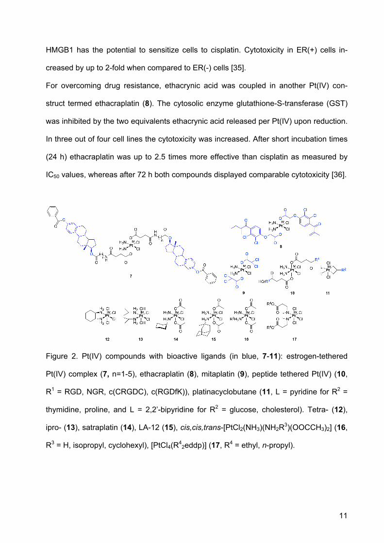
For targeting estrogen-receptor positive (ER(+)) cancers like breast and ovarian, estra-
diol-tethered Pt(IV) complexes have been employed (7, Figure 2, targeting groups are
depicted in blue). Reduction of the Pt(IV) complex in the intracellular environment re-
leases cisplatin and two equivalents of estradiol. The latter can upregulate expression
of the protein HMGB1, which can prevent repair of cisplatin-damaged DNA by shielding
the platinated DNA from the excision repair machinery [34]. Such upregulation of
11
HMGB1 has the potential to sensitize cells to cisplatin. Cytotoxicity in ER(+) cells in-
creased by up to 2-fold when compared to ER(-) cells [35].
For overcoming drug resistance, ethacrynic acid was coupled in another Pt(IV) con-
struct termed ethacraplatin (8). The cytosolic enzyme glutathione-S-transferase (GST)
was inhibited by the two equivalents ethacrynic acid released per Pt(IV) upon reduction.
In three out of four cell lines the cytotoxicity was increased. After short incubation times
(24 h) ethacraplatin was up to 2.5 times more effective than cisplatin as measured by
IC50 values, whereas after 72 h both compounds displayed comparable cytotoxicity [36].
Figure 2. Pt(IV) compounds with bioactive ligands (in blue, 7-11): estrogen-tethered
Pt(IV) complex (7, n=1-5), ethacraplatin (8), mitaplatin (9), peptide tethered Pt(IV) (10,
R1 = RGD, NGR, c(CRGDC), c(RGDfK)), platinacyclobutane (11, L = pyridine for R2 =
thymidine, proline, and L = 2,2’-bipyridine for R2 = glucose, cholesterol). Tetra- (12),
ipro- (13), satraplatin (14), LA-12 (15), cis,cis,trans-[PtCl2(NH3)(NH2R3)(OOCCH3)2] (16,
R3 = H, isopropyl, cyclohexyl), [PtCl4(R42eddp)] (17, R4 = ethyl, n-propyl).
12
In pursuit of a similar prodrug strategy, the Pt(IV) compound mitaplatin, cis,cis,trans-
[PtCl2(NH3)2(OOCCHCl2)2] (9), was prepared. Mitaplatin combines the orphan drug di-
chloroacetate (DCA) with cisplatin. DCA alters the mitochondrial membrane potential
gradient in cancer but not normal cells. As a consequence, cytochrome c is released
and apoptosis inducing factor is translocated to the nucleus [37]. Mitaplatin can thus
efficiently target both nuclear DNA with released cisplatin and mitochondria with re-
leased DCA in cancer cells. The cytotoxicity of mitaplatin equals or exceeds that of
most other Pt(IV) compounds and is comparable to that of cisplatin in a variety of can-
cer cell lines [38].
Small peptides recognizable by cancer tissue-specific receptors, the integrins, have
also been used as targeting moieties. In order to target cells expressing integrins on
their surface, Pt(IV) was coupled to several small peptides (3-5 amino acids) containing
RGD or NGR by either one or two amide linkages through either one or two succinato
groups (10, R1 = RGD, NGR, c(CRGDC), c(RGDfK)) [39]. Cytotoxicity was tested in
several endothelial and human cancer cells. RGD-tethered Pt(IV) complexes were more
cytotoxic than non-targeting Pt(IV) compounds and RGD tri- and pentapeptide moieties
alone. NGR conjugates were less inhibitory than RGD counterparts, but were still more
active than nonspecific Pt(IV)-peptide analogues.
Platinacyclobutane complexes (11), in which biologically relevant molecules like
thymidine, cholesterol, glucose, and proline (R2), are linked to cyclobutane moiety in an
equatorial position, have also been synthesized. The biocomponents were coupled to
cyclopropylmethanol and then allowed to react with Zeise’s dimer ([Pt(C2H4)Cl2]2) result-
ing in platinacycles. It is anticipated that the presence of the biomolecules would lead to
increased water solubility and cancer targeting, but thus far there has been no evalua-
tion of these compounds either in vitro or in vivo [40, 41].
13
Pt(IV) compounds lacking biologically active ligands have also been quite successful in
vitro. Four such octahedrally coordinated Pt(IV) complexes have even entered clinical
trials, namely, tetra- (12), ipro- (13) and satraplatin (14, formerly JM-216, diacetatoam-
minedichlorido(cyclohexylamine)platinum(IV)) as well as LA-12 (15). LA-12, however,
failed in phase I. Tetraplatin could not be investigated further after phase I studies due
to its high neurotoxicity. Iproplatin showed only limited success in phase II due to its low
reactivity. Satraplatin, the first orally available Pt-based drug candidate, had to be
abandoned recently in phase III [42]. Thus none of the platinum(IV) compounds has yet
found its way to the clinics because of lower efficacy than cisplatin, variability in drug
uptake, or the production of severe side-effects [6]. Satraplatin, although failing to pro-
vide an increase in overall survival above statistical significance when compared to cis-
platin, was better tolerated and showed no signs of inducing nephrotoxicity [43].
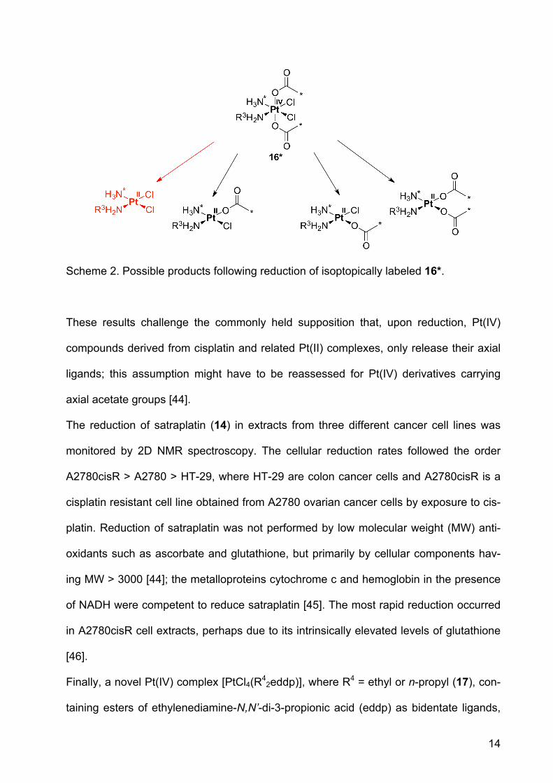
The initial success of the acetato complexes satraplatin (14) and LA-12 (15) motivated
researchers to investigate reduction pathways for Pt(IV) complexes having acetato
ligands in the axial position. Reduction of 13C- and 15N-labelled cis,cis,trans-
[PtCl2(15NH3)(NH2R3)(OOC13CH3)2] (16*, R3 = H, isopropyl or cyclohexyl) was effected
by different reducing agents like ascorbate, cytochrome c, NADH, or glutathione. Four
reduction products were identified by monitoring the reactions using two-dimensional
NMR spectroscopy and ESI-MS. In addition to the anticipated cisplatin analogue gener-
ated by loss of the axial acetato ligands (Scheme 2, red), additional products formed by
elimination of one acetate and equatorial chloride (middle structures, in Scheme 2) or
two chlorido ligands (structure at the right, Scheme 2).
14
Scheme 2. Possible products following reduction of isoptopically labeled 16*.
These results challenge the commonly held supposition that, upon reduction, Pt(IV)
compounds derived from cisplatin and related Pt(II) complexes, only release their axial
ligands; this assumption might have to be reassessed for Pt(IV) derivatives carrying
axial acetate groups [44].
The reduction of satraplatin (14) in extracts from three different cancer cell lines was
monitored by 2D NMR spectroscopy. The cellular reduction rates followed the order
A2780cisR > A2780 > HT-29, where HT-29 are colon cancer cells and A2780cisR is a
cisplatin resistant cell line obtained from A2780 ovarian cancer cells by exposure to cis-
platin. Reduction of satraplatin was not performed by low molecular weight (MW) anti-
oxidants such as ascorbate and glutathione, but primarily by cellular components hav-
ing MW > 3000 [44]; the metalloproteins cytochrome c and hemoglobin in the presence
of NADH were competent to reduce satraplatin [45]. The most rapid reduction occurred
in A2780cisR cell extracts, perhaps due to its intrinsically elevated levels of glutathione
[46].
Finally, a novel Pt(IV) complex [PtCl4(R42eddp)], where R4 = ethyl or n-propyl (17), con-
taining esters of ethylenediamine-N,N’-di-3-propionic acid (eddp) as bidentate ligands,

15
was described. Although the ligands themselves were not cytotoxic, the complexes had
IC50 values at least ten times higher when compared to cisplatin. The cytotoxic activity
was correlated with Pt uptake. The compound having R4 = n-propyl reacted with plas-
mid DNA as judged by gel electrophoresis studies [47].
3. Ruthenium
The biological activity of Ru compounds was first recognized in the 1950s [48], and re-
ports of their anticancer activity appeared in the 1960s [49]. Several Ru-based com-
pounds show significant efficacy against various types of tumors in vivo [50] while hav-
ing lower toxicity than cisplatin in vitro [51, 52].
Of particular interest are Ru(II) arene compounds and bioreducible Ru(III) complexes
with heterocyclic N-donor ligands. The only non-platinum transition metal compounds
currently in clinical trials are two Ru coordination compounds of the latter class,
[ImH][trans-RuCl4(DMSO)Im] (NAMI-A, 18, Figure 3) and [InH][trans-RuCl4In2] (KP1019,
19) [53, 54]. The first Ru-based anticancer drug candidate in clinical trials was NAMI-A,
followed by KP1019 in 2003. Both have successfully completed phase I. This achieve-
ment focused much attention on the medicinal properties of Ru compounds, reviews of
which are available [55-60]. KP1019 is active against colon cancer [5]; NAMI-A has only
low activity against primary tumors. NAMI-A is anti-angiogenic; its anti-invasive proper-
ties render it active against metastatic cancer.

16
Figure 3. Ru(III) compounds NAMI-A (18) and KP1019 (19).
The mechanism of action of these Ru(III) compounds remains unknown and their in vivo
chemistry is ambiguous [52]. Although Ru(III), like Pt(IV), can be reduced by ascorbate
or glutathione under physiological conditions, the resulting Ru(II) complexes maintain
their octahedral ligand set (Scheme 3) [54]. The biological target of Ru compounds has
not been discovered. DNA adducts can be formed by both NAMI-A and KP1019. The
resulting Ru-DNA adducts alter the duplex conformation [61]. KP1019 can unwind and
bend DNA [62]. As for Pt(II) compounds, sulfur-containing biomolecules might bind to
reduced Ru(II) species before they reach nuclear DNA.
NAMI-A undergoes aquation reactions within minutes [54], whereas KP1019 is more
stable, and better taken up by cells. About half of the intracellular Ru delivered in the
form of KP1019 appears in the nucleus, which is quite high by comparison to other
metal complexes; only 10% of cisplatin accumulates within the nucleus [63]. Ru com-
pounds not only target DNA but also proteins. KP1019 binds to transferrin [64] (Scheme
3), an Fe(III) transport protein, and is released from the protein as Ru(II) after reduction
by ascorbate or glutathione [6]. Interference with the Fe metabolism may also be an
explanation for the anticancer activity of Ru(III) complexes [54].
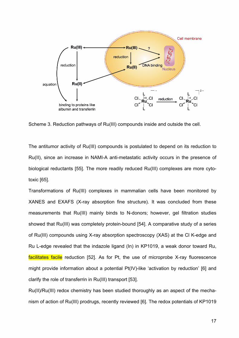
17
Scheme 3. Reduction pathways of Ru(III) compounds inside and outside the cell.
The antitumor activity of Ru(III) compounds is postulated to depend on its reduction to
Ru(II), since an increase in NAMI-A anti-metastatic activity occurs in the presence of
biological reductants [55]. The more readily reduced Ru(III) complexes are more cyto-
toxic [65].
Transformations of Ru(III) complexes in mammalian cells have been monitored by
XANES and EXAFS (X-ray absorption fine structure). It was concluded from these
measurements that Ru(III) mainly binds to N-donors; however, gel filtration studies
showed that Ru(III) was completely protein-bound [54]. A comparative study of a series
of Ru(III) compounds using X-ray absorption spectroscopy (XAS) at the Cl K-edge and
Ru L-edge revealed that the indazole ligand (In) in KP1019, a weak donor toward Ru,
facilitates facile reduction [52]. As for Pt, the use of microprobe X-ray fluorescence
might provide information about a potential Pt(IV)-like ‘activation by reduction’ [6] and
clarify the role of transferrin in Ru(III) transport [53].
Ru(II)/Ru(III) redox chemistry has been studied thoroughly as an aspect of the mecha-
nism of action of Ru(III) prodrugs, recently reviewed [6]. The redox potentials of KP1019

18
and NAMI-A are 30 mV and 25 mV, respectively, vs. NHE. The redox potentials are tun-
able by modification of the azole ligand (In and Im, respectively) [6].
These redox potential values reveal that NAMI-A and KP1019 can be reduced by glu-
tathione and ascorbate under physiological conditions [66, 67]. As described in the in-
troduction, in addition to providing reducing agents, cancer cells also harbor a hypoxic
environment that promotes reduction and subsequent reactivity [52]. Increased levels of
Ru-DNA adducts occurred when the O2 partial pressure was low, indicating a greater
amount of reduced, Ru(II), species [68].
Ru(III) phosphane complexes in which the metal is coordinated by two P,O,O-tridentate
tris(o-anisyl) phosphane ligands are highly cytotoxic even in cisplatin-resistant cell lines
[69]. Another construct involves NAMI A-type ligands conjugated through pyridyl or
bipyridyl rings to yield Ru porphyrin conjugates (meso-4’-tetrapyridylporphyrin or meso-
(p-bipyridyl-phenyl)porphyrin). The number of Ru fragments attached to the porphyrins
ranged from 1 to 4. Conjugation of porphyrins to the Ru center was an attempt to obtain
additive antitumor effects originating from the phototoxic and tumor-localizing properties
of the porphyrin together with the cytotoxic properties of Ru(III) [70]. Thus far, however,
the compounds have not been tested in vitro or in vivo.
An interesting conjugate was generated by coupling tamoxifen, a chemotherapeutic
agent for patients with hormone-dependent breast cancer, with the organometallic Ru
compound ruthenocene. This tamoxifen analog acts as an antiestrogen by competitive
binding to the estrogen receptor in ER(+) breast cancer cells but not in ER(-) cells [71,
72]. This result is in contrast to that for the Fe analog ferrocifen (20), which is active in
both cell lines (vide infra). Since the structural differences between the Fe and Ru de-
rivative are marginal, the different redox properties of the metal ions may be responsible
for the different patterns of activity [71].

19
Although Os belongs to the same group in the periodic table as Fe and Ru, and thus
should be also able to interact with biomolecules like proteins and nucleic acids, there
are only a few studies on the biological activity of Os complexes. In comparison to
analogous Ru compounds, Os complexes are more substitution inert and less prone to
hydrolysis and interactions with nucleobases. Nevertheless, they also have potential as
anticancer agents. By examining homologous Os compounds, one might even obtain
useful information about the mechanism of action of Ru drugs [73-75].
Os(III)-NAMI-A-type compounds (LH)[trans-Os(III)Cl4(DMSO)(L)] with L = 1H-indazole
(In), 1H-pyrazole, 1H-benzimidazole, 1H-imidazole (Im), 1H-1,2,4-triazole, and DMSO,
are kinetically stable in aqueous solution, but exhibit similar or even higher cytotoxicity
when compared with the analogous Ru compounds. For example, the indazole complex
(L = In) had an IC50 value ca. ten times lower than that of the analogous Ru complex
against HT-29 cells. Whereas hydrolysis is a prerequisite for the antimetastatic activity
of NAMI-A in vivo, hydrolyzed species were not required for the in vitro antiproliferative
activity of analogous Os(III) complexes [76, 77].
4. Iron, Cobalt, Copper
Ferrocifen (20, Figure 4), like the corresponding Ru compound described above, is an
organometallic derivative of the breast cancer drug tamoxifen. The ferrocene analog of
hydroxytamoxifen, however, acts against both ER(+) and ER(-) human breast cancer
cells, in contrast to the properties of hydroxytamoxifen alone [62, 78]. This behavior is
surprising since the latter cells lack an obvious molecular target for tamoxifen. It can be
concluded that the antiproliferative effect stems from the antiestrogenic effect of the ta-
moxifen moiety plus the cytotoxicity of the redox-active ferrocenyl group. Ferrocene in-
terconverts inside the cell between oxidation states II and III, represented by ferrocene
20
and ferrocenium ions. The cytotoxicity is not caused by direct linkage to DNA but by
formation of reactive oxygen species, which can damage DNA.
Not only ferrocene derivatives containing Fe(II) but also ferrocenium derivatives con-
taining Fe(III) are cytotoxic, generating radicals and thus inducing DNA damage [79].
Pyrazole conjugated to ferrocene serves as a ligand (L) for Co, Ni, and Fe (M), forming
ML2 and ML3 complexes, respectively. The ligand L and the three ML2 metal complexes
induced cytotoxicity in MCF-7 breast cancer cells, with IC50 values ranging from 46 to 73
µM; CoL2 exhibited the lowest IC50 value. As the redox potential increased, the toxicity
of the metal complexes decreased [80].
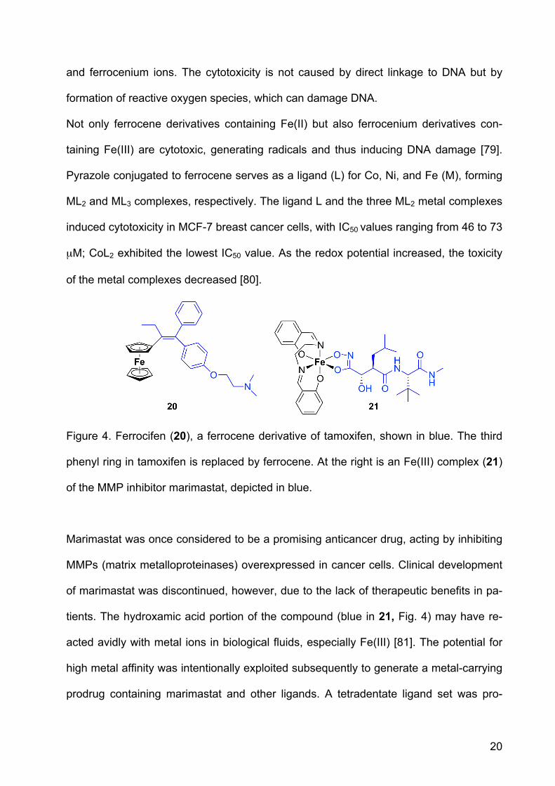
Figure 4. Ferrocifen (20), a ferrocene derivative of tamoxifen, shown in blue. The third
phenyl ring in tamoxifen is replaced by ferrocene. At the right is an Fe(III) complex (21)
of the MMP inhibitor marimastat, depicted in blue.
Marimastat was once considered to be a promising anticancer drug, acting by inhibiting
MMPs (matrix metalloproteinases) overexpressed in cancer cells. Clinical development
of marimastat was discontinued, however, due to the lack of therapeutic benefits in pa-
tients. The hydroxamic acid portion of the compound (blue in 21, Fig. 4) may have re-
acted avidly with metal ions in biological fluids, especially Fe(III) [81]. The potential for
high metal affinity was intentionally exploited subsequently to generate a metal-carrying
prodrug containing marimastat and other ligands. A tetradentate ligand set was pro-

21
vided by salen (N,N-bis(salicylidene)-ethane-1,2-diimine) (cf. black part in 21). The oc-
tahedral Fe(III) marimastat salen complex (21) inhibited MMP activity in vitro approxi-
mately 30-times less efficiently than marimastat alone. Reduced inhibitory activity by Fe
complexation of the hydroxamate functionality in marimastat and electrochemical data
indicate a potential for developing a prodrug that might be activated bioreductively [82].
Co(III) has also been complexed by the hydroxamate moiety in marimastat, although
the mechanism of action of such Co complexes is expected to differ from that of analo-
gous Fe(III) compounds, which are prone to generate reactive oxygen species. Instead,
Co(III) can be reduced to Co(II) in biological environments, leading to the release of ma-
rimastat. Co(III) thus provides an inert framework for the transport of the MMP inhibitor,
protecting the hydroxamate moiety prior to reaching the tumor. In this case, Co is not
the active species itself but only a protecting group for an otherwise cytotoxic ligand.
Similarly, Au(I) has been claimed to protect phosphane ligands from oxidation in Au
complexes like auranofin used for the treatment of rheumatoid arthritis [83].
As in the Fe(III) complex, a tetradentate ligand, here tpa for tris(methylpyridyl)-amine,
was combined with the hydroxamate to form the octahedral complex (22, Figure 5) [81,
84]. Increased cytotoxicity was observed for the prodrug in comparison to the inhibitor
alone. In an in vivo study free marimastat and its Co complex inhibited tumor growth in
the mammary fat pad in mice; but only the Co complex showed a statistically relevant
difference compared to the control group. The complex was 2-3 times more effective in
reducing the tumor growth than the MMP inhibitor alone. Both marimastat and its Co
complex, however, potentiated metastasis when compared to controls [81].
22
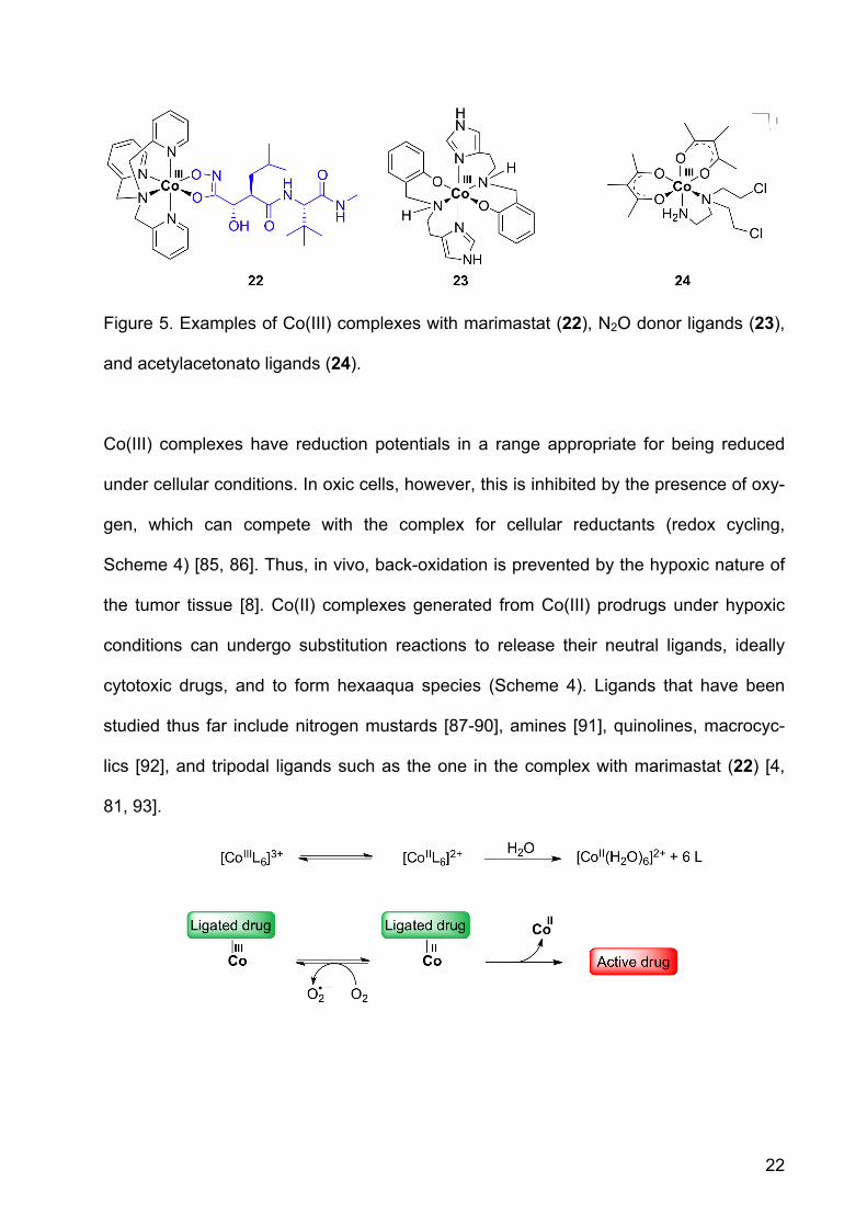
Figure 5. Examples of Co(III) complexes with marimastat (22), N2O donor ligands (23),
and acetylacetonato ligands (24).
Co(III) complexes have reduction potentials in a range appropriate for being reduced
under cellular conditions. In oxic cells, however, this is inhibited by the presence of oxy-
gen, which can compete with the complex for cellular reductants (redox cycling,
Scheme 4) [85, 86]. Thus, in vivo, back-oxidation is prevented by the hypoxic nature of
the tumor tissue [8]. Co(II) complexes generated from Co(III) prodrugs under hypoxic
conditions can undergo substitution reactions to release their neutral ligands, ideally
cytotoxic drugs, and to form hexaaqua species (Scheme 4). Ligands that have been
studied thus far include nitrogen mustards [87-90], amines [91], quinolines, macrocyc-
lics [92], and tripodal ligands such as the one in the complex with marimastat (22) [4,
81, 93].
23
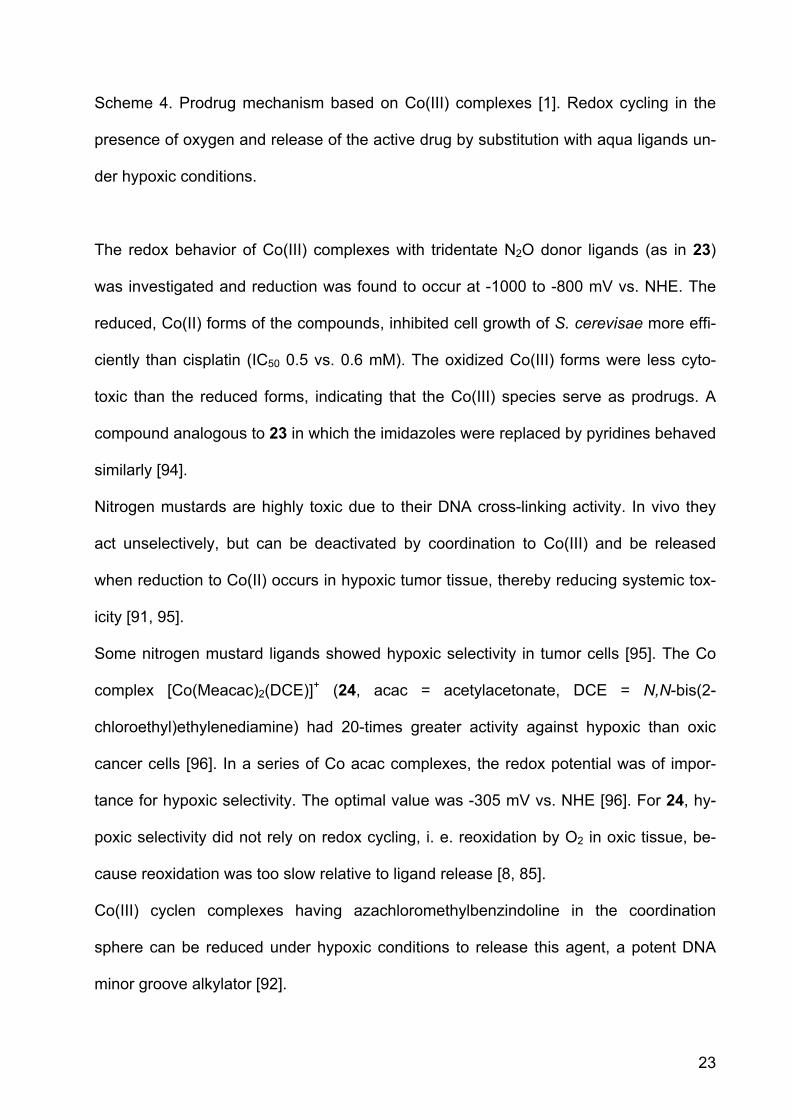
Scheme 4. Prodrug mechanism based on Co(III) complexes [1]. Redox cycling in the
presence of oxygen and release of the active drug by substitution with aqua ligands un-
der hypoxic conditions.
The redox behavior of Co(III) complexes with tridentate N2O donor ligands (as in 23)
was investigated and reduction was found to occur at -1000 to -800 mV vs. NHE. The
reduced, Co(II) forms of the compounds, inhibited cell growth of S. cerevisae more effi-
ciently than cisplatin (IC50 0.5 vs. 0.6 mM). The oxidized Co(III) forms were less cyto-
toxic than the reduced forms, indicating that the Co(III) species serve as prodrugs. A
compound analogous to 23 in which the imidazoles were replaced by pyridines behaved
similarly [94].
Nitrogen mustards are highly toxic due to their DNA cross-linking activity. In vivo they
act unselectively, but can be deactivated by coordination to Co(III) and be released
when reduction to Co(II) occurs in hypoxic tumor tissue, thereby reducing systemic tox-
icity [91, 95].
Some nitrogen mustard ligands showed hypoxic selectivity in tumor cells [95]. The Co
complex [Co(Meacac)2(DCE)]+ (24, acac = acetylacetonate, DCE = N,N-bis(2-
chloroethyl)ethylenediamine) had 20-times greater activity against hypoxic than oxic
cancer cells [96]. In a series of Co acac complexes, the redox potential was of impor-
tance for hypoxic selectivity. The optimal value was -305 mV vs. NHE [96]. For 24, hy-
poxic selectivity did not rely on redox cycling, i. e. reoxidation by O2 in oxic tissue, be-
cause reoxidation was too slow relative to ligand release [8, 85].
Co(III) cyclen complexes having azachloromethylbenzindoline in the coordination
sphere can be reduced under hypoxic conditions to release this agent, a potent DNA
minor groove alkylator [92].

24
The chemistry of copper is attractive for the development of hypoxia selective drugs,
because Cu has two oxidation states and the reduction potential in accessible within the
cellular potential range [97].
As for Co(III), Cu(II) complexes can be reduced to form Cu(I) complexes of low stability
to release an active ligand. Furthermore, radioactive isotopes of Cu can be used, pro-
viding the combination of radiation therapy with bioreduction. For this purpose, the tira-
pazamine ligand has been suggested as a hypoxic cytotoxin for use with radioactive
Cu, combining the antitumor activity of the copper complex with its radioactivity, the Cu
isotopes being 64Cu or 67Cu [98]. Radiolabelled 64Cu complexes carried by
bis(thiocarbazone) and bis(salicylaldimine) ligands showed hypoxic selectivity against
Chinese hamster ovary cancer cells [99].
Macrocyclic ligand systems used for Cu(II) include cyclen, cyclam and tacn. Complexes
with nitrogen mustards based on these macrocyclics are cytotoxic against leukemia
cells. The complexes were also cytotoxic in lung cancer cells selectively under hypoxic
conditions. The stability of the reduced Cu(I) species was investigated by cyclic volt-
ammetry. Its redox chemistry is reversible, indicating that the reduced complex is sta-
ble. A Cu(II) complex with a mustard derivative of cyclen (1,4,7-tetraazacyclodecane,
25, Figure 6) exhibited good aqueous stability and, in vitro a 24-fold increased cytotoxic-
ity under hypoxic conditions vs. oxic conditions. In aqueous solution, the redox behavior
of the complex and its stability correlated well with hypoxia selectivity. The hypoxia se-
lectivity is thought to originate from redox cycling in oxic tissue, similar to the behavior
of Co(III) prodrugs described above (Scheme 5). Complexes 26 and 27, derived from
the macrocyclic ligands cyclam and tacn, respectively, release the ligand independent
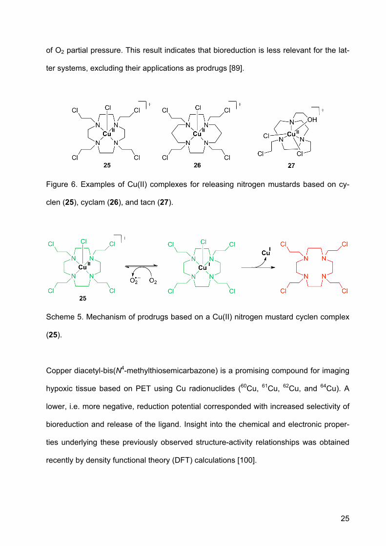
25
of O2 partial pressure. This result indicates that bioreduction is less relevant for the lat-
ter systems, excluding their applications as prodrugs [89].
Figure 6. Examples of Cu(II) complexes for releasing nitrogen mustards based on cy-
clen (25), cyclam (26), and tacn (27).
Scheme 5. Mechanism of prodrugs based on a Cu(II) nitrogen mustard cyclen complex
(25).
Copper diacetyl-bis(N4-methylthiosemicarbazone) is a promising compound for imaging
hypoxic tissue based on PET using Cu radionuclides (60Cu, 61Cu, 62Cu, and 64Cu). A
lower, i.e. more negative, reduction potential corresponded with increased selectivity of
bioreduction and release of the ligand. Insight into the chemical and electronic proper-
ties underlying these previously observed structure-activity relationships was obtained
recently by density functional theory (DFT) calculations [100].
26
Cu(II) complexes with ligands based on 3-aminoquinoxaline-2-carbonitrile-N1,N4-dioxide
were developed to serve as selective hypoxic cytotoxins [101]. The complexes were
evaluated under hypoxic and aerobic conditions in V79 cells. The complexes were
equally low cytotoxic with the ligands under aerobic conditions, but more cytotoxic un-
der hypoxic conditions [102].
Quinoxaline-N1,N4-dioxides themselves are bioreductively activatable drugs. However,
they exhibit low aqueous solubility and a short half life [103]. Their vanadyl complexes
VOL2, with L being a quinoxaline-N1,N4-dioxide ligand, however, showed improved
solubility and greater cytotoxicity than the free quinoxaline ligands, with high hypoxia
selectivity [104].
Pyrophosphate-bridged binuclear complexes of Cu(II) display low nanomolar toxicity
against adriamycin-resistant ovarian cancer cells. The complexes were tested in glu-
tathione assays to simulate cellular conditions. Cu(I) was formed in this reductive envi-
ronment and the system produced hydrogen peroxide from molecular oxygen, produc-
ing oxidative stress. The generation of oxidative stress might explain the high cytotoxic-
ity of this class of coordination compounds [105].
5. Nanobased Drug Delivery Systems
Delivery systems can mediate transport of drug molecules to desired cell populations.
Nanosized systems have been successfully applied in the last years, exploiting the
EPR, or enhanced permeability and retention, effect as a means of passively targeting
cancer tissue. A combination of metal prodrugs with nano-vehicles can be achieved
either by surface tethering or encapsulation. Encapsulation protects the drug from deg-
radation before reaching cancer cells as well as the non-cancerous cells from the ef-
fects of the drug. Depending on the encapsulating material, controlled release of the
27
drug from the nanoparticles is possible. Peptides, antibodies, or aptamers can be at-
tached to the particle surface for active targeting of cancer tissue. In this section, we
describe accumulation and active targeting as strategies for selectively delivering Pt(IV)
prodrugs to cancer cells with the help of nanoparticle materials. Although more stable in
the +IV oxidation state, Pt(IV) compounds are not impervious to degradation, as we
have already discussed (section 2). Their combination with nanomaterials offers a
promising opportunity to transport and protect these complexes from premature reduc-
tion in the blood stream.
In one manifestation of this strategy, Pt(IV) was attached to single-walled carbon nano-
tubes (SWNT) for shuttling into cells (28, Figure 7). Such soluble functionalized SWNTs
nanotubes cross the cell membrane by clathrin-dependent endocytosis. A substantial
increase in cytotoxicity was obtained when compared to cisplatin and the untethered
Pt(IV) complex. In one experiment, the SWNT “longboat” carried 65 Pt “passengers” as
determined by atomic absorption spectroscopy [106]. For targeting of folate receptors
on cancer cells, the Pt(IV) prodrug was also coupled to folate (29) by using a bisucci-
nate precursor, Pt(succ)2, Figure 7. One carboxyl group allowed coupling to folate via
an amino linker, while the other allowed tethering to the aminated nanotube surface.
The toxicity in cell lines expressing the folate receptor increased by up to 9-fold by
comparison to cisplatin [107].
Gold nanoparticles have also been used as delivery systems for potentially therapeutic
antisense oligonucleotides as well as Pt(IV) via covalent attachment (30). The nanopar-
ticles were functionalized with thiolated 28mer oligonucleotides containing a terminal
dodecyl amine for conjugation. The Pt(IV) complex Pt(succ) (Figure 7) was tethered to
the amino-functionalized DNA-Au-NP surface by forming an amide linkage [108]. The
28
constructs showed high levels of cellular uptake in different cell types and higher cyto-
toxicity when compared to cisplatin and the Pt(IV) precursor Pt(succ).
Gold nanorods, on the other hand, are even more promising vehicles than gold
nanoparticles due to longer circulation times when compared to the spherical particles.
PEGylated nanorods were loaded with disuccinato Pt(IV) (Pt(succ)2, 31, Figure 7). Cyto-
toxicity in HeLa, A549, and MCF7 cells was increased by up to 66-fold when compared
to cisplatin. Cytotoxicity could be correlated with Pt uptake as measured by ICP-MS
[109].
Better protection of the Pt(IV) prodrug can potentially be provided by encapsulation into
particles as opposed to tethering on their surface. Encapsulation protects the prodrug
but also can ameliorate unwanted side-effects of the drug. Cisplatin itself shows only
low loading efficiency (< 1 wt%) within the hydrophobic interiors of a polymer [110], but
an increase of internalization into nanoparticles could be achieved by using a hydro-
phobic Pt(IV) compound instead. Nanoparticles based on poly(D,L-lactic-co-glycolic
acid)-block-poly(ethylene glycol) (PLGA-b-PEG) combine hydrophobic PLGA for encap-
sulation and hydrophilic PEG for aqueous solubility [111]. A Pt(IV) compound carrying
hydrophobic hexyl chains, Pt(hex)2 (Figure 7), was released from PLGA-PEG nanopar-
ticles in a controlled fashion over 60 h. The nanoparticles were taken up by cells by re-
ceptor-mediated endocytosis. Besides being designed for passive targeting (EPR), the
nanoparticles were also expected to be guided to tumor tissue by active targeting. For
targeting prostate-specific membrane antigen (PSMA) over-expressed on prostate can-
cer cells, an A10 2-fluoropyrimidine RNA aptamer was coupled to the surface of the
nanoparticle (32) [111]. This system showed better cytotoxic activity in vitro than cis-
platin and selectivity for the cells expressing the receptors targeted. In vivo studies with
the PSMA-decorated Pt(IV) nanoparticle (32) demonstrated enhanced pharmacokinet-

29
ics, biodistribution, and tolerability in rats and mice when compared to cisplatin. Efficacy
in a PSMA-expressing LNCaP xenograft mouse model of prostate cancer was also
higher. For obtaining the same degree of tumor volume reduction only 1/3 the dose of
cisplatin was required [112].
Conjugation of dilevulinate Pt(IV) complexes Pt(levul)2 (Figure 7, levulinic acid = 4-
oxopentanoic acid) with a hydrazinated PEG-PLA diblock copolymer resulted in Pt(IV)
loaded nanoparticles (33). The nanoparticles were of sub-100 nm size and exhibited a
cisplatin loading yield of 1.1 wt%. Due to the acid-labile hydrazone bond, the system
showed acid-responsive drug release kinetics. The ability to kill ovarian cancer cells
was enhanced when compared to that of cisplatin [113].
A silica shell coating of coordination polymer nanoparticles based on Pt(succ)2 in-
creased half-release time for Pt by factor 9 - from 1 h for the uncoated Pt loaded poly-
mer to 9 h. Cytotoxicity was similar to that of cisplatin in breast cancer cells. Upon con-
jugation of integrin-targeting peptides c(RGDfK), in vitro toxicity against colon cancer
cells could be slightly increased in comparison to cisplatin [114].
Lastly, ethoxysuccinato Pt(IV), the prodrug also used for coupling to SWNT in 28, was
allowed to react postsynthetically with a dispersion of amino functionalized iron-
carboxylate nanoscale metal-organic frameworks. Once Pt was loaded into the frame-
work, the particles were coated with silica. The resulting material was then evaluated for
its cytotoxicity in HT-29 cells and found to be slightly less cytotoxic than cisplatin. In or-
der to increase the cytotoxicity, the silica shell was functionalized with a silyl derivative
of c(RGDfK) (34). As in the other examples described above, it was thereby armed for
targeting integrins, over-expressed in many anigiogenic tumors. Cytotoxicity was thus
increased, approaching that of cisplatin [115].

30
Figure 7. Metal-based prodrugs based on carbon nanotubes (28, 29), Au nanoparticles
and -rods (30, 31), polymeric nanoparticles made of PLGA-PEG (32) and PLA-PEG
(33), and iron-carboxylate nanoscale metal-organic frameworks with silica coating (34).
Loaded or coupled Pt(IV) compounds are shown below. As above (Figures 2, 4 and 5),
targeting units are shown in blue.
6. Conclusion
Therapeutically active complexes of Pt and Ru demonstrate that metal complexes can
play an important role in the treatment of cancer. It has been estimated that approxi-
31
mately half the patients being treated for cancer today by chemotherapy receive a plati-
num compound. The motivation for seeking other metal complexes with therapeutic po-
tential and for developing new metal-based drugs comes especially from the success of
the anticancer drug cisplatin. Also bioorganometallic chemistry, a relatively new branch
of medicinal inorganic chemistry, has contributed to the field, as exemplified by the ta-
moxifen analog ferrocifen.
In such complexes the tunable redox properties of the metal ion and ligand modifica-
tions can be exploited to control biological action. Ligand loss during reduction in hy-
poxic cancer tissue can activate metal complexes for binding to target molecules. Fur-
thermore, as for Co and Cu, the loss of the ligand in a reductive environment can also
trigger the release of an active species, a molecule that would have shown systemic
toxicity if not bound to the deactivating metal center.
In the future, the design of metal drugs should focus on molecular targeting agents to
provide greater selectivity and more effective drug administration. The activation by re-
duction strategy presented in this review is one strategy by which this goal can be ap-
proached. Metal complexes exhibiting targeting, both to cancer cells as well as to sub-
cellular targets therein, and selective activation properties can reduce side effects in
therapy and potentially cure a wider range of cancers by circumventing resistance. The
many opportunities that metal complexes have by comparison to organic molecules,
especially their versatile redox chemistry, should be exploited for creating more efficient
anticancer drugs.
Acknowledgements
32
N.G. thanks DAAD (German Academic Exchange Service) for a fellowship and a rein-
tegration grant. This work was supported by grant CA034992 from the National Cancer
Institute.
Abbreviations
acac, acetylacetonato; cyclam, 1,4,8,11-tetraazacyclotetradecane; cyclen, 1,4,7,10-
tetraazacyclododecane; DCE, N,N-bis(2-chloroethyl)ethylenediamine; eddp, ethyle-
nediamine-N,N’-di-3-propionic acid; en, ethylenediamine; EPR, enhanced permeability
and retention; EXAFS, extended X-ray absorption fine structure; hex, hexanoato;
HMGB, high-mobility group box; Im, 1H-imidazole; In, 1H-indazole; levul, levulinato (4-
oxopentanoato); MMP, matrix metalloproteinase; MW, molecular weight; NAMI-A, New
Anti-tumor Metastasis Inhibitor, -A means that this is the first of a series; NHE, normal
hydrogen electrode; PSMA, prostate-specific membrane antigen; QSAR, quantitative
structure-activity relationship; salen, N,N’-bis(salicylidene)-ethane-1,2-diimine; SRIXE,
synchrotron radiation-induced X-ray emission; succ, succinato (butanedioato); SWNT,
single-walled nanotube; tacn, 1,4,7-triazacyclononane; tpa, tris(2-methylpyridyl)-amine;
XANES, X-ray Absorption Near Edge Structure; XAS, X-ray absorption spectroscopy.
Graphical Abstract

33
References
[1] T.W. Hambley, Chemistry - Metal-based therapeutics, Science, 318 (2007) 1392-
1393.
[2] P.C.A. Bruijnincx, P.J. Sadler, New trends for metal complexes with anticancer
activity, Curr. Opin. Chem. Biol., 12 (2008) 197-206.
[3] Cisplatin. Chemistry and Biochemistry of a Leading Anticancer Drug, Wiley-VCH,
1999.
[4] T.W. Hambley, Developing new metal-based therapeutics: challenges and
opportunities, Dalton Trans., (2007) 4929-4937.
[5] C. Sanchez-Cano, M.J. Hannon, Novel and emerging approaches for the delivery of
metallo-drugs, Dalton Trans., (2009) 10702-10711.
[6] E. Reisner, V.B. Arion, B.K. Keppler, A.J.L. Pombeiro, Electron-transfer activated
metal-based anticancer drugs, Inorg. Chim. Acta, 361 (2008) 1569-1583.
34
[7] B.A. Teicher, Hypoxia and drug resistance, Cancer Metastasis Rev., 13 (1994) 139-
168.
[8] Y. Chen, L. Hu, Design of Anticancer Prodrugs for Reductive Activation, Med. Res.
Rev., 29 (2009) 29-64.
[9] J.M. Brown, A.J. Giaccia, The unique physiology of solid tumors: opportunities (and
problems) for cancer therapy, Cancer Res., 58 (1998) 1408-1416.
[10] M. Galanski, M.A. Jakupec, B.K. Keppler, Update of the preclinical situation of
anticancer platinum complexes: novel design strategies and innovative analytical
approaches, Curr. Med. Chem., 12 (2005) 2075-2094.
[11] M.D. Hall, C.T. Dillon, M. Zhang, P. Beale, Z. Cai, B. Lai, A.P.J. Stampfl, T.W.
Hambley, The cellular distribution and oxidation state of platinum(II) and platinum(IV)
antitumour complexes in cancer cells, J. Biol. Inorg. Chem., 8 (2003) 726-732.
[12] M.D. Hall, R.A. Alderden, M. Zhang, P.J. Beale, Z. Cai, B. Lai, A.P. Stampfl, T.W.
Hambley, The fate of platinum(II) and platinum(IV) anti-cancer agents in cancer cells
and tumours, J. Struct. Biol., 155 (2006) 38-44.
[13] A.R. Khokhar, Y. Deng, Y. Kido, Z.H. Siddik, Preparation, characterization, and
antitumor activity of new ethylenediamine platinum(IV) complexes containing mixed
carboxylate ligands, J. Inorg. Biochem., 50 (1993) 79-87.
[14] M.D. Hall, T.W. Hambley, Platinum(IV) antitumour compounds: their bioinorganic
chemistry, Coord. Chem. Rev., 232 (2002) 49-67.
[15] M.D. Hall, S. Amjadi, M. Zhang, P.J. Beale, T.W. Hambley, The mechanism of
action of platinum(IV) complexes in ovarian cancer cell lines, J. Inorg. Biochem., 98
(2004) 1614-1624.
[16] D. Gibson, The mechanism of action of platinum anticancer agents-what do we
really know about it?, Dalton Trans., (2009) 10681-10689.
35
[17] S. Choi, C. Filotto, M. Bisanzo, S. Delaney, D. Lagasee, J.L. Whitworth, A. Jusko,
C. Li, N.A. Wood, J. Willingham, A. Schwenker, K. Spaulding, Reduction and anticancer
activity of platinum(IV) complexes, Inorg. Chem., 37 (1998) 2500-2504.
[18] L.T. Ellis, H.M. Er, T.W. Hambley, The Influence of the Axial Ligands of a Series of
Platinum(IV) Anticancer Complexes on Their Reduction to Platinum(II) and Reaction
with DNA, Aust. J. Chem., 48 (1995) 793-806.
[19] A.R. Battle, G.B. Deacon, R.C. Dolman, T.W. Hambley, Electrochemistry, protein
binding and crystal structures of platinum(II) and platinum(IV) carboxylato complexes,
Aust. J. Chem., 55 (2002) 699-704.
[20] Y. Kido, A.R. Khokhar, Z.H. Siddik, Glutathione-mediated modulation of tetraplatin
activity against sensitive and resistant tumor cells, Biochem. Pharmacol., 47 (1994)
1635-1642.
[21] G.R. Gibbons, S. Wyrick, S.G. Chaney, Rapid reduction of tetrachloro(D,L-
trans)1,2-diaminocyclohexaneplatinum(IV) (tetraplatin) in RPMI 1640 tissue culture
medium, Cancer Res., 49 (1989) 1402-1407.
[22] A. Eastman, Glutathione-mediated activation of anticancer platinum(IV) complexes,
Biochem. Pharmacol., 36 (1987) 4177-4178.
[23] D.L. Rabenstein, R. Guevremont, C.A. Evans, in: H. Sigel (Ed.) Metal Ions in
Biological Systems, CRC Press, 1979, pp. 103-141.
[24] J.L. Vanderveer, A.R. Peters, J. Reedijk, Reaction-Products from Platinum(IV)
Amine Compounds and 5'-GMP Are Mainly Bis(5'-GMP)Platinum(II) Amine Adducts, J.
Inorg. Biochem., 26 (1986) 137-142.
[25] W. Zhong, Q. Zhang, Y. Yan, S. Yue, B. Zhang, W. Tang, Reaction of a
platinum(IV) complex with native Cd,Zn-metallothionein in vitro, J. Inorg. Biochem., 66
(1997) 179-185.
36
[26] R. Hong, G. Han, J.M. Fernández, B.-j. Kim, N.S. Forbes, V.M. Rotello,
Glutathione-mediated delivery and release using monolayer protected nanoparticle
carriers, J. Am. Chem. Soc., 128 (2006) 1078-1079.
[27] L. Pendyala, J.W. Cowens, G.B. Chheda, S.P. Dutta, P.J. Creaven, Identification of
cis-dichloro-bis-isopropylamine platinum(II) as a major metabolite of iproplatin in
humans, Cancer Res., 48 (1988) 3533-3536.
[28] E.G. Talman, Y. Kidani, L. Mohrmann, J. Reedijk, Can Pt(IV)-amine complexes act
as 'prodrugs'?, Inorg. Chim. Acta, 283 (1998) 251-255.
[29] R.M. Roat, J. Reedijk, Reaction of mer-Trichloro(diethylenetriamine)-platinum(IV)
Chloride, (mer-[Pt(dien)Cl3]Cl), with Purine Nucleosides and Nucleotides Results in
Formation of Platinum(II) as Well as Platinum(IV) Complexes, J. Inorg. Biochem., 52
(1993) 263-274.
[30] O. Nováková, O. Vrána, V.I. Kiseleva, V. Brabec, DNA Interactions of Antitumor
Platinum(IV) Complexes, Eur. J. Biochem., 228 (1995) 616-624.
[31] P. Gramatica, E. Papa, M. Luini, E. Monti, M.B. Gariboldi, M. Ravera, E. Gabano,
L. Gaviglio, D. Osella, Antiproliferative Pt(IV) complexes: synthesis, biological activity,
and quantitative structure-activity relationship modeling, J. Biol. Inorg. Chem., 15 (2010)
1157-1169.
[32] T.W. Hambley, A.R. Battle, G.B. Deacon, E.T. Lawrenz, G.D. Fallon, B.M.
Gatehouse, L.K. Webster, S. Rainone, Modifying the properties of platinum(IV)
complexes in order to increase biological effectiveness, J. Inorg. Biochem., 77 (1999) 3-
12.
[33] H.R. Mellor, S. Snelling, M.D. Hall, S. Modok, M. Jaffar, T.W. Hambley, R.
Callaghan, The influence of tumour microenvironmental factors on the efficacy of

37
cisplatin and novel platinum(IV) complexes, Biochem. Pharmacol., 70 (2005) 1137-
1146.
[34] K.R. Barnes, A. Kutikov, S.J. Lippard, Synthesis, characterization, and cytotoxicity
of a series of estrogen-tethered platinum(IV) complexes, Chem. Biol., 11 (2004) 557-
564.
[35] Q. He, C.H. Liang, S.J. Lippard, Steroid hormones induce HMG1 overexpression
and sensitize breast cancer cells to cisplatin and carboplatin, Proc. Natl. Acad. Sci., 97
(2000) 5768-5772.
[36] W.H. Ang, I. Khalaila, C.S. Allardyce, L. Juillerat-Jeanneret, P.J. Dyson, Rational
design of platinum(IV) compounds to overcome glutathione-S-transferase mediated
drug resistance, J. Am. Chem. Soc., 127 (2005) 1382-1383.
[37] S. Bonnet, S.L. Archer, J. Allalunis-Turner, A. Haromy, C. Beaulieu, R. Thompson,
C.T. Lee, G.D. Lopaschuk, L. Puttagunta, S. Bonnet, G. Harry, K. Hashimoto, C.J.
Porter, M.A. Andrade, B. Thebaud, E.D. Michelakis, A mitochondria-K+ channel axis is
suppressed in cancer and its normalization promotes apoptosis and inhibits cancer
growth, Cancer Cell, 11 (2007) 37-51.
[38] S. Dhar, S.J. Lippard, Mitaplatin, a potent fusion of cisplatin and the orphan drug
dichloroacetate, Proc. Natl. Acad. Sci., 106 (2009) 22199-22204.
[39] S. Mukhopadhyay, C.M. Barnes, A. Haskel, S.M. Short, K.R. Barnes, S.J. Lippard,
Conjugated platinum(IV)-peptide complexes for targeting angiogenic tumor vasculature,
Bioconjugate Chem., 19 (2008) 39-49.
[40] B.L. Stocker, J.O. Hoberg, Synthesis of platinacyclobutanes bearing biological
components for targeted, cisplatin prodrugs, Organometallics, 25 (2006) 4537-4541.

38
[41] B.W. Harper, A.M. Krause-Heuer, M.P. Grant, M. Manohar, K.B. Garbutcheon-
Singh, J.R. Aldrich-Wright, Advances in Platinum Chemotherapeutics, Chem. Eur. J., 16
(2010) 7064-7077.
[42] N.J. Wheate, S. Walker, G.E. Craig, R. Oun, The status of platinum anticancer
drugs in the clinic and in clinical trials, Dalton Trans., 39 (2010) 8113-8127.
[43] H. Choy, C. Park, M. Yao, Current status and future prospects for satraplatin, an
oral platinum analogue, Clin. Cancer Res., 14 (2008) 1633-1638.
[44] A. Nemirovski, I. Vinograd, K. Takrouri, A. Mijovilovich, A. Rompel, D. Gibson, New
reduction pathways for ctc-[PtCl2(CH3CO2)2(NH3)(Am)] anticancer prodrugs, Chem.
Commun., 46 (2010) 1842-1844.
[45] J.L. Carr, M.D. Tingle, M.J. McKeage, Satraplatin activation by haemoglobin,
cytochrome C and liver microsomes in vitro, Cancer Chemother. Pharmacol., 57 (2006)
483-490.
[46] A. Nemirovski, Y. Kasherman, Y. Tzaraf, D. Gibson, Reduction of cis,trans,cis-
[PtCl2(OCOCH3)2(NH3)2] by aqueous extracts of cancer cells, J. Med. Chem., 50 (2007)
5554-5556.
[47] G.N. Kaluderović, H. Kommera, S. Schwieger, A. Paethanom, M. Kunze, H.
Schmidt, R. Paschke, D. Steinborn, Synthesis, characterization, in vitro antitumoral
investigations and interaction with plasmid pBR322 DNA of R2eddp-platinum(IV)
complexes (R = Et, n-Pr), Dalton Trans., (2009) 10720-10726.
[48] F.P. Dwyer, E.C. Gyarfas, W.P. Rogers, J.H. Koch, Biological Activity of Complex
Ions, Nature, 170 (1952) 190-191.
[49] M.J. Clarke, Oncological implications of the chemistry of ruthenium, in: H. Sigel
(Ed.) Metal Ions in Biological Systems, Marcel Dekker, 1980, pp. 231-283.
39
[50] A. Bergamo, B. Gava, E. Alessio, G. Mestroni, B. Serli, M. Cocchietto, S. Zorzet, G.
Sava, Ruthenium-based NAMI-A type complexes with in vivo selective metastasis
reduction and in vitro invasion inhibition unrelated to cell cytotoxicity, Int. J. Oncol., 21
(2002) 1331-1338.
[51] D. Pluim, R.C.A.M. van Waardenburg, J.H. Beijnen, J.H.M. Schellens, Cytotoxicity
of the organic ruthenium anticancer drug NAMI-A is correlated with DNA binding in four
different human tumor cell lines, Cancer Chemother. Pharmacol., 54 (2004) 71-78.
[52] T.V. Harris, R.K. Szilagyi, K.L. McFarlane Holman, Electronic structural
investigations of ruthenium compounds and anticancer prodrugs, J. Biol. Inorg. Chem.,
14 (2009) 891-898.
[53] C.G. Hartinger, M.A. Jakupec, S. Zorbas-Seifried, M. Groessl, A. Egger, W. Berger,
H. Zorbas, P.J. Dyson, B.K. Keppler, KP1019, A New Redox-Active Anticancer Agent -
Preclinical Development and Results of a Clinical Phase I Study in Tumor Patients,
Chem. Biodivers., 5 (2008) 2140-2155.
[54] A. Levina, A. Mitra, P.A. Lay, Recent developments in ruthenium anticancer drugs,
Metallomics, 1 (2009) 458-470.
[55] M.J. Clarke, Ruthenium metallopharmaceuticals, Coord. Chem. Rev., 236 (2003)
209-233.
[56] C.G. Hartinger, S. Zorbas-Seifried, M.A. Jakupec, B. Kynast, H. Zorbas, B.K.
Keppler, From bench to bedside - preclinical and early clinical development of the
anticancer agent indazolium trans-[tetrachlorobis(1H-indazole)ruthenate(III)] (KP1019 or
FFC14A), J. Inorg. Biochem., 100 (2006) 891-904.
[57] I. Kostova, Ruthenium complexes as anticancer agents, Curr. Med. Chem., 13
(2006) 1085-1107.

40
[58] E. Alessio, G. Mestroni, A. Bergamo, G. Sava, Ruthenium antimetastatic agents,
Curr. Top. Med. Chem., 4 (2004) 1525-1535.
[59] M. Galanski, V.B. Arion, M.A. Jakupec, B.K. Keppler, Recent developments in the
field of tumor-inhibiting metal complexes, Curr. Pharm. Des., 9 (2003) 2078-2089.
[60] E. Alessio, G. Mestroni, A. Bergamo, G. Sava, Ruthenium Anticancer Drugs, in: H.
Sigel (Ed.) Metal Ions in Biological Systems, CRC Press, 2004, pp. 323-351.
[61] J. Malina, O. Novakova, B.K. Keppler, E. Alessio, V. Brabec, Biophysical analysis
of natural, double-helical DNA modified by anticancer heterocyclic complexes of
ruthenium(lll) in cell-free media, J. Biol. Inorg. Chem., 6 (2001) 435-445.
[62] I. Ott, R. Gust, Non platinum metal complexes as anti-cancer drugs, Arch. Pharm.
Chem. Life Sci., 340 (2007) 117-126.
[63] E. Lindauer, E. Holler, Cellular distribution and cellular reactivity of platinum(II)
complexes, Biochem. Pharmacol., 52 (1996) 7-14.
[64] M. Pongratz, P. Schluga, M.A. Jakupec, V.B. Arion, C.G. Hartinger, G. Allmaier,
B.K. Keppler, Transferrin binding and transferrin-mediated cellular uptake of the
ruthenium coordination compound KP1019, studied by means of AAS, ESI-MS and CD
spectroscopy, J. Anal. At. Spectrom., 19 (2004) 46-51.
[65] M.A. Jakupec, E. Reisner, A. Eichinger, M. Pongratz, V.B. Arion, M. Galanski, C.G.
Hartinger, B.K. Keppler, Redox-active antineoplastic ruthenium complexes with
indazole: Correlation of in vitro potency and reduction potential, J. Med. Chem., 48
(2005) 2831-2837.
[66] P. Schluga, C.G. Hartinger, A. Egger, E. Reisner, M. Galanski, M.A. Jakupec, B.K.
Keppler, Redox behavior of tumor-inhibiting ruthenium(III) complexes and effects of
physiological reductants on their binding to GMP, Dalton Trans., (2006) 1796-1802.

41
[67] G. Sava, A. Bergamo, S. Zorzet, B. Gava, C. Casarsa, M. Cocchietto, A. Furlani, V.
Scarcia, B. Serli, E. Iengo, E. Alessio, G. Mestroni, Influence of chemical stability on the
activity of the antimetastasis ruthenium compound NAMI-A, Eur. J. Cancer, 38 (2002)
427-435.
[68] D. Frasca, J. Ciampa, J. Emerson, R.S. Umans, M.J. Clarke, Effects of hypoxia and
transferrin on toxicity and DNA binding of ruthenium antitumor agents in HeLa cells,
Met.-Based Drugs, 3 (1996) 197-209.
[69] J.A. van Rijn, P. Marqués-Gallego, J. Reedijk, M. Lutz, A.L. Spek, E. Bouwman, A
novel ruthenium(III) complex with a tridentate dianionic P,O,O-ligand showing high
cytotoxic activity, Dalton Trans., (2009) 10727-10730.
[70] T. Gianferrara, I. Bratsos, E. Iengo, B. Milani, A. Oštric, C. Spagnul, E. Zangrando,
E. Alessio, Synthetic strategies towards ruthenium-porphyrin conjugates for anticancer
activity, Dalton Trans., (2009) 10742-10756.
[71] U. Schatzschneider, N. Metzler-Nolte, New principles in medicinal organometallic
chemistry, Angew. Chem. Int. Ed., 45 (2006) 1504-1507.
[72] P. Pigeon, S. Top, A. Vessières, M. Huché, E.A. Hillard, E. Salomon, G. Jaouen,
Selective estrogen receptor modulators in the ruthenocene series. Synthesis and
biological behavior, J. Med. Chem., 48 (2005) 2814-2821.
[73] C.S. Allardyce, A. Dorcier, C. Scolaro, P.J. Dyson, Development of organometallic
(organo-transition metal) pharmaceuticals, Appl. Organomet. Chem., 19 (2005) 1-10.
[74] A. Dorcier, W.H. Ang, S. Bolaño, L. Gonsalvi, L. Juillerat-Jeannerat, G. Laurenczy,
M. Peruzzini, A.D. Phillips, F. Zanobini, P.J. Dyson, In vitro evaluation of rhodium and
osmium RAPTA analogues: The case for organometallic anticancer drugs not based on
ruthenium, Organometallics, 25 (2006) 4090-4096.
42
[75] A.F.A. Peacock, A. Habtemariam, R. Fernández, V. Walland, F.P.A. Fabbiani, S.
Parsons, R.E. Aird, D.I. Jodrell, P.J. Sadler, Tuning the reactivity of osmium(II) and
ruthenium(II) arene complexes under physiological conditions, J. Am. Chem. Soc., 128
(2006) 1739-1748.
[76] A. Egger, B. Cebrián-Losantos, I.N. Stepanenko, A.A. Krokhin, R. Eichinger, M.A.
Jakupec, V.B. Arion, B.K. Keppler, Hydrolysis and cytotoxic properties of
osmium(II)/(III)-DMSO-azole complexes, Chem. Biodivers., 5 (2008) 1588-1593.
[77] B. Cebrián-Losantos, A.A. Krokhin, I.N. Stepanenko, R. Eichinger, M.A. Jakupec,
V.B. Arion, B.K. Keppler, Osmium NAMI-A analogues: synthesis, structural and
spectroscopic characterization, and antiproliferative properties, Inorg. Chem., 46 (2007)
5023-5033.
[78] E. Hillard, A. Vessières, L. Thouin, G. Jaouen, C. Amatore, Ferrocene-mediated
proton-coupled electron transfer in a series of ferrocifen-type breast-cancer drug
candidates, Angew. Chem. Int. Ed., 45 (2006) 285-290.
[79] D. Osella, M. Ferrali, P. Zanello, F. Laschi, M. Fontani, C. Nervi, G. Cavigiolio, On
the mechanism of the antitumor activity of ferrocenium derivatives, Inorg. Chim. Acta,
306 (2000) 42-48.
[80] W.C.M. Duivenvoorden, Y.-n. Liu , G. Schatte, H.-B. Kraatz, Synthesis of redox-
active ferrocene pyrazole conjugates and their cytotoxicity in human mammary
adenocarcinoma MCF-7 cells, Inorg. Chim. Acta, 358 (2005) 3183-3189.
[81] T.W. Failes, C. Cullinane, C.I. Diakos, N. Yamamoto, J.G. Lyons, T.W. Hambley,
Studies of a cobalt(III) complex of the MMP inhibitor marimastat: A potential hypoxia-
activated prodrug, Chem. Eur. J., 13 (2007) 2974-2982.
43
[82] T.W. Failes, T.W. Hambley, Towards bioreductively activated prodrugs: Fe(III)
complexes of hydroxamic acids and the MMP inhibitor marimastat, J. Inorg. Biochem.,
101 (2007) 396-403.
[83] S.P. Fricker, Medical Uses of Gold Compounds: Past, Present and Future, Gold
bulletin, 29 (1996) 53-60.
[84] T.W. Failes, C.I. Diakos, C.K. Underwood, T.W. Hambley, C.M. Cullinane, J.G.
Lyons, Can metal complexes serve as hypoxia activated prodrugs? Investigations of a
Co(III) complex of the MMP inhibitor marimastat, J. Inorg. Biochem., 96 (2003) 128-128.
[85] R.F. Anderson, W.A. Denny, D.C. Ware, W.R. Wilson, Pulse radiolysis studies on
the hypoxia-selective toxicity of a colbalt-mustard complex, Br. J. Cancer Suppl., 27
(1996) S48-S51.
[86] W.A. Denny, Prodrug strategies in cancer therapy, Eur. J. Med. Chem., 36 (2001)
577-595.
[87] W.A. Denny, W.R. Wilson, Bioreducible mustards: a paradigm for hypoxia-selective
prodrugs of diffusible cytotoxins (HPDCs), Cancer Metastasis Rev., 12 (1993) 135-151.
[88] D.C. Ware, H.R. Palmer, P.J. Brothers, C.E.F. Rickard, W.R. Wilson, W.A. Denny,
Bis-tropolonato derivatives of cobalt(III) complexes of bidentate aliphatic nitrogen
mustards as potential hypoxia-selective cytotoxins, J. Inorg. Biochem., 68 (1997) 215-
224.
[89] L.L. Parker, S.M. Lacy, L.J. Farrugia, C. Evans, D.J. Robins, C.C. O'Hare, J.A.
Hartley, M. Jaffar, I.J. Stratford, A novel design strategy for stable metal complexes of
nitrogen mustards as bioreductive prodrugs, J. Med. Chem., 47 (2004) 5683-5689.
[90] P.R. Craig, P.J. Brothers, G.R. Clark, W.R. Wilson, W.A. Denny, D.C. Ware,
Anionic carbonato and oxalato cobalt(III) nitrogen mustard complexes, Dalton Trans.,
(2004) 611-618.
44
[91] D.C. Ware, P.J. Brothers, G.R. Clark, W.A. Denny, B.D. Palmer, W.R. Wilson,
Synthesis, structures and hypoxia-selective cytotoxicity of cobalt(III) complexes
containing tridentate amine and nitrogen mustard ligands, Dalton Trans., (2000) 925-
932.
[92] G-O. Ahn, K.J. Botting, A.V. Patterson, D.C. Ware, M. Tercel, W.R. Wilson,
Radiolytic and cellular reduction of a novel hypoxia-activated cobalt(III) prodrug of a
chloromethylbenzindoline DNA minor groove alkylator, Biochem. Pharmacol., 71 (2006)
1683-1694.
[93] T.W. Failes, T.W. Hambley, Models of hypoxia activated prodrugs: Co(III)
complexes of hydroxamic acids, Dalton Trans., (2006) 1895-1901.
[94] E.T. Souza, L.C. Castro, F.A.V. Castro, L. do Canto Visentin, C.B. Pinheiro, M.D.
Pereira, S. de Paula Machado, M. Scarpellini, Synthesis, characterization and biological
activities of mononuclear Co(III) complexes as potential bioreductively activated
prodrugs, J. Inorg. Biochem., 103 (2009) 1355-1365.
[95] D.C. Ware, W.R. Wilson, W.A. Denny, C.E.F. Rickard, Design and Synthesis of
Cobalt(III) Nitrogen-Mustard Complexes as Hypoxia Selective Cytotoxins. The X-Ray
Crystal-Structure of Bis(3-chloropentane-2,4-dionato)(RS-N,N'-bis(2-
chloroethyl)ethylenediamine)cobalt(III) Perchlorate, [Co(Clacac)2(bce)]ClO4, Chem.
Commun., (1991) 1171-1173.
[96] D.C. Ware, B.D. Palmer, W.R. Wilson, W.A. Denny, Hypoxia-Selective Antitumor
Agents. 7. Metal-Complexes of Aliphatic Mustards as a New Class of Hypoxia-Selective
Cytotoxins - Synthesis and Evaluation of Cobalt(III) Complexes of Bidentate Mustards,
J. Med. Chem., 36 (1993) 1839-1846.
45
[97] P.J. Blower, J.R. Dilworth, R.I. Maurer, G.D. Mullen, C.A. Reynolds, Y. Zheng,
Towards new transition metal-based hypoxic selective agents for therapy and imaging,
J. Inorg. Biochem., 85 (2001) 15-22.
[98] P.-S. Lin, K.-C. Ho, CuTira brachytherapy: A new combination of radioactive copper
isotopes and the hypoxic cytotoxin, tirapazamine, for targeted tumor therapy, J. Nucl.
Med., 39 (1998) 677-678.
[99] J.L.J. Dearling, J.S. Lewis, G.E.D. Mullen, M.T. Rae, J. Zweit, P.J. Blower, Design
of hypoxia-targeting radiopharmaceuticals: selective uptake of copper-64 complexes in
hypoxic cells in vitro, Eur. J. Nucl. Med., 25 (1998) 788-792.
[100] R.I. Maurer, P.J. Blower, J.R. Dilworth, C.A. Reynolds, Y. Zheng, G.E.D. Mullen,
Studies on the mechanism of hypoxic selectivity in copper bis(thiosemicarbazone)
radiopharmaceuticals, J. Med. Chem., 45 (2002) 1420-1431.
[101] M.H. Torre, D. Gambino, J. Araujo, H. Cerecetto, B. González, M.L. Lavaggi, A.
Azqueta, A.L. de Cerain, A.M. Vega, U. Abram, A.J. Costa-Filho, Novel Cu(II)
quinoxaline N1,N4-dioxide as selective hypoxic cytotoxins, Eur. J. Med. Chem., 40
(2005) 473-480.
[102] C. Urquiola, D. Gambino, M. Cabrera, M.L. Lavaggi, H. Cerecetto, M. González,
A.L. de Cerain, A. Monge, A.J. Costa-Filho, M.H. Torre, New copper-based complexes
with quinoxaline N1,N4-dioxide derivatives, potential antitumoral agents, J. Inorg.
Biochem., 102 (2008) 119-126.
[103] A. Monge, J.A. Palop, A.L. de Ceráin, V. Senador, F.J. Martínez-Crespo, Y. Sainz,
S. Narro, E. García, C. de Miguel, M. González, E. Hamilton, A.J. Barker, E.D. Clarke,
D.T. Greenhow, Hypoxia-Selective Agents Derived from Quinoxaline 1,4-Di-N-Oxides,
J. Med. Chem., 38 (1995) 1786-1792.

46
[104] M. Vieites, P. Noblía, M.H. Torre, H. Cerecetto, M.L. Lavaggi, A.J. Costa-Filho, A.
Azqueta, A.L. de Cerain, A. Monge, B. Parajón-Costa, M. González, D. Gambino,
Selective hypoxia-cytotoxins based on vanadyl complexes with 3-aminoquinoxaline-2-
carbonitrile-N1,N4-dioxide derivatives, J. Inorg. Biochem., 100 (2006) 1358-1367.
[105] O.F. Ikotun, E.M. Higbee, W. Ouellette, R.P. Doyle, Pyrophosphate-bridged
complexes with picomolar toxicity, J. Inorg. Biochem., 103 (2009) 1254-1264.
[106] R.P. Feazell, N. Nakayama-Ratchford, H. Dai, S.J. Lippard, Soluble single-walled
carbon nanotubes as longboat delivery systems for Platinum(IV) anticancer drug
design, J. Am. Chem. Soc., 129 (2007) 8438-8439.
[107] S. Dhar, Z. Liu, J. Thomale, H. Dai, S.J. Lippard, Targeted single-wall carbon
nanotube-mediated Pt(IV) prodrug delivery using folate as a homing device, J. Am.
Chem. Soc., 130 (2008) 11467-11476.
[108] S. Dhar, W.L. Daniel, D.A. Giljohann, C.A. Mirkin, S.J. Lippard, Polyvalent
Oligonucleotide Gold Nanoparticle Conjugates as Delivery Vehicles for Platinum(IV)
Warheads, J. Am. Chem. Soc., 131 (2009) 14652-14653.
[109] Y. Min, C. Mao, D. Xu, J. Wang, Y. Liu, Gold nanorods for platinum based prodrug
delivery, Chem. Commun., 46 (2010) 8424-8426.
[110] K. Avgoustakis, A. Beletsi, Z. Panagi, P. Klepetsanis, A.G. Karydas, D.S.
Ithakissios, PLGA-mPEG nanoparticles of cisplatin: in vitro nanoparticle degradation, in
vitro drug release and in Vivo drug residence in blood properties, J. Controlled Release,
79 (2002) 123-135.
[111] S. Dhar, F.X. Gu, R. Langer, O.C. Farokhzad, S.J. Lippard, Targeted delivery of
cisplatin to prostate cancer cells by aptamer functionalized Pt(IV) prodrug-PLGA-PEG
nanoparticles, Proc. Natl. Acad. Sci., 105 (2008) 17356-17361.
47
[112] S. Dhar, N. Kolishetti, S.J. Lippard, O.C. Farokhzad, Targeted delivery of a
cisplatin prodrug for safer and more effective prostate cancer therapy in vivo, Proc. Natl.
Acad. Sci., 108 (2011) 1850-1855.
[113] S. Aryal, C.-M.J. Hu, L. Zhang, Polymer-Cisplatin Conjugate Nanoparticles for
Acid-Responsive Drug Delivery, ACS Nano, 4 (2010) 251-258.
[114] W.J. Rieter, K.M. Pott, K.M.L. Taylor, W. Lin, Nanoscale coordination polymers for
platinum-based anticancer drug delivery, J. Am. Chem. Soc., 130 (2008) 11584-11585.
[115] K.M.L. Taylor-Pashow, J. Della Rocca, Z. Xie, S. Tran, W. Lin, Postsynthetic
Modifications of Iron-Carboxylate Nanoscale Metal-Organic Frameworks for Imaging
and Drug Delivery, J. Am. Chem. Soc., 131 (2009) 14261-14263.