graphene nanoribbons in electrochemical sensors and
TRANSCRIPT

Int. J. Electrochem. Sci., 13 (2018) 6643 – 6654, doi: 10.20964/2018.07.51
International Journal of
ELECTROCHEMICAL SCIENCE
www.electrochemsci.org
Mini Review
Graphene Nanoribbons in Electrochemical Sensors and
Biosensors: A Review
Umamaheswari Rajaji
1, Rameshkumar Arumugam
2, Shen-Ming Chen
1,* , Tse-Wei Chen
1,
Tien-Wen Tseng1, Sathishkumar Chinnapaiyan
3, Shih-Yi Lee
4,*, Wen-Han Chang
5,6,7
1 Department of Chemical Engineering and Biotechnology, National Taipei University of Technology,
No.1, Section 3, Chung-Hsiao East Road, Taipei 106, Taiwan 2
Department of Chemistry, Bannari Amman Institute of Technology, Sathyamangalam, Erode, India 3
Department of Mechanical & Electrical Engineering, National Taipei University of Technology,
No.1, Section 3, Chung-Hsiao East Road, Taipei 106, Taiwan. 4
Division of Pulmonary and Critical Care Medicine, MacKay Memorial Hospital; MacKay Medicine,
Nursing and Management College. 5
MacKay Memorial College Department of Cardiology, MacKay Memorial Hospital, Taiwan. 6Department of Emergency Medicine, MacKay Memorial Hospital; Institute of Mechatronic
Engineering, National Taipei University of Technology, Taiwan 7Graduate Institute of Injury Prevention and Control, Taipei Medical University; Department of
Medicine, Taiwan *E-mail: [email protected] (S.-M.Chen),
[email protected] (S.-Y. Lee)
Received: 13 March 2018 / Accepted: 10 May 2018 / Published: 5 June 2018
Recently, graphene nanoribbons (GNRs) are narrow strips of graphene sheets with width in
nanometers (<50 nm) and they are becoming attractive material in a variety of electrochemical
applications owing to their outstanding electronic and catalytic properties. The most fascinating
property of GNRs that distinguish them from other members of graphene family is their higher area-
normalized edge-plane structures. In addition, GNRs have large surface area, high conductivity,
residual oxygen functionalities, and accessible sites for catalysis, biocompatibility and good stability.
Therefore, GNRs based nanomaterials are recently getting much popularity in electrochemically
sensing and biosensing applications. In this review, we are presenting a summary of recent reports on
GNRs based nanomaterials for electrochemical sensors and biosensors.
Keywords: Graphene, Layered nanomaterials, Electroanalytical chemistry, Electrochemical sensors
and biosensors, Modified electrodes
1. INTRODUCTION
Over the past years, carbonaceous materials, such as graphite, diamond, fullerene, carbon
nanotubes, amorphous carbon, active carbon, carbon black, and graphene have been widely employed

Int. J. Electrochem. Sci., Vol. 13, 2018
6644
in different applications owing to their interesting physico-chemical properties [1, 2]. Most recently,
graphene based materials are extensively applied in electrochemical applications including, sensors,
biosensors, batteries, supercapacitors, fuel cells, and solar cells [3-6]. Recently, graphene nanoribbons
(GNRs), narrow strips of graphene nanosheets with width in nanometers (<50 nm) have become
attractive material in a variety of applications particularly in electronics owing to its outstanding
electronic and spin transport properties [4, 7]. GNRs possess quasi one dimensional (1D) structure in
nature, which lies between 1D structure of carbon nanotubes and 2D structure of graphene and the
final structure is predominately depends on the degree of multiwalled carbon nanotubes (MWNTs)
unzipping. The band gap significantly varies as the width of GNRs is minimized. GNRs display a
finite band gap when their width is less than 10 nm which make it interesting materials for carbon
based nanoelectronics [4, 8]. The electronic states of GNRs largely depend on the edge structures,
wherein zigzag edges give rise to metallic, while armchair edges give rise to semiconducting/metallic
properties [9].
2. PREPARATION OF GRAPHENE NANORIBBONS
GNRs can be prepared by several methods including sonochemical [10], cutting form graphene
using lithography [4, 11], chemical vapour deposition [12], chemically oxidizing/longitudinal
unzipping CNTs [13], plasma etching [14], ionic liquid-assisted splitting of CNTs under microwave
irradiation [15], lithium intercalation/exfoliation of carbon nanotubes [16], longitudinal cutting method
using metal clusters as nanoscalpels [17], and microwave assisted synthesis methods [18]. The recent
progress in fabrication techniques of graphene nanoribbons on a substrate are reviewed [19]. Although,
major portion of GNRs research is focusing on its applicability in nanoelectronics, spintronics,
optoelectronics and nanoelectromechanical systems [20], they are also attractive material in
electrochemical devices such as, batteries [21], supercapacitors [22, 23], solar cells [1, 8], fuel cells
[24], microbial fuel cells [25], sensors [26], and biosensors [27]. Unlike graphene sheets that feature a
zero band gap, GNRs own an open band gap. Designed edges of GNRs could effectively modulate the
electronic energy gap, which hold significant impact on chemical and biological detection [28]. GNRs
have large surface area, high electrical conductivity and electrochemical stability, which make this
suitable for electrochemical applications [29]. In any graphitic materials, the electrochemical reactivity
at edge planes is several orders of magnitude higher than that at basal planes. Thus, rich edge defects
are responsible for the fast electron transfer process. GNRs have the highest edge density over all the
other graphene-based materials [30]. Highly expensive high-tech instrumentations are required to
prepare high quality GNRs with controlled widths.
Among all the reported methods, longitudinal unzipping through acid-treated oxidation
MWNTs is the suitable method for the mass production of GNRs with low-cost. This method of GNRs
production creates plethora of structural defects that are actually beneficial for electrocatalytic sensing
applications. The functional groups located at the edges of GNRs facilitate the adsorption of analytes
by π–π stacking, electrostatic, hydrogen bonding, and covalent interactions [31]. Interestingly, GNRs
Int. J. Electrochem. Sci., Vol. 13, 2018
6645
have higher area-normalized edge-plane structures and chemically active sites compared with either
MWNTs or graphene; as a result, they can be better electrocatalysts in electroanalytical methods.
3. GRAPHENE OXIDE NANORIBBONS
Graphene oxide nanoribbons (GONRs), oxygenated derivative of GNRs are the intermediate
compound in the longitudinal unzipping method. GONRs are amphiphilic in nature. The oxygen
functionalities present at the basal and edge planes of GONRs allow them to form aqueous dispersion,
while aromatic regions of GONRs allow them to form organic dispersion. On the other hand, the
aromatic regions offer sites for non-covalent interactions and functionalization and oxygen moieties
provide sites for functionalization [32]. GONRs can be reduced to GNRs via electrochemical, thermal
and chemical methods. GNRs have high electrical conductivity, chemical stability and high specific
surface. In addition, GONRs exhibits good biocompatibility and enhanced sensitivity, which makes
them as qualified substrate for enzymatic biosensing applications.
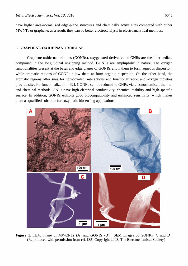
Figure 1. TEM image of MWCNTs (A) and GONRs (B). SEM images of GONRs (C and D).
(Reproduced with permission from ref. [35] Copyright 2003, The Electrochemical Society)
Int. J. Electrochem. Sci., Vol. 13, 2018
6646
The abundant oxygen functionalities, such as hydroxyl, epoxide, carbonyl, and carboxyl groups
located on the surfaces and edges of GONRs offer great potential for attaching biorecognition
molecules such as antibodies [33]. The carboxyl groups located on GONRs can be cross-linked to the
amine groups of proteins through well-established carbodiimide crosslinker chemistry. Recently, our
research group described a GONRs modified screen-printed carbon electrode (SPCE) for folic acid
detections [34]. Our studies revealed that the GONRs exhibited superior electrocatalytic ability over
MWNTs ascribed to their rich edge chemistry and catalytic properties.
In another report, we described GONRs modified SPCE for sensitive determination of methyl
parathion (Figure 1) [35]. GONRs/SPCE displayed significantly improved electrocatalytic ability
towards methyl parathion in comparison with MWCNTs. The modified electrode was shown excellent
real-time applicability in food analysis, which was successfully demonstrated in food samples such as,
Ugli and tomato fruits, Beetroot and Broccoli indicating its excellent practical applicability.
4. GNRs BASED NANOMATERIALS IN ELECTROCHEMICAL SENSORS
Several GNRs based nanomaterials have been developed in recent times for electroanalytical
sensing applications [29]. The hybridization of GNRs with metal nanoparticles can provide
nanocomposites of synergic properties and this behaviour is similar to the graphene-based composites
[26, 29].
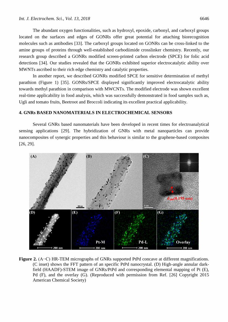
Figure 2. (A−C) HR-TEM micrographs of GNRs supported PtPd concave at different magnifications.
(C inset) shows the FFT pattern of an specific PtPd nanocrystal. (D) High-angle annular dark-
field (HAADF)-STEM image of GNRs/PtPd and corresponding elemental mapping of Pt (E),
Pd (F), and the overlay (G). (Reproduced with permission from Ref. [26] Copyright 2015
American Chemical Society)

Int. J. Electrochem. Sci., Vol. 13, 2018
6647
Besides, GNRs can be a good support for making nanoparticles dispersions because of their
large surface area, high electrical conductivity, and electrochemical stability in acidic and alkaline
electrolytes. For instance, highly stable and catalytically active PtPd concave nanocubes were
anchored on GNRs through a hydrothermal process [26]. the TEM image of GNRs/PtPd revealed that
the average particle size of PtPd concave cubes was 11 nm and the particles are uniformly placed on
the surface of GNRs (figure 2).
Recently, we have described silver particles decorated GNRs prepared through simple wet
chemical method and the resulting nanomaterial have shown excellent sensing attributes towards
Organophosphorus pesticide methyl parathion [36]. A highly sensitive (sub-nanomolar level) methyl
parathion sensor was fabricated using Ag@GNRs/SPCE and its practical applicability was
successfully demonstrated in in food samples such as, cabbage, green beans, strawberry, and nectarine
samples. The synergic effects between Ag and excellent physicochemical properties of GNRs make
the composite highly suitable for pesticide sensing. A nanocomposite of polyaniline (PANI)-GNRs
was prepared by in situ polymerization of aniline in presence of GNRs [37]. Using this cost-effective
method, highly ordered and vertically aligned PANI nanorods can be grown on GNRs (Figure 3).
Figure 3. SEM images of PANI-GNRs composite at (a) low resolution and (b) high resolution. TEM
image of PANI-GNRs composite (c) and (d). (Reproduced with permission from Ref. [37]
Copyright 2013 American Chemical Society)
Several GNRs based nanomaterials have been prepared and demonstrated in electrochemical
sensing applications; polyaniline nanorods grown on GNRs [37], GNRs supported PtPd concave
nanocubes [26], PdNi nanoparticles and N-doped GNRs [27, 38], graphene quantum dots (GQDs)
Int. J. Electrochem. Sci., Vol. 13, 2018
6648
supported GNRs [39], Ag/GNRs composite[40], PtPd/GNRs alloy [41], GNR-Pt nanocomposite [24],
PdAg alloy nanoparticles modified ionic liquid functionalized GNRs [42], GNR/Co coordination
polymer nanohybrids [43], GNRs/polyaniline [44], GNR/polypyrrole [45], GONRs/Au nanoparticle
hybrid [46], GONR/poly(diallyldimethylammonium chloride)/Au nanoparticle [47], GNRs/nafion
nanocomposite [48], Pd-functionalized GNRs [49], heteroatoms doped GNRs [50], graphene
sheet/GNRs [51], Core–shell MWNTs/GONR heterostructure [18], poly(l-arginine)-β-
cyclodextrin/carbon nanotubes@GNRs [52], electrochemically reduced GONR [53], and molecularly
imprinted polymer (MIP)–GNR composite [54].
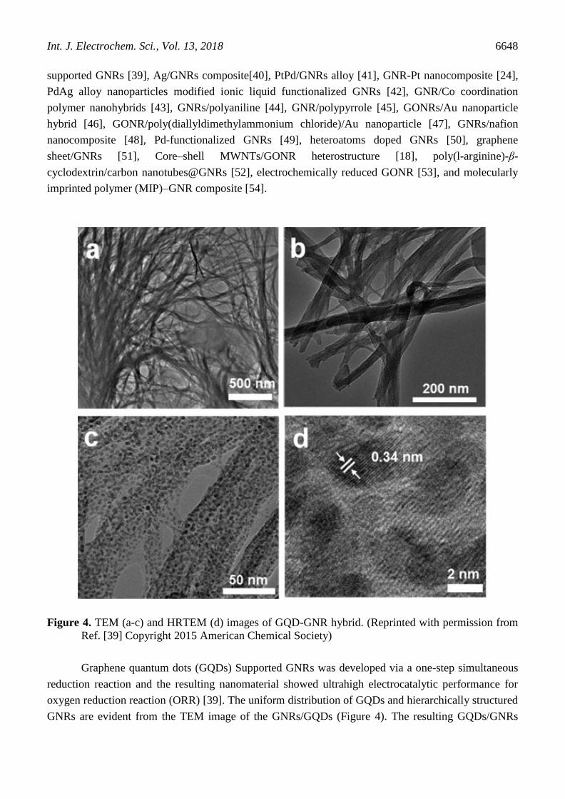
Figure 4. TEM (a-c) and HRTEM (d) images of GQD-GNR hybrid. (Reprinted with permission from
Ref. [39] Copyright 2015 American Chemical Society)
Graphene quantum dots (GQDs) Supported GNRs was developed via a one-step simultaneous
reduction reaction and the resulting nanomaterial showed ultrahigh electrocatalytic performance for
oxygen reduction reaction (ORR) [39]. The uniform distribution of GQDs and hierarchically structured
GNRs are evident from the TEM image of the GNRs/GQDs (Figure 4). The resulting GQDs/GNRs

Int. J. Electrochem. Sci., Vol. 13, 2018
6649
hybrid delivered higher limiting current density and lower overpotential than those of platinum and
high selectivity and stability in alkaline media. An ultra-sensitive, reproducible, stable and practically
applicable modified electrode based on electrochemically reduced (ER)-GONRs was fabricated for the
determination of nimesulide, a non-steroidal anti-inflammatory drug [55]. The sensor has excellent
sensor performance and exhibited wide linear range (1.0×10⁻⁸
– 1.50×10⁻³
), low detection limit (3.50
nM), high sensitivity (1.20 µA. µM–1
.cm–2
) and fast response (<5 s) towards nimesulide detection. The
assay demonstrated in drug and urine samples revealed the good practical feasibility of the ER-
GONRs/SPCE in drug and clinical analysis.
A PtPd/reduced GONRs nanocomposite based modified electrode displayed excellent sensing
performance towards trinitrotoluene (TNT) [26]. It exhibited a high sensing performance for TNT
detection with a linear range of 0.01 ppm to 3 ppm and a detection limit of 0.8 ppb through adsorption
stripping voltammetry (Figure 5)
Figure 5. (A) Electrochemical impedance spectra (EIS) obtained at PtPd-rGONRs/GCE,
rGONRs/GCE and unmodified GCE in 0.1 M KCl containing 5 mM K4[Fe(CN)6]/K3[Fe(CN)6].
(B) Stripping voltammograms obtained at PtPdrGONRs/GCE, rGONRs/GCE, and bare GCE in
0.1 M phosphate buffer (pH =6.5) containing 0.4 M KCl and 3 ppm of TNT. (C) Stripping
voltammograms obtained at PtPd-rGONRs/GCE with varied concentrations of TNT in 0.1 M
phosphate buffer (pH = 6.5) with 0.4 M KCl. (D) Linear regression plot between peak current
density and concentration of TNT concentrations at −0.33 V. The error bars represent the
standard deviation of three individual measurements. (Reproduced with permission from Ref.
[26] Copyright 2015 American Chemical Society)
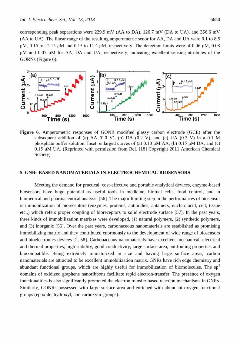
A microwave-assisted unzipping process was reported for the rapid synthesis of GONRs [18].
The GONRs able to separate the voltammetric peaks of ascorbic acid, dopamine, and uric acid and the
Int. J. Electrochem. Sci., Vol. 13, 2018
6650
corresponding peak separations were 229.9 mV (AA to DA), 126.7 mV (DA to UA), and 356.6 mV
(AA to UA). The linear range of the resulting amperometric senor for AA, DA and UA were 0.1 to 8.5
μM, 0.15 to 12.15 μM and 0.15 to 11.4 μM, respectively. The detection limits were of 0.06 μM, 0.08
μM and 0.07 μM for AA, DA and UA, respectively, indicating excellent sensing attributes of the
GORNs (Figure 6).
Figure 6. Amperometric responses of GONR modified glassy carbon electrode (GCE) after the
subsequent addition of (a) AA (0.0 V), (b) DA (0.2 V), and (c) UA (0.3 V) in a 0.1 M
phosphate buffer solution. Inset: enlarged curves of (a) 0.10 μM AA, (b) 0.15 μM DA, and (c)
0.15 μM UA. (Reprinted with permission from Ref. [18] Copyright 2011 American Chemical
Society)
5. GNRs BASED NANOMATERIALS IN ELECTROCHEMICAL BIOSENSORS
Meeting the demand for practical, cost-effective and portable analytical devices, enzyme-based
biosensors have huge potential as useful tools in medicine, biofuel cells, food control, and in
biomedical and pharmaceutical analysis [56]. The major limiting step in the performances of biosensor
is immobilization of bioreceptors (enzymes, proteins, antibodies, aptamers, nucleic acid, cell, tissue
etc.,) which refers proper coupling of bioreceptors to solid electrode surface [57]. In the past years,
three kinds of immobilization matrixes were developed, (1) natural polymers, (2) synthetic polymers,
and (3) inorganic [56]. Over the past years, carbonaceous nanomaterials are established as promising
immobilizing matrix and they contributed enormously to the development of wide range of biosensors
and bioelectronics devices [2, 58]. Carbonaceous nanomaterials have excellent mechanical, electrical
and thermal properties, high stability, good conductivity, large surface area, antifouling properties and
biocompatible. Being extremely miniaturized in size and having large surface areas, carbon
nanomaterials are attracted to be excellent immobilization matrix. GNRs have rich edge chemistry and
abundant functional groups, which are highly useful for immobilization of biomolecules. The sp2
domains of oxidized graphene nanoribbons facilitate rapid electron-transfer. The presence of oxygen
functionalities is also significantly promoted the electron transfer based reaction mechanisms in GNRs.
Similarly, GONRs possessed with large surface area and enriched with abundant oxygen functional
groups (epoxide, hydroxyl, and carboxylic groups).

Int. J. Electrochem. Sci., Vol. 13, 2018
6651
Recently, a field effect transistor (FET) biosensor was developed using solution-processed
GONRs and the sensor was applied in sensing methylene blue [28] and adenosine triphosphate [59].
Studies revealed that GNRs possess intrinsic peroxidase-like catalytic activity and hence it is a desired
material for biosensing applications [60]. GNRs based nanocomposites are reported to be
biocompatible and suitable for immobilizing bio-receptors such as, biomolecules, antibodies, aptamers
etc. [61]. The use of PdNi nanoparticles/N-doped graphene nanoribbons for immobilizing adamantine-
1-carboxylic acid functionalized primary anti-AFP (ADA–Ab1) was described, which was used to
develop an immunosensor for the detection of alpha-fetoprotein. A sensitive sandwich-type
electrochemical immunosensor was described using Au nanoparticles/NH2–GNRs as immobilizing
matrix for quantitative detection of squamous cell carcinoma antigen [62]. GNRs were used to develop
an ultrasensitive electrochemical immunoassay for rapid detection of interleukin-6 and matrix
metallopeptidase-9 [63]. An electrochemical DNA biosensor was developed for the detection of
genetically modified soybean by anchoring DNA on Au nanoparticles/MWNTs-reduced GONR.[64]
Guanine was electrochemically immobilized on GNR modified electrode, which was used as a DNA
biosensor for the evaluation of total antioxidant capacities (TAC) in fruit juices [65]. A surface
enhanced laser desorption/ionization mass spectrometry probe based on GONRs-polyethylene glycol-
antibody biomatrix was prepared for chloramphenicol detection [66]. A label-free impedimetric
aptasensor that detects femtomolar level of acetamiprid was developed using Au decorated MWNTs/
reduced GONR composite [67]. An electrochemical carbaryl biosensor was developed using acetyl
cholinesterase immobilized MWNTs/GONRs nanostructure [68]. A third generation glucose biosensor
was developed based on wiring of apo-enzyme of glucose oxidase with GNR bound to FAD at a
screen-printed carbon electrode [69]. A magneto-controlled electrochemical immunoassay was
developed for the detection of brevetoxin B in seafood using guanine-functionalized GNR molecular
tag [31] An ultrasensitive immunoassay was developed to for detect human cardiopathy biomarkers
cardiac troponin I (cTnI) and heart-type fatty-acid-binding protein (FABP) using GONRs as
immobilization matrix.
5. CONCLUSIONS
In this review, we have summarized remarkable advances in the development of novel
ultrasensitive electrochemical sensors based on graphene nanoribbon based modified electrodes. Major
preparation routes that can produce GNRs/GONRs are described and discussed. GNRs/GONRs based
nanomaterials are ideal immobilization matrix owing to their large surface area, high conductivity,
biocompatibility, high stability, good adhesion, inertness, affordability, physical strength,
regenerability, ability to increase enzyme specificity/activity and hindrance to product inhibition and
nonspecific adsorption. GONRs/GNRs based selective and sensitive sensors are presented and
discussed. Sensors ranging from electrocatalytic to affinity-based are discussed. Additional
electrocatalyst such as, metal nanoparticles, metal oxides, polymers and other nanomaterials are
attached on GONRs/GNRs surfaces to enable additional sensitivity and selectivity.

Int. J. Electrochem. Sci., Vol. 13, 2018
6652
ACKNOWLEDGEMENTS
The authors gratefully acknowledge the financial support of the National Taipei University of
Technology, and Mackay Memorial Hospital Joint Research Program(NTUT-MMH-No.107-01). The
National Science Council, and The Ministry of Education, Taiwan supported this work. We would also
like to acknowledge The Ministry of Science and Technology, Taiwan (MOST 106-2113-M-027-003)
for its financial support.
References
1. M.-H. Yeh, L.-Y. Lin, C.-L. Sun, Y.-A. Leu, J.-T. Tsai, C.-Y. Yeh, R. Vittal, K.-C. Ho, J. Phys.
Chem. C, 118 (2014) 16626.
2. V. Mani, B. Devadas, S.-M. Chen, Biosens. Bioelectron., 41 (2013) 309.
3. D.R. Dreyer, S. Park, C.W. Bielawski, R.S. Ruoff, Chem. Soc. Rev., 39 (2010) 228.
4. M.Y. Han, B. Özyilmaz, Y. Zhang, P. Kim, Phys. Rev. Lett., 98 (2007) 206805.
5. Y. Shao, J. Wang, H. Wu, J. Liu, I.A. Aksay, Y. Lin, Electroanalysis, 22 (2010) 1027.
6. X. Huang, X. Qi, F. Boey, H. Zhang, Chem. Soc. Rev., 41 (2012) 666.
7. M.A. Rafiee, W. Lu, A.V. Thomas, A. Zandiatashbar, J. Rafiee, J.M. Tour, N.A. Koratkar, ACS
nano, 4 (2010) 7415.
8. S. JooáLee, H. PiláKim, W.J. Silva, F. KurtáSchneider, A.R. bináMohd Yusoff, Chem. Commun.,
51 (2015) 9185.
9. L. Ma, J. Wang, F. Ding, ChemPhysChem, 14 (2013) 47.
10. X. Li, X. Wang, L. Zhang, S. Lee, H. Dai, Science, 319 (2008) 1229.
11. L. Tapasztó, G. Dobrik, P. Lambin, L.P. Biró, Nat. Nanotechnol., 3 (2008) 397.
12. J. Campos-Delgado, J.M. Romo-Herrera, X. Jia, D.A. Cullen, H. Muramatsu, Y.A. Kim, T.
Hayashi, Z. Ren, D.J. Smith, Y. Okuno, Nano Lett., 8 (2008) 2773.
13. D.V. Kosynkin, A.L. Higginbotham, A. Sinitskii, J.R. Lomeda, A. Dimiev, B.K. Price, J.M. Tour,
Nature, 458 (2009) 872.
14. L. Jiao, L. Zhang, X. Wang, G. Diankov, H. Dai, Nature, 458 (2009) 877.
15. S. Vadahanambi, J.-H. Jung, R. Kumar, H.-J. Kim, I.-K. Oh, Carbon, 53 (2013) 391.
16. A.G. Cano-Márquez, F.J. Rodríguez-Macías, J. Campos-Delgado, C.G. Espinosa-González, F.
Tristán-López, D. Ramírez-González, D.A. Cullen, D.J. Smith, M. Terrones, Y.I. Vega-Cantú,
Nano Lett., 9 (2009) 1527.
17. A.L. Elías, A.R. Botello-Méndez, D. Meneses-Rodríguez, V. Jehová González, D. Ramírez-
González, L. Ci, E. Muñoz-Sandoval, P.M. Ajayan, H. Terrones, M. Terrones, Nano Lett., 10
(2009) 366.
18. C.-L. Sun, C.-T. Chang, H.-H. Lee, J. Zhou, J. Wang, T.-K. Sham, W.-F. Pong, ACS nano, 5 (2011)
7788.
19. W. Xu, T.-W. Lee, Mater. Horiz., 3 (2016) 186.
20. B. Chitara, L. Panchakarla, S. Krupanidhi, C. Rao, Adv. Mater., 23 (2011) 5419.
21. L. Li, A.R.O. Raji, J.M. Tour, Adv. Mater., 25 (2013) 6298.
22. L.-Y. Lin, M.-H. Yeh, J.-T. Tsai, Y.-H. Huang, C.-L. Sun, K.-C. Ho, J. Mater. Chem. A, 1 (2013)
11237.
23. M. Liu, W.W. Tjiu, J. Pan, C. Zhang, W. Gao, T. Liu, Nanoscale, 6 (2014) 4233.
24. C. Wang, H. Li, J. Zhao, Y. Zhu, W.Z. Yuan, Y. Zhang, Inter. J. Hydrogen Energy, 38 (2013)
13230.
25. C. Zhao, P. Gai, C. Liu, X. Wang, H. Xu, J. Zhang, J.-J. Zhu, J. Mater. Chem. A, 1 (2013) 12587.
26. R. Zhang, C.-L. Sun, Y.-J. Lu, W. Chen, Anal. Chem., 87 (2015) 12262.
27. N. Li, H. Ma, W. Cao, D. Wu, T. Yan, B. Du, Q. Wei, Biosens. Bioelectron., 74 (2015) 786.
28. T.-C. Lin, Y.-S. Li, W.-H. Chiang, Z. Pei, Biosens. Bioelectron., 89 (2017) 511.
29. G. Zhu, Y. Yi, Z. Liu, H.J. Lee, J. Chen, Electrochem. Commun., 66 (2016) 10.
Int. J. Electrochem. Sci., Vol. 13, 2018
6653
30. F. Valentini, D. Romanazzo, M. Carbone, G. Palleschi, Electroanalysis, 24 (2012) 872.
31. A. Martín, J. Hernández-Ferrer, L. Vázquez, M.-T. Martínez, A. Escarpa, RSC Adv., 4 (2014) 132.
32. S. Zhang, S. Tang, J. Lei, H. Dong, H. Ju, J. Electroanal. Chem., 656 (2011) 285.
33. L.-N. Feng, Z.-P. Bian, J. Peng, F. Jiang, G.-H. Yang, Y.-D. Zhu, D. Yang, L.-P. Jiang, J.-J. Zhu,
Anal. Chem., 84 (2012) 7810.
34. V. Mani, R. Umamaheswari, S.-M. Chen, M. Govindasamy, C. Su, A. Sathiyan, J.P. Merlin, M.
Keerthi, Int. J. Electrochem. Sci., 12 (2017) 475.
35. M. Govindasamy, R. Umamaheswari, S.-M. Chen, V. Mani, C. Su, J. Electrochem. Soc., 164 (2017)
B403.
36. M. Govindasamy, V. Mani, S.-M. Chen, T.-W. Chen, A.K. Sundramoorthy, Sci. Rep., 7 (2017)
46471.
37. L. Li, A.-R.O. Raji, H. Fei, Y. Yang, E.L. Samuel, J.M. Tour, ACS Appl. Mater. Interfaces, 5
(2013) 6622.
38. L. Shi, X. Niu, T. Liu, H. Zhao, M. Lan, Microchim. Acta, 182 (2015) 2485.
39. H. Jin, H. Huang, Y. He, X. Feng, S. Wang, L. Dai, J. Wang, J. Am. Chem. Soc., 137 (2015) 7588.
40. D.J. Davis, A.R.O. Raji, T.N. Lambert, J.A. Vigil, L. Li, K. Nan, J.M. Tour, Electroanalysis, 26
(2014) 164.
41. Y. Liu, L. Wei, Y. Hu, X. Huang, J. Wang, J. Li, X. Hu, N. Zhuang, J. Alloys Compd., 656 (2016)
452.
42. L. Shang, F. Zhao, B. Zeng, Electrochim. Acta, 168 (2015) 330.
43. S.K. Ujjain, P. Ahuja, R.K. Sharma, J. Mater. Chem. B, 3 (2015) 7614.
44. E. Asadian, S. Shahrokhian, E. Jokar, Sens. Actuators, B, 196 (2014) 582.
45. K. Harpale, S. Bansode, M. More, D. Late, J. Appl. Polymer. Sci., 134 (2017) 45170
46. N.S. Ismail, Q.H. Le, H. Yoshikawa, M. Saito, E. Tamiya, Electrochim Acta, 146 (2014) 98.
47. X.-P. Liu, J. Tong, Z. Yuan, Y. Yang, C.-J. Mao, H.-L. Niu, B.-K. Jin, S.-Y. Zhang, J. Nanosci.
Nanotechnol., 16 (2016) 1645.
48. S. Wu, X. Lan, F. Huang, Z. Luo, H. Ju, C. Meng, C. Duan, Biosens. Bioelectron., 32 (2012) 293.
49. J.L. Johnson, A. Behnam, S. Pearton, A. Ural, Adv. Mater., 22 (2010) 4877.
50. A. Zehtab Yazdi, E.P.L. Roberts, U. Sundararaj, Carbon, 100 (2016) 99.
51. J. Lavanya, N. Gomathi, Talanta, 144 (2015) 655.
52. Y. Yi, G. Zhu, X. Wu, K. Wang, Biosens. Bioelectron., 77 (2016) 353.
53. N.L. Teradal, J. Seetharamappa, A. Satpati, RSC Adv., 5 (2015) 55550.
54. Y. Pan, F. Zhao, B. Zeng, RSC Adv., 5 (2015) 57671.
55. M. Govindasamy, V. Mani, S.-M. Chen, T. Maiyalagan, S. Selvaraj, T.-W. Chen, S.-Y. Lee, W.-H.
Chang, RSC Adv., 7 (2017) 33043.
56. S. Datta, L.R. Christena, Y.R.S. Rajaram, 3 Biotech, 3 (2013) 1.
57. B. Unnikrishnan, S. Palanisamy, S.-M. Chen, Biosens. Bioelectron., 39 (2013) 70.
58. V. Mani, B. Dinesh, S.-M. Chen, R. Saraswathi, Biosens. Bioelectron., 53 (2014) 420.
59. X. Dong, Q. Long, J. Wang, M.B. Chan-Park, Y. Huang, W. Huang, P. Chen, Nanoscale, 3 (2011)
5156.
60. J. Qian, X. Yang, Z. Yang, G. Zhu, H. Mao, K. Wang, J. Mater. Chem. B, 3 (2015) 1624.
61. Y.-R. Liang, Z.-M. Zhang, Z.-J. Liu, K. Wang, X.-Y. Wu, K. Zeng, H. Meng, Z. Zhang, Biosens.
Bioelectron., 91 (2017) 199.
62. Y. Li, Y. Zhang, J. Han, P.K. Chu, J. Feng, Y. Dong, RSC Adv., 7 (2017) 2242.
63. J.-J. Shi, T.-T. He, F. Jiang, E. Abdel-Halim, J.-J. Zhu, Biosens. Bioelectron., 55 (2014) 51.
64. S. Wang, Q. Liu, H. Li, Y. Li, N. Hao, J. Qian, W. Zhu, K. Wang, J. Electroanal. Chem., 782
(2016) 19.
65. Y. Yang, J. Zhou, H. Zhang, P. Gai, X. Zhang, J. Chen, Talanta, 106 (2013) 206.
66. J. Wang, M. Cheng, Z. Zhang, L. Guo, Q. Liu, G. Jiang, Chem. Commun., 51 (2015) 4619.

Int. J. Electrochem. Sci., Vol. 13, 2018
6654
67. A. Fei, Q. Liu, J. Huan, J. Qian, X. Dong, B. Qiu, H. Mao, K. Wang, Biosens. Bioelectron., 70
(2015) 122.
68. Q. Liu, A. Fei, J. Huan, H. Mao, K. Wang, J. Electroanal. Chem., 740 (2015) 8.
69. E. Mehmeti, D.M. Stanković, S. Chaiyo, J. Zavasnik, K. Žagar, K. Kalcher, Microchim. Acta,
(2017) 1.
© 2018 The Authors. Published by ESG (www.electrochemsci.org). This article is an open access
article distributed under the terms and conditions of the Creative Commons Attribution license
(http://creativecommons.org/licenses/by/4.0/).