green synthesis and characterizations of silver and gold
TRANSCRIPT

8
Green Synthesis and Characterizations of Silver and Gold Nanoparticles
Nora Elizondo et al.* Facultad de Ciencias Físico-Matemáticas, N. L., CP 66451,
México
1. Introduction
Metallic nanoparticles (nps) are of great interest because of the modification of properties
observed due to size effects, modifying the catalytic, electronic, and optical properties of the
monometallic nps.[Bronstein et al., 2000; Chushak & Bartell, 2003; Tomas, 2003]
In the last years, biosynthesis of nps have been received considerable attention due to the
growing need to develop clean, nontoxic chemicals, environmentally benign solvents and
renewable materials [Gericke and Pinches, 2006; Harris and Bali, 2008]. As a result,
researchers in the field of nanoparticle synthesis and assembly have turned towards the
utilization of biological system such as yeast, fungi, bacteria and plant extracts for the
synthesis of biocompatible metal and semiconductor nps through control nucleation and
growth of inorganic nps [Kasthuri et al., 2009; Lee et al., 2011; Shankar et al., 2003].
The green method employing plant extracts have drawn attention as a simple and viable
alternative to chemical procedures and physical methods, which consist of a low
concentration of gold or silver precursor that is added to plant extract in solution to make
up a final solution and centrifuged. The supernatant is heated at 50°C to 95°C. A change in
the color of solution is observed during the heating process. Bioreduction of silver ions to
yield metal nanoparticles using living plants, geranium leaf [Shankar et al., 2003], Neem leaf
[Shankar et al., 2004a]. Very recently, they have demonstrated synthesis of gold
nanotriangles and silver nps using Aloevera plant extracts [Chandran et al., 2006], Emblica
officinalis (amla, Indian Gooseberry).[Amkamwar et al., 2005] Most of the above research on
the synthesis of silver or gold nps utilizing plant extracts employed broths resulting from
boiling fresh plant leaves. The green synthesis of silver nps using Capsicum annuum leaf
extract has been reported.[Li et al., 2007] According to previous reports, the polyol
components and the water-soluble heterocyclic components are mainly responsible for the
* Paulina Segovia1,3, Víctor Coello3, Jesús Arriaga1, Sergio Belmares1, Aracelia Alcorta1, Francisco Hernández1, Ricardo Obregón1, Ernesto Torres2 and Francisco Paraguay4
1Facultad de Ciencias Físico-Matemáticas, México 2Facultad de Medicina, México Universidad Autónoma de Nuevo León, San Nicolás de los Garza, N. L., México 3CICESE, Monterrey, PIIT, Apodaca, N. L., México 4 CIMAV, Chihuahua, Complejo Ind. Chih., Chihuahua, Chih., México
www.intechopen.com

Green Chemistry – Environmentally Benign Approaches
140
reduction of silver ions and the stabilization of the nps, respectively[Arangasamy &
Munusamy, 2008; Nagajyoti et al., 2011].
Specific synthesis of nps and nanostructured materials are attracting attention in recent research because of their valuable properties which make them useful for catalysis, [El-Sayed & Narayanan, 2004] sensor technology, [Gomez-Romero, 2001] biological labeling, [Shankar et al., 2003] optoelectronics recording media and optics.[Qiu et al. 2004] The size, shape and surface morphology play pivotal roles in controlling the physical, chemical, optical and electronic properties of these nanoscopic materials.[Gracias et al., 2002; Kamat, 2002 ] This is particularly important for noble metals such as Au and Ag which have strong surface plasmon resonance (SPR) oscillations. The shape-selective metal nps such as rods, tubes, wires, triangles, prisms, hexagons and cubes can be regularly synthesized by chemical, biological and physical methods. [El-Sayet, 2001; Lim et al., 2008]
Many colloidal methods of synthesis have been approached to obtain metallic nps for this purpose, such as homogeneous reduction in aqueous solutions,[Shankar et al., 2004b]or phase transfer reactions,[Liz-Marzan & Philipse, 1995] with sodium citrate, hydrazine, NaBH4, and lithium triethylborohydride (LiBEt3H) as reducing agents, each of them yielding products with different physicochemical and structural characteristics.[Han et al., 1998] Among these, the polyol method has been reported to produce small nps as the final product, easily changing composition and surface modifiers. This technique does not require an additional reducing agent since the solvent by itself reduces the metallic species. However, besides the stoichiometry and order of addition of reagents in the synthesis process, one of the most important parameters in the preparation is the temperature. Modifications in temperature influence the reaction by changing the stabilization of the nps formed and the surface modifiers, e.g., PVP, and the nucleation rate of the reduced metallic atoms.[Schmid, 1994]
Gold (Au) and silver (Ag) nps have a diversity of interesting properties between which they emphasize the electrical ones, optical, catalytic and the applications in biomedicine like antibacterial and antiviral, same that depend on their morphology and size.
Characterization of these systems has been a difficult process where researchers have employed indirect measurements to identify the localization of the elements within the nps. A novel approach to study this kind of particles is based on the use of a high angle annular dark field (HAADF) technique, in a transmission electron microscope (TEM), which allows the observation of the elements due to atomic number, densities, or the presence of strain fields due to differences in lattice parameters, structure, the presence of surfactants or any other surface modifier besides the size of the particle and also by near-field scanning optical microscopy (NSOM) we determine the size of the particles.[Henglein, 2000; Turkevich et al., 1951]
The nps were synthesized using polyol and green methods. We made a comparison of these methods in order to investigate the influence of reaction parameters on the resulting particle size and its distribution. In the first method we use polyol process with poly (vinylpyrrolidone) (PVP) acting as a stabilizer and ethylenglycol as a reductor.[ Cao, 2004; Park et al., 2008] Such procedure yield different morphologies of metal nps (including gold and silver). [Burda et al., 2005; Gonzalez et al., 2009; Kasthuri et al., 2009; Rosi & Mirkin, 2005; Safaepour et al., 2009; Xia & Halas, 2005] The green method is an ecological synthesis
www.intechopen.com

Green Synthesis and Characterizations of Silver and Gold Nanoparticles
141
technique. There, we made use of chemical compounds of plants like Rosa Berberifolia and Geranium Maculatum in order to obtain ascorbic acid and polyphenols as reductor agents. Ascorbic acid (C6H8O6) and polyphenols like hydroxyphenol compounds are abundant components of plants. Ascorbic acid reaches a concentration of over 20 milimols in chloroplasts and occurs in all cell compartments including the cell wall. Additionally the acid has functions in photosynthesis as an enzyme cofactor (including synthesis of ethylene, gibberellins and anthocyanins) and in the control of cell growth. [Altansukha, 2010; Smirnoff & Wheeler, 2000] In nature, polyphenol is one of the most important chemicals in many reductive biological reactions widely found in plants and animals. The hydroxyphenol compounds and their derivatives could be used as versatile reducing agents for facile one-pot synthesis of gold and silver nanoparticles with diverse morphological characters. Most of the reports on the biological synthesis of metal nps utilizing plant extracts employed broths obtained from boiled fresh plant leaves. In this present study, we report on the synthesis of silver and gold nps using Aloe Barbadensis and Cucurbita Digitata extracts at 60°C. The approach is a simple, cost-effective, stable for long time, reproducible and previously unexploited method excellent for nanofabrication. These plants are predominant species in America especially in Mexico. In the case of Aloe Barbadensis it has been used for medical applications such as there is some preliminary evidence that Aloe Vera extracts may be useful in the treatment of wound and burn healing, minor skin infections, sebaceous cysts, diabetes, and elevated blood lipids in humans(from Wikipedia).[Harris & Bali 2008] These positive effects are thought to be due to the presence of compounds such as polysaccharides, mannans, anthraquinones, and lectins.[Boudreau & Beland 2006; Eshun & He 2004; King et al., 1995; Vogler & Ernst 1999]
We use for the synthesizes plants like Aloe Barbadensis and cactus plants like Cucurbita Digitata that is a reminder plant in Mexico, also with compounds that have surfactant properties like saponins. Here, we show that green method reduces the temperature requirement which is in contrast to the obtained with the polyol method. The use of these natural components allows synthesize gold and silver nps.
2. Experimental section
The polyol method was followed to obtain nps passivated with poly(vinylpyrrolidone) (PVP). Hydrogen tetrachloroaurate (HAuCl4) (III) hydrate (99.99%), silver nitrate (AgNO3) (99.99%), and poly (N-vinyl-2-pyrrolidone) (PVP-K30, MW = 40000) were purchased from Sigma Aldrich, and 1, 2-ethylenediol (99.95%) was purchased from Fischer Chemicals; all the materials were used without any further purification. A 0.4 g sample of Poly (N-vinyl-2-pyrrolidone) (PVP) was dissolved in 50 mL of 1,2-ethylenediol (EG) under vigorous stirring, heating in reflux, until the desired temperature was reached (working temperatures ranged from 140°C to 190°C in increments of 10°C). For the monometallic nps, a 0.1 mM aqueous solution of the metal precursor was added to the EG-PVP solution, with continuous agitation for 3 h in reflux.
The green method is an ecological synthesis technique. There, we made use of chemical compounds of plants like Rosa Berberifolia, Geranium Maculatum, Aloe Barbadensis and Cucurbita Digitata in order to obtain ascorbic acid as reductor agent from the extracts of these plants as can be seen from figure 2. Ascorbic acid (C6H8O6) is an abundant component of plants, which reaches a concentration of over 20 milimols in chloroplasts and occurs in all
www.intechopen.com
Green Chemistry – Environmentally Benign Approaches
142
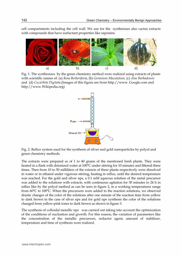
cell compartments including the cell wall. We use for the synthesizes also cactus extracts with compounds that have surfactant properties like saponins.
a) b) c) d)
Fig. 1. The synthesizes by the green chemistry method were realized using extracts of plants with scientific names of: (a) Rosa Berberiforia, (b) Geranium Maculatum, (c) Aloe Barbadensis and (d) Cucúrbita Digitata.(Images of this figure are from http://www. Google.com and http://www.Wikipedia.org)
Fig. 2. Reflux system used for the synthesis of silver and gold nanoparticles by polyol and green chemistry methods.
The extracts were prepared as of 1 to 40 grams of the mentioned fresh plants. They were heated in a flask with deionized water at 100°C under stirring for 10 minutes and filtered three times. Then from 10 to 50 milliliters of the extracts of these plants respectively were dissolved in water or in ethanol under vigorous stirring, heating in reflux, until the desired temperature was reached. For the gold and silver nps, a 0.1 mM aqueous solution of the metal precursor was added to the solutions with extracts, with continuous agitation for 30 minutes to 24 h in reflux like by the polyol method as can be seen in figure 2, in a working temperatures range from 60°C to 100°C. When the precursors were added to the reaction solutions, we observed drastic changes of the color of the solutions after one minute of the reaction time from yellow to dark brown in the case of silver nps and for gold nps synthesis the color of the solutions changed from yellow-pink tones to dark brown as shown in figure 3.
The synthesis of colloidal metallic nps was carried out taking into account the optimization of the conditions of nucleation and growth. For this reason, the variation of parameters like the concentration of the metallic precursors, reductor agent, amount of stabilizer, temperature and time of synthesis were realized.
www.intechopen.com

Green Synthesis and Characterizations of Silver and Gold Nanoparticles
143
Fig. 3. Photography of monometallic colloidal dispersions of gold nanoparticles in the solutions with the extracts of Aloe Barbadensis, the change of color is characteristic of gold and a function of the physical properties of metallic nanoparticles obtained by green method.
For the electron microscopy analysis of the metallic nps, samples were prepared over carbon
coated copper TEM grids. HAADF and HRTEM images were taken with a JEOL 2010F and a
FEI TITAN microscopes in the STEM mode, with the use of a HAADF detector with
collection angles from 50 mrad to 110 mrad. Also by near-field scanning optical microscopy
(NSOM) we determine the size of the particles. UV-vis spectra were obtained using a 10 mm
path length quartz cuvette in a Cary 5000 equipment.
3. Results and discussion
It is well known that the morphology and size distribution of metallic particles produced by
the reduction of metallic salts in solution depends on various reaction conditions such as
temperature, time, concentration, molar ratio of metallic salt/reducing agent, mode and
order of addition of reagents, presence and type of protective agents, degree and type of
agitation, and whether nucleation is homogeneous or heterogeneous [Sanguesa et al., 1992].
Following the polyol method with ethylene glycol as solvent reductor, it was possible to
obtain monometallic nanoparticles with narrow size distributions in systems and different
structures depending on the temperature of reaction. The monometallic synthesis of
nanoparticles by itself showed distinctive morphologies of the nanoparticles depending on
the temperature of reaction.
Reaction proceeds in general as an oxidation of the ethylene glycol reducing the metallic
precursor to its zero-valence state. [Carotenuto et al. 2000; Sun et al., 2002]
OH-CH2-CH2-OH → CH3-CHO + H2O (1)
6(CH3-CHO) + 2HAuCl4 → 3(CH3-CO-CO-CH3) + 2Au0 + 8HCl (2)
This reaction describes the reduction of Au+ to Au0.
OH-CH2-CH2-OH → CH3-CHO + H2O (3)
2(CH3-CHO) + AgNO3 → (CH3-CO-CO-CH3) + Ag0 + HNO3 (4)
www.intechopen.com
Green Chemistry – Environmentally Benign Approaches
144
This reaction describes the reduction of Ag+ to Ag0.
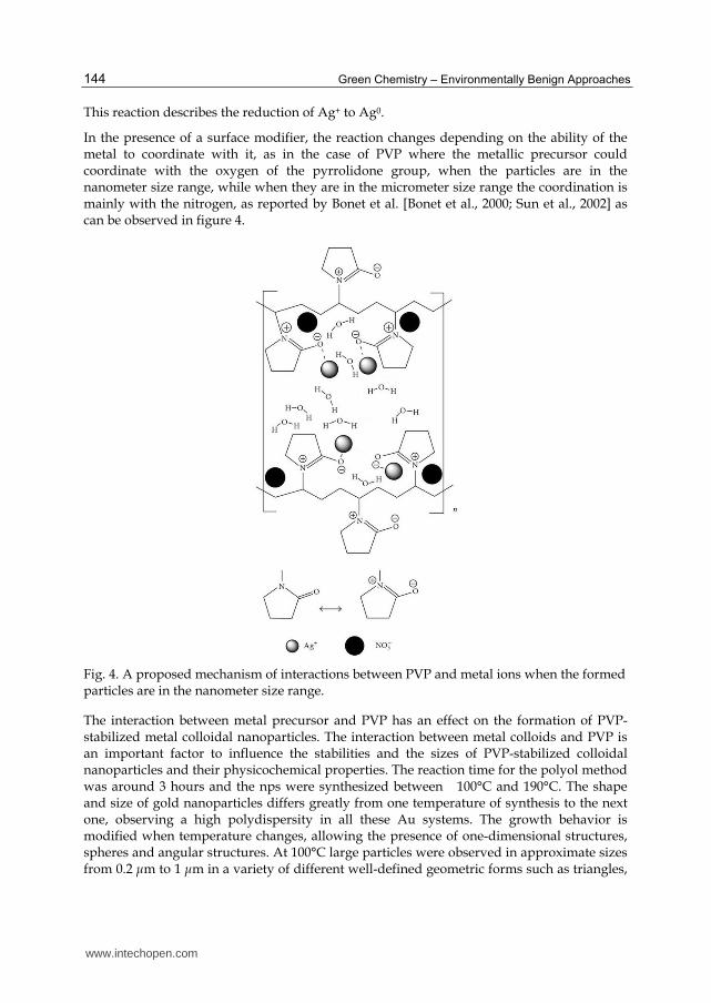
In the presence of a surface modifier, the reaction changes depending on the ability of the metal to coordinate with it, as in the case of PVP where the metallic precursor could coordinate with the oxygen of the pyrrolidone group, when the particles are in the nanometer size range, while when they are in the micrometer size range the coordination is mainly with the nitrogen, as reported by Bonet et al. [Bonet et al., 2000; Sun et al., 2002] as can be observed in figure 4.
Fig. 4. A proposed mechanism of interactions between PVP and metal ions when the formed particles are in the nanometer size range.
The interaction between metal precursor and PVP has an effect on the formation of PVP-stabilized metal colloidal nanoparticles. The interaction between metal colloids and PVP is an important factor to influence the stabilities and the sizes of PVP-stabilized colloidal nanoparticles and their physicochemical properties. The reaction time for the polyol method was around 3 hours and the nps were synthesized between 100°C and 190°C. The shape and size of gold nanoparticles differs greatly from one temperature of synthesis to the next one, observing a high polydispersity in all these Au systems. The growth behavior is modified when temperature changes, allowing the presence of one-dimensional structures, spheres and angular structures. At 100°C large particles were observed in approximate sizes from 0.2 ┤m to 1 ┤m in a variety of different well-defined geometric forms such as triangles,
www.intechopen.com

Green Synthesis and Characterizations of Silver and Gold Nanoparticles
145
truncated triangles, and decahedrons. Also rods with diameters between 50 and 150 nm and a few micrometers in length were observed. All of these structures had very well-defined shapes. The final product was a clear solution with large Au precipitates, some of them visible to the bare eye. At 140°C more rounded particles were observed, with shapes less defined. These particles were also smaller than in the 100°C case, approximately from 300 nm to 500 nm in sizes. At this temperature the structures observed tend to be more spherical than in the previous case. The final product at this temperature also was a clear solution with an evident precipitation of Au at the bottom of the flask. Finally, at 190°C the particles observed were smaller than in the last two cases mentioned from 200 nm to 250 nm in approximate sizes. At this temperature we can observe again particles with more geometric shapes than the ones observed at 140°C, as we can notice in Figure 5 a and b. Some rods with less than 100 nm in diameter and less than 1 ┤m in length were observed. The final product at this temperature had a purple color with an observable precipitation of Au at the bottom of the flask.
The reaction scheme for producing fine and monodisperse metallic nanoparticles using the polyol process involves the following successive reactions: reduction of the soluble silver nitrate and tetrachloroauric acid by ethylene glycol, nucleation of metallic silver and gold, and growth of individual nuclei in the presence of a protective agent, PVP. The fully reacted particle sizes synthesized from the polyol process depended strongly on the ramping rate of the precursor solutions to the reaction temperature; at a lower heating rate larger particles were generated, most likely due to a slower nucleation rate, while at a higher rate faster nucleation produced smaller-sized particles. At a heating rate of 2°C min−1, the mean size of silver particles was 50 nm, and increasing the heating rate to 10°C min−1 yielded smaller and more monodisperse particles with a mean size of 25 nm as can be seen in figure 5c. The particle size of the silver decreased slightly when the reaction temperature was decreased from 150°C to 100°C.
In order to obtain monodisperse metal particles, generally, rapid nucleation in a short period of time is important; that is, almost all ionic species have to be reduced rapidly to metallic species simultaneously, followed by conversion to stable nuclei so as to be grown [Dongjo, Kim et al., 2006]. In the method of heating a precursor solution, however, both nucleation and growth can proceed gradually with increasing temperature. As such, it is difficult to synthesize particles with high monodispersity.
Therefore, the rapid injection of silver nitrate aqueous solution into ethylene glycol maintained at the reaction temperature would guarantee a short burst of nucleation after which the nuclei would continue to grow without additional nucleation, thus ensuring monodispersity.
Upon addition of the silver nitrate and tetrachloroauric acid aqueous solutions to hot ethylene glycol, the Ag+ and Au+ species are reduced to metallic silver and gold nanoparticles.
The concentrations of metallic silver and gold in solution increase, reaching the supersaturation conditions and finally the critical concentration to nucleate. Spontaneous nucleation then takes place very rapidly and many nuclei are formed in a short time, lowering the silver and gold concentrations below the nucleation and supersaturation levels into the saturation concentration region. After a short period of nucleation, the nuclei grow
www.intechopen.com

Green Chemistry – Environmentally Benign Approaches
146
by the deposition of metallic silver and gold until the system reaches the saturation concentration. At the end of the growth period, all the metal nanoparticles have grown at almost the same rate and the systems exhibit a narrow particle size distribution.
This temperature dependence on particle size can be explained as follows. Because of the relatively high temperature used in the synthesis of silver and gold nanoparticles by polyol method, the Brownian motion and mobility of surface atoms increase. This enhances the probability of particle collision, adhesion, and subsequent coalescence. However, PVP is added to protect the particles from agglomeration. Particle coalescence is the means by which the system tries to attain thermodynamic equilibrium by reducing its total surface area.
Spherical silver nanoparticles with a controllable size and high monodispersity were synthesized by the polyol method as can be seen from figure 5c. Two different synthesis methods for producing the Ag nanoparticles were compared in terms of particle size and monodispersity. Silver nanoparticles with a size of 25 ± 4 nm were obtained at a reaction temperature of 120°C and a heating rate of 10°C min−1 in the precursor heating method, where the heating rate was a critical parameter affecting particle size.
In the precursor injection method, on the other hand, the injection rate and reaction temperature were important factors for producing uniform-sized Ag with a reduced size. Silver nanoparitlces with a size of 18 ± 2 nm were obtained at an injection rate of 2.0 ml s−1 and a reaction temperature of 100°C. The injection of the precursor solution into a hot solution is an effective means to induce rapid nucleation in a short period of time, ensuring the fabrication of silver and gold nanoparticles with a smaller size and a narrower size distribution by the polyol method.
The effect of temperature in the polyol method is crucial because at lower temperatures the oxidation potential of ethylene glycol is bigger than at higher temperatures. This means that at lower temperatures the oxidation of ethylene glycol is less favored, which traduces in fewer electrons available in the reaction environment to reduce the metals. As the temperature keeps increasing, the oxidation potential of the ethylene glycol decreases, indicating that this electrochemical reaction is favored at higher temperatures. This translates as an increment in the electrons concentration in the reaction environment. [Bonet et al., 1999]
In contrast, the reduction potential of the metals is not affected by the temperature, according to Bonet et al. It is insensitive to the reaction temperature, but there still is an effect on the reduction of the precursors related to the temperature dependence of the diffusion of metal species. Another effect of temperature on the reduction of the metal precursors is that the energy barrier that opposes to the reduction of the precursor is equal to the difference between the oxidation potential of the ethylene glycol and the reduction potential of the metal species. [Bonet et al., 1999]
Once the oxidation potential of the ethylene glycol is lowered down to the same value of the
reduction potential of the metal precursor, the reduction of the metal precursor will occur
spontaneously and followed by the nucleation of metal nanoparticles. [Bonet et al., 1999]
From this analysis one can conclude that at higher temperatures the reduction of the metal
species will be favored and also the oxidation of the ethylene glycol. This will decrease the
time needed to reduce the metal precursor and the nucleation time needed to the formation
of metal particles.
www.intechopen.com

Green Synthesis and Characterizations of Silver and Gold Nanoparticles
147
From this study it was found that by the polyol method the temperature plays a decisive role in the synthesis of gold and silver nanoparticles protected with PVP. It does not only affect the rates of reduction and nucleation of the metals, but it also affects the coordination between the metals and the polymeric protective agent, the distribution of elements in the nanoparticles, and the final particle size.
In the green method the reaction time is reduced from 3 hours to 30 minutes until 1 hour at 60°C, but we carried out the reaction during 24 hours in order to observe the growth of the particles.
a) b) c)
Fig. 5. TEM images of gold nanoparticles synthesized at 190 °C (a) 200 nm in size and (b) 250 nm in size and silver nanoparticles synthesized at 120°C (c) by the polyol method.
The TEM characterization reveal the formation of nps of these metals, independent of the employed method, with a size distribution between 20 and 120 nm for gold (see figure 5) and between 10 and 27 nm for the silver.
The NSOM showed that the size of gold nps synthesized was of 25 nm with very narrow distribution.
Plants contain a complex network of antioxidant metabolites and enzymes that work together to prevent oxidative damage to cellular components. Isolated quercetin [Wu, 2008] and polysaccharides [Ahmad et al, 2009; Collera et al., 2005; Vedpriya, 2010; Jagadeesh et al., 2004] have been used for the synthesis of silver and gold nanoparticles. Plants Extracts like Aloe Barbadensis is reported to contain chemically different groups of compounds: polyphenols, flavonoids, sterols, triterpenes, triterpenoid saponins, beta-phenylethylamines, tetrahydroisoquinolines, reducing sugars like glucose and fructose, and proteins, in all extracts.
The plant extract is reported to have activities of scavenging superoxide anion radicals and 1, 1-diphenyl-2-picrylhydrazyl radicals (DPPH). It could be that these water-soluble scavenging superoxide anion radicals and 1, 1-diphenyl-2-picrylhydrazyl (DPPH) radicals present in the plant extract be responsible for the reduction of silver and synthesis of nanoparticles through biogenic routes. The exact mechanism of the formation of these
www.intechopen.com

Green Chemistry – Environmentally Benign Approaches
148
nanoparticles in these biological media is unknown. Presumably, biosynthetic products or reduced cofactors play an important role in the reduction of respective salts to nanoparticles. However, it seems probable that some glucose and ascorbate reduce AgNO3 and HAuCl4 to form nanoparticles. [Ahmad et al. 2011; Hu et al., 2003]
The probability of reduction of AgNO3 to silver may be illustrated due to the mechanism known as glycolysis. Plants fix CO2 in presence of sunlight. Carbohydrates are the first cellular constituent formed by the photosynthesizing organism on absorption of light. This carbohydrate is utilized by the cell as glucose by Glycolysis. This is the metabolic pathway that converts glucose C6H12O6 into pyruvate and hydrogen ion:
CH3COCOO− + H+ (5)
The free energy released in this process is used to form the high-energy compounds, ATP adenosine triphosphate and NADH (reduced nicotinamide adenine dinuleotide). Glycolysis can be represented by the following simple equation:
Glucose + 2ADP + 2Pi + 2NAD+ = 2 Pyruvate + 2ATP + 2 NADH + 2H+ (6)
Glycolysis is a definite sequence of ten reactions involving ten intermediate compounds [Ahmad et al. 2011]. Large amount of H+ ions are produced along with ATP. Nicotinamide adenine dinucleotide, abbreviated NAD+, is a coenzyme found in all living cells. NAD is a strong reducing agent. NAD+ is involved in redox reactions, carrying electrons from one reaction to another. The coenzyme is therefore found in two forms in cells. NAD+ is an oxidizing agent—it accepts electrons from other molecules and becomes reduced. This reaction forms NADH, which can donate electrons. These electron transfer reactions are the main function of NAD:
AgNO3→ Ag+ +NO3− or 2HAuCl4 → 2Au+ + 4HCl
NAD+ + e−→ NAD,
NAD + H+ → NADH + e−, (7)
e− +Ag+ → Ag0 or e− +Au+ → Au0
NAD+ keeps on getting reoxidized and gets constantly regenerated due to redox reactions. This might have led to transformations of Ag or Au ions to Ag0 or Au0. Another mechanism for the reduction of Ag or Au ions to silver or gold could be due to the presence of water-soluble antioxidative substances like ascorbate. This acid is present at high levels in all parts of plants. Ascorbic acid is a reducing agent and can reduce, and thereby neutralize, reactive oxygen species leading to the formation of ascorbate radical and an electron.
This free electron reduces the Ag+ or Au+ ions to Ag0 or Au0 as can be seen in scheme 1.
In accordance with the studies of UV visible spectroscopy, whose plasmons are in figure 6 for Au and Ag synthesized nps the results shown an absorption energy in 547 nm and 415 nm respectively.
The use of these natural components allows synthesize metallic nps. In the green method, gold and silver nps were prepared by the same reduction of HAuCl4 and AgNO3
www.intechopen.com

Green Synthesis and Characterizations of Silver and Gold Nanoparticles
149
respectively using extracts of plants, ascorbic acid and polyphenols as reducing agents obtained from Geranium Maculatum leaves and Rosa Berberiforia petals and like natural surfactants saponins and simultaneous reducing agents in some cases were used Aloe Barbadensis and cactus extracts from Cucúrbita Digitata.
Scheme 1. Ascorbic acid reduction mechanism of gold and silver ions to obtain Ag0 and Au0 nps.
a) b)
Fig. 6. UV-Visible absorption spectrum of the Au (1a) and Ag (1b) nps synthesized by polyol and green chemistry respectively.
It is important to know the exact nature of the silver and gold nanoparticles formed, and this
can be deduced from the XRD Spectrum of the Sample. XRD patterns of derived Ag nps
from Figure 7(a) show four intense peaks in the whole spectrum of 2θ° values ranging from
20° to 90°. XRD spectra of pure crystalline silver structures have been published by the Joint
Committee on Powder Diffraction Standards (file no. 04-0787). A comparison of our XRD
spectrum with the Standard confirmed that the silver nanoparticles formed in our
experiments were in the form of face centered cubic nanocrystals, as evidenced by the peaks
www.intechopen.com

Green Chemistry – Environmentally Benign Approaches
150
at 2θ values of 38.52°, 44.49°, 64.70°, and 77.63°, corresponding to [111], [200], [220], and
[311] planes for silver, respectively. In the case of gold nanoparticles in the whole spectrum
of 2θ° values ranging from 35° to 80°, four new reflection signals appear at ca. 38.10°, 44.40°,
64.87°, and 77.84° in the XRD pattern of the Au, corresponding to the [111], [200], [220] and
[311] planes of the Au, respectively as can be seen in Figure 7 (b), indicating that crystal
structure of the gold nanoparticles was face centered cubic(JCPDS 4-0783)in this case also.
Scherrer’s equation for broadening resulting from a small crystalline size, the mean, effective, or apparent dimension of the crystal composing the powder is:
Phkl = k┣/β1/2 cosθ (8)
a) b)
Fig. 7. X-ray diffractograms of silver (a) and gold (b) nanoparticles synthesized as of extracts of Aloe Barbadensis at 1 hour and 60°C.
where θ is the Bragg angle, ┣ is the wavelength of the X ray used, β is the breadth of the pure
diffraction profile in radians on 2θ scale, and k is a constant approximately equal to unity
and related both to the crystalline shape and to the way in which θ is defined. The best
possible value of k has been estimated as 0.89. The Full Width at Half Maximum (FWHM)
values measured for [111], [200], [220], and [311] planes of reflection were used with the
Debye-Scherrer equation (8) to calculate the size of the nanoparticles. [Ahmad, N. et al. 2011]
Moreover, the average size of the gold nanoparticles was also determined from the width of
the reflection according to the Scherrer formula. The value of D calculated from the (111)
reflection were k is 0.90 of the cubic phase of Au was ca. 25 nm, which is basically in
agreement with the results of transmission electron microscopy (TEM) experiments for Aloe
Barbadensis at 1 hour and 60°C.
Further analysis of the silver and gold nanoparticles by energy dispersive spectroscopy
confirmed the presence of the signals characteristic of silver and gold respectively. Figure 8
shows the Energy-Dispersive Absorption Spectroscopy photographs of derived Ag nps and
Au nps. All the peaks of Ag and Au respectively are observed and are assigned. Peaks for
Cu and C are from the grid used, and the peaks for S, P, and Si (in the case of Au)
correspond to the protein capping over the Ag nps and Au nps.
www.intechopen.com

Green Synthesis and Characterizations of Silver and Gold Nanoparticles
151
a) Image corresponding to Spectrum 1 and EDX for silver nanoparticles.
b) Image corresponding to Spectrum 34 and EDX bright field for gold nanoparticles.
Fig. 8. a) Image corresponding to select area 1 and Energy-Dispersive Absorption Spectroscopy photograph for silver nanoparticles, and b) Image corresponding to select area 34 and Energy-Dispersive Absorption Spectroscopy photograph for gold nanoparticles.
a) b) c)
Fig. 9. Transmission electron microscopy images of Au nps at same magnification of (a) Rosa Berberiforia, (b) Geranium Maculatum, and (c) Cucúrbita Digitata using the same low concentration of plants extracts approximately (0.002M) with HAuCl4 (1 X 10⁻³ M) at 1 hours and 60°C.
www.intechopen.com

Green Chemistry – Environmentally Benign Approaches
152
a) Quasi-spherical shape. b) Triangle shape. c) Rhombohedral shape.
d) Rod shape. e) Hexagonal shape. f) Cubic shape.
Fig. 10. Images of gold nanoparticles observed with different shapes synthesized as of Aloe Barbadensis extracts at different conditions varying the concentration of the extract from 0.0015 to 0.004 M, using: high resolution TEM: a) Quasi-spherical, b) Triangle, c) Rhombohedral shapes; HAADark Field TEM image: d) Rod shape; and Bright field TEM images: e) hexagonal shape and f) cubic shape gold nanoparticles.
In the figures 9 and 10, it is possible to identify large population of polydispersed gold nps synthesized as of Rosa Berberiforia petals, Geranium Maculatum leads, Cucúrbita Digitata cactus at same reaction conditions, and Aloe Barbadensis varying the concentration of plant extracts from 0.0015 to 0.004M, the consisted of spherical-, quasi-spherical-, ellipsoidal-, triangular-, hexagonal-, rombohedral-, trapezhoidal- and rod-shaped with irregular contours.
The morphology of the Ag nps was predominantly spherical and quasi-spherical as shown in figure 11, and they appear to be monodisperse for Aloe Barbadensis with AgNO3 (1 X 10⁻³ M) at 1 hours and 60°C at different concentration of plant extract. Some of the nps were found to be oval and/or elliptical at high concentration of plant extract. Such variation in shape and size of nanoparticles synthesized by biological systems is common.
The figure 12. shows high resolution transmission electron micrographs of gold nps, synthesized with extracts of Aloe Barbadensis at 1 hour and 60°C using a concentration of plant extract of 0.0025 M approximately with an average size distribution of 25nm (figure 12a), the figure 12b shows a gold nanoparticle of 150 nm in size, synthesized with extracts of Cucúrbita Digitata using a concentration of plant extract of 0.0018M approximately and
www.intechopen.com

Green Synthesis and Characterizations of Silver and Gold Nanoparticles
153
figure 12c a gold nanoparticle of 4nm in size, synthesized with extracts of Cucúrbita Digitata at high concentration of plant extract of 0.003M. The gold nps synthesized with extracts of Aloe Barbadensis were used to prepare nanoarrays for the study of optical plasmonic phenomena in another work. [Coello et al., 2010]
a) b) c)
Fig. 11. Transmission electron microscopy images of Ag nps at different magnifications of (a) High Resolution -TEM image at a concentration of plant extract of 0.004 M, (b) High-TEM image at a concentration of plant extract of 0.002 M, (c) HAADF image at a concentration of plant extract of 0.0015 M approximately of Aloe Barbadensis with AgNO3 (1 X 10⁻³ M) at 1 hours and 60°C.
a) b) c)
Fig. 12. High resolution transmission electron microscopy images of (a) gold nps
synthesized with extracts of Aloe Barbadensis at 1 hour and 60°C using a concentration of
plant extract of 0.0025 M approximately with a size distribution of 25 nm approximately, (b)
a gold nanoparticle synthesized with extracts of Cucúrbita Digitata using a concentration of
plant extract of 0.0018M approximately with a size of 150 nm and (c) a gold nanoparticle
synthesized with extracts of Cucúrbita Digitata with a size of 4 nm at 60°C using a high
concentration of plant extract of 0.003M.
The anisotropic gold and spherical–quasi-spherical silver nps were synthesized by reducing
aqueous chloroauric acid (HAuCl4) and silver nitrate (AgNO3) solution with the extract of
Aloe Barbadensis at 60°C temperature. The size and shape of the nps can be controlled by
varying the concentration of plants extracts like Aloe Barbadensis.
www.intechopen.com

Green Chemistry – Environmentally Benign Approaches
154
The case of low concentration of extract with HAuCl4 offers the aid of electron-donating group containing extract leads to formation of hexagonal-or triangular-shaped gold nps. Transmission electron microscopy (TEM) analysis revealed that the shape changes on the gold nps from hexagonal to spherical particles with increasing initial concentration of Aloe Barbadensis.
The electron-donating methoxy (–OCH3) groups containing Aloe Barbadensis can provide a suitable environment for the formation of nps. A bioreductive approach of anisotropic gold and silver nps utilizing the Aloe Barbadensis has been demonstrated which provides a simple and efficient way for the synthesis of nanomaterials with tunable optical properties directed by particle shape.
The presence of small amount of Aloe Barbadensis leads to slow reduction of HAuCl4 ions which facilitated the formation of triangular- or hexagonal-shaped nps. Whereas greater amount of Aloe Barbadensis leads to higher population of spherical nps and was confirmed from the UV–visible and TEM analysis. The electron-donating nature of –OCH3 group of the Aloe Barbadensis plays a leading role for the formation and stabilization of nps, respectively results in accordance with Kasthuri et al.[Kasthuri, et al., 2009] as shown in scheme 2.
Scheme 2. The presence of small amount of Aloe Barbadensis leads to slow reduction of HAuCl4 ions which facilitated the formation of triangular- or hexagonal-shaped nps. Whereas greater amount of Aloe Barbadensis leads to higher population of spherical nps and was confirmed from TEM analysis.
4. Conclusion
One-step green synthesis of gold (Au) and silver (Ag) nanostructures is described using naturally occurring biodegradable plant-based surfactants, without any special reducing agent/capping agents. This green method uses water as a benign solvent and surfactant/plant extract as a reducing agent. Depending upon the Au and Ag concentration used for the
www.intechopen.com

Green Synthesis and Characterizations of Silver and Gold Nanoparticles
155
preparation and the temperature, Au and Ag crystallizes in different shapes and sizes to form spherical in the case of Ag, prisms, and hexagonal structures in the case of Au. Sizes vary from the nanometer to micrometer scale level depending on the plant extract used for preparation. Synthesized Au and Ag nanostructures were characterized using scanning electron microscopy, transmission electron microscopy, X-ray diffraction, and UV spectroscopy.
In this original work, we show that green method reduces the temperature requirement, which is in contrast to the obtained with the polyol method. In the green method the size and shape of the nps can be controlled by varying the concentration of plant extracts and the reaction time. The use of these natural components allows synthesize metallic nps with very narrow distribution.
5. Acknowledgment
Authors would like to acknowledge to Facultad de Ciencias Físico Matemáticas and Microscopy Laboratory of CIIDIT de la Universidad Autónoma de Nuevo León, to Nanotechnology Laboratory of CIMAV Chihuahua, México.
6. References
Ahmad, N.; Alam, M. K.; Singh, V. N. and Sharma, S. Journal of Bionanoscience, 2009, 3, 2, 97–104. Ahmad, N.; Sharma, S.; Singh, V. N.; Shamsi, S. F.; Fatma, A.; Mehta, B. R.; Biotechnology
Research International, 2011, Article ID 454090,1- 8. Alvarez, M. M.; Khoury, J. T.; Schaaff, G.; Shafigullin, M. N.; Vezmar, I.; Whetten, R. L. J.
Phys. Chem. B 1997, 101, 3706. Altansukha, B.; Burmaa, G.; Zhianshi, J.; Van, Dan; Antsiferova, S. A. Theoretical Foundations
of Chemical Engineering, 2010, 44, 4, 511. Amkamwar, B.; Damle, C.; Ahmad, A.; Sastry.; M. J. Nanosci. Nanotechnol. 2005, 5,1665. Arangasamy, L.; Munusamy, V.; Afr. J. Biotech. 2008, 7, 3162. Bonet, F.; Tekaia-Elhsissen, K.; Sarathy, K. V. Bull. Mater. Sci. 2000, 23, 165. Bonet, F.; Guery, C.; Guyomard, D.; Herrera-Urbina, R.; Tekaia-Elhsissen, K.; Tarascon, J. M.
Intl. J. of Inorg. Mater. 1999, 1, 47. Boudreau, M.D.; Beland, F.A. Journal of environmental science and health. Part C, Environmental
carcinogenesis & ecotoxicology reviews, 2006, 24, 1, 103–54. Bronstein, L. M.; Chernyshov, D. M.; Volkov, I. O.; Ezernitskaya, M. G.; Valetsky, P. M.;
Matveeva, V. G.; Sulman, E. M. J. Catal. 2000, 196, 302. Burda, C.; Chen, X.; Narayanan, R.; El-Sayed, M. A. Chem. Rev., 2005, 105, 1025. Cao, G. Nanostructures and Nanomaterials, synthesis, properties and applications,
Imperial College Press, 2004. Carotenuto, G.; Pepe, G. P.; Nicolais, L. Eur. Phys. J. B 2000, 16, 11. Chandran, S.P.; Chaudhary, M.; Pasricha, R.; Ahmad, A.; Sastry, M. Biotechnol. Prog. 2006,
22, 577. Chushak; Y. G.; Bartell, L. S. J. Phys. Chem B 2003, 107, 3747. Coello V., Cortes R., Segovia P., Garcia C. and Elizondo N., In Plasmons: Theory and
Applications, Editor: Kristina N. Helsey, Chapter 9, 2010, Nova Science Publishers. Collera-Zuniga O., Garcia Jimenez F., and Melendez Gordillo R., Food Chemistry, 2005, 90, 1-
2, 109–114. Dongjo, Kim; Sunho, J.; Jooho, M., Nanotechnology, 2006, 17, 4019–4024.
www.intechopen.com

Green Chemistry – Environmentally Benign Approaches
156
EI-Sayed, M.A.; Acc Chem Res, 2001, 34, 257–264. Eshun, K.; He, Q. Critical reviews in food science and nutrition, 2004, 44, 2, 91–6. Gericke, M.; Pinches, A. Hydrometallurgy, 2006, 83, 132–140. Gomez-Romero, P., Adv. Mater, 2001, 13, 163–174. González, C. M.; Liu, Y; Scaiano, J. C. J. Phys. Chem. C, 2009, 113, 11861. Gracias, D.H.; Tien, J.; Breen, T.; Hsu, C.; Whitesides, G. M.; Science, 2002, 289,1170–1172. Han, S. W.; Kim, Y.; Kim, K. J. Colloid Interface Sci. 1998, 208, 272. Harris, A.T.; Bali, R.; J. Nanopart. Res. 2008, 10, 691–695. Henglein, A. J. Phys. Chem. B 2000, 104, 2201. Hu, Y.; Xu, J.;Hu, Q.; J. Agric. Food Chem.2003,51,7788-7791. Jagadeesh, B. H.; Prabha, T. N.; Srinivasan, K. Indian Journal of Plant Physiology, 2004, 9,
2,164–168. Kamat, P.V.; J. Phys. Chem. B 2002, 106, 7729–7744. Kasthuri, J.; Kathiravan, K.; Rajendiran, N. J Nanopart Res, 2009, 11,1075–1085. King, G.K.; Yates, K.M.; Greenlee, P.G. et al. Journal of the American Animal Hospital
Association, 1995, 31, 5, 439–47. Lee, Y.; Gwan-Park, T. Langmuir, 2011, 27, 2965–2971. Li, S.; Shen, Y.; Xie, A.; Yu, X.; Qiu, L.; Zhang, L.; Zhang, O. Green. Chem. 2007, 9, 852. Lim, J.K.; Kim, Y.; Lee, S.Y.; Joo, S.W.; 2008, Spectrochim Acta A, 69, 286–289. Liz-Marzan, L. M.; Philipse, A. P. J. Phys. Chem. 1995, 99, 15120. Nagajyoti, P.C.; Prasad,T.N.V.K.V; Sreekan, T.V.M; Lee, K. D. Digest Journal of Nanomaterials
and Biostructure. 2011, 6, 1, 121–133. Narayanan, R.; EI-Sayed, M.A. Nano Lett. 2004, 4, 1343–1348. Park, H. K.; Lim, Y. T.; Kim, J. K.; Park, H. G.; Chung, B. H. Ultramicroscopy, 2008, 108,10, 1115. Qiu, H.; Rieger, B; Gilbert, R.; Jerome, C.; Chem. Mater. 2004, 16, 850–856. Rosi, N. L.; Mirkin, C. A. Chem. Rev., 2005, 105, 1547. Safaepour , M.; Reza Shahverdi, A.; Reza Shahverdi, H.; Reza Khorramizadeh, M.; Reza
Gohari, A. Avicenna J. Med. Biotech., 2009, 1, 2, 111. B. Sanguesa, C. D.; Urbina, R. H.; Figlarz, M. J. Solid State Chem. 1992, 100, 272. Schmid, G. Clusters and Colloids, From theory to Applications; VCH Publishers: Weinheim,
Germany, 1994. Shankar, S. S.; Ahmad, A.; Pasricha, R.; Sastry, M.; J. Mater. Chem. 2003, 13, 1822–1826. Shankar, S.S.; Rai, Ahmad, A. A.; Sastry, M. J. Colloid. Interface. Sci. 2004a, 275, 496. Shankar, S. S.; Rai, A.; Ankamwar, B.; Singh, A.; Ahmad, A.; Sastry, M.; 2004b, Nat. Mater. 3,
482–488. Smirnoff, N.; Wheeler G. L.; Critical Reviews in Biochemistry and Molecular Biology, 2000, 35, 4, 291. Sun, Y.; Yin, Y.; Mayers, B. T.; Herricks, T.; Xia, Y. Chem. Mater. 2002, 14, 4736. Thomas, J. M.; Raja, R.; Johnson, B. F. G.; Hermans, S.; Jones, M. D.; Khimyak, T. Ind. Eng.
Chem. Res. 2003, 42, 1563. Turkevich, J.; Stevenson, P.; Hillier, J. Discuss. Faraday Soc. 1951, 11, 55. Vedpriya, A.; Digest Journal of Nanomaterials and Biostructures, 2010, 5, 1, 9–21. Vogler, B.K.; Ernst, E. The journal of the Royal College of General Practitioners, 1999, 49, 447, 823–8. Wu, T.H.; Yen, F.L.; Lin, L.T.; Tsai, T.R.; Lin, C.C.; Cham, T.M., International Journal of
Pharmaceutics, 2008, 346, 1-2, 160–168. Xia, Y.; Halas, N. J. Mater. Res. Soc. Bull., 2005, 30, 338. Xia, Y. ; Yang, P.; Sun, Y.; Wu,Y.; Mayers, B.; Gates, B.; Yin, Y.; Kim, F.; Yan, H. Adv. Mater.,
2003,15, 353.
www.intechopen.com

Green Chemistry - Environmentally Benign ApproachesEdited by Dr. Mazaahir Kidwai
ISBN 978-953-51-0334-9Hard cover, 156 pagesPublisher InTechPublished online 23, March, 2012Published in print edition March, 2012
InTech EuropeUniversity Campus STeP Ri Slavka Krautzeka 83/A 51000 Rijeka, Croatia Phone: +385 (51) 770 447 Fax: +385 (51) 686 166www.intechopen.com
InTech ChinaUnit 405, Office Block, Hotel Equatorial Shanghai No.65, Yan An Road (West), Shanghai, 200040, China
Phone: +86-21-62489820 Fax: +86-21-62489821
Green chemistry is chemistry for the environment. It is really a philosophy and way of thinking that can helpchemistry in research and production to develop more eco-friendly solutions. Green chemistry is consideredan essential piece of a comprehensive program to protect human health and the environment. In its essence,green chemistry is a science-based non-regulatory and economically driven approach to achieving the goals ofenvironmental protection and sustainable development. Combining the technological progress withenvironmental safety is one of the key challenges of the millennium. In this context, this book describes theenvironmentally benign approaches for the industries as well as chemical laboratories. In order to provide aninsight into step change technologies, this book was edited by green organic chemists.
How to referenceIn order to correctly reference this scholarly work, feel free to copy and paste the following:
Nora Elizondo, Paulina Segovia, Víctor Coello, Jesús Arriaga, Sergio Belmares, Aracelia Alcorta, FranciscoHernández, Ricardo Obregón, Ernesto Torres and Francisco Paraguay (2012). Green Synthesis andCharacterizations of Silver and Gold Nanoparticles, Green Chemistry - Environmentally Benign Approaches,Dr. Mazaahir Kidwai (Ed.), ISBN: 978-953-51-0334-9, InTech, Available from:http://www.intechopen.com/books/green-chemistry-environmentally-benign-approaches/green-synthesis-and-characterization-of-gold-and-silver-nanoparticles-