green synthesis of silver nanoparticles ......green synthesis of silver nanoparticles using...
TRANSCRIPT
International Journal of BioChemiPhysics, Vol. 22, December 2014
21
GREEN SYNTHESIS OF SILVER NANOPARTICLES USING
EUCALYPTUS CORYMBIA LEAVES EXTRACT AND ANTIMICROBIAL
APPLICATIONS
J.M. Sila1*, I. Kiio4, F.B. Mwaura2, I. Michira1, D. Abongo1, E. Iwuoha3 and G.N. Kamau1* 1Department of Chemistry, University of Nairobi, P.O Box 30197-00100, Nairobi, Kenya 2School of Biological Sciences, University of Nairobi, P.O Box 30197-00100, Nairobi, Kenya 3Sensor research laboratory Department of Chemistry, University of Western Cape, private bag Bellville, South Africa. 4School of medicine,college of health sciences,department of biochemistry,University of Nairobi p.o box 30197-00100 Nairobi,Kenya.
ABSTRACT
In this study biosynthesis of silver nanoparticles (AgNPs) using Eucalyptus corymbia and their antimicrobial
activities have been reported. This work reveals that Eucalyptus corymbia leaf extract contains a variety of
bio-molecules responsible for reduction of metal ions and stabilization of nanoparticles. These bio-molecules
are believed to contain polyphenols and water soluble heterocyclic compounds. Optimized experimental
conditions included using extraction temperature of 90˚C; plant extract pH 5.7 and silver nitrate to plant
extract ratio of 4:1. These conditions favoured the formation of higher number of nanoparticles, which were
stable within the study period. The synthesized nanoparticles were polydispersed with average mean size of
18-20 nm and were spherical in shape without significant agglomeration, as revealed from the TEM analysis.
FT-IR spectra of the plant extract revealed that functional groups OH and –C=C– are responsible for
reduction and stabilization of the nanoparticles. Anti-Microbial activity of the synthesized silver
nanoparticles were studied against gram negative bacteria Escherichia coli (E.coli) and gram positive
bacteria staphylococcus aureus. In the medium treated with silver nanoparticles, E.coli and Staphylococcus
aureus growth was inhibited, as these particles have an excellent biocidal effects and hence effective in
inhibiting bacterial growth. These nontoxic nanomaterials, which can be prepared in a simple and cost-
effective manner may be suitable for the formulation of new types of bactericidal materials.
Key words: Silver nanoparticles, Eucalyptus corymbia, Green synthesis, Escherichia coli, Staphylococcus
aureus.
INTRODUCTION
Metal nanoparticles have received significant
attention in recent years owing to their unique
properties and practical applications. They exhibit
properties that differ significantly from those of bulk
materials as a result of small particle dimension, high
surface area, quantum confinement and other effects
[1]. Metal nanoparticles size and shape dependent
properties are of interest due to wide applications as
catalyst, optical sensors, in data storage and
antibacterial properties [2]. Nanoparticles can be
synthesised through different methods; chemical,
physical and biological methods. Conventionally,
chemical synthesis has been the method of choice
because it offers faster synthetic route. However,
chemical synthesis has raised environmental concerns
because of the nature of chemicals used, such as
reducing agents (sodium borohydride), organic
solvents and non – biodegradable stabilizing agents
(sodium citrate dehydrate). These chemicals are
potentially hazardous to the environment and
biological systems [3]. Majority of the conventional
methods makes use of organic solvents because of
the hydrophobicity of the capping agents. Capping
and stabilizing agents are used to prevent aggregation
which may hinder production of small sized silver
nanoparticles [4] Due to the increasing interest in
nanoparticles synthesis and applications; there is a
need for eco-friendly approaches based on green
chemistry principles [5].

International Journal of BioChemiPhysics, Vol. 22, December 2014
22
Green method employs principles of green chemistry
which involves exploitation of natural resources for
metal nanoparticle synthesis, which is a competent
and environmentally benign approach [6].This
involves three main steps, which must be evaluated
based on green chemistry perspectives, including
selection of solvent medium, environmentally benign
reducing agent, and non-toxic stabilisizing agents [7].
These bio–inspired methods utilize plant extracts and
micro-organisms for synthesis of nanoparticles
intracellularly or extracellularly [8]. The use of plant
in nanoparticles synthesis is more advantageous over
environmentally benign biological processes because
it eliminates elaborate process of maintaining cell
cultures.
In addition, green synthesis using plants offers a
better synthetic protocol because of the vast reserves
of plants that are easily accessible, widely distributed,
safe to handle with wide range of metabolites. Unlike
conventional methods bio-inspired methods are
economical and restrict the use of toxic chemicals
and do not require high pressure, energy and
temperatures.
The bioreduction of metal ions is done by
combinations of biomolecules found in plant extracts
(e.g. enzymes/proteins, amino acids, polysaccharides,
and vitamins) in an environmentally benign, yet
chemically complex process [9].
Depending on the origin there are three types of NPs:
natural, incidental and engineered. Natural NPs have
existed since the earth’s beginnings and still occur in
the environment, for example volcanic dusts and
mineral composites. Incidental NPs are typically
represented by engine exhaust particles, coal
combustion, or other fractions or airborne
combustion by-products [10]. Engineered
nanomaterials are defined as those nanomaterials that
are designed with specific properties and
intentionally produced via chemical or physical
processes. They are further divided into four types
[10], namely:
• Carbon-based materials, usually including
fullerenes, single walled carbon nanotubes
(SWCNT) and multi-walled carbon
nanotubes (MWCNT). Fullerenes are made
of pure carbon and represent a new carbon
allotrope discovered in 1985 (Kroto et al.,
1993).
• Metal-based materials such as quantum dots,
nanogold, nanozinc, nanoaluminum, and
nanoscale metal oxides like TiO2, ZnO and
Al2O3. Quantum dot is a closely packed
semiconductor crystal comprised of
hundreds or thousands of atoms, whose size
is in the order of a few nanometers to a few
hundred nanometers.
• Dendrimers, which are nanosized polymers
built from branched units capable of being
tailored to perform specific chemical
functions. The surface of a dendrimer has
numerous chain ends, which can be tailored
to perform specific chemical functions.
• Composites, which combine nanoparticles
with other nanoparticles or with larger, bulk-
type materials.
Silver nanoparticles (AgNps) have been proven to
have diverse importance and thus have been
extensively studied. In the recent years, there has
been an upsurge in studying AgNPs on account of
their inherent antimicrobial efficacy. Many bacteria
develop resistance to antibiotics hence the need to
develop a substitute. So far no literature has reported
any bacteria able to develop immunity against silver.
Generally the nanoparticles are designed with surface
modifications tailored to meet the needs of specific
applications they are going to be used for [9].
The exact mechanism which silver nanoparticles
employ to cause antimicrobial effect is not clearly
known. However, it has been hypothesized that silver
nanoparticles can act on microbes to cause the
microbicidal effect through various ways. In one of
the ways, silver nanoparticles are said to anchor on
the bacterial cell wall and subsequently penetrate it,
thereby causing structural changes in the cell
membrane like the permeability of the cell
membrane. This leads to formation of ‘pits’ on the
cell surface, and consequently accumulation of the
nanoparticles on the cell surface [11]. It has also been
proposed that silver nanoparticles can release silver
ions (Feng et al., 2008) and these ions can interact
with the thiol groups of many vital enzymes and
inactivate them [12] i.e., Ag+ works through
suppression of respiratory enzymes and electron
transport components which interfere with DNA
functions [13]. Silver ions are powerful
antimicrobials but are easily sequested by chloride,
phosphate and other cellular components [14]. Silver
International Journal of BioChemiPhysics, Vol. 22, December 2014
23
nanoparticles are less susceptible to being intercepted
and therefore offer a more effective delivery
mechanism [15]. Silver ions are released from the
nanoparticles in presence of oxygen [14].
EXPERIMENTAL SECTION
Materials and reagents
100g Silver nitrate (AgNO3) crystals and 2.5 Litres of
HPLC grade Methanol, Ethanol and Diethyl Ether
were purchased from Fischer Scientific Chemicals
(United Kingdom). 50 g of oven dried AgNO3 (Sigma
Aldrich USA) was used as received for the study.
Distilled de-ionized water and Nutrient broth (Sigma-
Aldrich, USA) was obtained from the Biochemistry
laboratory at the University of Nairobi. Folin-
ciocalteus’s phenol reagent (2N), NaOH, FeCl3, and
Gallic Acid were purchased from Sigma-Aldrich
(Germany).
Extraction of polyphenols from Eucalyptus
corymbia
A leaf extract of Eucalyptus corymbia was prepared
by weighing 5g of green leaves. The leaves were
properly washed with distilled water, cut into fine
pieces and transferred to 250ml Erlenmeyer flask
containing 100ml of distilled water. The mixture was
boiled for 5 minutes before filtering using a filter
paper. The filtrate obtained was centrifuged at 15000
revolutions per minute for 10 minutes and stored at
4oC in a refrigerator for subsequent use within 7 days
after extraction.
Confirmatory test for phenolic compound in the
leaf extract
An aliquot of Folin-ciocalteus’s phenol reagent (2N)
was added to 5mLs of the leaf extract and colour
change recorded [16].
Synthesis of silver nanoparticles
1.7g of silver nitrate was dissolved in 10mL of de-
ionised water. Aqueous solution of 1mM AgNO3 was
prepared by diluting 1 ml of 1M AgNO3 in a litre of
distilled water. Different volumes of the leaf extract
were added slowly to varying amounts of aqueous
silver nitrate solution with stirring [17]. This was
repeated with 0.8mM, 0.6mM, 0.4mM and 0.2mM of
silver nitrate solution. Analysis was done on the
resulting solution.
Procedure for calculating Percent yield of silver
nanoparticles
The efficiency of the synthetic procedure in this work
was determined by calculating the percent yield of
the synthesized silver nanoparticles. 10 ml aliquot of
the mixture of plant extract and silver nitrate were
centrifuged at 15000rpm and washed with distilled
water, then dried in an oven at 60oC for 24 hours. The
nanoparticles were weighed and the mass recorded in
grams. The weight was divided by the mass of Ag+
ions in 10 ml of 1mM AgNO3. The answer above was
multiplied by 100 to get percentage yield;
Mass of Ag0
Percent yield = ----------------- x 100
Mass of Ag+
Uv-vis Spectroscopy procedure
The solution for UV-Vis analysis was prepared by
taking 1ml of silver nitrate –plant extract mixture and
diluting it ten times. UV-VIS spectra analysis was
performed using UV-VIS double beam
spectrophotometer [UV-1700 pharmaspec UV-Vis
spectrophotometer (shimadzu)] at university of
Nairobi. Scanning of the spectra was done between
200-700nm at a resolution of 1 nm using quartz
cuvette. Baseline correction was done using de-
ionized water as the blank.
FT-IR Spectroscopy Procedure
Dry powder of the sample was crushed with KBr and
the mixture pressed in a mechanical press to form a
thin and transparent pellet. The collar and the pellet
were put onto the sample holder. FTIR of plant
extract was obtained by dropping a sample between
two plates of sodium chloride (salt) and analyzed in a
liquid cell. Finally, the dried nanoparticles were
analyzed by FTIR-JAS-CO 4100 spectrophotometer
in the range 4000–400 cm-1
.
Transmission Electron Microscopy Procedure
Samples for transmission electron microscopy (TEM)
analysis were prepared by drop coating biologically
synthesized silver nanoparticles solution on to
carbon-coated copper TEM grids [18].
The films on the TEM grid were allowed to stand for
2 minutes. The excess solution was removed using a
blotting paper and the grid allowed to dry under a
lamp prior to measurement. TEM images were
International Journal of BioChemiPhysics, Vol. 22, December 2014
24
acquired with Philips Technai-FE 12 TEM
instrument, operated at an accelerating voltage of 120
kV, equipped with an Energy dispersive X-ray
(EDAX) detector (Oxford LINK-ISIS 300) for
elemental composition analysis and the EDAX
spectra was measured at an accelerating voltage of 10
Kv.
Determination of Antimicrobial Activity
Nutrient broth (Sigma, St. Louis, USA) was prepared
by adding distilled water to 3.25 gm of the powder to
make 250 ml as recommended by the manufacturer.
The medium was sterilized by autoclaving at 121o C
for 15 minutes (All American, Hillsville, USA).
Escherichia coli and Staphylococcus aureus cells
were separately inoculated and cultured overnight at
37o
C. Incubation was done in a thermo-shaker
(Gallenkamp, London, England). A disk diffusion
test was carried out according to the Kirby- Bauer
disk diffusion susceptibility test protocol [19]. An
inoculum of the bacteria culture was applied
uniformly on the surface of Muller Hinton agar
(MHA) plates.
Sterile paper discs of 6mm diameter were
impregnated with 20µl nanoparticles of three
different concentrations (0.6mM, 0.8mM and
1.0mM) of nanoparticles suspended in distilled water
and placed on the plate with inoculums. A positive
control was prepared by impregnating a sterile disc of
6mm diameter with an antibiotic (Kanamycin
10mg/ml)
The plates were incubated for 15 hours at 37o
C in a
research CO2 incubator ( LEEC limited, Nottingham,
United Kingdom). The plates were observed at the
end of the incubation period.
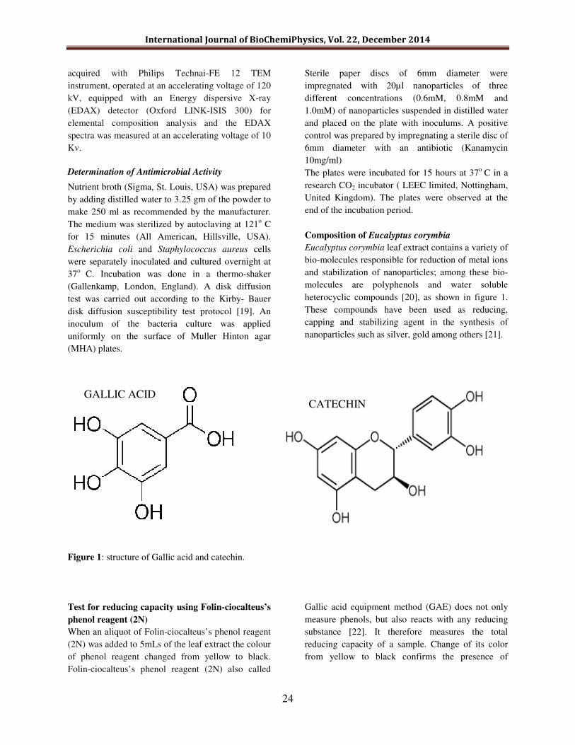
Composition of Eucalyptus corymbia
Eucalyptus corymbia leaf extract contains a variety of
bio-molecules responsible for reduction of metal ions
and stabilization of nanoparticles; among these bio-
molecules are polyphenols and water soluble
heterocyclic compounds [20], as shown in figure 1.
These compounds have been used as reducing,
capping and stabilizing agent in the synthesis of
nanoparticles such as silver, gold among others [21].
Figure 1: structure of Gallic acid and catechin.
Test for reducing capacity using Folin-ciocalteus’s
phenol reagent (2N)
When an aliquot of Folin-ciocalteus’s phenol reagent
(2N) was added to 5mLs of the leaf extract the colour
of phenol reagent changed from yellow to black.
Folin-ciocalteus’s phenol reagent (2N) also called
Gallic acid equipment method (GAE) does not only
measure phenols, but also reacts with any reducing
substance [22]. It therefore measures the total
reducing capacity of a sample. Change of its color
from yellow to black confirms the presence of
GALLIC ACID CATECHIN

International Journal of BioChemiPhysics, Vol. 22, December 2014
25
reducing compounds in Eucalyptus corymbia leaf
extract.
RESULTS & DISCUSSION The percentage yield of silver nanoparticles was
81.64%. The percent yield was calculated by dividing
the mass of AgNPs by the mass of Ag+ ions in 10ml
aqueous solution. The above calculated value
demonstrated that 81.6 ± 0.3 % of the silver ions
were converted to atomic state hence forming silver
nanoparticles.
Uv-vis analysis
Formation of silver nanoparticles from the plant
extract and AgNO3 was noted by visual observation,
a gradual colour change, which took less than ten
minutes from colorless solution to yellow then deep
red/brown on addition of the leaf extract of
Eucalyptus corymbia, indicating formation of AgNPs
which was further confirmed by Uv-Vis analysis
(figure 2). The observed results are in accordance
with what was reported earlier by Chandan Tamuly,
et al. [23].
The biosynthesized silver nanoparticles were found
to have absorbance peak at around 425nm as shown
in figure 1. Typically AgNPs have surface plasmons
resonance peaks with λmax values in the visible range
of 400–500 nm [24].
Figure 2: The absorbance spectra of silver nanoparticles synthesized with varying silver nitrate concentrations from
0.2 mM to 1mM at a wavelength range of 200nm to 700nm.
The appearance of the deep red/brown color was due
to collective oscillation of the conduction electrons in
resonance with the wavelength of irradiated light
[25].
Transmission Electron Microscopy and energy
dispersive spectroscopy results
The grid for the TEM analysis of Ag-nanoparticles
was prepared by placing a drop of the nanoparticles
suspension on the carbon-coated copper grid and
allowing the water to evaporate inside a vacuum
dryer. Scanning under TEM (Philips CM-10)
revealed that the average mean size of silver
nanoparticles was 18-20 nm and the particles were
spherical in shape without significant agglomeration
(figure 3a).

International Journal of BioChemiPhysics, Vol. 22, December 2014
26
Figure 3: ( a )TEM image showing spherical silver nanoparticles and (b)An EDS spectrum showing two peaks of
elemental silver in the silver region.
Energy Dispersive X-ray Spectroscopy was used to
verify the presence of silver in the sample. Figure 3b
showed two peaks at 3.0 keV and 3.15 keV, which
are due to the elemental silver. The typical optical
absorption band peaked nearly at 3 KeV confirms
formation of metallic silver nanoparticles [26].
Fourier Transform Infra-Red (FT-IR)
spectroscopy Analysis
The FT-IR spectra of Eucalyptus corymbia leaf
extract and synthesized nanoparticles were done to
identify the possible biomolecules responsible for the
reduction of the Ag+ ions and capping of the bio-
reduced Ag-NPs. Figure 4 shows the FT-IR spectrum
of pure Eucalyptus corymbia leaf extract and bio-
synthesized AgNPs.
Figure 4: FT-IR spectra of plant extract and silver nanoparticles.
a b

International Journal of BioChemiPhysics, Vol. 22, December 2014
27
The major absorbance bands present in the spectrum
of Eucalyptus corymbia were at 3270.82, 1634.24,
428.15 and 422.09 cm-1
. The extract containing
AgNPs showed transmission peaks at 3260.7,
1634.62, 1376.62, and 1243.76 and at 425.25 cm-1
.
The broad and strong bands at 3260 and 3270 cm-1
were due to bonded hydroxyl (–OH) stretch from
phenol group or alcohol group. The medium peak
centered at 1634 corresponds to –C=C– stretch from
alkenes. The peak at 1376 cm-1
and 1243cm-1
is
attributed to –C–H rocking and C–O from alkoxy
group, respectively. The functional groups mainly
OH and –C=C– are derived from heterocyclic
compounds or alkanols e.g. alkaloid, flavones and
tannins present in Eucalyptus corymbia leaf extract
and are the capping ligands of the nanoparticles [27].
The peaks at 425cm−1
suggests the presence of van
der Waals forces of interaction between oxygen
groups in alkanol structures in eucalyptus leaf extract
on the surface of Ag-NPs [28].
Therefore, the FT-IR results imply that the (–C=C)
and hydroxyl (–OH) groups of Eucalyptus corymbia
leaf extracts are mainly involved in fabrication of
AgNPs. On the other hand, additional research work
is needed to pin down the specific phenolic
compound responsible for the reduction of silver
ions.
Effect of Synthesized AgNPS on E.coli and
Staphylococus aureus
Silver has been employed most extensively since
ancient times to fight infections and control spoilage
[29]. The antibacterial activity of green synthesized
silver nanoparticles was tested on E.coli and multi-
resistant strains, specifically methicilin-resistant
Staphylococus aureus (MRSA). Clear halos were
observed for all nanoparticle concentrations used, i.e.
0.6mM, 0.8mM, 1.0mM and kanamycin 10 (mg/ml).
This is a clear indication that the growth of the two
microorganisms was inhibited by the synthesised
AgNPs. However, more tests are required to establish
the effective amount of nanoparticles and the
expected kinetics.
Figure 5: A Muller Hinton Agar (MHA) plate with Escherichia coli growth. Growth inhibition zones are indicated
by the clear halos for the three AgNps concentration and a positive control (Kanamycin 10mg/ml).

International Journal of BioChemiPhysics, Vol. 22, December 2014
28
Figure 6: A Muller Hinton Agar plate with Staphylococus aureus growth Growth inhibition zones are indicated by
the clear halos for the three AgNps concentration and a positive control (Kanamycin 10mg/ml).
The results showed that in MHA medium treated
with silver nanoparticles, Escherichia coli and
Staphylococcus aureus growth was inhibited (figures
5 and 6). The diameters of zones of inhibition of
nanoparticles, especially those of 0.8mM and 1.0mM
concentration, compared relatively well with that of
antibiotic kanamycin, an indication of their excellent
biocidal effect.
This observation is in accordance with what was
reported earlier that silver nanoparticles can release
silver ions [30] and these ions can interact with the
thiol groups of many vital enzymes and inactivate
them [12], i.e., Ag+ works through suppression of
respiratory enzymes and electron transport
components which interfere with DNA functions
[13]. In the present study silver nanoparticles were
found to exhibit an excellent biocidal impact and
effectiveness in inhibiting bacterial growth.
CONCLUSIONS
The use of Eucalyptus corymbia leaf extract offers a
simple synthetic protocol devoid of chemicals either
as reducing, stabilizing or capping agents which is in
line with green chemistry principles. Ferric chloride
and Folin-ciocalteus’s phenol reagent tests tested
positive for presence of reducing compounds. FT-IR
spectra of the plant extract revealed that functional
groups OH and –C=C– could be the responsible
candidates for reduction and stabilization of the
nanoparticles. The particles were polydispersed with
average mean size of 18-20 nm and were spherical in
shape without significant agglomeration as revealed
from the TEM analysis. EDX spectrum revealed the
strong signal in the silver region, hence confirming
the formation of silver nanoparticles. Moreover, the
results showed that E.coli and Staphylococcus aureus
growth was inhibited on MHA plates impregnated
with known concentrations of nanoparticles. A
similar observation was made when Kanamycin
(10mg/ml) was used as a positive control. These non-
toxic nanomaterials, which can be prepared in a
simple and cost-effective manner, may be suitable for
the formulation of new types of bactericidal
materials.
ACKNOWLEDGMENTS
The authors wishes to acknowledge the National
commission for science, technology and
Innovation (NACOSTI) for funding this research
work and Department of SensorLab, university of
Western Cape (South Africa), for providing

International Journal of BioChemiPhysics, Vol. 22, December 2014
29
laboratory facilities and key instruments needed for
the research.
REFERENCES
1. Emil Roduner, Chem. Soc. Rev., 2006, 35,
583-592 (2006).
2. O. Choi, K.K. Deng, N.J. Kim, L. Ross Jr,
R.Y. Surampalli, and Z. Hu, Water
Research 42 (12), 3066-3074 (2008).
3. M. Dubey, S. Bhadauria, and B.S.
Kushwah, Dig. J. Nanomaterials &
Biostructures , 4 (3), 537-543 (2009).
4. S. Aryal, C-M. Hu, V. Fu, L. Zhang, Journal
of Materials Chemistry, 22, 994-999 (2012).
5. Singhal Garima, R.B. Kunal Kasariya ,
Ashish Ranjan Sharma , Rajendra Pal Singh,
Journal of Nanoparticles Resources,
13:2981-2988 (2011).
6. M.S. Akhtar, J. Panwar, and Y.S. Yun, ACS
Sustainable Chemistry & Engineering, , 1
(6), 591-602 (2013).
7. V.K. Sharma, R.A. Yngard, and Y. Lin,
Advances in colloid and interface science, ,
145 (1), 83-96 (2009).
8. P.A.A. Mukherjee, D.S. Mandal S. Senapati
R. Sainkar M.I. Khan MI, R. Parishcha, P.V.
Ajaykumar, M. Alam, R. Kumar and M.
Sastry, Nanoparticle Letter 515-519 (2001).
9. Lamin S. Kassama, Abiola J. Kuponiyi,
Tatiana Kukhtareva, American International
Journal of Contemporary Research Vol. 5,
No. 2; 30-39 (2015).
10. M. Jeff, and W. Jim, (2007).
Nanotechnology white paper. Washington:
U.S. Environmental Protection Agency.
11. Bhumi Gaddala, Savithramma Nataru, Appl
Nanosci 5:99–104 (2015)
12. Y. Matsumura, K. Yoshikata, S. Kunisaki,
and T. Tsuchido, Applie Environmental
microbiology, 4278–4281(2003).
13. Y. Li, P. Leung, and L.S. Yao,
“Antimicrobial effect of surgical mask
coated with nanoparticles”. Hospital
infections , 58–63 (2006).
14. Z.M. Xiu, Q.B. Zhang, H.L. Puppala, V.L.
Colvin, and P.J. Alvarez, Nano letters , 12
(8), 4271-4275 (2012).
15. J. Chen, Z. Xiu, G. V. Lowry and P.J. J.
Alvarez, Wat. Res. 45: 1995-2001 (2010).
16. N. Tamilselvi, P. Krishnamoorthy, R.
Dhamotharan, P. Arumugam, and E.
Sagadevan, journal of chemistry and
pharmaceutical research , 3259-3262
(2012).
17. Hong Wei, Zhipeng Li, Xiaorui Tian,
Zhuoxian Wang, Fengzi Cong, Ning Liu,
Shunping Zhang, Peter Nordlander, Naomi
J. Halas, and Hongxing Xu, Nano Lett. 11,
471-475 (2011).
18. Prameela Kandra, Hemalatha Padma and
Jyoti Kalangi, Appl Microbiol Biotechnol
99:2055–2064 (2015.
19. J. Hudzicki, Kirby-Bauer Disk Diffusion
Susceptibility Test Protocol. ASM Microbe
Library. American Society for Microbiology
(2009).
20. S. Shiv Shankar, Akhilesh Rai, Balaprasad
Ankamwar, Amit Singh, Absar Ahmad and
Murali Sastry, Nature Materials 3, 482 - 488
(2004).
21. S. Iravani, Green Chemistry , 13 (10), 2638-
2650 (2011).
22. A. Mahfoudhil, F.P. Prencipe, Z Mighri,
and F. Pellati, Journal of Pharmaceutical
and Biomedical Analysis , 61-63 (2014).
23. Chandan Tamuly, Moushumi Hazarika,
Sarat Ch Borah, Manash R Das, Manas
P.Boruah, situ biosynthesis of Ag, Au and
bimetallic nanoparticles using piper
pedicellatum C.DC. Elsevier journal of
Green synthesis approach (2012).
24. S.S. Shankar, A. Rai, A. Ahmad, and M.
Sastry, Journal of colloid and interface
science, 275 (2), 496-502 (2004).
25. C. Mohandass, A.S. Vijayaraj, R.
Rajasabapathy, S. Satheeshbabu, S.V. Rao,
and C. Shiva, Indian journal
Pharmaceutical Science. , 606-610 (2013).
26. S. Hamedi, S.M. Ghaseminezhad, S.A.
Shojaosadati, and S. Shokrollahzadeh,
Iranian journal of biotechnology , 6-7
(2012).
27. L.Y. Luo, S. Qian, and T. Zhou, chemical
society , 2822-2823 (2005).
28. M.B. Ahmad, K. Shameli, Y.W. Wan, and
N.A. Ibrahim, Basic application sciences ,

International Journal of BioChemiPhysics, Vol. 22, December 2014
30
2158-2165 (2014).
29. R. Mahendra, Y. Alka, and G. Aniket
biotechnology advances , 27 (1), 76-83
(2009).
30. Q.L. Feng, J. Wu, G.Q. Chen, F.Z. Cui,
T.N.Kim, J.Q. Kim, Journal of Biomedical
Material Resources. , 662–668. (2008).