gutell 002.nar.1981.09.06167
TRANSCRIPT

Volume 9 Number 22 1981 Nucleic Acids Research
Secondary structure model for 23S ribosomal RNA
Harry F.Noller, JoAnn Kop, Virginia Wheaton, Jiirgen Brosius, Robin R.Gutell, Alexei M.Kopylov,Ferdinand Dohme and Winship HerrThimann Laboratories, University of California, Santa Cruz, CA 95064, and
David A.Stahl, Ramesh Gupta and Carl R.WoeseDepartment of Genetics and Development, University of Illinois, Urbana, IL 61801, USA
Received 27 July 1981
ABSTRACTA secondary structure model for 23s ribosomal RNA has been constructed on
the basis of comparative sequence data, including the complete sequences fromE. coli, Bacillus stearothermophilis, human and mouse sitochondria and severalpartial sequences. The model has been tested extensively with single strand-specific chemical and enzymatic probes. Long range base-paired interactionsorganize the molecule into six major structural domains containing over 100individual helices in all. Regions containing the sites of interaction withseveral ribosomal proteins and 5S RNA have been located. Segments of the 23SRNA structure corresponding to eucaryotic 5.8S and 2S RNA have been identi-fied, and base paired interactions in the model suggest how they are attachedto 28S RNA. Functionally important regions, including possible sites of con-tact with 30S ribosomal subunits, the peptidyl transferase center and loca-tions of intervening sequences in various organisms are discussed. Models formolecular 'switching' of RA molecules based on coaxial stacking of helicesare presented, including a scheme for tRNA-23S RMA interaction.
IWIRODUCTIOn
The last few years have seen a rapid development of the study of the
large ribosonal RNas (1, 2). In the main this reflects the introduction of
rapid nucleic acid sequencing technology (3, 4) and the power of comparative
sequence analysis in deducing secondary structure (5, 6). Although complete
elucidation of the role of ribosomal MMA in ribosome function and assembly
will doubtless require considerable three dimensional structural information,
our present level of understanding of the 16S INA structure has already pro-
vided significant insight into several aspects of ribosome biology (1). In
this paper we present a model for the secondary structure of 23S ribosomal
MMl. As in the case of 16S rRNA (6) evidence for the correctness of the model
comes largely from comparative sequence analysis. The latter is based mainly
on the nucleotide sequences of the E. coli (7) and Bacillus stearothermophilus
(8) 23S rRNA genes. The two organisms represent the phylogenetic extremes of
the eubacteria; their 23S rENA sequences differ in 26% of their analogous
positions. These data are supplemented by sequences of the corresponding
© IRL Press Umited, 1 Falconberg Court, London W1V 5FG, U.K. 6167
Nucleic Acids Research
large subunit rRNAs from mamalian mitochondria (9, 10) and other partial
sequences from the literature. Sites accessible to single strand-specific
chemical and enzymatic probes are given as further evidence for our model.
The 23S rRNA molecule is organized by long-range base paired interactions into
six major structural domains, and exhibits many of the same kinds of helical
structures seen in 16S rRNA.
METHODS
23S rRNA gene sequences
The E. coli 23S rRNA sequence was reported by Brosius et al. (7). A 23S
rRNA gene from B. stearothermophilus strain 1054 was cloned in pBR 313 and pBR
322 and sequenced (8) by the method of Maxam and Gilbert (4). The two
sequences were aligned for maximum homology. Additionally, the sequences of
the human and mouse mitochondrial large subunit rRNAs were used (9, 10) as
well as numerous partial sequences (11-19). After initial completion of our
studies, the nucleotide sequence of the maize chloroplast 23S RNA gene became
available (66). Use of this additional information resulted in changes in ten
of the helices in our earlier model.
Secondary structure strategy
All nucleotide differences between the two aligned sequences were marked
according to whether they were transitions or transversions. Using such nota-
tion on the aligned sequences, one can readily detect base paired regions com-
mon to the two RNAs that differ significantly in sequence. Thus the deriva-
tion of the secondary structure begins with those helices for which there is
the strongest comparative evidence. In this way the number of potential hel-
ices in the molecule (which number is enormous) could be reliably reduced,
greatly simplifying the task of deciding among the remaining helical possibil-
ities, for which less or no comparative evidence existed. At the same time,
fitting of the mammalian mitochondrial and other sequences to the developing
structure brought additional data to bear on the remaining unstructured part
of the sequence. [In cases where no base replacements are found between the
two bacterial sequences, the more highly diverged sequences often show differ-
ences.] Computer-generated arrays (34) were used as a source of potential hel-
ices. As with the 16S rRNA (6) only helices containing four or more base5
pairs were compiled; this amounts to about 4 x 10 possibilities, of which
only about 100 are the true helices.
Chemical and Enzymatic Probes
Bisulfite was used as a probe of single stranded cytosines in naked 23S
6168
Nucleic Acids Research
RNa and in 50S subunits (26). Kethoxal was used as a probe of single-stranded
guanines in 50S subunits (24).
Naked 23S RNA was also probed with RNase T1 under mild conditions (1:150
ratio of T1:RNA, 10 MN MgCl2, 100liMN%4Cl, 10 U TriS pH 7.2 for 30 min at
0°C. The reaction was stopped by addition of diethylpyrocarbonate (0.1%) and
SDS (0.5%)). Products were 3' labeled with pCp (21), fractionated by two
dimensional gel electrophoresis (22) and sequenced by the chemical cleavage
method of Peattie (23). In most cases 5' and 3' termini could be determined,
giving the precise positions of RNase T1 attack.
RESULTS AND DISCUSSION
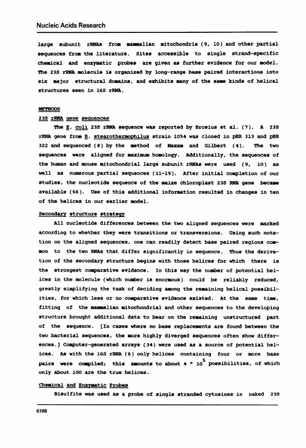
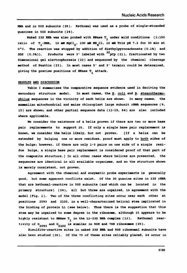
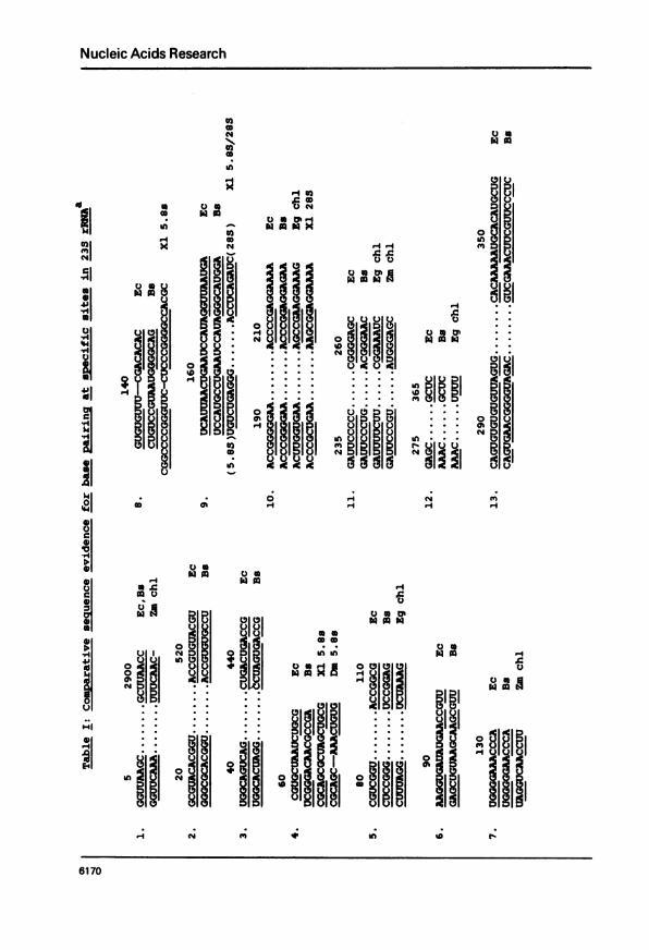
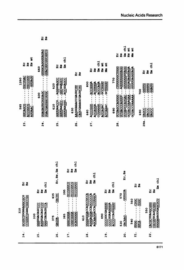
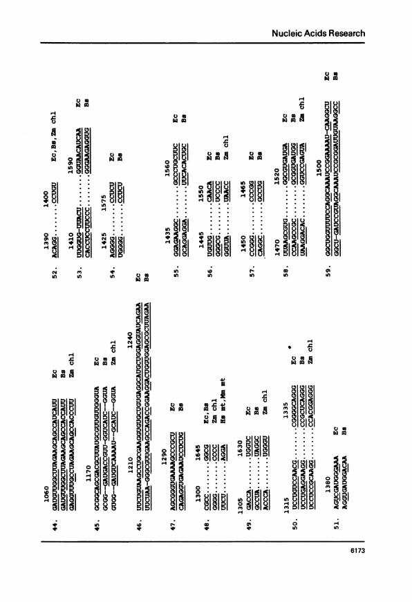
Table I summarizes the comparative sequence evidence used in deriving the
secondary structure model. In most cases, the E. coli and B. stearothermo-
philus sequences in the vicinity of each helix are shown. In many cases, the
mammalian mitochondrial and maize chloroplast large subunit rRNA sequences (9,
10) are shown; and other partial sequence data (11-19, 64) are also included
where applicable.
We consider the existence of a helix proven if there are two or more base
pair replacements to support it. If only a single base pair replacement is
known, we consider the helix likely, but not proven. [If a helix can be
extended by bulging one or more residues, proof must apply to both sides of
the bulge; however, if there are only 1-3 pairs on one side of a single resi-
due bulge, a single base pair replacement is considered proof of that part of
the composite structure.] In all other cases where helices are presented, the
sequences are identical in all available organisms, and so the structure shown
is merely consistent, not proven.
Agreement with the chemical and enzymatic probe experiments is generally
good, but some apparent conflicts exist. Of the 30 guanine sites in 23S rRNA
that are kethoxal-reactive in 50S subunits (and which can be located in the
primary structure) (24), all but three are unpaired, in agreement with the
model (Fig. 1). Two of the three conflicting sites occur near each other at
positions 2093 and 2102, in a well-characterized helical stem implicated in
the binding of protein LI (see below). Thus there is the suggestion that this
stem may be unpaired to some degree in the ribosome, although it appears to be
highly resistant to ENAse T1 in the L1-23S RNA complex (13). Kethoxal reac-
tivity of G2093 and G2102 is similar in 50S and 70S ribosomes (25).
Bisulfite-reactive sites in naked 23S MNA and 50S ribosonal subunits have
also been studied (26). Of the 75 of these sites reliably placed, 64 occur in
6169
Nucleic Acids Research
U)0N
U)
0
a)
a la6n
H
Ioii't) 2
$ * 4
p110
HaSCon0
u ala
0
8:
,-
I I
Ri5i10InI.
V
° u1111° 11001HmH
... ...........00 C
... ....... ....,,,55H
'xx i001I MNS
0Hi
H4p-
0 (m in
**
'I ~II
*
018
1 1gN4 (PH- PH
H
H- N S' 4 In
0 a
InS
I
0
CS
S
.0
Ia
u
*-Pa
E
6170
Nucleic Acids Research
z
0
N1-C * * *
I I I
* * *
o0llElllab
8%* ** *
*1 *
°° 1M0
U Z ~H
C)
U H
W4m 4~~~~D
H
'C
D C.
* .
* *
A 1- go
C)Z
A
0 a
m A
D
s'CIC
w m
0Ululu0s8lululVIU
Hq
m
o
n
0
to
Sc.C~~~~
0 0 gm
N N
6171
I a
Ho
I I
N
*
*
°1;N
C) 01
N (A0||
*m.
N * *
* .
I I I1
'-4
.C0
o 0i0I0i1a AN *to I I I1
0 .I1I .I
I-i"
t)0
1 t
6a ANam
* . . .°000u10IN
u 4o
I gI
%in%DN

Nucleic Acids Research
H
00)
u E
m0N
(900
000
o E1 Eco m V U
en
u toW01
Ul V
LA 0 0
.
6 u
0(9u) u1U
00u0 U
r~N
4J4J
01 VUl Ulm0:0 9Ul 91c
OO
0 . .
0 0 <1<
N
6172
0) u
o0
000
u cn u
~8 8 8
LA8O00O'000
en
0)
M.4J 4J.a 0
3.
:D eC 0
*H-
4nUu u
w Z) 4)u L) 9
.0w =NU V V 3
3. 8j8j e .
LA 000HOOOB0).0.0
Nen .
.u0
00)PC4mNW1 01: N
000000000V
~8 8 OvA0luv~000000
en
H
u u0 0
Nu N
N u
H00 N
Vi u
80 vD3D3V
0 000
::
.~0000
4.
00
Lo) Ul V
H *.
B B
8u4.
10,8
02)N m
AO O
H-*
s
0 u
4 .
° 812rn
V) m00)
000
LOO
Uo2181
0
4.
UZ4 Ulu
o %
4J)
t)mI
U U] f
0) u
e) n
rnPC1 81 13183:COV O <
m 1
5gm1
.0
009
001
0190
001
1.4 I:C)BN
r- U U U0 , 0
0
0.)I
0)
O1t
01
::
0 u4.'C
ru1H
u U2 E
00)0
00
.000
O a
-B
LO O
en.
uL L
.
.
<: m00)D
aa5019
00
0 uu u
0
u B
en:DIO
U) glElLI)
Nucleic Acids Research
urq~~~ ~ ~ ~ ~ ~ ~ M
C)
,"4 e8qC)
|| iE f̂ ~ M X 0110 01
H~~~Crq ~ ~ H
*0 *.
010 Ii;2M0$Ug^800
NOM1 1'iniNgx +00 ' 002un in in in in in irn
oMf H851M , s
MrnL~~~~~~~~~~~~~~~~~~~~~~~~~r
H~~~~~~~
888|18* ° Mm ^§ lgl3 f
*~ ~ ~ 8~* * * * 0
1' in ID CD 01 0' 4 4' 4 . 4' in in
6173

Nucleic Acids Research
ffigN ffi la ,., ^ur pp U~SeU
kj n B ~ B|0| |||| |3"|00
0%jur 18 1 I
o -4 N go
1~~0 ODl M fta o uf o Ou 1 l
tn~~~~~~~~~~~~r
0coSI0I@010|l* * . * 0
In to f".vlrn
WI0 | II I O0 qoOi8||0 °00 i 0
UoUU O : mDm UOwm: t t
Oe uH 00 00 b UL) o
-~~~~~~~iI617o
%D O t %O to DH6174NIB0 ~

Nucleic Acids Research
*rn -
0%t
t0Aa
0
a
IL)UAIL)
W X
01 01 0
.
0 *
N
0;r,
0
NN4
N o'
IiI"010Xi
L) II41
O N
N ..'.0-
oim
U' 5
g888a'
u aa Nw0
I)
I0
0
U . .
N * -I
'N
C) ZX
(4 01|
1
*
*Q*D
0(a CW m uxurn1 X j >
in m
CD 00s
D
W su xa
. .
. *
. .
04
01 1 1I
. *
'01 III
cm I LH0
92ONN N
0uin
. .
N- |
a A
0
In In04J
NBU
u I
* *I
or
o 8~ 1 |4|1
N
~III0 X0Q
I I
N
S. I
0
r1
0I
0 inN m
089
9
N 18
6175

Nucleic Acids Research
au u Cla m oC
0iIt0I I II
1311N
0
0
H mel-
I I
I*In
N
%OI
U B
On . .I*
* 8'0* -~
N|
0Hq
U4OQ 4)UU
E jw2IjilC) 55 C
3I:I> * ' ' I
83 010'3 u110. N
C)SC)
6176 i i
H
C* .4dli.
0
HD I |
N
H
rti
suu
0
0H
C )r m
N '
C. -
0..
0
1')
0;H
00
N
'IC
* a
N .'
. A
0r- 01g0
.I'4
goi
EP t
0)u101 u0Wm
to to
(NN
C) a C)Hum tomx
N0
to0H 6HC)W i 'II
N||810
0,m,.
0)
r4iC)
UI
C),C)wmc0 a gl
In
0)
.qIn.
* *
.
.;
.%
04'InN1
0 I
I6~I40r-
I
in IIn *
. .
* *
* *
Nucleic Acids Research
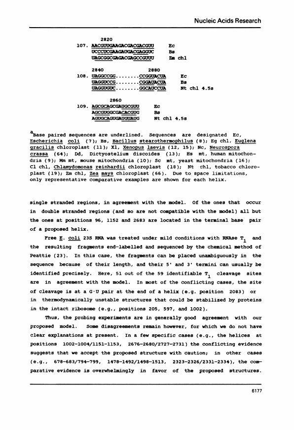
2820107. AACGUUGAAGACGACGACGUU Ec
UCCCUCGAAGAUGRCGAGGUC BsUAGCGGCGAGACGAGCCGUUU Zm chl
2840 2880108. UAGGCCGG........CCGGUACUA Ec
UAGGUCCG........CGGAUACUA BsUAGGUGUC........ GGCAUCCUA Nt chl 4.58
2860109. AGCGCAGCGAUGCGUU Ec
AGCGUGGCGACACGUG BsAGUGC&GUG&UGUAUG Nt chl 4.58
Base paired sequences are underlined. Sequences are designated Ec,Escherichia coli (7); Bs, Bacillus stearothermophilus (8); Eg chl, Euglenagracilis chloroplast (11); Xl, Xenopus laevis (12, 15); Nc, Neurosporacrassa (64); Dd, Dictyostelium discoides (13); Hs mt, human mitochon-dria (9); Mm mt, mouse mitochondria (10); Sc mt, yeast mitochondria (16);Cl chl, Chlamydomonas reinhardii chloroplast (18); Nt chl, tobacco chloro-plast (19); Zm chl, Zea mays chloroplast (66). Due to space limitations,only representative comparative examples are shown for each helix.
single stranded regions, in agreement with the model. Of the ones that occur
in double stranded regions (and so are not compatible with the model) all but
the ones at positions 96, 1152 and 2683 are located in the terminal base pair
of a proposed helix.
Free E. coli 23S RNA was treated under mild conditions with RNAse T and
the resulting fragments end-labelled and sequenced by the chemical method of
Peattie (23). In this case, the fragments can be placed unambiguously in the
sequence because of their length, and their 5' and 3' termini can usually be
identified precisely. Here, 51 out of the 59 identifiable T1 cleavage sites
are in agreement with the model. In most of the conflicting cases, the site
of cleavage is at a G-U pair at the end of a helix (e.g. position 2083) or
in thermodynamically unstable structures that could be stabilized by proteins
in the intact ribosome (e.g., positions 205, 597, and 1002).
Thus, the probing experiments are in generally good agreement with our
proposed model. Some disagreements remain however, for which we do not have
clear explanations at present. In a few specific cases (e.g., the helices at
positions 1002-1004/1151-1153, 2676-2680/2727-2731) the conflicting evidence
suggests that we accept the proposed structure with caution; in other cases
(e.g., 678-683/794-799, 1478-1492/1498-1513, 2323-2326/2331-2334), the com-
parative evidence is overwhelmingly in favor of the proposed structures.
6177

Nucleic Acids Research
GAA UUC'A C.~
AUUUCCGAAAU c GAA
AAUGG^
6G
C-
C G* AAACGGGGCG A UGC-CCCCCI A Ul' AAUuU
G
A6AU
CGgUAGA ,C
6178

Nucleic Acids Research
Figure 1. Secondary structure model for E. coli 23S ribosomal FAA. Primarystructure is from Brosius et al. (7). 'ihe molecule is arbitrarily displayedin two halves (positions 1-1647 and 1648-2904); the pairing of the 5' and 3'ends is shown in both drawings. I-VI refer to the six major structuraldomains. Guanines reactive with kethoxal in 50S subunits (24 and W. B. andB.F .N., unpublished) are indicated by a circled K. Cytosines reactive withbisulfite in naked 238 UM (26) are shaown by a filled circle; unreactive cyto-sines are shown by open circles. Positions of cleavage of 235 UNK by RUase Tunder mild conditions are indicated by arrows. Helices that we consider pro-ven by comparative sequence criteria (see text) are stippled.
6179
Nucleic Acids Research
Here, we must conclude either that the true structure became disrupted under
the experimental conditions (e.g., in the case of naked 23S RNA) or that mul-
tiple conformations are possible at these sites.
General architecture of the 23S ribosomal RN
Our secondary structure model is displayed in Fig. 1. About 52% of the
residues exist in paired structures. The 5' and 3' terminal sequences are
base paired, giving the whole the form of a closed loop. As in the 16S RNA
(1, 6), long range base paired interactions partition the chain into readily
identifiable large domains. There are six of these in the 23S RNA, defined by
their respective long range interactions: domain I (16-25/515-524), domain II
(579-585/1255-1261), domain III (1295-1298/1642-1645), domain IV (1656-
1664/1997-2004), domain V (2043-2054/2615-2625) and domain VI (2630-
2637/2781-2788). These domains project from the central loop created by pair-
ing of the 5' and 3' ends of the molecule.
The 23S RNA chain is readily cleaved into a 13S and an 18S fragment (20),
and the 18S is further cleavable into an 8S and a 12S fragment (63). Com-
parison of oligonucleotide compositions of the various fragments with the com-
plete 23S RNA sequence suggests that the 13S fragment corresponds roughly to
domains I and II (although the 13S/18S cleavage site appears to occur within
domain II), the 8S fragment to domains III and IV, and the 12S fragment to
domains v and VI. No "knots" (27) appear in our model; however, it would be
premature to rule out their existence in the structure. Nor is any convincing
evidence available to suggest how the structure might be organized into the
compact, roughly spherical shape demanded by the SOS ribosomal subunit.
Electron microscopic analysis of unfolded 50S subunits or partially dena-
tured 23S RMA has shown characteristic structural features that can be com-
pared with our secondary structure scheme. 50S subunits unfolded by removal
of magnesium ions appear as asterisk-like shapes, with five prominent arms
radiating from a common center (28). These could well correspond to the five
largest domains (I-V; Fig. 1), which radiate from the central loop. Large
loop structures have been seen in electron micrographs of partially denatured
23S RNA (29), presumably corresponding to strong long-range base paired
interactions between approximately positions 1/500, 1700/2100 and 2200/2600.
These agree well with the strong long range interactions enclosing domains I
(20/520), IV (1650/2000) and V (2050/2620). Hairpin structures visualized at
approximately positions 950, 1550 and 2800 probably correspond to the extended
compound helical stems centered at positions 890, 1SOO and 2700.
As observed previously in 16S RNA, there are no long, uninterrupted regu-
6180
Nucleic Acids Research
lar helices. Instead, the 23S rRNI architecture appears to comprise an
arrangement of small helices, many of which contain characteristic irregulari-ties. As in the 16S MNM case, the helices in 23S RNA appear to be of several
types. Some are regular (involve only Watson-Crick base pairs). Others are
notable for a high content of G-U pairs. Still others are even more irregu-
lar, containing non-Watson-Crick "pairings" (especially A-G "pairs") and
bulged residues. In 16S MNL there appeared a marked tendency for highly con-
served residues to be concentrated in the non-helical regions. This tendency
is not so pronounced in the present case. What is noticeable in the present
instance, however, is that some of the helices are particularly variable in
sequence phylogenetically (again a property of some helices in 16S rRNA).
Sites of interaction with ribosomal proteins and 5S MN
Considerable data have been reported on the ribosomal protein binding
sites in 23S MM, although generally not as extensive as for 16S MNM (reviewed
in ref. 37). These data come either from nuclease protection studies, in
which a specific RNa fragment is shielded by a bound protein or group of pro-
teins, or from covalent crosslinking. The regions bound by proteins LI (35),
L20 (36), L23 (38), and L24 (31, 38) can be located within their protected
fragments, and the position of L4 can be inferred from ultraviolet
irradiation-induced crosslinking of this protein to position 615 of the 23S
MNI (39). Approximate locations of these proteins in the 23S RNI secondary
structures are shown in Figure 2.
An interesting and characteristic feature noted previously in 16S rRNI
(1) and 5S rRNI (30) is a class of helix containing a single bulged nucleo-
tide, usually an A. The occurrence of this type of structure in regions of
r m known to contain recognition sites for ribosomal proteins has been noted
and it has been suggested that the bulged base could form part of the recogni-
tion signal for certain proteins (1, 30). There are fifteen singly bulged
nucleotides in our 23S TNI model; ten of these are adenylate residues. We
suggest that the bulged adenylate at position 443 is involved in the recogni-
tion of protein L24 (31), and that other helices of this type may also be
involved in protein binding.
The type of helix containing multiple G-U pairs may constitute another
class of protein recognition sites. Examples of proteins whose binding sites
contain this type of helix include S8 in 16S RNI (32) and L25 in 5s NI (24).
In 23s NI a number of these G-U helices occur, including 588-601/656-669
[implicated in binding protein L4 (34)] and the pair 2093-2103/2186-2196 and
2127-2143/2148-2161 [both implicated in binding protein L1 (13, 35)].
6181

Nucleic Acids Research
I XL24VIK
Figure 2. Schematic diagram of the 23S RNA secondary structure model. Re-gions containing binding sites for certain ribosomal proteins and 5S RNM areshaded. Helices that have clearly recognizable counterparts in mammalian mi-tochondrial large subunit RNAs are indicated by a dark bar. Post-transcriptionally modified nucleotides are shown by asterisks. I-VI are thesix major structural domains. IVS, intervening sequence; Di, D. melanoqaster;Dv, D. virilis; Pp, Physarum polycephalum; Sc, S. cerevisiae; Cr, Chlamydomo-nas reinhardii; Tp, Tetrahymena pigmentosa; mt, mitochondria; chlp, chloro-plas; 'Cm indicates sites of nucleotide substitutions in chloramphenicol-resistant uitochondrial ribosomes (see text). Kethoxal-reactive sites pro-tected in 70S ribosomes (25) are shown by a circled k. Positions correspond-ing to the points of demarcation of 5.8S, 2S and 4.5S RNAs are indicated.
RNA binding site fragments for proteins L20 and L23 are not well docu-
mented, but they can be placed approximately within the structure from the
available data in the literature. Protein L20 is located in domain II and
probably binds to the 23S RNA somewhere between positions 1000 and 1150 (36).
Protein L23 binds in domain III and can be placed approximately between posi-
tions 1320 and 1600 (38).
The mechanism of association of 5S RNA with the 50S ribosomal subunit is
not yet understood. Although it binds proteins L5, L18, L25 and L31' (33,
65), and requires L5 and 18 to associate with 23S RNK, it is not known
whether or not it interacts directly with 23S RNM. Comparative sequence evi-
6182
Nucleic Acids Research
dence from B. stearothersophilus shows that the rarkable complementarity of
base pairing observed between E. coli 5S and 23S RNUB involving position 143-
154 of 23S UNA (40) is not maintained in general. Thus, it is unlikely that
base pairing between SS and 23S ULs, although seemingly possible, actually
occurs at this site. Protein-mediated binding between the two RNAs is, of
course, a possibility.
The site of interaction of the complex between 5S RNA, L5, L18 and L25
with 23S UM has been studied by the nuclease protection approach in two
laboratories (41, 42). Both groups isolated a protected fragment of 23S RNU
spanning approximately positions 2250-2350. Significantly, this region of 23S
UM is deleted in the ma alian mitochondrial large subunit RNU, the ribosomes
of which lack 5S UNA.
5.8S, 2S and 4.5S rRUAsIn eucaryotic cytoplasmic 60S ribosomal subunits, a 5.8S RNA is found, in
addition to a 26S (or 28S) and a 5S U. Although previously thought to be a
5S UM analogue (43) more recent evidence suggests that 5.8S UM is in fact
analogous to the 5' terminus of bacterial 23S RN (44), whereas the eucaryotic
5S UM is at least structurally a close analogue of procaryotic 5S RNA (33,
45). Alignment of 5.8S Us with 23S RNAs so as to maximize their sequence
homology shows that both can form similar base paired structures within their
comon regions (see Table I). Attachment of 5.8S RNU to 28S RNA, known to
involve the 3' terminus of 5.8S UNA (47), is accounted for by its pairing with
the 5' terminus of 28S UN, which is a close analogue of the helical stem
150-158/168-176 in the E. coli structure (Table I; Figure 1).
Similarly, 2S UM, found in some insect 80S ribosomes (46, 48),
corresponds approximately to positions 140 to 165 of 23S UNA, and is probably
bound to 5.3S UN by base pairing analogous to the 132-137/142-147 pairing of
23S RU. The 4.5S UM found in certain higher plant chloroplasts has previ-
ously been postulated to be analogous to the 3' terminus of 23S UM (49).
This is supported by the finding that these two molecules can also form
closely analogous secondary structures involving their homologous regions (R.
Ballick, personal cowunication; Table I). By analogy with 5.8S RNU, it is
possible that binding of 4.5S UN to 23S UM occurs via base pairing of resi-
dues homologous to the helix 2791-2796/2800-2805 of E. coli.
Intervening sequences in large subunit ribosomal UNsIn several, but certainly not all cases, intervening sequences have been
discovered in the large subunit RUas of eucaryotic cytoplamic, mitochondrial
and chloroplast ribosomes (16-18, 50-53). Positions of six intervening
6183
Nucleic Acids Research
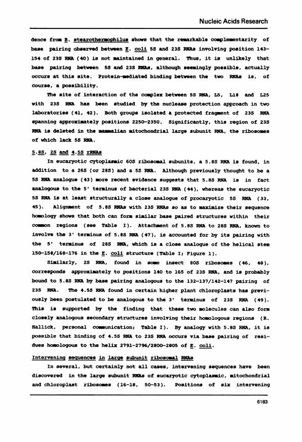
sequences are shown in Figure 2 in the analogous positions in the E. coli 23S
INA structure. Inspection of the primary and secondary structure around these
sites shows that (1) there is no obvious common primary sequence among them,
(2) all of the sites occur in single stranded positions, although nearly all
are closely adjacent to helical elements, (3) all are located in highly con-
served structures in the 3' half of the molecule, which include the peptidyl
transferase region (see below).
The role of intervening sequences in ribosomal RNA genes is not yet
clear; with the possible exception of the Drosophila virilis site (51), theydo not appear to demarcate structural domains, as has been suggested in the
case of protein genes (54). A more striking correlate is their appearance in
what would seem to be functionally indispensable regions of the structure. It
is likely that a ribosomal RNA containing an intervening sequence at any of
the observed positions would be non-functional. Perhaps, then, they are
involved in the regulation of the size of the active ribosome population in
certain cells. Alternatively, they might constitute a proof-reading mechanism
for ribosome maturation: only those ribosomes which have assembled properly
would be recognized and processed. In any event, the fact that they have all
been localized thus far in the 3' half of the large subunit RNA is probably
significant.
Functional Sites
There exists a growing body of evidence implicating the 23S RNA in ribo-somal function. Affinity labelling experiments, in which chemically reactive
groups attached to tRNA or antibiotics are allowed to react with ribosomes,
have shown that some of these functionally important ligands bind in the
immediate vicinity of the 23S RNA (55). In two cases, attempts have been made
to localize the precise sites of reaction with 23S RNA. In the case of
iodoacetyl-Phe-tRNA (56), an RNA sequence was reported which, however, is not
found in the complete 23S RNA sequence. Another affinity reagent, 5'-O-(N-bromoacetyl p-aminophenylphosphoryl)-3'-N-L-phenylalanyl puromycin
aminonucleoside, an analogue of the antibiotic puromycin, was localized to the
sequence GUCCG or GUUCG (57). Further studies are required to establish the
precise sites of attack by these reagents.
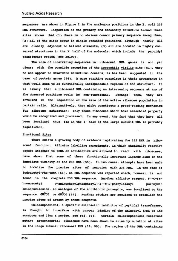
Chloramphenicol, a specific antibiotic inhibitor of peptidyl transferase,is thought to interfere with proper binding of the aminoacyl tRNA at its
acceptor end (for a review, see ref. 58). Certain chloramphenicol-resistantmutant mitochondrial ribosomes have been shown to arise by mutation at sites
in the large subunit ribosomal MMl (16, 59). The region of the ENA containing
6184
Nucleic Acids Research
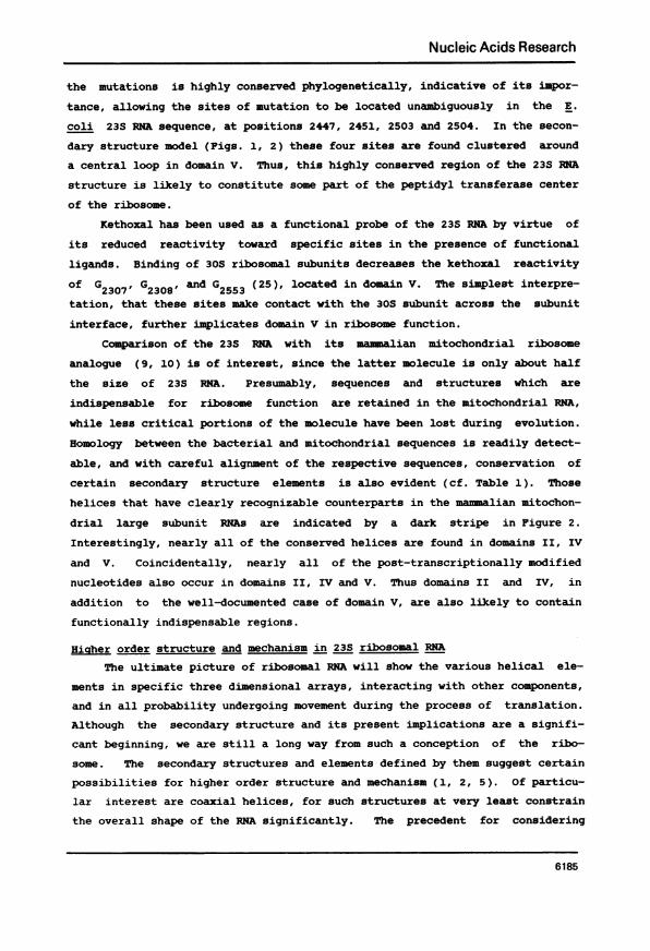
the mutations is highly conserved phylogenetically, indicative of its impor-
tance, allowing the sites of mutation to be located unambiguously in the E.
coli 23S YOM sequence, at positions 2447, 2451, 2503 and 2504. In the secon-
dary structure model (Figs. 1, 2) these four sites are found clustered around
a central loop in domain V. Thus, this highly conserved region of the 23S RNI
structure is likely to constitute some part of the peptidyl transferase center
of the ribosome.
Kethoxal has been used as a functional probe of the 23S RNA by virtue of
its reduced reactivity toward specific sites in the presence of functional
ligands. Binding of 30S ribosomal subunits decreases the kethoxal reactivity
of G2307, G2308, and G2553 (25), located in domain V. The simplest interpre-
tation, that these sites make contact with the 30S subunit across the subunit
interface, further implicates domain V in ribosome function.
Comparison of the 23S RNA with its mamalian mitochondrial ribosome
analogue (9, 10) is of interest, since the latter molecule is only about half
the size of 23S Rm. Presumably, sequences and structures which are
indispensable for ribosome function are retained in the mitochondrial RNA,
while less critical portions of the molecule have been lost during evolution.
Homology between the bacterial and mitochondrial sequences is readily detect-
able, and with careful alignment of the respective sequences, conservation of
certain secondary structure elements is also evident (cf. Table 1). Those
helices that have clearly recognizable counterparts in the mammalian mitochon-
drial large subunit RNAs are indicated by a dark stripe in Figure 2.
Interestingly, nearly all of the conserved helices are found in domains II, IV
and V. Coincidentally, nearly all of the post-transcriptionally modified
nucleotides also occur in domains II, IV and V. Thus domains II and IV, in
addition to the well-documented case of domain V, are also likely to contain
functionally indispensable regions.
Higher order structure and mechanism in 23S ribosomal RNA
The ultimate picture of ribosomal RNA will show the various helical ele-
ments in specific three dimensional arrays, interacting with other components,
and in all probability undergoing movement during the process of translation.
Although the secondary structure and its present implications are a signifi-
cant beginning, we are still a long way from such a conception of the ribo-
some. The secondary structures and elements defined by them suggest certain
possibilities for higher order structure and mechanism (1, 2, 5). of particu-
lar interest are coaxial helices, for such structures at very least constrain
the overall shape of the RNA significantly. The precedent for considering
6185
Nucleic Acids Research
coaxial helices is, of course, the tRNA molecule, which contains two such
juxtapositions-the coaxial TIC and CCA arms, and the nearly coaxial anticodon
and D arms. Any two adjacent helices (i.e., having no unpaired residues
between them in one of the strands) should be considered as potentially coax-
ial. There are about 13 such possible coaxial situations (not so drawn) in
Fig. 1.
One type of comparative evidence strongly suggests coaxiality. Suppose
two adjacent helices in an RN of organism A are of lengths n and m pairs. If
the analogous helices in organism B are of lengths n+a and i-a (i.e., the sum
of pairs in the two, n+m, is constant) then the two helices may be coaxial.
An example of such a case for the 23S rRNA is helix 79 in Table 1.
Going further, the forming and unforming of such coaxial structures, and
the alternate forming of mutually exclusive coaxial structures, should be con-
sidered as a possible basis for mechanism in the ribosome. It is conceivable
that such a mechanism operates in the tRNA molecule during translation and in
the SS RNA as well.
As an example of how coaxial stacking might be employed in translation,
we consider the following. At the 3' terminus of all tRNAs is found the
invariant sequence CCA, likely to play a role in the precise positioning of
aminoacyl and peptidyl moieties in the peptidyl transferase site of the 50S
ribosomal subunit. The two cytidine residues have been shown to be crucial
for productive binding of oligonucleotide analogues of peptidyl tRNA to the
peptidyl transferase site (60) (although the 3 -terminal adenosine of f-Net-
tRNA can be replaced by inosine or guanine and still carry out peptidyl
transfer (61]). Peattie and Herr (62) have demonstrated that the two 3'-
terminal C residuces in ribosome-bound tRNA are strongly protected from chemi-
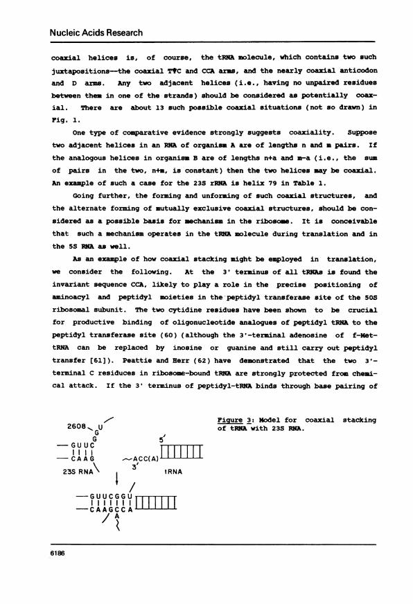
cal attack. If the 3' terminus of peptidyl-tRNA binds through base pairing of
Figure 3: Model for coaxial stacking2608" GU of tRNA with 23S RNA.
GG 5'- GU UClIiIIIIIl-CA AG AACC(A)
23S RNA\ tRNA
-GUUCGGU
-C AAGCC A/A
6186
Nucleic Acids Research
these two C residues, an invariant GG sequence is demanded in the ribosome. A
suitable candidate is the pair of guanosines at positions 2607 and 2608. Sig-
nificantly, U2605 is one of four possible candidates for the site of attack of
the above-mentioned puromycin analogue (57). In this instance, pairing of the
3'-terninal cytosines with G2607 and G2608 creates an extended coaxial helix
in which the two newly formed G-C pairs are stacked on the final C-G pair of
the pre-existing helix 2588-2594/2599-2606 (Fig. 3).
(In the case of tRBfAmet and the majority of other tENdas, an additional A-U
or G-U pair can be made, as shown in Fig. 3). Coaxial stacking of this kind
could afford the advantage of precise structural alignment of two MRA
molecules, in addition to the stability gained from the stacking itself. This
type of mechanism is also attractive for the reason that it makes use of two
well-known structural properties of nucleic acids: base-pairing and stacking.
Such a mechanism should soon be testable.
Recently, two other models for 23S RNA secondary structure models have
been proposed (67, 68). There are significant differences between these
models and the one presented here. These differences will be discussed in a
subsequent paper.
We wish to thank R. Hallick, I. Eperon and R. Brimacombe for sharingtheir unpublished results, Carol Kubitz for drawing figure 1, and N. Turnerfor preparing plasmid DIA. This work was supported by NIH grant GM 17129 (toH.F.N.) and NASA grant NSG 7044 (to C.R.W.).
REFERENCES
1 Noller, H. F. & Woese, C. R. (1981) Science 212, 403-4112 Noller, H. F. (1980) in Ribosomes (eds. Chambliss, G. et al.) Univ. Park
Press, Baltimore3 Sanger, F. & Coulson, A. R. (1975) J. Nol. Biol. 94, 441-4484 Naxam, A. N. & Gilbert, W. (1977) Proc. Natl. Acad. Sci. USA 74, 560-5645 Fox, G. & Woese, C. R. (1975) Nature 256, 505-5066 Woese, C. R., Nagrum, L. J., Gupta, R., Siegel, R. B., Stahl, D. A., top,
J., Crawford, N., Brosius, J., Gutell, R., Hogan, J. J. & Noller, H. F.(1980) Proc. Natl. Acad. Sci. USA 8, 2275-2293
7 Brosius, J., Dull, T. J. & Noller, H. F. (1980) Proc. Nat. Acad. Sci. USA77, 201-204
8 lop, J., Wheaton, V., Gupta, R., Turner, M., Woese, C. R. & Noller, B. F.unpublished
9 Eperon, I. C., Anderson, S. and Nierlich, D. P. (1980) Nature 286, 460-46710 VanEtten, R. A., Walberg,.M. W. and Clayton, D. A. (1980) Cell 22, 157-17011 Orozco, E. M., Rushlow, K. E., Dodd, J. R. and Hallick, R. B. (1980) J.
Biol. Chem. 255, 10997-1100312 Gourse, R. L. and Gerbi, S. A. (1980) Nucleic Acids Res. 8, 3623-363713 Gourse, R. L., Thurlow, D. L., Gerbi, S. A. and Zimrmann, R. A. (1981)
Proc. Natl. Acad. Sci. USA 78, 2722-272614 Hall, L. M. C. and Haden, B. E. H. (1980) Nucleic Acids Res 8, 5993-6004
6187

Nucleic Acids Research
15 Sollner-Webb, B. and Reeder, R. B. (1979) Cell 18, 485-49916 Dujon, B. (1980) Cell 20, 185-19717 Bos, J. L., Osinga, K. A., Van der Horst, G., Becht, N. B., Tabak, H. F.,
Von Ommen, G.-J. B. and Borst, P. (1980) Cell 20, 207-21418 Allet, B. and Rochaix, J. D. (1979) Cell 18, 55-6019 Takaiwa, F. and Sugiura, M. (1980) Molec. gen. Genet. 18, 55-6020 Allet, B. & Spahr, P. P. (1971) Eur. J. Biochem. 19, 250-25521 Bruce, A. G. & Uhlenbeck, 0. C. (1978) Nucleic Acids Res. 5, 3665-367722 Contreras, R. & Piers, W. (1979) FEBS Lett. 16, 281-28323 Peattie, D. A. (1979) Proc. Nat. Acad. Sci. USA 76, 1760-176424 Herr, W. & Noller, H. F. (1978) Biochem. 17, 307-31525 Herr, W. & Noller, H. F. (1979) J. Mol. Biol. 130, 421-43226 Stahl, D. A. (1978) Ph.D. Thesis, Univ. of Illinois, Urbana, Illinois27 Cantor, C. R. (1980) in Ribosomes (eds. Chambliss, G. et al.) Univ. Park
Press, Baltimore, pp. 23-4928 King, T. C., Rucinsky, T., Schlessinger, D. & Milanovich, F. (1981)
Nucleic Acids Res. 9, 647-66129 Edlind, T. D. & Bassel, A. R. (1980) J. Bact. 141, 365-37330 Noller, H. F., Douthwaite, S., Garrett, R. A., & Peattie, D. A. (in
preparation)31 Krol, A., Machatt, M. A., Branlant, C. & Ebel, J. P. (1978) Nucleic Acids
Res. 5, 4933-494732 Muller, R., Garrett, R. A. & Noller, H. F. (1979) J. Biol. Chem. 254,
3873-387833 Garrett, R. A., Douthwaite, S. & Noller, B. F. (1981) Trends in Biochem.
Sci. 6, 137-13934 Stauss, W., Gutell, R., Crawford, N. & Noller, B. F. (unpublished)35 Branlant, C., Krol, A., Sriwidada, J., Sloof, P. & Garrett, R. (1976) Eur.
J. Biochem. 70, 457-46936 Branlant, C., Sriwidada, J., Krol, A. & Ebel, J. P. (1977) Nucleic Acids
Res. 4, 4323-434537 Zimmermann, R. A. (1980) in Ribosomes (eds. Chambliss et al.) Univ. Park
Press, Baltimore, p. 135-16938 Sloof, P., Hunter, J. B., Garrett, R. A. & Branlant, C. (1978) Nucleic
Acids Res. 5, 3503-351339 Maly, P., Rinke, J., Ulmer, E., Zwieb, C. & Brimacombe, R. (1980) Biochem-
istry 19, 4179-418840 Herr, W. & Noller, B. F. (1975) FEBS Lett. 53, 248-25241 Branlant, C., Krol, A., Sriwidada, J. & Brimacombe, R. (1976) Eur. J.
Biochem. 70, 483-49242 Sloof, P. (1977) Ph.D. Thesis, Univ. of Amsterdam43 Wrede, P. & Erdmann, V. A. (1977) Proc. Nat. Acad. Sci. USA 74, 2706-270944 Nazar, R. N. (1980) FEBS Lett. 119, 212-21445 Luehrsen, K. R. & Fox, G. E. (1981) Proc. Nat. Acad. Sci. USA 78, 2150-
215446 Jordan, B. R. (1974) FEBS Lett. 44, 39-4247 Pace, N. R., Walker, T. A. & Schroeder, E. (1977) Biochem. 16, 5321-532848 Pavlakis, G. N., Jordan, B. R., Wurst, R. M. & Vournakis, J. N. (1979)
Nucleic Acids Res. 7, 2213-223849 MacKay, R. M. (1981) FEBS Lett. 123, 17-1850 Nomiyama, B., Sakaki, Y. and Takagi, Y. (1981) Proc. Natl. Acad. Sci. USA
78, 1376-138051 Rae, P. M. M., Kohorn, B. D. & Wade, R. P. (1980) Nucleci Acids Res. 8,
3491-350452 Roiha, H., Miller, J. R., Woods, L. C. & Glover, D. M. (1981) Nature 290,
749-75353 Wild, M. A. & So_er, R. (1980) Nature 283, 693-694
6188
Nucleic Acids Research
54 Gilbert, W. (1978) Nature 271, 50155 Cooperuan, B. S. (1980) in Ribosomes, eds. Chambliss et al., Univ. Park
Press, Baltimore, pp. 531-55456 Yukioka, N., Hatayama, T. & Omari, K. (1977) Eur. J. Biochem. 73, 449-45957 Eckermann, D. J. & Symons, R. H. (1978) Eur. J. Biochem. 82, 225-23458 Cundliffe, E. (1980) in Ribosomes, eds. Charbliss, G. et al., Univ. Park
Press, Baltimore, pp. 555-58159 Kearsay, S. E. & Craig, I. W. (1981) Nature 290, 607-60860 Monro, R. E., Cern&, J. & Narcker, K. A. (1968) Proc. Nat. Acad. Sci. USA
61,61 Cern&, J., Rychlik, I., Krayevsky, A. A. and Gottikh, B. P. (1974) Acta
Biol. Ned. Ger. 33, 877.62 Peattie, D. A. & Herr, W. (1981) Proc. Nat. Acad. Sci. USA 78, 2273-227763 Spierer, P., Zimermann, R. A. & Branlant, C. (1976) FEBS Lett. 68, 71-7564 Selker, E. and Yanofsky, C. (1979) Nucleic Acids Res. 6, 2561-256765 Fanning, T. G. and Traut, R. R. (1981) Nucleic Acids Res. 9, 993-100466 Edwards, K. & KOrsel, B. (1981) Nucleic Acids Res. 9, 2853-286967 Glotz, C., Zwieb, C. & Brimacombe, R. (1981) Nucleic Acids Res. 9, 3287-
330668 Branlant, C., Krol, A., Machatt, M. A., Pouyet, J. & Ebel, J. B. (1981)
Nucleic Acids Res., in press.
6189