haematuria, proteinuria and chronic renal failure · •24-hour versus timed urine collection...
TRANSCRIPT

Haematuria, Proteinuria and Chronic Renal Failure
Procare Group: Wednesday 12 October 2011 @ 19:00
St Heliers Presbyterian Church Community Centre
David Voss ED** BSc MBChB FRACP MRCP(UK) RNZAMC
Renal Physician Eastcare & Waitemata Specialists Centres, Ascot Office Park (Remuera)

Haematuria

Haematuria - macroscopic
• Clots => lower urinary tract origin
• Trauma and UT stones
• Tumour
• Glomerulonephritis
• UTI
• Meatal or perineum irritation
• Contamination from menstrual periods
• Structural: stricture; VUR; RN
• Drugs
– cyclophosphamide

Haemoglobinuria
• Haemoglobulinuria
– Dimer – M. Wt = 34,000
• Protein bound (haptoglobin)
– From intravascular lysis
• Occurs once haptoglobin saturated
– Met-haemoglobulinuria (“Coke colour”)
• Slow nephron transit time
• Acid urine

Myoglobinuria
• Myoglobinuria
– Monomer – M. Wt = 17,000
– Not protein bound
– Readily filtered in glomerulus
• Therefore clears serum rapidly
• Discolours urine
• Rhabdomyolysis
– CK (creatine phosphokinase) levels

Other red urine (1)
• Beet – beta(al)nin (14% of population)
– Not seen in ileostomy patients
– ?colonic absorption
– Worse in:
• Fe deficiency
• Oxalaturia (rhubarb, oysters, chocolate, tea,
spinach)
• Pernicious anaemia induced achlorhydria
• Food dyes
• Porphyria

Other red urine (2)
• Food dyes
• Porphyria
• B12
– In treatment of cyanide poisoning


Detection/diagnosis
• Dipstick
– More sensitive than microscopy
• False positives
– Semen (positive haem reaction)
– Alkaline urine
– Myoglobinuria
• Very dilute urine can haemolyse RBCs
– False negatives
• Excessive vitamin C
– Factitious (rare)

Clues from history
• Vigorous exercise or trauma
• Dysuria and pyuria
• Family history
• Unilateral flank pain
• Prostatic obstructive symptoms
• Bleeding disorders
– The case of anti-coagulants
• Travel
• Sterile pyuria (e.g. Tb; analgesics; IN)

Site/level of haematuria
• Glomerular
– RBC casts
– Proteinuria > 500mg/24 hours
– Dysmorphic RBCs
• Especially acanthocytes
– “Coke” coloured urine
• Non-glomerular
– Lower UT symptoms
– clots

Investigations
• Cytology
– 90% sensitivity for CA bladder
– < 35% for upper UT tumours
• Imaging
– USS
– IVP / IVU
– CT
– MR
– pyelography

Imaging
• CT urogram (radiation dose is high)
– Replaced IVU / IVP
• USS
– Not so good for small urothelial lesions
• MR urography
– Misses smaller lesions and non-
obstructing stones
• Retrograde pyelography
– Similar detection to CT urography

The 40 year-old cut off age
• < 40 year old
– Non-tumour
– Renal origin
– USS
• CT if high risk
• >40 (maybe 45 year old)
– CT urography

Cystoscopy
• In combination with CT urogram
– Excellent screening in combination
• Only method to visualise prostate and
urethra
• Source of bleeding
– And treatment

“No cause found”
• Intermittent?
– How far do we investigate?
• 5-10% of cases
• Hypercalciuria and hyperuricosuria
– Family history
• Rare:
– Schistosomiasis
– Radiation
– AV malformations/fistulae
– Haemorrhagic telangiectasia
– Loin pain syndrome
– Nutcracker syndrome

Follow-up / treatment
• Treat the cause
• No specific treatment for haematuria
per se
• No cause found
– 3 to 6 monthly monitoring
– ?duration
• 3-5 years

Summary – haematuria investigation
• Renal function, MSU, urine microscopy
– Urine RBC morphology
• Quantify proteinuria
– 24 hr; protein-creatinine ratio (PCR); random
• Urinary cytology
• Others: Ca and PO4; SPE; BJ proteinuria;
LFT; ANA, dsDNA; ANCA; Ig; PTH;
complements.
• If < 40 year old renal USS
– otherwise CT urogram

Summary – haematuria referral
• Isolated microscopic
– With symptoms -> urologist
– Painless
• < 40 year old -> renal physician
• > 40 year old => cancer (urologist)
• With proteinuria and/or impaired renal
function
-> Renal physician / nephrologist

Proteinuria

Proteinuria
• Marker of urinary tract inflammation
– Primarily renal in origin
• Glomerulonephritis
• Tubulo-interstitial nephritis
• “Transient contamination”
– Prostatitis
– UTI
• Minor
– Seminal fluid
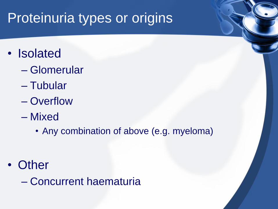
Proteinuria types or origins
• Isolated
– Glomerular
– Tubular
– Overflow
– Mixed
• Any combination of above (e.g. myeloma)
• Other
– Concurrent haematuria

Glomerular proteinuria
• Serum macromolecular proteins
– Albumin
– Glomerular diseases
– Diabetic nephropathy
– “Benign” causes
• Usually < 2 gram/24 hours proteinuria
• Orthostatic
• Exercise induced

Tubular proteinuria
• Low molecular weight proteins
– Not detected by dipsticks
– Freely filtered
• Usually reabsorbed unless tubular overload or
dysfunction
– β2 microglobulin
– Immunoglobulin light chains
– Retinol protein
– Amino acids

Overflow proteinuria
• Myeloma (most common)
– Immunoglobulin light chains
• Less common causes:
– Lysosomal (a. myelomonocytic leukaemia)
– Myoglobin (rhabdomyolysis)
– Haemoglobin (haemolysis)

Detection of Proteinuria
• Dipstick
– Albuminuria
– Misses tubular proteins
• Laboratory quantification
– Pros and cons

Detection of Proteinuria
• 24-hour versus timed urine collection
– Accuracy of 24 hour collection
– Total urine volume in timed collections
• Untimed urinary protein-creatinine ratio
– Pragmatic
– Reproducible
– Affordable and effective

Quantification
dipstick 24 hour
Trace 150 – 300mg
+ 300 – 1,000mg
++ 1 – 3G
+++ 3 – 10G
++++ > 10G

Quantification
24 hour
collection
mg/L or g/L
(spot urine)
mg/mmol
(PCR)
< 300mg 20 – 30 mg/L 20
500mg 40 mg/L 40
1G 0.6 (600mg/L) 60 – 80
3G 2 G/L 180 – 250
10G 6 G/L 600 – 800


Albumin / ACR
• ACR problems
– 24-hour urine and spot urines do not correlate
well
• Orthostatic proteinuria
• Pragmatically first-am and mid-pm are best times
– Depends upon serum creatinine (denominator)
• Muscular people will have a low value
– Vigorous exercise leads to increased albuminuria
• Wait 24 hours

The risk of proteinuria…
• Meta-analysis of general population
cohort: n = 105,872 (ACR) , plus n =
1,128,310 (dipstick proteinuria); mean
follow-up of 7.9 years (cf. a group with
mean eGFR of 95ml/min/1.73m2 BSA)
– Hazard ratios for all cause mortality
• eGFR 60 1.18 (CI 1.05-1.32)
• eGFR 45 1.57 (CI 1.39-1.78)
• eGFR 15 3.14 (CI 2.30-4.13)

The risk of proteinuria……
• Presence of proteinuria alone -> RR of cardiovascular event = 1.3
• Nat Health and Nutrition Examination Survey cardiovascular death rate (unadjusted):
– Proteinuria <30 6.2 deaths / 1000 person-yr
– Proteinuria 30-299 17.9 deaths / 1000 person-yr
– Proteinuria >300 37.2 deaths / 1000 person-yr
• When adjusted:
– Relative hazard 1.57 (30-299 cf. <30 cohort)
– Relative hazard 1.8 (>300 cf. <30 cohort)

Microalbuminuria
• Associated with LV dysfunction; stroke; MI
• Doubles mortality in DM
– Especially in type I DM
• Indicator of inflammation
– Within renal tissue
– Endothelium

Proteinuria
• Concurrent disease
– Inflammation elsewhere (only → small increase)
• E.g. ACR < 10mg/mmol
– Long-standing poorly controlled hypertension
• Rarely above < 3G per day
– Diabetes mellitus
• Usually concurrent diabetic retinopathy
– Connective tissue diseases
– Medications

When to refer
• Unexplained proteinuria
• Worrisome concurrent diagnosis
– Connective tissue diseases
• Nephrotic
• Concurrent impaired renal function
• >1G per 24 hours
~ spot > 600mg/L
~ PCR > 60mg/mmol (60g/mol)

Investigations
• Renal function, uric acid, Ca and PO4
• FBC; ?LFT
• Serum & urine protein electrophoresis
• Renal ultrasound scan
• Others
– Serology - CT diseases
• Renal biopsy

Management
• Generic
– Cease offending agent(s)
• E.g. Nephrotoxins → interstitial nephritis
– ACEi and/or ARBs
• Type I diabetes mellitus
• Other glomerulonephritides
• Specific
– Immunosuppressives

Management
• Goal is to minimise proteinuria
• General rule:
– Double ACEi or ARB dose
→ 30-50% reduction in proteinuria
• Limitation of ACEi & ARB
– hyperkalaemia – when eGFR < 30
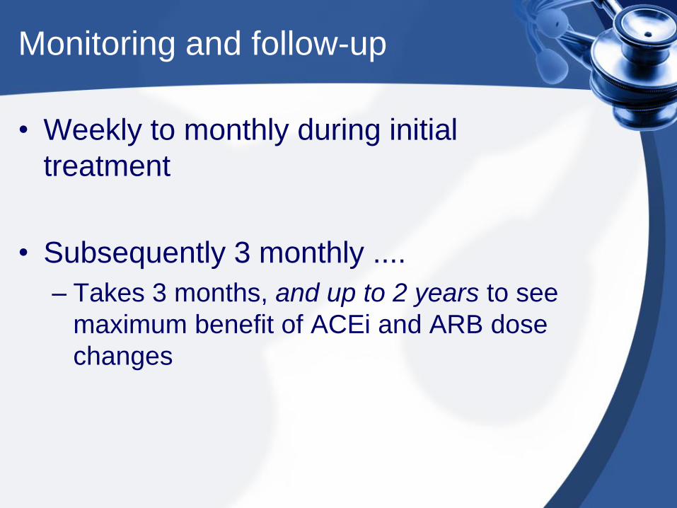
Monitoring and follow-up
• Weekly to monthly during initial
treatment
• Subsequently 3 monthly ....
– Takes 3 months, and up to 2 years to see
maximum benefit of ACEi and ARB dose
changes
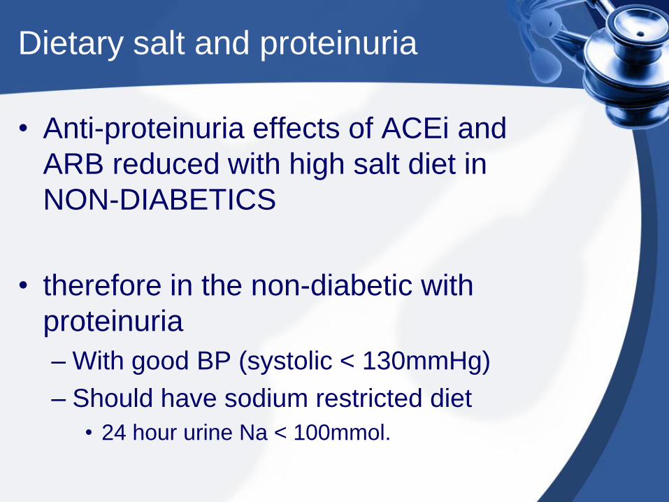
Dietary salt and proteinuria
• Anti-proteinuria effects of ACEi and
ARB reduced with high salt diet in
NON-DIABETICS
• therefore in the non-diabetic with
proteinuria
– With good BP (systolic < 130mmHg)
– Should have sodium restricted diet
• 24 hour urine Na < 100mmol.

Summary - proteinuria
• Dipsticks test for ALBUMIN
– Will miss tubular protein and myeloma and
light chain disease
• Spot urine for proteinuria is satisfactory
• ACEi and ARBs for proteinuria
• Consider salt restriction if treatment
failure
• 3-monthly monitoring of progress
• Refer if proteinuria > 1G/24 hours

CKD – Chronic Renal Failure

[Cre
ati
nin
e] s
μ
mo
l/L
GFR ml/min/1.73m2 BSA
20 40 60 80 100 120
20
0
4
00
60
0
80
0

0
20
40
60
80
100
120
140
160
180 16-2
9
30-3
9
40-4
9
50-5
9
60-6
9
70-7
9
80-8
9
90-9
9
Age (years)
eG
FR
(m
L/m
in/1
.73m
2)
0
10
20
30
40
50
60
High Limit
Median
Low Limit
60 mL/min
%<60 mL/min

eGFR deteriorates with age (median eGFR for age – a GUIDE)
AGE (years) Male Female
60 80mL/min/1.73 m2 60mL/min/1.73 m2
65 75mL/min/1.73 m2 55mL/min/1.73 m2
70 70mL/min/1.73 m2 50mL/min/1.73 m2
75 65mL/min/1.73 m2 45mL/min/1.73 m2
80 60mL/min/1.73 m2 40mL/min/1.73 m2
85 55mL/min/1.73 m2 35mL/min/1.73 m2
90 50mL/min/1.73 m2 30mL/min/1.73 m2

CRF stages
Stage Description GFR
(ml/min/1.73m2)
1 Kidney damage with normal or ↑ GFR >= 90
2 Kidney damage with
mild ↓GFR
60 – 89
3 Moderate ↓GFR 30 – 59
4 Severe ↓GFR 15 - 29
5 ESRF < 15

Chronic Renal Failure
• Deterioration rates
– >60 yo
• 0.75 to 1 ml/min per annum • Lindeman RD, et al. Kidney Int, 26:861; 1984.
• In CRD - depends upon aetiology
diabetes > PVD / analgesics > gn / PCK

Avoiding Nephrotoxins
• Common toxins
– Lithium.
– Chemotherapeutic agents.
– NSAIDs; COX-2; allopurinol.
– H2 / proton pump blockers - omeprazole.
– OTC/herbals.
– Fibrates.

Summary – The Management
(1)
• Type 1 and 2 probably the same
(renally)
• ACEI or ARB
– Probably both together (type 1 and 2)
• Good glycaemic control
– HbA1c < 6.5%; certainly <7%
• Good BP control
– BP <125/75
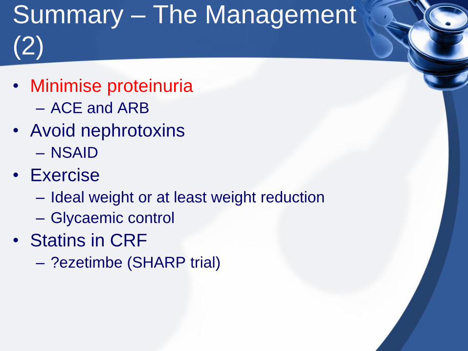
Summary – The Management
(2)
• Minimise proteinuria – ACE and ARB
• Avoid nephrotoxins – NSAID
• Exercise – Ideal weight or at least weight reduction
– Glycaemic control
• Statins in CRF – ?ezetimbe (SHARP trial)

Summary – The Management
(3)
• Aspirin
– Macrovascular disease; IHD
• Folate
– >2mg daily (once CRF)
• Refer when eGFR < 30ml/min (MoH)
– <60ml/min
– Erythropoietin (GFR at 45ml/min)

Medication Dosing
• Renally cleared medications
– Needs dose reduction
• Either dose
• Or frequency
• Avoid nephrotoxins
– NSAIDs vs COX2 inhibitors
• Non-prescription OTC & herbals
– “natural”

Medication Dosing
• Care with combinations
– E.g. NSAIDs and PPI
• Antibiotics
– Penicillins and cephalosporins
• Long courses
– Aminoglycosides
• High trough levels
– Vancomycin
• Long courses and high trough levels

Avoiding Nephrotoxins
• Commonly prescribed nephrotoxins
– lithium
– NSAIDs
– COX-2 inhibitors
– Allopurinol
– Cyclosporine; tacrolimus
– H2 / proton pump blockers
– Fibrates