hdaar 4189516 1. - science
TRANSCRIPT

Review ArticleThe Dynamic Inflammatory Tissue Microenvironment: Signalityand Disease Therapy by Biomaterials
Rani Mata,1,2 Yuejun Yao,1 Wangbei Cao,1 Jie Ding,1 Tong Zhou,1 Zihe Zhai,1
and Changyou Gao 1,2
1MOE Key Laboratory of Macromolecular Synthesis and Functionalization, Department of Polymer Science and Engineering,Zhejiang University, Hangzhou 310027, China2Dr. Li Dak Sum & Yip Yio Chin Center for Stem Cell and Regenerative Medicine, Zhejiang University, Hangzhou 310058, China
Correspondence should be addressed to Changyou Gao; [email protected]
Received 5 October 2020; Accepted 22 December 2020; Published 3 February 2021
Copyright © 2021 Rani Mata et al. Exclusive Licensee Science and Technology Review Publishing House. Distributed under aCreative Commons Attribution License (CC BY 4.0).
Tissue regeneration is an active multiplex process involving the dynamic inflammatory microenvironment. Under a normalphysiological framework, inflammation is necessary for the systematic immunity including tissue repair and regeneration as wellas returning to homeostasis. Inflammatory cellular response and metabolic mechanisms play key roles in the well-orchestratedtissue regeneration. If this response is dysregulated, it becomes chronic, which in turn causes progressive fibrosis, improperrepair, and autoimmune disorders, ultimately leading to organ failure and death. Therefore, understanding of the complexinflammatory multiple player responses and their cellular metabolisms facilitates the latest insights and brings novel therapeuticmethods for early diseases and modern health challenges. This review discusses the recent advances in molecular interactions ofimmune cells, controlled shift of pro- to anti-inflammation, reparative inflammatory metabolisms in tissue regeneration,controlling of an unfavorable microenvironment, dysregulated inflammatory diseases, and emerging therapeutic strategiesincluding the use of biomaterials, which expand therapeutic views and briefly denote important gaps that are still prevailing.
1. Introduction
Tissue regeneration is a fundamental biological task essentialfor the survival of all organisms. Tissue repair and regenera-tion after mechanical injury or infection are firmly regulatedcomplex processes involving a highly efficient inflammatorymicroenvironment. Inflammatory response is a body’s indis-pensable defensive mechanism against tissue damage orpathogens [1]. After tissue damage, a quick reciprocalinflammatory response is generated in the local tissue micro-environment by the damage-associated molecular patterns(DAMPs) or pathogen-associated molecular patterns(PAMPs) via the dying and invading organisms [2, 3]. Theinflammatory microenvironment facilitates various stagesto restore the normal tissue framework including an earlyproinflammatory acute stage (initiation of recruitment ofvital inflammatory cells by the innate immune response com-ponents to start the repair response), a second crucial stage(subsiding proinflammatory response by switching key pro-inflammatory macrophages to a repairing phenotype), and
the last stage (disappearance of inflammatory cells from theinjury site or elimination by apoptosis to restore tissuehomeostasis). However, a sustained chronic inflammationoften impairs the repair/regenerative process and formsfibrosis and scarring. It also dysregulates normal tissue func-tions and eventually leads to organ failure and death [4].
The initial acute inflammatory reaction has an intrinsicfunction in healing tissue injury and plays an essential rolein restoring tissue homeostasis [5]. The principal goal ofacute inflammation is to eliminate dead cells and pathogensat the injury site. Different types of immune cells includingnonhematopoietic and hematopoietic cells collectivelyrespond in the tissue microenvironment and togetherorchestrate tissue repair and regeneration [6] (Figure 1(a)).Although various cell types embrace tissue regenerative func-tions, the resilient macrophages play an important regulatoryrole. The acute inflammatory stage in skin injury encom-passes stimulation of the innate immune system, resultingin initial entry of neutrophils, followed by monocytes thatcan be transformed to macrophages. Macrophages and other
AAASResearchVolume 2021, Article ID 4189516, 31 pageshttps://doi.org/10.34133/2021/4189516

immune cells together clear the cell debris, combat againstpathogens, and also organize cellular mechanisms. Such out-set following the stage of new tissue formation takes placewithin 2-10 days after injury [7]. Multiplication and differen-tiation of stromal and parenchymal cells could then recon-struct tissue integrity. However, if the inflammation is notproperly resolved, the granulated tissue may transform intoscar tissue.
Both the migrating and local macrophages multiply andundergo remarkable phenotypic and functional modifica-tions towards cytokines and growth factors at a local tissuemicroenvironment [8, 9]. Nevertheless, macrophage dys-function could attenuate the proper tissue regeneration pro-cess and activate fibrosis formation, type I and type IIIcollagen deposition, and myofibroblast activation. Therefore,the knowledge on how the immune cells modulate
Tissue injury site
Debris
Effer
ocyt
osis
ROS/hypoxiaDAMPsIL-1, IL-6, IL-12,IL-23,IFN-𝛾, TNF-𝛼ProteasesGrowth factors
Apoptotic neutrophil
Growth factorsPDGFVEGFIGF-1 M
igra
tion
Interleukins
T-cell
Neutrophil Monocyte Monocyte
(a)
(b)
DAMPs NF-𝜅B inhibitorsCrosslinkingDegradabilityHydrophobicityTopography e.g.: TNF-𝛼
SDF-1,...PGE2
Chemokines
Chemokines
Extracellular vesicles
siRNAS
Biomaterial / Delivery system
Injured tissue Regeneration
T helper 2
POLARIZATION
NeutrophilCONVERTION
Monocyte𝛾𝛿T cell
T cell
Residentmacrophage
Stem cellactivation
Anti-inflammatorymacrophage
Inflammatoryfactors
Cell differentiationProliferationAngiogenesis
Pro-inflammatorymacrophage
Physicochemicalproperties
Pro-inflammatorymodulators
Anti-inflammatorymodulators
Synthetic vsnaturally derived
Inflammatorymolecules
Anti-inflammatorycytokines e.g.: IL-10, IL-4,...
Pro-resolvingmediators
Anti-inflammatorymiRNAS
Anti-TNF-𝛼
Blood orlymphaticvessel
Tissuemacrophage
Inflammatorymacrophage
Anti-fibroticmacrophage
RegulatoryT cell
Figure 1: (a) Schematic illustration of the tissue microenvironment at the site of injury. Tissue injury is sensed by the resident macrophagesvia the released DAMPs and neutrophils that are primary infiltrating cells recruited to the damage site, which in turn recruit monocytes andmacrophages. The inflammatory microenvironment is formed by the released inflammatory cytokines, growth factors, and proteases in theearlier stage. It is then shifted to the anti-inflammatory microenvironment that exploits tissue repair and homeostasis in the later stage. (b)Illustrating how the physiochemical properties of biomaterials regulate the tissue immune system. Biomaterials aid in the regulation ofinflammatory cells towards the regeneration/repair phase. They are involved in the polarization of M1 inflammatory macrophages to M2anti-inflammatory/profibrotic/proregenerative macrophages, which is a critical process for tissue regeneration. They also play a crucialrole in converting T-cells into T-regulatory cells. Reprinted with permission from [21] Copyright © Elsevier 2017.
2 Research

inflammation, tissue fibrosis, and neoangiogenesis wouldilluminate the development of promising therapies that tar-get tissue regeneration.
A close examination on the metabolisms of immune cellsover recent years has revealed a strong correlation prevailingamong the metabolic state and phenotype of cells. In partic-ular, macrophages are a notable model of this phenomenon.The M1 macrophages depend on aerobic glycolysis and fattyacid synthesis. Conversely, the M2 macrophages rely onoxidative phosphorylation (OXPHOS), tricarboxylic acid(TCA), and fatty acid oxidation (FAO) [10, 11]. Although itwas believed earlier that the M1macrophages exclusively relyon glycolysis and the M2 macrophages depend on OXPHOSas well as FAO, it has been evident that the proportion is notmerely simple, and the recent evidences favor glycolysis inM2 and FAO in M1 cells [12, 13]. Therefore, the knowledgeon metabolic phenotype switching provides important cuesfor targeting immune metabolic constituents to tune immunecell functions.
Biomaterials play a vital role in the immune modulationand macrophage polarization based on their uniquephysiological properties whether they stimulate or reduceinflammation. The forms of biomaterials such as micro/na-noparticles, scaffolds, and hydrogels and other propertiessuch as crosslinking capacity, topography, and wettabilityas well as nature of biomaterials influence their functions[14]. The biomaterials play a significant role in regulating cellresponses solely or as a part of a complicated system. Theinnate immune cells are the foremost cells to counter theimplanted biomaterials in the vascularized tissue and conse-quent cell recruitment. The intensity of the acute or chronicinflammation depends on the implanted biomaterials, matrixformation, and duration. Biomaterials can be shaped intoscaffolds or hydrogels for cell culture [15] and used for drugdelivery [16]. They can also be easily surface modified [17] orinjected [18] for different applications. The biomaterials withunique biochemical and biophysical characteristics can inter-act with the body and regulate the local tissue microenviron-ment by modulating the immune system from scarring tototal regeneration. The biochemical properties of biomate-rials include delivery of signaling biomolecules such as pro-teins and small drugs. These signaling elements releasedcan stimulate cell-receptor proteins, which regulate theprocess especially protein transport, signaling, and cellmorphology. For instance, biomaterials can induce angio-genesis in vivo within scaffolds by releasing proangiogenicgrowth factors [19]. Moreover, the biophysical characteris-tics, in particular topology, stiffness, degradation, andstructure, can modulate local microenvironments via inter-and intracellular signaling [20] (Figure 1(b)). The alter-ations of the tissue microenvironment consist of tempera-ture, pH, ions, and radicals. The scaffolds may directcellular infiltration by facilitating the transport of oxygen,nutrients, and waste products and induce angiogenesis.However, when the biomaterial biophysical properties aredisproportioned with tissues, it will result in low optimalhealing and defective functionality of regenerated tissue.Therefore, potential biomaterials with advanced architec-tures (biomimicking) and surface topography (bioactive)
provide well-orchestrated biomaterial-immune systeminteractions for optimal functionality.
This review article discusses the inflammatory microen-vironment, crucial macrophage regulatory mechanisms,unfavorable inflammatory microenvironment, dysregulatedinflammatory diseases, and insights into effective therapiesand finally highlights recent biomaterial therapies for propertissue repair and regeneration. Due to the increasing signifi-cance of immune-regulating biomaterials in tissue repairand regeneration and therapy of many other diseases, wewish this review can fill up the gap between immunologicalknowledge and biomaterials and thus promote the develop-ment of tissue microenvironment-modulating biomaterialsfor better medicinal applications, in particular for tissuerepair and regeneration.
2. Inflammatory Microenvironment SignifiesTissue Repair and Regeneration
The inflammatory microenvironment at the tissue damagesite is a complex interlinked framework of immune cells thatplay a critical role in tissue healing and homeostasis.
2.1. Neutrophils. Neutrophils are the primary immune cellsarriving at the injury site and are critical to detecting woundsand host defence. These short-lived immune cells are brisklyrecruited by DAMP signals to restore barrier integrity andfacilitate tissue homeostasis [22]. They can then recruitmonocytes and macrophages to the inflammation site. Theyexpress a large number of chemokine receptors such asCXCR1 and CXCR2 related to the G protein-coupled recep-tor (GPCR) family [23]. A recent study has indicated criticalroles of CXCL8 and CXCR1 or CXCR2 in fostering neutro-phil stimulation and induction to the inflammation site [24].
The activated neutrophils release nuclear and granularconstituents to create web-like structures of DNA, calledneutrophil extracellular traps (NETs) [25, 26]. NETs com-prise double-stranded DNA, histones, and granular proteinsincluding myeloperoxidase (MPO), elastase, and cathepsin G[27, 28]. NETs are associated with pulmonary [29], inflam-matory [30], and cardiovascular [31] thrombosis diseases[32]. NETs are involved in the phagocytosis of their owndying neutrophils and other cellular debris that enhance theirown removal and thereby provide resolution of inflammationand tissue repair [23]. This removal process creates an antic-ipatory proresolution strategy characterized by the release oftissue-repairing cytokines, including interleukin 10 (IL-10)and transforming growth factor-β (TGF-β). Sofoluwe et al.have recently demonstrated that adenosine triphosphate(ATP) channel pannexin 1 (Panx1) and ATP support NETformation or NETosis, which is functionally expressed inmarrow-derived neutrophils of mice (wild type) induced bythe calcium ionophore A23187 or phorbol 12-myristate13-acetate (PMA) in vitro [33]. They also found delayedinduction of NETosis in Panx1−/− mice. Therefore, thebiomaterials that stimulate neutrophil apoptosis wouldhave a great therapeutic effect on promoting tissue repairand regeneration.
3Research
However, the overall effect of neutrophil response appar-ently relies on the conditions including the activation ofinflammatory response, tissue microenvironment, and otherassociated cell types. Nevertheless, various neutrophil subsetsare identified based on their different functions in cancersincluding proinflammatory, antitumoral (N1), and anti-inflammatory protumoral (N2) phenotypes [34]. In addition,Ma et al. found neutrophil polarization during myocardialinfarction (MI) and suggested that lipopolysaccharide(LPS)/interferon gamma (INF-γ) stimulates the polarizationof blood peripheral neutrophils to proinflammatory N1,whereas IL-4 triggers the polarization of anti-inflammatoryN2 similar to macrophage phenotypes [35]. The N1 neutro-phil phenotype is predominant in the myocardium duringMI, whereas the N2 phenotype increases over time, mitigat-ing inflammation and promoting tissue repair. Cuarteroet al. used a peroxisome proliferator-activated receptorgamma (PPAR-γ) agonist to polarize neutrophils to an N2anti-inflammatory subtype, leading to beneficial results instroke [36]. However, further investigations are required toevidently demonstrate the distinctive N2 phenotype func-tions. It would be quite interesting to study whether this phe-notypic subset polarization takes place in variousinflammatory diseases. Moreover, their phenotypic plasticityto alter from one subset to another one is a fascinating chal-lenge because of their short life span.
2.2. Mast Cells. Mast cells are vital immune cells of hemato-poietic lineage residing in all vascularized tissues except forthe retina and nervous system [37]. They are a large sourceof inflammatory factors such as histamine, heparin, numer-ous types of cytokines, neutral proteases, and chondroitinsulfate. Migration of mast cells to target sites is achieved bythe coordinated functions of chemokines, cytokines, integ-rins, adhesion molecules, and growth factors [38]. Althoughthe mast cells are usually engaged in allergic responses, theyare also activated during initial tissue injury and release anample number of proinflammatory mediators including his-tamine and vascular endothelial growth factor (VEGF). Mastcells play an important role in recruiting inflammatory neu-trophils to the injury site through protease-4 (chymase)[39]. Similar to neutrophils, the mast cells secrete numerouseffector elements to recruit eosinophils and monocytes. Agreat number of mast cells are lethal for the tissue regenera-tion since they intensify acute inflammation and support scarformation in the central nervous system [40]. Moreover, theyare highly present in chronic inflammatory wounds [41].However, they also secrete anti-inflammatory molecules,indicating their bilateral and active role during the tissuerepair process [40]. Nevertheless, regulation of mast cells toencourage tissue regeneration rather than scar formation isstill limited.
2.3. Dendritic Cells. Dendritic cells are bone marrow-derivedefficient antigen-presenting cells (APC) that are importantmodulators of innate and adaptive immunity. After tissueinjury, in situ immature dendritic cells captivate dischargedantigens and thereafter travel back to lymphoid organsthrough chemokine gradient, whereby initiating clonal selec-
tion and development of specific T-cells. Additionally,antigen-specific T-cells, B-cells, macrophages, natural killercells, and eosinophils are recruited to the injured site to elim-inate invasive dangers. Even though their specific roles dur-ing tissue repair and regeneration are poorly understood upto present [42], they play a crucial role during the tissue heal-ing process [43]. Dendritic cells apparently function asimmune modulators during tissue recovery via controllingmacrophage homeostasis. As a subset of the dendritic cellpopulation, the plasmacytoid dendritic cells can sense skininjury through host-generated nucleic acids identified bytoll-like receptors 7 and 9 (TLR7 and TLR9) to enhancewound healing via type I INF. Wound closure of burning isconsiderably delayed in a dendritic cell-deficient mousemodel [42]. The defective wound healing is characterizedby the substantial inhibition of initial cellular proliferation,granulated tissue formation, high TGF-β level at the woundsite, and blood vessel configuration. In a murine skeletalmuscle model, dendritic cells gathering together at the injurysite during regeneration undergo maturation following a tar-get with antigen or with the exposure of LPS and migrate intoa lymph node, whereby supporting antigen-specific T helper1 (Th1) priming [44].
2.4. Monocytes and Macrophages. Under normal physiologi-cal conditions, macrophages reside abundantly in hemato-poietic circulation and all types of tissues, which executescavenging and maintain tissue homeostatic functions. Uponinjury, huge numbers of monocytes depart from circulationand reach to the injury site. All macrophages begin their lifeas circulating monocytes and further reside in different tis-sues and habitate the local microenvironment. Both the resi-dent and infiltrating macrophages are stimulated by the localmicroenvironmental cues and further mature into subpopu-lations characterized by the distinctive functional pheno-types. Macrophages are a great source of several cytokines,growth mediators, proteases, extracellular matrix (ECM)constituents, and soluble factors that support tissue repair,regeneration, and fibrosis [45, 46]. Macrophages are differen-tiated from circulatory monocytes that reach to the injury siteafter neutrophils in 1-3 days [47]. They reach a peak level atthe injury site in around 7 days and increase to a high level upto 21 days [48]. The circulatory monocytes are classified intotwo different subsets based on their expression of the lym-phocyte antigen 6 complex (Ly6C) marker in mice andhumans. The Ly6C+/hi (high) arises instantly from blood cir-culation, while the Ly6C-/lo (low) possibly evolves from circu-lating Ly6C+/hi monocytes [49]. They can be differentiateddepending on functions. For instance, the Ly6C+/hi classicalmonocytes respond to injury by infiltrating tissue and dif-ferentiate into macrophages during tissue regeneration. Bycontrast, the Ly6C-/lo monocytes patrol endothelial integ-rity in the vascular system, but they are not differentiatedinto macrophages [50, 51]. After an extensive discussionon the rise of blood macrophages found at the injury site,it is now widely accepted that the infiltrating macrophagesoriginated only from Ly6C+/hi monocytes, which executeclearing damage components, wound healing, and regener-ative functions [52].
4 Research
However, it is still uncertain that a specific (local orrecruited) macrophage can adopt a local tissue microenvi-ronment at distinct times in response to stimulation, or theyare certainly different functional monocyte subsets toregulate divergent activities. Considering these constraintsof subset populations of macrophages such as M1 and M2,here we discriminate these M1/M2 subpopulations usingtheir surface phenotype markers such as Ly6Chi and Ly6Clo.Nevertheless, a subset of the macrophage population charac-terized by specific markers has divergent functions in differ-ent milieu. During the course of injury to repair,macrophages frequently shift their phenotypes betweenLy6Chi and Ly6Clo. For instance, in a CCl4 (carbon tetrachlo-ride) liver injury model, the Ly6Chi macrophages areproinflammatory and profibrotic, whereas the Ly6Clo macro-phages are anti-inflammatory and antifibrotic. Moreover, invarious tissues including skeletal muscle and renal tissue,the Ly6Chi macrophage promotes inflammation and theLy6Clo macrophage stimulates fibrogenesis [53, 54].
A proinflammatory Ly6Chi CX3CR1lo macrophage pop-ulation derived from monocytes acquires homing at theinjury site in a chemokine ligand 2- (CCL2-) dependent fash-ion. The reparative Ly6Clo CX3CR1hi subpopulation movesalong the way led by cluster of differentiation 73 (CD73), vas-cular cell adhesion protein 1 (VCAM-1), very late antigen-4(VLA-4), endothelial-cell-surface enzymes (of leukocyteextravasation), and adhesion proteins [55]. Lorchner et al.identified that regenerative islet-derived 3β (Reg3β) is animportant modulator of macrophage to cardiac tissue afterinjury [56]. The Reg3β facilitates recruitment of the repairmacrophage population that assists neutrophil removal, orelse it would stimulate matrix degeneration and delay colla-gen accumulation and myocyte rupture.
In many tissues, an individual subset of monocytes canfunction as proinflammatory and proreparatory cells, indi-cating that in situ modulation rather than provision of theproreparative LyC6- population is crucial in many contexts.For instance, the protein activin-A that guides differentiationof oligodendrocytes during remyelination of the central ner-vous system (CNS) is a critical macrophage-derived prore-parative factor, and inhibition of M2-derived activin-Aprotein blocks the differentiation of oligodendrocytes duringremyelination in culture of cerebellar slices [57]. In responseto the stimuli, macrophages have a broad range of activitiesparticularly in the transition of innate immune responsethrough the mediation of various factors. Indeed, some ofthese functions are distinct.
The opposing roles of macrophages, for example, pro-and anti-inflammatory nature, rely on stimuli that triggerto procure a divergent phenotype. Despite the fact that themicroenvironmental factors and phenotypes are divergent,they are broadly classified into two phenotypes dependingon the type of T helper cell type 1 or 2 (Th1/Th2) cell polar-ization [58]. The IFN-γ and tumor necrosis factor alpha(TNF-α) excreted by Th1 cells stimulate macrophages to aclassically activated M1 phenotype, and the Th2 cytokinesincluding IL-4 and IL-10 can inhibit stimulation of macro-phages, which are identified as an anti-inflammatory M2phenotype [59]. The M1 macrophages primarily assist in
phagocytosis, as well as in IL-12-associated Th1 effects,whereas the anti-inflammatory M2 phenotype performsTh2 responses and serves in the later phases of tissue repair.By means of the microenvironmental factors, the M2 pheno-type is additionally subclassified into M2a, M2b, M2c, andM2d subtypes. The M2a subtype is notably a profibrotic sub-set activated via IL-4 and IL-13 [60]. The M2b subtype is trig-gered by the combined exposure with immune complexes aswell as toll-like receptors (TLR)/IL-1 antagonist [61]. TheM2c subtype is triggered by the IL-10 and TGF-β/glucocorti-coids that suppress inflammation and promote neovascular-ization [60]. The M2d macrophages are considered tumor-associated macrophages (TAMs) due to their involvementin the tumor growth, angiogenesis, and metastasis [62]. Theyare activated by IL-6, TLR, and A2 adenosine receptor (A2R)agonists and produce higher levels of IL-10, TGF-β, andVEGF but minimal levels of IL-12, TNF-α, and IL-1β. Theclassification paradigm of the M1/M2 axis encounters severalcontroversies because it is based on the in vitro constructionon stimulating macrophages in culture with a specified set offactors and hence may miss the in vivo setting of tissue-specific gene expression programs. For example, in vitrostimulation of monocytes using macrophage colony-stimulating factor (M-CSF) or granulocyte macrophagecolony-stimulating factor (GM-CSF) indicates deviationfrom the M1/M2 concept, because free fatty acids and high-density lipoproteins are within such stimuli, which isspecially applicable to the cardiovascular investigators.Therefore, the classification needs more keen observation onnatural habitats over simply naming according to functionsbecause they may be changed at different environments [63].
Overall, the earlier investigations with various experi-mental schemes in different organ systems properly highlightthe unique and opposing role of inflammatory monocytesand tissue-resident macrophages in the tissue repair andregenerative process [63]. The critical decisive roles of mac-rophages from proinflammatory to anti-inflammatory sub-sets during regenerative response are discussed extensivelyin the following section.
2.5. T-Cells.Over the past decade, innate immunity includingthe macrophage phenotype polarization has been addressedas a critical player in the tissue regeneration process. Never-theless, recent studies indicate that adaptive immunityincluding T-cells also performs a central role. The T-celltypes and subpopulations accumulating at the injury sitegreatly vary in different tissues. For example, in bone, boththe Th1 CD4+ and cytotoxic CD8+ subpopulations restrictregeneration. T-cells suppress major histocompatibility com-plex- (MHC-) derived bone generation in a mouse throughregulating IFN-γ and TNF-α [64]. The investigation inhumans shows that the excretion of IFN-γ and TNF-α resultsin delayed osteogenesis and fracture healing [63].
Although the regulatory T-cells (T-regs) have an indirectrole in regulating inflammation induced by injury, they alsoact directly through amphiregulin, which is a molecule pro-duced by different immune cells and plays a central role inorgan development and supports tissue regeneration in thecontext of inflammation [65]. The highly accepted reliable
5Research

specific T-reg marker is forkhead box P3 (FOXP3), which isvital for the maturation and function of T-regs. An excessiveinflammation after tissue injury causes defective tissue heal-ing and remodeling. T-regs from various tissues are recruitedto the injury site to promote inflammation resolution andcontrol immunity [66], where they can indirectly modulateregeneration by regulating neutrophils, stimulating macro-phage polarization, and controlling T helper cells [67, 68].Furthermore, T-regs can directly support regeneration viastimulating local progenitor cells [69]. A recent in vitro studyhas highlighted that the activated T-regs facilitate neutro-phils to excrete anti-inflammatory mediators such as hemeoxygenase-1, indoleamine 2, 3-dioxygenase (IDO), IL-10,and TGF-β through the preinhibition of IL-6 production,suggesting that T-regs can modulate inflammation via inhi-biting neutrophil function [70]. Moreover, T-regs can controlinfiltration of neutrophils to the damage site. For example,removal of T-regs can cause the increase in neutrophil infil-tration upon cardiac injury and consequently results in adefective healing process [66]. Many studies have evidencedthat CD4+ T-regs are essential for the repair and regenerationof various tissues such as skeletal muscle, lung, myocardium,bone, kidney, and skin [71–75].
2.6. Pro- to Anti-Inflammatory Macrophage Modulation forTissue Regeneration. The proinflammatory macrophagesknown as M1 macrophages can be polarized into anti-inflammatory macrophages or alternatively activated M2macrophages. This M1-M2 macrophage modulation is cru-cial for scavenging inflammation and promoting tissuerepair. In case if the early inflammation is not regulated,excessive inflammation can impair the tissue repair process.Alternatively, an immature early anti-inflammation processcould interrupt the tissue repair and regeneration process.IFN-γ is an important cytokine involved in the activation ofM1 macrophages. The IL-4 and IL-13 are typical stimulatingfactors for inducing the M2 phenotype. Alternatively, theanti-inflammatory IL-10, intermediates of glucose and lipidmetabolism, prostaglandins, and glucocorticoids, may alsoinduce the M2-like phenotype. The anti-inflammatory IL-10plays an essential role in the polarization of macrophagesfrom proinflammation to anti-inflammation, which boostsmuscle regeneration [76]. Nevertheless, IL-4 is often trans-mitted as a protein to trigger M(IL-4)-like macrophagetransition. For instance, in an in vivo rat model withperipheral nerve damage, IL-4 is supplied through theinjectable agarose hydrogels to enhance the number ofM(IL-4) macrophages [77].
Moreover, the controlled delivery of IL-4 supports therepair of peripheral nerve via M(IL-4) macrophages. Overall,the release of IL-4 possibly improves tissue repair and regen-eration in various contexts by inducing M(IL-4) macro-phages. However, the IL-4 effect on the T-regs still has tobe explored. The T-reg accumulation at the injury site sug-gests the modulation of macrophage phenotypes, supportingtheir contribution to the transition of macrophages throughIL-10 and other regulators. In a previous study, early reduc-tion in T-regs upon injury fosters NK cells and effector T cellsto produce IFN-γ, resulting in increased inflammatory stim-
ulation of macrophages [78]. Similar to anti-inflammatoryM(IL-4) macrophages, mesenchymal stem cells also playa regulatory role by switching resident macrophages froma proinflammatory M(IFN-γ) phenotype to a tissue-regenerative phenotype [79]. However, the M(IL-4) cellssimultaneously develop an anti-inflammatory microenvi-ronment that facilitates persistence and growth of bothMHCs and progenitor cells at the injury site, indicating theexistence of a reciprocal helpful feedback relationship amonganti-inflammatory macrophages and pluripotent stem cellsubsets that promotes tissue regeneration [80, 81].
Indeed, impairment in the polarization of M1 to M2macrophages has been involved in the pathogenic chronicwounds. In the diabetic wound repair process, activation ofPPAR-γ is critical for the phenotype transition of macro-phages defected by the steady manifestation of IL-1β inmouse and human wounds. This causes impaired macro-phage shifting from M1 to M2, leading to a delayed woundhealing process [82]. The later stage of the wound healingprocess involves removal of neutrophils, because the over-time accumulated neutrophils produce proteases. The over-produced proteases can deteriorate the complement system,ECM, clotting factors, immunoglobulins, and cytokines,which are essential for tissue repair and regeneration. More-over, they also produce reactive oxygen species (ROS) thatfurther impair and delay the tissue healing process. Hence,the clearing of neutrophils is demanded to advance the repairprocess into a proliferative stage. The macrophages play amajor role in the removal of neutrophils through apoptosisand then by phagocytosis referred to as efferocytosis [83,84]. It is noteworthy that the efferocytosis of neutrophils bymacrophages is a critical element to stimulate modulationfrom a pro- to anti-inflammatory phenotype [85, 86]. Forinstance, lung macrophages induce the engulfment of apo-ptotic bodies by the epithelial cells of the lung airway viainsulin-like growth factor 1 (IGF-1) [87]. A recent studyshows that the developmental endothelial locus-1- (DEL-1-)secreted protein inhibits adhesion of leukocytes and endothe-lial cells and initiation of inflammation and thus functions inresolving inflammation. The proresolving action of DEL-1can be attributed to the efferocytosis via reprogrammingliver X receptor- (LXR-) dependent macrophages to a pro-resolving phenotype with the help of specific proresolvingmediators [88].
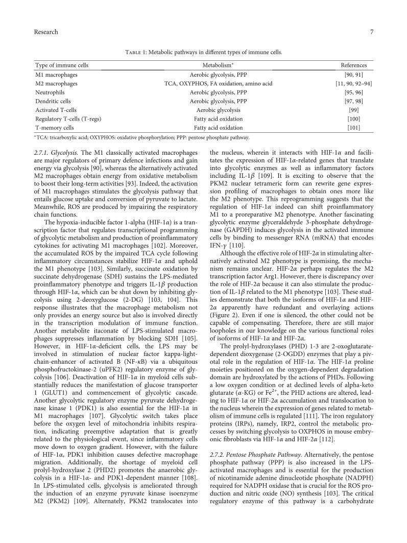
2.7. Metabolisms Linked to Proinflammatory and ProrepairFunctions of Immune Cells. Macrophages are familiar inembracing the challenging circumstances to fight infectionand promote repair, but their precise energetic needs in thissetting are not fully understood. Nevertheless, the macro-phages receive their bioenergy via glycolysis or oxidativemetabolism, which can cause various phenotypes. Mitochon-dria are the powerhouses of immune cells [89]. Metabolicintermediates not only are a source of energy but alsoare directly involved in metabolic switching of phenotypesof different immune cells including macrophages, neutro-phils, dendritic cells, T-cells, T-regs, and memory T-cells(Table 1).
6 Research
2.7.1. Glycolysis. The M1 classically activated macrophagesare major regulators of primary defence infections and gainenergy via glycolysis [90], whereas the alternatively activatedM2 macrophages obtain energy from oxidative metabolismto boost their long-term activities [93]. Indeed, the activationof M1 macrophages stimulates the glycolysis pathway thatentails glucose uptake and conversion of pyruvate to lactate.Meanwhile, ROS are produced by impairing the respiratorychain functions.
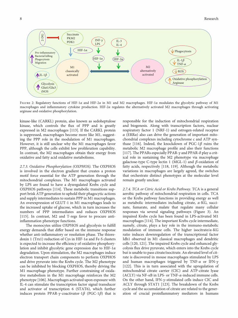
The hypoxia-inducible factor 1-alpha (HIF-1α) is a tran-scription factor that regulates transcriptional programmingof glycolytic metabolism and production of proinflammatorycytokines for activating M1 macrophages [102]. Moreover,the accumulated ROS by the impaired TCA cycle followinginflammatory circumstances stabilize HIF-1α and upholdthe M1 phenotype [103]. Similarly, succinate oxidation bysuccinate dehydrogenase (SDH) sustains the LPS-mediatedproinflammatory phenotype and triggers IL-1β productionthrough HIF-1α, which can be shut down by inhibiting gly-colysis using 2-deoxyglucose (2-DG) [103, 104]. Thisresponse illustrates that the macrophage metabolism notonly provides an energy source but also is involved directlyin the transcription modulation of immune function.Another metabolite itaconate of LPS-stimulated macro-phages suppresses inflammation by blocking SDH [105].However, in HIF-1α-deficient cells, the LPS may beinvolved in stimulation of nuclear factor kappa-light-chain-enhancer of activated B (NF-κB) via a ubiquitousphosphofructokinase-2 (uPFK2) regulatory enzyme of gly-colysis [106]. Deactivation of HIF-1α in myeloid cells sub-stantially reduces the manifestation of glucose transporter1 (GLUT1) and commencement of glycolytic cascade.Another glycolytic regulatory enzyme pyruvate dehydroge-nase kinase 1 (PDK1) is also essential for the HIF-1α inM1 macrophages [107]. Glycolytic switch takes placebefore the oxygen level of mitochondria inhibits respira-tion, indicating preemptive adaptation that is greatlyrelated to the physiological event, since inflammatory cellsmove down to oxygen gradient. However, with the failureof HIF-1α, PDK1 inhibition causes defective macrophagemigration. Additionally, the shortage of myeloid cellprolyl-hydroxylase 2 (PHD2) promotes the anaerobic gly-colysis in a HIF-1α- and PDK1-dependent manner [108].In LPS-stimulated cells, glycolysis is ameliorated throughthe induction of an enzyme pyruvate kinase isoenzymeM2 (PKM2) [109]. Alternately, PKM2 translocates into
the nucleus, wherein it interacts with HIF-1α and facili-tates the expression of HIF-1α-related genes that translateinto glycolytic enzymes as well as inflammatory factorsincluding IL-1β [109]. It is exciting to observe that thePKM2 nuclear tetrameric form can rewrite gene expres-sion profiling of macrophages to obtain ones more likethe M2 phenotype. This reprogramming suggests that theregulation of HIF-1α indeed can shift proinflammatoryM1 to a proreparative M2 phenotype. Another fascinatingglycolytic enzyme glyceraldehyde 3-phosphate dehydroge-nase (GAPDH) induces glycolysis in the activated immunecells by binding to messenger RNA (mRNA) that encodesIFN-γ [110].
Although the effective role of HIF-2α in stimulating alter-natively activated M2 phenotype is promising, the mecha-nism remains unclear. HIF-2α perhaps regulates the M2transcription factor Arg1. However, there is discrepancy overthe role of HIF-2α because it can also stimulate the produc-tion of IL-1β related to the M1 phenotype [103]. These stud-ies demonstrate that both the isoforms of HIF-1α and HIF-2α apparently have redundant and overlaying actions(Figure 2). Even if one is silenced, the other could not becapable of compensating. Therefore, there are still majorloopholes in our knowledge on the various functional rolesof isoforms of HIF-1α and HIF-2α.
The prolyl-hydroxylases (PHD) 1-3 are 2-oxoglutarate-dependent dioxygenase (2-OGDD) enzymes that play a piv-otal role in the regulation of HIF-1α. The HIF-1α prolinemoieties positioned on the oxygen-dependent degradationdomain are hydroxylated by the actions of PHDs. Followinga low oxygen condition or at declined levels of alpha-ketoglutarate (α-KG) or Fe2+, the PHD actions are altered, lead-ing to HIF-1α or HIF-2α accumulation and translocation tothe nucleus wherein the expression of genes related to metab-olism of immune cells is regulated [111]. The iron regulatoryproteins (IRPs), namely, IRP2, control the metabolic pro-cesses by switching glycolysis to OXPHOS in mouse embry-onic fibroblasts via HIF-1α and HIF-2α [112].
2.7.2. Pentose Phosphate Pathway. Alternatively, the pentosephosphate pathway (PPP) is also increased in the LPS-activated macrophages and is essential for the productionof nicotinamide adenine dinucleotide phosphate (NADPH)required for NADPH oxidase that is crucial for the ROS pro-duction and nitric oxide (NO) synthesis [103]. The criticalregulatory enzyme of this pathway is a carbohydrate
Table 1: Metabolic pathways in different types of immune cells.
Type of immune cells Metabolism∗ References
M1 macrophages Aerobic glycolysis, PPP [90, 91]
M2 macrophages TCA, OXYPHOS, FA oxidation, amino acid [11, 90, 92–94]
Neutrophils Aerobic glycolysis, PPP [95, 96]
Dendritic cells Aerobic glycolysis, PPP [97, 98]
Activated T-cells Aerobic glycolysis [99]
Regulatory T-cells (T-regs) Fatty acid oxidation [100]
T-memory cells Fatty acid oxidation [101]∗TCA: tricarboxylic acid; OXYPHOS: oxidative phosphorylation; PPP: pentose phosphate pathway.
7Research
kinase-like (CARKL) protein, also known as sedoheptulosekinase, which controls the flux of PPP and is greatlyexpressed in M2 macrophages [113]. If the CARKL proteinis suppressed, macrophages become more like M1, suggest-ing the PPP role in the modulation of M1 macrophages.However, it is still unclear why the M1 macrophages favorPPP, although the cells exhibit low proliferation capability.In contrast, the M2 macrophages obtain their energy fromoxidative and fatty acid oxidative metabolisms.
2.7.3. Oxidative Phosphorylation (OXPHOS). The OXPHOSis involved in the electron gradient that creates a protonmotif force essential for the ATP generation through themitochondrial complexes. The M1 macrophages activatedby LPS are found to have a dysregulated Krebs cycle andOXPHOS pathways [114]. These metabolic transitions sup-port brisk ATP generation to uphold their phagocytic actionsand supply intermediates to sustain PPP inM1macrophages.An overexpression of GLUT-1 in M1 macrophages leads tothe increased uptake of glucose, which in turn increases thenumbers of PPP intermediates and reduces OXPHOS[115]. In contrast, M2 and T-regs favor to procure anti-inflammation phenotype functions.
The monocytes utilize OXPHOS and glycolysis to fulfillenergy demands that differ based on the immune responsewhether anti-inflammatory or resolution phase. The thiore-doxin 1 (Trx1) reduction of Cys in HIF-1α and Fe-S clustersis expected to increase the efficiency of oxidative phosphory-lation and inhibit glycolytic gene expression due to HIF-1αdegradation. Upon stimulation, the M2 macrophages induceelectron transport chain components to perform OXPHOSand drive pyruvate into the Krebs cycle. The M2 phenotypecan be inhibited by blocking OXPHOS, thereby driving theM1 macrophage phenotype. Further constraining of oxida-tive metabolism in the M1 macrophage reinforces the M2phenotype [106]. Macrophages activated upon exposure withIL-4 can stimulate the transcription factor signal transducerand activator of transcription 6 (STAT6), which furtherinduces protein PPAR-γ-coactivator-1β (PGC-1β) that is
responsible for the induction of mitochondrial respirationand biogenesis. Along with transcription factors, nuclearrespiratory factor 1 (NRF-1) and estrogen-related receptorα (ERRα) also can drive the generation of important mito-chondrial complexes including cytochrome c and ATP syn-thase [116]. Indeed, the knockdown of PGC-1β ruins themetabolic M2 macrophage profile and also their functions[117]. The PPARs especially PPAR-γ and PPAR-δ play a crit-ical role in sustaining the M2 phenotype via macrophagegalactose-type C-type lectin 1 (MGL-1) and β-oxidation offatty acids, respectively [118, 119]. Although the metabolicvariations in macrophages are largely agreed, the switchesthat orchestrate distinct phenotypes at the molecular levelremain greatly unclear.
2.7.4. TCA or Citric Acid or Krebs Pathway. TCA is a generalaerobic pathway of mitochondrial respiration in cells. TCAor the Krebs pathway functions in providing energy as wellas metabolic intermediates including citrate, α-KG, succi-nate, fumarate, and malate that regulate many cellularresponses via several signaling pathways (Figure 3). Animpaired Krebs cycle has been found in LPS-activated M1macrophages [114]. The important Krebs cycle intermediate,namely, citrate, plays a key role in the immuno-metabolicmodulation of immune cells. The higher isocitrate/α-KGratio induces downregulation of the transcriptional factorIdh1 observed in M1 classical macrophages and dendriticcells [120, 121]. The impaired Krebs cycle and enhanced gly-colysis flux drive pyruvate, which enters into the Krebs cyclebut is unable to pass citrate/isocitrate. An elevated level of cit-rate is discovered in mouse macrophages stimulated by LPSand human macrophages triggered by TNF-α or IFN-γ[122]. This is in turn associated with the upregulation ofmitochondrial citrate carrier (CIC) and ATP-citrate lyase(ACLY) via NF-κB in LPS- or TNF-α-induced immune cells.On the other hand, IFN-γ-stimulated cells induce CIC andACLY through STAT1 [123]. The breakdown of the Krebscycle and the accumulation of citrate are related to the gener-ation of crucial proinflammatory mediators in humans
Phagocytosis
ROS
2-D
G SuccinatePKM2PDK1 INF-𝛾
LPSIL-3IL-4
HIF-2𝛼HIF-1𝛼
HIF-1𝛼
M1clsassicallyactivatedIL-1𝛽
Pro-inflammatoryBacterial killingPhagocytosisMigration
GlycolysisGlut1/Glu3iNOS
M2alternatively
activated
IL-1𝛽TNF-𝛼
Oxidative phosphorylationArginase
Figure 2: Regulatory functions of HIF-1α and HIF-2α in M1 and M2 macrophages. HIF-1α modulates the glycolytic pathway of M1macrophages and inflammatory cytokine production. HIF-2α regulates the alternatively activated M2 macrophages through activatingarginase and oxidative phosphorylation.
8 Research

including prostaglandin E2 (PGE2), NO, and ROS [122, 123].Intriguingly, the Akt-mammalian target of rapamycin 1(mTORC1) signaling pathway also regulates the function ofACLY and protein levels in IL-4-stimulated macrophages[124]. Alternatively, the accumulated citrate formed by thekey aspect of IDH1 can be utilized to stimulate fatty acidsand acetylation of histone, and the other fate is the generationof itaconate [125].
Itaconate has recently come under spotlight in the area ofimmune cell metabolisms because of its efficient anti-inflammatory regulator capacity. Upon LPS exposure, theM1 macrophages largely produce itaconate metabolite inthe Krebs cycle from citrate [105]. The mitochondrialenzyme aconitase 2 (ACO2) acts on citrate and producescis-aconitate [126], which is further decarboxlylated to driveitaconate by the enzyme cis-aconitate decarboxylase, referred
ACO2Cis-
aconitate
IRG1Itaconate
Isocitrate
IDH
Hif-1𝛼 Proglycolytic genesIL-1𝛽
Mitochondrial ROSIL-1𝛽
RET
NA
DH
NA
D+
Pro-inflammatory
Pro-inflammatory
OGDH
Succinate
𝛼-Ketoglutarate
Succinyl-CoAsynthetase
TCA cycle
Succinyl-CoA
SDH
Fumarate
ATP
ATP
synt
hase
IV
O2
H2O
AD
P
Fum
arat
e
Malate
MDH
Pyruvate
Glycolysis
Fatty acidsynthesis
NOROS
Prostaglandins
SDH Nrf2 ATF3 Other
Anti-inflammatory
Pro-inflammatory
Citrate Itaconate
CytoplasmaMitochondria
Oxaloacetate
Acetyl-CoA
CIC
CS
Citrate
FH
Succinate
DMM
Elec
tron
tran
spor
t cha
in
SDH
Succ
inat
e
Anti-inflammatory
Figure 3: Overview of the principal metabolic regulation of TCA, glycolysis, electron transport chain, and fatty acid synthesis in macrophagesinvolved in switching macrophage phenotypes. Glycolysis is primarily involved in the activation of M1 that further secretes proinflammatorycytokines. The TCA cycle mainly supports the stimulation of M2 macrophages, consequently inducing secretion of prorepair cytokines. Theimportant metabolites or enzymes involved in the phenotype switching are highlighted in orange color. Reprinted with permission from [134]Copyright © 2020 Springer Nature.
9Research
to as immune-responsive gene 1 (IRG1). Treatment withdimethyl itaconate (DMI), a permeable analog of itaconatein murine bone marrow-derived macrophages (BMDMs)stimulated with LPS, can downregulate the expression of var-ious proinflammatory genes along with inducible nitric oxidesynthase (iNOS) and suppress the production of IL-6, IL-18,IL-1β, ROS, and NO [105]. It has been shown that itaconatecan also prevent the functions of SDH that is mitochondrialcomponent complex II of the electron transport chain(ETC) [127] and further inhibit ROS generation by reverseelectron transport (RET) [128]. Although the usage of DMIprovides insights into the regulatory function of itaconate,there are still some questions that need to be answered, forexample, how the gene silencing of IRG1 and prominentamount of IRG1 mRNA as well as itaconate synthesis areenhanced in immune cells. Itaconate also triggers electro-philic stress and binds with glutathione and consequentlystimulates both nuclear factor erythroid 2-related factor2- (Nrf2-) dependent [129] and independent reactions. Bam-bouskova et al. found that this selective electrophilic stressregulates secondary transcriptional response rather than pri-mary transcriptional response to activate toll-like receptorvia inhibiting inhibitor of kappa B-ζ (IκB-ζ) protein induc-tion that is independent of Nrf2 but dependent on a keymediator activating transcription factor 3 (ATF3) [130].
The key enzyme of the Krebs cycle α-KG plays criticalfunctional roles in promoting an anti-inflammatoryM2mac-rophage phenotype while suppressing proinflammatoryresponses [131]. The M2 macrophages activate the expres-sion of an array of scavenging receptors including mannosereceptors that function in identification and phagocytosis ofapoptotic cells. In IL-4-activated M2 macrophages, theTCA pathway imparts in the production of uridine 5′-diphospho-N-acetylglucosamine (UDP-GlcNAc), an essen-tial intermediate necessary for the glycosylation of the man-nose receptor [120]. In contrast, chemical inhibition of theOXPHOS pathway enzyme ATP synthase in IL-4-activatedM2 macrophages reduces the functional expression of M2genes such as Arg1, C-type mannose receptor 1 (Mrc1),and markers CD206 and arginase-1 [132]. The main causeof the increased glutaminolysis in the proinflammatoryIL-4-activated macrophages is the high level of α-KG,resulting in the promotion of the anti-inflammatory M2phenotype through regulating the histone demethylaseJumonji domain-containing histone demethylase 3 (Jmjd3)or T5-methylcytosine hydroxylases (TET). In contrast, inLPS-activated M1 macrophages, the low level of α-KGreduces proinflammatory functions. The α-KG repressesthe inhibitor of nuclear factor kappa-B kinase (IKKβ) activa-tion needed for the proinflammatory functions driventhrough the NF-κB pathway depending on PHD activity.These results emphasize the targeting schemes involved inα-KG production, suggesting a fascinating therapeutic possi-bility in impaired macrophage-associated diseases.
The alternatively activated M2 macrophages also inducearginine metabolism through arginase-1 (Arg-1), resultingin the generation of ornithine, urea, and polyamines thatare crucial for the wound healing functions of M2 macro-phages [133]. Recently, a key protein TNF-α-induced protein
8-like 2 also known as TIPE2 has been reported, which cantrigger an M2 phenotype through inducing arginine metabo-lism. Fascinatingly, TIPE2 strives these actions upon long-term classical stimulation with LPS rather than alternativeactivation. Therefore, TIPE2 can be a critical switch that neg-atively modulates arginine metabolism and rewrites classicalM1 into an anti-inflammatory phenotype. On the otherhand, iNOS is enhanced in M1 macrophages, resulting inthe catabolism of arginine into citrulline and NO that areimportant for intracellular killing of pathogens. These resultsillustrate that examining the metabolic profiles of macro-phages can develop more potential therapeutic target inswitching phenotypes.
NO produced in murine macrophages is involved in theregulation of macrophage phenotypes via TCA cycle modifi-cations and citrate accumulation. NO inhibits the TCAenzyme aconitase. Furthermore, the inflammatory macro-phages reroute pyruvate far away from pyruvate dehydroge-nase (PDH) depending on NO but not HIF-1α,consequently promoting glutamine anaplerosis and eventu-ally leading to the suppression of ETC complexes [135].
2.7.5. Fatty Acid Metabolism. An increased fatty acid synthe-sis is observed in LPS-stimulated macrophages [136]. Cas-toldi et al. found that triacylglycerol synthesis promotes themacrophage inflammation and decreases FA oxidation[137]. The inhibition of triacylglycerol synthesis significantlyblocks lipid droplet (LD) formation, which further affects theproduction of PGE2, IL-6, IL-1β, and phagocytic capability.A recent report indicated that fatty acids are preferable sub-strate in prorepair M2 macrophages after cardiac injury[138]. An increased arachidonic acid metabolism and eicosa-noid synthesis activated by the cyclooxygenase 2/1 (COX2/-COX1) ratio, isoform of microsomal prostaglandin Esynthase (mPGE2S), arachidonate 5-lipoxygenase, leukotri-ene A4 hydrolase, and thromboxane A synthase 1 areprominent in M1 macrophages. Conversely, COX1 and15-lipoxygenase are stimulated, whereas the mPGE2S isinhibited in the M2 macrophage [139]. Moreover, TLRsalso stimulate the production of nonclassical eicosanoidsincluding resolvins and lipoxins, which support anti-inflammatory or proresolving activities [140]. An elevatedFA metabolism and corresponding stimulation of PPAR-α/β/δ in macrophages seem to play a significant role inswitching macrophages into a reparative phenotype. Inhumans, M1 macrophages induce glycolysis and result inthe production of proinflammatory IL-6, IL-12, p40, andTNF-α similar to murines. However, the M2 phenotypeoxidative metabolism and fatty acid oxidation are not pre-vailed instead of the gluconeogenesis induced by Fbp1largely involved in macrophages [141]. The key enzymefatty acid synthase (FAS) plays a critical regulatory rolein M1 stimulation [142]. This study illustrates that theFAS is essential for the membrane remodeling, and theimpaired FAS can cause alterations in the plasma mem-brane composition. The stimulation of FAS by the mito-chondrial uncoupling protein 2 in macrophages alsointervenes the activation of the NOD-like receptor (NLR)family pyrin domain containing 3 (NLRP3) inflammasome
10 Research
and the subsequent secretion of IL-1β and IL-18 inresponse to the LPS stimuli [143]. However, the mechanis-tic pathways of endogenous and exogenous FAs thatinduce inflammasome activation in macrophages are stillunclear. A recent study shows that the saturated fatty acidsrather than unsaturated fatty acids activate the inflamma-some via increasing phosphatidylcholine levels and therebycause suppression of membrane fluidity and subsequentdisruption of Na+/K+ ATPase, which in turn leads to aK+ efflux [144].
Namgaladze and Brüne reported that in humans, IL-4-induced macrophages do not stimulate PGC-1β, an essentialtranscription factor required to induce fatty acid oxidation.In murine studies, blockers of fatty acid oxidation have noimpact on the production of M2-type factors such as Mrc1and CCL18 [145], indicating the principal variances in themetabolic needs of macrophage phenotype modificationsbetween humans and mice. The fatty acid oxidation canindeed regulate the proinflammatory functions of macro-phages. A recent study shows that larger intracellular levelsof unsaturated fatty acids such as arachidonic acid, oleic acid,and linoleic acids rather than saturated fatty acids trigger thegeneration of IL-1α in foam cells, which in turn causes severeinflammation [146]. Particularly, the M2 macrophages acti-vated with IL-4 depend on fatty acid oxidation throughSTAT6 and PGC1β and suppress inflammatory responses[117, 147]. The influence of FA oxidation on M2 macro-phages is primarily noticed by reducing fatty acid oxida-tion via regulating AMP kinase, a probe responsible formetabolic alterations to elevate ATP levels, which in turnimpairs the inflammatory resolution functions of macro-phages [148].
2.7.6. Amino Acid Metabolism. In recent years, the roles ofamino acids in development and effector actions of immunecells have been considered especially for immune modulation[149]. L-Arginine is a crucial amino acid that can modulateimmune cells. In M1macrophages, L-arginine is metabolizedinto NO and citrulline by iNOS. In contrast, suppression ofiNOS leads the M1 macrophage metabolic and phenotypeswitch towards M2 macrophages. By contrast, in M2 macro-phages activated with IL-4, L-arginine metabolized byarginase-1 results in the production of urea and L-ornithine[90]. Thereafter, it serves as a precursor for the polyaminesand proline that are required for the synthesis of collagenand tissue remodeling of the M2 macrophage [150]. Gluta-mine is another nonessential amino acid that significantlycontributes to the polarization of M2 macrophages by acti-vating the glutamine-UDPN-acetylglucosamine (GlcNAc)pathway to produce α-ketoglutarate [94]. On the other hand,succinate produced from the glutamine-dependent aner-plerosis or γ-aminobutyric acid (GABA) triggers polarizationof M1 macrophages [131]. Glutamine plays an importantrole in M2 macrophages by providing almost third portioncarbon to replenish the TCA cycle. In contrast, suppressionof glutamine synthetase swifts M2 macrophages towardsM1 polarization, suggesting its integral role in theM2macro-phages [151]. However, the current data available on the glu-tamine metabolism in the regulation of macrophage
phenotypes is largely from in vitro studies. Moreover, howthey modulate the phenotype functions of the human macro-phage is still unclear.
3. Unfavorable InflammatoryMicroenvironment in Tissue Regeneration
Although the tissue repair and regeneration process criticallyinvolves the resolution of inflammation as well as tight con-trol of immune response, this process is often dysregulated,leading to fibrosis and scar formation that disturb tissuearchitecture and functions.
3.1. Fibrosis. Fibrosis is a pathological state due to chronicand uncleared inflammation that triggers the production ofsynchronic inflammatory, regenerative, and angiogenic com-ponents in an uncontrolled manner [152]. Development offibrosis ultimately leads to organ dysfunction and death. Inresponse to the DAMPs and inflammatory factors of macro-phages during injury, the inflammation and associated func-tions of fibroblasts will be enhanced to promote theirmigration, differentiation, and synthesis activities [153]. Ingeneral, they synthesize new proteins and ECM proteoglycanconstituents to reconstruct a typical tissue structure. Anexcessive deposition of ECM during the process of fibrosisdrastically impairs tissue structure and functions [46]. Wynnand Ramalingam hypothesized that constant stimulation andsustained mobilization of M(IL-4)-like cells may be involvedin the production of pathological fibrosis [4]. Nonetheless, sofar the majority of studies on fibrosis have engrossed on theinflammatory functions of macrophages. Murray et al.demonstrated that serum amyloid P (SAP) inhibits TGF-β1-induced pathologies including airway inflammation,apoptosis, pulmonary fibrocyte accumulation, and collagendeposition, without affecting TGF-β1 level [154]. In addition,SAP minimizes pulmonary M2 macrophages and enhanceschemokine IP10/CXCL10 expression in a SMAD3-independent manner.
Fibrosis is predominantly accompanied by the crippledangiogenesis and persistent generation of local tissue hyp-oxia. The principal oxygen homeostasis regulator HIF-1α isinstantly associated with the TGF-β1 during fibrogenesis[155]. Certainly, attenuation of HIF-1α expression signifi-cantly declines TGF-β1 generation in alveolar macrophagesand impairs the production of bleomycin-induced fibrosis,substantiating the important role of TGF-β1 in the develop-ment of fibrosis. However, fibrosis can also be independent ofTGF-β [156], wherein type 2 cytokine, namely IL-13, per-forms a major role in many cases [157]. The involvement ofmacrophages in the production and stimulation of IL-13cytokine is still unclear because macrophages are thought tobe not a major source of IL-13 [158]. An endoplasmic retic-ulum (ER) protein, disulfide isomerase containing thiore-doxin domain 5 (TXNDC5), contributes to fibrogenesis bystimulating TGF-β1 signaling cascade through binding andstabilizing lung TGFBR1. Furthermore, activation ofTGF-β1 upregulates TXNDC5 through an ER stress/ATF6-dependent regulation of lung fibroblasts [159]. A transcrip-tion factor JUN plays a crucial role in the lung fibrogenesis
11Research
by increasing the expression of CD47, programmed death-ligand 1 (PD-L1), and IL-6. The inhibition of CD47, PD-L1, and IL-6 reverses fibrosis by enhancing phagocytosis offibroblasts and removing suppressive functions on the adap-tive immune system [160]. The oxidative stress-inducedhepatocyte premature senescence implicated in liver fibrosisis attenuated by IGF-1 via promoting cytoplasmic Akt1-p53interaction, which in turn blocks nuclear p53-progerin(farnesylated mutant lamin A protein) interaction [161].A bioactive chitosan hydrogel prepared with immobilizingthe C domain of IGF-1 peptide (IGF-1C) and adipose-derived mesenchymal stem cell (ADSCs) cotransplantationagainst ischemic kidneys attenuates fibrosis and amelio-rates renal functions [162]. Therefore, IGF-1 embracingparacrine effects may be a promising therapeutic targetto reduce fibrosis. Perhaps, the activated macrophages triggerfibrosis by producing cytokines that stimulate fibroblasts toproduce collagen and ECM. The activated profibrotic mye-loid cells solely express folate receptor β. The folate-targeted TLR7 agonist (FA-TLR7-54) rewrites M2 similarcells that produce fibrosis cytokines. This results in the sup-pression of profibrotic cytokine secretion, biosynthesis ofhydroxyproline, deposition of collagen, and consequentexpansion of alveolar airspaces [163]. One challenge for theregenerative investigators is to regulate phenotypes of fibro-blasts and mesenchymal cells that orchestrate tissue regener-ation. In this regard, novel strategies using biomaterialsenriched with growth factors, stem cells, and decellularizedECM scaffolds can significantly improve the quality of regen-erated tissues.
3.2. Chronic Inflammation. Chronic inflammation is charac-terized by the prolonged response to inflammatory signalsinvolved in continuous recruitment of lymphocytes, mono-cytes, and macrophages with neovascularization as well asconnective tissue proliferation. Chronic inflammation sus-tains tissue remodeling and impairs tissue functions in sev-eral diseases. It is evident that the resolution phase ofinflammation encompassing the control of extravasation ofimmune cells, regulation of production of chemokines andcytokines, shutdown of signaling pathways connected to leu-kocytes, and succeeding leukocyte removal via efferocytosisafter an injury is essential to restore tissue homeostasis. How-ever, an impaired resolution of inflammation causes chronicinflammatory diseases such as colitis and asthma and is alsoinvolved in immutable tissue damage as well as enhanced riskfor the development of cancer, osteoporosis, and cardiovas-cular diseases [164–166]. Unlike the acute inflammation thatis initiated by the DAMPs and PAMPs, the systematicchronic inflammation is classically triggered by DAMPs inthe absence of PAMPs [167, 168]. Therefore, the develop-ment of drugs and therapies is chronically weighed on anti-inflammatory steroids, nonsteroidal anti-inflammatorydrugs (NSAIDS), and furthermore targeted strategies includ-ing TNF monoclonal antibodies.
In this review, we insinuate that novel approaches thatmitigate the negative functions of inflammation in a tissuemicroenvironment may allow constructive functions in theimmune response. Another systematic emerging idea is to
regulate the polarization of immune response, and the con-temporary data also indicate that modulating the entry portof inflammatory cells could be one scheme, although macro-phages may also activate matrix metalloproteinases (MMPs)and other degenerative enzymes to affect ECM. Several typesof MMPs are involved in the clearance of fibrosis, but someothers function in the development of fibrosis. The chronicinflammation-associated molecular composition of diseasesand current therapies are reviewed in detail in the followingsection.
4. Chronic Inflammation-Associated Diseasesand Therapies
4.1. Myocardial Infarction (MI). MI is a type of acute coro-nary syndromes caused by acute and persistent ischemiaand hypoxia [169] and has recently become one of the lead-ing causes of death and disability. In recent years, the clinicaltherapeutic methods of MI mainly contain pharmaceuticaltherapies for thrombolysis, antiplatelet and antihypertension,and interventional therapies such as percutaneous coronaryintervention (PCI) and coronary artery bypass grafting(CABG) [170]. Longer time of coronary artery blockage leadsto a larger area of irreversible myocardial necrosis and finallydeath. Therefore, the patients with acute MI should betreated with thrombolysis as soon as possible to make theinfarcted blood vessels reopen completely and continuouslyfor saving the dying myocardium, preserving the cardiacfunction, and reducing mortality [171]. Thrombolytic drugsinclude streptokinase, urokinase, and recombinant streptoki-nase. Urokinase is widely used due to the absence of antige-nicity, definite curative effect of turning plasminogen intoplasmin to degrade fibrin and dissolve thrombus, and conve-nient application without a skin test [172]. Compared to theconservative and single pharmaceutical therapies, interven-tional therapies are universally considered more effectiveMI reperfusion methods for further reducing mortality from10% to 5% [173]. However, the slow flow/no-reflow phenom-enon occasionally occurring after the interventional thera-pies leads to ineffective treatments, which remains achallenge and difficulty due to its undefined mechanism[174]. Advances have been achieved recently in the possiblemechanisms that are related to the fracture and shedding ofthe thrombus tissue during the mechanical compression ofthe balloon or stent, resulting in microvascular damage,microcirculation embolism, microvascular spasm, reperfu-sion injury, inflammation, release of ROS, and/or swellingof cardiomyocytes [175]. To deal with this phenomenon, pre-operative and intraoperative application of some drugs suchas tirofiban, alprostadil, and nitroprusside together with athrombus aspiration device makes much sense for inhibitingmicrocirculation embolism and promoting microvascularexpansion to some degree [176–179].
Considering that the clinical therapeutic methods stillhave some limitations in irreversible myocardial necrosisand declined cardiac function, other effective regenerationmedicine strategies including stem cell therapy [180, 181]and regenerative biomaterial (such as nanoparticles, patches,scaffolds, and hydrogels) therapy have been developed
12 Research

recently. In particular, the ROS-responsive biomaterials playan important role in the tissue microenvironment and regen-eration [182]. For example, implantation of thioketal-(PUTK-) based ROS-responsive polyurethane patchesloaded with glucocorticoid methylprednisolone (MP)improved significantly the reconstruction of functions ofthe myocardium including elevated ejection fraction,reduced infarction size, and increased revascularization inMI rats [183].
Inflammation participates in and plays a key role in thepathophysiological process of heart damage, repair, andregeneration after MI [184–186], which provides a basisand guidance for regenerative medicine strategies. Generally,initial ischemia and hypoxia after MI result in massive cellapoptosis, and the released DAMPs jointly induce acuteinflammation in the tissue microenvironment [187]. Then,the immune cells such as neutrophils and macrophages arerecruited successively to secrete more inflammatory factorssuch as TNF-α, IL-6, and IL-1β, which further exacerbatethe inflammation and abnormal ventricular remodeling inthe MI area [21]. Stem cells are a type of cells with high plas-ticity and self-renewing ability and are applied in a largenumber of clinical studies. Efficiency of stem cell therapiesin MI is mainly based on two hypotheses: differentiation intomyocardial tissue-related cells such as cardiomyocytes andparacrine effect. Lee et al. injected 2 million ADSCs into thecoronary artery in a pig MI model [188]. The ADSC treat-ment group could increase the capillary density and reducethe infarction area to improve the final cardiac function,likely due to the differentiation of ADSCs into vascular endo-thelial cells instead of cardiomyocytes. Gnecchi et al. foundthat implanted bone marrow-derived mesenchymal stemcells (BMSCs) are able to activate endogenous stem cellsand promote the proliferation of original cardiomyocytesthrough a paracrine effect to achieve the inhibition of MI[189]. However, simple injection of stem cells faces someinevitable problems such as low cell retention, time-consuming cell preparation, and potential allogeneicimmune response [190]. Hao et al. fabricated an injectablefullerenol/alginate hydrogel loaded with ADSCs for cardiacrepair [180]. They found that fullerenol nanoparticles canscavenge excessive ROS to reduce microenvironment inflam-mation and then improve the survival capacity of ADSCs,which are beneficial for increased vascularization and cardiacfunction. Moreover, the direct usage of noncell regenerationmedicinal biomaterials having a property of modulating theMI inflammatory environment presents a greater potentialand convenience for MI treatment. Han et al. synthesizedgraphene oxide (GO) particles of 150 nm loaded with IL-4plasmid DNA (IL-4 pDNA) to treat MI [191]. The GO parti-cles can reduce inflammation as an antioxidant, and the IL-4pDNA increases the ratio of anti-inflammatory M2 macro-phages to inflammatory M1 ones, leading to significant myo-cardial repair. Fan et al. prepared a glutathione- (GSH-)modified collagen hydrogel loaded with recombinant proteinglutathione-S-transferase- (GST-) TIMP-basic fibroblastgrowth factor (bFGF) [192]. TIMP is a peptide PLGLAG thatcan respond to upregulated MMP-2/9 and break to releaseGST and bFGF on demand, which inhibits the degradation
of ECM byMMP-2/9 and contributes to the final vasculariza-tion and MI repair.
In summary, the most clinically used treatment for MI isPCI therapy together with thrombolytic drugs and a throm-bus aspiration device for better reperfusion efficiency. Con-sidering the limitations of irreversible myocardial necrosisand declined cardiac function of clinical methods, regenera-tive biomaterial therapies deserve more attention.
4.2. Atherosclerosis. Atherosclerosis is a chronic inflamma-tory vascular disease, characterized by lipid deposition andfibrosis underneath the inner wall of vessels, which mayresult in serious clinical symptoms such as sudden cardiacdeath, acute myocardial infraction, and stroke [193].Atherosclerosis-related inflammation is associated withimmune activation and induction of inflammatory mediatorsand signaling pathways [194]. The formation of atherosclero-sis is a continuously changing process, and the pathologicaldevelopment progress mainly includes three stages. Theinflammation plays an important role in all stages of the ath-erosclerotic process (Figure 4(a)) [195]. In the first stage, theaccumulation of low-density lipoprotein (LDL) on the arte-rial wall takes place and further passively diffuses via endo-thelial cell (EC) junctions, leading to pathological intimalthickening [196]. In the nascent atheroma, monocytes canbe observed adhering to the surface of the endothelium.Withtime prolongation, the monocytes pass through the endothe-lial monolayer to the intima, where they proliferate and dif-ferentiate into macrophages and foam cells (Figure 4(b)).Macrophages in the atheroma may also have the characteris-tics and probably the antigen-presenting functions. Duringatherogenesis, the smooth muscle cells (SMCs) migrate fromthe media into the intima and produce ECM molecules suchas collagen and elastin (Figure 4(c)). Then, the fibrous capruptures to trigger thrombus. Therefore, the therapies of ath-erosclerosis in its early stage play an important role in slow-ing down the lesions and saving the lives of patients.
The promising therapeutic strategies include nonspecificanti-inflammatory drugs such as allopurinol, colchicine,methotrexate, and aspirin; biologic therapies targeting che-mokines and cytokines such as tumor necrosis factor inhibi-tors and IL-1 neutralization; and small-molecule enzymeinhibitors such as phospholipase inhibitors and antileuko-trienes, as well as targeting of inflammatory signaling path-ways (inhibition of NADPH oxidase, p38 mitogen-activatedprotein kinase (MAPK), or phosphodiesterase) [197]. Simul-taneously, medications to lower lipid, anticoagulation, hyper-tension control, and prevention of thrombosis as well asantiplatelet drugs are used as routine treatments for anti-atherosclerosis [198].
However, severe atherosclerosis always induces a largearea of lumen occlusion, which requires surgical interventionsuch as the technique of percutaneous coronary intervention(PCI) or coronary artery bypass grafting (CABG) surgery[199]. Considering that stents are unsuitable for those withsmall or tortuous vessels or lesions at vessel bifurcation andhigh risk of restenosis, their widespread use may be limited.CABG surgery is the standard of care for patients, havingthe better long-term patency and showing a reduction in
13Research

morbidity andmortality compared to PCI.However, themor-tality, duration of operation, and risk of sternal wound prob-lems are reported for this type of graft [200]. Both carotidendarterectomy (CEA) and carotid artery stenting (CAS) arethe standard treatment for carotid atherosclerosis [201].
Nanocarriers or polymer-based therapeutics used in thecontext of atherosclerosis treatment have been widelyreported, which can overcome the limited efficacy and plentyof side effects related to the existing strategies based on con-ventional drug delivery systems [202]. Inflammatory signal-ing plays an important role in atherosclerosis and is one ofthe most promising targets for the nanosystems. For exam-ple, the ROS-responsive PEG-b-PPS micelles are used as asmart drug delivery system to treat atherosclerosis, whichnot only quickly release the encapsulated drug and rographo-lide but also consume ROS by themselves. The micelles cansimultaneously decrease the inflammatory response andROS level to treat atherosclerosis (Figure 5(a)) [203]. More-over, by taking the physicochemical and biological character-istics of thrombus, fibrin-targeted and H2O2-responsivenanoparticles have been developed (Figure 5(b)) [204]. Afibrin-targeted imaging and antithrombotic nanomedicine(FTIAN) can target fibrin specifically to image thrombus,scavenge H2O2, and prevent platelet activation, which canreduce the formation of thrombus. Consequently, the inflam-matory response biomaterials combined with different drugscan be a more effective antiatherosclerotic therapy anddeserved further study.
4.3. Inflammatory Bowel Disease. Inflammatory bowel dis-ease (IBD) is a chronic inflammatory disorder of the gastro-intestinal tract, which can be divided into chronic relapsinginflammatory disorders (Crohn’s disease) and ulcerativecolitis. IBD is considered an inappropriate and continuinginflammatory response to commensal microbes in a geneti-cally susceptible host [205]. Nonsteroidal anti-inflammatorydrugs and corticosteroids as well as immuno-suppressiveand immuno-regulatory agents such as methotrexate, azathi-oprine, and its metabolite 6-mercaptopurine are used to cureIBD, aimed at reducing intestinal inflammation and immunesystem hyperresponsiveness [206]. Besides, a selective TNF-αblocker, the monoclonal antibody infliximab, is considered amajor advance in IBD therapy [207]. Conventional oral for-mulations are limited for use in IBD due to the adverse effectsand toxicity following distribution of drug among the body[208]. Compared with the conventional agents, small interfer-ing RNA (siRNA) can effectively alleviate IBD progressionand promote intestinal mucosa recovery through precise reg-ulation of the expression of proinflammatory cytokinesrelated to IBD [209]. Nanoparticle delivery systems for siRNAconfer stability of RNA during delivery in vivo, and the nano-particles with ligand modification can target macrophages inthe inflamed intestinal mucosa [210] (Figure 6).
For the treatment of IBD, nanoparticles show specialadvantages including protecting drugs and increasing drugrelease/retention at diseased sites. The nanoparticle deliverysystems include liposomes and polymer-based nanoparticles
�rombusformation
(a) (b)
(c) (d)
Platelet
Fibrous caprupture
Dividing SMCApoptoticbodies
Apoptoticmacrophage
Vasavasorum
MigratingSMC
Cholesterolcrystal
Foamcell
Foamcell
T cellMacrophage
Dendriticcell
Monocyte
Endothelial cell
Intima
Media
SMCs
AdventitiaFibroblastMast cell
Collagen
Lipidcore
Figure 4: (a, b) Stages in the development of atherosclerotic lesions. Reprinted with permission from [195] Copyright © 2021 SpringerNature.
14 Research

[210]. For instance, a kind of nanoparticles formulated fromROS-responsive poly(1,4-phenleneacetonedimethylene thio-ketal) (PPADT) is synthesized [211]. The NPs successfully
release loaded TNF-α-siRNA that knocks down the proin-flammatory cytokine TNF-α in response to the high ROSlevel in the inflammatory gut region. A nanosystem designed
A-MC
ROS
ROSSH5044
PEG-PPS+
Andrographolide
Inflammatoryfactors
Macrophage
Foam cell
(a)
(b)
oxLDL
NecrosisPlaques
SO O
O44 50
O OS
SH
LumenLDL
Intima
Andrographolide PEG-PPS
Antithrombotic activity
Fluorescence/Photoacousticdual imaging
Platelet
fBAP
mN+
O
O
O O
OB
NBO
O
OO O
O x
NaOO
O
OSO
N
So
o
o oDSPE-PEG-CREKA
Tirofiban
Self assembly
FTIAN
FeCl3-treatedartery
i.v. injection
o o nN
yo
Fibrin
ROSNF-𝜅B activation
Inflammation Oxidative stress
Atherosclerosis
ROS
Endothelial injury
Monocyte
O
Figure 5: (a) Schematic illustration of the therapeutic mechanism by PEG-b-PPS micelles for reducing ROS and proinflammatory cytokinesin the atherosclerotic process. Reprinted with permission from [203] Copyright 2018 Elsevier Ltd. (b) Schematic diagram of FTIAN as athrombus-specific theranostic agent that is able to image thrombus and exert potent antithrombotic activity. Reprinted with permissionfrom [204] Copyright © 2021 American Chemical Society.
Endosome/lysosome
Epithelial cell
Receptor on epithelial cell
Receptor on macrophage
Macrophage
RNAi nanoparticleIntestinal lumen
Mucus layer
M-cell
Endosome/lysosome escape
Figure 6: siRNA nanoparticles target epithelial cells or macrophages in the intestinal lumen. Reprinted with permission from [210] Copyright© Dove Press 2016.
15Research

based on Tempol (Tpl) and a biocompatible β-cyclodextrin-derived material (Tpl/OxbCD NP) was reported [212]. Thisnanomedicine is stable in the gastrointestinal tract and caneffectively scavenge multiple components of ROS and selec-tively release Tpl in the inflamed intestinal tissues. AnotherpH and ROS-sequential responsive nano-in-micro compos-ite for targeted therapy of IBD can selectively release rifaxi-min (an intestine-specific antibiotic, RIF) to the inflamedtissues [213].
4.4. Chronic Diabetic Wound.Millions of people get diabetesworldwide, and about 25% of them suffer from diabetic footulcer (DFU), which is a typical chronic wound and can leadto amputation [214]. Common acute wound caused bymechanical factors can heal naturally via a series of continu-ous and overlapping physical stages such as haemostasis,inflammation, proliferation, and remodeling [215]. But thechronic wound can hardly recover because of its abnormalwound microenvironment. The chronic wounds are con-firmed by persistent inflammation, impaired angiogenesisand epithelialization, disordered cytokine/growth factorsecretion, and excessive degradation of ECM, which delaythe normal progression of wound healing [216]. The com-mon features of the chronic diabetic wounds are high levelof ROS [217], formation of bacterial biofilms [218], excessiveneutrophil infiltration [219], and the maladjustment of thenumber of different macrophage phenotypes in the wound[62]. Aimed at these symptoms in diabetic wounds, variousstrategies have been developed to adjust the chronic inflam-mation and accelerate skin regeneration.
The nanoparticles have been extensively used for woundhealing, which may have different functions such as anti-infection, antioxidant, immunoregulation, and controlledrelease of drugs/cytokines/genes [220]. Qiao et al. preparedcopper sulfide nanodots that can eradicate multidrug-resistant bacteria owing to the photothermal effect [221].Meanwhile, the controlled release of copper ions can acceler-ate wound healing by promoting fibroblast migration andendothelial cell angiogenesis. The excessive ROS in chronicwounds lead to severe damage to nucleic acids and proteins[222]. Liu et al. developed a simple and efficient one-stepway to get ultrasmall Cu5.4O nanoparticles. This nanoparticlehas multiple enzyme-mimicking properties and can scavengebroad-spectrum ROS, which is beneficial to wound healing[223]. Gan et al. prepared a kind of konjac glucomannan-modified SiO2 nanoparticles with immunoregulation func-tion, which can reduce the inflammation level and promotewound healing by inducing macrophages to differentiateinto M2 phenotypes in situ [224]. This material provides anew endogenous regulation method to treat diabetic woundwithout adding drugs or exogenous M2 phenotype macro-phages. Due to their unique biochemical characteristics,lipid-based vesicles are applied to the constructions of anadvance drug delivery system. A recent study indicates thatdexamethasone-loaded liposomes can achieve local deliveryto primary human macrophages, induce an anti-inflamma-tory/proresolution phenotype, and promote diabetic woundhealing [225].
Hydrogels are three dimensional networks of hydrophilicpolymers filled with water [226]. Due to their high hydration(similar to native ECM), tunable properties, and porousarchitecture, they are highly recognized in biomedical appli-cations [227]. A gelatin methacryloyl- (GelMA-) based adhe-sive hydrogel for the local delivery of miR-223 5p mimic(miR − 223 ∗) encapsulated in hyaluronic acid- (HA-) basednanoparticles was developed [228]. This hydrogel deliverysystem with miR − 223 ∗ efficiently promotes the formationof uniform vascularized skin at the wound site, which ismainly due to the polarization of macrophages to theM2 phenotype. Zhao et al. prepared a multifunctionalhydrogel (PDA@AgNPs/CPHs) withmany desirable features,including antibacterial property, tunable mechanical andelectrochemical properties, repeatable adhesiveness, goodprocessability, and self-healing ability [229]. This hydrogelshows a significant effect on promoting angiogenesis, acceler-ating collagen deposition, inhibiting bacterial growth, andcontrolling wound infection in a diabetic wound healing ani-mal test. Besides, this conductive hydrogel is a biocompatibledetector that can monitor movements of the human body inreal time. Xiao et al. designed an antioxidant thermorespon-sive citrate-based hydrogel (H-HKUST-1) embedded withcopper metal organic framework nanoparticles, which candecrease toxicity of copper ion and accelerate diabetic woundhealing [230]. The animal test result shows that theH-HKUST-1 hydrogels can induce angiogenesis, collagendeposition, and reepithelialization during wound healing.
Electrospinning technology provides a convenient anddirect approach to prepare scaffolds consisting of microfi-bers/nanofibers [231]. Various types of electrospun fiberssuch as core-shell structure [232] and hollow [233] structurehave been prepared so far. Since the microfibers/nanofiberspossess a high specific surface area and a highly porous 3Dnetwork, the electrospun nanofiber scaffolds have a wellinteraction with cells [234]. Different chemical structuresand modifications also endow the fibers with plenty of func-tions [235]. Augustine et al. reported the development ofelectrospun poly(3-hydroxybutyrate-co-3-hydroxyvalerate)(PHBV) membrane incorporated with cerium oxide nano-particles (nCeO2) for diabetic wound healing application[236]. The nCeO2-PHBV membranes can enhance cellproliferation and vascularization to promote the healingof diabetic wounds. Liu et al. prepared a kind of electrospunthioether-grafted hyaluronic acid nanofibers (FHHA-S/Fe)that can form a nanofibrous hydrogel in situ on thewound, which combine the advantages of both hydrogeland nanofibers [237]. The in vivo test shows that thismaterial can synergistically modulate the inflammationmicroenvironment to help diabetic wound healing by scav-enging ROS effectively in the early inflammation phaseand promoting transformation of the gathered M1 macro-phages to the M2 phenotype.
Dysfunction of HIF-1α is one of the reasons that hinderthe healing of diabetic wound [238]. Gao et al. preparedpoly(ε-caprolactone)/type I collagen electrospun nanofiberwound dressings loaded with dimethyloxalylglycine(DMOG), which can stabilize HIF-1α by inhibiting its degra-dation [239]. The in vivo test shows that the DMOG-loaded
16 Research
nanofibers can improve diabetic wound healing by accelerat-ing reepithelialization, angiogenesis, and wound closure.
The electrospun fibers can also be applied to deliver pep-tides. Liraglutide (Lira), a GLP-1R receptor agonist, has beenreported to promote the angiogenic ability of endothelial cells[240]. Yu et al. prepared a poly(lactide-co-glycolide)/gelatin(PLGA/Gel) nanofibrous membrane that achieves controlledrelease of liraglutide [241]. The PLGA/Gel/Lira can shortenwound closure time and increase blood vessel density, colla-gen deposition, and alignment, efficiently improving thehealing process of diabetic wounds.
In summary, the recent studies show that more and morebiomaterials for diabetic wound healing attach importance towound microenvironment adjustment, including the regula-tion of inflammation, cytokines/nucleic acids, phenotypes ofmacrophages, and elimination of excessive ROS/reactivenitrogen species (RNS). Instead of delivery of drugs orgrowth factors directly, a suitable wound microenvironmentcan activate the natural wound healing process correctly,which involves a series of exquisitely complex physiologicalactivity.
4.5. Osteoarthritis. Osteoarthritis (OA) is a whole joint dis-order that involves structural changes in hyaline articularcartilage, subchondral bone, ligaments, capsules, synovium,and muscles around the joints [242], which is the mostprevalent joint disease afflicting millions of wordwide peo-ple [243, 244]. Accumulating research results indicate thatchronic low-grade inflammation is the key to the patho-logical process and symptoms of OA [245], instead ofthe traditional view that osteoarthritis is only a degenera-tive disease. The pathogenesis of osteoarthritis is quitecomplicated, including many factors such as mechanics,inflammation, and metabolism [242].
In OA joint tissue and synovial fluid, not only are thelevels of plasma proteins, complement components, andcytokines abnormally high but also the chondrocytes andsynovial cells are activated to overproduce redundant inflam-matory mediators [246] such as IL-1 and TNF-α, prostaglan-dins, NO, and much too high concentration of ROS [247].The overexpressed inflammatory mediators in response tojoint structural breakdown alter metabolism and differentia-tion behaviors of chondrocytes, inducing secretion and acti-vation of MMPs [248] and aggrecanases [249], both ofwhich can degrade the ECM. The abnormality of a varietyof cells and inflammatory mediators worsens the inflamma-tory microenvironment of osteoarthritis, disrupts thedynamic balance in the joints, and might eventually lead tojoint failure.
Nonsurgical therapies such as physical treatment, anti-inflammatory drugs, analgesic or chondroitin sulfate supple-mentation, and local injection of sodium hyaluronate arefirstly adopted to temporarily alleviate pain and relievesymptoms for the early OA [242], which are difficult toinhibit the progress of the disease and completely repair thedamaged cartilage. Early diagnosis and timely treatment ofOA play critically important roles in delaying the onset andprogression of severe OA, of which the treatments have torely on surgical interventions [243, 244] such as joint replace-
ment, marrow stimulating techniques, and joint debride-ment. Since all of these surgical procedures have defects,tissue engineering has led to the development of moreadvanced regenerative techniques [250]. Emerging treat-ments combined with biologicals, ranging from growth fac-tors, blood derivatives, and genes to stem cells orchondrocyte transplantation and stem cell-derived exo-somes, are advocated as the promising tools, which make adifference in regulating immune function, lowering inflam-matory mediators, and thus reducing joint damage and pro-moting cartilage regeneration [251, 252].
Various types of biomaterials including hydrogels[253–255], nanoparticles [256–260], electrospun membranes[261], 3D printed materials [262], drug or bioactive factorcarriers, and tissue engineering scaffolds have been appliedfor the treatment of OA. In particular, the hydrogel has beendeemed as an attractive option, attributed to the capabilitiesto stimulate the ECM environment and to incorporate bioac-tive molecules and drugs as well as the convenience of han-dling and minimally invasive repair of cartilaginous tissuesvia adjusting chemical structure, injectability, and crosslink-ing properties [263]. Madry et al. have prepared an injectablehydrogel based on thermosensitive poloxamers, which isloaded with and able to release a therapeutic recombinantadeno-associated virus (rAAV) vector overexpressing thechondrogenic SRY-box transcription actor 9 (SOX9) in full-thickness chondral defects [253]. The hydrogel-virus com-plex is applied in a clinically relevant minipig modelin vivo, which may represent a major step towards improvedcartilage repair in the near future. Besides, the hydrogel scaf-folds with the capabilities to promote cartilage tissue repairbased on regulating the cartilage microenvironment presenta novel perspective for cartilaginous tissue regeneration. Fenget al. have fabricated an aggrecanase-1 cleavable hydrogelconjugated with N-cadherin mimetic peptides HAV-DIGGGC to mediate the degradation of hydrogels and cell-cell interactions of encapsulated BMSCs, thus achieving abetter regeneration of osteochondral defects [254].
The intra-articular treatment systems in virtue of inject-able hydrogels and nanobiomaterials provide effective long-term symptom relief and disease-modifying properties viaenhancing whole joint retention and regulating the OAinflammatory microenvironment, especially for the manage-ment of the early or midstage OA [264]. For instance, Jinet al. have developed a hyaluronate acid-based injectablehydrogel physically mixing epigallocatechin-3-gallate toscavenge ROS, which inhibits inflammation and enhancescartilage regeneration [255].
Recently, nanoparticles have been modified throughactive and passive targeting strategies to facilitate localizationand interactions with specific joint tissues such as cartilageand synovium [265]. In this way, the therapeutic systembased on nanoparticles with the capabilities of regulatingthe OA microenvironment can intelligently enter the tar-geted cells and thereby efficiently suppress inflammationand delay the progression of OA. Li et al. have demonstratedthat polyethyleneimine-modified mesoporous silica nano-spheres can successfully bring hyaluronan synthase type 2into synoviocytes to synthesize endogenous HA with high
17Research

molecular weight and downregulate the synovial inflamma-tory mediators in the OA microenvironment, hence promot-ing the self-repairing mechanism and putting forward atotally different method for OA management [258]. Chenet al. designed a novel MMP-13/pH-responsive andcartilage-targeting ferritin nanocages encapsulating hydroxy-chloroquine for OA imaging and therapy, which can smartlymonitor the overproduced MMP-13 in OA and releasehydroxychloroquine induced by weak acid. Such theranosticnanoplatform holds promise for smart and precise OA clini-cal application [259]. In addition, specifically targeting andregulating macrophages are other crucial sources of inflam-matory responses in OA joint, representing a fresh trendtowards therapeutic platform construction. Liang et al. havefabricated a multifunctional anti-inflammatory drug deliverysystem based on a carbonic oxide (CO) release moleculeloaded in peptide dendrimer nanocages, the surfaces ofwhich are wrapped with folic acid- (FA-) modified HA[261]. The system can be smartly delivered into activatedmacrophages via FA- and HA-mediated specific targetingeffects and then rapidly release CO by depleting hydrogenperoxide to inhibit the expression of IL-1β, IL-6, and TNF-α, thus to inhibit the degradation of articular cartilage ECM.
In summary, OA is still an incurable joint disease. Theever-improving biomaterial-based OA therapeutic platformshave been designed to effectively regulate the OA inflamma-tory microenvironment to suppress the secretion of inflam-matory mediators, relieve symptoms, and promote jointtissue repair and regeneration, which turns out to be highlyefficient options for OA management. Besides, the emergingtherapies via targeting and regulating the key inflammatorycells to improve the OA microenvironment provide newinsight into OA management and hold promise on the pre-cise treatment of every stage of OA.
4.6. Acute Lung Injury (ALI). Inflammation is the body’sdefence against internal and external harmful stimuli. How-ever, uncontrolled inflammation can injure cells, tissues,and organs and cause damage to the body [266]. Acute lunginjury (ALI) is diffuse alveolar damage induced by excessiveinflammatory response, which may further develop intoacute respiratory syndrome (ARDS) and thereby threatento life health if not timely treated [267, 268].
Various internal and external injury factors such as tissuenecrosis, bacteria, and viruses can cause damage to epithelialcells of alveoli. The macrophages in alveoli will recognizethese factors and release inflammatory mediators such asTNF-α and IL-1 that can activate capillary endothelial cellsand recruit white blood cells (WBCs) such as neutrophilsand monocytes from the blood vessel and bone marrow tothe inflamed tissue [269]. The activated endothelial cells willoverexpress intercellular adhesion molecular-1 (ICAM-1)that can bind to integrin expressed on WBCs’ membraneand then help WBCs migrate and adhere onto the vascularwall. Meanwhile, endothelial cells will shrink under the stim-ulation of inflammatory mediators, leading to the increase invascular permeability [270], followed by the WBCs and pro-tein edema fluid exuding to the alveoli. More WBCs willgather in the inflammatory site because of chemotaxis and
are activated by the inflammatory mediators. Furthermore,the activated WBCs will take the phagocytosis role andrelease ROS and lysosomal enzymes to kill and degrade thepathogens. If the pathogens cannot be eliminated in a shorttime, the WBCs will not stop to migrate to the infected tissueand release more ROS and lysosomal enzymes not onlywithin the cells but also outside cells, resulting damage tonormal lung tissue and cells. Therefore, there are high levelsof cytokines and chemokines such as ROS, lysosomalenzymes, TNF-α, IL-1, and more WBCs especially neutro-phils in the ALI microenvironment.
There are no effective methods to treat ALI at present,because the available drugs with a high dose are poorly tar-geted to the lung and thus bring side effects on other organs[271]. When ALI is caused by bacteria or viruses, antibioticsand antiviral drugs are widely used in clinic. However, themultidrug-resistant bacteria and new viruses (such asSARS-CoV-2) are huge challenges [272, 273]. Apart fromsymptomatic treatment, supportive care is also importantfor recovery, including lung protection ventilation, nutri-tional support, and fluid management [274]. Therefore,anti-inflammatory therapy for pneumonia and lung injuryis a necessary and effective method.
Nowadays, the drug delivery systems (DDS) that respondto the ALI microenvironment have attracted much concern[275]. Wang et al. used 4-(hydroxymethyl) phenylboronicacid pinacol ester-modified alpha-cyclodextrin (Oxi-alphaCD) as a carrier to obtain a kind of ROS-responsive moxi-floxacin- (MXF-) loaded nanoparticles, which release MXFtriggered by H2O2 and are effective in a mouse model of pul-monary P. aeruginosa infection [276]. Zhang and hiscoworkers developed an acid-sensitive nanoparticle to targetinflamed lungs. The nanoparticle is composed of a lowpH-sensitive polymer, poly(beta-amino esters), as a corefor drug loading and controlled release, and its surface ismodified with anti-ICAM-1 antibodies for targeting thelung. This kind of nanoparticles can target the inflamedsite and release the loaded drug, reducing inflammationin an ALI mouse model [277]. Ma et al. provided a DDSthat can not only responsively release drug but also havediagnosis property. The anti-inflammation drug predniso-lone is conjugated to a two-photon AIE bioimaging com-pound via ROS-responsive linker, which is furtherencapsulated into a core-shell micelle. The micelle is com-posed of an amphipathic polymer that can release the drugthrough hydrophobic-to-hydrophilic transition of the coretriggered by ROS [278]. In addition, our lab also preparedPPADT nanoparticles loaded with FK-506, which can sup-press the lung inflammation caused by PM 2.5 [279].
The polymer-based DDS has good targeting property,which can reduce dose and side effects to drugs. Further-more, the stimulus responsiveness enables the drugs to bereleased in a controlled manner and also eliminates inflam-matory mediators in the lesion site, offering a promisingtherapy for the treatment of ALI.
4.7. Inflammatory Disorders of the Central Nervous System.The inflammatory-related diseases of CNS include a widespectrum of inflammatory or autoimmune disorders of
18 Research

CNS distinguished by the immune-mediated tissue injuryand existence of inflammatory infiltrations in the brain andspinal cord [280, 281]. The brain injury milieu is allied withimmune reactions, i.e., the activation of innate immunitycomprised of resident monocytes, astrocytes, and microgliaas well as released cytokines and chemokines, and recruitedmacrophages, granulocytes, and lymphocytes from circula-tion. Although convincing evidence suggests that the adap-tive immune cells are activated by the native antigenrecognition, the consequent multiplication and clonaldevelopment are highly absent. The acute and chronicinflammatory brain disorders are predominated by theclass I MHC-restricted CD8+ T-lymphocytes [282]. Atlater stages, astrocytes turn up in a reactive state to createglial scar tissue [283]. In general, CNS has minimal capac-ity to naturally regenerate after traumatic injury/disorders.Therefore, advanced tissue regenerative therapies arerequired to promote tissue and functional repair of CNS.One such strategy is transplantation of cells includingmesenchymal stem cells (MSCs), neural stem cells,induced pluripotent stem (iPS) cells, and endothelial pro-genitor cells [284–287]. Although cell transplantation islargely preferred to regenerate CNS, several limitationsremained due to less cell viability, lower cell survival,and uncontrolled cell multiplication and integration [288].
The conventional delivery of therapeutic molecules byoral and intravenous administration has also been restrainedby diffusion over the blood-brain/spinal cord barrier [289].By contrast, the composite strategies integrating cells, bioac-tive molecules, and biomaterials have thus attracted signifi-cant interest over the past few years to improve cellsurvival, reduce side effects across the blood-brain barrier,and achieve local delivery. For example, implantation ofporous collagen-based scaffolds (PCSs) seeded with neuralstem cells can enhance cell delivery and differentiation, acti-vate robust axonal elongation, and decrease astrogliosis inanimals [290]. HA is an extensively considered and modifiednatural polymer for a regenerative scaffold of CNS because itplays an important role in neural tissues by regulating cellmigration, proliferation, and differentiation. For instance,HA interacts with the CD44 cell surface receptor of glialand neuronal cells, which regulates cell behavior at tissuedamage conditions [291]. Moreover, HA-CD44 pairing inastrocytes triggers the Rac 1-dependent PKN (protein kinaseN) pathway, leading to enhanced astrocyte migration, andactivated Src family kinases (SFKs) and cascade of focal adhe-sion kinase (FAK) [292, 293]. Combination of HA with syn-thetic peptides in particular RGD and IKVAV potentiallysupports differentiation of neural progenitor cells (NPC) intooligodendrocytes and synapse-forming neurons [294].Hydrogels have been employed to recapitulate and improveregeneration and functions of CNS [295]. Injectable self-assembling peptide-based hydrogel (SAPH) used directlyon the rat brain after traumatic brain injury significantly trig-gers VEGF-receptor 2 and thereby enhances angiogeniceffect and new blood vessel formation. Furthermore, thevon-Willebrand factor (vWF) and α-smooth muscle actin(α-SMA) also increase along with blood vessel density[296]. Coupled self-assembling peptide (SAP) and neural
stem/progenitor cell (NSPC) transplantation enhancesengraftment of NSPC, synaptic connectivity, and behavioraloutcomes and reduces CSPG deposition and astrogliosis[297]. The composite 3D biomimetic CNS scaffolds consist-ing of polyethylene glycol-gelatin-methacrylate (PEG-GelMa) can promote the repair of rat spinal cord injury[298]. The NSPS derived from the spinal cord are suspendedin a fibrin matrix consisting of brain-derived neurotrophicfactor, VEGF, fibroblast growth factor, and calpain inhibitor,which are loaded into a scaffold and then incorporated into athoracic cord transection lesion of rats. After implantation,the 3D scaffold maintains its architecture for 6 months, theengrafted NCPS survive, the axons expand into the host spi-nal cord, and the synaptic transmission recovers, leading toelectrophysiological and functional development. Thesestudies show that with the suitable combination of ideallyengineered scaffolds, stem cells, and growth factors, theadverse environment of brain/spine cord injury can beimproved, resulting in the regeneration of CNS. Moreover,the use of electrically conductive biomaterials and integrationof different functional scaffolds/hydrogels are more apprecia-ble to achieve better neural tissue repair and regeneration.
5. Conclusions and Future Perspectives
The primary role of the inflammatory microenvironmentparticularly immune cells at the tissue injury/damage site isto establish and orchestrate proregenerative milieu. Differenttypes of immune cells and several immune regulators partic-ipate in all the phases of tissue repair/regeneration andhomeostasis. As discussed in this review, among diversetypes of immune cells, monocytes and macrophages are crit-ical players, which secure many functional traits that are crit-ical for the tissue regeneration. However, the mechanisticstimulations by which they modulate phenotypes for proin-flammation, anti-inflammation, prorepair, and profibrogenicmechanisms remain unclear. Further efforts are needed tofully understand the functional characteristics of distinctmacrophage phenotypes in several organs.
The immune responses in potential tissue regenerationare crucial to regulate the disease environment. One suchemerging strategy is the modulation of macrophage pheno-type responses. Development of therapies specially targetingthe immune system is restrained because of deficient of spe-cific markers that discriminate within the subsets of immunecells, causing a break in our knowledge that how these sub-populations of immune cells act among normal and diseaseconditions. However, one strategic plan to determine newimmune regulatory targets for regeneration therapeutics isto determine the specific markers and function of tissue-resident immune cells under homeostasis and injury.Although different phenotypes of pro/anti-inflammatorymacrophages are extensively studied in the context of woundhealing, fibrosis, and regeneration, other phenotypic subpop-ulations, for instance, nuetrophils and T-cells, need to beinvestigated to know their precise roles in tissue regeneration.Modulation of macrophage polarization seems to be success-ful in application. Thus, the current knowledge on the meta-bolic profiles of distinctly activated macrophages could be a
19Research

promising target for therapeutic applications. Although met-abolic molecular aspects of macrophages are still at the back,the metabolic switches are much more crucial.
There is little knowledge on the role of the adaptiveimmune system particularly T-cells in tissue regeneration.Does adaptive immunity facilitate tissue regenerationdepending on antigen specificity? If so, how the T-cells areassigned and stimulated and function to counter self-antigens during injury, which are distinctive from reactingnonself antigens? At present, several therapies are hamperedbecause of the lack of specific markers that identify differentsubpopulations of immune cells. T-regs in skeletal muscle,adipose, and colonic lamina propria are known to maintaintissue homeostasis [299]. It has been reported that IL-33expands T-regs in the lamina propria, which facilitate localtissue repair with stimulation of amphiregulin. The preciserole of T-regs in intestinal regeneration needs furtherexamination.
The T-regs’ suppressive actions play a role in supportingtissue repair. However, the allied contributions of skin-resident T-regs versus lymph nodes to suppress severeinflammation still need to be addressed. There is still farmore to be discovered related to the metabolic reprogram-ming of immune cells because a diverse range of switches isinvolved in the phenotype transition. For example, L-PFK2to the more active u-PFK2 during classical activation causesaccumulation of fructose-2,6-bisphosphaste that drives gly-colysis and simultaneous downregulation of sedoheptulose-7-phosphate.
Although citrate connects various critical pathwaysincluding carbohydrate, fatty acid, and protein transitions,its key role in generating acetyl-CoA that is involved in theacetylation of histones in modulating immune cell functionsneeds further investigation. Several studies discussed herehave provided new mechanistic insight into how the deliveryof immune signals can program tolerance and how materialcharacteristics impact immune cell functions in diseasesettings.
Furthermore, the chronic inflammation sustains tissueremodeling and dysfunction in many diseases. Althoughthe immune modulation by biomaterials has great capacityin therapy of several diseases, coordinating control overimmune activities is urgently required. The majority ofrecent findings about the interactions of biomaterials includ-ing scaffolds, nanoparticles, and hydrogels with immune cellsand stem cells suggest that appropriate design of biomaterialsmay modulate immune signality, which in turn affects regen-erative capacity of stem cells and inflammation resolution ina complex microenvironment. Several studies have pointedout the initial immune response towards implanted biomate-rials, but detailed strategies would be necessary to achieveoptimal functional incorporation. The future biomaterialshaving structural and functional properties that resembleantigen self-assembling peptides may modulate adaptiveimmune response and thereby achieve appealing regenera-tion results. Failure to provide itemized understanding ofbiomaterial-immune cell interactions governing consequen-tial pathological alterations in the microenvironment is acentral barrier to the development of potential biomaterial-
based therapies. Emerging strategies and solutions to preventor decrease undesirable side effects in the usage of biomedicaldevices/implants still designate a major challenge in the tis-sue engineering field. Moreover, biomaterials with the inte-gration of stem cells/bioactive elements provide an excellentscope to design novel therapies especially for inflammatorydiseases of CNS. Finally, in the future, biomaterials can beused in emerging areas that illuminate immune tolerance.These include immune metabolisms, wherein exciting con-nections between immune functions and metabolisms arebeing discovered.
Abbreviations
2-DG: 2-Deoxyglucose2-OGDD: 2-Oxoglutarate-dependent dioxygenaseA2R: A2 adenosine receptorACLY: ATP-citrate lyaseACO2: Aconitase 2ADSCs: Adipose-derived mesenchymal stem cellsALI: Acute lung injuryAPC: Antigen-presenting cellsARDS: Acute respiratory syndromeArg-1: Arginase-1ATF3: Activating transcription factor 3ATP: Adenosine triphosphatebFGF: Basic fibroblast growth factorBMDMs: Bone marrow-derived macrophagesBMSCs: Bone marrow-derived mesenchymal stem
cellsCABG: Coronary artery bypass graftingCARKL: Carbohydrate kinase-likeCAS: Carotid artery stentingCCl4: Carbon tetrachlorideCD-73: Cluster of differentiation 73CEA: Carotid endarterectomyCIC: Citrate carrierCNS: Central nervous systemCOX2/COX1: Cyclooxygenase 2/1DAMPs: Damage-associated molecular patternsDDS: Drug-delivered systemsDEL-1: Developmental endothelial locus-1DFU: Diabetic foot ulcerDMI: Dimethyl itaconateDMOG: DimethyloxalylglycineECM: Extracellular matrixECs: Endothelial cellsER: Endoplasmic reticulumERRα: Estrogen-related receptor-αETC: Electron transport chainFA: Folic acidFAK: Focal adhesion kinaseFAO: Fatty acid oxidationFAS: Fatty acid synthaseFOXP3: Forkhead box P3FTIAN: Fibrin-targeted imaging and antithrombotic
nanomedicineFA-TLR7-54: Folate-targeted TLR7 agonistGABA: γ-Aminobutyric acid
20 Research
GAPDH: Glyceraldehyde 3-phosphate dehydrogenaseGelMA: Gelatin methacryloylGlcNAc: Glutamine-UDPN- acetylglucosamineGLUT1: Glucose transporter 1GO: Graphene oxideGPCR: G protein-coupled receptorGSH: GlutathioneGST: Glutathione-S-transferaseHA: Hyaluronic acidHIF-1α: Hypoxia-inducible factor 1-alphaIBD: Inflammatory bowel diseaseICAM-1: Intercellular adhesion molecular-1IDO: Indoleamine 2,3-dioxygenaseIGF-1: Insulin-like growth factor 1IGF-1C: C domain of IGF-1 peptideIKKβ: Inhibitor of nuclear factor kappa-B kinaseIL: InterleukinIL-4 pDNA: IL-4 plasmid DNAINF-γ: Interferon gammaiNOS: Inducible nitric oxide synthaseiPSC: Induced pluripotent stem cellsIRG1: Immune-responsive gene 1IRPs: Iron regulatory proteinsIκBζ: Inhibitor of kappa BJmjd3: Jumonji domain-containing histone
demethylase 3LDL: Low-density lipoproteinLDs: Lipid dropletsLira: LiraglutideLPS: LipopolysaccharideLXR: Liver X receptorLy6C: Lymphocyte antigen 6 complexMAPK: Mitogen-activated protein kinaseMGL-1: Macrophage galactose-type C-type lectin 1MHC: Major histocompatibility complexMI: Myocardial infarctionMMPs: MetalloproteinasesMP: MethylprednisolonemPGE2S: Microsomal prostaglandin E synthaseMPO: MyeloperoxidaseMrc1: C-type mannose receptor 1mRNA: Messenger RNAMSCs: Mesenchymal stem cellsmTORC1: Mammalian target of rapamycin 1MXF: MoxifloxacinNADPH: Nicotinamide adenine dinucleotide
phosphatenCeO2: Cerium oxide nanoparticlesNETs: Neutrophil extracellular trapsNF-κB: Nuclear factor kappa-light-chain-enhancer
of activated BNK: Natural killerNLR: NOD-like receptorNLRP3: NLR family pyrin domain containing 3NO: Nitric oxideNPC: Neural progenitor cellsNRF-1: Nuclear respiratory factor 1Nrf2: Nuclear factor erythroid 2-related factor 2NSAIDS: Nonsteroidal anti-inflammatory drugs
OXPHOS: Oxidative phosphorylationPAMPs: Pathogen-associated molecular patternsPCI: Percutaneous coronary interventionPCSs: Porous collagen-based scaffoldsPDH: Pyruvate dehydrogenasePDK1: Pyruvate dehydrogenase kinase 1PD-L1: Programmed death-ligand 1PEG-GelMa: Polyethylene glycol-gelatin-methacrylatePGC-1β: PPARγ-coactivator-1βPGE2: Prostaglandin E2PHBV: Poly(3-hydroxybutyrate-co-3-
hydroxyvalerate)PHD2: Prolyl-hydroxylase 2PKM2: Pyruvate kinase isoenzyme M2PKN: Protein kinase NPLGA/Gel: Poly(lactide-co-glycolide)/gelatinPPADT: Poly(1,4-phenleneacetonedimethylene
thioketal)PPAR-γ: Peroxisome proliferator-activated receptor
gammaPPP: Pentose phosphate pathwayPUTK: Polyurethane containing thioketalrAAV: Recombinant adeno-associated virusReg3β: Regenerative islet-derived 3βRET: Reverse electron transportROS: Reactive oxygen speciesSAP: Self-assembling peptideSAP: Serum amyloid PSDH: Succinate dehydrogenasesiRNA: Small interfering RNASMCs: Smooth muscle cellsSOX9: SRY-box transcription actor 9Src: Family kinasesSTAT6: Signal transducer and activator of transcrip-
tion 6TAMs: Tumor-associated macrophagesTCA: Tricarboxylic acidTET: T5-methylcytosine hydroxylasesTGF-β: Transforming growth factor-βTh1/Th2: T helper cell type 1/2TLR: Toll-like receptorsTNF-α: Tumor necrosis factor alphaTpl: TempolT-regs: Regulatory T-cellsTrx1: Thioredoxin 1TXNDC5: Thioredoxin domain 5UDP-GlcNAc: Uridine 5′-diphospho-N-acetylglucosamineuPFK2: Ubiquitous phosphofructokinase-2VCAM-1: Vascular cell adhesion protein 1VEGF: Vascular endothelial growth factorVLA-4: Very late antigen-4vWF: von Willebrand factorWBCs: White blood cellsα-KG: Alpha-keto glutarateα-SMA: α-Smooth muscle actin.
Conflicts of Interest
There are no conflicts of interest to declare.
21Research
Acknowledgments
This study is financially supported by the National NaturalScience Foundation of China (51873188), the NationalKey Research and Development Program of China(2016YFC1100403, 2018YFC1105401), and the Fundamen-tal Research Funds for the Central Universities of China(2020XZZX004-01).
References
[1] T. A. Wynn and K. M. Vannella, “Macrophages in tissuerepair, regeneration, and fibrosis,” Immunity, vol. 44, no. 3,pp. 450–462, 2016.
[2] M. Huber-Lang, J. D. Lambris, and P. A. Ward, “Innateimmune responses to trauma,” Nature Immunology, vol. 19,no. 4, pp. 327–341, 2018.
[3] Q. Zhang, M. Raoof, Y. Chen et al., “Circulating mitochon-drial DAMPs cause inflammatory responses to injury,”Nature, vol. 464, no. 7285, pp. 104–107, 2010.
[4] T. A. Wynn and T. R. Ramalingam, “Mechanisms of fibrosis:therapeutic translation for fibrotic disease,” Nature Medicine,vol. 18, no. 7, pp. 1028–1040, 2012.
[5] G. M. Slavich and M. R. Irwin, “From stress to inflammationand major depressive disorder: a social signal transductiontheory of depression,” Psychological Bulletin, vol. 140, no. 3,pp. 774–815, 2014.
[6] T. A. Wynn, “Cellular and molecular mechanisms of fibro-sis,” Journal of Pathology, vol. 214, no. 2, pp. 199–210, 2008.
[7] T. A. Wynn, A. Chawla, and J. W. Pollard, “Macrophage biol-ogy in development, homeostasis and disease,” Nature,vol. 496, no. 7446, pp. 445–455, 2013.
[8] S. Watanabe, M. Alexander, A. V. Misharin, and G. R. S.Budinger, “The role of macrophages in the resolution ofinflammation,” Journal of Clinical Investigation, vol. 129,no. 7, pp. 2619–2628, 2019.
[9] S. J. Jenkins, D. Ruckerl, P. C. Cook et al., “Local macrophageproliferation, rather than recruitment from the blood, is a sig-nature of TH2 inflammation,” Science, vol. 332, no. 6035,pp. 1284–1288, 2011.
[10] J. Van den Bossche, L. A. O’Neill, and D. Menon, “Macro-phage immunometabolism: where are we (going)?,” Trendsin Immunology, vol. 38, no. 6, pp. 395–406, 2017.
[11] M. Nomura, J. Liu, I. I. Rovira et al., “Fatty acid oxidation inmacrophage polarization,” Nature Immunology, vol. 17,no. 3, pp. 216-217, 2016.
[12] S. C. C. Huang, A. M. Smith, B. Everts et al., “Metabolicreprogramming mediated by the mTORC2-IRF4 signalingaxis is essential for macrophage alternative activation,”Immunity, vol. 45, no. 4, pp. 817–830, 2016.
[13] J. Korbecki and K. Bajdak-Rusinek, “The effect of palmiticacid on inflammatory response in macrophages: an overviewof molecular mechanisms,” Inflammation Research, vol. 68,no. 11, pp. 915–932, 2019.
[14] S. Browne and A. Pandit, “Biomaterial-mediated modifica-tion of the local inflammatory environment,” Frontiers inBioengineering and Biotechnology, vol. 3, article 67, 2015.
[15] C. Loebel, R. L. Mauck, and J. A. Burdick, “Local nascentprotein deposition and remodelling guide mesenchymalstromal cell mechanosensing and fate in three-dimensional
hydrogels,” Nature Materials, vol. 18, no. 8, pp. 883–891,2019.
[16] E. R. Ruskowitz and C. A. DeForest, “Photoresponsive bio-materials for targeted drug delivery and 4D cell culture,”Nature Reviews Materials, vol. 3, no. 2, article 17087, 2018.
[17] H. J. Chung and T. G. Park, “Surface engineered and drugreleasing pre-fabricated scaffolds for tissue engineering,”Advanced Drug Delivery Reviews, vol. 59, no. 4-5, pp. 249–262, 2007.
[18] L. R. Nih, S. Gojgini, S. T. Carmichael, and T. Segura, “Dual-function injectable angiogenic biomaterial for the repair ofbrain tissue following stroke,” Nature Materials, vol. 17,no. 7, pp. 642–651, 2018.
[19] P. S. Briquez, L. E. Clegg, M. M. Martino, F. M. Gabhann, andJ. A. Hubbell, “Design principles for therapeutic angiogenicmaterials,” Nature Review Materials, vol. 1, no. 1, article15006, 2016.
[20] S. Mukherjee, S. Darzi, K. Paul, J. A. Werkmeister, and C. E.Gargett, “Mesenchymal stem cell-based bioengineered con-structs: foreign body response, cross-talk with macrophagesand impact of biomaterial design strategies for pelvic floordisorders,” Interface Focus, vol. 9, no. 4, article 20180089,2019.
[21] Z. Julier, A. J. Park, P. S. Briquez, and M. M. Martino, “Pro-moting tissue regeneration by modulating the immune sys-tem,” Acta Biomaterialia, vol. 53, pp. 13–28, 2017.
[22] P. Niethammer, C. Grabher, A. T. Look, and T. J. Mitchison,“A tissue-scale gradient of hydrogen peroxide mediates rapidwound detection in zebrafish,” Nature, vol. 459, no. 7249,pp. 996–999, 2009.
[23] E. Kolaczkowska and P. Kubes, “Neutrophil recruitment andfunction in health and inflammation,”Nature Reviews Immu-nology, vol. 13, no. 3, pp. 159–175, 2013.
[24] M. Akdis, A. Aab, C. Altunbulakli et al., “Interleukins (fromIL-1 to IL-38), interferons, transforming growth factor β,and TNF-α: Receptors, functions, and roles in diseases,” Jour-nal of Allergy and Clinical Immunology, vol. 138, no. 4,pp. 984–1010, 2016.
[25] E. Laridan, F. Denorme, L. Desender et al., “Neutrophil extra-cellular traps in ischemic stroke thrombi,” Annals of Neurol-ogy, vol. 82, no. 2, pp. 223–232, 2017.
[26] Y. Wang, L. Luo, O. Ö. Braun et al., “Neutrophil extracellulartrap-microparticle complexes enhance thrombin generationvia the intrinsic pathway of coagulation in mice,” ScientificReports, vol. 8, no. 1, article 4020, 2018.
[27] C. F. Urban, D. Ermert, M. Schmid et al., “Neutrophil extra-cellular traps contain calprotectin, a cytosolic protein com-plex involved in host defense against candida albicans,”PLoS Pathology, vol. 5, no. 10, article e1000639, 2009.
[28] R. J. Roth Flach and M. P. Czech, “NETs and traps delaywound healing in diabetes,” Trends in Endocrinology &Metabolism, vol. 26, no. 9, pp. 451-452, 2015.
[29] F. Ebrahimi, S. Giaglis, S. Hahn et al., “Markers of neutrophilextracellular traps predict adverse outcome in community-acquired pneumonia: secondary analysis of a randomisedcontrolled trial,” European Respiratory, vol. 51, no. 4, article1701389, 2018.
[30] C. Schauer, C. Janko, L. E. Munoz et al., “Aggregated neutro-phil extracellular traps limit inflammation by degrading cyto-kines and chemokines,” Nature Medicine, vol. 20, no. 5,pp. 511–517, 2014.
22 Research

[31] A. Mangold, S. Alias, T. Scherz et al., “Coronary neutrophilextracellular trap burden and deoxyribonuclease activity inst-elevation acute coronary syndrome are predictors of st-segment resolution and infarct size,” Circulation Research,vol. 116, no. 7, pp. 1182–1192, 2015.
[32] K. Martinod and D. D. Wagner, “Thrombosis: tangled up inNETs,” Blood, vol. 123, no. 18, pp. 2768–2776, 2014.
[33] A. Sofoluwe, M. Bacchetta, M. Badaoui, B. R. Kwak, andM. Chanson, “ATP amplifies NADPH-dependent and -inde-pendent neutrophil extracellular trap formation,” ScientificReports, vol. 9, no. 1, article 16556, 2019.
[34] S. de Oliveira, E. E. Rosowski, and A. Huttenlocher, “Neutro-phil migration in infection and wound repair: going forwardin reverse,” Nature Reviews Immunology, vol. 16, no. 6,pp. 378–391, 2016.
[35] Y. Ma, A. Yabluchanskiy, R. P. Iyer et al., “Temporal neutro-phil polarization following myocardial infarction,” Cardio-vascular Research, vol. 110, no. 1, pp. 51–61, 2016.
[36] M. I. Cuartero, I. Ballesteros, A. Moraga et al., “N2 neutro-phils, novel players in brain inflammation after Stroke,”Stroke, vol. 44, no. 12, pp. 3498–3508, 2013.
[37] E. Z. M. da Silva, M. C. Jamur, and C. Oliver, “Mast cell func-tion: a new vision of an old cell,” Journal of Histochemistryand Cytochemistry, vol. 62, no. 10, pp. 698–738, 2014.
[38] S. J. Collington, T. J. Williams, and C. L. Weller, “Mecha-nisms underlying the localisation of mast cells in tissues,”Trends in Immunology, vol. 32, no. 10, pp. 478–485, 2011.
[39] L. Lin, E. Bankaitis, L. Heimbach et al., “Dual targets formouse mast cell protease-4 in mediating tissue damage inexperimental bullous pemphigoid,” Journal of BiologicalChemistry, vol. 286, no. 43, pp. 37358–37367, 2011.
[40] S. D. Skaper, L. Facci, and P. Giusti, “Mast cells, glia and neu-roinflammation: partners in crime?,” Immunology, vol. 141,no. 3, pp. 314–327, 2014.
[41] B. C.Wulff and T. A.Wilgus, “Mast cell activity in the healingwound: more than meets the eye?,” Experimental Dermatol-ogy, vol. 22, no. 8, pp. 507–510, 2013.
[42] M. Vinish,W. Cui, E. Stafford et al., “Dendritic cells modulateburn wound healing by enhancing early proliferation,”Wound Repair and Regeneration, vol. 24, no. 1, pp. 6–13,2016.
[43] A. Anzai, T. Anzai, S. Nagai et al., “Regulatory role of den-dritic cells in postinfarction healing and left ventricularremodeling,” Circulation, vol. 125, no. 10, pp. 1234–1245,2012.
[44] F. Wirsdörfer, J. M. Bangen, E. Pastille et al., “Dendritic cell-like cells accumulate in regenerating murine skeletal muscleafter injury and boost adaptive immune responses only upona microbial challenge,” PLoS ONE, vol. 11, no. 5, articlee0155870, 2016.
[45] Y. Okabe and R. Medzhitov, “Tissue biology perspective onmacrophages,” Nature Immunology, vol. 17, no. 1, pp. 9–17,2016.
[46] M. Karin and H. Clevers, “Reparative inflammation takescharge of tissue regeneration,” Nature, vol. 529, no. 7586,pp. 307–315, 2016.
[47] G. C. Gurtner, S. Werner, Y. Barrandon, and M. T. Longaker,“Wound repair and regeneration,” Nature, vol. 453, no. 7193,pp. 314–321, 2008.
[48] M. L. Novak, E. M. Weinheimer-Haus, and T. J. Koh, “Mac-rophage activation and skeletal muscle healing following
traumatic injury,” Journal of Pathology, vol. 232, no. 3,pp. 344–355, 2014.
[49] S. Yona, K. W. Kim, Y. Wolf et al., “Fate mapping reveals ori-gins and dynamics of monocytes and tissue macrophagesunder homeostasis,” Immunity, vol. 38, no. 1, pp. 79–91, 2013.
[50] C. Jakubzick, E. L. Gautier, S. L. Gibbings et al., “Minimal dif-ferentiation of classical monocytes as they survey steady-statetissues and transport antigen to lymph nodes,” Immunity,vol. 39, no. 3, pp. 599–610, 2013.
[51] L. M. Carlin, E. G. Stamatiades, C. Auffray et al., “Nr4a1-Dependent Ly6Clow Monocytes Monitor Endothelial Cellsand Orchestrate Their Disposal,” Cell, vol. 153, no. 2,pp. 362–375, 2013.
[52] G. Juban, M. Saclier, H. Yacoub-Youssef et al., “AMPK Acti-vation Regulates LTBP4-Dependent TGF-β1 Secretion byPro-inflammatory Macrophages and Controls Fibrosis inDuchenne Muscular Dystrophy,” Cell Reports, vol. 25, no. 8,pp. 2163–2176.e6, 2018.
[53] J. S. Duffield, “Macrophages and immunologic inflammationof the kidney,” Seminars in Nephrology, vol. 30, no. 3,pp. 234–254, 2010.
[54] P. Ramachandran, A. Pellicoro, M. A. Vernon et al., “Differ-ential Ly-6C expression identifies the recruited macrophagephenotype, which orchestrates the regression of murine liverfibrosis,” Proceedings of the National Academy of Sciences ofthe United States of America, vol. 109, no. 46, pp. E3186–E3195, 2012.
[55] R. Shechter, O. Miller, G. Yovel et al., “Recruitment of bene-ficial M2 macrophages to injured spinal cord is orchestratedby remote brain choroid plexus,” Immunity, vol. 38, no. 3,pp. 555–569, 2013.
[56] H. Lörchner, J. Pöling, P. Gajawada et al., “Myocardial heal-ing requires Reg3β-dependent accumulation of macrophagesin the ischemic heart,” Nature Medicine, vol. 21, no. 4,pp. 353–362, 2015.
[57] V. E. Miron, A. Boyd, J. W. Zhao et al., “M2 microglia andmacrophages drive oligodendrocyte differentiation duringCNS remyelination,” Nature Neuroscience, vol. 16, no. 9,pp. 1211–1218, 2013.
[58] S. K. Biswas, M. Chittezhath, I. N. Shalova, and J. Y. Lim,“Macrophage polarization and plasticity in health and dis-ease,” Immunologic Research, vol. 53, no. 1-3, pp. 11–24,2012.
[59] R. D. Stout and J. Suttles, “Functional plasticity of macro-phages: reversible adaptation to changing microenviron-ments,” Journal of Leukocyte Biology, vol. 76, no. 3,pp. 509–513, 2004.
[60] M. Lech, R. Gröbmayr, M. Weidenbusch, and H. J. Anders,“Tissues use resident dendritic cells and macrophages tomaintain homeostasis and to regain homeostasis upon tissueinjury: the immunoregulatory role of changing tissue envi-ronments,” Mediators of Inflammation, vol. 2012, Article ID951390, 15 pages, 2012.
[61] A. Mantovani, A. Sica, S. Sozzani, P. Allavena, A. Vecchi, andM. Locati, “The chemokine system in diverse forms of macro-phage activation and polarization,” Trends In Immunology,vol. 25, no. 12, pp. 677–686, 2004.
[62] A. Shapouri-Moghaddam, S. Mohammadian, H. Vazini et al.,“Macrophage plasticity, polarization, and function in healthand disease,” Journal of Cellular Physiology, vol. 233, no. 9,pp. 6425–6440, 2018.
23Research
[63] M. Nahrendorf and F. K. Swirski, “Abandoning M1/M2 for anetwork model of macrophage function,” CirculationResearch, vol. 119, no. 3, pp. 414–417, 2016.
[64] Y. Liu, L. Wang, T. Kikuiri et al., “Mesenchymal stem cell-based tissue regeneration is governed by recipient T lympho-cytes via IFN-γ and TNF-α,” Nature Medicine, vol. 17, no. 12,pp. 1594–1601, 2011.
[65] J. van der Veeken, A. J. Gonzalez, H. Cho et al., “Memory ofinflammation in regulatory T cells,” Cell, vol. 166, no. 4,pp. 977–990, 2016.
[66] T. J. Murphy, N. N. Choileain, Y. Zang, J. A. Mannick,and J. A. Lederer, “CD4+ CD25+ regulatory T cells controlinnate immune reactivity after injury,” Journal of Immu-nology, vol. 174, no. 5, pp. 2957–2963, 2005.
[67] J. Weirather, U. D. Hofmann, N. Beyersdorf et al., “Foxp3+CD4+ T cells improve healing after myocardial infarctionby modulating monocyte/macrophage differentiation,” Cir-culation Research, vol. 115, no. 1, pp. 55–67, 2014.
[68] Y. Dombrowski, T. O'Hagan, M. Dittmer et al., “Regulatory Tcells promote myelin regeneration in the central nervous sys-tem,” Nature Neuroscience, vol. 20, no. 5, pp. 674–680, 2017.
[69] N. Ali, B. Zirak, R. S. Rodriguez et al., “Regulatory T cells inskin facilitate epithelial stem cell differentiation,” Cell,vol. 169, no. 6, pp. 1119–1129.e11, 2017.
[70] N. Lewkowicz, M. Klink, M. P. Mycko, and P. Lewkowicz,“Neutrophil - CD4+CD25+ T regulatory cell interactions: Apossible new mechanism of infectious tolerance,” Immuno-biology, vol. 218, no. 4, pp. 455–464, 2013.
[71] E. Rigamonti, P. Zordan, C. Sciorati, P. Rovere-Querini, andS. Brunelli, “Macrophage plasticity in skeletal muscle repair,”BioMed Research International, vol. 2014, Article ID 560629,9 pages, 2014.
[72] B. T. Garibaldi, F. R. D’Alessio, J. R. Mock et al., “RegulatoryT cells reduce acute lung injury fibroproliferation by decreas-ing fibrocyte recruitment,” American Journal of RespiratoryCell and Molecular Biology, vol. 48, no. 1, pp. 35–43, 2013.
[73] X. Meng, J. Yang, M. Dong et al., “Regulatory T cells in car-diovascular diseases,” Nature Reviews Cardiology, vol. 13,no. 3, pp. 167–179, 2016.
[74] A. Nosbaum, N. Prevel, H.-A. Truong et al., “Cutting edge:regulatory T cells facilitate cutaneous wound healing,” Jour-nal of Immunology, vol. 196, no. 5, pp. 2010–2014, 2016.
[75] L.-W. Lai, K.-C. Yong, and Y.-H. H. Lien, “Pharmacologicrecruitment of regulatory T cells as a therapy for ischemicacute kidney injury,” Kidney International, vol. 81, no. 10,pp. 983–992, 2012.
[76] B. Deng, M. Wehling-Henricks, S. A. Villalta, Y. Wang, andJ. G. Tidball, “IL-10 triggers changes in macrophage pheno-type that promote muscle growth and regeneration,” TheJournal of Immunology, vol. 189, no. 7, pp. 3669–3680, 2012.
[77] N. Mokarram, A. Merchant, V. Mukhatyar, G. Patel, andR. V. Bellamkonda, “Effect of modulating macrophage phe-notype on peripheral nerve repair,” Biomaterials, vol. 33,no. 34, pp. 8793–8801, 2012.
[78] M. Panduro, C. Benoist, and D. Mathis, “Tregcells limit IFN-γ production to control macrophage accrual and phenotypeduring skeletal muscle regeneration,” Proceedings of theNational Academy of Sciences of the United States of America,vol. 115, no. 11, pp. E2585–E2593, 2018.
[79] D. I. Cho, M. R. Kim, H. Y. Jeong et al., “Mesenchymal stemcells reciprocally regulate the M1/M2 balance in mouse bone
marrow-derived macrophages,” Experimental & MolecularMedicine, vol. 46, no. 1, article e70, 2014.
[80] D. O. Freytes, J. W. Kang, I. Marcos-Campos, and G. Vunjak-Novakovic, “Macrophages modulate the viability and growthof human mesenchymal stem cells,” Journal of Cellular Bio-chemistry, vol. 114, no. 1, pp. 220–229, 2013.
[81] R. Mounier, M. Théret, L. Arnold et al., “AMPKα1 RegulatesMacrophage Skewing at the Time of Resolution of Inflamma-tion during Skeletal Muscle Regeneration,” Cell Metabolism,vol. 18, no. 2, pp. 251–264, 2013.
[82] R. E. Mirza, M. M. Fang, M. L. Novak et al., “MacrophagePPARγ and impaired wound healing in type 2 diabetes,”Journal of Pathology, vol. 236, no. 4, pp. 433–444, 2015.
[83] M. Casanova-Acebes, J. A. Nicolás-Ávila, J. L. Li et al., “Neu-trophils instruct homeostatic and pathological states in naivetissues,” Journal of Experimental Medicine, vol. 215, no. 11,pp. 2778–2795, 2018.
[84] S. Khanna, S. Biswas, Y. Shang et al., “Macrophage dysfunc-tion impairs resolution of inflammation in the wounds of dia-betic mice,” PLoS One, vol. 5, no. 3, article e9539, 2010.
[85] R. E. Voll, M. Herrmann, E. A. Roth, C. Stach, J. R. Kalden,and I. Girkontaite, “Immunosuppressive effects of apoptoticcells,” Nature, vol. 390, no. 6658, pp. 350-351, 1997.
[86] L. Bosurgi, Y. G. Cao, M. Cabeza-Cabrerizo et al., “Macro-phage function in tissue repair and remodeling requires IL-4 or IL-13 with apoptotic cells,” Science, vol. 356, no. 6342,pp. 1072–1076, 2017.
[87] C. Z. Han, I. J. Juncadella, J. M. Kinchen et al., “Macrophagesredirect phagocytosis by non-professional phagocytes andinfluence inflammation,” Nature, vol. 539, no. 7630,pp. 570–574, 2016.
[88] I. Kourtzelis, X. Li, I. Mitroulis et al., “DEL-1 promotes mac-rophage efferocytosis and clearance of inflammation,”NatureImmunology, vol. 20, no. 1, pp. 40–49, 2019.
[89] E. L. Mills, B. Kelly, and L. A. J. O'Neill, “Mitochondria arethe powerhouses of immunity,” Nature Immunology,vol. 18, no. 5, pp. 488–498, 2017.
[90] A. Viola, F. Munari, R. Sánchez-Rodríguez, T. Scolaro, andA. Castegna, “The metabolic signature of macrophageresponses,” Frontiers in Immunology, vol. 10, article 01462,2019.
[91] C. Diskin and E. M. Pålsson-McDermott, “Metabolic modu-lation in macrophage effector function,” Frontiers in Immu-nology, vol. 9, article 270, 2018.
[92] F. Wang, S. Zhang, I. Vuckovic et al., “Glycolytic stimulationis not a requirement for M2macrophage differentiation,” CellMetabolism, vol. 28, no. 3, pp. 463–475.e4, 2018.
[93] A. A. J. Hamers and A. B. Pillai, “A sweet alternative: main-taining M2 macrophage polarization,” Science Immunology,vol. 3, no. 29, article eaav7759, 2018.
[94] W. Ren, Y. Xia, S. Chen et al., “Glutamine metabolism inmacrophages: a novel target for obesity/type 2 diabetes,”Advances in Nutrition, vol. 10, no. 2, pp. 321–330, 2019.
[95] P. Amini, D. Stojkov, A. Felser et al., “Neutrophil extracellu-lar trap formation requires OPA1-dependent glycolytic ATPproduction,” Nature Communications, vol. 9, no. 1, article2958, 2018.
[96] S. Kumar and M. Dikshit, “Metabolic insight of neutrophilsin health and disease,” Frontiers in Immunology, vol. 10, arti-cle 2099, 2019.
24 Research

[97] E. J. Pearce and B. Everts, “Dendritic cell metabolism,”Nature Reviews Immunology, vol. 15, no. 1, pp. 18–29, 2015.
[98] S. K. Wculek, S. C. Khouili, E. Priego, I. Heras-Murillo, andD. Sancho, “Metabolic control of dendritic cell functions:digesting information,” Frontiers in Immunology, vol. 10,article 775, 2019.
[99] R. J. Salmond, “mTOR regulation of glycolytic metabolism inT cells,” Frontiers in Cell and Developmental Biology, vol. 6,article 122, 2018.
[100] R. Newton, B. Priyadharshini, and L. A. Turka, “Immunome-tabolism of regulatory T cells,” Nature Immunology, vol. 17,no. 6, pp. 618–625, 2016.
[101] B. Raud, P. J. McGuire, R. G. Jones, T. Sparwasser, andL. Berod, “Fatty acid metabolism in CD8+ T cell memory:challenging current concepts,” Immunology Reviews,vol. 283, no. 1, pp. 213–231, 2018.
[102] C. Li, Y. Wang, Y. Li et al., “HIF1α-dependent glycolysis pro-motes macrophage functional activities in protecting againstbacterial and fungal infection,” Scientific Reports, vol. 8,no. 1, article 3603, 2018.
[103] G. M. Tannahill, A. M. Curtis, J. Adamik et al., “Succinate isan inflammatory signal that induces IL-1β through HIF-1α,”Nature, vol. 496, no. 7444, pp. 238–242, 2013.
[104] E. L. Mills, B. Kelly, A. Logan et al., “Succinate dehydrogenasesupports metabolic repurposing of mitochondria to driveinflammatory macrophages,” Cell, vol. 167, no. 2, pp. 457–470.e13, 2016.
[105] V. Lampropoulou, A. Sergushichev, M. Bambouskova et al.,“Itaconate links inhibition of succinate dehydrogenase withmacrophage metabolic remodeling and regulation ofinflammation,” Cell Metabolism, vol. 24, no. 1, pp. 158–166, 2016.
[106] J. C. Rodríguez-Prados, P. G. Través, J. Cuenca et al., “Sub-strate fate in activated macrophages: a comparison betweeninnate, classic and alternative activation,” Journal of Immu-nology, vol. 185, no. 1, pp. 605–614, 2010.
[107] H. Semba, N. Takeda, T. Isagawa et al., “HIF-1α-PDK1 axis-induced active glycolysis plays an essential role in macro-phage migratory capacity,” Nature Communications, vol. 7,no. 1, article 11635, 2016.
[108] A. Guentsch, A. Beneke, L. Swain et al., “PHD2 is a regulatorfor glycolytic reprogramming in macrophages,” Molecularand Cellular Biology, vol. 37, no. 1, article e00236, 2016.
[109] E. M. Palsson-McDermott, A. M. Curtis, G. Goel et al., “Pyru-vate kinase M2 regulates Hif-1α activity and IL-1β inductionand is a critical determinant of the warburg effect in LPS-activated macrophages,” Cell Metabolism, vol. 21, no. 1,pp. 65–80, 2015.
[110] C. H. Chang, J. D. Curtis, L. B. Maggi Jr. et al., “Posttranscrip-tional control of T cell effector function by aerobic glycoly-sis,” Cell, vol. 153, no. 6, pp. 1239–1251, 2013.
[111] A. Singh, J. Wilson, C. J. Schofield, and R. Chen, “Hypoxia-inducible factor (HIF) prolyl hydroxylase inhibitors induceautophagy and have a protective effect in an _in-vitro_ischaemia model,” Scientific Reports, vol. 10, no. 1, article1597, 2020.
[112] H. Li, Y. Liu, L. Shang et al., “Iron regulatory protein 2 mod-ulates the switch from aerobic glycolysis to oxidative phos-phorylation in mouse embryonic fibroblasts,” Proceedings ofthe National Academy of Sciences of the United States ofAmerica, vol. 116, no. 20, pp. 9871–9876, 2019.
[113] A. Haschemi, P. Kosma, L. Gille et al., “The sedoheptulosekinase CARKL directs macrophage polarization through con-trol of glucose metabolism,” Cell Metabolism, vol. 15, no. 6,pp. 813–826, 2012.
[114] A. K. Jha, S. C.-C. Huang, A. Sergushichev et al., “Networkintegration of parallel metabolic and transcriptional datareveals metabolic modules that regulate macrophage polari-zation,” Immunity, vol. 42, no. 3, pp. 419–430, 2015.
[115] A. J. Freemerman, A. R. Johnson, G. N. Sacks et al., “Meta-bolic reprogramming of macrophages: glucose transporter 1(GLUT1)-mediated glucose metabolism drives a proinflam-matory phenotype,” Journal of Biological Chemistry,vol. 289, no. 11, pp. 7884–7896, 2014.
[116] D. Shao, Y. Liu, X. Liu et al., “PGC-1β-Regulated mitochon-drial biogenesis and function in myotubes is mediated byNRF-1 and ERRα,” Mitochondrion, vol. 10, no. 5, pp. 516–527, 2010.
[117] D. Vats, L. Mukundan, J. I. Odegaard et al., “Oxidativemetabolism and PGC-1β attenuate macrophage-mediatedinflammation,” Cell Metabolism, vol. 4, no. 1, pp. 13–24,2006.
[118] J. I. Odegaard, R. R. Ricardo-Gonzalez, M. H. Goforth et al.,“Macrophage-specific PPARγ controls alternative activationand improves insulin resistance,” Nature, vol. 447, no. 7148,pp. 1116–1120, 2007.
[119] K. Kang, S. M. Reilly, V. Karabacak et al., “Adipocyte-derivedTh2 cytokines and myeloid PPARδ regulate macrophagepolarization and insulin sensitivity,” Cell Metabolism, vol. 7,no. 6, pp. 485–495, 2008.
[120] A. K. Jha, S. C. Huang, A. Sergushichev et al., “Network inte-gration of parallel metabolic and transcriptional data revealsmetabolic modules that regulate macrophage polarization,”Immunity, vol. 42, no. 3, pp. 419–430, 2015.
[121] B. Everts, E. Amiel, S. C. C. Huang et al., “TLR-driven earlyglycolytic reprogramming via the kinases TBK1-IKKɛ sup-ports the anabolic demands of dendritic cell activation,”Nature Immunology, vol. 15, no. 4, pp. 323–332, 2014.
[122] V. Infantino, V. Iacobazzi, A. Menga, M. L. Avantaggiati, andF. Palmieri, “A key role of the mitochondrial citrate carrier(SLC25A1) in TNFα\- and IFNγ-triggered inflammation,”Biochimica et Biophysica Acta, vol. 1839, no. 11, pp. 1217–1225, 2014.
[123] V. Infantino, V. Iacobazzi, F. Palmieri, and A. Menga, “ATP-citrate lyase is essential for macrophage inflammatoryresponse,” Biochemical and Biophysical Research Communi-cations, vol. 440, no. 1, pp. 105–111, 2013.
[124] A. J. Covarrubias, H. I. Aksoylar, J. Yu et al., “Akt-mTORC1signaling regulates Acly to integrate metabolic input to con-trol of macrophage activation,” Cell Biology Immunologyand Inflammation, vol. 5, article e11612, 2016.
[125] X. H. Yu, D. W. Zhang, X. L. Zheng, and C. K. Tang, “Itaco-nate: an emerging determinant of inflammation in activatedmacrophages,” Immunology & Cell Biology, vol. 97, no. 2,pp. 134–141, 2018.
[126] L. A. J. O’Neill and M. N. Artyomov, “Itaconate: the posterchild of metabolic reprogramming in macrophage function,”Nature Reviews Immunology, vol. 19, no. 5, pp. 273–281, 2019.
[127] T. Cordes, M. Wallace, A. Michelucci et al., “Immunorespon-sive gene 1 and itaconate inhibit succinate dehydrogenase tomodulate intracellular succinate levels,” Journal of BiologicalChemistry, vol. 291, no. 27, pp. 14274–14284, 2016.
25Research

[128] M. P. Murphy, “How mitochondria produce reactive oxy-gen species,” Biochemical Journal, vol. 417, no. 1, pp. 1–13,2009.
[129] E. L. Mills, D. G. Ryan, H. A. Prag et al., “Itaconate is an anti-inflammatory metabolite that activates Nrf2 via alkylation ofKEAP1,” Nature, vol. 556, no. 7699, pp. 113–117, 2018.
[130] M. Bambouskova, L. Gorvel, V. Lampropoulou et al., “Elec-trophilic properties of itaconate and derivatives regulate theIκBζ-ATF3 inflammatory axis,” Nature, vol. 556, no. 7702,pp. 501–504, 2018.
[131] P. S. Liu, H. Wang, X. Li et al., “α-Ketoglutarate orchestratesmacrophage activation through metabolic and epigeneticreprogramming,” Nature Immunology, vol. 18, no. 9,pp. 985–994, 2017.
[132] J. van den Bossche, J. Baardman, N. A. Otto et al., “Mitochon-drial dysfunction prevents repolarization of inflammatorymacrophages,” Cell Reports, vol. 17, no. 3, pp. 684–696, 2016.
[133] I. M. Corraliza, G. Soler, K. Eichmann, and M. Modolell,“Arginase Induction by Suppressors of Nitric Oxide Synthesis(IL-4, IL-10 and PGE2) in Murine Bone-Marrow-DerivedMacrophages,” Biochemical and Biophysical Research Com-munications, vol. 206, no. 2, pp. 667–673, 1995.
[134] E. M. Pålsson-McDermott and L. A. J. O’Neill, “Targetingimmunometabolism as an anti-inflammatory strategy,” CellResearch, vol. 30, no. 4, pp. 300–314, 2020.
[135] E. M. Palmieri, M. Gonzalez-Cotto, W. A. Baseler et al.,“Nitric oxide orchestrates metabolic rewiring in M1 macro-phages by targeting aconitase 2 and pyruvate dehydroge-nase,” Nature Communications, vol. 11, no. 1, article 698,2020.
[136] K. R. Feingold, J. K. Shigenaga, M. R. Kazemi et al., “Mecha-nisms of triglyceride accumulation in activated macro-phages,” Journal of Leukocyte Biology, vol. 92, no. 4,pp. 829–839, 2012.
[137] A. Castoldi, L. B. Monteiro, N. van Teijlingen Bakker et al.,“Triacylglycerol synthesis enhances macrophage inflamma-tory function,” Nature Communications, vol. 11, no. 1, article4107, 2020.
[138] S. Zhang, S. Weinberg, M. DeBerge et al., “Efferocytosis fuelsrequirements of fatty acid oxidation and the electron trans-port chain to polarize macrophages for tissue repair,” CellMetabolism, vol. 29, no. 2, pp. 443–456.e5, 2019.
[139] M. Mosca, N. Polentarutti, G. Mangano et al., “Regulation ofthe microsomal prostaglandin E synthase-1 in polarizedmononuclear phagocytes and its constitutive expression inneutrophils,” Journal of Leukocyte Biology, vol. 82, no. 2,pp. 320–326, 2007.
[140] O. Werz, J. Gerstmeier, S. Libreros et al., “Human macro-phages differentially produce specific resolvin or leukotrienesignals that depend on bacterial pathogenicity,” Nature Com-munications, vol. 9, no. 1, article 59, 2018.
[141] J. A. Reales-Calderon, N. Aguilera-Montilla, A. L. Corbi,G. Molero, and C. Gil, “Proteomic characterization of humanproinflammatory M1 and anti-inflammatory M2 macro-phages and their response to Candida albicans,” Proteomics,vol. 14, no. 12, pp. 1503–1518, 2014.
[142] X. Wei, H. Song, L. Yin et al., “Fatty acid synthesis configuresthe plasma membrane for inflammation in diabetes,” Nature,vol. 539, no. 7628, pp. 294–298, 2016.
[143] J.-S. Moon, S. Lee, M. A. Park et al., “UCP2-induced fatty acidsynthase promotes NLRP3 inflammasome activation during
sepsis,” Journal of Clinical Investigation, vol. 125, no. 2,pp. 665–680, 2015.
[144] M. A. Gianfrancesco, J. Dehairs, L. L'homme et al., “Saturatedfatty acids induce NLRP3 activation in human macrophagesthrough K+ efflux resulting from phospholipid saturationand Na, K-ATPase disruption,” Biochimica et BiophysicaActa (BBA) - Molecular and Cell Biology of Lipids, vol. 1864,no. 7, pp. 1017–1030, 2019.
[145] D. Namgaladze and B. Brüne, “Fatty acid oxidation is dis-pensable for human macrophage IL-4-induced polarization,”Biochimica et Biophysica Acta (BBA) - Molecular and CellBiology of Lipids, vol. 1841, no. 9, pp. 1329–1335, 2014.
[146] S. Freigang, F. Ampenberger, A. Weiss et al., “Fatty acid-induced mitochondrial uncoupling elicits inflammasome-independent IL-1α and sterile vascular inflammation in ath-erosclerosis,” Nature Immunology, vol. 14, no. 10, pp. 1045–1053, 2013.
[147] S. C. C. Huang, B. Everts, Y. Ivanova et al., “Cell-intrinsiclysosomal lipolysis is essential for alternative activation ofmacrophages,” Nature Immunology, vol. 15, no. 9, pp. 846–855, 2014.
[148] G. R. Steinberg and J. D. Schertzer, “AMPK promotes macro-phage fatty acid oxidative metabolism to mitigate inflamma-tion: implications for diabetes and cardiovascular disease,”Immunology Cell Biology, vol. 92, no. 4, pp. 340–345, 2014.
[149] H. Lemos, L. Huang, G. C. Prendergast, and A. L. Mellor,“Immune control by amino acid catabolism during tumori-genesis and therapy,” Nature Reviews Cancer, vol. 19, no. 3,pp. 162–175, 2019.
[150] C. D. Mills, “Macrophage arginine metabolism to ornithi-ne/urea or nitric oxide/ citrulline: a life or death issue,” Crit-ical Reviews™ in Immunology, vol. 21, no. 5, pp. 28–425,2001.
[151] E. M. Palmieri, A. Menga, R. Martín-Pérez et al., “Pharmaco-logic or genetic targeting of glutamine synthetase skews mac-rophages toward an M1-like phenotype and inhibits tumormetastasis,” Cell Reports, vol. 20, no. 7, pp. 1654–1666,2017.
[152] S. Ueha, F. H. W. Shand, and K. Matsushima, “Cellular andmolecular mechanisms of chronic inflammation-associatedorgan fibrosis,” Frontiers in Immunology, vol. 3, no. 71, 2012.
[153] R. Nowarski, R. Jackson, and R. A. Flavell, “The StromalIntervention: Regulation of Immunity and Inflammation atthe Epithelial-Mesenchymal Barrier,” Cell, vol. 168, no. 3,pp. 362–375, 2017.
[154] L. A. Murray, Q. Chen, M. S. Kramer et al., “TGF-beta drivenlung fibrosis is macrophage dependent and blocked by serumamyloid P,” International Journal of Biochemistry & Cell Biol-ogy, vol. 43, no. 1, pp. 154–162, 2011.
[155] M. Ueno, T. Maeno, M. Nomura et al., “Hypoxia-induciblefactor-1α mediates TGF-β-induced PAI-1 production inalveolar macrophages in pulmonary fibrosis,” American Jour-nal of Physiology-Lung Cellular and Molecular Physiology,vol. 300, no. 5, pp. L740–L752, 2011.
[156] M. Kaviratne, M. Hesse, M. Leusink et al., “IL-13 activates amechanism of tissue fibrosis that is completely TGF-β inde-pendent,” Journal of Immunology, vol. 173, no. 6, pp. 4020–4029, 2004.
[157] T. A. Wynn, “Common and unique mechanisms regulatefibrosis in various fibroproliferative diseases,” Journal of Clin-ical Investigation, vol. 117, no. 3, pp. 524–529, 2007.
26 Research

[158] T. A. Wynn, “Fibrotic disease and the TH1/TH2 paradigm,”Nature Reviews Immunology, vol. 4, no. 8, pp. 583–594, 2004.
[159] T. H. Lee, C. F. Yeh, Y. T. Lee et al., “Fibroblast-enrichedendoplasmic reticulum protein TXNDC5 promotes pulmo-nary fibrosis by augmenting TGFβ signaling throughTGFBR1 stabilization,” Nature Communications, vol. 11,no. 1, article 4254, 2020.
[160] L. Cui, S. Chen, T. Lerbs et al., “Activation of JUN in fibro-blasts promotes pro-fibrotic programme and modulates pro-tective immunity,” Nature Communications, vol. 11, no. 1,article 2795, 2020.
[161] X. Luo, X. Jiang, J. Li et al., “Insulin-like growth factor-1attenuates oxidative stress-induced hepatocyte prematuresenescence in liver fibrogenesis via regulating nuclear p53-progerin interaction,” Cell Death Disease, vol. 10, no. 6, article451, 2019.
[162] G. Feng, J. Zhang, Y. Li et al., “IGF-1 C domain–modifiedhydrogel enhances cell therapy for AKI,” Journal of the Amer-ican Society of Nephrology, vol. 27, no. 8, pp. 2357–2369, 2016.
[163] F. Zhang, E. A. Ayaub, B. Wang et al., “Reprogramming ofprofibrotic macrophages for treatment of bleomycin-induced pulmonary fibrosis,” EMBO Molecular Medicine,vol. 12, no. 8, article e12034, 2020.
[164] O. H. Nielsen and M. A. Ainsworth, “Tumor necrosis factorinhibitors for inflammatory bowel disease,” The New EnglandJournal of Medicine, vol. 369, no. 8, pp. 754–762, 2013.
[165] I. B. McInnes and G. Schett, “Pathogenetic insights from thetreatment of rheumatoid arthritis,” The Lancet, vol. 389,no. 10086, pp. 2328–2337, 2017.
[166] G. Schett, D. Elewaut, I. B. McInnes, J. M. Dayer, and M. F.Neurath, “How cytokine networks fuel inflammation: towarda cytokine-based disease taxonomy,” Nature Medicine,vol. 19, no. 7, pp. 822–824, 2013.
[167] D. Frank and J. E. Vince, “Pyroptosis versus necroptosis: sim-ilarities, differences, and crosstalk,” Cell Death Differentia-tion, vol. 26, no. 1, pp. 99–114, 2019.
[168] A. Liston and S. L. Masters, “Homeostasis-altering molecularprocesses as mechanisms of inflammasome activation,”Nature Reviews Immunology, vol. 17, no. 3, pp. 208–214, 2017.
[169] J. Ding, R. Venkatesan, Z. Zhai, W. Muhammad, J. R. Nak-kala, and C. Gao, “Micro- and nanoparticles-based immuno-regulation of macrophages for tissue repair andregeneration,” Colloid and Surface B: Biointerfaces, vol. 192,article 111075, 2020.
[170] D. Lloyd-Jones, R. J. Adams, T. M. Brown et al., “Heart dis-ease and stroke statistics-2010 Update,” Circulation,vol. 121, no. 7, pp. e46–e215, 2010.
[171] K. A. Eagle, B. K. Nallamothu, R. H. Mehta et al., “Trends inacute reperfusion therapy for ST-segment elevation myocar-dial infarction from 1999 to 2006: we are getting better butwe have got a long way to go,” European Heart Journal,vol. 29, no. 5, pp. 609–617, 2008.
[172] S. Carlson, D. Helterline, L. Asbe et al., “Cardiac macro-phages adopt profibrotic/M2 phenotype in infarcted hearts:role of urokinase plasminogen activator,” Journal of Molecu-lar and Cellular Cardiology, vol. 108, pp. 42–49, 2017.
[173] M. C. Kim, J. Y. Cho, H. C. Jeong et al., “Long-term clinicaloutcomes of transient and persistent no reflow phenomenafollowing percutaneous coronary intervention in patientswith acute myocardial infarction,” Korean Circulation Jour-nal, vol. 46, no. 4, pp. 490–498, 2016.
[174] S. Papapostolou, N. Andrianopoulos, S. J. Duffy et al., “Long-term clinical outcomes of transient and persistent no-reflowfollowing percutaneous coronary intervention (PCI): a multi-centre Australian registry,” Eurointervention, vol. 14, no. 2,pp. 185–193, 2018.
[175] J. K. Fajar, T. Heriansyah, andM. S. Rohman, “The predictorsof no reflow phenomenon after percutaneous coronary inter-vention in patients with ST elevation myocardial infarction:A meta- analysis,” Indian Heart Journal, vol. 70, Supplement3, pp. S406–S418, 2018.
[176] N. Liu, M. Hou, W. Ren, J. Cao, H. Wu, and W. Zhou, “Clin-ical research of treatment with tirofiban for high-risk non-ST-segment elevation acute coronary syndrome duringperi-operative intervention operation period,” Cell Biochem-istry and Biophysics, vol. 71, no. 1, pp. 43–47, 2015.
[177] Z. Y. Chen, X. H. Chen, S. Li, X. Huo, X. Fu, and X. Dong,“Nicorandil improves myocardial function by regulatingplasma nitric oxide and endothelin-1 in coronary slow flow,”Coronary Artery Disease, vol. 26, no. 2, pp. 114–120, 2015.
[178] Q. Qi, J. H. Niu, T. Chen, H. Yin, T. Wang, and Z. Jiang,“Intracoronary nicorandil and the prevention of the no-reflow phenomenon during primary percutaneous coronaryintervention in patients with acute ST-segment elevationmyocardial infarction,” Medical Science Monitor, vol. 24,pp. 2767–2776, 2018.
[179] A. K. M. Hassan, H. S. E. Mohamed, A. M. Mohamed, T. A.N. Ahmed, and Y. T. Kishk, “Predictors of no-reflow inpatients undergoing primary percutaneous coronary inter-vention. Thrombus aspiration was protective,” The EgyptianHeart Journal, vol. 70, no. 4, pp. 421–426, 2018.
[180] T. Hao, J. Li, F. Yao et al., “Injectable fullerenol/alginatehydrogel for suppression of oxidative stress damage in brownadipose-derived stem cells and cardiac repair,” ACS Nano,vol. 11, no. 6, pp. 5474–5488, 2017.
[181] Y. Zhang, J. Mignone, and W. R. MacLellan, “Cardiac regen-eration and stem cells,” Physiological Reviews, vol. 95, no. 4,pp. 1189–1204, 2015.
[182] Y. Yao, H. Zhang, Z. Wang et al., “Reactive oxygen species(ROS)-responsive biomaterials mediate tissue microenviron-ments and tissue regeneration,” Journal of Materials Chemis-try B, vol. 7, no. 33, pp. 5019–5037, 2019.
[183] Y. Yao, J. Ding, Z. Wang et al., “ROS-responsive polyure-thane fibrous patches loaded with methylprednisolone (MP)for restoring structures and functions of infarcted myo-cardium in vivo,” Biomaterials, vol. 232, article 119726,2020.
[184] E. H. Choo, J. H. Lee, E. H. Park et al., “Infarctedmyocardium-primed dendritic cells improve remodelingand cardiac function after myocardial infarction by modulat-ing the regulatory T cell and macrophage polarization,” Cir-culation, vol. 135, no. 15, pp. 1444–1457, 2017.
[185] Y. Zhang, P. Murugesan, K. Huang, and H. Cai, “NADPHoxidases and oxidase crosstalk in cardiovascular diseases:novel therapeutic targets,” Nature Reviews Cardiology,vol. 17, no. 3, pp. 170–194, 2020.
[186] N. G. Frangogiannis, C. W. Smith, and M. L. Entman, “Theinflammatory response in myocardial infarction,” Cardiovas-cular Research, vol. 53, no. 1, pp. 31–47, 2002.
[187] N. G. Frangogiannis, “The inflammatory response inmyocar-dial injury, repair, and remodelling,”Nature Reviews Cardiol-ogy, vol. 11, no. 5, pp. 255–265, 2014.
27Research

[188] H.W. Lee, H. C. Lee, J. H. Park et al., “Effects of intracoronaryadministration of autologous adipose tissue-derived stemcells on acute myocardial infarction in a porcine model,”Yonsei Medical Journal, vol. 56, no. 6, pp. 1522–1529, 2015.
[189] M. Gnecchi, H. M. He, N. Noiseux et al., “Evidence support-ing paracrine hypothesis for Akt-modified mesenchymalstem cell-mediated cardiac protection and functionalimprovement,” Federation of American Societies for Experi-mental Biology Journal, vol. 20, no. 6, pp. 661–669, 2006.
[190] D. Hou, E. A.-S. Youssef, T. J. Brinton et al., “Radiolabeledcell distribution after intramyocardial, intracoronary, andinterstitial retrograde coronary venous delivery - implicationsfor current clinical trials,” Circulation, vol. 112, no. 9,pp. I150–I156, 2005.
[191] J. Han, Y. S. Kim, M. Y. Lim et al., “Dual roles of grapheneoxide to attenuate inflammation and elicit timely polarizationof macrophage phenotypes for cardiac repair,” ACS Nano,vol. 12, no. 2, pp. 1959–1977, 2018.
[192] C. Fan, J. Shi, Y. Zhuang et al., “Myocardial-infarction-responsive smart hydrogels targeting matrix metalloprotein-ase for on-demand growth factor delivery,” Advanced Mate-rials, vol. 31, no. 40, article 1902900, 2019.
[193] T. Dai, W. He, C. Yao et al., “Applications of inorganic nano-particles in the diagnosis and therapy of atherosclerosis,” Bio-materials Science (Baltimore, Md. : 1950), vol. 8, no. 14,pp. 3784–3799, 2020.
[194] Y. Zhu, X. Xian, Z. Wang et al., “Research progress on therelationship between atherosclerosis and inflammation,” Bio-molecules, vol. 8, no. 3, p. 80, 2018.
[195] P. Libby, P. M. Ridker, and G. K. Hansson, “Progress andchallenges in translating the biology of atherosclerosis,”Nature, vol. 473, no. 7347, pp. 317–325, 2011.
[196] A. J. Lusis, “Atherosclerosis,” Nature, vol. 407, no. 6801,pp. 233–241, 2000.
[197] D. A. Chistiakov, A. A. Melnichenko, A. V. Grechko, V. A.Myasoedova, and A. N. Orekhov, “Potential of anti-inflammatory agents for treatment of atherosclerosis,”Experimental and Molecular Pathology, vol. 104, no. 2,pp. 114–124, 2018.
[198] M. Bäck and G. K. Hansson, “Anti-inflammatory therapiesfor atherosclerosis,” Nature Reviews Cardiology, vol. 12,no. 4, pp. 199–211, 2015.
[199] J. C. Tardif, J. Grégoire, M. A. Lavoie, and P. L. LʼAllier,“Pharmacologic prevention of both restenosis and athero-sclerosis progression: AGI-1067, probucol, statins, folic acidand other therapies,” Current Opinion in Lipidology, vol. 14,no. 6, pp. 615–620, 2003.
[200] K. Yahagi, F. D. Kolodgie, F. Otsuka et al., “Pathophysiologyof native coronary, vein graft, and in-stent atherosclerosis,”Nature Reviews Cardiology, vol. 13, no. 2, pp. 79–98, 2016.
[201] A. Lamanna, J. Maingard, C. D. Barras et al., “Carotid arterystenting: current state of evidence and future directions,”Acta Neurologica Scandinavica, vol. 139, no. 4, pp. 318–333,2019.
[202] F. Jiang, Y. Zhu, C. Gong, and X. Wei, “Atherosclerosis andnanomedicine potential: current advances and future oppor-tunities,” Current Medicinal Chemistry, vol. 27, no. 21,pp. 3534–3554, 2020.
[203] T. Wu, X. Chen, Y. Wang et al., “Aortic plaque-targetedandrographolide delivery with oxidation-sensitive micelleeffectively treats atherosclerosis _via_ simultaneous ROS cap-
ture and anti-inflammation,” Nanomedicine : nanotechnol-ogy, biology, and medicine, vol. 14, no. 7, pp. 2215–2226,2018.
[204] C. Kang, S. Gwon, C. Song et al., “Fibrin-targeted and H2O2-responsive nanoparticles as a theranostics for thrombosedvessels,” ACS Nano, vol. 11, no. 6, pp. 6194–6203, 2017.
[205] B. Khor, A. Gardet, and R. J. Xavier, “Genetics and pathogen-esis of inflammatory bowel disease,” Nature, vol. 474,no. 7351, pp. 307–317, 2011.
[206] G. Aviello and U. G. Knaus, “ROS in gastrointestinal inflam-mation: rescue or sabotage?,” British Journal of Pharmacol-ogy, vol. 174, no. 12, pp. 1704–1718, 2017.
[207] G. Uzel, J. S. Orange, N. Poliak, B. E. Marciano, T. Heller, andS. M. Holland, “Complications of tumor necrosis Factor–αblockade in chronic granulomatous disease-related colitis,”Clinical Infectious Diseases, vol. 51, no. 12, pp. 1429–1434,2010.
[208] S. Hua, E. Marks, J. J. Schneider, and S. Keely, “Advances inoral nano-delivery systems for colon targeted drug deliveryin inflammatory bowel disease: selective targeting to diseasedversus healthy tissue,” Nanomedicine: Nanotechnology, Biol-ogy and Medicine, vol. 11, no. 5, pp. 1117–1132, 2015.
[209] M. T. J. Olesen, B. Ballarín-González, and K. A. Howard,“The application of RNAi-based treatments for inflammatorybowel disease,” Drug Delivery and Translational Research,vol. 4, no. 1, pp. 4–18, 2014.
[210] J. Guo, X. Jiang, and S. Gui, “RNA interference-based nano-systems for inflammatory bowel disease therapy,” Interna-tional Journal of Nanomedicine, vol. 11, pp. 5287–5310, 2016.
[211] D. S. Wilson, G. Dalmasso, L. Wang, S. V. Sitaraman,D. Merlin, and N. Murthy, “Orally delivered thioketal nano-particles loaded with TNF-α-siRNA target inflammationand inhibit gene expression in the intestines,” Nature Mate-rials, vol. 9, no. 11, pp. 923–928, 2010.
[212] Q. Zhang, H. Tao, Y. Lin et al., “A superoxide dismutase/ca-talase mimetic nanomedicine for targeted therapy of inflam-matory bowel disease,” Biomaterials, vol. 105, pp. 206–221,2016.
[213] S. Bertoni, Z. Liu, A. Correia et al., “pH and reactive oxygenspecies-sequential responsive nano-in-micro composite fortargeted therapy of inflammatory bowel disease,” AdvancedFunctional Materials, vol. 28, no. 50, article 1806175, 2018.
[214] M. M. Iversen, K. Midthjell, G. S. Tell et al., “The associationbetween history of diabetic foot ulcer, perceived health andpsychological distress: the Nord-Trøndelag Health Study,”BMC Endocrine Disorders, vol. 9, no. 1, article 18, 2009.
[215] M. Xue, R. Zhao, H. Lin, and C. Jackson, “Delivery systems ofcurrent biologicals for the treatment of chronic cutaneouswounds and severe burns,” Advanced Drug Delivery Reviews,vol. 129, pp. 219–241, 2018.
[216] P. Martin and R. Nunan, “Cellular and molecular mecha-nisms of repair in acute and chronic wound healing,” BritishJournal of Dermatology, vol. 173, no. 2, pp. 370–378, 2015.
[217] S. Schreml, R. M. Szeimies, L. Prantl, S. Karrer,M. Landthaler, and P. Babilas, “Oxygen in acute and chronicwound healing,” British Journal of Dermatology, vol. 163,no. 2, pp. 257–268, 2010.
[218] S. A. Eming, P. Martin, and M. Tomic-Canic, “Wound repairand regeneration: mechanisms, signaling, and translation,”Science Translational Medicine, vol. 6, no. 265, article265sr6, 2014.
28 Research

[219] J. B. Acosta, D. Garcia del Barco, D. Cibrian Vera et al., “Thepro-inflammatory environment in recalcitrant diabetic footwounds,” International Wound Journal, vol. 5, no. 4,pp. 530–539, 2008.
[220] B. Blanco-Fernandez, O. Castaño, M. Á. Mateos-Timoneda,E. Engel, and S. Pérez-Amodio, “Nanotechnology approachesin chronic wound healing,” Advances in Wound Care,2020.
[221] Y. Qiao, Y. Ping, H. Zhang et al., “Laser-activatable CuSnanodots to treat multidrug-resistant bacteria and releasecopper ion to accelerate healing of infected chronic nonheal-ing wounds,” ACS Applied Materials Interfaces, vol. 11, no. 4,pp. 3809–3822, 2019.
[222] J. H. Kim, B. Yang, A. Tedesco et al., “High levels of oxidativestress and skin microbiome are critical for initiation anddevelopment of chronic wounds in diabetic mice,” ScientificReports, vol. 9, no. 1, article 19318, 2019.
[223] T. Liu, B. Xiao, F. Xiang et al., “Ultrasmall copper-basednanoparticles for reactive oxygen species scavenging and alle-viation of inflammation related diseases,” Nature Communi-cations, vol. 11, no. 1, article 2788, 2020.
[224] J. Gan, C. Liu, H. Li et al., “Accelerated wound healing in dia-betes by reprogramming the macrophages with particle-induced clustering of the mannose receptors,” Biomaterials,vol. 219, article 119340, 2019.
[225] A. Gauthier, A. Fisch, K. Seuwen et al., “Glucocorticoid-loaded liposomes induce a pro-resolution phenotype inhuman primary macrophages to support chronic woundhealing,” Biomaterials, vol. 178, pp. 481–495, 2018.
[226] A. Jayakumar, V. K. Jose, and J. M. Lee, “Hydrogels for med-ical and environmental applications,” Small Methods, vol. 4,no. 3, article 1900735, 2020.
[227] Y. S. Zhang and A. Khademhosseini, “Advances in engineer-ing hydrogels,” Science, vol. 356, no. 6337, article eaaf3627,2017.
[228] B. Saleh, H. K. Dhaliwal, R. Portillo-Lara et al., “Local immu-nomodulation using an adhesive hydrogel loaded withmiRNA-laden nanoparticles promotes wound healing,”Small, vol. 15, no. 36, article e1902232, 2019.
[229] Y. Zhao, Z. Li, S. Song et al., “Skin-inspired antibacterial con-ductive hydrogels for epidermal sensors and diabetic footwound dressings,” Advanced Functional Materials, vol. 29,no. 31, article 1901474, 2019.
[230] J. Xiao, S. Chen, J. Yi, H. F. Zhang, and G. A. Ameer, “A coop-erative copper metal–organic framework-hydrogel systemimproves wound healing in diabetes,” Advanced FunctionalMaterials, vol. 27, no. 1, article 1604872, 2017.
[231] J. Ding, J. Zhang, J. Li et al., “Electrospun polymer biomate-rials,” Progress in Polymer Science, vol. 90, pp. 1–34, 2019.
[232] Y. Yang, S. Chang, Y. Bai, Y. Du, and D. Yu, “Electrospun tri-axial nanofibers with middle blank cellulose acetate layers foraccurate dual-stage drug release,” Carbohydrate Polymers,vol. 243, article 116477, 2020.
[233] L. Li, S. Peng, J. K. Y. Lee, D. Ji, M. Srinivasan, andS. Ramakrishna, “Electrospun hollow nanofibers foradvanced secondary batteries,” Nano Energy, vol. 39,pp. 111–139, 2017.
[234] S. Chen, R. Li, X. Li, and J. Xie, “Electrospinning: an enablingnanotechnology platform for drug delivery and regenerativemedicine,” Advanced Drug Delivery Reviews, vol. 132,pp. 188–213, 2018.
[235] B. Sun, Y. Z. Long, H. D. Zhang et al., “Advances in three-dimensional nanofibrous macrostructures via electrospin-ning,” Progress in Polymer Science, vol. 39, no. 5, pp. 862–890, 2014.
[236] Q. Augustine, A. Hasan, N. K. Patan et al., “Cerium oxidenanoparticle incorporated electrospun poly(3-hydroxybuty-rate-co-3-hydroxyvalerate) membranes for diabetic woundhealing applications,” ACS Biomaterials Science & Engineer-ing, vol. 6, no. 1, pp. 58–70, 2019.
[237] S. Liu, Q. Zhang, J. Yu et al., “Absorbable thioether graftedhyaluronic acid nanofibrous hydrogel for synergistic modula-tion of inflammation microenvironment to acceleratechronic diabetic wound healing,” Advanced Healthcare Mate-rials, vol. 9, no. 11, article 2000198, 2020.
[238] S. B. Catrina, K. Okamoto, T. Pereira, K. Brismar, andL. Poellinger, “Hyperglycemia regulates hypoxia-inducibleFactor-1α Protein Stability and function,” Diabetes, vol. 53,no. 12, pp. 3226–3232, 2004.
[239] W. Gao, L. Sun, X. Fu et al., “Enhanced diabetic wound heal-ing by electrospun core–sheath fibers loaded with dimethy-loxalylglycine,” Journal of Materials Chemistry B, vol. 6,no. 2, pp. 277–288, 2018.
[240] Y. Di, J. He, P. Ma et al., “Liraglutide promotes the angiogenicability of human umbilical vein endothelial cells through theJAK2/STAT3 signaling pathway,” Biochemical and Biophysi-cal Research Communications, vol. 523, no. 3, pp. 666–671,2020.
[241] M. Yu, J. Huang, T. Zhu et al., “Liraglutide-loaded PLGA/ge-latin electrospun nanofibrous mats promote angiogenesis toaccelerate diabetic wound healingviathe modulation of miR-29b-3p,” Biomaterials Science, vol. 8, no. 15, pp. 4225–4238,2020.
[242] J. Martel-Pelletier, A. Barr, F. Cicuttini et al., “Osteoarthritis,”Nature Reviews Disease Primers, vol. 2, no. 1, article 16072,2016.
[243] S. Glyn-Jones, A. J. R. Palmer, R. Agricola et al., “Osteoarthri-tis,” The Lancet, vol. 386, no. 9991, pp. 376–387, 2015.
[244] D. J. Hunter and S. Bierma-Zeinstra, “Osteoarthritis,” TheLancet, vol. 393, no. 10182, pp. 1745–1759, 2019.
[245] W. H. Robinson, C. M. Lepus, Q. Wang et al., “Low-gradeinflammation as a key mediator of the pathogenesis of osteo-arthritis,” Nature Reviews Rheumatology, vol. 12, no. 10,pp. 580–592, 2016.
[246] C. R. Scanzello, “Chemokines and inflammation in osteoar-thritis: Insights from patients and animal models,” Journalof Orthopaedic Research, vol. 35, no. 4, pp. 735–739, 2017.
[247] P. Lepetsos and A. G. Papavassiliou, “ROS/oxidative stresssignaling in osteoarthritis,” Biochimica et Biophysica Acta,vol. 1862, no. 4, pp. 576–591, 2016.
[248] E. E. Mehana, A. F. Khafaga, and S. S. el-Blehi, “The role ofmatrix metalloproteinases in osteoarthritis pathogenesis: anupdated review,” Life Sciences, vol. 234, article 116786,2019.
[249] J. El Bakali, H. Gras-Masse, L. Maingot et al., “Inhibition ofaggrecanases as a therapeutic strategy in osteoarthritis,”Future Medicinal Chemistry, vol. 6, no. 12, pp. 1399–1412,2014.
[250] H. Kwon, W. E. Brown, C. A. Lee et al., “Surgical and tissueengineering strategies for articular cartilage and meniscusrepair,” Nature Reviews Rheumatology, vol. 15, no. 9,pp. 550–570, 2019.
29Research

[251] X. Chevalier, F. Eymard, and P. Richette, “Biologic agents inosteoarthritis: hopes and disappointments,” Nature ReviewsRheumatology, vol. 9, no. 7, pp. 400–410, 2013.
[252] B. G. Cooper, Catalina Bordeianu, A. Nazarian, B. D. Snyder,and M. W. Grinstaff, “Active agents, biomaterials, andtechnologies to improve biolubrication and strengthen softtissues,” Biomaterials, vol. 181, pp. 210–226, 2018.
[253] H. Madry, L. Gao, A. Rey-Rico et al., “Thermosensitivehydrogel based on PEO-PPO-PEO poloxamers for a con-trolled in situ release of recombinant adeno-associatedviral vectors for effective gene therapy of cartilagedefects,” Advanced Materials, vol. 32, no. 2, articlee1906508, 2019.
[254] X. Feng, T. Zhou, P. Xu, J. Ye, Z. Gou, and C. Gao, “Enhancedregeneration of osteochondral defects by using anaggrecanase-1 responsively degradable and N-cadherinmimetic peptide-conjugated hydrogel loaded with BMSCs,”Biomaterials Science, vol. 8, no. 8, pp. 2212–2226, 2020.
[255] Y. Jin, R. H. Koh, S. H. Kim, K. M. Kim, G. K. Park, and N. S.Hwang, “Injectable anti-inflammatory hyaluronic acidhydrogel for osteoarthritic cartilage repair,”Materials Scienceand Engineering: C, vol. 115, article 111096, 2020.
[256] Y. Yan, T. Sun, H. Zhang et al., “Euryale FeroxSeed-Inspiredsuperlubricated nanoparticles for treatment of osteoarthri-tis,” Advanced Functional Materials, vol. 29, no. 4, article1807559, 2019.
[257] H. Chen, T. Sun, Y. Yan et al., “Cartilage matrix-inspired bio-mimetic superlubricated nanospheres for treatment of osteo-arthritis,” Biomaterials, vol. 242, article 119931, 2020.
[258] H. Li, H. Guo, C. Lei et al., “Nanotherapy in joints: increasingendogenous hyaluronan production by delivering hyaluro-nan synthase 2,” Advanced Materials, vol. 31, no. 46, articlee1904535, 2019.
[259] H. Chen, Z. Qin, J. Zhao et al., “Cartilage-targeting and dualMMP-13/pH responsive theranostic nanoprobes for osteoar-thritis imaging and precision therapy,” Biomaterials, vol. 225,article 119520, 2019.
[260] G. Yang, M. Fan, J. Zhu et al., “A multifunctional anti-inflammatory drug that can specifically target activated mac-rophages, massively deplete intracellular H2O2, and producelarge amounts CO for a highly efficient treatment of osteoar-thritis,” Biomaterials, vol. 255, article 120155, 2020.
[261] R. Liang, J. Zhao, B. Li et al., “Implantable and degradableantioxidant poly(ε-caprolactone)-lignin nanofiber mem-brane for effective osteoarthritis treatment,” Biomaterials,vol. 230, article 119601, 2020.
[262] J. Radhakrishnan, A. Subramanian, U. M. Krishnan, andS. Sethuraman, “Injectable and 3D bioprinted polysaccharidehydrogels: from cartilage to osteochondral tissue engineer-ing,” Biomacromolecules, vol. 18, no. 1, pp. 1–26, 2016.
[263] Y. J. Chuah, Y. Peck, J. E. Lau, H. T. Hee, and D. A. Wang,“Hydrogel based cartilaginous tissue regeneration: recentinsights and technologies,” Biomaterials Science, vol. 5,no. 4, pp. 613–631, 2017.
[264] I. A. Jones, R. Togashi, M. L. Wilson, N. Heckmann, and C. T.Vangsness Jr., “Intra-articular treatment options for kneeosteoarthritis,” Nature Reviews Rheumatology, vol. 15, no. 2,pp. 77–90, 2019.
[265] S. Brown, S. Kumar, and B. Sharma, “Intra-articular targetingof nanomaterials for the treatment of osteoarthritis,” ActaBiomaterialia, vol. 93, pp. 239–257, 2019.
[266] C. Nathan, “Points of control in inflammation,” Nature,vol. 420, no. 6917, pp. 846–852, 2002.
[267] L. K. Reiss, A. Schuppert, and S. Uhlig, “Inflammatory pro-cesses during acute respiratory distress syndrome,” CurrentOpinion in Critical Care, vol. 24, no. 1, pp. 1–9, 2018.
[268] M. A. Matthay, L. B. Ware, and G. A. Zimmerman, “Theacute respiratory distress syndrome,” Journal of ClinicalInvestigation, vol. 122, no. 8, pp. 2731–2740, 2012.
[269] M. A. Matthay and R. L. Zemans, “The acute respiratory dis-tress syndrome: pathogenesis and treatment,” Annual Reviewof Pathology, vol. 6, no. 1, pp. 147–163, 2011.
[270] D. Mehta and A. B. Malik, “Signaling mechanisms regulatingendothelial permeability,” Physiological Reviews, vol. 86,no. 1, pp. 279–367, 2006.
[271] Z. Wang, J. Li, J. Cho, and A. B. Malik, “Prevention of vascu-lar inflammation by nanoparticle targeting of adherent neu-trophils,” Nature Nanotechnology, vol. 9, no. 3, pp. 204–210,2014.
[272] A. C. Kalil, M. L. Metersky, M. Klompas et al., “Managementof adults with hospital-acquired and ventilator-associatedpneumonia: 2016 clinical practice guidelines by the InfectiousDiseases Society of America and the American Thoracic Soci-ety,” Clinical Infectious Diseases, vol. 63, no. 5, pp. e61–e111,2016.
[273] M. Bassetti, E. Righi, A. Russo, and A. Carnelutti, “New anti-biotics for pneumonia,” Clinics in Chest Medicine, vol. 39,no. 4, pp. 853–869, 2018.
[274] A. Ceccato, M. Ferrer, E. Barbeta, and A. Torres, “Adjunctivetherapies for community-acquired pneumonia,” Clinics inChest Medicine, vol. 39, no. 4, pp. 753–764, 2018.
[275] F. Lebre, C. H. Hearnden, and E. C. Lavelle, “Modulation ofimmune responses by particulate materials,” AdvancedMate-rials, vol. 28, no. 27, pp. 5525–5541, 2016.
[276] Y. Wang, Q. Yuan, W. Feng et al., “Targeted delivery of anti-biotics to the infected pulmonary tissues using ROS-responsive nanoparticles,” Journal of Nanobiotechnology,vol. 17, no. 1, article 103, 2019.
[277] C. Y. Zhang, W. Lin, J. Gao et al., “pH-responsive nanoparti-cles targeted to lungs for improved therapy of acute lunginflammation/injury,” ACS Applied Materials & Interfaces,vol. 11, no. 18, pp. 16380–16390, 2019.
[278] B. Ma, H. Xu, W. Zhuang, Y. Wang, G. Li, and Y. Wang,“Reactive oxygen species responsive theranostic nanoplat-form for two-photon aggregation-induced emission imagingand therapy of acute and chronic inflammation,” ACS Nano,vol. 14, no. 5, pp. 5862–5873, 2020.
[279] Y. Zhang, H. Zhang, Z. Mao, and C. Gao, “ROS-responsivenanoparticles for suppressing the cytotoxicity and immuno-genicity caused by PM2.5 particulates,” Biomacromolecules,vol. 20, no. 4, pp. 1777–1788, 2019.
[280] R. Höftberger and H. Lassmann, “Immune-mediated disor-ders,” Handbook of Clinical Neurology, vol. 145, pp. 285–299, 2018.
[281] R. Höftberger and H. Lassmann, “Inflammatory demyelinat-ing diseases of the central nervous system,” Handbook ofClinical Neurology, vol. 145, pp. 263–283, 2018.
[282] J. Machado-Santos, E. Saji, A. R. Tröscher et al., “Thecompartmentalized inflammatory response in the multiplesclerosis brain is composed of tissue-resident CD8+ T lym-phocytes and B cells,” Brain, vol. 141, no. 7, pp. 2066–2082, 2018.
30 Research
[283] M. C. Morganti-Kossmann, L. Satgunaseelan, N. Bye, andT. Kossmann, “Modulation of immune response by headinjury,” Injury, vol. 38, no. 12, pp. 1392–1400, 2007.
[284] G. H. Li, F. B. Yu, T. Lei et al., “Bone marrow mesenchymalstem cell therapy in ischemic stroke: mechanisms of actionand treatment optimization strategies,” Neural RegenerationResearch, vol. 11, no. 6, pp. 1015–1024, 2016.
[285] D. L. Haus, L. López-Velázquez, E. M. Gold et al., “Trans-plantation of human neural stem cells restores cognition inan immunodeficient rodent model of traumatic brain injury,”Experimental Neurology, vol. 281, pp. 1–16, 2016.
[286] K. Kajikawa, K. Imaizumi, M. Shinozaki et al., “Cell therapyfor spinal cord injury by using human iPSC-derived region-specific neural progenitor cells,” Molecular Brain, vol. 13,no. 1, article 120, 2020.
[287] Y. Lin, L. L. Luo, J. Sun et al., “Relationship of circulatingCXCR4+EPC with prognosis of mild traumatic brain injurypatients,” Aging and Disease, vol. 8, no. 1, pp. 115–127, 2017.
[288] T. Führmann, R. Y. Tam, B. Ballarin et al., “Injectable hydro-gel promotes early survival of induced pluripotent stem cell-derived oligodendrocytes and attenuates longterm teratomaformation in a spinal cord injury model,” Biomaterials,vol. 83, pp. 23–36, 2016.
[289] R. Y. Tam, T. Fuehrmann, N. Mitrousis, and M. S. Shoichet,“Regenerative therapies for central nervous system diseases:a biomaterials approach,” Neuropsychopharmacology,vol. 39, no. 1, pp. 169–188, 2014.
[290] A. Kourgiantaki, D. S. Tzeranis, K. Karali et al., “Neural stemcell delivery via porous collagen scaffolds promotes neuronaldifferentiation and locomotion recovery in spinal cordinjury,” NPJ Regenerative Medicine, vol. 5, no. 1, article 12,2020.
[291] W. Su, S. C. Foster, R. Xing et al., “CD44 transmembranereceptor and hyaluronan regulate adult hippocampal neuralstem cell quiescence and differentiation,” Journal of BiologicalChemistry, vol. 292, no. 11, pp. 4434–4445, 2017.
[292] J. Dzwonek and G. M. Wilczynski, “CD44: molecular interac-tions, signaling and functions in the nervous system,” Fron-tiers in Cellular Neuroscience, vol. 9, p. 175, 2015.
[293] A. Skupien, A. Konopka, P. Trzaskoma et al., “CD44 regulatesdendrite morphogenesis through Src tyrosine kinase-dependent positioning of the Golgi,” Journal of Cell Science,vol. 127, no. 23, pp. 5038–5051, 2014.
[294] S. K. Seidlits, J. Liang, R. D. Bierman et al., “Peptide-modified,hyaluronic acid-based hydrogels as a 3D culture platform forneural stem/progenitor cell engineering,” Journal of Biomed-ical Materials Research, vol. 107, no. 4, pp. 704–718, 2019.
[295] N. Hlavac, M. Kasper, and C. E. Schmidt, “Progress towardfinding the perfect match: hydrogels for treatment of centralnervous system injury,” Materials Today Advances, vol. 6,article 100039, 2020.
[296] X. Ma, A. Agas, Z. Siddiqui et al., “Angiogenic peptide hydro-gels for treatment of traumatic brain injury,” Bioactive Mate-rials, vol. 5, no. 1, pp. 124–132, 2020.
[297] K. Zweckberger, C. S. Ahuja, Y. Liu, J. Wang, and M. G.Fehlings, “Self-assembling peptides optimize the post-traumatic milieu and synergistically enhance the effects ofneural stem cell therapy after cervical spinal cord injury,”Acta Biomaterialia, vol. 42, pp. 77–89, 2016.
[298] J. Koffler, W. Zhu, X. Qu et al., “Biomimetic 3D-printed scaf-folds for spinal cord injury repair,” Nature Medicine, vol. 25,no. 2, pp. 263–269, 2019.
[299] M. Panduro, C. Benoist, and D. Mathis, “Tissue Tregs,”Annual Review of Immunology, vol. 34, no. 1, pp. 609–633,2016.
31Research