hdaar 9816594 1. - science
TRANSCRIPT

Review ArticleLow-Temperature Photothermal Therapy:Strategies and Applications
Xiulin Yi , Qiu-Yi Duan, and Fu-Gen Wu
State Key Laboratory of Bioelectronics, School of Biological Science and Medical Engineering, Southeast University, 2 Sipailou Road,Nanjing 210096, China
Correspondence should be addressed to Fu-Gen Wu; [email protected]
Received 3 January 2021; Accepted 1 March 2021; Published 7 May 2021
Copyright © 2021 Xiulin Yi et al. Exclusive Licensee Science and Technology Review Publishing House. Distributed under aCreative Commons Attribution License (CC BY 4.0).
Although photothermal therapy (PTT) with the assistance of nanotechnology has been considered as an indispensable strategy inthe biomedical field, it still encounters some severe problems that need to be solved. Excessive heat can induce treated cells todevelop thermal resistance, and thus, the efficacy of PTT may be dramatically decreased. In the meantime, the uncontrollablediffusion of heat can pose a threat to the surrounding healthy tissues. Recently, low-temperature PTT (also known as mild PTTor mild-temperature PTT) has demonstrated its remarkable capacity of conquering these obstacles and has shown excellentperformance in bacterial elimination, wound healing, and cancer treatments. Herein, we summarize the recently proposedstrategies for achieving low-temperature PTT based on nanomaterials and introduce the synthesis, characteristics, andapplications of these nanoplatforms. Additionally, the combination of PTT and other therapeutic modalities for defeatingcancers and the synergistic cancer therapeutic effect of the combined treatments are discussed. Finally, the current limitationsand future directions are proposed for inspiring more researchers to make contributions to promoting low-temperature PTTtoward more successful preclinical and clinical disease treatments.
1. Introduction
In the past few decades, photothermal therapy (PTT) whichusually utilizes photothermal agents (PTAs) capable ofgenerating abundant heat under light irradiation has beenfrequently applied to kill/inactivate tumors [1] or bacteria[2], and is thus acknowledged as one of the primary treat-ment strategies in clinical and preclinical phases. With therapid development of nanotechnology, various nanomater-ials have been constructed for a broad range of biomedicalapplications and have exhibited excellent performance intreating a variety of diseases, including bacterial infection[3], cancer [4], neural diseases [5], and cardiovascular dis-eases [6]. Compared to traditional thermal therapy methods,the nanomaterial-mediated hyperthermia can realize bacte-rium- or tumor-specific location by designing passive and/oractive targeting nanoplatforms and achieve on-demandtreatments via controlling the external energy sources likelight. Under the external energy sources, the nanomaterialslocated at the targeted tissues can produce heat and theinside-out heating direction remarkably decreases undesirable
damage to normal tissues, and moreover, the nanomaterial-mediated hyperthermia therapy holds great promise forspatiotemporally controllable disease treatments [1, 2, 7].Additionally, especially for cancer treatments, PTT basedon nanotechnology has been combined with other therapeu-tic modalities including chemotherapy, radiotherapy (RT),photodynamic therapy (PDT), gene therapy, immunother-apy, and chemodynamic therapy (CDT) to achieve synergis-tic treatments [8–10], which has been considered as a robuststrategy for improving therapeutic efficacies. In the pastdecades, researchers have found a large number of nanoma-terials possessing photothermal conversion capacity, whichcan be categorized into two groups—inorganic PTAs andorganic PTAs. The inorganic PTAs include metal or metal-containing nanomaterials including gold nanostructures[11, 12], palladium-based nanostructures [13–15], iron orcopper-containing nanoparticles (NPs) [16–19], transitionmetal chalcogenides [20], and quantum dots [21, 22]. Onthe other hand, organic materials [23] including near-infrared (NIR) dyes represented by cyanine dyes [24–32], con-jugated polymers (e.g., polydopamine (PDA), polyaniline,
AAASResearchVolume 2021, Article ID 9816594, 38 pageshttps://doi.org/10.34133/2021/9816594

polypyrrole, and poly(3,4-ethylenedioxythiophene)) [33–36],and some carbon-based nanomaterials (most of them areorganic materials; as represented by graphite-related nano-structures) [37–41] have also been utilized for PTT.
There are several properties that dictate the satisfactoryPTT efficacy of PTAs: (1) strong absorbance in the NIRregion and superb photothermal conversion capacity, (2)acceptable biocompatibility and biosafety, and (3) the acces-sibility of surface modification for realizing quality improve-ment and multifunctionality. For (3), in terms of conqueringbacterial infections, factors like the balance between hydro-phobicity and cationic charges of nanomaterials should beconsidered; as for cancer treatments, factors including pro-longed circulation time, low retention in liver and kidney,and pronounced tumor-accumulating/targeting ability forprecise therapy need to be emphasized. Compared with otherconventional therapeutic approaches, PTT possesses thetypical advantages in terms of noninvasiveness, negligibletoxicity to normal cells in the dark, and spatiotemporallycontrolled administration. Besides, through the employmentof NIR light irradiation, much deeper tissue penetration fortreating interior or deep-seated tumors, bacterial infections,or wounds can be achieved.
However, excessive hyperthermia poses an unavoidablethreat to surrounding healthy tissues and may induce unde-sirable inflammation because of the difficulty in blockingheat diffusion. Moreover, in cancer treatments, there aresome undesirable biological effects induced by high-temperature thermal ablation. On the one hand, since PTTthat takes effect at a temperature above 50°C compels cellsto death mainly through necrosis [7], which is supposed tocause the release of cellular fragments and intracellular bio-molecules, violent local inflammation may appear resultingin the further damage to normal tissues and the increase intumor metastasis [42]. On the other hand, it is consideredthat overheating at such high temperatures has the risk ofimpairing the immune antigens capable of evoking antitu-mor immunity and immune cells exceuting immuneresponses in the tumor microenvironment (TME), thus pre-venting immune system from conquering cancers [43, 44].Further, to reach a temperature high enough to thoroughlykill cancer cells or bacteria requires high-quality NIR lasersand/or high-performance PTAs, which may increase thecost/consumption and then restrain the application in clinic.
For surmounting these bottlenecks, low-temperaturePTT (also termed mild-temperature PTT or mild PTT)which preferentially eliminates bacteria or tumors and pro-motes wound healing under low temperatures with inconsid-erable damage to normal tissues and inflammation has beenproposed. A large number of studies have elucidated thatlow-temperature PTT can achieve fantastic therapeuticperformance to enable practical biomedical applications.Although there are different temperature thresholds of low-temperature PTT, it is widely recognized that most of themild hyperthermia treatments are conducted at temperaturesbelow 48°C [7, 45]. Meanwhile, low-temperature PTT can beachieved under mild NIR laser irradiation with a laser densitynot higher than the permitted value 0.329W/cm2 (for808nm) for skin exposure [46]. Although it has been widely
considered that the efficacy of mild hyperthermia can be dra-matically compromised due to the elevated expression of heatshock proteins (HSPs) able to repair the heat injury and pro-tect cells from apoptosis through multiple stress-relatedpathways, which leads to tumor thermal resistance [45, 47],there have been various strategies proven to be capable ofsolving this problem. Importantly, a large number of studieshave demonstrated that mild heat can render the cancer cellsand bacteria more vulnerable to other treatment modalities,thus elevating the effectiveness of other treatments [44, 45].For example, hyperthermic perfusion chemotherapy (includ-ing hyperthermic isolated limb perfusion and hyperthermicintraperitoneal chemotherapy) systems have been utilizedfor cancer treatments in clinic for many years and exhibitedsatisfactory therapeutic effects [48, 49]. Furthermore, similarto conventional PTT, the rapid development of nanotechnol-ogy enables the use of various nanomaterials for achievinglow-temperature PTT.
In this review, we comprehensively summarize and com-ment on the recent advances regarding the development ofstrategies for realizing low-temperature PTT and the utiliza-tion of low-temperature PTT for combating tumors and bac-terial infections and facilitating wound healing (Figure 1). Itis believed that the summarization of the low-temperaturePTT strategies will inspire more researchers to adopt suchan efficacious treatment modality to treat a variety of diseases(beyond bacterial infections, wounds, and cancers). Regard-ing the applications of low-temperature PTT, we will dividethis part into three sections: (1) low-temperature PTT fortreating bacterial infections, (2) low-temperature PTT forpromoting wound healing, and (3) low-temperature PTTfor cancer treatments. For the first and second parts, we willintroduce the preparation and composition of the reportedplatforms and discuss the effectiveness of mild PTT in antibi-osis and wound healing applications. For the third section,we will pay specific attention to the proposed strategies forrealizing effective mild PTT and the combined uses of low-temperature PTT and other cancer therapeutic modalitiesfor achieving enhanced anticancer outcomes. Finally, we willlist the current challenges faced in this field and proposesome future research directions aiming at improving theexisting strategies and developing new strategies for realizinglow-temperature PTT.
2. Applications of Low-Temperature PTT
2.1. Low-Temperature PTT for Treating Bacterial Infections.There are various kinds of pathogenic bacteria posing severethreats to human health, and the fight against the infectionscaused by these bacteria has become one of the most impor-tant biomedical directions for many decades [50]. Althoughthe development of antibiotics has relieved the unfavorablecondition to a considerable extent, we have not conqueredbacterial infections fundamentally, especially in the situa-tion where the bacteria have the staggering capability ofmutation to adapt to extremely harsh environments result-ing in the transformation to antibiotic-resistant bacteria.Additionally, many bacteria live in a sticky and multicellu-lar community called biofilm which consists of plentiful
2 Research

biochemical molecules making an indispensable contribu-tion to bacterial growth and reproduction [51]. The bio-films serve as a natural obstacle to prevent the invasionof life-threatening substances including antibiotics andsome designed antibacterial nanomaterials. Accordingly,developing effective and practical methods able to sur-mount bacterial infections thoroughly is a challengingand urgent task.
PTT associated with nanotechnology has been wellemployed for antibacterial applications in recent decades,due to its broad-spectrum bacterial killing activity, avoid-ance of drug resistance, and satisfactory controllability. Itis widely recognized that the mechanism of employinghyperthermia for killing microorganisms is destroyingdiverse life-fundamental molecules or structures, includingprotein denaturation, lipid evaporation, and cell membranebreakage [2, 52]. To date, many kinds of PTAs, such ascarbon-based nanomaterials [53, 54], noble metal nanoma-terials [55, 56], other metal-containing nanocomposites[57], and conjugated polymers [35, 36], have been usedfor antimicrobial applications and have established theirunique advantages. Moreover, owing to the uneven heatdistribution in the bacterial biofilms, it is difficult tocompletely eradicate the bacteria within the biofilms viaPTT alone. In recent years, as shown in Table 1, an increas-ing number of related studies have demonstrated that thecombination of low-temperature PTT and other therapeuticmodalities including PDT and gas therapy or thecombination of PTT and antimicrobial peptides (AMPs)or antibiotics has a remarkable performance in eliminatingbacteria [58–61].
One of the most frequently used nanomaterials for PTT-based antibacterial application is PDA, which is formed bythe self-polymerization of dopamine. PDA has been appliedextensively in the biomedical field because of its high bio-compatibility and facile functionalization. Furthermore,PDA nanomaterials possessing fascinating NIR photother-mal conversion efficiency are excellent PTAs that can addressnumerous issues including the treatment of bacterial infec-tions. Hence, associating PDA with other components whichcan endow the assembled platforms with the capacity to facil-itate the efficacy of low-temperature PTT has attracted tre-mendous attention. For instance, Fan et al. synthesized anovel nanoplatform composed of PDA NPs modified bymagainin I (MagI), a typical antimicrobial peptide capableof preferentially interacting with the bacterial membrane,and thiolated poly(ethylene glycol) (PEG), a widely used bio-compatible polymer employed to promote the NPs’ disper-sity and stability in hydrophilic environments [58]. Afterbeing irradiated by NIR light for only 500 s, the NPs elevatedthe temperature to 45°C in the suspension of Escherichia coli(E. coli) and presented exciting competence in targeting andkilling bacteria. This work emphasizes that the close interac-tion between bacteria and PTAs can facilitate the effect oflow-temperature PTT. Similarly, Yuan et al. reported a nano-platform to get rid of the tough bacterial biofilm with nitricoxide- (NO-) enhanced PDT and low-temperature PTT viaemploying mesoporous PDA (MPDA) with surface modifi-cation of L-arginine (L-Arg) and further adsorption of indo-cyanine green (ICG) (the resultant product was termed AI-MPDA NPs) [59]. In the presence of NO synthase (NOS)existing in most cells, L-Arg could serve as a biocompatible
Light
Low-temperature
PTT
≤48°C
Antibios
isCancer tre atm
entW
ound healing
downregulation
HSPMSC-based
therapy
Autophagy
modulation
Organelle-targeting
strategiesSy
nerg
istic
ther
apy
Bioa
ctive
ion-m
ediat
edth
erap
y
Synergistic
therapyemployment
AntibioticandAMP
Figure 1: Scheme illustrating the use of low-temperature PTT for antibiosis, wound healing, and cancer treatment via various strategies.
3Research

donor of NO which has potential in interfering with bacterialDNA to disturb the bacteria’s basic life activities. In the mean-time, the reactive oxygen species (ROS) generated by ICG fur-ther oxidized NO to reactive peroxynitrite (ONOO−),consequently strengthening the PDT’s efficiency, which fur-ther triggered the subsequent catalysis of L-Arg to releaseNO gas. Moreover, because NO-enhanced PDT could promi-nently disrupt the bacterial membranes to make the bacterialcells in biofilms sensitive to hyperthermia, low-temperaturePTT induced by MPDA effectively led to the death of suscep-tible bacteria and the destruction of biofilms. Thus, after beinginjected to the bacteria-infected tissue where biofilms werealready formed and being exposed to the NIR light, the AI-MPDA NPs residing in the focal area increased the tempera-ture to 45°C and successfully killed bacteria and eliminatedbiofilms without eliciting notable inflammatory responsesin vivo (Figure 2(a)). Further, the authors also demonstratedthat AI-MPDA NPs could evoke a rapid wound healing effect.
Besides PDA, a two-dimensional molybdenum disulfide(MoS2) nanoflake (an NIR PTA) was also used by Huanget al. for defeating bacterial infections at mild tempera-tures [60]. The authors modified MoS2 nanoflakes withpositively-charged quaternized chitosan (QCS) (yieldingQCS-MoS2) to increase the dispersion stability and assistthe MoS2 nanoflakes to adhere onto the bacterial surfacemore tightly. Further, the authors loaded ofloxacin, an anti-biotic, onto QCS-MoS2. Under NIR light irradiation, theheat generated by QCS-MoS2 rendered methicillin-resistantStaphylococcus aureus (MRSA) more susceptible to ofloxacinat a low dose. The resulting nanoplatform (denoted as QCS-MoS2-OFLX) exhibited outstanding MRSA bacterium erad-ication effect, proving that the combined low-temperaturePTT and antibiotic therapy could alleviate the drug resis-tance of bacteria (Figure 2(b)). Similarly, by employing redphosphorus NPs to realize low-temperature PTT, Tan et al.demonstrated that low-temperature photothermal treatmentcould make MRSA susceptible to conventional aminoglyco-side antibiotics [61]. In this example, through exploring theunderlying mechanisms, the authors proposed that low-temperature PTT owned the advantages of conqueringmultidrug-resistant (MDR) bacterial infection by restrainingthe activity of 2-aminoglycoside phosphotransferase. Thus,low-temperature PTT may pave a new way for the elimina-tion of MDR bacteria and the improvement of the antibi-otics’ bactericidal effect.
2.2. Low-Temperature PTT for Promoting Wound Healing. Inthe past few decades, effectively promoting wound healingwhich is a common but complicated process in animals hasbeen considered as an important task in the field of biomed-ical engineering. There exist several important problems inthe wound healing issue, including bacterial contamination[62], chronic wound [63], and excessive wound healing[64]. Fortunately, with the deeper understanding of wound’spathophysiology and the development of biomaterials, amyriad of novel strategies have been proposed and diversematerials have been introduced into the wound healing ther-apeutic platforms. However, as shown in Table 1, up till now,only a few studies utilized low-temperature PTT to improvethe effectiveness of wound therapy and they all exhibited sat-isfactory performance in facilitating wound healing [65–68].
For example, to conquer bacterial infection and alleviatesurrounding inflammation for effective wound treatment,Xu et al. synthesized a gold NP-decorated hydroxyapatite(Hap) nanorod and modified the surface of the nanorod withPDA [65]. Through controlling the working time of NIRlaser, the authors found that this nanoplatform could achievelow-temperature PTT for killing bacteria. Besides, thisnanoplatform exhibited peroxidase-like activity and wascapable of catalyzing hydrogen peroxide (H2O2) to producehighly reactive hydroxyl radical (·OH), thus rendering bacte-ria more sensitive to mild heat. Importantly, the release ofCa2+ and PO4
3− from Hap could facilitate the expression oftissue repairing-related genes and promote the formation ofgranulation tissue and collagen synthesis, which realized therapid tissue regeneration and accelerated wound healing.Likewise, Li et al. fabricated a Hap/nitrogen-doped carbondot- (NCD-) modified graphene oxide (GO) heterojunction(GO/NCD/Hap) film which showed the enhanced photocat-alytic and photothermal effects [66]. Under NIR light irradi-ation, this film could not only achieve PDT and mild PTT forkilling bacteria but also repair vascular injury via the Ca2+-activated PLCγ1/ERK pathway and avoid excessive inflam-mation by activating the PI3K/P-AKT pathway to promotewound healing. The above two studies both verify that theemployment of low-temperature PTT can help to eliminatebacteria and facilitate wound healing, and demonstrate thatCa2+ plays a crucial role in regulating tissue regeneration.
Besides wounds with bacterial infections, there are othertypes of wounds like chronic wounds which also need to betreated with. Lately, it has been found that the presence of
Table 1: Strategies for low-temperature PTT for treating bacterial infections and promoting wound healing.
Applications Strategies Agents for strategies PTAs Reference
Antibiosis
Antimicrobial peptides MagI PDA [58]
NO-enhanced PDT L-Arg and ICG PDA [59]
Antibiotic therapyOfloxacin QCS-MoS2 nanoflakes [60]
Aminoglycoside antibiotics Red phosphorus NPs [61]
Wound healingBioactive ion-mediated treatment
Hap nanorods PDA [65]
Hap GO/NCD/Hap films [66]
FA FA [67]
MSC-based therapy MSCs CuS@BSA NPs [68]
4 Research

both appropriate mild heat and bioactive elements can ele-vate the vascular density of granulation tissue and achievesatisfactory wound healing promotion, which is known as“hot spring” effect [69]. Inspired by this fact, Sheng et al. pre-pared a new photothermal hydrogel using oxidized sodiumalginate, N,O-carboxymethyl chitosan (NOCS), and fayalite(FA) composed of Fe2+ and SiO4
4− (Figure 2(c)) and evalu-ated the wound healing effect of this hydrogel by using
full-thickness excisional wound model in diabetic mice[67]. Under mild NIR laser, the hydrogel showed satisfac-tory low-temperature photothermal effect and successfullyreleased ferrous and silicate ions nearby the wound in vivo(Figure 2(d)). It was verified that the combination of mildheat and bioactive ions could promote endothelial cellproliferation and angiogenesis through activating differentangiogenic factors and signaling pathways, finally realizing
Normal bacterialmembrane
Damaged bacterialmembrane
MoS2 nanoflakes
QCS modifying
QCS-MoS2 QCS-MoS2-OFLX
Ofloxacin:
EfficientadhesionPTT
Antibioticrelease
MRSAEnhanced bactericidal effect
&Reduction of drug resistance
NN N
F
O
O O
OHQCS:
Controllableofloxacin loading
Deadbacteria
Accelerated ICG release
Design of hot spring-mimetic hydrogel
Hot spring FA-NOCS hydrogel
Inspiring
Mineral ions Mild heat
Application of the hydrogel with “hot spring effect” for wound healing
Fe2+ / SiO44−
ions release
Fe2+
Fe2SiO4
Fe2SiO4
NOCS
Ion release
Heat stimulation
FibroblastEndothelial cellBlood vessel
Fe2+
Fe2+
Fe2+
Fe2+
Fe2+
Photothermaleffect
NIR laser
CuS or mild heating
CuS or mild heating
Heating orNIR laser
Improve skinwound regeneration
MSC
Fibroblast
CuS
Matrigel
NO-enhanced PDT Low-temperature PTT
NIR
Alivebacteria
AI-MPDA
Sensitivebacteria
NO ROS ONOO−
SiO44−SiO4
4−
SiO44− SiO4
4−
SiO44−
Wound bed
SiO44−
(a) (b)
(c)
(e)(d)
Figure 2: (a) Schematic illustration of NO-enhanced PDT and low-temperature PTT for biofilm eradication based on AI-MPDA NPs underNIR light irradiation. Reprinted with permission from Reference [59]. Copyright 2020, American Chemical Society. (b) Scheme of thesynthesis of QCS-MoS2-OFLX and the synergistic low-temperature photothermal/antibiotic therapy against MRSA bacteria. Reprintedwith permission from Reference [60]. Copyright 2020, Springer. (c) Design of hot spring-mimetic hydrogel. (d) Application of theFA-NOCS hydrogel with “hot spring effect” for wound healing. (c, d) Reprinted with permission from Reference [67]. Copyright2021, Elsevier Ltd. (e) Scheme illustrating the differentiation of MSCs that were treated with CuS@BSA (in the presence or absenceof NIR light irradiation) or mild heating into fibroblasts, and the skin wound closure function of these MSC-containing systems.Reprinted with permission from Reference [68]. Copyright 2020, Ivyspring.
5Research

wound healing and tissue reconstruction. This examplefurther emphasizes that some bioactive ions significantlycontribute to wound healing and mild PTT can enhancethe therapeutic effect of these bioactive ions. Therefore,the combination of bioactive ion-mediated treatment andlow-temperature PTT may provide a new way for repair-ing chronic wounds under mild conditions.
Moreover, given that mesenchymal stem cells (MSCs)have the advantages of ease of isolation and expansion, greatproliferative capability, multidirectional differentiation capac-ity, and immunomodulatory property [70, 71], MSC-basedtherapies have attracted increasing interest from manyresearchers working on the treatments of different kinds ofdiseases, like wounds [71] and cancer [72]. It has been foundthat both the MSC-induced vascular endothelial growth factor(VEGF) production and the MSC-involved collagen deposi-tion can make contribution to skin tissue regeneration forwound healing [73]. Additionally, it is reported that copperhas the potential in not only stimulating cell proliferation[74] but also boosting angiogenesis [75]. Recently, Xiao et al.used CuCl2⋅2H2O and bovine serum albumin (BSA) to con-struct ultrasmall CuS@BSA NPs via a biomineralization strat-egy in aqueous solution at a physiological temperature [68].They isolated MSCs from the bone marrow of newborn mice.On the one hand, the CuS@BSA NPs showed the ability toinduce MSCs to differentiate into fibroblasts. On the otherhand, under NIR laser irradiation, the mild heat generatedby CuS@BSA NPs which owned the photothermal conversionproperty also promoted the differentiation of MSCs. Further-more, the authors encapsulated MSCs (preheated or not) withor without CuS@BSA NPs into Matrigel and seeded the mix-ture in the wound area to evaluate the wound healing efficacyof these therapeutic platforms. They found that in the presenceof CuS@BSA NPs, both the MSCs preheated by a 42°C waterbath and the MSCs in situ treated by mild heat generated byCuS@BSA NPs under NIR light irradiation showed thesatisfactory capacity in differentiating into fibroblasts. Thein vivo experimental results further demonstrated that thecombination of CuS@BSA NPs and mild heating (preheatingor in situ heating through NIR light irradiation) couldenable MSCs to promote wound healing (Figure 2(e)). Thiswork suggests that mild hyperthermia may be able to con-trol the direction of MSC differentiation to some extent,and provides a novel strategy, i.e., the employment oflow-temperature PTT, for improving MSC-based therapiesfor treating other diseases.
Although there are only limited studies paying attentionto employing mild PTT to facilitate wound healing, we haveobtained new insights into the synergistic utilization of low-temperature PTT and other therapeutic agents/strategies forwound treatments. These published examples may inspiremore researchers to introduce other types of functional mate-rials and alternative approaches into the mild PTT systemsfor more efficient and durable wound healing.
2.3. Low-Temperature PTT for Cancer Treatments. Cancertherapy has always been an extremely tough task since theadvent of modern biology and medicine. In the past fewdecades, PTT for cancer treatments has achieved great
improvement and showed good therapeutic performancewith advantages of controllable administration, minimalinvasiveness, and reduced side effects [1]. However, as men-tioned above, there are diverse problems in desperate need tobe solved in this region. Mild-temperature PTT has beenrecently recognized as a robust approach that can meet therequirement of defeating cancer with alleviated side effects.With the help of nanotechnology, more and more engineeredtherapeutic platforms can be orchestrated, such as those withstimulus-responsive characteristic for improving the thera-peutic accuracy and those integrated with other functionalmolecules or motifs through a series of manufacturingmethods for the accomplishment of multimodal cancer ther-anostics. As illustrated in Table 2 (which summarizes thecommon strategies for low-temperature cancer PTT) andTable 3 (which summarizes some typical examples that com-bine low-temperature PTT with other therapeutic modali-ties), there have been some smart and promising strategiesto control the temperature during cancer PTT, which willbe elaborated below.
2.3.1. HSP Downregulation/Starvation Therapy. HSPs, ahighly evolutionarily conserved group of chaperone proteinsthat can be observed in cells exposed to elevated tempera-tures, are found to be extremely important in the folding,maintenance of structural integrity, and appropriate regula-tion of a series of cytosolic proteins, especially under thestressful circumstances [76, 77]. Classified by their molecularmass with the kDa unit, HSPs are usually divided into smallHSPs (molecularmass < 40 kDa), and the HSP60, HSP70,HSP90, and HSP100 families [47]. Unfortunately, in the pastfew decades, numerous studies have proven that HSPs,especially HSP90 and HSP70 families, can significantly helpcancer cells cope with harsh environments, enabling the sur-vival of cancer cells [76, 78]. Regarding HSP90 capable ofmaintaining the conformation, stability, and function of keyclient proteins related to oncogenic signal transduction (i.e.,mutant epidermal growth factor receptor), angiogenesis(i.e., VEGF), antiapoptosis (i.e., AKT), and metastasis (i.e.,matrix metalloproteinase 2 and CD91), processes importantfor maintaining the cancer phenotype, it is substantiallyexpressed at 2- to 10-fold higher levels in tumor cells com-pared to their normal counterparts, suggesting that it maybe crucial for tumor growth and/or survival [76, 79–81]. Sim-ilarly, in normal cells, proteins of the HSP70 family aremainly expressed under stressful conditions and there arealso some constitutively expressed HSP70 proteins to main-tain cellular homeostasis. In contrast to normal cells, it hasbeen found that some proteins of HSP70 family arefundamentally overexpressed in malignant human tumorcells of various origins for tumor growth and survival[77, 82]. With its working mechanism being extensivelyinvestigated, HSP70 has been suggested to be highly relevantto apoptosis through not only interfering with the intrinsicpathway based on the successive activation of caspases [78]but also inhibiting caspase-independent apoptosis [82, 83]and lysosomal membrane permeabilization [84], resultingin the escape of cancer cells from programmed cell death.Consequently, the downregulation of HSPs has been
6 Research

Table2:Strategies
forlow-tem
perature
PTTforcancer
treatm
ents.
Strategies
Agentsforstrategies
PTAs
Reference
HSP
downregulation
HSP
inhibition
Small-moleculeHSP
inhibitors
GA
dc-IR825
[27]
ICG
[93]
Bi@
ZIF-8
NPs
[94]
HMCSs
[95]
Semicon
ductingpo
lymer
[96]
BPQDs
[97]
GO
[98]
Qu
Qu-Fe
IIPs
[99]
AuN
Rs
[100]
17-A
AG
Cypate
[101]
VER-155008
AuN
Rs
[102]
PES
PEDOT
[103]
EGCG
Lu:Nd@
NiS2NPs
[104]
BIIB021
IR780
[105]
siRNAHSP
70inhibitors
Cypate
[106]
Goldnano
shells
[107]
PDA
[108]
Cancerstarvation
therapy
DC
GNRs
[113]
GOx
ICG
[32]
ICG
[117]
PBNPs
[118]
M-N
SM-N
S[119]
siPKM2
ICG
[120]
Autop
hagy-m
ediatedcancer
therapy
Autop
hagy
inhibition
CQ
orchloroqu
inediph
osph
ate
PDA
[129]
PDA
[130]
PDA
[131]
PDA
[132]
Autop
hagy
augm
ent
Beclin
1-derivedpeptide
PDA
[133]
Organelle-targeting
strategies
Nucleus
TATpeptides
Pdnano
sheets
[14]
Vanadium
carbideQDs
[140]
Chitosan-coated
ruthenium(IV)oxideNPs
Chitosan-coated
ruthenium(IV)oxideNPs
[141]
Hf-HI-4C
OOH-based
NCPs
Hf-HI-4C
OOH
[142]
Mitocho
ndrion
Lipo
philiciridium(III)
Fe3O
4NPs
[143]
Integrin
andcytoskeleton
RGDpeptides
AuN
Rs
[144]
Multipleorganelles
PEG-PtCNPs
PtCNPs
[145]
7Research

Table3:Lo
w-tem
perature
PTT-involving
synergistictherapies.
Therapeuticmod
alityin
combination
withPTT
Agents(exceptPTAs)
PTAs
Reference
Chemotherapy
DOX
PEG
GCshell
[159]
HA
Gd-hybridized
plasmon
icAu-nano
compo
sites
[160]
PVP
Rb xWO3nano
rods
[162]
Low-tem
perature-sensitive
liposom
esGoldnano
antenn
as[163]
PEG
PDAandICG
[166]
HSA
ICG
[167]
Poly(acrylic
acid-b-N
-isoprop
ylam
ide-b-acrylic
acid)
PPy
[169]
Fe3+
carboxylateMOFs
[173]
—Au@
SiO2
[171]
Meso-2,3-dimercaptosuccinicacid
Fe3O
4[174]
Hydroxyprop
yl-β-cyclodextrin(H
P-β-C
D)
Fe3O
4/carbon
NPs
[175]
Periodicmesop
orou
sorgano
silicaNPs
CuS
[176]
Cam
ptothecin
HollowCuS
NPs
[161]
PaclitaxelandHSA
Black
phosph
orus
nano
sheets
[164]
SN38
andalendron
ate
PDA
[165]
DTX,P
LGA,and
PEG
PPy
[168]
Gem
citabine,17-AAG,h
ollowmesop
orou
sorgano
silica
nano
capsule,andPEG
ICG
[170]
DOXandcisplatin
PDA
[172]
Cisplatin
andam
phiphilic
polymer
containing
Pt(IV
)prod
rugs
andpend
antiodides
IR780
[177]
RT
131 IandPEG
RGO
[186]
PEG
Gd3
+-dop
edWS 2
nano
flakes
[187]
PEG
BiN
Ps
[188]
HAandGA
Bi 2Se
3HNCs
[189]
PDT
PEGandHSP
70siRNA
Zr-FePMOFnano
shuttles
[194]
Ce6
andPEG
MoS
2nano
sheets
[196]
HPPH
BCCGnano
dots
[197]
Ce6
Amino-rich
redem
issive
CDs
[198]
HMONs,BSA
,PEG,and
17-A
AG
IrO2
[199]
DTX,m
PEG-PCL,
PCL-g-PEI,andLyp-1
IR820
[200]
SDT
PVP
TiH
1.924nano
dots
[202]
PEDT
PVP
SnSe
[203]
CDT
PEG
FeS 2@RBCs
[208]
—W
18O49nano
rods
[209]
8 Research

Table3:Con
tinu
ed.
Therapeuticmod
alityin
combination
withPTT
Agents(exceptPTAs)
PTAs
Reference
Gas
therapy
BNN
andTween-20
Bi 2S 3
NPs
[214]
S-nitrosothiol,P
ES,andPEG
Au@
SiO2
[215]
Genetherapy
HSP
-regulated
gene
plasmids
Semicon
ductingbackbones
[217]
Therapeuticplasmid
DNAandPEI
PB
[218]
HSP
-nucleaseprotein9(Cas9)
plasmids
Cationicpo
lymer-coatedAunano
rods
[219]
OncolyticAds
AuN
Rs
[223]
BAG3-targetingsiRNAoligom
ers
AuN
Rs
[225]
Immun
otherapy
CSP
G4-specificCARTcells
ICG
[242]
aPD-L1,folic
acid,and
SNX-2112
GO
[255]
aPD-L1andLG
depo
tIR820
[256]
siRNAagainstPD-L1,mPEG,and
PEI
Bismuthene
[257]
R848andGCS
PANI
[262]
aPD-L1
WO2.9-WSe
2-PEGNPs
[272]
FeOOH
nano
dotsandST
A-9090
Cu-LD
H[43]
Oxalip
latinandEGCG
PCSnano
materials
[273]
9Research

regarded as an alternative strategy for cancer treatment, andplenty of studies have suggested that the inhibition of HSPscan give rise to tumor suppression [85–87]. Since hyperther-mia can trigger the overexpression of HSPs, which is the vitalreason for the thermoresistance of cancer cells, it is reason-able to associate HSP downregulation with PTT to achievehigh-performance low-temperature PTT. Inspiringly, theprosperity of nanotechnology has pushed HSP inhibition-mediated mild PTT to an unprecedented climax. Herein,we divide the well-known strategies of HSP suppression intotwo types, i.e., the use of HSP inhibitors and cancer starvationtherapy, and will introduce the corresponding action mecha-nisms and related advancements in the following parts.
(1) HSP Inhibitors. With a deeper understanding of HSPs atthe cell and molecular biology level, researchers encoun-tered the advent of some highly specific HSP inhibitorsand successfully employed them to experiments. Forexample, the benzoquinone ansamycin geldanamycin and17-allylaminogeldanamycin were identified and put intoclinical trials many years ago [88, 89]. Lately, scientistsalso found several other HSP inhibitors, represented bygambogic acid (GA), and figured out the mechanism atthe molecular level [81, 90]. GA, a universally usedHSP90 inhibitor which can weaken thermoresistance andinduce apoptosis of cancer cells, has been applied in clin-ical and preclinical trials. With the development of geno-mics, small interfering RNA (siRNA) HSP inhibitorsexerting their activity on transcription have attracted con-siderable attention [91, 92]. However, similar to chemo-therapeutic drugs, the utility of HSP inhibitors still facessome problems, such as wide body distribution, low bio-availability, and unsatisfactory pharmacokinetics. To copewith the dilemma, as mentioned above, changing the formu-lation of a delivery system through nanotechnology andcombining HSP inhibitors with other treatment methodolo-gies have shed new light on these issues.
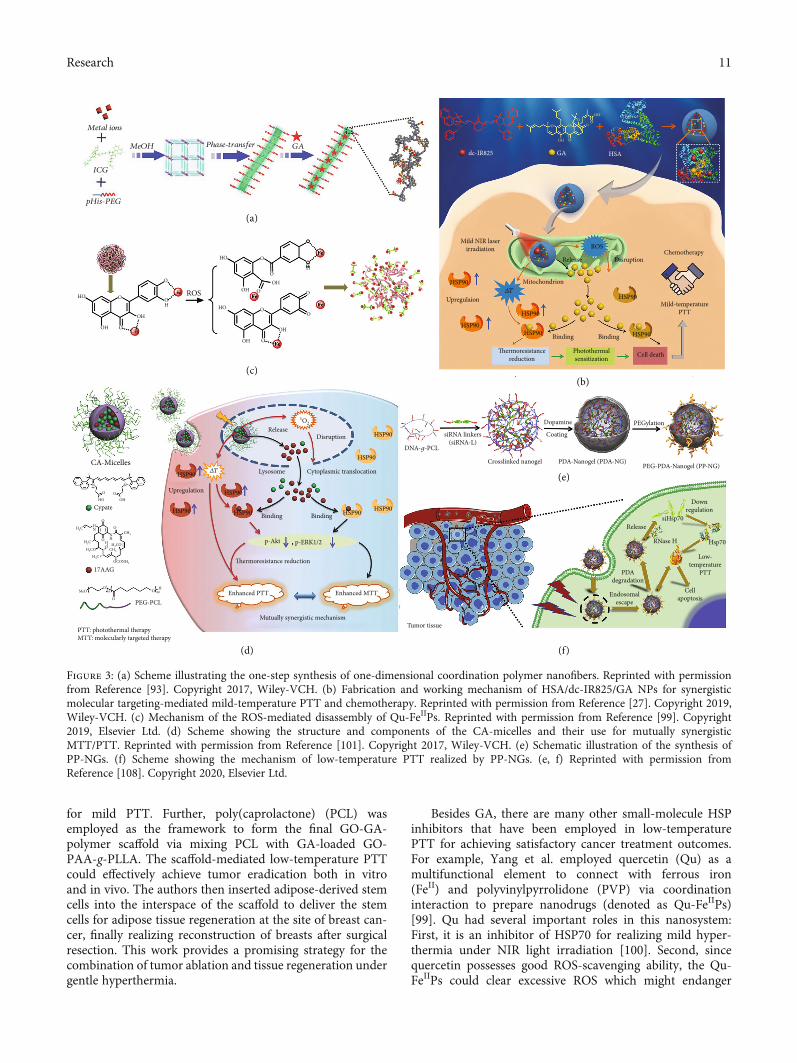
Small-Molecule HSP Inhibitors. Up till now, scientistshave taken advantage of dozens of the existing nanomaterialsto endowmild-temperature PTT platforms containing small-molecule HSP inhibitors with increased drug loading capac-ity and decreased nonspecific cytotoxicity. For example,Yang et al. proposed a facile method to synthesize a PEGy-lated one-dimensional coordination polymer via a one-stepreaction and phase transfer [93]. ICG (an NIR dye), poly-L-histidine-PEG (pHis-PEG), and Mn2+ (a frequently usedcontrast agent in magnetic resonance (MR) imaging, MRI)were first self-assembled to build a three-dimensional porousframework in the methanol solution via coordination inter-action, and after being transferred into the aqueous solution,the three-dimensional porous materials finally became one-dimensional nanofibers. Then, the authors integrated GAinto the nanofibers to afford Mn-ICG@pHis-PEG/GA(Figure 3(a)) and uncovered that this nanoplatform couldprominently facilitate cancer cell apoptosis and realize effi-cient low-temperature PTT. Moreover, the nanoplatformshowed enhanced cell internalization and tumor retentiondue to its pH-responsive ability, which was attributed to the
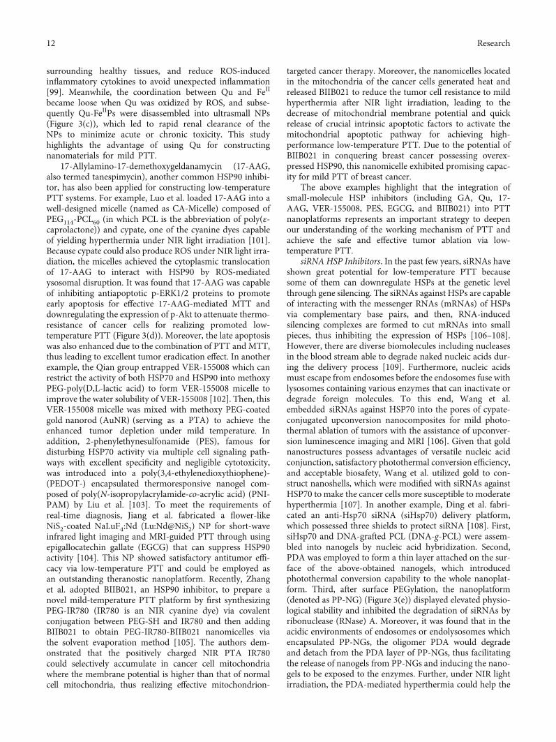
protonation of imidazole groups in pHis-PEG under theacidic environment. Recently, our group established anintelligent molecular targeting-mediated nanoplatform com-posed of GA, dc-IR825 (a fluorescent probe and a PTA), andhuman serum albumin (HSA) (a biocompatible carrier forhydrophobic molecules) [27]. The nanoplatform (HSA/dc-IR825/GA NPs) showed intrinsic low pH-induced chargereversal which was beneficial for the notably increased cellu-lar uptake and tumor accumulation under the acidic tumormicroenvironment (TME). It was found that the NPs werewell located in mitochondria after endocytosis. Under NIRlight irradiation, the ROS generated by dc-IR825 in the NPseffectively destructed mitochondrial membranes, whichresulted in the escape of GA from the mitochondria to thecytoplasm and subsequent HSP90 inhibition for low-temperature PTT (Figure 3(b)). Additionally, we have alsodemonstrated that the nanoplatform could realize synergisticlow-temperature PTT/GA-mediated molecularly targetedtherapy (MTT) in vivo and efficiently eliminate tumors andinhibit tumor metastasis. In another example, Li et al. fabri-cated zeolitic imidazole framework-8 (ZIF-8) NPs andembedded bismuth nanodots in the ZIF-8 NPs (abbreviatedas BZ) by a simplified one-step reduction method [94]. Thiscomposite nanomaterial possessed satisfactory photothermalconversion efficiency in the NIR-II (1000–1350 nm) region,which was important for realizing deep tissue penetration.The authors further loaded GA onto BZ to afford GBZ,and demonstrated that GBZ could achieve markedly accel-erated GA release under acidic conditions and NIR lightexposure. Moreover, the authors revealed that GBZ couldsuccessfully treat hepatocellular carcinoma in mousemodels. These studies all emphasize that GA-containingnanoplatforms can remarkably alleviate the thermal resis-tance of cancer cells and induce cancer cell apoptosis forenhanced low-temperature PTT. Moreover, the low pH-responsive property of nanomaterials is crucial for realiz-ing specific tumor accumulation and/or GA release, whichmay inspire researchers to design more smart nanoagentswith multiple exogenous and/or endogenous stimulus-responsive capacity for improving the performance of low-temperature PTT systems.
Recently, Sun et al. proposed a novel one-pot synthesismethod for fabricating hollow mesoporous carbon spheres(HMCSs), which had the eminent potential in photothermalconversion and photoacoustic (PA) imaging (PAI) [95].After PEG modification and GA loading, the resultantHMCS-PEG-GA displayed its capacity in ablating tumorsunder moderate temperature environments controlled bythe NIR laser. Semiconducting polymer NPs and black phos-phorus quantum dots have also been employed to encapsu-late GA for promoting the efficiency of PTT under mildNIR light irradiation [96, 97]. In addition, the Chen groupfabricated a scaffold to achieve low-temperature PTT ofbreast cancer. They first synthesized poly(acrylic acid)-g-poly(lactic acid) (PAA-g-PLLA) and then connected PAA-g-PLLA with graphene oxide (GO) via a low-pH cleavablebond, forming GO-PAA-g-PLLA [98]. After GA was loadedto GO-PAA-g-PLLA, the obtained complex could releaseGO/GA in the acidic TME due to the low-pH cleavable bond
10 Research
for mild PTT. Further, poly(caprolactone) (PCL) wasemployed as the framework to form the final GO-GA-polymer scaffold via mixing PCL with GA-loaded GO-PAA-g-PLLA. The scaffold-mediated low-temperature PTTcould effectively achieve tumor eradication both in vitroand in vivo. The authors then inserted adipose-derived stemcells into the interspace of the scaffold to deliver the stemcells for adipose tissue regeneration at the site of breast can-cer, finally realizing reconstruction of breasts after surgicalresection. This work provides a promising strategy for thecombination of tumor ablation and tissue regeneration undergentle hyperthermia.
Besides GA, there are many other small-molecule HSPinhibitors that have been employed in low-temperaturePTT for achieving satisfactory cancer treatment outcomes.For example, Yang et al. employed quercetin (Qu) as amultifunctional element to connect with ferrous iron(FeII) and polyvinylpyrrolidone (PVP) via coordinationinteraction to prepare nanodrugs (denoted as Qu-FeIIPs)[99]. Qu had several important roles in this nanosystem:First, it is an inhibitor of HSP70 for realizing mild hyper-thermia under NIR light irradiation [100]. Second, sincequercetin possesses good ROS-scavenging ability, the Qu-FeIIPs could clear excessive ROS which might endanger
Metal ions
pHis-PEG
O
ROSO
O
OH
OH
HO
Fe
Fe
Fe
FeFe
Fe
dc-IR825 GA
OH
OH
O
OON
Cl
NBr− OOO
HSA
HSP90
Mild NIR laserirradiation
HSP90
HSP90
HSP90Mild-temperature
PTT
HSP90HSP90
Upregulaion
Mitochondrion
Release DisruptionChemotherapy
ROS
𝛥T
Binding Binding
Cell deathPhotothermalsensitization
�ermoresistancereduction
O
O
O
O
O
O
O
OH
OHOH
HO
OH
HO
OOH
OH
ICG
MeOH GA
CA-Micelles
Cypate
17AAG
PEG-PCL
Mutually synergistic mechanism
Enhanced PTT Enhanced MTT
�ermoresistance reduction
p-Akt , p-ERK1/2
Binding BindingHSP90 HSP90
HSP90
HSP90
HSP90DNA-g-PCL
siRNA linkers(siRNA-L)
Crosslinked nanogel PDA-Nanogel (PDA-NG)PEG-PDA-Nanogel (PP-NG)
Dopamine
Coating
PEGylation
HSP90
HSP90
CellapoptosisEndosomal
escape
PDAdegradation
Low-temperature
PTT
RNase H Hsp70
ReleasesiHsp70
DownregulationHSP90
Upregulation
Lysosome Cytoplasmic translocation
ReleaseDisruption
1O2
PTT: photothermal therapyMTT: molecularly targeted therapy
O
O
O
O
O
OH
CH3
CH3
H2C
H3COH3C
H3C
MeOO
O
45 OH
60
OCONH2
H
NH
HN
H3CO
HO
N N
O
(c)
(d) (f)
(a)
(b)
(e)
Tumor tissue
𝛥T
Figure 3: (a) Scheme illustrating the one-step synthesis of one-dimensional coordination polymer nanofibers. Reprinted with permissionfrom Reference [93]. Copyright 2017, Wiley-VCH. (b) Fabrication and working mechanism of HSA/dc-IR825/GA NPs for synergisticmolecular targeting-mediated mild-temperature PTT and chemotherapy. Reprinted with permission from Reference [27]. Copyright 2019,Wiley-VCH. (c) Mechanism of the ROS-mediated disassembly of Qu-FeIIPs. Reprinted with permission from Reference [99]. Copyright2019, Elsevier Ltd. (d) Scheme showing the structure and components of the CA-micelles and their use for mutually synergisticMTT/PTT. Reprinted with permission from Reference [101]. Copyright 2017, Wiley-VCH. (e) Schematic illustration of the synthesis ofPP-NGs. (f) Scheme showing the mechanism of low-temperature PTT realized by PP-NGs. (e, f) Reprinted with permission fromReference [108]. Copyright 2020, Elsevier Ltd.
11Research
surrounding healthy tissues, and reduce ROS-inducedinflammatory cytokines to avoid unexpected inflammation[99]. Meanwhile, the coordination between Qu and FeII
became loose when Qu was oxidized by ROS, and subse-quently Qu-FeIIPs were disassembled into ultrasmall NPs(Figure 3(c)), which led to rapid renal clearance of theNPs to minimize acute or chronic toxicity. This studyhighlights the advantage of using Qu for constructingnanomaterials for mild PTT.
17-Allylamino-17-demethoxygeldanamycin (17-AAG,also termed tanespimycin), another common HSP90 inhibi-tor, has also been applied for constructing low-temperaturePTT systems. For example, Luo et al. loaded 17-AAG into awell-designed micelle (named as CA-Micelle) composed ofPEG114-PCL60 (in which PCL is the abbreviation of poly(ε-caprolactone)) and cypate, one of the cyanine dyes capableof yielding hyperthermia under NIR light irradiation [101].Because cypate could also produce ROS under NIR light irra-diation, the micelles achieved the cytoplasmic translocationof 17-AAG to interact with HSP90 by ROS-mediatedysosomal disruption. It was found that 17-AAG was capableof inhibiting antiapoptotic p-ERK1/2 proteins to promoteearly apoptosis for effective 17-AAG-mediated MTT anddownregulating the expression of p-Akt to attenuate thermo-resistance of cancer cells for realizing promoted low-temperature PTT (Figure 3(d)). Moreover, the late apoptosiswas also enhanced due to the combination of PTT and MTT,thus leading to excellent tumor eradication effect. In anotherexample, the Qian group entrapped VER-155008 which canrestrict the activity of both HSP70 and HSP90 into methoxyPEG-poly(D,L-lactic acid) to form VER-155008 micelle toimprove the water solubility of VER-155008 [102]. Then, thisVER-155008 micelle was mixed with methoxy PEG-coatedgold nanorod (AuNR) (serving as a PTA) to achieve theenhanced tumor depletion under mild temperature. Inaddition, 2-phenylethynesulfonamide (PES), famous fordisturbing HSP70 activity via multiple cell signaling path-ways with excellent specificity and negligible cytotoxicity,was introduced into a poly(3,4-ethylenedioxythiophene)-(PEDOT-) encapsulated thermoresponsive nanogel com-posed of poly(N-isopropylacrylamide-co-acrylic acid) (PNI-PAM) by Liu et al. [103]. To meet the requirements ofreal-time diagnosis, Jiang et al. fabricated a flower-likeNiS2-coated NaLuF4:Nd (Lu:Nd@NiS2) NP for short-waveinfrared light imaging and MRI-guided PTT through usingepigallocatechin gallate (EGCG) that can suppress HSP90activity [104]. This NP showed satisfactory antitumor effi-cacy via low-temperature PTT and could be employed asan outstanding theranostic nanoplatform. Recently, Zhanget al. adopted BIIB021, an HSP90 inhibitor, to prepare anovel mild-temperature PTT platform by first synthesizingPEG-IR780 (IR780 is an NIR cyanine dye) via covalentconjugation between PEG-SH and IR780 and then addingBIIB021 to obtain PEG-IR780-BIIB021 nanomicelles viathe solvent evaporation method [105]. The authors dem-onstrated that the positively charged NIR PTA IR780could selectively accumulate in cancer cell mitochondriawhere the membrane potential is higher than that of normalcell mitochondria, thus realizing effective mitochondrion-
targeted cancer therapy. Moreover, the nanomicelles locatedin the mitochondria of the cancer cells generated heat andreleased BIIB021 to reduce the tumor cell resistance to mildhyperthermia after NIR light irradiation, leading to thedecrease of mitochondrial membrane potential and quickrelease of crucial intrinsic apoptotic factors to activate themitochondrial apoptotic pathway for achieving high-performance low-temperature PTT. Due to the potential ofBIIB021 in conquering breast cancer possessing overex-pressed HSP90, this nanomicelle exhibited promising capac-ity for mild PTT of breast cancer.
The above examples highlight that the integration ofsmall-molecule HSP inhibitors (including GA, Qu, 17-AAG, VER-155008, PES, EGCG, and BIIB021) into PTTnanoplatforms represents an important strategy to deepenour understanding of the working mechanism of PTT andachieve the safe and effective tumor ablation via low-temperature PTT.
siRNA HSP Inhibitors. In the past few years, siRNAs haveshown great potential for low-temperature PTT becausesome of them can downregulate HSPs at the genetic levelthrough gene silencing. The siRNAs against HSPs are capableof interacting with the messenger RNAs (mRNAs) of HSPsvia complementary base pairs, and then, RNA-inducedsilencing complexes are formed to cut mRNAs into smallpieces, thus inhibiting the expression of HSPs [106–108].However, there are diverse biomolecules including nucleasesin the blood stream able to degrade naked nucleic acids dur-ing the delivery process [109]. Furthermore, nucleic acidsmust escape from endosomes before the endosomes fuse withlysosomes containing various enzymes that can inactivate ordegrade foreign molecules. To this end, Wang et al.embedded siRNAs against HSP70 into the pores of cypate-conjugated upconversion nanocomposites for mild photo-thermal ablation of tumors with the assistance of upconver-sion luminescence imaging and MRI [106]. Given that goldnanostructures possess advantages of versatile nucleic acidconjunction, satisfactory photothermal conversion efficiency,and acceptable biosafety, Wang et al. utilized gold to con-struct nanoshells, which were modified with siRNAs againstHSP70 to make the cancer cells more susceptible to moderatehyperthermia [107]. In another example, Ding et al. fabri-cated an anti-Hsp70 siRNA (siHsp70) delivery platform,which possessed three shields to protect siRNA [108]. First,siHsp70 and DNA-grafted PCL (DNA-g-PCL) were assem-bled into nanogels by nucleic acid hybridization. Second,PDA was employed to form a thin layer attached on the sur-face of the above-obtained nanogels, which introducedphotothermal conversion capability to the whole nanoplat-form. Third, after surface PEGylation, the nanoplatform(denoted as PP-NG) (Figure 3(e)) displayed elevated physio-logical stability and inhibited the degradation of siRNAs byribonuclease (RNase) A. Moreover, it was found that in theacidic environments of endosomes or endolysosomes whichencapsulated PP-NGs, the oligomer PDA would degradeand detach from the PDA layer of PP-NGs, thus facilitatingthe release of nanogels from PP-NGs and inducing the nano-gels to be exposed to the enzymes. Further, under NIR lightirradiation, the PDA-mediated hyperthermia could help the
12 Research

nanogels escape from endolysosomes to the cytoplasm wherethey could be degraded by RNase H (which recognizes DNA-RNA hybrid duplex and digests the RNA strand), and then,siRNAs were released to the cytoplasm to downregulateHSP70 to realize effective low-temperature PTT (Figure 3(f)).This work exemplifies a feasible solution to realize safesiRNA delivery for low-temperature PTT.
(2) Cancer Starvation Therapy. Lately, cancer starvation ther-apy has been introduced for the ablation of malignant tumors[110]. As is well known, tumors display fast nutrition con-sumption to maintain the rapid speed of cancer cells’ growthand proliferation. On the one hand, cancer cells secrete sev-eral proangiogenic growth factors and express the counter-part receptors of the factors. Therefore, there are abundantblood vessels around and inside tumors that ensure thetumors’ sufficient nutrition supply for their survival andgrowth. On the other hand, cancer cells tend to obtain energyby an abnormal type of metabolism (the Warburg effect) thatmainly sustains energy supply through anaerobic glycolysis,which exacerbates their reliance on glucose [111]. Hence,two major principles of designing platforms for cancer star-vation therapy have been proposed [110]: (1) inhibitingangiogenesis or blocking tumor blood vessels and (2) reduc-ing the cellular uptake of or exhausting life-necessary nutri-ents, especially glucose. Unfortunately, starvation therapyonly partially succeeds due to a series of obstacles likeincreased tumor hypoxia, unsatisfactory targeting capacity,induced drug resistance, and elevated tumor metastasis risk,which all impede their further applications in clinic [110].To overcome these challenges (or some of these challenges),combining starvation therapy with PTT serves as a goodchoice since it can maximize the anticancer therapeutic effi-ciency [110, 112]. More importantly, given that both the pro-duction of HSPs and the execution of their bioactiveprocesses need ATP, developing strategies that can lead tothe lack of ATP via blocking or decreasing the supply ofenergy sources like glucose provides an efficacious way torealize HSP inhibition and hence mild-temperature PTT.Therefore, cancer starvation therapy represents one promis-ing strategy that can be combined with low-temperaturePTT to achieve potentiated anticancer effect.
Considering that there are overexpressed glucose trans-porters on the membranes of tumor cells, blocking or down-regulating these transporters acts as an emerging solution todeprive cancer cells of cytoplasmic glucose for cancer starva-tion therapy. This strategy has been employed for enhancedmild PTT. For example, Chen et al. used diclofenac (DC),which served as a small-molecule inhibitor of Glut1 (one ofthe major glucose transporters (Gluts)), and hyaluronic acid-(HA-) modified plasmonic gold nanorod (GNR) to constructGNR/HA-DC for realizing low-temperature PTT [113]. Assome cancer cells overexpress CD44 receptors capable ofinteracting with HA and TME is highly abundant in hyal-uronidase (HAase), this platform could target CD44-overexpressed tumors and achieve the HAase-mediatedrelease of DC to the cytoplasm to inhibit Glut1. Thanks toits surface plasmon resonance property, GNR/HA-DC exhib-
ited noticeable photothermal conversion capacity, which wasbeneficial for realizing cancer-specific mild-temperature PTTespecially for CD44-overexpressed tumor cells (Figure 4(a)).
Moreover, glucose oxidase (GOx), which can catalyzeglucose, O2, and H2O into gluconic acid and H2O2, has beenwidely used in cancer therapy [114–116]. In the past fewyears, due to the ability of GOx to achieve cancer starvation,some researchers utilized GOx to realize low-temperaturePTT of tumor [32, 117, 118]. For instance, a nanoreactorwas prepared by Cao et al. to combine tumor starvation ther-apy with multiple mechanism-enhanced mild-temperaturephototherapy for cancer treatment [117]. They loaded bothGOx and ICG onto the Fe-doped polydiaminopyridine (Fe-PDAP) nanozyme via electrostatic adsorption and π-π inter-action to obtain the Fe-PDAP/GOx/ICG nanoreactor. Onthe one hand, the Fe-PDAP nanozyme could catalyze H2O2produced during GOx-mediated glucose consumption as wellas supplied by tumor cells into O2 to improve O2-dependentICG-based PDT. On the other hand, the Fe-PDAP nanozymewas capable of depleting GSH (which is the key antioxidantagent capable of reducing ROS) by a metal-reducing reactionafter releasing Fe3+, thus further enhancing the efficiency ofPDT. Meanwhile, under NIR light irradiation, mild PTT wasachieved due to GOx-mediated HSP downregulation andICG-based hyperthermia. To be noted, the Fe-PDAP/GOx/-ICG showed satisfactory performance in multimodal fluores-cence imaging (FLI)/PAI/MRI. This work verifies that theGOx-mediated cancer starvation therapy has advantages innot only downregulating HSPs for low-temperature PTT butalso remarkably improving the synergistic cancer therapeuticefficiency. However, protecting GOx from deterioration andleakage during the circulation and endocytosis processes toexecute catalysis validly is a disturbing problem. To deal withthis issue, researches have designed a variety of GOx-containing nanomaterials and further optimized the GOxrelease process by the introduction of stimulus responsiveness[32, 118]. Zhou et al. entrapped GOx into a porous hollowPrussian blue (PB) NP (termed PHPBN) which is a biologi-cally friendly NIR PTA and can protect GOx from beinginactivated [118]. Further, they conjugated the surface ofGOx-loaded PHPBNs with HA through disulfide bonds,which can be cleaved by intracellular glutathione (GSH) torealize the GOx release into the cytoplasm, and meanwhile,the HA can help the nanoplatform to target the CD44 recep-tors overexpressed on the plasma membranes of HepG2 cells.Finally, PEG was grafted to the outer shell to prolong theblood circulation time. Importantly, the PHPBNs were ableto catalyze H2O2 generated during GOx-mediated glucose oxi-dation to produce oxygen for alleviating tumor hypoxia andelevating the catalytic efficacy of GOx, finally achieving effec-tive cancer starvation therapy and low-temperature PTT. Inanother example, our group prepared thermosensitive lipo-somes which were loaded with GA, GOx, and ICG to affordGOIGLs [32]. Upon NIR light irradiation, ICG within theliposomes could efficiently convert light to heat, and whenthe temperature was elevated to above 42°C, the liposomesunderwent the gel-to-fluid phase transition and subsequentlyreleased the loaded GA, GOx, and ICG (Figure 4(b)). Addi-tionally, under visible light illumination (400–750nm), H2O2
13Research

which was produced during the GOx-catalyzed glucose oxida-tion reaction could be transformed into ⋅OH, which is one ofthe most lethal ROS to cells, thus showing enzyme-enhancedphototherapy (EEPT) effect. Besides, after NIR light irradia-tion, the GOIGLs could successfully escape from lysosomesvia ROS-mediated lysosomal disruption, which contributedto the release of GOx and GA to the cytoplasm to consumeglucose and downregulate HSPs, respectively, thus realizingsynergistic starvation therapy and low-temperature PTT(Figure 4(c)). This work emphasizes the robust anticancertherapeutic effect of the combined use of HSP inhibitor-promoted mild PTT and GOx-mediated cancer starvationtherapy. Besides GOx, GOx-like nanoagents can also be usedto construct the starvation therapy/mild PTT-based platformsfor defeating cancer. As an example, Tang et al. fabricated a
two-dimensional MnO2 nanosheet (M-NS) through a uniqueone-step wet-chemical method, which could control the sizeand thickness of M-NSs by adjusting the ratio of BSA toMnCl2 in oxygen-containing alkaline solutions [119]. More-over, the M-NSs exhibited excellent GOx-like catalytic perfor-mance and NIR-absorbing ability. Thus, the M-NSs couldconsume intracellular glucose leading to the downregulationof HSPs and generate heat under NIR light irradiation, finallyachieving low-temperature PTT (Figure 4(d)). The authorsalso developed a novel sono-chemical way to coat the surfaceof M-NSs with 2-S-(4-isothiocyanatobenzyl)-1,4,7-triazacy-clononane-1,4,7-triacetic acid-modified BSA (BSA-NOTA)and found that the modified M-NSs possessed improvedstability and accessibility of further functionalization, suchas chelation with radioactive 64Cu for positron emission
Glucose
Cancer cell ReceptorGlut1 Normal cell
DPPCICG
GA
DSPE-PEG2000
GOx
Light
Endocytosis
NIR laser
Disruption
Lysosome𝛥T
HSP90HSP90
HSP90
�ermoresistancereductionUpregulation
Low-temperaturePTT
Enzyme-enhancedphototherapy
•OH
H2O2
GlucoseO2 + H2O
NIR laserirradiation
Inhibition
Glucose deprivation
Inhibited anaerobic glycolysisReduced ATP level
Laser
Heat
GNRDiclofenac
Cancer cell Laser
Enhancedphotothermal therapy
Starvationtherapy
HA
HAaseGNR/HA-DC
Elevatedphotothermal
effect
HSP70 HSP90
H2O H2O2
O2
𝛽-D glucose
Gluconic acid
HEAT
HSP70 HSP90
Inhi
bit
(a)
(b)
(c)(d)
ATP
Figure 4: (a) Scheme showing the use of GNR/HA-DC to sensitize cancer cells to mild PTT by interfering with the anaerobic glycolysismetabolism. Reprinted with permission from Reference [113]. Copyright 2017, American Chemical Society. (b) Scheme illustrating thetransformation of GOIGLs upon NIR light irradiation. (c) Scheme depicting the mechanism of EEPT and low-temperature PTT. (b, c)Reprinted with permission from Reference [32]. Copyright 2020, Wiley-VCH. (d) Scheme showing the utilization of M-NS for cancerstarvation therapy and PTT. Reprinted with permission from Reference [119]. Copyright 2019, Wiley-VCH.
14 Research

tomography imaging. The above reports all utilize the advan-tages of GOx or its mimics to construct cancer therapeuticnanoplatforms and emphasize the eminent potential ofGOx/GOx mimics-mediated tumor starvation in promotingeffective low-temperature PTT.
Besides employing the Glut inhibitors and GOx, Danget al. exploited a new way to accomplish synergistic cancerstarvation therapy/low-temperature PTT. The authors usedan siRNA against pyruvate kinase M2 (siPKM2, which iscapable of interfering with tumor glycolysis) and ICG to fab-ricate a novel nanocomplex based on a spherical dendrimerpolypeptide (DPP) [120]. Specifically, amine-terminatedpolyamidoamine (PAMAM) was utilized as a macroinitiatortemplate to synthesize DPP via controlled ring-opening poly-merization (ROP) of N-carboxyanhydride followed by side-chain functionalization with guanidines. The DPP could loadICG into its hydrophobic cavity, and meanwhile, siPKM2was condensed by DPP via electrostatic interaction to formpositively charged nanocomplexes. Further, to improve thestability and biosafety, the nanocomplexes were envelopedby human serum albumin to obtain the final product termedD-I/P@HSA NCs (NCs: nanocomplexes). Under NIR lightirradiation, the NCs exhibited enhanced tumor photother-mal ablation efficiency by the aid of the siPKM2-mediatedcancer starvation effect. This research points out that disturb-ing tumor glycolysis via molecular or nanoscale agents mayrepresent a novel approach to realize the combination of can-cer starvation therapy and low-temperature PTT.
2.3.2. Autophagy-Mediated Cancer Therapy. Autophagy, anessential catabolic process, can degrade and recycle mis-folded proteins or damaged organelles to conserve cellularhomeostasis [121]. Nevertheless, there have been argumentsabout the role of autophagy in the occurrence and progres-sion of cancers. On the one hand, autophagy can acceleratetumor development and shield cancer cells from diversecellular stresses like chemical drugs and radiation, eventu-ally making tumors develop chemo- and radioresistanceand be difficult to be cured [122–125]. On the other hand,autophagy can restrict tumorigenesis via regulating manyoncogenes and tumor suppressor genes and inhibit tumorinvasion and metastasis from the primary sites via limitingnecrosis and inflammation [122–124]. Thus, both the pro-motion and inhibition of autophagy have been applied todefeat cancers.
Given that inhibition of autophagy may contribute totumor eradication, researchers have recently invented manyapproaches to inhibit autophagy for improved cancer treat-ments [126–128]. Among these approaches, the combinationof autophagy-modulating molecules and mild PTT shows thesatisfactory ability of eliminating tumors at gentletemperatures [129–132]. Some studies have figured out thatlow-temperature PTT can induce both apoptosis andautophagy in cancer cells, and autophagy provides a survivalway for malignant tumors [129, 130]. Further, it has beenrevealed that the inhibition of autophagy can augmentapoptosis and apparently strengthen the efficiency of PTT,verified by the lifted malignant cells’ fatality under mildhyperthermia using the autophagy inhibitor chloroquine
(CQ) [129] and chloroquine diphosphate-loaded NPs [130].It is noteworthy that the research carried out by Zhou et al.is the first report of using CQ-mediated autophagy inhibitionto enhance PTT [129]. The authors loaded CQ on the surfaceof PEG-modified PDA NPs (the resultant product wastermed PDA-PEG/CQ NPs) and found that CQ could bereleased from PDA NPs in the moderately acidic environ-ment (pH = 6). The authors demonstrated that this nanoplat-form could realize effective CQ release in the TME andcytoplasm to inhibit autophagy, leading to the elevated can-cer cell sensitization to mild PTT for defeating cancer(Figure 5(a)). Lately, Shao et al. synthesized a rattle-structured NP by encapsulating a PDA nanocore in a hollowmesoporous silica nanoshell to afford PDA@hm [131]. Then,CQ was loaded into the hollow cavity of PDA@hm and GOxwas conjugated onto the surface of the above obtained NP,thus obtaining PDA@hm@CQ@GOx (Figure 5(b)). It wasdemonstrated that this nanoplatform could successfullyachieve enhanced photothermal eradication of tumors underthe moderate temperature with the assistance of GOx-mediated starvation and CQ-mediated autophagy inhibition,consequently reducing cancer survival rates (Figure 5(c)).Additionally, the CQ-mediated autophagy inhibition madecontribution to compensating for the loss of mild PTT and star-vation therapy efficacies due to PTT- and starvation-activatedautophagy, showing the outstanding synergistic effect of thePDA@hm@CQ@GOx. Moreover, the nanoplatform was capa-ble of realizing PAI. This trimodal synergistic treatment strategy(i.e., autophagy inhibition, starvation therapy, and mild PTT)has a great potential in overcoming PTT’s side effects and ele-vating the efficiency of low-temperature PTT. Recently, to breakthe cycle between tumor cell proliferation and bone resorption(this cycle plays an important role in the promotion of bonetumors’ progression and metastasis), Wang et al. fabricatedPEG-conjugated alendronate-functionalized and CQ-loadedPDA NPs (PPA/CQ) for bone cancer therapy [132]. It wasfound that, owing to the alendronate, PAA had the advantageof strongly interacting with hydroxyapatite abundant in bonetissues, thus realizing the bone-targeting function. Besides facil-itating the PDA-mediated photothermal ablation of bonetumors at mild temperatures, CQ could also attenuate tumor-associated bone resorption via interfering with the differentia-tion and activation of osteoclasts and subsequently inhibitingosteoclastogenesis. The in vivo experiments showed that thePPA/CQ nanoplatform achieved both tumor elimination andosteolysis inhibition. This work verifies that CQ-mediatedautophagy inhibition provides a feasible method for malignantbone tumor treatments.
On the other hand, the augment of autophagy in cancercells has also been exploited to be combined with mild PTTfor defeating tumors. Zhou et al. fabricated a PEGlyatedmelanin-like PDA NP decorated with a cyclic Arg-Gly-Asp(RGD) peptide which is famous for binding integrin overex-pressed in tumor tissues, and a beclin 1-derived peptide(denoted as beclin 1) which is the product of a putative generelated to the upregulation of autophagy [133]. As expected,the NPs showed tumor-targeting capability and enhancedlow-temperature PTT efficiency (resulting from the promo-tion of autophagy) (Figure 5(d)).
15Research

Collectively, the above examples highlight that wemust pay more attention to figuring out and utilizingthe complicated relationship between mild PTT andautophagy regulation to elevate the efficiency of tumoreradication.
2.3.3. Organelle-Targeting Strategy. Undoubtedly, the integ-rity of the structure and function of all cellular organellesis the crucial support for cell growth and proliferation,and it is also of great significance in the invasion andmetastasis of cancer cells [134, 135]. It is worth noting
PDA-PEG/CQ
Autophagosome
CQ release
Lysosome
Autophagy inhibition
Autolysosome Complete autophagy
Beclin 1 RGD
RGD-mediatedintracellular uptake
Beclin 1 + PTT
Cell death
Protectiveautophagy
LC3-l
LC3-ll
Autophagy promotion
Autophagy induction
Self-assembly
808 nmNIR (0.3 W/cm2)
>50°C danger
48°C
Temp. Safe temperature
Enhanceduptake
Efficientuptake
Hydrothermal
160°C, 1.5 h
Modification
Nucleus
PTT
CellNucleus
PEG-PtCNPs LaserCell migration
Defectivecytoskeleton
PT imaging& PTT
Tumor
Tumor
Lamin A/Cover-
espression ∆T
FLimaging
PA imaging
Plasma membraneMitochondrionGolgi apparatus
ERLysosome
Before treatment(Cell migration)
Hyperthermia
Nuclear entry
HI-4COOH
Hf4+
OO
O O
OH
OH
Cl
HO
+
-O
NN
Autophagosomematuration
Lysosomal fusion
Beclin 1
Beclin 1Beclin 1
NIR
PEG
Cell survival
Cell deathPhotothermalsensitization
CQCl
HN
N
N
NIR laser
Heat
𝛼v𝛽3 integrin
∆T
24
135
Modification
RITCPEG
PEG-PtCNPs PEG-PtCNPs-RITC
PEG-PtCNPs (orPEG-PtCNPs
-RITC)
PtCNPs[PtCl4]2
NH2
NH2
Heat
NIRirradiation
Low-temperaturephotothermal therapy
Autophagosome
Autolysosome
Lysosome Activation
Enhancedapoptosis
Starvation therapy
GOx
GlucoseO2+H2O
Gluconic acidH2O2
Biologicalcatalysis
Tumor cell
Tumor tissue
Blood vessel
Photoacoustic imaging
HSP
CQ releas
e
Activation
Autophag
yinhibi
tion
PDA PDA@dSiO2 PDA@dSiO2@mSiO2 PDA@hm PDA@hm@CQ@GOx
GOxCQ
(a)
(d)
(b)
(c)
(e) (f)
1
2
3
4 5
Figure 5: (a) Scheme of the enhanced cancer therapeutic efficiency of mild PTT resulting from photothermal sensitization throughCQ-mediated autophagy inhibition. Reprinted with permission from Reference [129]. Copyright 2017, Elsevier Ltd. (b) Multisteppreparation process of the rattle-structured PDA@hm@CQ@GOx NP. (c) Schematic illustration of the working mechanism ofPDA@hm@CQ@GOx NPs that can achieve enhanced low-temperature PTT under NIR light irradiation. (b, c) Reprinted with permissionfrom Reference [131]. Copyright 2020, Ivyspring. (d) Scheme depicting the beclin 1-induced autophagy rendering cancer cells vulnerableto photothermal elimination. Reprinted with permission from Reference [133]. Copyright 2019, Elsevier Ltd. (e) Scheme illustrating theHf-HI-4COOH NCP structure and nucleus-targeted low-temperature PTT. Reprinted with permission from Reference [142]. Copyright2020, Elsevier Ltd. (f) Scheme depicting the synthesis of PEG-PtCNPs and PEG-PtCNPs-RITC and their application for multimodalimaging-guided and multiorganelle-targeted PTT. Reprinted with permission from Reference [145]. Copyright 2018, Elsevier Ltd.
16 Research

that a large number of studies have attached great impor-tance to developing biomedical engineering platforms withawesome organelle-targeting capacity for bothering or evendevastating the organelles and finally deteriorating tumors[24, 25, 30, 126, 136–139]. Most of the organelles are verysusceptible to elevated temperatures, especially the cellnucleus enveloping approximately all the genetic materialseasy to denaturation, which brings hope to low-temperaturePTT. On the other hand, there are numerous pores consist-ing of nuclear pore complexes (NPCs) on the nucleusenvelop, playing an important role in facilitating substancetransportation and signal conduction between the nucleusand cytoplasm. Numerous studies spared no effort to develophighly reliable and effective drugs or delivery systems posses-sing the capability of taking action in the nucleus followingpassing through these tiny pores. For instance, Cao et al.engineered a cell nucleus-targeting TAT peptide onto thesurface of a vanadium carbide quantum dot to obtain V2C-TAT, which could serve as a robust NIR-II PTA [140]. Afterencapsulating V2C-TATs into RGD peptide-decoratedendogenous exosomes which were beneficial for the elevatedbiocompatibility, nonimmunogenicity, and long blood circu-lation ability, the authors found that the nanosystem couldgather nearby the tumor and enter the nucleus to destroythe genetic materials directly for low-temperature PTT withthe dual guidance from PAI and MRI. Similarly, our groupfound that the palladium nanosheets modified by TAT (Pd-TAT) mainly accumulated in the perinuclear region andshowed increased endocytosis and reduced efflux [14].Importantly, Pd-TAT in the perinuclear region could triggerthe overexpression of lamin A/C proteins to elevate nuclearstiffness and inhibit cancer metastasis, because lamin A/Cproteins are highly related to nuclear stiffness and lack ofthe proteins facilitates cell migration. Under mild NIRlight irradiation, the Pd-TAT presented perinuclear-to-intranuclear translocation and exhibited enhanced cancer cellmetastasis inhibition and low-temperature PTT effect. Inaddition, Liu et al. synthesized an ultrasmall chitosan-coated ruthenium(IV) oxide NP with excellent NIR-IIphotothermal conversion ability and suitability for PAI[141]. The NPs could accumulate in the cell nucleus andrelease mild heat enough for destroying nuclear DNA underNIR-II light irradiation. Jiang et al. synthesized an Hf-heptamethine indocyanine dye- (Hf-HI-4COOH-) basednanoscale coordination polymer (NCP) which possessedstrong NIR absorption and excellent photothermal conversioncapacity to serve as an intrinsic nucleus-targeting PTA [142].Under low-power NIR light irradiation, the NCP could gener-ate enough heat to destroy the nucleus at a mild temperature,leading to the elimination of cancer cells. This study expandsthe application of NCPs and provides a new choice for con-ducting nucleus-targeted low-temperature PTT (Figure 5(e)).
Additionally, mitochondrion which is considered as thecellular energy factory and apoptosis control center has beenregarded as a key location where mild hyperthermia canachieve a satisfactory cancer cell killing effect. Based on this,Qiu et al. constructed mitochondrion-targeting lipophilic iri-dium(III) cation-modified Fe3O4 NPs [143]. The NPs notonly showed superb MRI/photothermogenic ability but also
worked as a nanozyme for executing the Fenton reaction bycatalyzing H2O2 generated from the mitochondrial respira-tory electron transport chain (this catalysis process could bestimulated by the heat stress of mild PTT) into ⋅OH for kill-ing cancer cells, thus realizing excellent MRI-guided andnanozyme-mediated mild-temperature PTT. In addition,the cytoskeleton, which is highly related to tumor migration,is a proper target for a variety of therapeutics [144, 145]. Ourgroup fabricated platinum-doped carbon NPs (CNPs) by afacile one-pot hydrothermal method and modified them withPEG to afford PEG-PtCNPs (Figure 5(f)) [145]. To observe thebehaviors of PEG-PtCNPs both in vitro and in vivo, we conju-gated rhodamine B isothiocyanate (RITC) onto PEG-PtCNPsto afford PEG-PtCNPs-RITC. The PEG-PtCNPs showedstrong photothermal conversion ability and could bind to mul-tiple organelles. Additionally, it was found that under mild NIRlight irradiation, PEG-PtCNPs impaired the cellular cytoskele-ton and induced the overexpression of lamin A/C, leading tothe inhibition of tumor metastasis. When the cells were irradi-ated by the NIR laser at a higher power density, irrecoverabledestruction to the nuclear membranes was observed and thenuclear delivery of PEG-PtCNPs in the absence of nucleus-targeting ligands was subsequently realized. Thus, this nano-platform achieved the damage to the cytoskeleton and nucleusby multiorganelle-targeted PTT and showed exceptional per-formance in tumor ablation and metastasis restriction. Thiswork provides a new solution for targeting multiple organ-elles to elevate the efficiency of low-temperature PTT.
2.3.4. Synergistic Therapy. Since numerous preclinic andclinic experiments have disclosed that many monothera-pies show low efficiency in defeating tumor metastasis/reoccurrence and some adverse side effects like high toxic-ity to normal cells and damage to immune system duringthe past few decades, many researchers have devotedthemselves to conquering these obstacles. The rationalcombination of conventional strategies has been appreci-ated as an important breakthrough verified by a myriadof studies, which has opened a new chapter in the fieldof cancer therapy [146].
As is well known, the presence of hypoxic cells in tumorsis considered to be one of the main reasons for the reductionof the effectiveness of radiotherapy, certain chemotherapeu-tic drugs, and phototherapy [147], and the insufficient bloodflow within tumor tissues impedes the delivery of varioustherapeutic agents [111]. Fortunately, the association of mildhyperthermia treatment with other therapeutic modalitieshas obtained many encouraging results. It was found that ata slightly higher temperature the tumor hypoxic environ-ment can be ameliorated due to the accelerated blood flow[147–149]. Additionally, increased vascular permeabilitynearby the heated region can be observed, which may con-tribute to the enhanced permeability and retention (EPR)effect (the key of the passive accumulation of drugs in tumortissues). Thus, low-temperature PTT, which can induce mildhyperthermia within tumors, may have considerable poten-tial in improving the effects of other therapies. In the mean-time, it has been widely recognized that NPs can passivelyaccumulate in tumors due to the EPR effect and actively
17Research

target tumors through being modified by some molecules ormotifs capable of recognizing and interacting with tumors orTME [150]. Moreover, the utility of nanostructures consist-ing of PTAs and other agents like chemodrugs, radiosensiti-zers, and photosensitizers can integrate multimodaltreatments in one nanoplatform. Therefore, with the assis-tance of nanotechnology, we can remarkably improve thecancer cell-killing performance of other therapies in combi-nation with low-temperature PTT. On the other hand, othertreatment results can sensitize cancer cells to be more vulner-able to mild heat, thus enhancing the tumor eradication effectof low-temperature PTT. Besides, low-temperature PTT pos-sesses other unique advantages when it is combined with dif-ferent therapeutic modalities, which will be discussed in thefollowing parts.
(1) Chemotherapy. Chemotherapy, surgical resection, andradiotherapy (RT) are considered as the three major tacticsfor cancer treatments. Chemotherapy plays an importantrole in clinical cancer therapy and a large number of novelcancer therapeutic strategies and agents have been developedup till now [151–154]. Nevertheless, there are several prob-lems that are desperately imperative to be solved, e.g., indis-criminative accumulation and cellular uptake leading toincomplete tumor eradication and unavoidable damage tonormal tissues, the development of multidrug resistance(MDR) compromising the therapeutic performance of thechemodrugs, and unsatisfactory therapeutic effects of thedrugs used at safe doses [155–157]. Thus, it is of great signif-icance to develop a platform which can efficiently kill tumorsat a relative low drug dose.
To date, an agreement has been reached that combiningchemotherapy with PTT can make a practical contributionto improving the anticancer outcomes [158]. First, PTTmay achieve spatiotemporally-controlled drug delivery byadjusting the location, power density, and working time ofNIR/visible light. Second, hyperthermia can render cancercells more accessible and vulnerable to chemotherapeuticdrugs. Third, the heat generated by PTAs can stimulate thechemodrugs’ intratumoral penetration and accumulationdue to increased tumor blood flow and vascular permeability.In addition, due to the reduced phototoxicity and enhancedstability of some chemodrugs at moderate temperatures,decreasing the temperature used to kill tumors by controllinglight intensity, blocking HSPs, or interfering with otherthermoresistance-related signal pathways makes the com-bined use of chemotherapy and mild PTT an appealingchoice for cancer treatments. Furthermore, the administra-tion of chemodrugs can compensate for the insufficientheat damage, thus realizing low-temperature PTT [159–177]. For instance, Sherlock et al. fabricated an ultrasmallnanocrystal (FeCo/GC) containing a single crystalline iron-cobalt core surrounded by a single- or few-layer graphiticcarbon (GC) shell, which could serve as a PTA and an MRIcontrast agent [159]. Further, doxorubicin (DOX) wasloaded on the GC shell by π-π stacking interaction to formFeCo/GC-DOX. Under acidic conditions in TME and endo-somes/lysosomes, the π-π stacking interaction was weak-
ened, leading to the pH-sensitive release of DOX. UnderNIR light irradiation, FeCo/GC-DOX exhibited enhancedcellular uptake, which was determined byMRI, DOX fluores-cence measurements, and flow cytometry. The enhanced cel-lular uptake and the elevated chemotherapeutic efficacy ofDOX under mild hyperthermia dramatically enhanced thetoxicity of FeCo/GC-DOX toward breast cancer cells. Wanget al. also found that mild hyperthermia could help DOX topenetrate the extravascular space and transiently alter thetumor interstitial permeabilization by using DOX-loadedGd-hybridized plasmonic Au nanocomposites [160]. Theauthors also confirmed that after further HA modifica-tion, the obtained nanocomposites could target CD44-overexpressing cancer cells and achieve drug releasemediated by HAase, further leading to enhanced tumor elim-ination (Figure 6(a)). Besides, a large number of other PTAs,including hollow CuS NPs [161], RbxWO3 nanorods [162],gold nanomaterials [163], black phosphorus nanosheets[164], PDA NPs [165, 166], ICG or polypyrrole- (PPy-) con-taining nanomaterials [166–169], and hollow mesoporousorganosilica nanocapsule [170], have been employed tocooperate with various clinical chemotherapeutic drugs, suchas paclitaxel, docetaxel (DTX), and gemcitabine, to realizesynergistic low-temperature PTT/chemotherapy.
Drug-resistant cancer cells tend to express a series ofmembrane transporters represented by the ATP-bindingcassette transporter (ABC) family to export diverse cyto-toxic drugs, and in the meantime induce the mutation ofproapoptosis genes like TP53 to escape from apoptosis,then resulting in the elevation of their endurance to thetherapeutic agents. Fortunately, it is reported that the mildheat produced by PTAs may bring us encouraging hope.Wang et al. found that the moderate heat produced byAuNR-based mesoporous silica nanocarriers (Au@SiO2)under the 780nm femtosecond pulsed laser irradiation(3.2Wcm−2, 10min) not only evoked the obvious andlong-lasting expression of the heat shock factor-1 (HSF-1)capable of restricting the NF-κB pathway, which is involvedin the formation of P-glycoproteins (Pgps), but also elicitedthe downregulation and decomposition of mutant p53 inresistant cells to render cancer cells susceptible to chemo-drugs [171]. The authors also loaded DOX inside the meso-porous shell of Au@SiO2, and found that the resultantnanoplatform (termed Au@SiO2-DOX) could avoid Pgp-mediated DOX efflux via the cellular internalization andlysosomal accumulation of Au@SiO2-DOX. Moreover,under localized laser, Au@SiO2-DOX enhanced the cancercells’ sensitiveness to DOX due to the suppressed mutationof p53 (Figure 6(b)). Further, Du et al. employed cocktailchemotherapy, i.e., administration of two or more thantwo drugs, and low-temperature PTT at the same time todefeat cancer MDR [172]. They synthesized a dual drug-paired polyprodrug NP via the precipitation copolymeriza-tion of dopamine-DOX prodrug and subsequent cisplatincoordination. The NPs showed intracellular pH-responsivedrug release and photothermal conversion capacity.Under mild hyperthermia, both the reversal of MDRand tumor eradication were achieved by the synergistictreatment.
18 Research

To further decrease undesirable drug leakage andincrease the drug release efficiency in targeted tumors,researchers have introduced some internal and external (asfor mild PTT, it is generally NIR light irradiation) stimulus-responsive properties to the drug delivery systems. Forexample, low pH is a widely used stimulus for specific drugdelivery. On the one hand, TME is weakly acidic mainlydue to the abnormal metabolism and proton pump overex-pression of tumor cells [111]. On the other hand, manyNPs undergo the clathrin-dependent endocytosis to entercells, which leads to the subsequent enzymatic degradationin the endosomes/lysosomes, where the pH is low [178]. Inthe past few years, some synergistic low-temperaturePTT/chemotherapy nanoplatforms combining NIR lightirradiation-triggerable and pH-responsive properties showedgreat capacity in controlling the delivery and release of drugsand achieved satisfactory cancer therapeutic effect [172–176].For example, Lu et al. prepared copper sulfide-doped peri-odic mesoporous organosilica nanoparticles (CuS@PMOs)with excellent drug loading capacity and biodegradability[176]. Then, DOX was loaded into CuS@PMOs to obtainDOX-CuS@PMOs. The nanosystem could release DOXmol-
ecules upon the triggering of three stimuli—intracellularGSH, acidic environment in TME, and external mild laserirradiation. Besides, it was found that mild heat generatedby CuS NPs exposed to the NIR laser could remarkablyenhance the cellular uptake of DOX-CuS@PMOs, leadingto the enhanced chemotherapeutic efficacy. In addition tothe weakly acidic pH of TME, the overexpression of GSHin cancer cells can be also used to achieve stimuli-responsive drug release. Recently, a NP based on the DNAalkylating agent cisplatin was prepared by using an amphi-philic polymer containing a platinum(IV) (Pt(IV)) prodrugand pendant iodides [177]. On the one hand, the Pt(IV) onthe main chain of the polymer could be reduced by GSH toobtain cisplatin and result in the breakage of the polymer.On the other hand, iodides on the side chains of the poly-mer depleted GSH via the iodo-thiol click chemistry,which circumvented the detoxification of cisplatin causedby GSH. Given that cisplatin is usually used in intraperito-neal hyperthermia perfusion therapy in clinical cancertreatment, the authors further introduced a PTA, IR780,into this platform. The obtained Pt-I-IR780 NPs underNIR light irradiation produced mild hyperthermia to
Mild thermal enhances the permeability of nanocomposites (drugs)from tumor margin into interior
Magneticfield
NIR laser
PAimaging
Tumorvessel
Imaging/analytical
techniques:CT
XRFSTXM X-ray
fs-pulselaser
InhibitedDOX export
Increased sensitiveness
Intrastrand crosslinks
Apoptosis
GSH
GSH
GSH
GSH
GSH
GSHGSH
GSH
DOX
PgpNF-𝜅B
HSF-1MDR-1
p53 Nucleus
Cell death
HAaseStimuli-responsive
drug release
Cisplatin
Pt–I–IR780 NPs
IR780
Reduction
Dissociation
NIR
Self-assembly Cisplatin(Pt(II))
Pt(IV)
BPD
mPEG5K
mPEG5KmPEG5K
P1
Pt–I–IR780 NPs
OH OH
ClCl
Cl Cl
HO OHPt
Pt
NH3
NH3
H3N
H3N
O
O
OOO
OO
I I
OOOO
O
O
O
OOO
O nr
Cl Cl
PtH3N NH3
mNH
NHN
HNH
NH
NH
CD44receptor Lysosome
Improved cellular uptakeby HA targeting
MRI42~43°C
NanocompositesDox
SH S
Iodo-thiol click chemistry
Interstrand crosslinks
Pt detoxification
Apoptosis
GSH depletion
3
4
1
2
43°C
43°C
(a)
(c)
(b) (d)
Figure 6: (a) Scheme illustrating the enhancement of tumor-interior permeability accomplished by Gd-hybridized plasmonicAu-nanocomposites in multimodal imaging-guided cancer therapy. Reprinted with permission from Reference [160]. Copyright 2016,Wiley-VCH. (b) Discipline of defeating cancer cell drug resistance under fs-pulse laser irradiation. Reprinted with permission fromReference [171]. Copyright 2014, Wiley-VCH. (c) Scheme showing the construction and NIR light- and GSH-mediated dissociation of Pt-I-IR780 NPs. (d) Scheme showing the significantly elevated cancer treatment efficiency of Pt-I-IR780 NPs (compared with cisplatin)achieved via low-temperature PTT-enhanced chemotherapy. (c, d) Reprinted with permission from Reference [177]. Copyright 2020,American Chemical Society.
19Research

promote GSH-mediated reduction of Pt(IV) and iodo-thiolclick reaction (Figure 6(c)). Importantly, it was found thatmild hyperthermia could facilitate cisplatin to form preferableinterstrand crosslinks with DNA rather than intrastrandcrosslinks. Since interstrand crosslinks are considerably moreharmful to cancer cells than intrastrand crosslinks because ofthe difficulty of DNA interstrand repair, the Pt-I-IR780 NPscould realize enhanced cisplatin-mediated cancer chemother-apy (Figure 6(d)). This platform not only solves the problemof the unsatisfactory cisplatin toxicity to cancer cells butalso achieves low-temperature PTT-enhanced chemother-apy. It is also believed that this work will pave the wayfor the further clinical translation of the combination ofcisplatin-based chemotherapy and hyperthermia.
(2) RT. Radicals have strong redox activity and can lead toirreversible damage to some vital biomolecules like proteins,DNA/RNA, and lipids [179–181]. RT, which uses ionizingradiation (e.g., X-ray) to directly and/or indirectly react withorganic molecules for generating toxic radicals to kill malig-nant cells, has become a key cancer therapeutic approach toelevate the cancer cells’mortality and decrease unwanted sideeffects. In addition, employing RT to modulate DNA repair,cell cycle checkpoints, and related signal transduction path-ways has also been considered as an encouraging tactic toaffect the cellular fates [182]. In the past decades, there havebeen a large number of studies combining RT and PTT forelevating tumor elimination efficiency [183, 184]. The effectof RT may render cancer cells more sensitive to mild hyper-thermia, so the association with RT provides the possibility ofrealizing low-temperature PTT. Actually, a host of researcheshave already demonstrated that the synergistic low-temperature PTT/RT has great potential in defeating cancers[185–189]. For example, Chen et al. fabricated an 131I-labeledand PEG-coated reduced GO (RGO) (131I-RGO-PEG) [186].Under mild NIR light irradiation, the RGO with strong NIRabsorption realized mild hyperthermia. Meanwhile, 131I-RGO-PEG exhibited high-energy X-ray emission capacitydue to the radionuclide 131I, thus killing cancer cells throughcombined low-temperature PTT/RT. Besides utilizing radio-nuclides, some nanomaterials capable of converting light intoheat and attenuating X-ray have also been exploited to fabri-cate nanoplatforms that show outstanding tumor eradicationperformance via synergistic low-temperature PTT/RT. Forinstance, Cheng et al. synthesized PEG-modified Gd3+-dopedWS2 (a typical type of two-dimensional transition metaldichalcogenides) nanoflakes with strong NIR absorptionand X-ray attenuation capacity (Figure 7(a)) [187]. Thenanoflakes could also be used as a competent contrast agentfor X-ray computed tomography (CT), PA, and MR imaging.Similarly, Yu et al. prepared an all-in-one theranostic plat-form for synergistic mild PTT/RT and CT imaging [188].The platform was composed of Bi NPs capped with thiols(via introducing 1-dodecanethiol) capable of protecting theNPs from oxidation and thenmodified with PEGylated phos-pholipids. In addition, inspired by the fact that mild hyper-thermia is able to improve the tumor oxygenation throughincreasing the blood flow thus relieving the hypoxia-associated radioresistance, Song et al. constructed a Bi2Se3
hollow nanocube (HNC) (an NIR PTA) and then loadedGA into the its cavity and decorated the HNC with HA viaredox-cleavable disulfide bonds to afford Bi2Se3 HNC-s-s-HA/GA [189]. The Bi2Se3 HNC-s-s-HA/GA could targetCD44-overexpressing cancer cells, achieve GSH-sensitiveGA release, and inhibit HSP90, and could hence accomplishmore efficient low-temperature PTT under mild NIR laserirradiation. Moreover, both the X-ray attenuation capabilityof Bi2Se3 and the relief of hypoxia of TME due to mild PTTnotably enhanced the efficacy of RT. It is noteworthy thatunder X-ray irradiation, Bi2Se3 HNCs could realize enhancedRT for eradicating tumor cells without the restriction ofdepth. Collectively, the above investigations demonstratethat RT represents a robust modality to enhance the effi-ciency of mild PTT to combat cancer.
(3) PDT. PDT is a treatment strategy that makes use of ROSgenerated by photosensitizers (PSs) exposed to certain lightirradiation to cause lethal damage to cancer cells and tissues.Similar to PTT, PDT has an outstanding advantage ofspatiotemporally-controlled selectivity and noninvasiveness.Nevertheless, PDT has been confronted with the obstacle thatthe administration of a high dose of PSs and the use of astrong laser may lead to unexpected phototoxicity to healthytissues and unsatisfactory cure efficiency. Thanks to thedevelopment of nanotechnology, PDT can be facilely com-bined with PTT by designing desirable nanoplatforms whichcan potentiate the therapeutic efficacies via a synergisticeffect [190–192]. On the one hand, mild heat can remarkablyfacilitate blood flow and supply sufficient oxygen for potenti-ating the efficacy of PDT [193, 194]. On the other hand, PDTcan elevate the thermal sensitivity of cancer cells [194, 195].In this way, mild laser irradiation can be adopted in PTT toachieve satisfactory anticancer performance with the assis-tance of PDT. In the past few years, the synchronous imple-mentation of mild hyperthermia induced by NIR lightirradiation and PDT has attracted increasing interest andexhibited remarkable credibility [196–200]. For instance,Liu et al. loaded a photodynamic agent chlorin e6 (Ce6) ontoPEGylated MoS2 nanosheets (in which MoS2 nanosheetsserved as a PTA) for synergistic PDT/mild PTT [196]. Inanother example, our group used a protein-templated reac-tion to fabricate a BSA-stabilized, carbon dot-mediated, andCu/Gd-doped nanodot (termed BCCG), which could serveas an eminent PTA [197]. We first synthesized CDsthrough the hydrothermal treatment of L-cysteine and o-phenylenediamine and then added these CDs into thepre-prepared BSA-Cu2+-Gd3+ complexes (Figure 7(b)).After covalently conjugating 2-(1-hexyloxyethyl)-2-devinylpyropheophorbide-α (HPPH), a PS, onto the surface ofBCCG, the obtained nanoagents (termed BCCGH) showedexcellent photothermal/photodynamic and fluorescence(FL)/magnetic properties to achieve FL/PA/MR/photother-mal imaging-guided cancer therapy (Figure 7(b)). Due totheir ultrasmall size and negatively charged surface,BCCGH had an advantage in enhanced tumor accumula-tion. Meanwhile, the ultrasmall size endowed the nanoa-gents with decreased systemic toxicity via their rapid renalexcretion and increased therapeutic efficacy via deep tumor
20 Research

penetration. Moreover, the nanoagents could escape fromlysosomes/endosomes and were mainly distributed in thelarge-volume organelle (endoplasmic reticulum) aroundthe nucleus (Figure 7(c)). After mild laser irradiation,BCCGH showed significantly increased cellular uptakeand exhibited nucleus-targeting capacity, thus greatlyimproving the anticancer therapeutic effect of PDT(Figure 7(d)). Finally, the nanoagents were found to be able
to significantly eradicate tumors and thoroughly restrainthe possible tumor recurrence. This work verifies that thecombination of low-temperature PTT and PDT provides apromising strategy for cancer treatments.
Besides, by choosing the PSs that have overlappingabsorption spectra with the PTAs and considering other issuessuch as the construction rationality of the combinational
Enhanced radiotherapy& photothermal therapy
Cu2+
Gd3+
CDs
55°C, reaction55°C, mixing
BSA
MRIHPPH
EDC/NHS
In vivo
PA imaging
TumorPT
imaging
FLimaging
Growth
BSA−Cu2+−Gd3+
complex
BCCGHIn vitro
BCCG
Nanoreactor
BCCGH
BCCGH
BCCGH LysoTracker
Golgi-TrackerER-TrackerBCCGH
X-ra
y
Rhod-123 Hoechst
Hoechst
LysoTrackerFree
HPPH Hoechst
Merged
Merged
Merged
BCCGH Hoechst
Hoechst
Hoechst
Merged
Merged
Merged
Chol-PEG-FITC
Lysosome
Laser-triggerednuclear entry808 nm
laser671 nmlaser PTT
∆TPDT
ROSNucleus
PEGsiRNA
siRNA
HSP70
PTT
Imaging
PLI
Control
Hoe
chst
𝛾-H
2AX
(DN
A d
amag
e)
BCCGHBCCGH
+PTTBCCGH
+PDTBCCGH
+PTT+PDT
CTI PAI
O2
•O2-•OH
H2O2
Zr-FeP MOF
(a) (c)
(d)
(e)(b)
Figure 7: (a) Schematic illustration of the PEG-modified Gd3+-doped WS2 nanoflake-mediated enhancement of RT and PTT under X-rayand NIR light irradiation. Reprinted with permission from Reference [187]. Copyright 2015, American Chemical Society. (b) Synthesis ofBCCGH and its application for multimodal imaging and combined PTT and PDT. (c) Confocal images showing the localization of freeHPPH and BCCGH in the different organelles of A549 cells. Hoechst 33342 (Hoechst), ER-Tracker Green (ER-Tracker), rhodamine 123(Rhod-123), LysoTracker Green (LysoTracker), Golgi-Tracker Green (Golgi-Tracker), and cholesterol-polyethylene glycol (PEG)2k-fluorescein isothiocyanate (Chol-PEG-FITC) were adopted to visualize the nucleus, ER, mitochondrion, lysosome, Golgi apparatus, andplasma membrane, respectively. Scale bar: 10 μm. (d) Confocal images of Hoechst- and FITC-labeled γ-H2AX-stained A549 cells aftervarious treatments as indicated. Scale bar: 10μm. (b–d) Reprinted with permission from Reference [197]. Copyright 2018, AmericanChemical Society. (e) Scheme illustrating the use of siRNA/Zr-FeP MOF nanoshuttles for multimodal imaging and synergistic low-temperature PTT and PDT. Reprinted with permission from Reference [194]. Copyright 2018, Wiley-VCH.
21Research
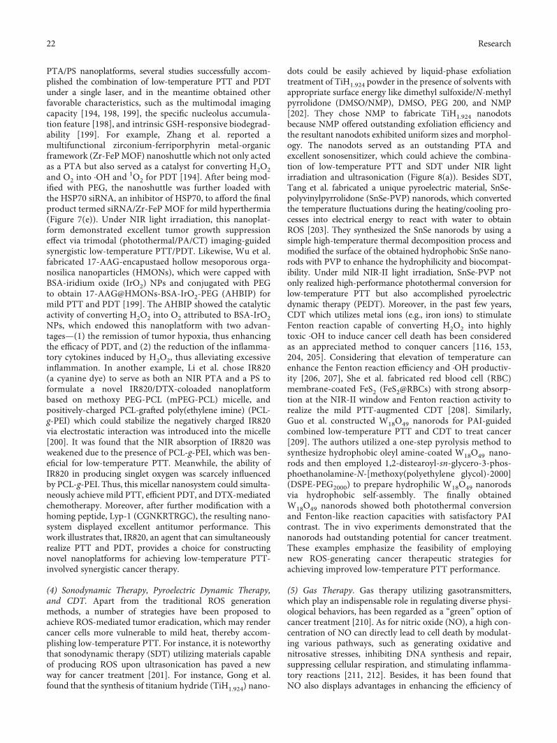
PTA/PS nanoplatforms, several studies successfully accom-plished the combination of low-temperature PTT and PDTunder a single laser, and in the meantime obtained otherfavorable characteristics, such as the multimodal imagingcapacity [194, 198, 199], the specific nucleolus accumula-tion feature [198], and intrinsic GSH-responsive biodegrad-ability [199]. For example, Zhang et al. reported amultifunctional zirconium-ferriporphyrin metal-organicframework (Zr-FePMOF) nanoshuttle which not only actedas a PTA but also served as a catalyst for converting H2O2and O2 into ⋅OH and 1O2 for PDT [194]. After being mod-ified with PEG, the nanoshuttle was further loaded withthe HSP70 siRNA, an inhibitor of HSP70, to afford the finalproduct termed siRNA/Zr-FeP MOF for mild hyperthermia(Figure 7(e)). Under NIR light irradiation, this nanoplat-form demonstrated excellent tumor growth suppressioneffect via trimodal (photothermal/PA/CT) imaging-guidedsynergistic low-temperature PTT/PDT. Likewise, Wu et al.fabricated 17-AAG-encapustaed hollow mesoporous orga-nosilica nanoparticles (HMONs), which were capped withBSA‐iridium oxide (IrO2) NPs and conjugated with PEGto obtain 17-AAG@HMONs-BSA-IrO2-PEG (AHBIP) formild PTT and PDT [199]. The AHBIP showed the catalyticactivity of converting H2O2 into O2 attributed to BSA-IrO2NPs, which endowed this nanoplatform with two advan-tages—(1) the remission of tumor hypoxia, thus enhancingthe efficacy of PDT, and (2) the reduction of the inflamma-tory cytokines induced by H2O2, thus alleviating excessiveinflammation. In another example, Li et al. chose IR820(a cyanine dye) to serve as both an NIR PTA and a PS toformulate a novel IR820/DTX-coloaded nanoplatformbased on methoxy PEG-PCL (mPEG-PCL) micelle, andpositively-charged PCL-grafted poly(ethylene imine) (PCL-g-PEI) which could stabilize the negatively charged IR820via electrostatic interaction was introduced into the micelle[200]. It was found that the NIR absorption of IR820 wasweakened due to the presence of PCL-g-PEI, which was ben-eficial for low-temperature PTT. Meanwhile, the ability ofIR820 in producing singlet oxygen was scarcely influencedby PCL-g-PEI. Thus, this micellar nanosystem could simulta-neously achieve mild PTT, efficient PDT, and DTX-mediatedchemotherapy. Moreover, after further modification with ahoming peptide, Lyp-1 (CGNKRTRGC), the resulting nano-system displayed excellent antitumor performance. Thiswork illustrates that, IR820, an agent that can simultaneouslyrealize PTT and PDT, provides a choice for constructingnovel nanoplatforms for achieving low-temperature PTT-involved synergistic cancer therapy.
(4) Sonodynamic Therapy, Pyroelectric Dynamic Therapy,and CDT. Apart from the traditional ROS generationmethods, a number of strategies have been proposed toachieve ROS-mediated tumor eradication, which may rendercancer cells more vulnerable to mild heat, thereby accom-plishing low-temperature PTT. For instance, it is noteworthythat sonodynamic therapy (SDT) utilizing materials capableof producing ROS upon ultrasonication has paved a newway for cancer treatment [201]. For instance, Gong et al.found that the synthesis of titanium hydride (TiH1.924) nano-
dots could be easily achieved by liquid-phase exfoliationtreatment of TiH1.924 powder in the presence of solvents withappropriate surface energy like dimethyl sulfoxide/N-methylpyrrolidone (DMSO/NMP), DMSO, PEG 200, and NMP[202]. They chose NMP to fabricate TiH1.924 nanodotsbecause NMP offered outstanding exfoliation efficiency andthe resultant nanodots exhibited uniform sizes and morphol-ogy. The nanodots served as an outstanding PTA andexcellent sonosensitizer, which could achieve the combina-tion of low-temperature PTT and SDT under NIR lightirradiation and ultrasonication (Figure 8(a)). Besides SDT,Tang et al. fabricated a unique pyroelectric material, SnSe-polyvinylpyrrolidone (SnSe-PVP) nanorods, which convertedthe temperature fluctuations during the heating/cooling pro-cesses into electrical energy to react with water to obtainROS [203]. They synthesized the SnSe nanorods by using asimple high-temperature thermal decomposition process andmodified the surface of the obtained hydrophobic SnSe nano-rods with PVP to enhance the hydrophilicity and biocompat-ibility. Under mild NIR-II light irradiation, SnSe-PVP notonly realized high-performance photothermal conversion forlow-temperature PTT but also accomplished pyroelectricdynamic therapy (PEDT). Moreover, in the past few years,CDT which utilizes metal ions (e.g., iron ions) to stimulateFenton reaction capable of converting H2O2 into highlytoxic ⋅OH to induce cancer cell death has been consideredas an appreciated method to conquer cancers [116, 153,204, 205]. Considering that elevation of temperature canenhance the Fenton reaction efficiency and ⋅OH productiv-ity [206, 207], She et al. fabricated red blood cell (RBC)membrane-coated FeS2 (FeS2@RBCs) with strong absorp-tion at the NIR-II window and Fenton reaction activity torealize the mild PTT-augmented CDT [208]. Similarly,Guo et al. constructed W18O49 nanorods for PAI-guidedcombined low-temperature PTT and CDT to treat cancer[209]. The authors utilized a one-step pyrolysis method tosynthesize hydrophobic oleyl amine-coated W18O49 nano-rods and then employed 1,2-distearoyl-sn-glycero-3-phos-phoethanolamine-N-[methoxy(polyethylene glycol)-2000](DSPE-PEG2000) to prepare hydrophilic W18O49 nanorodsvia hydrophobic self-assembly. The finally obtainedW18O49 nanorods showed both photothermal conversionand Fenton-like reaction capacities with satisfactory PAIcontrast. The in vivo experiments demonstrated that thenanorods had outstanding potential for cancer treatment.These examples emphasize the feasibility of employingnew ROS-generating cancer therapeutic strategies forachieving improved low-temperature PTT performance.
(5) Gas Therapy. Gas therapy utilizing gasotransmitters,which play an indispensable role in regulating diverse physi-ological behaviors, has been regarded as a “green” option ofcancer treatment [210]. As for nitric oxide (NO), a high con-centration of NO can directly lead to cell death by modulat-ing various pathways, such as generating oxidative andnitrosative stresses, inhibiting DNA synthesis and repair,suppressing cellular respiration, and stimulating inflamma-tory reactions [211, 212]. Besides, it has been found thatNO also displays advantages in enhancing the efficiency of
22 Research

other therapeutic modalities [213]. First, NO can sensitizethe multidrug-resistant cells to chemodrugs. Second, NO iscapable of acting as a radiosensitizer to assist RT in treatinghypoxic tumors. Third, in the presence of ROS, NO can beoxidized into highly reactive peroxynitrite (ONOO−) tostrengthen oxidative stress for killing cancer cells. Neverthe-less, how to deliver exogenous gasotransmitters safely andprecisely has become one of the most imperative questionsin the gas therapy field. Hence, some recent state-of-the-artstudies have focused on the design of various endogenousand/or exogenous stimulus-responsive NO-releasing nano-structures and their application in on-demand NO-sensitized synergistic cancer therapy [213]. Among them,the NIR light used in mild hyperthermia has been suggested
to be an appropriate exogenous stimulus and the combina-tion of low-temperature PTT and NO-mediated gas therapyhas shown greatly improved cancer therapeutic effect. Forexample, Zhang et al. proposed that utilizing NO in mildPTT could improve the mild PTT’s capability of eradicatingtumors [214]. They used Tween 20 (a commercial surfactant)to modify the surface of bismuth sulfide (Bi2S3) NPs toimprove Bi2S3 NPs’ dispersity in physiological solutionstal0and then added a hydrophobic NO donor, N,N ′-di-sec-butyl-N,N ′-dinitroso-1,4-phenylenediamine (BNN, whichis relatively stable below 60°C) to form the final productBNN-Bi2S3 nanocomposites via hydrophobic interaction.Upon NIR light irradiation, the high local temperature onthe surface of Bi2S3 NPs could cause on-demand NO release,
PTT & SDT
ROS
Heat
HSP-regulated
gene
2Release
Entry3
808 nm
Heat
DSP
HSFmonomer
HSFtrimer
4PhotothermalactivationH2O
NDsTiH1.924
1064 nm
O2
US
5Binding
RNApolymerase
HSE
Expression6Internalization
Nanocomplex
BNN-Bi2S3Bi2S3
Proteins
NO release
Mild PTT
Mild PTT Mild PTT + NO
Dead tumor cell
Survived tumor cell
Nitric oxide
Autophagosome
Cytochrome c
Caspase-3 Inhibiting
protective autophagy
Mitophagy
1
Enhanced apoptosis
(b)
(c)(a)
Figure 8: (a) Scheme showing the use of TiH1.924 nanodots (NDs) for SDT and PTT. Reprinted with permission from Reference [202].Copyright 2020, Nature Publishing Group. (b) Scheme describing the use of Bi2S3 for mild PTT, and the use and working mechanism ofBNN-Bi2S3 for combined NO-mediated gas therapy and mild PTT. Reprinted with permission from Reference [214]. Copyright 2019,Wiley-VCH. (c) Schematic illustration of DSP-mediated gene delivery and the photothermal activation of gene expression under NIR lightirradiation. Reprinted with permission from Reference [217]. Copyright 2017, Wiley-VCH.
23Research

and the surrounding temperature of the nanocomposites waskept at a low level to achieve mild PTT. Moreover, thereleased NO could suppress cancer cell autophagy-mediatedself-repair which was triggered by mild PTT, and promotecancer cell apoptosis, thus rendering tumor vulnerable tomild heat (Figure 8(b)). Meanwhile, NO could diffusehomogeneously in the solid tumor to further enhance theeffect of NO-sensitized low-temperature PTT for defeatingcancer. Likewise, S-nitrosothiol, a thermosensitive donor ofNO, was conjugated onto the core-shell structure ofAu@SiO2 nanomaterials (serving as PTAs) by You et al.[215]. Then, the authors decorated Au@SiO2 with PEG,and loaded PES, an inhibitor of HSP70, into the cavityof the SiO2 shell. Upon NIR light illumination, the NOproduced by S-nitrosothiol and PES-mediated inhibitionof HSP70 could collaboratively induce cell apoptosis ornecrosis, thereby strengthening the cancer therapeuticeffect of the synergistic low-temperature PTT/gas therapy.
(6) Gene Therapy. Given that nucleic acids are the determi-nant of almost all bioactivities from the molecular to physio-logical level, gene therapy delivering exogenous nucleic acidswith the help of suitable vectors has gradually become one ofthe most prevalent cancer therapeutic strategies. As men-tioned above, the major challenge in this field is designingdelivery tools capable of releasing nucleic acids accuratelyand controllably as well as protecting them from degrada-tion. It is suggested that PTT may provide an ideal way fornucleic acid delivery/release [109], because the heat inducedby light can be employed as a trigger of nucleic acid releaseand can be spatiotemporally controlled. Besides, it has beenfound that the mild photothermal effect of PTAs can signifi-cantly facilitate the intracellular uptake of therapeutic nucleicacids and thus elevate the cancer treatment efficacy of genetherapy [109, 216]. In the past few years, some studies havesuggested that the codelivery of PTAs and therapeutic nucleicacids encoded with HSP70 promoters provides the possibilityof precisely regulating the expression of therapeutic genesunder mild photothermal conditions [217–219]. The moder-ate increase of temperature caused by PTAs enables the heatshock factor (HSF) to transform from an inactive monomerto an active trimer, which is capable of translocating intothe nucleus, and meanwhile, the therapeutic nucleic acidsencoded with HSP70 promoters can enter the nucleus. Then,the intranuclear HSF trimer binds to the heat-shock elementof the HSP70 promoter and activate the expression of thetherapeutic genes cloned downstream of the HSP70 pro-moter. For example, Lyu et al. designed a dendronized semi-conducting polymer (DSP) consisting of three components:the hydrophobic semiconducting backbone, the cationicthird-generation polyamidoamine (PAMAM3) side chains,and the PEG segments, serving as the PTA, the gene vector,and the water-solubility enhancer, respectively [217]. Afterloading HSP-regulatable gene plasmids to DSPs, the authorsobtained the nanocomplexes which achieved not only the effi-cient delivery of genes but also mild heat-controlled geneexpression (Figure 8(c)). Similarly, Liu et al. selected PB andtherapeutic plasmid DNA consisting of HSP70 promoter,tumor suppressor p53 gene, and green fluorescent protein
(GFP) gene (acting as a reporter gene) to achieve thermocon-trolled synergistic gene therapy/PTT [218]. It was observedthat mild NIR light irradiation (temperature: ~41°C) couldstimulate the HSP70 promoter for activating tumor suppres-sor p53-dependent apoptosis, and meanwhile, strong NIRlight irradiation (temperature: ~50°C) resulted in the photo-thermal ablation of tumors through cellular dysregulationand necrosis. This work emphasizes the feasibility of combin-ing low-temperature PTT-assisted gene therapy and conven-tional PTT to achieve effective tumor eradication by bothapoptosis and necrosis.
Moreover, considering that oncolytic adenovirus (Ad) iswidely recognized as a promising candidate for cancer genetherapy, and some studies demonstrate that mild hyperther-mia can improve the anticancer effect of oncolytic Adsin vitro [220–222], Jung et al. fabricated an AuNR-mediated mild hyperthermia nanoplatform for enhancedoncolytic Ad gene delivery [223]. They combined the oncoly-tic Ad expressing VEGF promoter-targeted artificial transcrip-tional repressor zinc-finger protein with AuNR-mediatedmildPTT. Under NIR laser irradiation, the mild heat generated byAuNRs notably promoted the endocytosis of oncolytic Ads,transgene expression, viral replication, and subsequent cytoly-sis of head and neck cancer cells, thus realizing the elevatedefficiency of oncolytic Ads in cancer treatment.
Besides HSPs, Bcl2-associated athanogene domain 3(BAG3) is also responsible for protecting cells from thermalattacks and developing cellular thermoresistance [224].Inspired by this fact, Wang et al. used AuNRs to deliverBAG3-targeting siRNA oligomers to silence the expressionof BAG3, thus rendering cancer cells vulnerable to low-temperature PTT [225]. The AuNRs were prepared by aseed-mediated growth method with hexadecyltrimethylam-monium bromide and were sequentially modified withnegatively charged poly(sodium 4-styrenesulfonate) andpositively-charged poly(diallyldimethylammonium chloride)to form positive charged nanorods for connecting withBAG3-targeting siRNA oligomers through electrostaticinteraction. The experimental results demonstrated that theresultant nanocomplexes exhibited great NIR photothermalconversion ability and the cancer cells were more sensitiveto heat with the help of BAG3-targeting siRNA oligomers,thus realizing both gene delivery and low-temperature PTT.
(7) Immunotherapy. In the past few decades, immunotherapyhas played an unparalleled role in defeating cancers andexhibited satisfactory capability to eradicate both primaryand distant tumors [226, 227]. Since the immune system isa critical regulator of the tumor physiology and biology,which may result in the stimulation or suppression of tumordevelopment, growth, invasion, and metastasis, it is impor-tant to develop effective immunotherapeutic strategies withthe capacity to drive the immune system to pose a lethalthreat to tumors rather than help them survive [228–231].There have been numerous studies which focus on the explo-ration of cancer immune escape mechanisms and try to ele-vate the efficiency of immunotherapy [231]. Three majormechanisms have been proposed [231]: (1) the loss of
24 Research

antigenicity which has the potential to induce tumor-specificimmune responses, (2) the poor immunogenicity of cancers,which may endow tumors with additional immunosuppres-sive properties via the overexpression of immune check-points and the secretion of suppressive cytokines, etc., and(3) the generation of immunosuppressive TME where thetumor-infiltrating lymphocytes (TILs) like macrophages donot attack cancer cells. In view of these problems, variousimmunotherapeutic approaches represented by chimericantigen receptor (CAR) T cell therapy, immune check-point blockade (ICB), and cancer vaccines have been pro-posed and exhibited outstanding treatment performance[232–236]. Nevertheless, due to the heterogeneity and com-plexity of cancer, immunotherapy may not be able to activatethe immune system to prevent tumor reoccurrence andmetastasis, and its efficiency may be restrained by the denseextracellular matrix (ECM) and elevated interstitial fluidpressure (IFP) nearby the tumor tissue during the deliveryof therapeutic molecules or cells [231, 237, 238]. Fortunately,the collaboration with low-temperature PTT can shed newlight on the elevation of the immunotherapeutic efficiency.It has been found that mild hyperthermia can not onlydirectly kill cancer cells but also lead to the partial disruptionof ECM, the decrease of IFP, and the increase of the bloodperfusion, which is beneficial for the delivery of immuno-therapeutic agents and the recruitment and infiltration oflymphocytes. More importantly, mild hyperthermia can pro-duce a favorable TME for immune responses via makingdying cancer cells release tumor-associated antigens (TAAs)and damage-associated molecular pattern molecules (DAMPs)to modulate the immune system, consequently promotingthe maturation of antigen-presenting cells (APCs), facilitat-ing the secretion of immunostimulatory cytokines, andactivating the cytotoxic lymphocytes to defeat tumors[167, 239–242]. In addition, Deng et al. found that theGO-mediated low-temperature PTT could inhibit the M2polarization of tumor-associated macrophages (TAMs)(M2 phenotype TAMs are beneficial for tumor growth andmetastasis) and thus contribute to evoking the immune sys-tem [243]. However, the immune responses induced bymild hyperthermia alone are usually not enough to remark-ably reverse the tumor-mediated immunosuppressivemicroenvironment [244]. Therefore, on the one hand, thecombination of low-temperature PTT and immunotherapymay pave a promising way for significantly evoking immunesystems to eradicate cancers. On the other hand, the syner-gistic cancer treatment provides the possibility to achievelow-temperature PTT. For example, Chen et al. fabricatedpoly(lactic-co-glycolic acid) (PLGA)-ICG NPs to generatemild heat under NIR light irradiation and found that themild heat could trigger increased infiltration and accumula-tion of the chondroitin sulfate proteoglycan-4- (CSPG4-)specific CAR T cells in tumor tissues [242]. This platformshows the eminent performance of mild PTT in elevatingthe therapeutic efficacy of CAR T cells in solid tumors.
In addition, ICB with the aim of inhibiting the immunecheckpoints to activate immune responses has been widelyexploited for the eradication of various cancers. It has been
recognized that the two major determinants of the cancertherapeutic effect of ICB-based strategies are the expressionof programmed death-ligand 1 (PD-L1) in the tumor tissuesand the number of TILs [245–247]. However, a number ofstudies have demonstrated that some kinds of tumors werenot vulnerable to ICB treatments because there are few TILsand low PD-L1 content in these tumor tissues [248–251].Fortunately, due to its excellent potential for facilitating therecruitment of TILs and its capacity of inducing the elevatedexpression of PD-L1 on the plasma membranes of cancercells, mild PTT is able to provide a possibility of remodelingthe TME and enhancing the efficacy of ICB-based immuno-therapy [239, 251–255]. For instance, Huang et al. encapsu-lated both IR820 and PD-L1 antibody (aPD-L1), which iscapable of interfering with the interaction of programmeddeath protein 1 (PD-1) with PD-L1, into an injectable lipidgel (LG) depot which could undergo thermally reversiblegel-to-sol phase transition [256]. Under the mild heatinduced by the NIR light irradiation, the drug-loaded LGdepot in sol phase successfully released aPD-L1 for achievingthe symbiotic mild photothermal-assisted immunotherapy(SMPAI). This platform shows strong capacity in reversingthe immunosuppressive microenvironment via ICB andexhibiting noticeable primary and distal tumor ablation effect(Figure 9(a)). It is noteworthy that the characteristics of thisplatform such as a consistent retention time in differenttumors, the drug exposure dose, and the drug release amountand schedule could be precisely controlled through adjustingthe component proportion of LG, changing drug loadingamount, and manipulating NIR irradiation, which endowedthe platform with great potential for personalized cancertherapy. Recently, Guo et al. designed a “one-for-all” nano-system based on PEGylated PEI-coated monoelementalbismuthene loaded with siRNA against PD-L1 for mildphotothermal immunotherapy [257]. This nanosystem coulddetach PEG in acidic TME, which resulted in the decreasedsize and formation of positively charged surface (due to theexposure of PEI) of these NPs, thus enhancing cancer celluptake. After the endocytosis of the NPs, PEI assisted theNPs to escape from endosomes via the “proton sponge”effect, and meanwhile, the bismuthene-mediated mild hyper-thermia could also promote the endosomal escape andinduce the controllable release of siRNA against PD-L1.Then, under NIR light irradiation, the bismuthene releasedto the cytoplasm achieved low-temperature PTT, and it wasfound that mild hyperthermia stimulated a systemic immuneresponse, recruited abundant TILs, and upregulated theexpression of PD-L1 on tumor cells. The above two exampleshighlight the synergistic effect of low-temperature PTT andICB-based immunotherapy and provide implications forthe design of well-integrated nanoplatforms for cancertherapy.
In addition, cancer vaccines are designed to elicit long-lasting immune activation [258]. However, some studies havedemonstrated that most cancer vaccines composed ofdefined peptide/protein antigens rather than all antigenswhich are obtained from whole tumor cells have the inferiorability of inducing immunological memory cells to develop ananticancer immunologic memory [259, 260]. Additionally, it
25Research

Tumorinfiltration
Distaltumor
Mild PTT inducedimmunogenic tumor
“Cold” tumor “Hot” tumor
MCH
TCR
LG depot
IR820
ICD maximization
ICD boosting
ICD generation
H2O2 Fenton
�erapy-inducedICD amplifier
Cu-LDH
FeOOH
Hsp90 inhibitor
Hsp90
CRT
CTL
‘eat me’
‘eat me’
Distant tumor
Primary tumor
Anti-tumorimmunostimulation
•OH
Hsp90 inhibition Fever-type photothermaltreatment
Fenton •OH
aPD-L1
PD-1
PD-L1
PD-L1/PD-1blockade
CD4+ T cells
CD8+ T cells
Primarytumor
Tumor bloodvessel
Effector Tcells
Lymph node
Naive Tcells
Antigen presentation
Immature DC
DC maturationAntigen release
and uptake
In situgelation
<45°CMild PTT
(b)
(a)
Figure 9: (a) Scheme illustrating the use of the IR820/aPD-L1-encapsulated LG depot for SMPAI. Reprinted with permission from Reference[256]. Copyright 2019, Nature Publishing Group. (b) Scheme depicting the fever-type photothermal treatment and Fenton reaction-enhancedimmunotherapeutic effect by using the FeOOH@STA/Cu-LDH nanohybrid. Reprinted with permission from Reference [43]. Copyright 2020,Wiley-VCH.
26 Research

is very difficult to collect the whole tumor to prepare a stan-dardized vaccine line consisting of a variety of antigens fromwhole cancer cells. More importantly, TAAs may vary amongtumors and individuals, which further restrains the develop-ment of cancer vaccines [261]. With regard to these problems,Chen et al. chose an immunomodulator resiquimod (R848)rather than antigens to develop a low-temperature PTT-mediated cancer vaccine [262]. R848 is the agonist of Toll-like receptors 7 and 8 (TLR-7/8) which are abundant in theimmune cells that infiltrate TME, and it has been proved thatthe stimulation of TLR-7/8 by R848 can activate APCs viafacilitating the maturation of DCs and subsequently trans-forming DCs’ phenotype from immunosuppressive to immu-nogenic [263–265]. Then, with the assistance of diverseproinflammatory cytokines (e.g., interleukin 6 and tumornecrosis factor-α) secreted by matured DCs, the antitumorimmune responses can be activated. The authors used anamphiphilic copolymer consisting of hydrophobic polyaniline(PANI) and the hydrophilic glycol-chitosan (GCS) backboneto deliver R848 via loading R848 in the hydrophobic core[262]. PANI served as an NIR PTA and achieved low-temperature PTT, and mild hyperthermia showed satisfactoryperformance in rendering the primary tumor residues moresensitive to immunotherapy. Meanwhile, this platform suc-cessfully realized the development of robust and long-termimmunological memory against cancer and the effective inhi-bition of tumor reoccurrence and metastasis. This work ver-ifies that mild PTT can potentiate the immunotherapeuticefficiency of cancer vaccines.
Moreover, immunogenic cell death (ICD), a cell deathtype that can release DAMPs to activate the immune systemto fight against tumors and establish long-term immunosur-veillance, is gaining increasing research interest in cancertherapy [266]. It has been found that chemotherapy, PTT,RT, and PDT are all able to induce ICD and then stimulatethe tumor-specific immune responses [267–269]. DuringICD, calreticulin (a significant marker of ICD) can migratefrom the endoplasmic reticulum of tumor cells to the plasmamembrane and induce dendritic cells to engulf dying tumorcells and their debris. However, the undesirable toxicity toimmune cells in harsh treatment conditions may severelyaffect the process of ICD and the reduced toxicity to cancercells in mild treatment conditions can decrease the tumortherapeutic efficacy [270, 271]. Meanwhile, overheating candestroy antigens from dying tumor cells leading to thedecreased tumor-killing immune responses [272]. Thus, thedevelopment of efficacious cancer therapeutic strategies withrelatively mild treatment conditions is highly needed.Recently, the combination of low-temperature PTT andICD-mediated immunotherapy has demonstrated itsremarkable capacity of elevating the efficacy of cancer treat-ments [43, 272, 273]. For example, a triple therapeutic proto-col combining low X-ray dose RT, low-temperature PTT,and immunotherapy was developed based on semiconductorheterojunction structured WO2.9-WSe2-PEG NPs [272].Under X-ray irradiation, the NPs could catalyze H2O2 exist-ing in TME to generate nonoxygen-dependent ROS whichnot only damaged cancer cells but also further triggeredICD. Besides, the NPs exhibited excellent NIR photothermal
conversion capacity and it has been found that mild PTT wascapable of triggering ICD to some extent. With the adminis-tration of aPD-L1, this platform effectively achieved tumoreradication and metastasis inhibition under mild tempera-ture (induced by WO2.9-WSe2-PEG NPs upon mild NIRlaser irradiation (0.5Wcm−2)) and low X-ray radiation dose.This work further verifies the ICD-inducing ability of RT andmild PTT and provides a valuable strategy for defeating can-cer in a much safer condition. Additionally, Li et al. foundthat fever-type (i.e., low-temperature) PTT could facilitatethe ROS-induced ICD to enhance immunotherapy [43].They prepared an NIR PTA composed of Cu-containing lay-ered double hydroxide (Cu-LDH), anchored highly reactiveFeOOH nanodots which can generate ROS via Fenton reac-tion onto Cu-LDH, and inserted STA-9090 (an HSP90 inhib-itor) into the gaps between the layers of Cu-LDH to promotecancer cell apoptosis at low temperatures, finally obtainingthe FeOOH@STA/Cu-LDH nanohybrid. Under NIR lightirradiation, the Fenton reaction-induced ICD was sequen-tially enhanced by mild hyperthermia and HSP90 inhibitionand the primary and distant tumors were effectively elimi-nated by this synergistic therapeutic platform (Figure 9(b)).This work proposes a novel method for strengthening ICD-mediated immunotherapy via the synergistic Fenton reac-tion/low-temperature PTT. Moreover, the employment ofan ICD inducer with mild PTT also shows eminent perfor-mance in eradicating tumors. Zhou et al. prepared a porouscobalt sulfide nanomaterial (PCS) via a simple DNA-templated hydrothermal method for PTT and the deliveryof EGCG, an HSP70 inhibitor, and oxaliplatin, an ICDinducer [273]. The authors utilized the EGCG-mediatedlow-temperature PTT as the “first-hit” to kill tumors andthe oxaliplatin-mediated ICD as the “second-hit” to elimi-nate the potential threat of tumor reoccurrence. Moreover,the release of EGCG and oxaliplatin was induced by pH-and heat-trigger, respectively, which realized the controllabledrug delivery sequence. This work may inspire moreresearchers to employ low-temperature PTT to activate theimmune system and amplify the cancer elimination effectof immunotherapy.
3. Conclusions and Perspectives
In the past few years, low-temperature PTT based on nano-materials has attracted considerable attention and exhibitedpromising potential in conquering some important problemsin the biomedical region. It has been demonstrated that, withthe assistance of nanotechnology, mild hyperthermiainduced by NIR laser shows many advantages in elevatingthe therapeutic efficacy in diverse applications, because thewell-designed nanoplatforms containing PTAs can (1) pas-sively penetrate biofilms due to their nanoscale sizes andphysicochemical properties or accumulate in the tumor tis-sues via EPR effect and specifically target bacteria or cancercells via some integrated bacterium/tumor-targeting mole-cules or motifs, (2) possess some beneficial characteristicsincluding stimulus-responsive property, organelle-targetingcapability, and multimodal imaging capacity, (3) be com-bined with other therapeutic modalities to achieve synergistic
27Research

therapies, and (4), as for cancer treatments, achieve signifi-cantly increased cellular uptake and tumor accumulation ofthe administrated therapeutics with the help of mild heat topotentiate the tumor therapeutic effect.
In this review, we have summarized the recent progress ofnanomaterial-mediated low-temperature PTT from threeaspects—(1) bacterial elimination, (2) wound treatment,and (3) cancer treatment. As for the antibiosis, there aretwo major obstacles impeding us to develop more effectivestrategies to avoid bacterial infection. On the one hand, dueto the inappropriate utilization of diverse antibiotics, anincreasing number of bacteria have become antibiotic-resistant and the antibacterial efficiency of antibiotics hasbeen decreased dramatically. On the other hand, the trouble-some biofilms largely hinder the interaction of antibacterialdrugs and/or nanoagents with bacteria. Fortunately, it hasbeen noted that mild PTT may render bacteria more suscep-tible to other treatments, and some novel methods based onlow-temperature PTT have been proposed to solve theseproblems, including the synergistic NO-enhanced PDT/low-temperature PTT and QCS-MoS2 nanoflake-mediatedmild PTT for realizing bacterial resensitization to antibiotics[59, 60] as illustrated above. In regard to wound healing, it isinspiring that the combination of low-temperature PTT andother wound therapeutic strategies can achieve rapid andlong-term wound healing [65–68]. It has been shown that,in the presence of some tissue regeneration-stimulating sub-stances like bioactive ions, mild PTT can not only defeat bac-terial infection but also alleviate excessive inflammation andfacilitate the angiogenesis, which are crucial for wound heal-ing [65, 66]. Besides, the introduction of “hot spring” effectalso provides an encouraging method for treating chronicwounds [67]. To be noted, as being combined with MSC-based therapy, mild hyperthermia mediated by CuS@BSANPs can enhance the differentiation of MSCs to form fibro-blasts for promoting wound healing [68]. In view of cancertherapy, we first summarized the up-to-date strategies forrealizing low-temperature PTT for tumor eradication. First,given that HSPs are the major determinant of the generationof thermal resistance and they play an important role in help-ing cancer cells escape from apoptosis-mediated death, theinhibition of HSPs using small-molecule HSP inhibitors(e.g., GA, Qu, 17-AAG, VER-155008, PES, EGCG, andBIIB021) and siRNAs can not only render tumors more vul-nerable to mild heat for achieving low-temperature PTT butalso promote the apoptosis-mediated cell death, which mayfurther enhance the cancer treatment effect of mild PTT.Moreover, because both the synthesis of HSPs and the reali-zation of HSP biological functions need energy, cancer star-vation therapy which blocks/reduces the nutrient supplyand/or exhausts the nutrients can lead to the decreasedproduction of energy, thus inhibiting HSPs to accomplishlow-temperature PTT. Second, the combination withautophagy-mediated cancer therapy sheds new light on thetherapeutic effect enhancement of mild PTT. It has beenfound that the regulation of autophagy can contribute tolow-temperature PTT for more effective tumor eradication.Third, the combination of organelle-targeting strategy withlow-temperature PTT has also been exploited to achieve sat-
isfactory cancer cell-killing effect because many organellesare vulnerable to mild hyperthermia and each organelle playsan indispensable role in maintaining intracellular homeosta-sis. Fourth, we concluded the synergistic therapy strategiesthat combine low-temperature PTT with other treatmentmodalities (e.g., chemotherapy, RT, gene therapy, andimmune therapy). On the one hand, mild hyperthermia canmarkedly promote the tumor accumulation and retentionof NPs/drugs via increasing blood flow and changing thecompact tumor ECM into a loose one, thus elevating theintratumoral delivery of chemodrugs, radiosensitizers,nucleic acids, immunotherapeutic agents, etc. to achieve bet-ter cancer therapeutic outcomes. On the other hand, othertreatments can render cancer cells more susceptible to mildhyperthermia, thus realizing low-temperature PTT. Addi-tionally, it has also been verified that mild heat can alleviatethe hypoxia of tumor tissues, which paves a promising wayfor potentiating the effect of oxygen-dependent treatmentssuch as PDT and SDT. Furthermore, mild hyperthermiahas the potential for modulating immune systems, i.e., acti-vating the immunosuppressive microenvironment to formthe immunostimulatory one [167, 239–243]. Therefore, thecombination of low-temperature PTT and immunotherapycan significantly induce the immune responses to attack cancercells and inhibit the metastasis and reoccurrence of tumors.More importantly, it is noteworthy that nanotechnology playsan unparalleled role in achieving synergistic therapy. By utiliz-ing different synthesis and modification methods, variousmaterials and agents possessing diverse therapeutic effects canbe integrated into the nanoplatforms. Besides, the agents innanosystems usually exhibit increased therapeutic effects anddecreased side effects due to the strong drug-loading capacity,exogenous and/or endogenous stimulus-responsive property,and tumor targeting/accumulation ability of the nanocarriers.
Despite the above strategies developed, there are someproblems which we must solve. First, although a large num-ber of nanocarriers for low-temperature PTT have beendeveloped and exhibited satisfactory characteristics like theincreased drug encapsulation/loading capacity, decreasedtoxicity to normal cells, and good biocompatibility and bio-safety, we still need to develop nanocarriers with strongerdrug delivery capability, more desirable biodegradability,and enhanced stability in long-term blood circulation.Second, more strategies that can achieve low-temperaturePTT need to be developed. For example, we can designmore nanoplatforms for targeting other organelles exceptthe nucleus, mitochondrion, and cytoskeleton and com-pare the effectiveness of the different strategies to optimizethe organelle-targeted low-temperature PTT. Third, thereare some novel and promising therapeutic modalities,including ferroptosis, ion-interference therapy, and electro-dynamic therapy, which can also be adopted to be combinedwith low-temperature PTT for realizing satisfactory synergis-tic therapies. More importantly, although there have beenstudies focusing on the working mechanisms of the combina-tion of low-temperature PTT and other treatments, we aresupposed to figure out the more detailed action mechanismsof these synergistic therapies to propose more effective com-binational strategies and more appropriate administration
28 Research

sequences. Likewise, to elevate the effectiveness of using mildPTT to treat bacterial infections and wounds, researchers aresuggested to pay more attention to understanding and utiliz-ing the complex mechanisms from various aspects like thebehaviors and roles of bacteria or other indispensable typesof cells in bacterial invasion and wound development. More-over, there are few reports about using low-temperature PTTfor treating other diseases like neurological disorders, meta-bolic diseases, and cardiovascular diseases. Therefore, it issuggested that mild PTT can be applied in more biomedicalfields in the future.
To summarize, low-temperature PTT is a robust strategyfor eliminating bacteria and cancers. We sincerely hope thatthis reviewmay inspire more researchers to devote their effortsto this fascinating field and achieve significant advances inmild PTT-involved preclinical and clinical researches.
Conflicts of Interest
The authors declare no conflict of interest.
Acknowledgments
This work was supported by the National Natural ScienceFoundation of China (21673037) and the Open Project ofKey Laboratory of High-Incidence-Tumor Prevention andTreatment (Guangxi Medical University), Ministry of Educa-tion (GKE-KF202001).
References
[1] J. J. Hu, Y. J. Cheng, and X. Z. Zhang, “Recent advances innanomaterials for enhanced photothermal therapy oftumors,” Nanoscale, vol. 10, no. 48, pp. 22657–22672, 2018.
[2] P. C. Ray, S. A. Khan, A. K. Singh, D. Senapati, and Z. Fan,“Nanomaterials for targeted detection and photothermal kill-ing of bacteria,” Chemical Society Reviews, vol. 41, no. 8,pp. 3193–3209, 2012.
[3] J. M. V. Makabenta, A. Nabawy, C. H. Li, S. Schmidt-Malan,R. Patel, and V. M. Rotello, “Nanomaterial-based therapeu-tics for antibiotic-resistant bacterial infections,” NatureReviews Microbiology, vol. 19, no. 1, pp. 23–36, 2021.
[4] M. E. Davis, Z. Chen, and D. M. Shin, “Nanoparticle thera-peutics: an emerging treatment modality for cancer,” NatureReviews Drug Discovery, vol. 7, no. 9, pp. 771–782, 2008.
[5] S. H. Jo, T. Chang, I. Ebong, B. B. Bhadviya, P. Mazumder,and W. Lu, “Nanoscale memristor device as synapse in neu-romorphic systems,” Nano Letters, vol. 10, no. 4, pp. 1297–1301, 2010.
[6] F. Danhier, E. Ansorena, J. M. Silva, R. Coco, A. Le Breton,and V. Préat, “PLGA-based nanoparticles: an overview ofbiomedical applications,” Journal of Controlled Release,vol. 161, no. 2, pp. 505–522, 2012.
[7] D. Jaque, L. Martínez Maestro, B. del Rosal et al., “Nanopar-ticles for photothermal therapies,” Nanoscale, vol. 6, no. 16,pp. 9494–9530, 2014.
[8] Z. Xie, T. Fan, J. An et al., “Emerging combination strategieswith phototherapy in cancer nanomedicine,” Chemical Soci-ety Reviews, vol. 49, no. 22, pp. 8065–8087, 2020.
[9] J. Wang, X. Wu, P. Shen et al., “Applications of inorganicnanomaterials in photothermal therapy based on combina-tional cancer treatment,” International Journal of Nanomedi-cine, vol. 15, pp. 1903–1914, 2020.
[10] J. Yao, F. Zheng, C. Yao et al., “Rational design of nanomedi-cine for photothermal-chemodynamic bimodal cancer ther-apy,” WIREs Nanomedicine and Nanotechnology, no. articlee1682, 2020.
[11] X. Yang, M. Yang, B. Pang, M. Vara, and Y. Xia, “Gold nano-materials at work in biomedicine,” Chemical Reviews,vol. 115, no. 19, pp. 10410–10488, 2015.
[12] S. Yang, L. Zhou, Y. Su, R. Zhang, and C. M. Dong,“One-pot photoreduction to prepare NIR-absorbing plas-monic gold nanoparticles tethered by amphiphilic poly-peptide copolymer for synergistic photothermal-chemotherapy,” Chinese Chemical Letters, vol. 30, no. 1,pp. 187–191, 2019.
[13] Y. Liu, J. Li, M. Chen, X. Chen, and N. Zheng, “Palladium-based nanomaterials for cancer imaging and therapy,” Ther-anostics, vol. 10, no. 22, pp. 10057–10074, 2020.
[14] G. Gao, Y. W. Jiang, H. R. Jia et al., “From perinuclear tointranuclear localization: a cell-penetrating peptide modifica-tion strategy to modulate cancer cell migration under mildlaser irradiation and improve photothermal therapeutic per-formance,” Biomaterials, vol. 223, article 119443, 2019.
[15] Y. W. Jiang, G. Gao, P. Hu et al., “Palladium nanosheet-knotted injectable hydrogels formed via palladium−sulfurbonding for synergistic chemo-photothermal therapy,”Nanoscale, vol. 12, no. 1, pp. 210–219, 2020.
[16] S. Shen, S. Wang, R. Zheng et al., “Magnetic nanoparticleclusters for photothermal therapy with near-infrared irradia-tion,” Biomaterials, vol. 39, pp. 67–74, 2015.
[17] C. M. Hessel, V. P. Pattani, M. Rasch et al., “Copper selenidenanocrystals for photothermal therapy,” Nano Letters,vol. 11, no. 6, pp. 2560–2566, 2011.
[18] H. Y. Wang, X. W. Hua, F. G. Wu et al., “Synthesis of ultra-stable copper sulfide nanoclusters via trapping the reactionintermediate: potential anticancer and antibacterial applica-tions,” ACS Applied Materials & Interfaces, vol. 7, no. 13,pp. 7082–7092, 2015.
[19] Y. W. Bao, X. W. Hua, Y. H. Li, H. R. Jia, and F. G. Wu,“Hyperthemia-promoted cytosolic and nuclear delivery ofcopper/carbon quantum dot-crosslinked nanosheets: multi-modal imaging-guided photothermal cancer therapy,” ACSApplied Materials & Interfaces, vol. 10, no. 2, pp. 1544–1555, 2018.
[20] L. Gong, L. Yan, R. Zhou, J. Xie, W. Wu, and Z. Gu, “Two-dimensional transition metal dichalcogenide nanomaterialsfor combination cancer therapy,” Journal of Materials Chem-istry B, vol. 5, no. 10, pp. 1873–1895, 2017.
[21] B. del Rosal, E. Carrasco, F. Ren et al., “Infrared-emittingQDs for thermal therapy with real-time subcutaneous tem-perature feedback,” Advanced Functional Materials, vol. 26,no. 33, pp. 6060–6068, 2016.
[22] W. Tao, X. Ji, X. Xu et al., “Antimonene quantum dots:synthesis and application as near-infrared photothermalagents for effective cancer therapy,” Angewandte ChemieInternational Edition, vol. 56, no. 39, pp. 11896–11900,2017.
[23] H. S. Jung, P. Verwilst, A. Sharma, J. Shin, J. L. Sessler, andJ. S. Kim, “Organic molecule-based photothermal agents: an
29Research

expanding photothermal therapy universe,” Chemical SocietyReviews, vol. 47, no. 7, pp. 2280–2297, 2018.
[24] G. Y. Pan, H. R. Jia, Y. X. Zhu, R. H. Wang, F. G. Wu, andZ. Chen, “Dual channel activatable cyanine dye for mito-chondrial imaging and mitochondria-targeted cancer thera-nostics,” ACS Biomaterials Science & Engineering, vol. 3,no. 12, pp. 3596–3606, 2017.
[25] G. Y. Pan, H. R. Jia, Y. X. Zhu, W. Sun, X. T. Cheng, and F. G.Wu, “Cyanine-containing polymeric nanoparticles with ima-ging/therapy-switchable capability for mitochondria-targetedcancer theranostics,” ACS Applied Nano Materials, vol. 1,no. 6, pp. 2885–2897, 2018.
[26] G. Y. Pan, H. R. Jia, Y. X. Zhu, and F. G.Wu, “Turning doublehydrophilic into amphiphilic: IR825-conjugated polymericnanomicelles for near-infrared fluorescence imaging-guidedphotothermal cancer therapy,” Nanoscale, vol. 10, no. 4,pp. 2115–2127, 2018.
[27] G. Gao, Y. W. Jiang, W. Sun et al., “Molecular targeting-mediated mild-temperature photothermal therapy with asmart albumin-based nanodrug,” Small, vol. 15, no. 33, article1900501, 2019.
[28] X. Chen, X. Zhang, Y. Guo et al., “Smart supramolecular“Trojan horse”-inspired nanogels for realizing light-triggered nuclear drug influx in drug-resistant cancer cells,”Advanced Functional Materials, vol. 29, no. 13, article1807772, 2019.
[29] H. R. Jia, Y. X. Zhu, X. Liu et al., “Construction of duallyresponsive nanotransformers with nanosphere–nanofiber–nanosphere transition for overcoming the size paradox ofanticancer nanodrugs,” ACS Nano, vol. 13, no. 10,pp. 11781–11792, 2019.
[30] Y. X. Zhu, H. R. Jia, G. Gao et al., “Mitochondria-actingnanomicelles for destruction of cancer cells via excessivemitophagy/autophagy-driven lethal energy depletion andphototherapy,” Biomaterials, vol. 232, article 119668,2020.
[31] X. Liu, H. R. Jia, Y. X. Zhu et al., “Mitochondrion- andnucleus-acting polymeric nanoagents for chemo-photothermalcombination therapy,” Science China Materials, vol. 63, no. 5,pp. 851–863, 2020.
[32] G. Gao, Y. W. Jiang, Y. Guo et al., “Enzyme-mediated tumorstarvation and phototherapy enhance mild-temperaturephotothermal therapy,” Advanced Functional Materials,vol. 30, no. 16, article 1909391, 2020.
[33] L. Xu, L. Cheng, C. Wang, R. Peng, and Z. Liu, “Conjugatedpolymers for photothermal therapy of cancer,” PolymerChemistry, vol. 5, no. 5, pp. 1573–1580, 2014.
[34] G. Gao, Y. W. Jiang, H. R. Jia, and F. G. Wu, “Near-infraredlight-controllable on-demand antibiotics release usingthermo-sensitive hydrogel-based drug reservoir for combat-ing bacterial infection,” Biomaterials, vol. 188, pp. 83–95,2019.
[35] H. H. Ran, X. Cheng, G. Gao et al., “Colistin-loaded polydo-pamine nanospheres uniformly decorated with silver nano-dots: a nanohybrid platform with improved antibacterialand antibiofilm performance,” ACS Applied Bio Materials,vol. 3, no. 4, pp. 2438–2448, 2020.
[36] F. Lin, Q. Y. Duan, and F. G. Wu, “Conjugated polymer-based photothermal therapy for killing microorganisms,”ACS Applied Polymer Materials, vol. 2, no. 10, pp. 4331–4344, 2020.
[37] J. T. Robinson, S. M. Tabakman, Y. Liang et al., “Ultrasmallreduced graphene oxide with high near-infrared absorbancefor photothermal therapy,” Journal of the American ChemicalSociety, vol. 133, no. 17, pp. 6825–6831, 2011.
[38] K. Yang, L. Hu, X. Ma et al., “Multimodal imaging guidedphotothermal therapy using functionalized graphene nano-sheets anchored with magnetic nanoparticles,” AdvancedMaterials, vol. 24, no. 14, pp. 1868–1872, 2012.
[39] C. Liang, S. Diao, C. Wang et al., “Tumor metastasis inhibi-tion by imaging-guided photothermal therapy with single-walled carbon nanotubes,” Advanced Materials, vol. 26,no. 32, pp. 5646–5652, 2014.
[40] L. Zhao, Y. Jing, Y. Liu et al., “Mesoporous carbon nano-spheres as a multifunctional carrier for cancer theranostics,”Theranostics, vol. 8, no. 3, pp. 663–675, 2018.
[41] W. Sun, X. Zhang, H. R. Jia et al., “Water-dispersible candlesoot-derived carbon nano-onion clusters for imaging-guided photothermal cancer therapy,” Small, vol. 15, no. 11,article 1804575, 2019.
[42] P. Orosz, B. Echtenacher, W. Falk, J. Rüschoff, D. Weber, andD. N. Männel, “Enhancement of experimental metastasis bytumor necrosis factor,” Journal of Experimental Medicine,vol. 177, no. 5, pp. 1391–1398, 1993.
[43] B. Li, G. Hao, B. Sun, Z. Gu, and Z. P. Xu, “Engineering atherapy-induced “immunogenic cancer cell death” amplifierto boost systemic tumor elimination,” Advanced FunctionalMaterials, vol. 30, no. 22, article 1909745, 2020.
[44] B. Frey, E. M. Weiss, Y. Rubner et al., “Old and new factsabout hyperthermia-induced modulations of the immunesystem,” International Journal of Hyperthermia, vol. 28,no. 6, pp. 528–542, 2012.
[45] B. Hildebrandt, P. Wust, O. Ahlers et al., “The cellular andmolecular basis of hyperthermia,” Critical Reviews in Oncolo-gy/Hematology, vol. 43, no. 1, pp. 33–56, 2002.
[46] ANSI, American National Standard for Safe Use of Lasers,Z136.1-2014, Laser Institute of America, Orlando, FL, USA,2014.
[47] C. Jolly and R. I. Morimoto, “Role of the heat shock responseand molecular chaperones in oncogenesis and cell death,”Journal of the National Cancer Institute, vol. 92, no. 19,pp. 1564–1572, 2000.
[48] V. J. Verwaal, S. van Ruth, E. de Bree et al., “Randomized trialof cytoreduction and hyperthermic intraperitoneal chemo-therapy versus systemic chemotherapy and palliative surgeryin patients with peritoneal carcinomatosis of colorectal can-cer,” Journal of Clinical Oncology, vol. 21, no. 20, pp. 3737–3743, 2003.
[49] T. D. Yan, M. Deraco, D. Baratti et al., “Cytoreductive surgeryand hyperthermic intraperitoneal chemotherapy for malig-nant peritoneal mesothelioma: multi-institutional experi-ence,” Journal of Clinical Oncology, vol. 27, no. 36,pp. 6237–6242, 2009.
[50] Z. Shang, S. Y. Chan, Q. Song, P. Li, and W. Huang, “Thestrategies of pathogen-oriented therapy on circumventingantimicrobial resistance,” Research, vol. 2020, article2016201, 32 pages, 2020.
[51] W. Xiu, S. Gan, Q. Wen et al., “Biofilm microenvironment-responsive nanotheranostics for dual-mode imaging andhypoxia-relief-enhanced photodynamic therapy of bacterialinfections,” Research, vol. 2020, article 9426453, 15 pages,2020.
30 Research
[52] J. W. Xu, K. Yao, and Z. K. Xu, “Nanomaterials with a photo-thermal effect for antibacterial activities: an overview,” Nano-scale, vol. 11, no. 18, pp. 8680–8691, 2019.
[53] Q. Xin, H. Shah, A. Nawaz et al., “Antibacterial carbon-basednanomaterials,” Advanced Materials, vol. 31, no. 45, article1804838, 2019.
[54] M. Y. Xia, Y. Xie, C. H. Yu et al., “Graphene-based nanoma-terials: the promising active agents for antibiotics-independent antibacterial applications,” Journal of ControlledRelease, vol. 307, pp. 16–31, 2019.
[55] D. Hu, H. Li, B. Wang et al., “Surface-adaptive goldnanoparticles with effective adherence and enhancedphotothermal ablation of methicillin-resistant Staphylococ-cus aureus biofilm,” ACS Nano, vol. 11, no. 9, pp. 9330–9339, 2017.
[56] A. Nakal-Chidiac, O. García, L. García-Fernández et al.,“Chitosan-stabilized silver nanoclusters with luminescent,photothermal and antibacterial properties,” CarbohydratePolymers, vol. 250, article 116973, 2020.
[57] S. Cheeseman, A. J. Christofferson, R. Kariuki et al., “Antimi-crobial metal nanomaterials: from passive to stimuli-activated applications,” Advanced Science, vol. 7, no. 10, arti-cle 1902913, 2020.
[58] X. L. Fan, H. Y. Li, W. Y. Ye et al., “Magainin-modified poly-dopamine nanoparticles for photothermal killing of bacteriaat low temperature,” Colloids and Surfaces B: Biointerfaces,vol. 183, article 110423, 2019.
[59] Z. Yuan, C. Lin, Y. He et al., “Near-infrared light-triggerednitric-oxide-enhanced photodynamic therapy and low-temperature photothermal therapy for biofilm elimination,”ACS Nano, vol. 14, no. 3, pp. 3546–3562, 2020.
[60] Y. Huang, Q. Gao, X. Li et al., “Ofloxacin loaded MoS2 nano-flakes for synergistic mild-temperature photothermal/anti-biotic therapy with reduced drug resistance of bacteria,”Nano Research, vol. 13, no. 9, pp. 2340–2350, 2020.
[61] L. Tan, Z. Zhou, X. Liu et al., “Overcoming multidrug-resistant MRSA using conventional aminoglycoside antibi-otics,” Advanced Science, vol. 7, no. 9, article 1902070,2020.
[62] D. Simões, S. P. Miguel, M. P. Ribeiro, P. Coutinho, A. G.Mendonça, and I. J. Correia, “Recent advances on antimicro-bial wound dressing: a review,” European Journal of Pharma-ceutics and Biopharmaceutics, vol. 127, pp. 130–141, 2018.
[63] S. Saghazadeh, C. Rinoldi, M. Schot et al., “Drug delivery sys-tems and materials for wound healing applications,”Advanced Drug Delivery Reviews, vol. 127, pp. 138–166, 2018.
[64] P. H. Wang, B. S. Huang, H. C. Horng, C. C. Yeh, and Y. J.Chen, “Wound healing,” Journal of the Chinese Medical Asso-ciation, vol. 81, no. 2, pp. 94–101, 2018.
[65] X. Xu, X. Liu, L. Tan et al., “Controlled-temperature photo-thermal and oxidative bacteria killing and acceleration ofwound healing by polydopamine-assisted Au-hydroxyapatitenanorods,” Acta Biomaterialia, vol. 77, pp. 352–364, 2018.
[66] Y. Li, X. Xu, X. Liu et al., “Photoelectrons mediating angio-genesis and immunotherapy through heterojunction filmfor noninvasive disinfection,” Advanced Science, vol. 7,no. 17, article 2000023, 2020.
[67] L. Sheng, Z. Zhang, Y. Zhang et al., “A novel "hot spring"-mimetic hydrogel with excellent angiogenic properties forchronic wound healing,” Biomaterials, vol. 264, article120414, 2021.
[68] Y. Xiao, J. Peng, Q. Liu et al., “Ultrasmall CuS@BSA nanopar-ticles with mild photothermal conversion synergisticallyinduce MSCs-differentiated fibroblast and improve skinregeneration,” Theranostics, vol. 10, no. 4, pp. 1500–1513,2020.
[69] J. Liang, D. Kang, Y. Wang, Y. Yu, J. Fan, and E. Takashi,“Carbonate ion-enriched hot spring water promotes skinwound healing in nude rats,” PLoS One, vol. 10, no. 2, articlee0117106, 2015.
[70] Y. Jiang, B. N. Jahagirdar, R. L. Reinhardt et al., “Pluripotencyof mesenchymal stem cells derived from adult marrow,”Nature, vol. 418, no. 6893, pp. 41–49, 2002.
[71] R. R. Sharma, K. Pollock, A. Hubel, and D. McKenna,“Mesenchymal stem or stromal cells: a review of clinicalapplications and manufacturing practices,” Transfusion,vol. 54, no. 5, pp. 1418–1437, 2014.
[72] A. Dongre and R. A. Weinberg, “New insights into the mech-anisms of epithelial-mesenchymal transition and implica-tions for cancer,” Nature Reviews Molecular Cell Biology,vol. 20, no. 2, pp. 69–84, 2019.
[73] M. Markowicz, E. Koellensperger, S. Neuss, S. Koenigschulte,C. Bindler, and N. Pallua, “Human bone marrow mesenchy-mal stem cells seeded on modified collagen improved dermalregeneration in vivo,” Cell Transplantation, vol. 15, no. 8–9,pp. 723–732, 2006.
[74] M. L. Turski and D. J. Thiele, “New roles for coppermetabolism in cell proliferation, signaling, and disease,”Journal of Biological Chemistry, vol. 284, no. 2, pp. 717–721, 2009.
[75] S. J. Yang, J. Y. Uriu-Adams, C. L. Keen, R. B. Rucker, andL. Lanoue, “Effects of copper deficiency on mouse yolk sacvasculature and expression of angiogenic mediators,” BirthDefects Research B: Developmental and Reproductive Toxicol-ogy, vol. 77, no. 5, pp. 445–454, 2006.
[76] J. S. Isaacs, W. Xu, and L. Neckers, “Heat shock protein 90 asa molecular target for cancer therapeutics,” Cancer Cell,vol. 3, no. 3, pp. 213–217, 2003.
[77] M. Daugaard, M. Rohde, and M. Jäättelä, “The heat shockprotein 70 family: highly homologous proteins with overlap-ping and distinct functions,” FEBS Letters, vol. 581, no. 19,pp. 3702–3710, 2007.
[78] H. M. Beere, B. B. Wolf, K. Cain et al., “Heat-shock protein 70inhibits apoptosis by preventing recruitment of procaspase-9to the Apaf-1 apoptosome,” Nature Cell Biology, vol. 2, no. 8,pp. 469–475, 2000.
[79] D. Picard, “Heat-shock protein 90, a chaperone for foldingand regulation,” Cellular and Molecular Life Sciences,vol. 59, no. 10, pp. 1640–1648, 2002.
[80] A. Maloney and P. Workman, “HSP90 as a new therapeutictarget for cancer therapy: the story unfolds,” Expert Opinionon Biological Therapy, vol. 2, no. 1, pp. 3–24, 2002.
[81] D. S. Hong, U. Banerji, B. Tavana, G. C. George, J. Aaron,and R. Kurzrock, “Targeting the molecular chaperone heatshock protein 90 (HSP90): lessons learned and futuredirections,” Cancer Treatment Reviews, vol. 39, no. 4,pp. 375–387, 2013.
[82] J. Nylandsted, M. Rohde, K. Brand, L. Bastholm,F. Elling, and M. Jäättelä, “Selective depletion of heatshock protein 70 (Hsp70) activates a tumor-specificdeath program that is independent of caspases andbypasses Bcl-2,” Proceedings of the National Academy of
31Research

Sciences of the United States of America, vol. 97, no. 14,pp. 7871–7876, 2000.
[83] L. Ravagnan, S. Gurbuxani, S. A. Susin et al., “Heat-shockprotein 70 antagonizes apoptosis-inducing factor,” NatureCell Biology, vol. 3, no. 9, pp. 839–843, 2001.
[84] J. Nylandsted, M. Gyrd-Hansen, A. Danielewicz et al., “Heatshock protein 70 promotes cell survival by inhibiting lyso-somal membrane permeabilization,” Journal of ExperimentalMedicine, vol. 200, no. 4, pp. 425–435, 2004.
[85] J. Trepel, M. Mollapour, G. Giaccone, and L. Neckers, “Tar-geting the dynamic HSP90 complex in cancer,” NatureReviews Cancer, vol. 10, no. 8, pp. 537–549, 2010.
[86] G. Jego, A. Hazoumé, R. Seigneuric, and C. Garrido, “Target-ing heat shock proteins in cancer,” Cancer Letters, vol. 332,no. 2, pp. 275–285, 2013.
[87] M. E. Murphy, “The HSP70 family and cancer,” Carcinogen-esis, vol. 34, no. 6, pp. 1181–1188, 2013.
[88] L. Whitesell, E. G. Mimnaugh, B. De Costa, C. E. Myers, andL. M. Neckers, “Inhibition of heat shock protein HSP90-pp60v-src heteroprotein complex formation by benzoquinoneansamycins: essential role for stress proteins in oncogenictransformation,” Proceedings of the National Academy of Sci-ences of the United States of America, vol. 91, no. 18,pp. 8324–8328, 1994.
[89] A. Kamal, L. Thao, J. Sensintaffar et al., “A high-affinityconformation of Hsp90 confers tumour selectivity onHsp90 inhibitors,” Nature, vol. 425, no. 6956, pp. 407–410, 2003.
[90] J. Davenport, J. R. Manjarrez, L. Peterson, B. Krumm, B. S. J.Blagg, and R. L. Matts, “Gambogic acid, a natural productinhibitor of Hsp90,” Journal of Natural Products, vol. 74,no. 5, pp. 1085–1092, 2011.
[91] A. J. Massey, D. S. Williamson, H. Browne et al., “A novel,small molecule inhibitor of Hsc70/Hsp70 potentiates Hsp90inhibitor induced apoptosis in HCT116 colon carcinomacells,” Cancer Chemotherapy and Pharmacology, vol. 66,no. 3, pp. 535–545, 2010.
[92] M. Matokanovic, K. Barisic, J. Filipovic-Grcic, andD. Maysinger, “Hsp70 silencing with siRNA in nanocarriersenhances cancer cell death induced by the inhibitor ofHsp90,” European Journal of Pharmaceutical Sciences,vol. 50, no. 1, pp. 149–158, 2013.
[93] Y. Yang, W. Zhu, Z. Dong et al., “1D coordination poly-mer nanofibers for low-temperature photothermal ther-apy,” Advanced Materials, vol. 29, no. 40, article 1703588,2017.
[94] J. Li, D. Zhu, W. Ma et al., “Rapid synthesis of a Bi@ZIF-8composite nanomaterial as a near-infrared-II (NIR-II)photothermal agent for the low-temperature photothermaltherapy of hepatocellular carcinoma,” Nanoscale, vol. 12,no. 32, pp. 17064–17073, 2020.
[95] J. Sun, Y. Li, Y. Teng, S. Wang, J. Guo, and C. Wang, “NIR-controlled HSP90 inhibitor release from hollow mesoporousnanocarbon for synergistic tumor photothermal therapyguided by photoacoustic imaging,” Nanoscale, vol. 12,no. 27, pp. 14775–14787, 2020.
[96] T. Sun, X. Chen, X. Wang, S. Liu, J. Liu, and Z. Xie,“Enhanced efficacy of photothermal therapy by combining asemiconducting polymer with an inhibitor of a heat shockprotein,” Materials Chemistry Frontiers, vol. 3, no. 1,pp. 127–136, 2019.
[97] B. Q. Chen, R. K. Kankala, Y. Zhang et al., “Gambogic acidaugments black phosphorus quantum dots (BPQDs)-basedsynergistic chemo-photothermal therapy through downregu-lating heat shock protein expression,” Chemical EngineeringJournal, vol. 390, article 124312, 2020.
[98] G. Bai, P. Yuan, B. Cai et al., “Stimuli-responsive scaffold forbreast cancer treatment combining accurate photothermaltherapy and adipose tissue regeneration,” Advanced Func-tional Materials, vol. 29, no. 36, article 1904401, 2019.
[99] G. G. Yang, D. J. Zhou, Z. Y. Pan et al., “Multifunctional low-temperature photothermal nanodrug with in vivo clearance,ROS-scavenging and anti-inflammatory abilities,” Biomate-rials, vol. 216, article 119280, 2019.
[100] M. R. K. Ali, H. R. Ali, C. R. Rankin, and M. A. El-Sayed,“Targeting heat shock protein 70 using gold nanorodsenhances cancer cell apoptosis in low dose plasmonic photo-thermal therapy,” Biomaterials, vol. 102, pp. 1–8, 2016.
[101] H. Luo, Q. Wang, Y. Deng et al., “Mutually synergistic nano-particles for effective thermo-molecularly targeted therapy,”Advanced Functional Materials, vol. 27, no. 39, article1702834, 2017.
[102] X. Tang, L. Tan, K. Shi et al., “Gold nanorods together withHSP inhibitor-VER-155008 micelles for colon cancer mild-temperature photothermal therapy,” Acta PharmaceuticaSinica B, vol. 8, no. 4, pp. 587–601, 2018.
[103] D. Liu, L. Ma, Y. An et al., “Thermoresponsive nanogel-encapsulated PEDOT and HSP70 inhibitor for improvingthe depth of the photothermal therapeutic effect,” AdvancedFunctional Materials, vol. 26, no. 26, pp. 4749–4759, 2016.
[104] A. Jiang, Y. Liu, L. Ma et al., “Biocompatible heat-shock pro-tein inhibitor-delivered flowerlike short-wave infrarednanoprobe for mild temperature-driven highly efficienttumor ablation,” ACS Applied Materials & Interfaces,vol. 11, no. 7, pp. 6820–6828, 2019.
[105] T. Zhang, B. Wu, O. U. Akakuru et al., “Hsp90 inhibitor-loadedIR780 micelles for mitochondria-targeted mild-temperaturephotothermal therapy in xenograft models of human breast can-cer,” Cancer Letters, vol. 500, pp. 41–50, 2021.
[106] L. Wang, C. Gao, K. Liu et al., “Cypate-conjugated porousupconversion nanocomposites for programmed delivery ofheat shock protein 70 small interfering RNA for gene silenc-ing and photothermal ablation,” Advanced Functional Mate-rials, vol. 26, no. 20, pp. 3480–3489, 2016.
[107] Z. Wang, S. Li, M. Zhang et al., “Laser-triggered small inter-fering RNA releasing gold nanoshells against heat shock pro-tein for sensitized photothermal therapy,” Advanced Science,vol. 4, no. 2, article 1600327, 2017.
[108] F. Ding, X. Gao, X. Huang et al., “Polydopamine-coatednucleic acid nanogel for siRNA-mediated low-temperaturephotothermal therapy,” Biomaterials, vol. 245, article 119976,2020.
[109] J. Kim, J. Kim, C. Jeong, and W. J. Kim, “Synergisticnanomedicine by combined gene and photothermal ther-apy,” Advanced Drug Delivery Reviews, vol. 98, pp. 99–112, 2016.
[110] S. Yu, Z. Chen, X. Zeng, X. Chen, and Z. Gu, “Advances innanomedicine for cancer starvation therapy,” Theranostics,vol. 9, no. 26, pp. 8026–8047, 2019.
[111] H. S. El-Sawy, A. M. Al-Abd, T. A. Ahmed, K. M. El-Say, andV. P. Torchilin, “Stimuli-responsive nano-architecture drug-delivery systems to solid tumor micromilieu: past, present,
32 Research

and future perspectives,” ACS Nano, vol. 12, no. 11,pp. 10636–10664, 2018.
[112] S. Son, N. Kim, D. G. You et al., “Antitumor therapeuticapplication of self-assembled RNAi-AuNP nanoconstructs:combination of VEGF-RNAi and photothermal ablation,”Theranostics, vol. 7, no. 1, pp. 9–22, 2017.
[113] W. H. Chen, G. F. Luo, Q. Lei et al., “Overcoming the heatendurance of tumor cells by interfering with the anaerobicglycolysis metabolism for improved photothermal therapy,”ACS Nano, vol. 11, no. 2, pp. 1419–1431, 2017.
[114] L. H. Fu, C. Qi, J. Lin, and P. Huang, “Catalytic chemistry ofglucose oxidase in cancer diagnosis and treatment,” ChemicalSociety Reviews, vol. 47, no. 17, pp. 6454–6472, 2018.
[115] L. H. Fu, C. Qi, Y. R. Hu, J. Lin, and P. Huang, “Glu-cose oxidase-instructed multimodal synergistic cancertherapy,” Advanced Materials, vol. 31, no. 21, article1808325, 2019.
[116] Y. Guo, H. R. Jia, X. Zhang et al., “A glucose/oxygen-exhaust-ing nanoreactor for starvation- and hypoxia-activated sustain-able and cascade chemo-chemodynamic therapy,” Small,vol. 16, no. 31, article 2000897, 2020.
[117] J. Cao, B. Qian, Y. Luo et al., “A multimodal imaging-guidednanoreactor for cooperative combination of tumor starvationand multiple mechanism-enhanced mild temperature photo-therapy,” Biomaterials Science, vol. 8, no. 23, pp. 6561–6578,2020.
[118] J. Zhou, M. Li, Y. Hou et al., “Engineering of a nanosized bio-catalyst for combined tumor starvation and low-temperaturephotothermal therapy,” ACS Nano, vol. 12, no. 3, pp. 2858–2872, 2018.
[119] W. Tang, W. Fan, W. Zhang et al., “Wet/sono-chemical syn-thesis of enzymatic two-dimensional MnO2 nanosheets forsynergistic catalysis-enhanced phototheranostics,” AdvancedMaterials, vol. 31, no. 19, article 1900401, 2019.
[120] J. Dang, H. Ye, Y. Li, Q. Liang, X. Li, and L. Yin, “Multivalency-assisted membrane-penetrating siRNA delivery sensitizesphotothermal ablation via inhibition of tumor glycolysismetabolism,” Biomaterials, vol. 223, article 119463, 2019.
[121] N. Mizushima, “Autophagy: process and function,” Genes &Development, vol. 21, no. 22, pp. 2861–2873, 2007.
[122] S. K. Bhutia, S. Mukhopadhyay, N. Sinha et al., “Autophagy:Cancer's Friend or Foe?,” Advances in Cancer Research,vol. 118, pp. 61–95, 2013.
[123] J. M. M. Levy, C. G. Towers, and A. Thorburn, “Targetingautophagy in cancer,” Nature Reviews Cancer, vol. 17, no. 9,pp. 528–542, 2017.
[124] C. W. Yun and S. H. Lee, “The roles of autophagy in cancer,”International Journal of Molecular Sciences, vol. 19, no. 11,article 3466, 2018.
[125] R. K. Amaravadi, A. C. Kimmelman, and J. Debnath, “Tar-geting autophagy in cancer: recent advances and futuredirections,” Cancer Discovery, vol. 9, no. 9, pp. 1167–1181,2019.
[126] X. Zhang, X. Chen, Y. Guo, H. R. Jia, Y. W. Jiang, and F. G.Wu, “Endosome/lysosome-detained supramolecular nano-gels as an efflux retarder and autophagy inhibitor for repeatedphotodynamic therapy of multidrug-resistant cancer,” Nano-scale Horizons, vol. 5, no. 3, pp. 481–487, 2020.
[127] J. Lin, Z. Huang, H. Wu et al., “Inhibition of autophagyenhances the anticancer activity of silver nanoparticles,”Autophagy, vol. 10, no. 11, pp. 2006–2020, 2014.
[128] B. Yang, L. Ding, Y. Chen, and J. Shi, “Augmenting tumor-starvation therapy by cancer cell autophagy inhibition,”Advanced Science, vol. 7, no. 6, article 1902847, 2020.
[129] Z. Zhou, Y. Yan, K. Hu et al., “Autophagy inhibition enabledefficient photothermal therapy at a mild temperature,” Bio-materials, vol. 141, pp. 116–124, 2017.
[130] Y. Wang, H. Zhao, D. Wang et al., “Inhibition of autophagypromoted apoptosis and suppressed growth of hepatocellularcarcinoma upon photothermal exposure,” Journal of Biomed-ical Nanotechnology, vol. 15, no. 4, pp. 813–821, 2019.
[131] L. Shao, Y. Li, F. Huang et al., “Complementary autophagyinhibition and glucose metabolism with rattle-structuredpolydopamine@mesoporous silica nanoparticles for aug-mented low-temperature photothermal therapy and in vivophotoacoustic imaging,” Theranostics, vol. 10, no. 16,pp. 7273–7286, 2020.
[132] Y. Wang, H. Chen, K. Lin et al., “Breaking the vicious cyclebetween tumor cell proliferation and bone resorption bychloroquine-loaded and bone-targeted polydopamine nano-particles,” Science China Materials, vol. 64, no. 2, pp. 474–487, 2021.
[133] Z. Zhou, Y. Yan, L. Wang, Q. Zhang, and Y. Cheng, “Mela-nin-like nanoparticles decorated with an autophagy-inducing peptide for efficient targeted photothermal ther-apy,” Biomaterials, vol. 203, pp. 63–72, 2019.
[134] X. Guo, X. Wei, Z. Chen, X. Zhang, G. Yang, and S. Zhou,“Multifunctional nanoplatforms for subcellular delivery ofdrugs in cancer therapy,” Progress in Materials Science,vol. 107, article 100599, 2020.
[135] F. Lin, Y. W. Bao, and F. G. Wu, “Improving the photother-apeutic efficiencies of molecular and nanoscale materials bytargeting mitochondria,” Molecules, vol. 23, no. 11, article3016, 2018.
[136] X. W. Hua, Y. W. Bao, and F. G. Wu, “Fluorescent carbonquantum dots with intrinsic nucleolus-targeting capabilityfor nucleolus imaging and enhanced cytosolic and nucleardrug delivery,” ACS Applied Materials & Interfaces, vol. 10,no. 13, pp. 10664–10677, 2018.
[137] G. Gao, Y. W. Jiang, J. Yang, and F. G. Wu, “Mitochondria-targetable carbon quantum dots for differentiating cancerouscells from normal cells,” Nanoscale, vol. 9, no. 46, pp. 18368–18378, 2017.
[138] X. W. Hua, Y.W. Bao, Z. Chen, and F. G.Wu, “Carbon quan-tum dots with intrinsic mitochondrial targeting ability formitochondria-based theranostics,” Nanoscale, vol. 9, no. 30,pp. 10948–10960, 2017.
[139] X. W. Hua, Y. W. Bao, J. Zeng, and F. G. Wu, “Nucleolus-targeted red emissive carbon dots with polarity-sensitiveand excitation-independent fluorescence emission: high-resolution cell imaging and in vivo tracking,” ACS AppliedMaterials & Interfaces, vol. 11, no. 36, pp. 32647–32658,2019.
[140] Y. Cao, T. Wu, K. Zhang et al., “Engineered exosome-mediated near-infrared-II region V2C quantum dot deliveryfor nucleus-target low-temperature photothermal therapy,”ACS Nano, vol. 13, no. 2, pp. 1499–1510, 2019.
[141] Z. Liu, K. Qiu, X. Liao et al., “Nucleus-targeting ultrasmallruthenium(IV) oxide nanoparticles for photoacoustic imag-ing and low-temperature photothermal therapy in the NIR-II window,” Chemical Communications, vol. 56, no. 20,pp. 3019–3022, 2020.
33Research

[142] Z. Jiang, B. Yuan, Y. Wang et al., “Near-infrared hepta-methine cyanine dye-based nanoscale coordination poly-mers with intrinsic nucleus-targeting for low temperaturephotothermal therapy,” Nano Today, vol. 34, article100910, 2020.
[143] K. Qiu, J. Wang, T. W. Rees, L. Ji, Q. Zhang, and H. Chao, “Amitochondria-targeting photothermogenic nanozyme forMRI-guided mild photothermal therapy,” Chemical Commu-nications, vol. 54, no. 100, pp. 14108–14111, 2018.
[144] M. R. K. Ali, Y. Wu, Y. Tang et al., “Targeting cancer cellintegrins using gold nanorods in photothermal therapyinhibits migration through affecting cytoskeletal proteins,”Proceedings of the National Academy of Sciences of theUnited States of America, vol. 114, no. 28, pp. E5655–E5663, 2017.
[145] Y. W. Bao, X. W. Hua, X. Chen, and F. G. Wu, “Platinum-doped carbon nanoparticles inhibit cancer cell migrationunder mild laser irradiation: multi-organelle-targeted photo-thermal therapy,” Biomaterials, vol. 183, pp. 30–42, 2018.
[146] N. R. Datta, S. G. Ordóñez, U. S. Gaipl et al., “Local hyper-thermia combined with radiotherapy and-/or chemotherapy:recent advances and promises for the future,” Cancer Treat-ment Reviews, vol. 41, no. 9, pp. 742–753, 2015.
[147] C. W. Song, A. Shakil, J. L. Osborn, and K. Iwata, “Tumouroxygenation is increased by hyperthermia at mild tempera-tures,” International Journal of Hyperthermia, vol. 25, no. 2,pp. 91–95, 2009.
[148] C. W. Song, H. J. Park, C. K. Lee, and R. Griffin, “Implicationsof increased tumor blood flow and oxygenation caused bymildtemperature hyperthermia in tumor treatment,” InternationalJournal of Hyperthermia, vol. 21, no. 8, pp. 761–767, 2005.
[149] X. Sun, X. F. Li, J. Russell et al., “Changes in tumor hypoxiainduced by mild temperature hyperthermia as assessed bydual-tracer immunohistochemistry,” Radiotherapy andOncology, vol. 88, no. 2, pp. 269–276, 2008.
[150] J. Ding, J. Chen, L. Gao et al., “Engineered nanomedicineswith enhanced tumor penetration,”Nano Today, vol. 29, arti-cle 100800, 2019.
[151] E. Pérez-Herrero and A. Fernández-Medarde, “Advancedtargeted therapies in cancer: drug nanocarriers, the future ofchemotherapy,” European Journal of Pharmaceutics and Bio-pharmaceutics, vol. 93, pp. 52–79, 2015.
[152] Y. X. Zhu, H. R. Jia, Q. Y. Duan et al., “Photosensitizer-dopedand plasma membrane-responsive liposomes for nucleardrug delivery and multidrug resistance reversal,” ACSApplied Materials & Interfaces, vol. 12, no. 33, pp. 36882–36894, 2020.
[153] Y. W. Bao, X. W. Hua, J. Zeng, and F. G. Wu, “Bacterialtemplate synthesis of multifunctional nanospindles forglutathione detection and enhanced cancer-specific chemo-chemodynamic therapy,” Research, vol. 2020, article9301215, 15 pages, 2020.
[154] X. Zhang, X. Chen, H. Y. Wang, H. R. Jia, and F. G. Wu,“Supramolecular nanogel-based universal drug carriersformed by “soft−hard” co-assembly: accurate cancer diagno-sis and hypoxia-activated cancer therapy,” Advanced Thera-peutics, vol. 2, no. 5, article 1800140, 2019.
[155] N. R. Patel, B. S. Pattni, A. H. Abouzeid, and V. P. Torchilin,“Nanopreparations to overcome multidrug resistance in can-cer,” Advanced Drug Delivery Reviews, vol. 65, no. 13−14,pp. 1748–1762, 2013.
[156] B. Yang, Y. Chen, and J. Shi, “Tumor-specific chemotherapyby nanomedicine-enabled differential stress sensitization,”Angewandte Chemie International Edition, vol. 59, no. 24,pp. 9693–9701, 2020.
[157] W. Li, H. Zhang, Y. G. Assaraf et al., “Overcoming ABCtransporter-mediated multidrug resistance: molecular mech-anisms and novel therapeutic drug strategies,” Drug Resis-tance Updates, vol. 27, pp. 14–29, 2016.
[158] Z. Zhang, J. Wang, and C. Chen, “Near-infrared light-mediated nanoplatforms for cancer thermo-chemotherapyand optical imaging,” Advanced Materials, vol. 25, no. 28,pp. 3869–3880, 2013.
[159] S. P. Sherlock, S. M. Tabakman, L. Xie, and H. Dai, “Photo-thermally enhanced drug delivery by ultrasmall multifunc-tional FeCo/graphitic shell nanocrystals,” ACS Nano, vol. 5,no. 2, pp. 1505–1512, 2011.
[160] J. Wang, J. Liu, Y. Liu et al., “Gd-hybridized plasmonic Au-nanocomposites enhanced tumor-interior drug permeabilityin multimodal imaging-guided therapy,” Advanced Mate-rials, vol. 28, no. 40, pp. 8950–8958, 2016.
[161] K. Dong, Z. Liu, Z. Li, J. Ren, and X. Qu, “Hydrophobic antican-cer drug delivery by a 980 nm laser-driven photothermal vehiclefor efficient synergistic therapy of cancer cells in vivo,”AdvancedMaterials, vol. 25, no. 32, pp. 4452–4458, 2013.
[162] G. Tian, X. Zhang, X. Zheng et al., “Multifunctional RbxWO3nanorods for simultaneous combined chemo-photothermaltherapy and photoacoustic/CT imaging,” Small, vol. 10,no. 20, pp. 4160–4170, 2014.
[163] Y. C. Ou, J. A. Webb, S. Faley et al., “Gold nanoantenna-mediated photothermal drug delivery from thermosensitiveliposomes in breast cancer,” ACS Omega, vol. 1, no. 2,pp. 234–243, 2016.
[164] S. Wang, J. Weng, X. Fu et al., “Black phosphorus nanosheetsfor mild hyperthermia-enhanced chemotherapy and chemo-photothermal combination therapy,” Nanotheranostics,vol. 1, no. 2, pp. 208–216, 2017.
[165] Y. Wang, Q. Huang, X. He et al., “Multifunctional melanin-like nanoparticles for bone-targeted chemo-photothermaltherapy of malignant bone tumors and osteolysis,” Biomate-rials, vol. 183, pp. 10–19, 2018.
[166] Z. Dong, H. Gong, M. Gao et al., “Polydopamine nanoparti-cles as a versatile molecular loading platform to enableimaging-guided cancer combination therapy,” Theranostics,vol. 6, no. 7, pp. 1031–1042, 2016.
[167] H. Ma, X. Yang, J. Ke et al., “Smart assembled human serumalbumin nanocarrier enhanced breast cancer treatment andantitumor immunity by chemo-photothermal therapy,”ACS Biomaterials Science & Engineering, vol. 6, no. 5,pp. 3217–3229, 2020.
[168] J. Yuan, J. Liu, Q. Song et al., “Photoinduced mild hyperther-mia and synergistic chemotherapy by one-pot-synthesizeddocetaxel-loaded poly(lactic-co-glycolic acid)/polypyrrolenanocomposites,” ACS Applied Materials & Interfaces,vol. 8, no. 37, pp. 24445–24454, 2016.
[169] S. Geng, H. Zhao, G. Zhan, Y. Zhao, and X. Yang, “Injectablein situ forming hydrogels of thermosensitive polypyrrolenanoplatforms for precisely synergistic photothermo-chemo-therapy,” ACS Applied Materials & Interfaces, vol. 12, no. 7,pp. 7995–8005, 2020.
[170] J. Wu, D. H. Bremner, S. Niu et al., “Chemodrug-gated biode-gradable hollow mesoporous organosilica nanotheranostics
34 Research

for multimodal imaging-guided low-temperature photother-mal therapy/chemotherapy of cancer,” ACS Applied Mate-rials & Interfaces, vol. 10, no. 49, pp. 42115–42126, 2018.
[171] L. Wang, X. Lin, J. Wang et al., “Novel insights into combat-ing cancer chemotherapy resistance using a plasmonic nano-carrier: enhancing drug sensitiveness and accumulationsimultaneously with localized mild photothermal stimulusof femtosecond pulsed laser,” Advanced Functional Mate-rials, vol. 24, no. 27, pp. 4229–4239, 2014.
[172] C. Du, Y. Ding, J. Qian, R. Zhang, and C. M. Dong, “Dual drug-paired polyprodrug nanotheranostics reverse multidrug resis-tant cancers via mild photothermal-cocktail chemotherapy,”Journal of Materials Chemistry B, vol. 7, no. 35, pp. 5306–5319, 2019.
[173] Y. D. Zhu, S. P. Chen, H. Zhao et al., “PPy@MIL-100 nano-particles as a pH- and near-IR-irradiation-responsive drugcarrier for simultaneous photothermal therapy and chemo-therapy of cancer cells,” ACS Applied Materials & Interfaces,vol. 8, no. 50, pp. 34209–34217, 2016.
[174] Y. Oh, J. Y. Je, M. S. Moorthy, H. Seo, and W. H. Cho, “pHand NIR-light-responsive magnetic iron oxide nanoparticlesfor mitochondria-mediated apoptotic cell death induced bychemo-photothermal therapy,” International Journal ofPharmaceutics, vol. 531, no. 1, pp. 1–13, 2017.
[175] S. Song, Y. Chong, H. Fu, X. Ning, H. Shen, and Z. Zhang,“HP-β-CD functionalized Fe3O4/CNPs-based theranosticnanoplatform for pH/NIR responsive drug release andMR/NIRFL imaging-guided synergetic chemo/photothermaltherapy of tumor,” ACS Applied Materials & Interfaces,vol. 10, no. 40, pp. 33867–33878, 2018.
[176] N. Lu, P. Huang, W. Fan et al., “Tri-stimuli-responsive biode-gradable theranostics for mild hyperthermia enhanced che-motherapy,” Biomaterials, vol. 126, pp. 39–48, 2017.
[177] J. Zhang, B. Zhao, S. Chen et al., “Near-infrared light irradia-tion induced mild hyperthermia enhances glutathione deple-tion and DNA interstrand cross-link formation for efficientchemotherapy,” ACS Nano, vol. 14, no. 11, pp. 14831–14845, 2020.
[178] A. Chakraborty and N. R. Jana, “Clathrin to lipid raft-endocytosis via controlled surface chemistry and efficientperinuclear targeting of nanoparticle,” The Journal of PhysicalChemistry Letters, vol. 6, no. 18, pp. 3688–3697, 2015.
[179] S. Shen, C. Zhu, D. Huo, M. Yang, J. Xue, and Y. Xia, “A hybridnanomaterial for the controlled generation of free radicals andoxidative destruction of hypoxic cancer cells,”Angewandte Che-mie International Edition, vol. 56, no. 30, pp. 8801–8804, 2017.
[180] B. Ouyang, F. Liu, S. Ruan et al., “Localized free radicals bursttriggered by NIR-II light for augmented low-temperaturephotothermal therapy,” ACS Applied Materials & Interfaces,vol. 11, no. 42, pp. 38555–38567, 2019.
[181] X. Cheng, X. Zhang, P. Liu et al., “Sequential treatment of cellcycle regulator and nanoradiosensitizer achieves enhancedradiotherapeutic outcome,” ACS Applied Bio Materials,vol. 2, no. 5, pp. 2050–2059, 2019.
[182] A. C. Begg, F. A. Stewart, and C. Vens, “Strategies to improveradiotherapy with targeted drugs,” Nature Reviews Cancer,vol. 11, no. 4, pp. 239–253, 2011.
[183] Q. Xiao, X. Zheng, W. Bu et al., “A core/satellite multifunc-tional nanotheranostic for in vivo imaging and tumor eradi-cation by radiation/photothermal synergistic therapy,”Journal of the American Chemical Society, vol. 135, no. 35,pp. 13041–13048, 2013.
[184] N. Ma, Y.W. Jiang, X. Zhang et al., “Enhanced radiosensitiza-tion of gold nanospikes via hyperthermia in combined cancerradiation and photothermal therapy,” ACS Applied Materials& Interfaces, vol. 8, no. 42, pp. 28480–28494, 2016.
[185] B. Buckway, N. Frazier, A. J. Gormley, A. Ray, andH. Ghandehari, “Gold nanorod-mediated hyperthermiaenhances the efficacy of HPMA copolymer-90Y conjugatesin treatment of prostate tumors,” Nuclear Medicine and Biol-ogy, vol. 41, no. 3, pp. 282–289, 2014.
[186] L. Chen, X. Zhong, X. Yi et al., “Radionuclide 131I labeledreduced graphene oxide for nuclear imaging guided com-bined radio- and photothermal therapy of cancer,” Biomate-rials, vol. 66, pp. 21–28, 2015.
[187] L. Cheng, C. Yuan, S. Shen et al., “Bottom-up synthesis ofmetal-ion-doped WS2 nanoflakes for cancer theranostics,”ACS Nano, vol. 9, no. 11, pp. 11090–11101, 2015.
[188] N. Yu, Z. Wang, J. Zhang et al., “Thiol-capped Bi nanoparti-cles as stable and all-in-one type theranostic nanoagents fortumor imaging and thermoradiotherapy,” Biomaterials,vol. 161, pp. 279–291, 2018.
[189] Y. Song, Y. Wang, Y. Zhu et al., “Biomodal tumor-targetedand redox-responsive Bi2Se3 hollow nanocubes for MSOT/CT imaging guided synergistic low-temperature photother-mal radiotherapy,” Advanced Healthcare Materials, vol. 8,no. 16, article 1900250, 2019.
[190] S. Wang, A. Riedinger, H. Li et al., “Plasmonic copper sulfidenanocrystals exhibiting near-infrared photothermal and pho-todynamic therapeutic effects,” ACS Nano, vol. 9, no. 2,pp. 1788–1800, 2015.
[191] W. Chen, J. Ouyang, H. Liu et al., “Black phosphorusnanosheet-based drug delivery system for synergistic photo-dynamic/photothermal/chemotherapy of cancer,” AdvancedMaterials, vol. 29, no. 5, article 1603864, 2017.
[192] G. Li, R. Zhou, W. Zhao et al., “Photothermally responsiveconjugated polymeric singlet oxygen carrier for phasechange-controlled and sustainable phototherapy for hypoxictumor,” Research, vol. 2020, article 5351848, 14 pages, 2020.
[193] L. Zeng, Y. Pan, R. Zou et al., “808 nm-excited upconversionnanoprobes with low heating effect for targeted magnetic res-onance imaging and high-efficacy photodynamic therapy inHER2-overexpressed breast cancer,” Biomaterials, vol. 103,pp. 116–127, 2016.
[194] K. Zhang, X. Meng, Y. Cao et al., “Metal–organic frameworknanoshuttle for synergistic photodynamic and low-temperature photothermal therapy,” Advanced FunctionalMaterials, vol. 28, no. 42, article 1804634, 2018.
[195] S. Gai, G. Yang, P. Yang et al., “Recent advances in functionalnanomaterials for light-triggered cancer therapy,” NanoToday, vol. 19, pp. 146–187, 2018.
[196] T. Liu, C. Wang, W. Cui et al., “Combined photothermaland photodynamic therapy delivered by PEGylated MoS2nanosheets,” Nanoscale, vol. 6, no. 19, pp. 11219–11225,2014.
[197] X. W. Hua, Y. W. Bao, J. Zeng, and F. G. Wu, “Ultrasmall all-in-one nanodots formed via carbon dot-mediated andalbumin-based synthesis: multimodal imaging-guided andmild laser-enhanced cancer therapy,” ACS Applied Materials& Interfaces, vol. 10, no. 49, pp. 42077–42087, 2018.
[198] S. Sun, J. Chen, K. Jiang et al., “Ce6-modified carbon dots formultimodal-imaging-guided and single-NIR-laser-triggeredphotothermal/photodynamic synergistic cancer therapy by
35Research

reduced irradiation power,” ACS Applied Materials & Inter-faces, vol. 11, no. 6, pp. 5791–5803, 2019.
[199] J. Wu, S. Niu, D. H. Bremner et al., “A tumormicroenvironment-responsive biodegradable mesoporousnanosystem for anti-inflammation and cancer theranostics,”Advanced Healthcare Materials, vol. 9, no. 2, article1901307, 2020.
[200] W. Li, J. Peng, L. Tan et al., “Mild photothermal therapy/pho-todynamic therapy/chemotherapy of breast cancer by Lyp-1modified docetaxel/IR820 co-loaded micelles,” Biomaterials,vol. 106, pp. 119–133, 2016.
[201] S. Liang, X. Deng, P. Ma, Z. Cheng, and J. Lin, “Recentadvances in nanomaterial-assisted combinational sonody-namic cancer therapy,” Advanced Materials, vol. 32, no. 47,article 2003214, 2020.
[202] F. Gong, L. Cheng, N. Yang et al., “Preparation of TiH1.924nanodots by liquid-phase exfoliation for enhanced sonody-namic cancer therapy,” Nature Communications, vol. 11,no. 1, article 3712, 2020.
[203] Z. Tang, P. Zhao, D. Ni et al., “Pyroelectric nanoplatform forNIR-II-triggered photothermal therapy with simultaneouspyroelectric dynamic therapy,” Materials Horizons, vol. 5,no. 5, pp. 946–952, 2018.
[204] Y. Guo, X. Zhang, W. Sun et al., “Metal–phenolic network-based nanocomplexes that evoke ferroptosis by apoptosis:promoted nuclear drug influx and reversed drug resistanceof cancer,” Chemistry of Materials, vol. 31, no. 24,pp. 10071–10084, 2019.
[205] Z. Tang, Y. Liu, M. He, andW. Bu, “Chemodynamic therapy:tumour microenvironment-mediated Fenton and Fenton-like reactions,” Angewandte Chemie International Edition,vol. 58, no. 4, pp. 946–956, 2019.
[206] Y. Deng and J. D. Englehardt, “Treatment of landfill leachateby the Fenton process,” Water Research, vol. 40, no. 20,pp. 3683–3694, 2006.
[207] G. Guan, X. Wang, B. Li et al., ““Transformed” Fe3S4 tetrag-onal nanosheets: a high-efficiency and body-clearable agentfor magnetic resonance imaging guided photothermal andchemodynamic synergistic therapy,” Nanoscale, vol. 10,no. 37, pp. 17902–17911, 2018.
[208] D. She, S. Peng, L. Liu et al., “Biomimic FeS2 nanodrug withhypothermal photothermal effect by clinical approved NIR-II light for augmented chemodynamic therapy,” ChemicalEngineering Journal, vol. 400, article 125933, 2020.
[209] Q. Guo, D. Wang, and G. Yang, “Photoacoustic imagingguided photothermal and chemodynamic combined therapyfor cancer using “all in one”W18O49 nanorod agent,” Journalof Biomedical Nanotechnology, vol. 15, no. 10, pp. 2090–2099,2019.
[210] Y. Qian and J. B. Matson, “Gasotransmitter delivery via self-assembling peptides: treating diseases with natural signalinggases,” Advanced Drug Delivery Reviews, vol. 110–111,pp. 137–156, 2017.
[211] A.W. Carpenter andM. H. Schoenfisch, “Nitric oxide release:part II. Therapeutic applications,” Chemical Society Reviews,vol. 41, no. 10, pp. 3742–3752, 2012.
[212] W. Xu, L. Z. Liu, M. Loizidou, M. Ahmed, and I. G. Charles,“The role of nitric oxide in cancer,” Cell Research, vol. 12,no. 5–6, pp. 311–320, 2002.
[213] W. Fan, B. C. Yung, and X. Chen, “Stimuli-responsive NOrelease for on-demand gas-sensitized synergistic cancer ther-
apy,” Angewandte Chemie International Edition, vol. 57,no. 28, pp. 8383–8394, 2018.
[214] X. Zhang, J. Du, Z. Guo et al., “Efficient near infrared lighttriggered nitric oxide release nanocomposites for sensitizingmild photothermal therapy,” Advanced Science, vol. 6, no. 3,article 1801122, 2019.
[215] C. You, Y. Li, Y. Dong et al., “Low-temperature trigger nitricoxide nanogenerators for enhanced mild photothermal ther-apy,” ACS Biomaterials Science & Engineering, vol. 6, no. 3,pp. 1535–1542, 2020.
[216] L. Feng, X. Yang, X. Shi et al., “Polyethylene glycol and poly-ethylenimine dual-functionalized nano-graphene oxide forphotothermally enhanced gene delivery,” Small, vol. 9,no. 11, pp. 1989–1997, 2013.
[217] Y. Lyu, D. Cui, H. Sun, Y. Miao, H. Duan, and K. Pu, “Den-dronized semiconducting polymer as photothermal nanocar-rier for remote activation of gene expression,” AngewandteChemie International Edition, vol. 129, no. 31, pp. 9283–9287, 2017.
[218] Y. Liu, G. Shu, X. Li et al., “Human HSP70 promoter-basedPrussian blue nanotheranostics for thermo-controlled genetherapy and synergistic photothermal ablation,” AdvancedFunctional Materials, vol. 28, no. 32, article 1802026,2018.
[219] X. Chen, Y. Chen, H. Xin, T. Wan, and Y. Ping, “Near-infra-red optogenetic engineering of photothermal nanoCRISPRfor programmable genome editing,” Proceedings of theNational Academy of Sciences of the United States of America,vol. 117, no. 5, pp. 2395–2405, 2020.
[220] Y. S. Haviv, J. L. Blackwell, H. Li, M. Wang, X. Lei, and D. T.Curiel, “Heat shock and heat shock protein 70i enhance theoncolytic effect of replicative adenovirus,” Cancer Research,vol. 61, no. 23, pp. 8361–8365, 2001.
[221] S. Rohmer, A. Mainka, I. Knippertz, A. Hesse, and D. M. Net-telbeck, “Insulated hsp70B′ promoter: stringent heat-inducibleactivity in replication-deficient, but not replication-competentadenoviruses,” The Journal of Gene Medicine, vol. 10, no. 4,pp. 340–354, 2008.
[222] S. H. Thorne, G. Brooks, Y. L. Lee, T. Au, L. F. Eng, andT. Reid, “Effects of febrile temperature on adenoviralinfection and replication: implications for viral therapyof cancer,” Journal of Virology, vol. 79, no. 1, pp. 581–591, 2005.
[223] B. K. Jung, Y. K. Lee, J. Hong, H. Ghandehari, and C. O. Yun,“Mild hyperthermia induced by gold nanorod-mediated plas-monic photothermal therapy enhances transduction and rep-lication of oncolytic adenoviral gene delivery,” ACS Nano,vol. 10, no. 11, pp. 10533–10543, 2016.
[224] A. Rosati, V. Graziano, V. De Laurenzi, M. Pascale, and M. C.Turco, “BAG3: a multifaceted protein that regulates majorcell pathways,” Cell Death & Disease, vol. 2, no. 4, articlee141, 2011.
[225] B. K. Wang, X. F. Yu, J. H. Wang et al., “Gold-nanorods-siRNA nanoplex for improved photothermal therapy by genesilencing,” Biomaterials, vol. 78, pp. 27–39, 2016.
[226] I. Mellman, G. Coukos, and G. Dranoff, “Cancer immuno-therapy comes of age,” Nature, vol. 480, no. 7378, pp. 480–489, 2011.
[227] D. M. Pardoll, “The blockade of immune checkpoints in can-cer immunotherapy,” Nature Reviews Cancer, vol. 12, no. 4,pp. 252–264, 2012.
36 Research

[228] C. Ferrone and G. Dranoff, “Dual roles for immunity in gas-trointestinal cancers,” Journal of Clinical Oncology, vol. 28,no. 26, pp. 4045–4051, 2010.
[229] T. F. Gajewski, H. Schreiber, and Y. X. Fu, “Innate and adap-tive immune cells in the tumor microenvironment,” NatureImmunology, vol. 14, no. 10, pp. 1014–1022, 2013.
[230] D. S. Chen and I. Mellman, “Oncology meets immunology:the cancer-immunity cycle,” Immunity, vol. 39, no. 1, pp. 1–10, 2013.
[231] G. L. Beatty and W. L. Gladney, “Immune escape mecha-nisms as a guide for cancer immunotherapy,” Clinical CancerResearch, vol. 21, no. 4, pp. 687–692, 2015.
[232] K. M. Hargadon, C. E. Johnson, and C. J. Williams, “Immunecheckpoint blockade therapy for cancer: an overview of FDA-approved immune checkpoint inhibitors,” InternationalImmunopharmacology, vol. 62, pp. 29–39, 2018.
[233] S. A. Patel and A. J. Minn, “Combination cancer therapy withimmune checkpoint blockade: mechanisms and strategies,”Immunity, vol. 48, no. 3, pp. 417–433, 2018.
[234] J. S. O’Donnell, M. W. L. Teng, and M. J. Smyth, “Cancerimmunoediting and resistance to T cell-based immunother-apy,” Nature Reviews Clinical Oncology, vol. 16, no. 3,pp. 151–167, 2019.
[235] R. S. Riley, C. H. June, R. Langer, andM. J. Mitchell, “Deliverytechnologies for cancer immunotherapy,” Nature ReviewsDrug Discovery, vol. 18, no. 3, pp. 175–196, 2019.
[236] P. Romero, J. Banchereau, N. Bhardwaj et al., “The humanvaccines project: a roadmap for cancer vaccine development,”Science Translational Medicine, vol. 8, no. 334, article 334ps9,2016.
[237] I. Caruana, B. Savoldo, V. Hoyos et al., “Heparanase pro-motes tumor infiltration and antitumor activity of CAR-redirected T lymphocytes,” Nature Medicine, vol. 21, no. 5,pp. 524–529, 2015.
[238] C. Wang, Y. Ye, Q. Hu, A. Bellotti, and Z. Gu, “Tailoring bio-materials for cancer immunotherapy: emerging trends andfuture outlook,” Advanced Materials, vol. 29, no. 29, article1606036, 2017.
[239] Y. Li, L. He, H. Dong et al., “Fever-inspired immunother-apy based on photothermal CpG nanotherapeutics: thecritical role of mild heat in regulating tumor microenvi-ronment,” Advanced Science, vol. 5, no. 6, article1700805, 2018.
[240] T. Vo-Dinh, B. Inman, P. Maccarini, G. Palmer, and Y. Liu,“Synergistic immuno photothermal nanotherapy (SYM-PHONY) to treat metastatic cancers and induce anti-cancervaccine effect,” Proceedings of SPIE, vol. 10484, article UNSP1048408, 2018.
[241] Y. Liu, B. M. Crawford, and T. Vo-Dinh, “Goldnanoparticles-mediated photothermal therapy and immuno-therapy,” Immunotherapy, vol. 10, no. 13, pp. 1175–1188,2018.
[242] Q. Chen, Q. Hu, E. Dukhovlinova et al., “Photothermal ther-apy promotes tumor infiltration and antitumor activity ofCAR T cells,” Advanced Materials, vol. 31, no. 23, article1900192, 2019.
[243] X. Deng, H. Liang, W. Yang, and Z. Shao, “Polarization andfunction of tumor-associated macrophages mediate grapheneoxide-induced photothermal cancer therapy,” Journal of Pho-tochemistry and Photobiology, B: Biology, vol. 208, article111913, 2020.
[244] K. F. Chu and D. E. Dupuy, “Thermal ablation of tumours:biological mechanisms and advances in therapy,” NatureReviews Cancer, vol. 14, no. 3, pp. 199–208, 2014.
[245] H. Lin, S. Wei, E. M. Hurt et al., “Host expression of PD-L1determines efficacy of PD-L1 pathway blockade-mediatedtumor regression,” Journal of Clinical Investigation, vol. 128,no. 2, pp. 805–815, 2018.
[246] J. F. Anker, A. F. Naseem, H. Mok, A. J. Schaeffer, S. A.Abdulkadir, and P. Thumbikat, “Multi-faceted immunomod-ulatory and tissue-tropic clinical bacterial isolate potentiatesprostate cancer immunotherapy,” Nature Communications,vol. 9, no. 1, article 1591, 2018.
[247] J. Lau, J. Cheung, A. Navarro et al., “Tumour and host cellPD-L1 is required to mediate suppression of anti-tumourimmunity in mice,” Nature Communications, vol. 8, no. 1,article 14572, 2017.
[248] M. F. Sanmamed and L. Chen, “A paradigm shift in cancerimmunotherapy: from enhancement to normalization,” Cell,vol. 175, no. 2, pp. 313–326, 2018.
[249] C. Wang, W. Sun, Y. Ye, Q. Hu, H. N. Bomba, and Z. Gu, “Insitu activation of platelets with checkpoint inhibitors forpost-surgical cancer immunotherapy,” Nature BiomedicalEngineering, vol. 1, no. 2, article 0011, 2017.
[250] D. Wang, T. Wang, J. Liu et al., “Acid-activatable versatilemicelleplexes for PD-L1 blockade-enhanced cancer photody-namic immunotherapy,” Nano Letters, vol. 16, no. 9,pp. 5503–5513, 2016.
[251] J. Peng, Y. Xiao, W. Li et al., “Photosensitizer micellestogether with IDO inhibitor enhance cancer photothermaltherapy and immunotherapy,” Advanced Science, vol. 5,no. 5, article 1700891, 2018.
[252] T. Chen, D. Cen, Z. Ren et al., “Bismuth embedded silica nano-particles loaded with autophagy suppressant to promote photo-thermal therapy,” Biomaterials, vol. 221, article 119419, 2019.
[253] J. Li, J. Liu, and C. Chen, “Remote control and modulation ofcellular events by plasmonic gold nanoparticles: implicationsand opportunities for biomedical applications,” ACS Nano,vol. 11, no. 3, pp. 2403–2409, 2017.
[254] W. Jiang, C. A. von Roemeling, Y. Chen et al., “Designingnanomedicine for immuno-oncology,” Nature BiomedicalEngineering, vol. 1, no. 2, article 0029, 2017.
[255] X. Deng, W. Guan, X. Qing et al., “Ultrafast low-temperaturephotothermal therapy activates autophagy and recoversimmunity for efficient antitumor treatment,” ACS AppliedMaterials & Interfaces, vol. 12, no. 4, pp. 4265–4275, 2020.
[256] L. Huang, Y. Li, Y. Du et al., “Mild photothermal therapypotentiates anti-PD-L1 treatment for immunologically coldtumors via an all-in-one and all-in-control strategy,” NatureCommunications, vol. 10, no. 1, article 4871, 2019.
[257] M. Guo, X. Zhang, J. Liu et al., “Few-layer bismuthene forcheckpoint knockdown enhanced cancer immunotherapywith rapid clearance and sequentially triggered one-for-allstrategy,” ACS Nano, vol. 14, no. 11, pp. 15700–15713,2020.
[258] C. J. M. Melief, T. van Hall, R. Arens, F. Ossendorp, and S. H.van der Burg, “Therapeutic cancer vaccines,” Journal of Clin-ical Investigation, vol. 125, no. 9, pp. 3401–3412, 2015.
[259] I. Melero, G. Gaudernack, W. Gerritsen et al., “Therapeuticvaccines for cancer: an overview of clinical trials,” NatureReviews Clinical Oncology, vol. 11, no. 9, pp. 509–524,2014.
37Research

[260] B. P. Keenan and E. M. Jaffee, “Whole cell vaccines–pastprogress and future strategies,” Seminars in Oncology,vol. 39, no. 3, pp. 276–286, 2012.
[261] M. M. Gubin, M. N. Artyomov, E. R. Mardis, and R. D.Schreiber, “Tumor neoantigens: building a framework forpersonalized cancer immunotherapy,” Journal of ClinicalInvestigation, vol. 125, no. 9, pp. 3413–3421, 2015.
[262] P. M. Chen, W. Y. Pan, C. Y. Wu et al., “Modulation of tumormicroenvironment using a TLR-7/8 agonist-loaded nanopar-ticle system that exerts low-temperature hyperthermia andimmunotherapy for in situ cancer vaccination,” Biomaterials,vol. 230, article 119629, 2020.
[263] Y. Mi, C. T. Hagan IV, B. G. Vincent, and A. Z. Wang,“Emerging nano-/microapproaches for cancer immunother-apy,” Advanced Science, vol. 6, no. 6, article 1801847, 2019.
[264] T. Kawasaki and T. Kawai, “Toll-like receptor signaling path-ways,” Frontiers in Immunology, vol. 5, p. 461, 2014.
[265] A. Marabelle, H. Kohrt, C. Caux, and R. Levy, “Intratumoralimmunization: a new paradigm for cancer therapy,” ClinicalCancer Research, vol. 20, no. 7, pp. 1747–1756, 2014.
[266] L. Galluzzi, A. Buqué, O. Kepp, L. Zitvogel, and G. Kroemer,“Immunogenic cell death in cancer and infectious disease,”Nature Reviews Immunology, vol. 17, no. 2, pp. 97–111, 2017.
[267] L. He, T. Nie, X. Xia et al., “Designing bioinspired 2D MoSe2nanosheet for efficient photothermal-triggered cancer immu-notherapy with reprogramming tumor-associated macro-phages,” Advanced Functional Materials, vol. 29, no. 30,article 1901240, 2019.
[268] H. Y. Chen, L. Xu, L. F. Li, X. X. Liu, J. X. Gao, and Y. R. Bai,“Inhibiting the CD8+ T cell infiltration in the tumor microen-vironment after radiotherapy is an important mechanism ofradioresistance,” Scientific Reports, vol. 8, no. 1, article11934, 2018.
[269] G. Lan, K. Ni, Z. Xu, S. S. Veroneau, Y. Song, and W. Lin,“Nanoscale metal−organic framework overcomes hypoxiafor photodynamic therapy primed cancer immunotherapy,”Journal of the American Chemical Society, vol. 140, no. 17,pp. 5670–5673, 2018.
[270] M. C. Chang, J. C. H. Cheng, and Y. C. Chiang, “Local irradi-ation promotes antitumor immunity and abscopal effect ofantigen-specific immunotherapy by enhancing dendritic cellmaturation,” Cancer Research, vol. 77, no. 13, article 3674,2017.
[271] F. Teng, L. Kong, X. Meng, J. Yang, and J. Yu, “Radiotherapycombined with immune checkpoint blockade immunother-apy: achievements and challenges,” Cancer Letters, vol. 365,no. 1, pp. 23–29, 2015.
[272] X. Dong, R. Cheng, S. Zhu et al., “A heterojunction structuredWO2.9-WSe2 nanoradiosensitizer increases local tumor abla-tion and checkpoint blockade immunotherapy upon lowradiation dose,” ACS Nano, vol. 14, no. 5, pp. 5400–5416,2020.
[273] J. Zhou, Y. Liu, G. Zhang, Q. Jia, and L. Li, “DNA-templatedporous nanoplatform towards programmed "double-hit" can-cer therapy via hyperthermia and immunogenicity activa-tion,” Biomaterials, vol. 219, article 119395, 2019.
38 Research