head & spinal cord injury - fisiokinesiterapia · – blood flow to the brain remains adequate...
TRANSCRIPT

Head & Spinal Cord Injury
www.fisiokinesiterapia.biz

Objectives
♦Upon successful completion of this module, the ECRN should be able to:
– identify mechanisms of injury that can cause traumatic head and neck injuries
– describe the interventions performed in the field for patients with head and spinal injuries
– describe the signs and symptoms of increased intracranial pressure

Objectives continued
– describe field interventions performed for increased intracranial pressure
– discuss field care for the patient wearing a helmet
– review scoring of the Glasgow Coma Scale– review protocol for conscious sedation

EMS vs ED Care♦EMS must follow the Region’s SOP’s♦The ECRN can only give a verbal order to
EMS if it is written in the SOP’s/protocol♦Any deviation from the SOP’s/protocol
must be at the direction of the ED MD♦Many activities in the field (assessment,
interventions) can easily be duplicated or modified to be used in the hospital setting by the ED RN
♦This packet contains information to share what EMS will do as well as ED’s actions

Incidence, Morbidity, Mortality♦ 4 million people per year have a significant
head injury♦Severe head injury is the most frequent
cause of trauma death♦ 11,000 permanent spinal cord injuries occur
per year♦Populations most at risk are:
males between 15 and 24 years of age infants and young children elderly

Contributions to Injuries
Violence
Sports
Alcohol
MVC
RecreationalFalls

Prevention Is Key
♦Restraints - seat belts; car seats; boosters♦Helmets - organized sports; bicycles;
skateboarding; motorcycles♦Bike Rodeos - Rules of the Road; proper
sizing of bike to rider♦Educational programs regarding drinking
and driving♦Following safety practices in workplace and
in the home

Anatomy of the Head
♦Scalp– strong, flexible mass of skin– can absorb tremendous kinetic energy– extremely vascular therefore open injuries tend
to bleed heavily♦Skull
– cranium (collection of bones fused together) encloses the brain
– facial bones
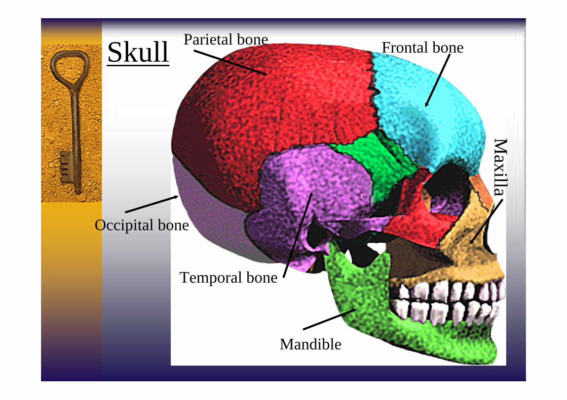
Skull Parietal bone Frontal bone
Occipital bone
Temporal bone
Mandible
Maxilla

Anatomy of Head continued♦Meninges
dura mater - outermost layer; connective tissue• bleeding between dura & skull are epidural bleeds• bleeding between dura & arachnoid space are
subdural bleeds
arachnoid membrane - suspends brain in cranial cavity; arachnoid space under membrane filled with cerebrospinal fluid (CSF)
• CSF provides cushioning & nutrients to brain• bleeding under this area are subarachnoid bleeds
pia mater - delicate tissue covering brain and spinal cord; highly vascular
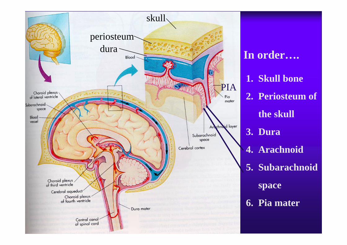
1. Skull bone
2. Periosteum of
the skull
3. Dura
4. Arachnoid
5. Subarachnoid
space
6. Pia mater
In order….
skull
periosteumdura
PIA
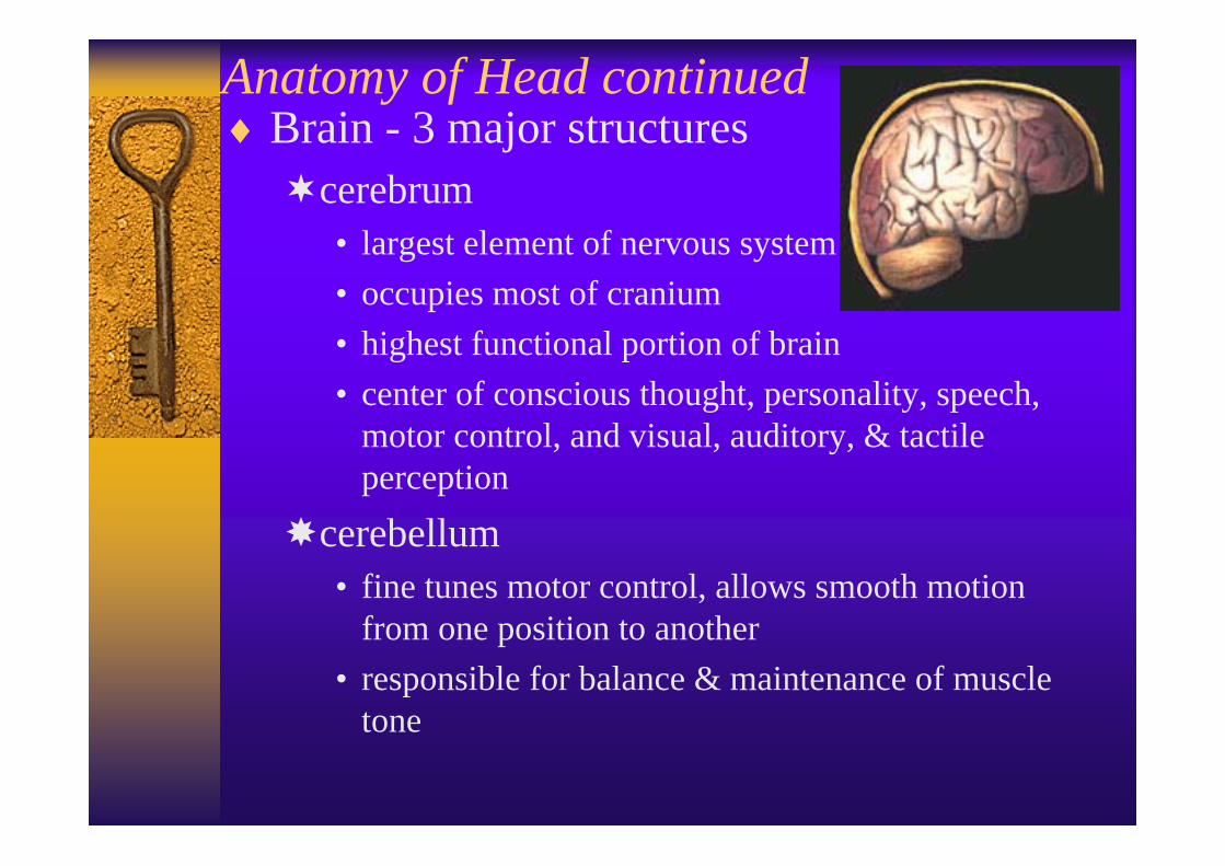
Anatomy of Head continued♦Brain - 3 major structures
cerebrum• largest element of nervous system• occupies most of cranium• highest functional portion of brain• center of conscious thought, personality, speech,
motor control, and visual, auditory, & tactile perception
cerebellum• fine tunes motor control, allows smooth motion
from one position to another• responsible for balance & maintenance of muscle
tone

Brainstem• central processing center &communication junction• midbrain
• hypothalamus• controls much of endocrine function,
vomiting reflex, hunger, thirst, kidney function, body temperature, emotions
• pons• medulla oblongata
• respiratory center (depth, rate, rhythm)• cardiac center (rate & strength of cardiac
contractions)• vasomotor center (control of distribution of
blood and maintenance of blood pressure)
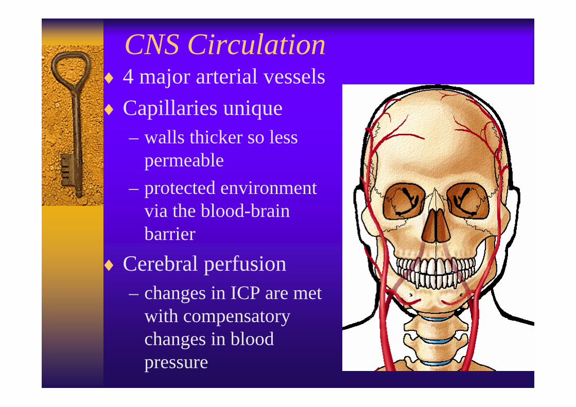
CNS Circulation♦ 4 major arterial vessels♦Capillaries unique
– walls thicker so less permeable
– protected environment via the blood-brain barrier
♦Cerebral perfusion– changes in ICP are met
with compensatory changes in blood pressure

Cerebral Perfusion Pressure♦ Intracranial pressure - pressure within cranium
– pressures within cranium create a natural resistance to control the amount of cerebral blood flow
– blood flow to the brain remains adequate as long as pressures within the cranium are appropriate
♦ 3 major cranial contents – brain, blood, & cerebrospinal fluid
♦Any changes in one of the 3 cranial contents is at the sacrifice to one of the others
♦When perfusion pressures drop, ICP rises to tryto maintain adequate cerebral perfusion

Cranial Nerves♦Cranial nerves are nerve roots that originate
in the cranium and along the brainstem♦ 12 distinct pathways known as CN I-XII
– control senses• smell; sight; touch; hearing; taste
– control the facial area• eye movement; facial muscle movement; chewing;
swallowing
– control significant body functions• monitors receptors in major blood vessels; major
nerve of parasympathetic nervous system (CN X - vagus nerve)

Form of Trauma: Blunt Trauma♦Blunt trauma - closed injury♦Transmission of energy causes damage to
tissues & organs beneath the skin♦True nature of injuries often hidden &
evidence of injury are often subtle♦Sources of blunt trauma
• MVC• falls• body to body contact• augmented forces (sticks, clubs)

Form of Trauma: Penetrating Trauma
♦Penetrating trauma - open wounds♦ Injuries influenced by degree of transfer of
kinetic energy & characteristic of the projectile
♦True knowledge of degree of bodily injury obtained after wound exploration
♦Sources of penetrating trauma –GSW, stabbings–bites - dog, human
Head Injuries♦Caused by blunt and penetrating forces♦Any injury above the level of the clavicles
is considered to involve the C-spine until proven otherwise
♦Repeated reassessments will be key in determining patient trends (VS, neuro signs)
♦Secondary insults - negative patient outcomes based on what we do or don’t do while caring for the patient– airway control, O2 therapy, fluids, c-spine
control, aspiration precautions
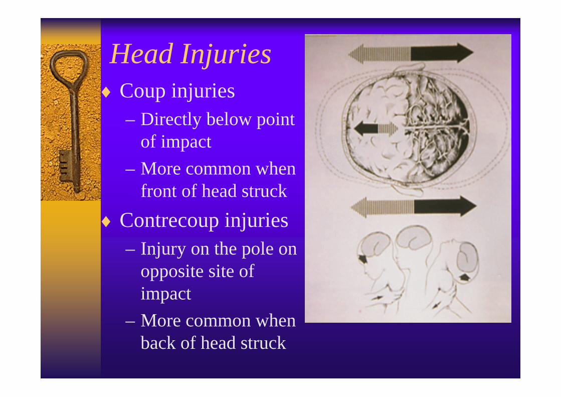
Head Injuries♦Coup injuries
– Directly below point of impact
– More common when front of head struck
♦Contrecoup injuries– Injury on the pole on
opposite site of impact
– More common when back of head struck

Levels of Head Injury♦Focal injury
– An identifiable site of injury limited to a particular area of the brainContusion
• blunt trauma• capillary bleeding into brain• often see prolonged confusion• neurological deficits related to site of injury
Intracranial hemorrhageepidural
– arterial bleed (often from artery in temporal area)– rapid build in intracranial pressure– quick onset altered level of consciousness

Focal Injuries continuedsubdural hematoma
• slow bleeding, usually venous• blood is above pia mater so do not get cerebral
irritation like in intracerebral hemorrhages• onset of signs & symptoms may be delayed for
hours or days• need to look for mechanism of injury; injury often
prior to day of patient interaction• increased incidence in elderly and chronic
alcoholism• reduced size of brain allows greater movement
of brain within the skull and increases the chance of injury & room to bleed
intracerebral hemorrhage• ruptured blood vessel within brain; local irritation

Levels of Head Injuries♦Diffuse axonal injury (DAI)
– Type of brain injury characterized by shearing, stretching or tearing of nerve fibers with subsequent axonal damage
– Axons are the communication pathways of nerve cells
– Injuries are spread over wider areas of the brain – More common with vehicular occupants and
pedestrians struck by vehicle due to acceleration/deceleration forces
– Injuries can range from mild to severe and life threatening

Diffuse Axonal Injury (DAI)Concussion
• Most common outcome of blunt trauma to the head• Nerve dysfunction without anatomical damage• Transient confusion, disorientation, amnesia of the
event• Management - quiet, calm atmosphere, constant
orientation, intact airway, adequate tidal volume
Moderate DAI• Accounts for 45% of all cases of DAI• Minute petechial bruising of brain tissue• May lead to unconsciousness• Commonly associated with basal skull fractures• Residual neurological impairment is common

Diffuse Axonal Injury (DAI)Moderate DAI continued
– Short and long term deficits• Immediate unconsciousness• Persistent confusion, disorientation • Retrograde amnesia - past memory affected• Anterograde amnesia - no memory of incident and
forward in time• Inability to concentrate• Frequent & significant mood swings & anxiety• Headache; other focal neurological deficits• Light sensitivity (photophobia)• Altered sense of smell and other senses

Diffuse Axonal Injury (DAI)Severe DAI
• Formerly called brain stem injury• Severe mechanical disruption of many axons in both
cerebral hemispheres and extending into brainstem• Accounts for 36% of all cases of DAI• Prolonged unconsciousness • Decorticate (flexion) or decerebrate (extension)
posturing common• Signs of ICP
– bradycardia, increasing B/P, altered respiratory pattern• High mortality rate• Significant neurological impairment for survivors

Intracranial Perfusion♦Brain has a high metabolic rate♦Brain needs constant fresh blood supply -
the brain has no stores of energy sources♦Brain consumes 20% of body’s oxygen♦Cranial volume fixed, does not vary
– 80% of the volume is the brain– 12% of the volume is blood flow– 8% of the volume is cerebrospinal fluid (CSF)
♦ Intracranial pressure (ICP) rises if any one of the cranial contents increases; an increase in one is at the sacrifice of another

ICP & Compensation♦ If a mass expands in the cranium, vessels are
compressed♦The next compensation is to push CSF out of
the cranium and into the spinal canal♦As ICP goes up, arterial blood flow is
restricted to reduce inflow of blood volume ♦ in cerebral blood flow rise in systemic
B/P to maintain cerebral perfusion ICP more resistance to cerebral blood flow more hypoxia, hypercarbia ( CO2) and
acidosis (unhealthy tissue/cell environment)

CO2 Levels and Head Injuries♦ CO2 level causes cerebral arteries to dilate
– blood flow volume is increased to the brain– increased volume of blood is detrimental– body’s response to try to lower CO2 is
hyperventilation & increasing B/P ♦Causes of or retained CO2 levels
– any thing that causes ineffective breathing (hypoventilation) causes CO2 to be retained• head injury with altered level of consciousness• drug and alcohol overdose• ineffective use of ambu bag

♦ CO2 level triggers cerebral arterial constriction– constriction minimizes blood flow to brain;
brain dependent on constant flow of oxygenated blood
– brain insult will develop due to lack of adequate blood flow from the vasoconstriction
♦Causes of or low levels of CO2– any thing that causes rapid breathing
(hyperventilation) causes CO2 to be blown off• from head injury reflex• overly aggressive use of ambu bag on patient

CO2 Levels continued♦ Major insults to brain occur in presence of low blood
pressure & poor ventilation – low B/P causes poor perfusion (hypoxia) & stimulates
anaerobic metabolism that results in acidosis – poor ventilation produces retained CO2 (acidosis) &
hypoxia– elevated levels of CO2 cause vasodilation which further
elevates intracranial pressure with increased blood flow
♦ Goal of respiratory care: keep CO2 levels normal by monitoring ETCO2
– immediate care provided after insult will positively or negatively affect outcome based on what is done or not done for the patient
– normal CO2 level is 35 - 45

Brain Stem Insults♦Upper brain stem
involvement– Cushing’s Triad: B/P rising;
pulse slowing; Cheyne-Stokes respirations
• alternating apnea/tachypnea
– Pupils small & reactive – Initially localizes pain &
tries to remove painful stimuli; then withdraws from pain; then flexed posturing (decorticate posturing - arms, wrists flexed & legs extended )
♦All effects reversible at this time

Middle Brain Stem Involvement– Widened pulse pressure (difference between
systolic & diastolic B/P) as systolic pressure increases
– Bradycardia (from head injury and not a diseased heart)
– Pupils nonreactive or sluggish bilaterally– Central neurogenic hyperventilation (CNH)
• respirations deep & rapid
– Extension posturing (decerebrate - rigid extension of arms & legs, backward arch of head)
– Few patients will be able to return to normal function once they reach this level of intracranial pressure

Lower Brainstem Involvement– Pupils dilated & unreactive– Respirations ataxic
(erratic, no pattern) or absent– Pulse rate often irregular
with great swings in rate– Flaccid; no response– EKG complex changes– High mortality rate for
patients who reach this level of function

Injuries of the Head & Neck♦Major concern will be airway patency♦Eye injury
– fracture - may entrap a nerve– hyphema - blood in anterior chamber, threat to
sight♦Nasal injury
– epistaxis may interfere with airway– swallowed blood can make a patient nauseated
♦Mandible injury– fracture and dislocation– immobility of jaw (watch airway); painful injury
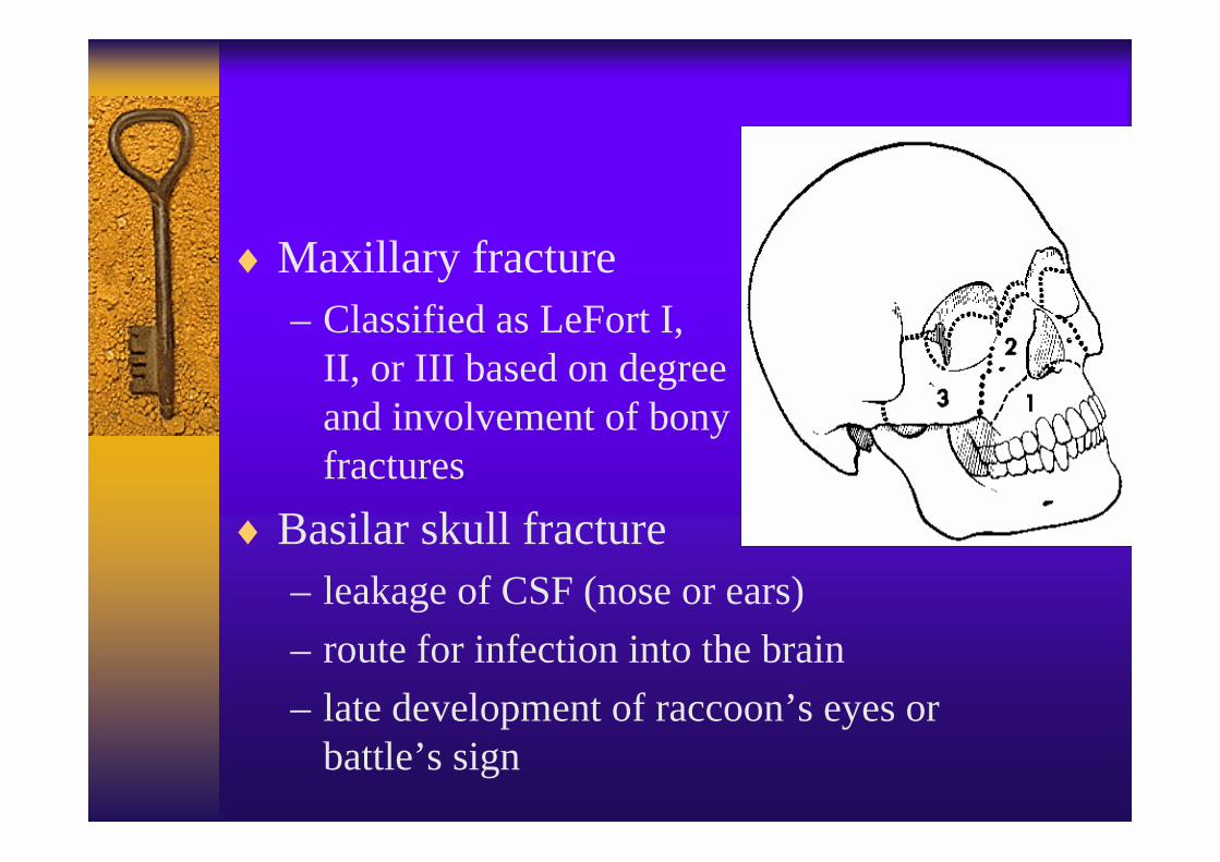
♦Maxillary fracture– Classified as LeFort I,
II, or III based on degree and involvement of bony fractures
♦Basilar skull fracture– leakage of CSF (nose or ears)– route for infection into the brain– late development of raccoon’s eyes or
battle’s sign

Soft Tissue Injury of Head & Neck♦Associated problems
– cosmetic importance of appearance– highly vascular region– potential for blood loss– airway involvement– potential for
hypoxia-induced secondary injury or insult
– potential for cervical spine injuries

Mechanisms of Spinal Injury♦Flexion - fall; MVC; diving♦Hyperextension - fall; MVC;
diving; football♦Flexion-rotation - fall;
tackled in football; MVC♦Compression - diving; fall
from height♦Distraction - hanging;
bungee jumping; clothesline♦Penetration - foreign object

Traumatic Spinal Cord Injury♦Cord transection
– Complete• All tracts of spinal cord completely disrupted• Cord-mediated functions below transection
permanently lost• Long term prognosis more accurately
determined at least 24 hours post injury– Incomplete
• Some tracts of spinal cord remain intact• Some cord-mediated functions intact• Function may be lost temporarily• Has potential for recovery

Spinal Cord Injury♦Cord transection
• Injury at cervical level – Quadriplegia– Loss of all normal function below injury site– Injuries from C3 to C5 increases risk for
respiratory paralysis due to involvement of phrenic nerve that is responsible for control of the diaphgram
• Injury below beginning of thoracic spine– Paraplegia– Loss of lower trunk function– Incontinence

Incomplete Spinal Cord Injuries♦Some spinal tracts remain; potential for
some recovery; 3 syndromes of injuryAnterior cord syndrome
• Bony fragments or pressure on spinal arteries
• Potential for recovery is poor• Loss of motor function and sensation to pain,
temperature and light touch• Likely to retain motion, positional, and
vibration sensation

Incomplete Spinal Cord Injuries
Central cord syndrome– Usually occurs with hyperextension of cervical
spine (ie: forward fall with facial impact)– Weakness/paresthesia upper extremities– Usually normal strength in lower extremities– Varying degrees of bladder function– Best prognosis for recovery of the 3 syndromes

Incomplete Spinal Cord InjuriesBrown-Sequard syndrome– Usually caused by penetrating injury affecting
one side of the cord (hemitransection) – Sensory and motor loss to same side of body
(ipsilateral) as the injury– Pain and temperature sensation lost on opposite
side of body (contralateral)– Injury rarest of the 3– May have some recovery

Neurogenic Shock♦Malfunction of autonomic nervous system
in regulating vessel tone & cardiac output♦Lack of sympathetic tone
– vasoconstriction limited so vessels dilate– reduced preload causes decrease in atrial filling
volume and weakens cardiac contractions– no release of epinephrine or norepinephrine
♦Assessment– normal skin color & temperature (warm & dry) – bradycardia (no catecholamines circulating)– hypotension (pooling of blood)– priapism

Treatment of Neurogenic Shock♦Airway control & supplemental O2
♦Spinal immobilization starting with manual control (document techniques/equipment used)
♦ IV - O2 - monitor♦Fluid bolus 20 ml/kg; reassess♦Dressings & splinting as needed and
potentially done enroute to the ED♦Watch for respiratory compromise due to loss
of phrenic nerve stimulation– adults with excessive belly breathing are using
alternate muscles to breathe and will tire & arrest

Non-traumatic Spinal Conditions♦ Low back pain
– 60 - 90 % of population have some form of low back pain• Affects men and women equally• Reported more commonly in women over 60 years
– Most causes of LBP are idiopathic• Precise diagnosis difficult to determine
– Affected area• Between lower rib cage and gluteal muscles• May radiate to thighs
– 1% of acute low back pain is sciatica• Usual cause is in lumbar nerve root• Pain accompanied by motor and sensory deficits
(ie: weakness) of lower extremities

Causes of Low Back Pain♦ Tension from tumors♦ Prolapsed disk♦ Bursitis♦ Synovitis♦ Rising venous
pressure♦ Tissue pressure from
degenerative joint disease (DJD)
♦ Abnormal bone pressure
♦ Problems with spinal mobility
♦ Inflammation from infection (osteomyelitis)
♦ Fractures♦ Ligament strains

Low Back Pain
♦Risk factors–Repetitious lifting–Vibrations from industrial machinery–Osteoporosis

Anatomical Considerations♦Pain from innervated
structures– Varies from person-to-
person♦Disk has no specific
innervation– Compresses cord if
herniated♦Pain in L-3,4,5 and S-1
may be interspinous bursae

Anatomical Considerations
♦Anterior and posterior longitudinal ligaments and other ligaments richly supplied with pain receptors
♦Muscles of spine vulnerable to sprains/strains

Degenerative Disk Disease♦Common over
age 50♦Causes
– Degeneration of disk•Biomechemical alterations of intervertebral disk
– Narrowing of disk•Results in variable segment stability

Spondylolysis♦Structural defect of
spine– Involves lamina or
vertebral arch♦Usually between
superior and inferior articulating facets
♦Heredity a significant factor
♦Rotational fractures common at affected site

Herniated Intervertebral Disk
♦Also called herniated nucleus pulposus
♦Tear in posterior rim of capsule enclosing the gelatinous center of the disk
Causes of Herniated Intervertebral Disk♦ Trauma♦ Degenerative disk
disease♦ Improper lifting
– Most common cause
♦ Men ages 30 - 50 most prone
♦ Commonly affects L-4, L-5, and S-1 disks
♦ May occur in C-5, C-6 and C-7

Spinal Cord Tumors
♦Problems noted:– Compression of cord– Degenerative changes in
bones/joints– Interruption of blood
supply♦Manifestations dependent
upon– Tumor type and location

Management of Non-traumatic Spinal Conditions♦Primarily palliative/supportive to decrease
pain from movement♦May elect to immobilize to aid in comfort
– Long back board - pad as needed– Vacuum type stretcher
♦Full spinal immobilization not required unless condition results from trauma– EMS will follow In-field Spinal Clearance
protocol to determine need for immobilization– ED walk-in may need immobilization

Assessment and Care of the Patient with Head and Neck Injuries

Trauma Patient Assessment♦Patients may present by private vehicle or
walk-in and not by EMS♦ED staff may adopt some or all of the
assessment steps used in a field assessment♦All assessments performed need to be a
systematic process used repeatedly by the individual– less likely to miss some detail– gives assessor a way to focus for the first few
minutes while gathering information

Trauma Patient Field Assessment♦Scene size-up - BSI, scene safety, determine
mechanism of injury, locate all patients♦Primary survey- initial assessment
– to identify immediate life threats– general impression, LOC (AVPU), ABC’s,
manual c-spine immobilization♦Decision: Is this critical? Interventions
needed right now including transport?♦Rapid trauma assessment head-to-toe or
focused if isolated injury♦A decision of when and where to transport to
made now if not done earlier

Trauma Patient Assessment cont’d♦Secondary survey
– Gather history (SAMPLE), GCS, vital signs• S - signs and symptoms• A - allergies• M- medications (prescription, over-the-counter,
herbal)• P - past pertinent medical history• L - last oral intake including food and water• E - events leading to the incident
– Pulse oximetry, ECG monitoring– If applicable: blood glucose level
♦Detailed assessment - head-to-toe again

♦Ongoing assessment - monitor for changes– will not be aware of patient deterioration unless
repeated reassessments are performed– document your findings – consider use of same rescuer for repeated
reassessment - will best pick up subtle changes– includes: vital signs, EKG monitor, pulse ox,
hands-on reassessment, asking the patient how they feel, reassessing any interventions already performed (ie: meds, fluids, splinting, dressings)

Region X Field Triage Criteria for Assessing Trauma Patients♦Criteria helps EMS determine transportation
of patient to Level I, II or closest hospital♦Evaluation of patient helps to determine
appropriate receiving facility– vital signs and level of consciousness– assessment for anatomy of injury– evaluation of mechanism of injury– assessment for co-morbid factors
♦ If Level I is >25 min away, transport to II♦No airway - transport to closest hospital

Ventilation Rates in Head Injuries♦ If rapid neurological deterioration of the
patient, the patient should be initially ventilated with BVM– adult (>8 years old) 20 bpm (every 3 seconds)– children (1-8 years old) 30 bpm (every 2 seconds)– infants (<1 years old) 35 bpm (every 1.7 seconds)
♦Avoid hyperventilation at higher rates♦Consider conscious sedation intubation♦ If seizure activity, give valium 5 mg IVP or
10 mg IM/rectally. May repeat to 10 mg max

Neurological assessmentAVPU - evaluates mental status
• alert meaning awake (may be oriented or confused)• responds to verbal prompts (includes moaning)• responds only to painful stimuli (may be to light
touch and not necessarily something painful)• unresponsive - comatose; absolutely no responses
glasgow coma scale (GCS)• evaluates level of consciousness
pupillary reaction• eyes are specialized tissue• eyes indicate problems with 4 cranial nerves• reflect adequacy of perfusion of cerebral blood flow
- perfusion and the eyes lose their luster

Glasgow Coma Scale - GCS♦Scale that awards points based on patient’s
best responsesmodified for developmental age
♦Moderately good predictor of head injury severity
♦Total score ranges 3-1513-15 - mild head injury9-12 - moderate head injury<8 - severe head injury (patient usually in coma)
♦Note differences right side to left side and upper versus lower extremities

Glasgow Coma Scale♦ Eye Opening
– spontaneous 4– to voice 3– to pain 2– none 1
♦ Verbal response– oriented 5– confused 4– inappropriate
words 3– incomprehensible
words 2– none 1
♦ Motor response– obeys commands 6– purposeful movement
to pain 5– withdraws to pain 4– abnormal flexion 3– abnormal extension 2– none 1

GCS Pearls & Pitfalls♦Eye opening
– don’t touch patient before calling their name -you will not be able to determine if they are responding to voice (3) or to touch (pain - 2)
♦Verbal response– inappropriate words (3) are beyond confusion (4)– muttering is incomprehensible words (2)
♦Motor response– purposeful is the patient pulling at what annoys
them (B/P cuff, cervical collar) (5)– withdrawal is trying to move away from pain &
annoyance (4)

Glasgow Coma Scale - GCS♦Per Region X SOP’s, EMS is to do GCS on
all patients♦CMC patient care run report provides space
to document two GCS scores– additional assessments would be in the
comments♦Components should be assessed and results
should be available at the time of the first radio contact to medical control
♦Components or the total score may be given during the radio report

Glasgow Coma Scale
♦EMS will not normally calculate the RTS (revised trauma score)
♦EMS will provide the components of the RTS in report for the ECRN to do the calculation– Glasgow coma scale score– systolic blood pressure– respiratory rate
In-field Spinal Clearance♦When in doubt, fully immobilize the patient♦EMS will evaluate:
mechanism of injurysigns & symptomspatient reliability
♦No spinal immobilization needed if:negative mechanism of injuryno neurological signs or symptomspatient is reliable
♦Spinal clearance is not a priority but restricting spinal motion is

Spinal Immobilization Required Related to Mechanism of Injury♦ High velocity MVC > 40mph♦ Unrestrained occupant in MVC♦ Passenger compartment intrusion > 12″♦ Ejection from vehicle♦ Rollover MVC♦ Motorcycle collision > 20mph♦ Death in same vehicle♦ Pedestrian struck by vehicle♦ Falls > 2 times patient’s height♦ Diving injury

Spinal Immobilization Required Related to Signs and Symptoms ♦Pain in neck or spine♦Tenderness/deformity of neck or spine upon
palpation♦Paralysis or abnormal motor exam♦Paresthesia (pins & needles) in extremities♦Abnormal response to painful stimuli

Spinal Immobilization Required Related to Patient Reliability♦Signs of intoxication♦Abnormal mental status♦Communication difficulty♦Abnormal stress reaction
∗ When in doubt, fully immobilize

Spinal Immobilization♦Cervical collars
– limit flexion, extension, & lateral movements– must be combined with additional pieces of
equipment to be effective– start with manual stabilization, neutral position
with eyes forward– do not move neck if movement:
• increases muscle spasms• neck pain increases• neurological deficits are aggravated• airway becomes compromised

Measuring C-Collar Sizes♦Measure with fingers held horizontally and
tucked in tight at base of neck (top of shoulder) to horizontal line drawn even with bottom of chin
♦Size the collar from bottom of the rigid plastic edge (not the foam edge)
♦Find window closest to top of your fingers♦Adjust sizing and snap to lock collar into
place♦ If a collar is too short it causes flexion♦ If a collar is too tall it causes extension

Cervical Collars♦ It is rare for the patient
to be sized a no-neck ♦ If the majority of your
patients are being sized as no-necks, then measurements are probably not accurate!!!
♦Directions are printed on the collars if you need a reminder

Conscious Sedation♦Procedure performed when the airway
needs to be secured and the patient is not in full arrest (inadequate airway; aspiration risk; GCS <8)– Note: not all patients with a GCS <8 need to be
intubated in the field or the ED; evaluate each individual situation (ie: patient with a GCS <8 under the influence of alcohol does not necessarily get intubated!)
♦Conscious Sedation can be utilized for trauma & medical patients (ie: stroke)

Conscious Sedation cont’d♦Contraindications - EMS to call medical
control if they feel need to intubate exists but a contraindication is present:– coma– B/P < 100 mmHg– known hypersensitivity/allergy to meds used– age < 13
♦Need to weigh the risks versus the benefits of spending extra time in the field to administer medications and perform this invasive procedure

Conscious Sedation Meds♦Lidocaine
– 1.5 mg/kg IVP bolus (no drip) to suppress cough reflex in head injured/insulted patient (ie: trauma and stroke)
• coughing increases intracranial pressures• can be given in presence of bradycardia because the
bradycardia is due to brain irritation versus sick heart
♦Morphine– given for relief of pain & reduce anxiety– 2 mg slow IVP for pain; repeat 2 mg every 3
minutes up to maximum of 10 mg– monitor for hypotension & resp depression

Conscious Sedation Medications♦Versed
– 2 mg slow IVP for sedation & amnesia– repeat 2 mg every minute until sedated-max 10mg– does not take away any pain sensations– need to call medical control for more versed to
maintain sedation if needed after intubation♦Benzocaine
– 1-2 short sprays using long red nozzle to spray back of throat
– suppresses the gag reflex– gagging stimulates vagus nerve (bradycardia) &
increases potential for vomiting

In-line Intubation♦Procedure performed to secure the airway if
neck injury is suspected♦Best when accomplished with 2 persons♦One person secures manual control of head♦ Intubator must position their body to be in-
line with anatomical structures– crouching down and leaning backwards– lying on belly; sitting on buttocks works in the
field♦ET tube position confirmed and secured in
normal manner

In-line Intubation continued♦Confirming ET tube placement
– direct visualization– 5 point auscultation (epigastric area, bilateral
upper lobes, lateral chest area bilaterally)– chest rise and fall– ETCO2 confirmation (yellow)– EDD if ETCO2 not definitive
♦ET tube position confirmed every time the patient is moved; document confirmation
♦Securing ET tube– collar patient to minimize/prevent head
movement which may move distal tip ET tube

Care of Soft Tissue Injuries ♦Dislodged/knocked out tooth
– gently rinse off gross contaminant with saliva or sterile saline
– only handle tooth by the crown– do not allow tooth to dry out
• transport tooth moist - best solution is in milk; can be covered with patient’s saliva or sterile saline gauze
• milk is used only if it were readily available at the scene
– tooth can be replaced into socket facing the correct way if airway will not be compromised
– referral to dentist important (< 2 hours)

Soft Tissue Injuries
♦Open neck wounds– risk of airway compromise due to injury and
swelling– risk of blood loss because area is vascular– risk of air embolism into open blood vessel– wounds must be immediately covered with
occlusive dressing– observe for changes in voice due to swelling
and any dyspnea– stabilize impaled objects in place

Pearls and Pitfalls of Head & Neck Injuries♦Any injury above the level of the clavicles
is considered to have a spinal injury until proven otherwise
♦Additional associated injuries to watch for– Airway compromise
• open airway using jaw thrust maneuver• intubation via in-line technique
– Brain injury• address hypoxia
– Dental trauma or avulsion - airway compromise
Distractions♦Evidence of alcohol (ETOH) on board
– Can make it difficult to determine true cause of altered level of consciousness
– Patient will often be uncooperative – EMS and ED will be challenged to do the right
thing and protect the patient from harming himself further• will most likely need longer manual control
of c-spine than usual– Remember to check blood sugar levels

Helmets♦Purpose of helmet
– protect head– protect brain– cervical spine remains
vulnerable♦Types of helmets
– Full face or open face• motorcycle,bicycle, roller blade
– Sports helmet• football, motor-cross

Helmets♦Helmet removal controversy: Scene vs
hospital– Priorities for rapid/early removal
• Airway management• Difficult spinal immobilization
– Determining factors for immediate removal• Helmet prevents airway care needed immediately• Presence of airway or breathing problems• Helmet does not immobilize head within• Inability to immobilize the helmet to long board• Helmet prevents assessment of anticipated injuries• Helmet removal will not compromise patient status

Helmets
♦Other considerations– Ready access of athletic trainer
• Need for special equipment to remove face piece
– Presence of garb such as shoulder pads• May compromise the cervical spine if only helmet
removed - additional space under head will need to be padded to avoid neck extension
– Firm fit of helmet may provide firm support for head
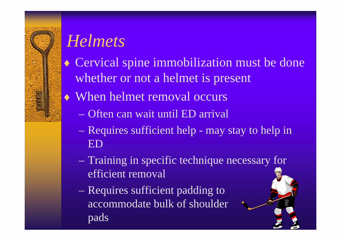
Helmets♦Cervical spine immobilization must be done
whether or not a helmet is present♦When helmet removal occurs
– Often can wait until ED arrival– Requires sufficient help - may stay to help in
ED– Training in specific technique necessary for
efficient removal– Requires sufficient padding to
accommodate bulk of shoulder pads

Helmet Removal♦Takes a minimum of 2 people♦Cut away or remove as much additional
pieces as possible (strap, face mask, visor)♦One person slides hands under helmet to
support occiput and immobilize head♦Second person spreads helmet laterally to
clear ears, then rotates helmet to clear chin, occiput, nose, and brow
♦First person needs to be sliding hands to constantly be supporting occiput as helmet is removed

Abbreviated Radio Report♦ In situations where manpower is limited and
the patient’s condition is critical, EMS should provide to the ED:– provider’s name, vehicle ID, and include name of
receiving hospital you are talking to– nature of situation & protocol you are following– age, sex, chief complaint; brief history of present
illness/injury– airway & vascular access status– current vital signs– major interventions completed or attempted– ETA

GCS Review - You Score The Pt♦Patient responds to their name being called
√ eye opening to voice - 3 points♦Patient asks repetitively “what happened”
√ verbal response confused - 4 points♦Patient obeys commands
√motor response - 6 points♦Total GCS - 13♦Needs to be watched for change in level of
consciousness & worsening condition

GCS Review - You Score The Pt♦Patient must be shook to respond to EMS;
flutters eyelids when touched√ eye opening is “to pain” - 2 points
♦Patient muttering words but not appropriate to the situation√ verbal response is inappropriate - 3 points
♦Patient is trying to pull off cervical collar and rip off blood pressure cuff√motor response is to purposeful
movement; patient knows what is bothering them - 5 pointsTotal 10 points
GCS Review - You Score The Pt♦Patients eyelids flutter when they are given a
sternal rub√ eye opening is to pain - 2 points
♦Patient mutters & moans when stimulated√ verbal response is incomprehensible - 2 pts
♦Patient pulls away when arm is touched to start an IV or take a B/P√motor response is withdrawal - 4 points
♦Total GCS is 8 points♦Need to consider airway protection-intubation

Documentation♦Any patient with an altered level of
consciousness must have a documented blood glucose level
♦Assess for and document a GCS (EMS does GCS on all patients)– guideline reminder on back side of run report
♦Neurological assessment includes:– level of consciousness (blood sugar if altered)– GCS– pupillary response– movement & sensory - right compared to left
and upper compared to lower extremities