heart-rate and ekg monitor using the msp430fg439 (rev. b)
TRANSCRIPT

1SLAA280B–October 2005–Revised March 2018Submit Documentation Feedback
Copyright © 2005–2018, Texas Instruments Incorporated
Heart-Rate and EKG Monitor Using the MSP430FG439
Application ReportSLAA280B–October 2005–Revised March 2018
Heart-Rate and EKG Monitor Using the MSP430FG439
Murugavel Raju .................................................................................................................... MSP430
ABSTRACTThis application report describes how to build a digital heart-rate monitor using a MSP430FG439microcontroller (MCU). The heartbeat rate per minute is displayed on an LCD. In addition, the applicationoutputs a digital data stream through an RS232 serial port to allow display of the EKG waveform on a PC.The entire application runs using a CR2032 3-V lithium battery.
PCB Gerber files, schematic, bill of materials, and firmware are provided at www.ti.com/lit/zip/SLAA280.
This application report uses the MOD-EKG Heart-Rate and EKG Monitor Using the MSP430FG439development board from Olimex.
Contents1 Introduction ................................................................................................................... 12 Circuit Description ........................................................................................................... 2
2.1 Front-End Amplifier ................................................................................................ 22.2 EKG Amplifier ....................................................................................................... 3
3 Signal Processing and Heartbeat Detection ............................................................................. 43.1 EKG Sampling....................................................................................................... 43.2 Filtering Line-Frequency Noise ................................................................................... 53.3 Detecting QRS Complexes ........................................................................................ 53.4 FIR Filters............................................................................................................ 63.5 Calculating Heartbeat Rate........................................................................................ 7
4 Software....................................................................................................................... 75 Testing the Application ...................................................................................................... 8
5.1 PC Scope EKG Display ............................................................................................ 96 References .................................................................................................................. 11
TrademarksMSP430 is a trademark of Texas Instruments.IAR Embedded Workbench is a registered trademark of IAR Systems.All other trademarks are the property of their respective owners.
1 IntroductionAn electrocardiogram (ECG), also called an EKG, is a graphic tracing of the voltage generated by thecardiac or heart muscle during a heartbeat. It provides very accurate evaluation of the performance of theheart.
The heart generates an electrochemical impulse that spreads out in the heart in such a fashion as tocause the cells to contract and relax in a timely order and, thus, give the heart a pumping characteristic.This sequence is initiated by a group of nerve cells called the sinoatrial (SA) node, resulting in apolarization and depolarization of the cells of the heart. Because this action is electrical in nature and thebody is conductive with its fluid content, this electrochemical action can be measured at the surface of thebody.

1 mV
P
Q
R
S
T
TIME
ATRIA
DEPOLARIZE
VENTRICLES
DEPOLARIZE
VENTRICLES
REPOLARIZE
DIFFERENTIAL VOLTAGEBETWEEN TWO ELECTRODES
Circuit Description www.ti.com
2 SLAA280B–October 2005–Revised March 2018Submit Documentation Feedback
Copyright © 2005–2018, Texas Instruments Incorporated
Heart-Rate and EKG Monitor Using the MSP430FG439
An actual voltage potential of approximately 1 mV develops between various body points. This can bemeasured by placing electrode contacts on the body. The four extremities and the chest wall havebecome standard sites for applying the electrodes. Standardizing electrocardiograms makes it possible tocompare them as taken from person to person and from time to time from the same person. The normalelectrocardiogram shows typical upward and downward deflections that reflect the alternate contraction ofthe atria (the two upper chambers) and of the ventricles (the two lower chambers) of the heart.
A typical single cardiac cycle waveform of a normal heartbeat is shown in Figure 1. The voltages producedrepresent pressures exerted by the heart muscles in one pumping cycle. The first upward deflection, P, isdue to atria contraction and is known as the atrial complex. The other deflections, Q, R, S, and T, are alldue to the action of the ventricles and are known as the ventricular complexes. Any deviation from thenorm in a particular electrocardiogram is indicative of a possible heart disorder.
In this application report, the EKG waveform is used by the MCU to measure the heartbeat rate. Becauseheartbeat calculation is the major focus, the electrodes are simplified to two connections, one to a rightarm and the other to the left arm. This type of setup can be frequently seen in exercise machines such astreadmills.
Figure 1. A Typical EKG Waveform
2 Circuit DescriptionFigure 9 shows the complete schematic diagram of this application.
2.1 Front-End AmplifierThe electrical signal derived from the electrodes is typically 1 mV peak-peak. An amplification of about1000× is necessary to make this signal usable for heart-rate detection. Realizing clean amplification of theEKG signal with such high gain is no easy task given that the human body acts as a huge antenna thatpicks up a lot of noise, including a dominant 50 to 60-Hz line-frequency noise. This noise must be filteredby a strong post filter after amplification. Unfortunately, any amplification amplifies the noise voltages inaddition to the desired EKG signal. In certain situations, the noise can completely override the EKG andrender the amplified signal useless.

LEFTELECTRODE
RIGHTELECTRODE
100k
2M
100k
2M
INA321EA
−
+
DAC112-bit DAC
OA1OOA1
0.1µF 1M
OA1IO
820 1M
OA0I1
−
+
OA0
10k
1M
4.7nF
OA0O
OA0I0
www.ti.com Circuit Description
3SLAA280B–October 2005–Revised March 2018Submit Documentation Feedback
Copyright © 2005–2018, Texas Instruments Incorporated
Heart-Rate and EKG Monitor Using the MSP430FG439
A better approach is to use a differential amplifier. Thanks to the identical common mode signals from theEKG pick up electrodes, the common mode noise is automatically cancelled out using an ideally matcheddifferential amplifier.
The differential amplifier used in the front end of this application is an INA321 instrumentation amplifierthat has perfectly matched and balanced integrated gain resistors. This device is specified to operate witha minimum of 2.7-V single-rail power supply. The INA321 provides a fixed amplification of 5× for the EKGsignal. With its CMRR specification of 94 dB extended up to 3 kHz, the INA321 rejects the common-modenoise signals including the line frequency and its harmonics. The quiescent current of the INA321 is40 µA, and the shutdown mode current is less than 1 µA.
2.2 EKG AmplifierThe EKG signal at the output of INA321 is further amplified by OA0, one of the three integratedoperational amplifiers in the MSP430FG439 MCU. Figure 2 shows the circuit diagram for the EKGamplifier front end.
Figure 2. EKG Amplifier Circuit Diagram
Signal Processing and Heartbeat Detection www.ti.com
4 SLAA280B–October 2005–Revised March 2018Submit Documentation Feedback
Copyright © 2005–2018, Texas Instruments Incorporated
Heart-Rate and EKG Monitor Using the MSP430FG439
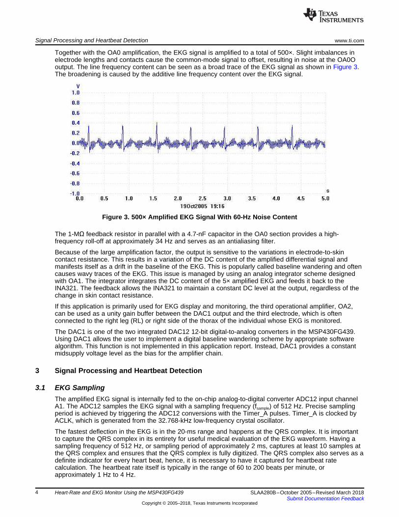
Together with the OA0 amplification, the EKG signal is amplified to a total of 500×. Slight imbalances inelectrode lengths and contacts cause the common-mode signal to offset, resulting in noise at the OA0Ooutput. The line frequency content can be seen as a broad trace of the EKG signal as shown in Figure 3.The broadening is caused by the additive line frequency content over the EKG signal.
Figure 3. 500× Amplified EKG Signal With 60-Hz Noise Content
The 1-MΩ feedback resistor in parallel with a 4.7-nF capacitor in the OA0 section provides a high-frequency roll-off at approximately 34 Hz and serves as an antialiasing filter.
Because of the large amplification factor, the output is sensitive to the variations in electrode-to-skincontact resistance. This results in a variation of the DC content of the amplified differential signal andmanifests itself as a drift in the baseline of the EKG. This is popularly called baseline wandering and oftencauses wavy traces of the EKG. This issue is managed by using an analog integrator scheme designedwith OA1. The integrator integrates the DC content of the 5× amplified EKG and feeds it back to theINA321. The feedback allows the INA321 to maintain a constant DC level at the output, regardless of thechange in skin contact resistance.
If this application is primarily used for EKG display and monitoring, the third operational amplifier, OA2,can be used as a unity gain buffer between the DAC1 output and the third electrode, which is oftenconnected to the right leg (RL) or right side of the thorax of the individual whose EKG is monitored.
The DAC1 is one of the two integrated DAC12 12-bit digital-to-analog converters in the MSP430FG439.Using DAC1 allows the user to implement a digital baseline wandering scheme by appropriate softwarealgorithm. This function is not implemented in this application report. Instead, DAC1 provides a constantmidsupply voltage level as the bias for the amplifier chain.
3 Signal Processing and Heartbeat Detection
3.1 EKG SamplingThe amplified EKG signal is internally fed to the on-chip analog-to-digital converter ADC12 input channelA1. The ADC12 samples the EKG signal with a sampling frequency (fsample) of 512 Hz. Precise samplingperiod is achieved by triggering the ADC12 conversions with the Timer_A pulses. Timer_A is clocked byACLK, which is generated from the 32.768-kHz low-frequency crystal oscillator.
The fastest deflection in the EKG is in the 20-ms range and happens at the QRS complex. It is importantto capture the QRS complex in its entirety for useful medical evaluation of the EKG waveform. Having asampling frequency of 512 Hz, or sampling period of approximately 2 ms, captures at least 10 samples atthe QRS complex and ensures that the QRS complex is fully digitized. The QRS complex also serves as adefinite indicator for every heart beat, hence, it is necessary to have it captured for heartbeat ratecalculation. The heartbeat rate itself is typically in the range of 60 to 200 beats per minute, orapproximately 1 Hz to 4 Hz.
www.ti.com Signal Processing and Heartbeat Detection
5SLAA280B–October 2005–Revised March 2018Submit Documentation Feedback
Copyright © 2005–2018, Texas Instruments Incorporated
Heart-Rate and EKG Monitor Using the MSP430FG439
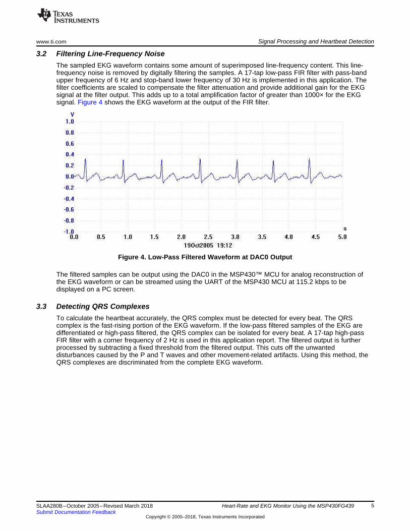
3.2 Filtering Line-Frequency NoiseThe sampled EKG waveform contains some amount of superimposed line-frequency content. This line-frequency noise is removed by digitally filtering the samples. A 17-tap low-pass FIR filter with pass-bandupper frequency of 6 Hz and stop-band lower frequency of 30 Hz is implemented in this application. Thefilter coefficients are scaled to compensate the filter attenuation and provide additional gain for the EKGsignal at the filter output. This adds up to a total amplification factor of greater than 1000× for the EKGsignal. Figure 4 shows the EKG waveform at the output of the FIR filter.
Figure 4. Low-Pass Filtered Waveform at DAC0 Output
The filtered samples can be output using the DAC0 in the MSP430™ MCU for analog reconstruction ofthe EKG waveform or can be streamed using the UART of the MSP430 MCU at 115.2 kbps to bedisplayed on a PC screen.
3.3 Detecting QRS ComplexesTo calculate the heartbeat accurately, the QRS complex must be detected for every beat. The QRScomplex is the fast-rising portion of the EKG waveform. If the low-pass filtered samples of the EKG aredifferentiated or high-pass filtered, the QRS complex can be isolated for every beat. A 17-tap high-passFIR filter with a corner frequency of 2 Hz is used in this application report. The filtered output is furtherprocessed by subtracting a fixed threshold from the filtered output. This cuts off the unwanteddisturbances caused by the P and T waves and other movement-related artifacts. Using this method, theQRS complexes are discriminated from the complete EKG waveform.

0 0.05 0.1 0.15 0.2 0.25
70
75
80
85
90
95
100
105
Frequency (kHz)
Mag
nitu
de (
dB)
Magnitude Response (dB)
Signal Processing and Heartbeat Detection www.ti.com
6 SLAA280B–October 2005–Revised March 2018Submit Documentation Feedback
Copyright © 2005–2018, Texas Instruments Incorporated
Heart-Rate and EKG Monitor Using the MSP430FG439
Figure 5 shows the signal that is output from the QRS discriminator to the input of the heartbeat detectionand heart-rate calculation algorithm.
Figure 5. Discriminated QRS Waveform
3.4 FIR FiltersLinear phase symmetrical FIR filters are used in this application. Using symmetrical FIR filters reduces thedemand on math multiplication operations to one half, because of the symmetrical nature of the filtercoefficients. The filter results are rounded to 16 bits.
Figure 6 shows the magnitude versus frequency response curve for the low-pass filter used in thisapplication. Note the amplification provided by the filter. This is achieved by multiplying all the coefficientsby a constant.
Figure 6. 17-Tap FIR Low-Pass Filter Magnitude vs Frequency Response

0 0.05 0.1 0.15 0.2 0.25
78
80
82
84
86
88
90
92
Frequency (kHz)
Mag
nitu
de (
dB)
Magnitude Response (dB)
www.ti.com Signal Processing and Heartbeat Detection
7SLAA280B–October 2005–Revised March 2018Submit Documentation Feedback
Copyright © 2005–2018, Texas Instruments Incorporated
Heart-Rate and EKG Monitor Using the MSP430FG439
Figure 7 shows the magnitude versus frequency response curve for the high-pass filter used in thisapplication. The filter coefficients were calculated using ScopeFIR, a filter designing and analyzingsoftware tool from Iowegian International. Any other filter design tool, including MATLAB, can be used fordesigning the filters and calculating the coefficients.
Figure 7. 17-Tap FIR High-Pass Filter Magnitude vs Frequency Response
3.5 Calculating Heartbeat RateThe number of heart beats per minute is calculated using a three-beat average. Two variables in the Cmain function, counter and pulseperiod, accurately track the time scale. Each output sample from the QRSdiscriminator is compared against a set threshold to detect the presence of a beat. Pulseperiod isincremented by one during every sample period. Because each sample occurs every 1/512 second, it iseasy to track the time scale based on the number of counts in the pulseperiod variable. A 128-sampletime window is used as a debounce time using counter. Every time a beat is detected, counter is resetand the LCD icon with four arrows is turned on to represent the heart beat. If a beat is not detected for128 consecutive samples, a separation between successive beats is identified and the LCD icon with fourarrows is turned off.
The pulseperiod is accumulated for three consecutive beats. On the third beat, pulseperiod is used for thecalculation of heart-rate per minute and reset.Heartbeat rate per minute = 1 / [pulseperiod / (3 × 512 × 60)] = 92160 / pulseperiod (1)
4 SoftwareThe software for this application is written in C using IAR Embedded Workbench® IDE KIckstart Edition.The source code for reproducing this application is provided in an accompanying zip file. The softwareuses a dedicated 16-×16-bit signed multiply routine written in assembly language for faster execution ofthe FIR filter calculations compared to the native C math library multiplication function. This function iscalled from the main C program using the syntax long mul16(register int x, register int y).
Three C source files ("Heart rate.c", "Heart rate with DAC output.c", and "Heart rate with EKG Demo.c")are provided in the zip file. The names of these files signify their functionality.
The project must include the C source file as per the required functionality and the mul.s43 assemblersource file for proper compilation.
The memory usage for the complete heart-rate with EKG project is 1168 bytes of code memory, 225 bytesof data memory, and 64 bytes of const memory. This is approximately one-fourth of the 4KB limit of thefree C compiler in the IAR Embedded Workbench Kickstart edition.

Testing the Application www.ti.com
8 SLAA280B–October 2005–Revised March 2018Submit Documentation Feedback
Copyright © 2005–2018, Texas Instruments Incorporated
Heart-Rate and EKG Monitor Using the MSP430FG439
The CPU runs at 2.097152 MHz using the FLL to source MCLK. The entire EKG program, including theFIR filters, QRS detection, and heart-rate calculation, uses approximately 1 MIPS of the CPU bandwidth.
5 Testing the ApplicationTwo square pads, one on the top layer and the other on the bottom layer of the double sided PCB, areprovided on either side of the LCD to serve as right and left hands contact electrodes. When in use, thepower jumper PWR must be installed, and the board must be held using both hands by placing the thumband index fingers of each hand on the square pads. Care must be taken not to touch any other electricalareas of the PCB. A good way is to keep the hold towards the edges of the board. The contact resistancebetween the fingers and the square pads must be low for good signal quality. A little bit of moisturizerspread and rubbed over the fingers helps users with dry skin. Figure 8 shows the picture of the EKGboard in action.
Figure 8. Picture of EKG Board in Action
NOTE: This application design is published for reference purpose only and is not intended for anylife-saving or medical monitoring use.
www.ti.com Testing the Application
9SLAA280B–October 2005–Revised March 2018Submit Documentation Feedback
Copyright © 2005–2018, Texas Instruments Incorporated
Heart-Rate and EKG Monitor Using the MSP430FG439
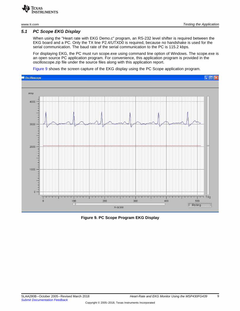
5.1 PC Scope EKG DisplayWhen using the "Heart rate with EKG Demo.c" program, an RS-232 level shifter is required between theEKG board and a PC. Only the TX line P2.4/UTXD0 is required, because no handshake is used for theserial communication. The baud rate of the serial communication to the PC is 115.2 kbps.
For displaying EKG, the PC must run scope.exe using command line option of Windows. The scope.exe isan open source PC application program. For convenience, this application program is provided in theoscilloscope.zip file under the source files along with this application report.
Figure 9 shows the screen capture of the EKG display using the PC Scope application program.
Figure 9. PC Scope Program EKG Display

Testing the Application www.ti.com
10 SLAA280B–October 2005–Revised March 2018Submit Documentation Feedback
Copyright © 2005–2018, Texas Instruments Incorporated
Heart-Rate and EKG Monitor Using the MSP430FG439
Figure 10. Schematic Diagram

www.ti.com References
11SLAA280B–October 2005–Revised March 2018Submit Documentation Feedback
Copyright © 2005–2018, Texas Instruments Incorporated
Heart-Rate and EKG Monitor Using the MSP430FG439
6 References• MSP430x4xx Family User’s Guide• MSP430FG43x Mixed-Signal Microcontrollers data sheet• INA321 microPower Single-Supply CMOS Instrumentation Amplifier data sheet• Digital Filters Design• Introduction to Medical Electronics, Burton R. Klein

Revision History www.ti.com
12 SLAA280B–October 2005–Revised March 2018Submit Documentation Feedback
Copyright © 2005–2018, Texas Instruments Incorporated
Revision History
Revision HistoryNOTE: Page numbers for previous revisions may differ from page numbers in the current version.
Changes from September 26, 2007 to March 9, 2018 ..................................................................................................... Page
• Editorial updates throughout ............................................................................................................. 1• Changed frequency from 250 Hz to 34 Hz in the paragraph that begins "The 1-MΩ feedback resistor..." in Section 2.2,
EKG Amplifier.............................................................................................................................. 4

IMPORTANT NOTICE AND DISCLAIMER
TI PROVIDES TECHNICAL AND RELIABILITY DATA (INCLUDING DATASHEETS), DESIGN RESOURCES (INCLUDING REFERENCEDESIGNS), APPLICATION OR OTHER DESIGN ADVICE, WEB TOOLS, SAFETY INFORMATION, AND OTHER RESOURCES “AS IS”AND WITH ALL FAULTS, AND DISCLAIMS ALL WARRANTIES, EXPRESS AND IMPLIED, INCLUDING WITHOUT LIMITATION ANYIMPLIED WARRANTIES OF MERCHANTABILITY, FITNESS FOR A PARTICULAR PURPOSE OR NON-INFRINGEMENT OF THIRDPARTY INTELLECTUAL PROPERTY RIGHTS.These resources are intended for skilled developers designing with TI products. You are solely responsible for (1) selecting the appropriateTI products for your application, (2) designing, validating and testing your application, and (3) ensuring your application meets applicablestandards, and any other safety, security, or other requirements. These resources are subject to change without notice. TI grants youpermission to use these resources only for development of an application that uses the TI products described in the resource. Otherreproduction and display of these resources is prohibited. No license is granted to any other TI intellectual property right or to any thirdparty intellectual property right. TI disclaims responsibility for, and you will fully indemnify TI and its representatives against, any claims,damages, costs, losses, and liabilities arising out of your use of these resources.TI’s products are provided subject to TI’s Terms of Sale (www.ti.com/legal/termsofsale.html) or other applicable terms available either onti.com or provided in conjunction with such TI products. TI’s provision of these resources does not expand or otherwise alter TI’s applicablewarranties or warranty disclaimers for TI products.
Mailing Address: Texas Instruments, Post Office Box 655303, Dallas, Texas 75265Copyright © 2019, Texas Instruments Incorporated