heart type fatty acid-binding protein
TRANSCRIPT

Heart-type Fatty Acid-Binding ProteinBiomarker of myocardial ischemia
2 www.randox.com/cardiology
H-FABP: The Protein
• Heart-type Fatty Acid Binding Protein (H-FABP) is an unbound, low molecular weight protein, located in the cytoplasm of cardiac myocytes.1
• The molecular weight is only 15kDa – smaller than Myoglobin (18kDa), Troponin I (22kDa), Troponin T (37kDa) and CK-MB (86kDa).
• The function of H-FABP is in the intracellular uptake of long chain fatty acids in the myocardium.
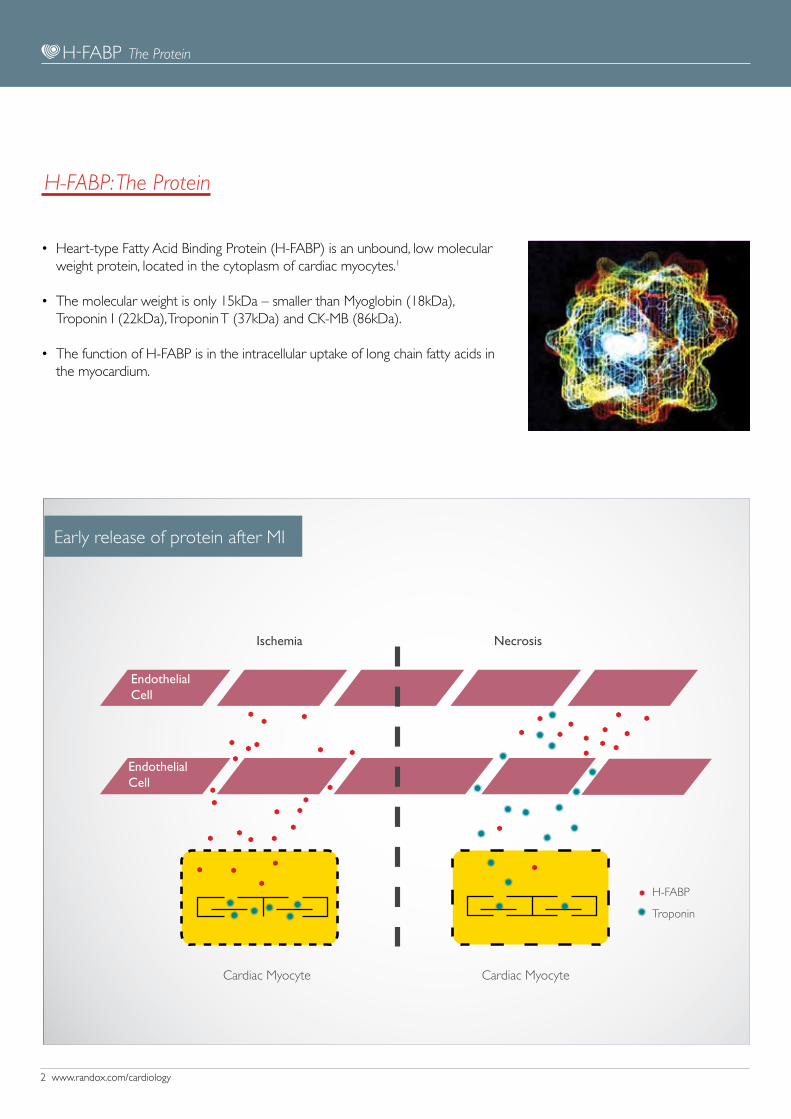
Early release of protein after MI
The Protein
EndothelialCell
EndothelialCell
Ischemia Necrosis
Cardiac Myocyte
H-FABP
Cardiac Myocyte
Troponin
www.randox.com/cardiology 3
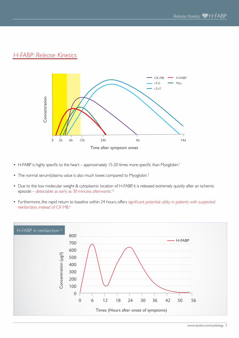
• H-FABP is highly specific to the heart – approximately 15-20 times more specific than Myoglobin.2
• The normal serum/plasma value is also much lower, compared to Myoglobin.3
• Due to the low molecular weight & cytoplasmic location of H-FABP, it is released extremely quickly after an ischemic episode – detectable as early as 30 minutes afterwards.4,5
• Furthermore, the rapid return to baseline within 24 hours, offers significant potential utility in patients with suspected reinfarction, instead of CK-MB.6
H-FABP: Release Kinetics
Times (Hours after onset of symptoms)
Con
cent
ratio
n (µ
g/l)
H-FABP in reinfarction 6
Release Kinetics
0 2h 6h 24h12h 4d 14d
CK-MB
cTnI
cTnT
H-FABP
Myo
Time after symptom onset
Con
cent
ratio
n
800700600500400300200100
00 6 12 18 24 30 36 42 50 56
H-FABP
4 www.randox.com/cardiology
Diagnostic Value in ACS
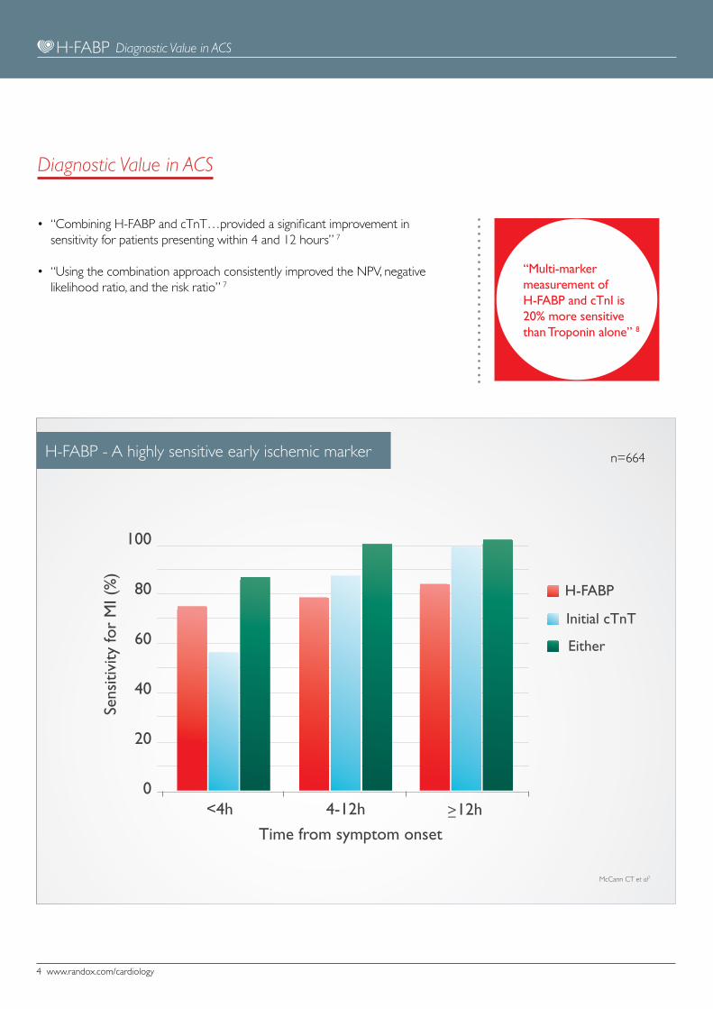
• “Combining H-FABP and cTnT…provided a significant improvement in sensitivity for patients presenting within 4 and 12 hours” 7
• “Using the combination approach consistently improved the NPV, negative likelihood ratio, and the risk ratio” 7
“Multi-marker measurement of H-FABP and cTnI is 20% more sensitive than Troponin alone” 8
H-FABP - A highly sensitive early ischemic marker n=664
Diagnostic Value in ACS
100
80
60
40
20
0
Sens
itivi
ty fo
r M
I (%
)
H-FABP
Initial cTnT
Either
<4h 4-12h ≥12h
Time from symptom onset
McCann CT et al7
www.randox.com/cardiology 5
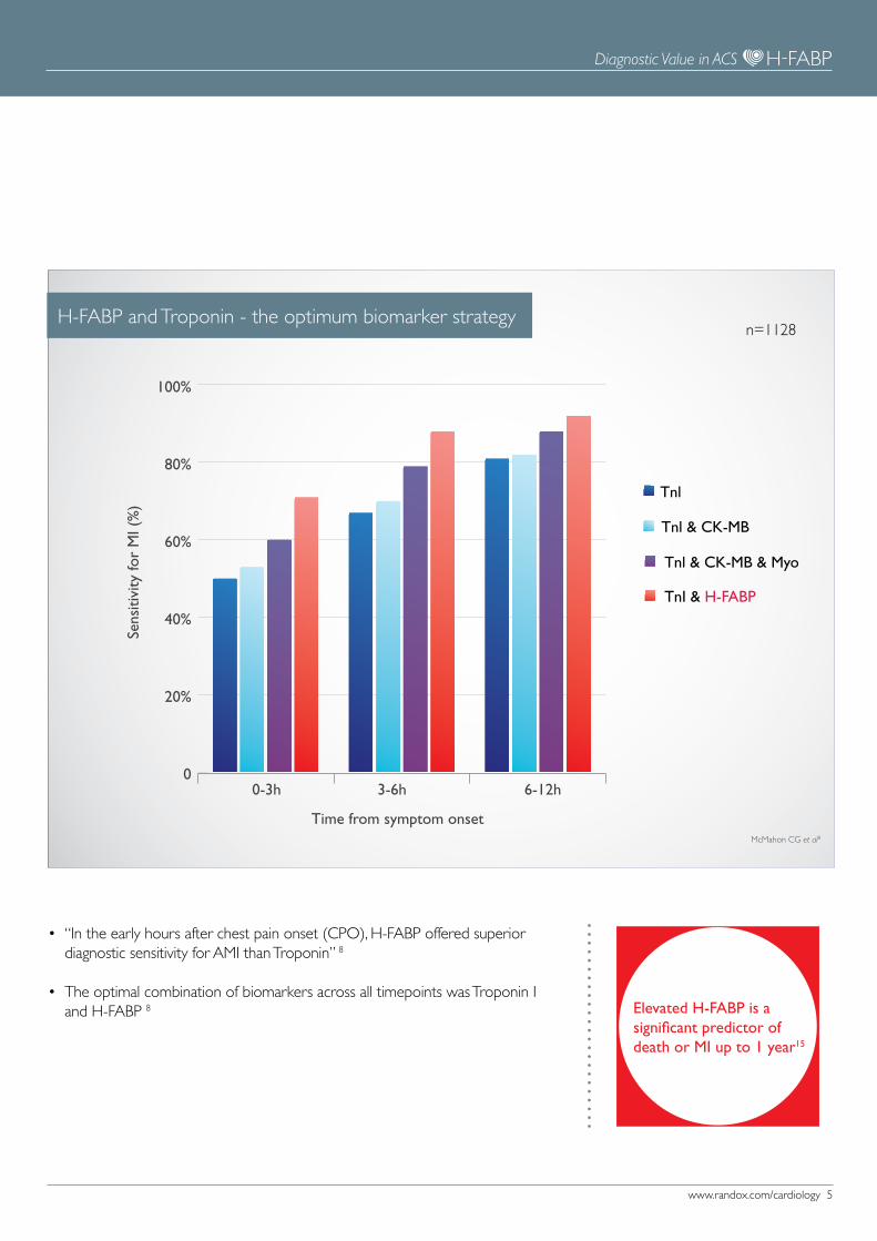
• “In the early hours after chest pain onset (CPO), H-FABP offered superior diagnostic sensitivity for AMI than Troponin” 8
• The optimal combination of biomarkers across all timepoints was Troponin I and H-FABP 8 Elevated H-FABP is a
significant predictor of death or MI up to 1 year15
n=1128H-FABP and Troponin - the optimum biomarker strategy
Diagnostic Value in ACS
0
20%
40%
60%
80%
100%
Sens
itivi
ty fo
r M
I (%
)
Time from symptom onset
0-3h 3-6h 6-12h
Tnl
Tnl & CK-MB
Tnl & CK-MB & Myo
TnI & H-FABP
McMahon CG et al8

6 www.randox.com/cardiology
• Even based on samples taken immediately after hospital admission (<24h after CPO), the combination of H-FABP & Troponin I was superior to the triple marker strategy across the measures of sensitivity, specificity, PPV & NPV. 9
H-FABP and Troponin - the optimum biomarker strategy n=705
H-FABP + Tnl CK-MB, myoglobin, Tnl
82.2
69.8
85.878.8
56.4
42.5
95.692.1
p<0.0001 p<0.0001
p<0.0001
p=0.02
Dia
gnos
is o
f MI (
%)
0
20
40
60
80
100
Sensitivity Specificity PPV NPV
Diagnostic Value in ACS
Body R et al9
www.randox.com/cardiology 7
H-FABP and Troponin enables early MI rule out
• “The combination of H-FABP & TnI can be used effectively as a rule-out test to exclude AMI within 6 hours of pain onset 8
• H-FABP & TnI offers a NPV of 98% at 3-6 hours after symptom onset 8
• Measuring H-FABP with Tnl also appears to identify at-risk patients with ACS more effectively than a single TnI assay 8
“H-FABP allows for more accurate risk stratification of low-medium risk chest pain patients”8
NPV for MI (%)
Time post pain 0-3h 3-6h 6-12h 12-24h
Individual markers
H-FABP 93 97 98 99
cTnI 92 95 97 99
2 marker combinations
H-FABP + cTnI 94 98 99 100
Diagnostic Value in ACS
McMahon CG et al8
n=1128
8 www.randox.com/cardiology
Diagnostic value of H-FABP combined with hsTnT
Diagnostic value of H-FABP combined with hsTnT
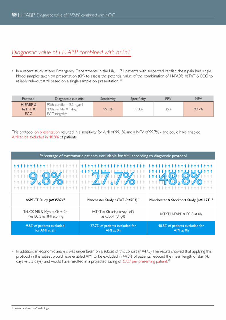
• In a recent study at two Emergency Departments in the UK, 1171 patients with suspected cardiac chest pain had single blood samples taken on presentation (0h) to assess the potential value of the combination of H-FABP, hsTnT & ECG to reliably rule-out AMI based on a single sample on presentation.10
Protocol Diagnostic cut-offs Sensitivity Specificity PPV NPV
H-FABP &hsTnT &
ECG
95th centile = 2.5 ng/ml99th centile = 14ng/lECG negative
99.1% 59.3% 35% 99.7%
This protocol on presentation resulted in a sensitivity for AMI of 99.1%, and a NPV of 99.7% - and could have enabled AMI to be excluded in 48.8% of patients.
• In addition, an economic analysis was undertaken on a subset of this cohort (n=473). The results showed that applying this protocol in this subset would have enabled AMI to be excluded in 44.3% of patients, reduced the mean length of stay (4.1 days vs 5.3 days), and would have resulted in a projected saving of £327 per presenting patient.13
Percentage of symtomatic patients excludable for AMI according to diagnostic protocol
ASPECT Study (n=3582)11 Manchester Study hsTnT (n=703)12 Manchester & Stockport Study (n=1171)10
TnI, CK-MB & Myo at 0h + 2hPlus ECG & TIMI scoring
hsTnT at 0h using assay LoD as cut-off (3ng/l)
hsTnT, H-FABP & ECG at 0h
9.8% of patients excluded for AMI at 2h
27.7% of patients excluded for AMI at 0h
48.8% of patients excluded for AMI at 0h
27.7%9.8% 48.8%
www.randox.com/cardiology 9
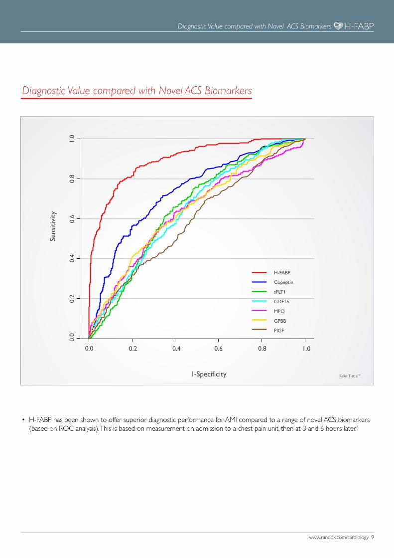
• H-FABP has been shown to offer superior diagnostic performance for AMI compared to a range of novel ACS biomarkers (based on ROC analysis). This is based on measurement on admission to a chest pain unit, then at 3 and 6 hours later.4
Diagnostic Value compared with Novel ACS Biomarkers
H-FABP
Copeptin
sFLT1
GDF15
MPO
GPBB
PIGF
1-Specificity
0.0 0.2 0.4 0.6 0.8 1.0
Sens
itivi
ty
0.0
0.2
0.4
0.6
0.8
1.0
Diagnostic Value compared with Novel ACS Biomarkers
Keller T et al14
10 www.randox.com/cardiology
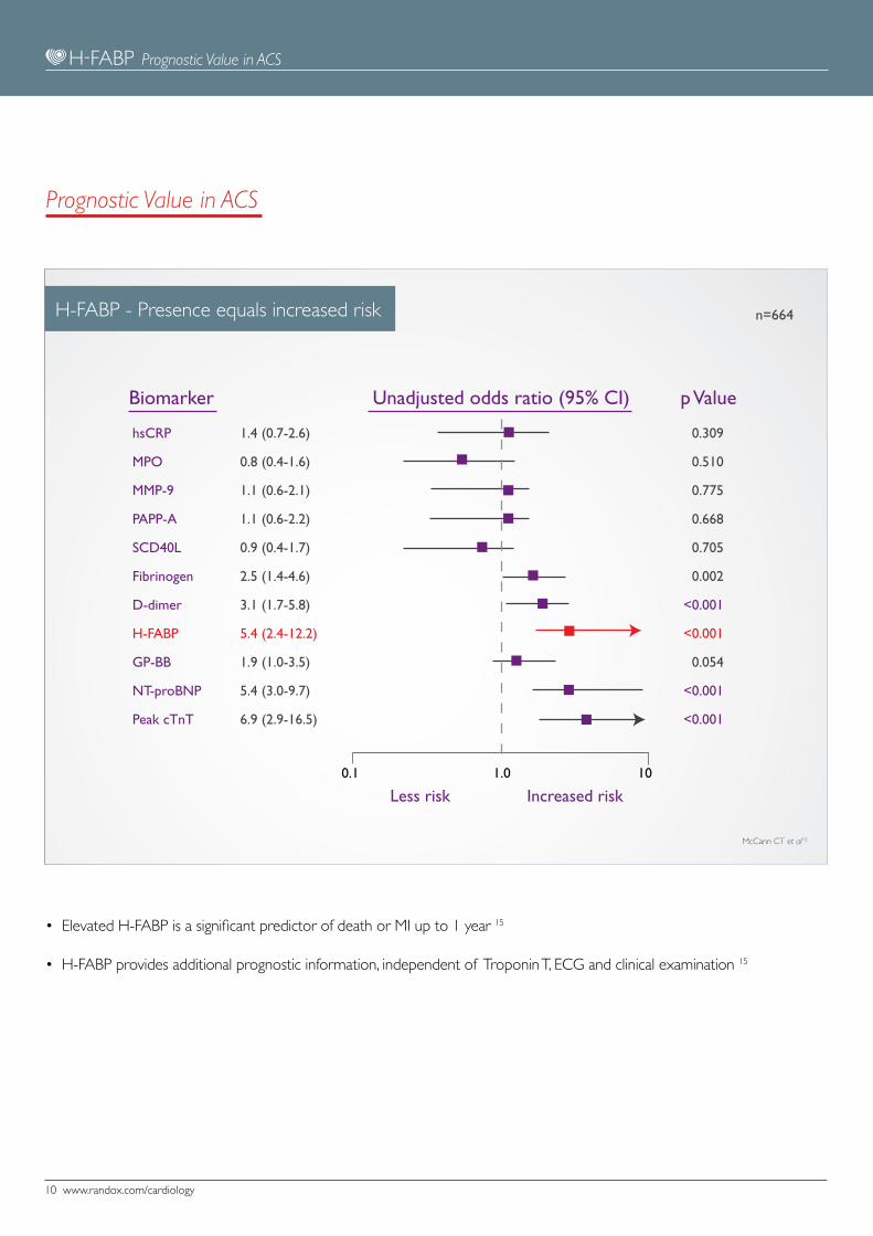
• Elevated H-FABP is a significant predictor of death or MI up to 1 year 15
• H-FABP provides additional prognostic information, independent of Troponin T, ECG and clinical examination 15
H-FABP - Presence equals increased risk n=664
hsCRP
MPO
MMP-9
PAPP-A
SCD40L
Fibrinogen
D-dimer
H-FABP
GP-BB
NT-proBNP
Peak cTnT
Biomarker
Less risk Increased risk
p ValueUnadjusted odds ratio (95% CI)
0.309
0.510
0.775
0.668
0.705
0.002
<0.001
<0.001
0.054
<0.001
<0.001
0.1 1.0 10
1.4 (0.7-2.6)
0.8 (0.4-1.6)
1.1 (0.6-2.1)
1.1 (0.6-2.2)
0.9 (0.4-1.7)
2.5 (1.4-4.6)
3.1 (1.7-5.8)
5.4 (2.4-12.2)
1.9 (1.0-3.5)
5.4 (3.0-9.7)
6.9 (2.9-16.5)
Prognostic Value in ACS
Prognostic Value in ACS
McCann CT et al15

www.randox.com/cardiology 11
• H-FABP allows identification of high risk patients across the full range of TnI concentrations16
• Negative test result for both TnI and H-FABP was associated with 0% mortality
at 6 months16
Raised concentrations of H-FABP are strongly predictive of mortality after ACS16
n=1448
Kilcullen N et al16
H-FABP predicts mortality after ACS
0 100 200 300 4000
5%
10%
15%
20%
25% Tnl+ / H-FABP+
Tnl- / H-FABP+
Tnl+ / H-FABP-
Tnl- / H-FABP-
Time (Days after event)
All
Cau
se M
orta
lity
(%)
Prognostic value in Troponin - Negative patients
Prognostic value in Troponin - Negative patients
Based on a single blood sample at 12-24 hrs after ACS system onset
12 www.randox.com/cardiology
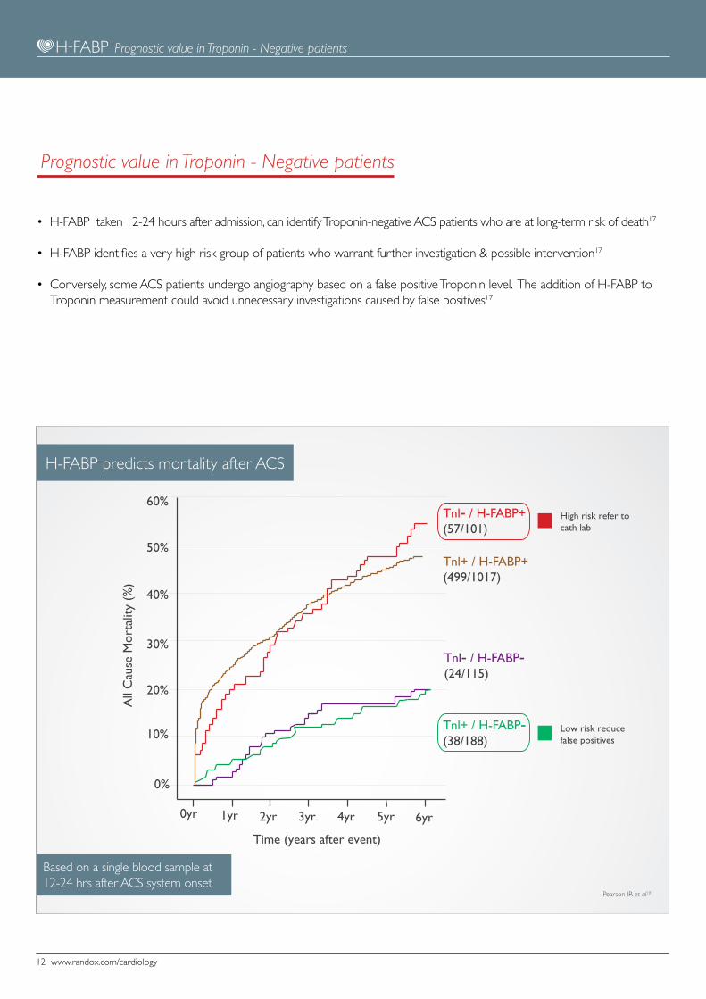
• H-FABP taken 12-24 hours after admission, can identify Troponin-negative ACS patients who are at long-term risk of death17
• H-FABP identifies a very high risk group of patients who warrant further investigation & possible intervention17
• Conversely, some ACS patients undergo angiography based on a false positive Troponin level. The addition of H-FABP to Troponin measurement could avoid unnecessary investigations caused by false positives17
H-FABP predicts mortality after ACS
Prognostic value in Troponin - Negative patients
Tnl- / H-FABP+ (57/101)
Tnl- / H-FABP- (24/115)
Tnl+ / H-FABP+ (499/1017)
Tnl+ / H-FABP- (38/188)
0yr 1yr 2yr 3yr 4yr 5yr 6yr
0%
10%
20%
30%
40%
50%
60%
Time (years after event)
All
Cau
se M
orta
lity
(%)
High risk refer to cath lab
Low risk reduce false positives
Prognostic value in Troponin - Negative patients
Pearson IR et al18
Based on a single blood sample at 12-24 hrs after ACS system onset
www.randox.com/cardiology 13
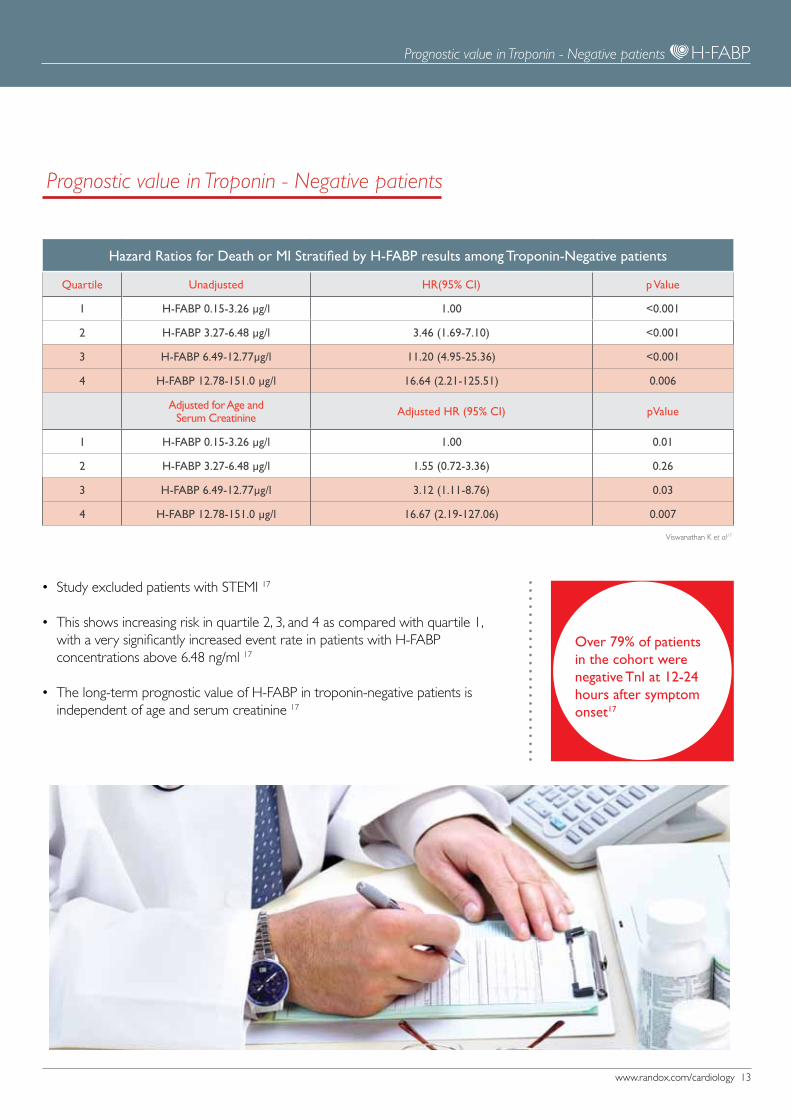
• Study excluded patients with STEMI 17
• This shows increasing risk in quartile 2, 3, and 4 as compared with quartile 1, with a very significantly increased event rate in patients with H-FABP concentrations above 6.48 ng/ml 17
• The long-term prognostic value of H-FABP in troponin-negative patients is independent of age and serum creatinine 17
Over 79% of patients in the cohort were negative TnI at 12-24 hours after symptom onset17
Hazard Ratios for Death or MI Stratified by H-FABP results among Troponin-Negative patients
Quartile Unadjusted HR(95% CI) p Value
1 H-FABP 0.15-3.26 µg/l 1.00 <0.001
2 H-FABP 3.27-6.48 µg/l 3.46 (1.69-7.10) <0.001
3 H-FABP 6.49-12.77µg/l 11.20 (4.95-25.36) <0.001
4 H-FABP 12.78-151.0 µg/l 16.64 (2.21-125.51) 0.006
Adjusted for Age and Serum Creatinine Adjusted HR (95% CI) pValue
1 H-FABP 0.15-3.26 µg/l 1.00 0.01
2 H-FABP 3.27-6.48 µg/l 1.55 (0.72-3.36) 0.26
3 H-FABP 6.49-12.77µg/l 3.12 (1.11-8.76) 0.03
4 H-FABP 12.78-151.0 µg/l 16.67 (2.19-127.06) 0.007
Prognostic value in Troponin - Negative patients
Viswanathan K et al17
Prognostic value in Troponin - Negative patients

14 www.randox.com/cardiology
• Patients with H-FABP concentrations >6.48µg/L had significantly increased risk of adverse events17
• Among Troponin negative patients, the cut-off of 6.48µg/L identified patients at very high risk of adverse outcomes independent of patient age and serum creatinine17
n= 955Increased H-FABP concentrations confers increased risk
Value of a fully quantitative H-FABP assay
Value of a fully quantitative H-FABP assay
This demonstrates a clear need for a fully quanitative H-FABP assay
Viswanathan K et al17
50
40
30
20
10
0
Dea
th o
r M
I (%
)
Time of death or MI (in months)
12.78 to 151.0 µg/L (24/54)
6.49 to 12.77 µg/L (24/63)
3.27 to 6.48 µg/L (24/203)
0.15 to 3.26 µg/L (24/635)
0 3 6 9 12 15

www.randox.com/cardiology 15
• “ This demonstrated the additive value of H-FABP, particularly for ACS subtypes such as Unstable Angina, traditionally considered to be associated with a low long-term risk “ 16
All Cause Mortality
Unstable Angina NSTEMI STEMI
H-FABP negative (≤5.8µg/l) 2.1% (2) 4.8% (9) 0% (0)
H-FABP positive (>5.8µg/l) 22.9% (19) 26.1% (189) 23.0% (77)
p Value 0.006 0.004 _*
ECG X X
Troponin X
H-FABP
H-FABP - The first true global ACS biomarker
Kilcullen N et al16
Prognostic value in Unstable Angina
Prognostic Value in Unstable Angina
16 www.randox.com/cardiology
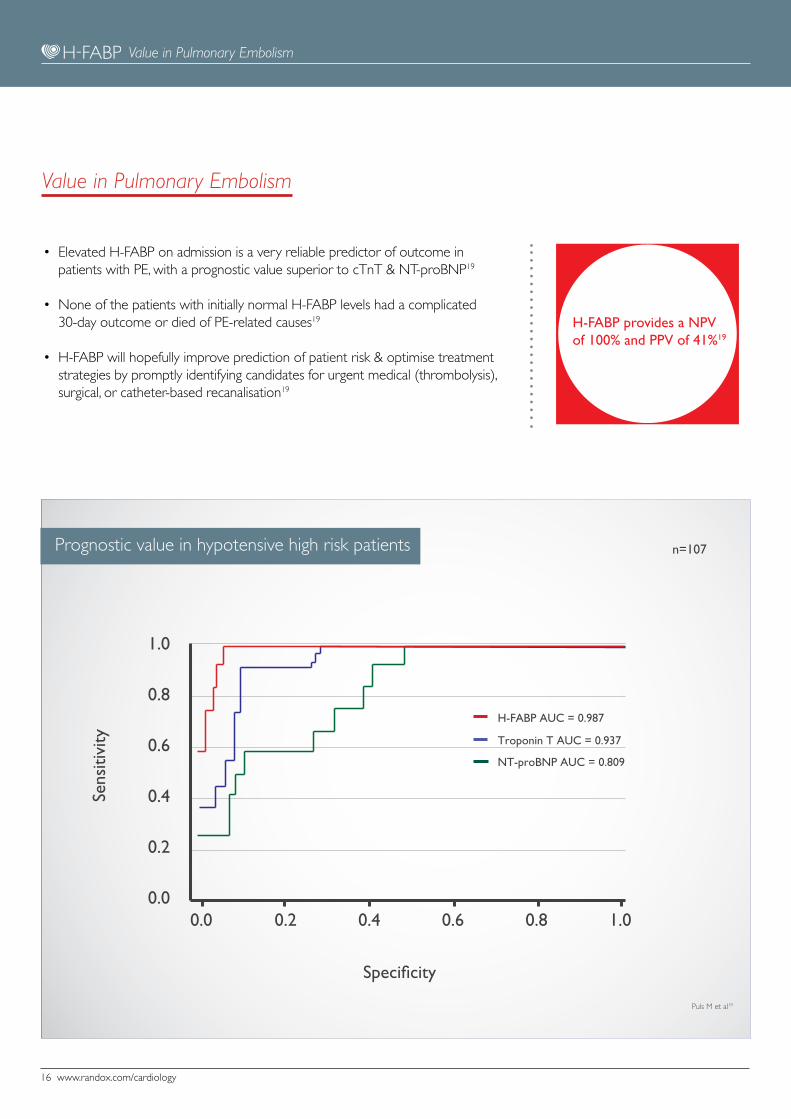
• Elevated H-FABP on admission is a very reliable predictor of outcome in patients with PE, with a prognostic value superior to cTnT & NT-proBNP19
• None of the patients with initially normal H-FABP levels had a complicated 30-day outcome or died of PE-related causes19
• H-FABP will hopefully improve prediction of patient risk & optimise treatment strategies by promptly identifying candidates for urgent medical (thrombolysis), surgical, or catheter-based recanalisation19
H-FABP provides a NPV of 100% and PPV of 41%19
n=107
1.0
0.8
0.6
0.4
0.2
0.00.0 0.2 0.4 0.6 0.8 1.0
Sens
itivi
ty
H-FABP AUC = 0.987
Troponin T AUC = 0.937
NT-proBNP AUC = 0.809
Specificity
Prognostic value in hypotensive high risk patients
Value in Pulmonary Embolism
Value in Pulmonary Embolism
Puls M et al19
www.randox.com/cardiology 17
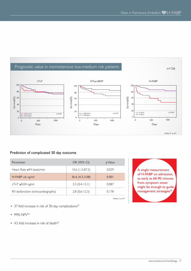
• 37 fold increase in risk of 30-day complications20
• 99% NPV20
• 4.5 fold increase in risk of death20
Parameter OR (95% CI) p Value
Heart Rate ≥94 beats/min 10.6 (1.3-87.2) 0.029
H-FABP ≥6 ng/ml 36.6 (4.3-3.08) 0.001
cTnT ≥0.04 ng/ml 3.3 (0.4-13.1) 0.087
RV dysfunction (echocardiography) 2.8 (0.6-12.3) 0.178
Prediction of complicated 30 day outcome
Days
Surv
ival
(%)
cTnT
DaysSu
rviv
al(%
)
H-FABP
Dellas C et al20
n=126
Days
Surv
ival
(%)
NT-proBNP
Prognostic value in normotensive low-medium risk patients
≥1.000 pg/ml<1.000 pg/ml
p=0.108
0 500 10000
20
40
60
80
100
0 500 10000
20
40
60
80
100
≥0.04 ng/ml<0.04 ng/ml
p=0.667
0 500 10000
20
40
60
80
100
≥6 ng/ml<6 ng/ml
p<0.0001
A single measurement of H-FABP on admission, as early as 60-90 minutes from symptom onset might be enough to guide management strategies20
Value in Pulmonary Embolism
Dellas C et al20

18 www.randox.com/cardiology
• The slower release of CK-MB and cTnI generates an inability to discriminate between graft failure with massive tissue necrosis and ischemia reperfusion injury within 24 hours after CABG surgery21
• H-FABP is superior to Troponin & CK-MB for the prediction of mortality and ventricular dysfunction21
• Enable clinicians to identify patients in need of further diagnostic or therapeutic procedures to reduce loss of myocardial mass or performance21
Muehlschlegel et al21
n=12985 year predictive value following CABG
CK-MB
MYOH-FABP
cTnI
60
50
40
30
20
10
0
1.6
1.4
1.2
1.0
0.8
0.6
0.4
0.2
0
54321PostPre
Myo
glob
in (
1/10
th),
CK
MB
and
H-F
ABP
(ng
/ml)
Time of blood draw after CPB (days)
Median Time Course Biomarkers
cTnl
(ng
/ml)
Value in Coronary Artery Bypass Grafting (CABG)
Value in CABG

www.randox.com/cardiology 19
References
References
1. Glatz JFC, van Bilsen M, Paulussen RJA, Veerkamp J, van der Vusse GJ, Reneman RS. Release of fatty acid-binding protein from isolated rat heart subjected to ischemia and reperfusion or the calcium paradox. Biochim Biophys Acta.1988;961:148-52
2. Data on file3. Ghani F, Wu A, Graff L, Petry C, Armstrong G, Prigent F, Brown M. Role of heart-type fatty acid-binding protein in early
detection of acute myocardial infarction. Clin Chem. 2000; 46: 718-7194. Pelsers MM, Hermens WT, Glatz JF. Fatty acid-binding proteins as plasma markers of tissue injury. Clin. Chem. Acta.
2005;352(1-2):15-35.5. Kleine AH, Glatz JF, van Nieuwenhoven FA. van der Vasse GJ. Release of heart type fatty acid binding protein into plasma
after acute myocardial infarction in man. Mol Cell Biochem.1992;116:155-162.6. Data on file7. McCann CJ, Glover BM, Menown IB, Moore MJ, McEneny J, Owens CG, Smith B, Sharpe PC, Young IS, Adgey JA.
Novel biomarkers in early diagnosis of acute myocardial infarction compared with cardiac troponin T. Eur Heart J. 2008;29(23):2843-50.
8. McMahon CG, Lamont JV, Curtin E, McConnell RI, Crockard M, Kurth MJ, Crean P, Fitzgerald SP. Diagnostic accuracy of heart-type fatty acid-binding protein for the early diagnosis of acute myocardial infarction. Am J Emerg Med. 2012;30(2):267-74.
9. Body R, McDowell G, Carley S, Wibberley C, Ferguson J, Mackway-Jones K. A FABP-ulous ‘rule out’ strategy? Heart fatty acid binding protein and troponin for rapid exclusion of acute myocardial infarction. Resuscitation 2011;82(8):1041-6.
10. Body R, Carley S, Burrows G, Pemberton P, Mackway-Jones K. Combining heart fatty acid binding protein and high sensitivity troponin in the emergency department.14th International Conference on Emergency Medicine. Acad Emerg Med. 2012;19(6):748-749.
11. Than M, Cullen L, Reid CM, Lim SH, Aldous S, Ardagh MW, Peacock WF, Parsonage WA, Ho HF, Ko HF, Kasliwal RR, Bansal M, Soerianata S, Hu D, Ding R, Hua Q, Seok-Min K, Sritara P, Sae-Lee R, Chiu TF, Tsai KC, Chu FY, Chen WK, Chang WH, Flaws DF, George PM, Richards AM. A 2-h diagnostic protocol to assess patients with chest pain symptoms in the Asia-Pacific region (ASPECT): a prospective observational validation study. Lancet. 2011;26;377(9771):1077-84.
12. Body R, Carley S, McDowell G, Jaffe AS, France M, Cruickshank K, Wibberley C, Nuttall M, Mackway-Jones K. Rapid exclusion of acute myocardial infarction in patients with undetectable troponin using a high-sensitivity assay. J Am Coll Cardiol. 2011;20;58(13):1332-9.
13. Body R, Dixon D, Burrows G, Cook G, Lewis PS. Economic evaluation of a heart fatty acid binding protein based protocol for rapid chest pain assessment.14th International Conference on Emergency Medicine. Acad Emerg Med. 2012;19(6):746-747.
14. Keller T, Zeller T, Ojeda F, Tzikas S, Lillpopp L, Sinning C, Wild P, Genth-Zotz S, Warnholtz A, Giannitsis E, Möckel M, Bickel C, Peetz D, Lackner K, Baldus S, Münzel T, Blankenberg S. Serial changes in highly sensitive troponin I assay and early diagnosis of myocardial infarction. JAMA. 2011;28;306(24):2684-93.
15. McCann CJ, Glover BM, Menown IB, Moore MJ, McEneny J, Owens CG, Smith B, Sharpe PC, Young IS, Adgey JA. Prognostic value of a multimarker approach for patients presenting to hospital with acute chest pain. Am J Cardiol. 2009:103(1):22-8.
16. Kilcullen N, Viswanathan K, Das R, Morrell C, Farrin A, Barth JH, Hall AS; EMMACE-2 Investigators. Heart-type fatty acid-binding protein predicts long-term mortality after acute coronary syndrome and identifies high-risk patients across the range of troponin values. J Am Coll Cardiol. 2007;50(21):2061-7.
17. Viswanathan K, Kilcullen N, Morrell C, Thistlethwaite SJ, Sivananthan MU, Hassan TB, Barth JH, Hall AS. heart-type fatty-acid binding-protein (H-FABP) predicts long-term mortality and re-infarction in consecutive patients with suspected acute coronary syndrome who are troponin negative. J Am Coll Cardiol. 2010;55(23): 2590-8
18. Pearson IR, Hall AS, Gale CP, Sivananthan MU, Viswanathan K, Kilcullen N, Barth JH, In Acute Coronary Syndromes, Heart-type Fatty Acid Binding Protein is a More Accurate Predictor of Long Term Prognosis than Troponin. Circulation. 2010;122:A11374
19. Puls M, Dellas C, Lankeit M, Olschewski M, Binder L, Geibel A, Reiner C, Schäfer K, Hasenfuss G, Konstantinides S. Heart-type fatty acid-binding protein permits early risk stratification of pulmonary embolism. Eur Heart J. 2007;28(2):224-9.
20. Dellas C, Puls M, Lankeit M, Schäfer K, Cuny M, Berner M, Hasenfuss G, Konstantinides S. Elevated heart-type fatty acid-binding protein levels on admission predict an adverse outcome in normotensive patients with acute pulmonary embolism. J Am Coll Cardiol. 2010;11;55(19):2150-7.
21. Muehlschlegel JD, Perry TE, Liu KY, Fox AA, Collard CD, Shernan SK, Body SC. Heart-type fatty acid binding protein is an independent predictor of death and ventricular dysfunction after coronary artery bypass graft surgery. Anesth Analg. 2010; 111(5):1101-9.
22. Nordestgaard BG, Chapman MJ, Ray K, Borén J, Andreotti F, Watts GF, Ginsberg H, Amarenco P, Catapano A, Descamps OS, Fisher E, Kovanen PT, Kuivenhoven JA, Lesnik P, Masana L, Reiner Z, Taskinen MR, Tokgözoglu L, Tybjærg-Hansen A; European Atherosclerosis Society Consensus Panel. Lipoprotein(a) as a cardiovascular risk factor: current status. Eur Heart J. 2010; 31(23):2844-53.
20 www.randox.com/cardiology
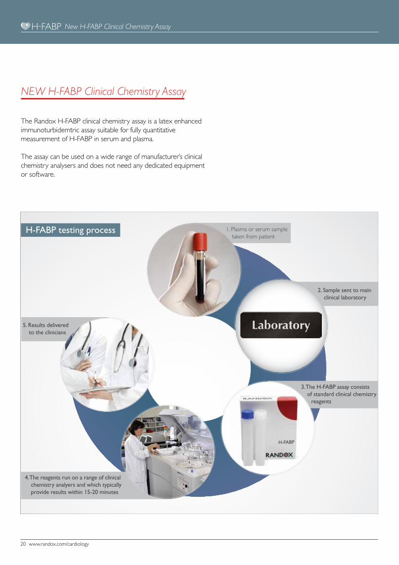
H-FABP testing process
The Randox H-FABP clinical chemistry assay is a latex enhanced immunoturbidemtric assay suitable for fully quantitative measurement of H-FABP in serum and plasma.
The assay can be used on a wide range of manufacturer’s clinical chemistry analysers and does not need any dedicated equipment or software.
New H-FABP Clinical Chemistry Assay
NEW H-FABP Clinical Chemistry Assay
1. Plasma or serum sample taken from patient
2. Sample sent to main clinical laboratory
3. The H-FABP assay consists of standard clinical chemistry reagents
4. The reagents run on a range of clinical chemistry analyers and which typically provide results within 15-20 minutes
5. Results delivered to the clinicians
www.randox.com/cardiology 21
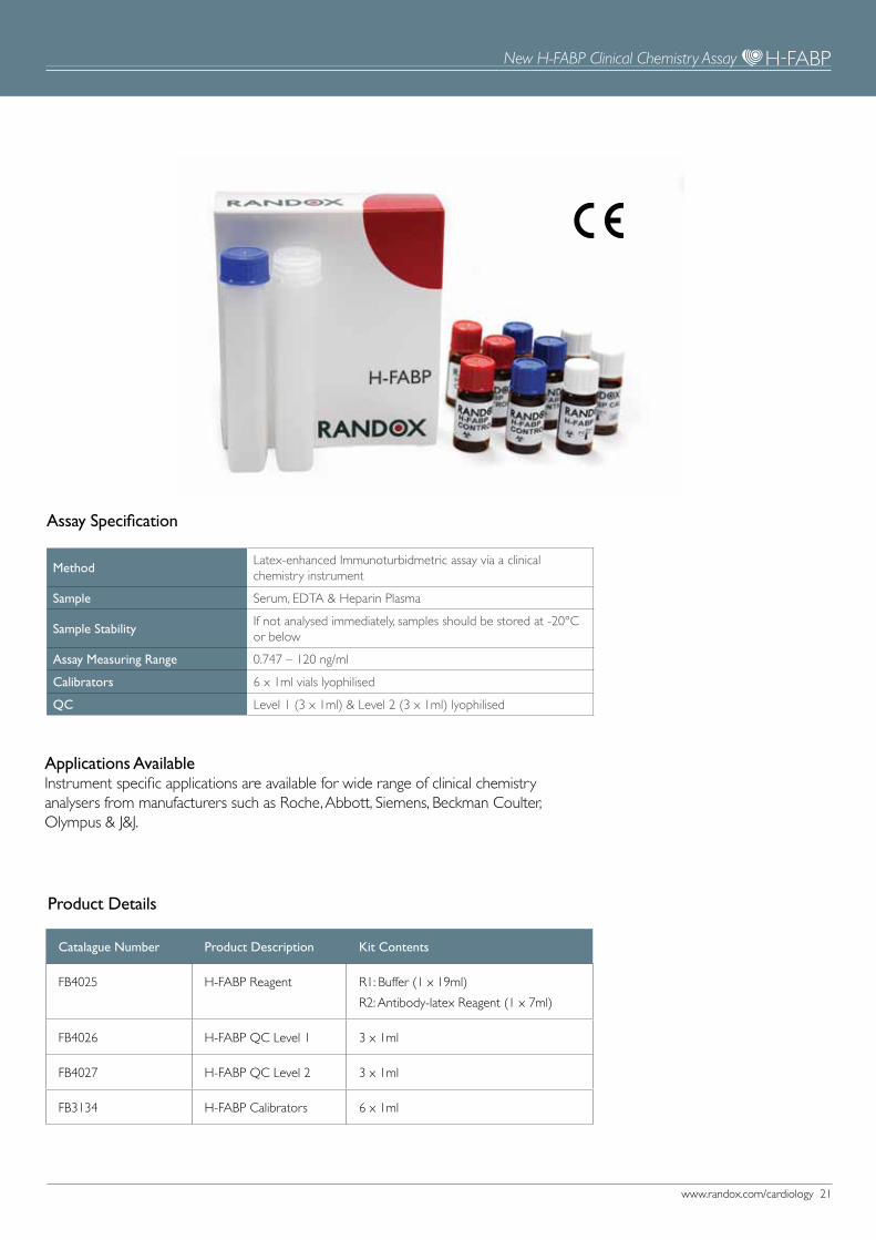
Assay Specification
MethodLatex-enhanced Immunoturbidmetric assay via a clinical chemistry instrument
Sample Serum, EDTA & Heparin Plasma
Sample StabilityIf not analysed immediately, samples should be stored at -20°C or below
Assay Measuring Range 0.747 – 120 ng/ml
Calibrators 6 x 1ml vials lyophilised
QC Level 1 (3 x 1ml) & Level 2 (3 x 1ml) lyophilised
Applications AvailableInstrument specific applications are available for wide range of clinical chemistry analysers from manufacturers such as Roche, Abbott, Siemens, Beckman Coulter, Olympus & J&J.
Product Details
Catalague Number Product Description Kit Contents
FB4025 H-FABP Reagent R1: Buffer (1 x 19ml)
R2: Antibody-latex Reagent (1 x 7ml)
FB4026 H-FABP QC Level 1 3 x 1ml
FB4027 H-FABP QC Level 2 3 x 1ml
FB3134 H-FABP Calibrators 6 x 1ml
New H-FABP Clinical Chemistry Assay
22 www.randox.com/cardiology
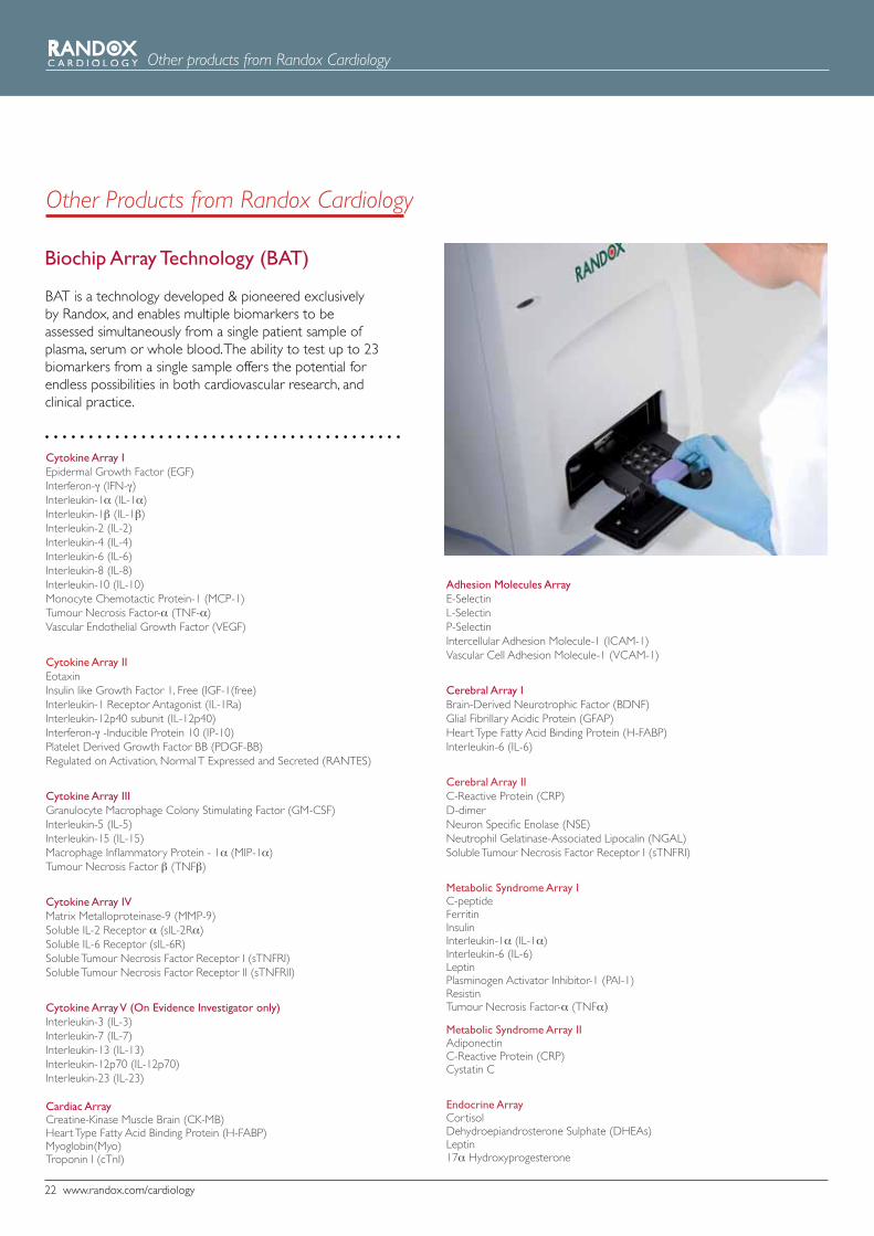
Biochip Array Technology (BAT)
BAT is a technology developed & pioneered exclusively by Randox, and enables multiple biomarkers to be assessed simultaneously from a single patient sample of plasma, serum or whole blood. The ability to test up to 23 biomarkers from a single sample offers the potential for endless possibilities in both cardiovascular research, and clinical practice.
Other products from Randox Cardiology
Other Products from Randox Cardiology
Cytokine Array I Epidermal Growth Factor (EGF) Interferon-g (IFN-g)Interleukin-1a (IL-1a)Interleukin-1b (IL-1b)Interleukin-2 (IL-2)Interleukin-4 (IL-4)Interleukin-6 (IL-6)Interleukin-8 (IL-8)Interleukin-10 (IL-10)Monocyte Chemotactic Protein-1 (MCP-1)Tumour Necrosis Factor-a (TNF-a)Vascular Endothelial Growth Factor (VEGF)
Cytokine Array IIEotaxin Insulin like Growth Factor 1, Free (IGF-1(free) Interleukin-1 Receptor Antagonist (IL-1Ra)Interleukin-12p40 subunit (IL-12p40)Interferon-g -Inducible Protein 10 (IP-10)Platelet Derived Growth Factor BB (PDGF-BB)Regulated on Activation, Normal T Expressed and Secreted (RANTES)
Cytokine Array IIIGranulocyte Macrophage Colony Stimulating Factor (GM-CSF)Interleukin-5 (IL-5)Interleukin-15 (IL-15)Macrophage Inflammatory Protein - 1a (MIP-1a)Tumour Necrosis Factor b (TNFb)
Cytokine Array IVMatrix Metalloproteinase-9 (MMP-9)Soluble IL-2 Receptor a (sIL-2Ra)Soluble IL-6 Receptor (sIL-6R)Soluble Tumour Necrosis Factor Receptor I (sTNFRI)Soluble Tumour Necrosis Factor Receptor II (sTNFRII)
Cytokine Array V (On Evidence Investigator only)Interleukin-3 (IL-3)Interleukin-7 (IL-7)Interleukin-13 (IL-13)Interleukin-12p70 (IL-12p70)Interleukin-23 (IL-23)
Cardiac Array Creatine-Kinase Muscle Brain (CK-MB)Heart Type Fatty Acid Binding Protein (H-FABP)Myoglobin(Myo) Troponin I (cTnI)
Adhesion Molecules Array E-Selectin L-Selectin P-Selectin Intercellular Adhesion Molecule-1 (ICAM-1) Vascular Cell Adhesion Molecule-1 (VCAM-1)
Cerebral Array I Brain-Derived Neurotrophic Factor (BDNF) Glial Fibrillary Acidic Protein (GFAP)Heart Type Fatty Acid Binding Protein (H-FABP)Interleukin-6 (IL-6)
Cerebral Array II C-Reactive Protein (CRP)D-dimer Neuron Specific Enolase (NSE)Neutrophil Gelatinase-Associated Lipocalin (NGAL)Soluble Tumour Necrosis Factor Receptor I (sTNFRI)
Metabolic Syndrome Array I C-peptideFerritinInsulinInterleukin-1a (IL-1a)Interleukin-6 (IL-6)LeptinPlasminogen Activator Inhibitor-1 (PAI-1)ResistinTumour Necrosis Factor-a (TNFa)
Metabolic Syndrome Array IIAdiponectinC-Reactive Protein (CRP)Cystatin C
Endocrine Array CortisolDehydroepiandrosterone Sulphate (DHEAs)Leptin17a Hydroxyprogesterone

www.randox.com/cardiology 23
Lipoprotein (a)
Other products from Randox Cardiology
Elevated Lp(a) concentration in plasma is an independent
genetic marker correlating with increased risk of atherosclerotic
disorders including myocardial and cerebral infarction. Levels
are also elevated in nephritic syndrome, patients undergoing
renal dialysis, patients with uncontrolled diabetes mellitus and
hypothyroidism.
Our highly successful Lipoprotein (a) test is the only method
in the world to accurately and reliably measure Lp(a), it is not
affected by Apo (a) size related bias like most other methods.
In June 2010, the European Atherosclerosis Society (EAS)
published a consensus paper on Lp(a), recommending its
widespread use as a screening tool in those at intermediate or
high risk of cardiovascular disease
“The evidence clearly supports Lp(a) as a priority for reducing
cardiovascular risk, beyond that associated with LDL cholesterol.
Clinicians should consider screening statin-treated patients with
recurrent heart disease, in addition to those considered at
moderate to high risk of heart disease - EAS Consensus Panel22
Automated chemistry assay for the casual genetic biomarker of CVD
Cardiac Risk Prediction Array
•Forassessmentofgeneticriskfordevelopmentof
cardiovascular disease
•Examinesspecificgeneticelementsthatwillnotchange
over an individual’s lifespan
Familial Hypercholesterolemia Array
•FHisageneticdisordercharacterisedbyhighlevelsofLDL
•MostcommondefectsareLDLR,ApoBandPCSK9gene
mutations
•Arrayassesses20SNPsknowntoinfluencethefunctionofthese
three genes
Hypertension Array
•Estimatedonebillionpeopleworldwideaffectedby
hypertension
•Leadingriskfactorforstroke,AMI,heartfailureandchronic
renal failure
•ArraycontainsanumberofSNPsindicatinggenetic
predisposition to hypertension
Biochip Array Technology - Genetic Arrays

LT23
7 A
PR13
Information correct at time of print. All Randox products are made in the UK. Randox Laboratories Limited is a company registered within Northern Ireland with company number N.I. 15738. VAT Registered Number: GB 151 6827 08. Product availability may vary from country to country. Please contact your local Randox representative for information.
Randox Laboratories Limited, 55 Diamond Road, Crumlin, County Antrim, BT29 4QY, United Kingdomt +44 (0) 28 9442 2413 f +44 (0) 28 9445 2912 e [email protected] I www.randox.com
RANDOX INTERNATIONAL HEADQUARTERSRandox Laboratories Limited, 55 Diamond Road, Crumlin, Co. Antrim, United Kingdom, BT29 4QY
T +44 (0) 28 9442 2413 F +44 (0) 28 9445 2912 E [email protected] I www.randox.com
USARandox Laboratories-US, Ltd.
515 Industrial Boulevard, Kearneysville, West Virginia, 25430
Tel: +1 304 728 2890 Toll Free: 8664 RANDOX Fax: +1 304 728 1890 Toll Free: 866 RANDOX 1
AustraliaRandox (Australia) Pty Ltd.
Suite 2/4 Charles Street, Paramatta, NSW 2150, Australia.
Tel: +61 (0) 2 9615 4640Fax: +61 (0) 2 9615 4644
France Laboratoires Randox
Roissy Parc, ZAC du Moulin24-26 rue du Noyer, BP 40, 95700 Roissy en France
Tel: +33 (0) 130 18 96 80Fax: +33 (0) 130 18 03 60
www.randox.fr
IndiaRandox Laboratories India Pvt Ltd.
3rd Floor, Godrej Coliseum, Somaiya Hospital Road,Off. Eastern Express Highway, Sion (East), Mumbai - 400 022, India
Tel: +91 22 6714 0600Fax: +91 22 2408 3803
Portugal Irlandox Laboratorios Quimica Analitica Ltda
Rua Agostinho de Jesus e Sousa 258, 4000-015 Porto, Portugal. Tel: +351 22 589 8320Fax: +351 22 589 8329
Switzerland Randox Laboratories Ltd. (Switzerland)C/O Wirtschafts-Treuhand Auctor Schwyz AG, Oberer Steisteg 18, 6430 Schwyz, Switzerland.
Tel: +41 (0) 41 810 48 89Fax: +41 (0) 41 560 81 41
BrazilRandox Brasil Ltda
Rua Fernandes Moreira, 415 CEP: 04716-000 - São Paulo / SP - Brasil.
Tel: +55 11 5181-2024Fax: +55 11 5181-0817
ChinaRandox Laboratories Ltd.
Shanghai Representative OfficeRoom 522-523, Fortune Times Tower, No.1438 North,
Shanxi Road, Putuo District, Shanghai, China 20060Tel: +86 (0) 21 6288 6240Fax: +86 (0) 21 6288 6246
www.randox.cn
GermanyRandox Laboratories GmbH
Wilhelmstr. 147a, 42489 Wülfrath, Germany. Tel: +49 (0) 2151/93 706-11
Fax: +49 (0)32 211089 91626
ItalyRandox Laboratories Ltd.
Corso Palestro 10, 10122 Torino, Italy.
Tel: +39 06 9896 8954Fax: +39 06 6051 3810
Puerto RicoClinical Diagnostics of Puerto Rico, LLC
PMB 590 PO Box 29029 San Juan, PR 00929-0029.
Tel: +1 787 701 7000 Fax: +1 787 701 6901
Czech RepublicRandox Laboratories S.R.O.
Bořivojova 35/878130 00 Praha 3, Czech Republic.
Tel: +420 2 1115 1661Fax: +420 2 1115 1662
Hong Kong Randox Laboratories Hong Kong Limited
Room 602, Skyline Commercial Centre,No 71-77 Wing Lok Street, Sheung Wan, Hong Kong
Tel: +852 3595 0515Fax: +852 3008 5133
Poland Randox Laboratories Ltd.
ul. Puławska 405 a 02-801 Warszawa, Polska Tel: +48 (0) 22 862 1080Fax: +48 (0) 22 862 1081
www.randox.pl
Slovakia Randox S.R.O.
Vilová 2, 851 01 Bratislava, Slovakia. Tel: +421 2 6381 3324Fax: +421 2 6381 2482
South AfricaRandox Laboratories SA (PTY) Ltd
Unit 69F Allandale Business Park Cnr. Le Roux Avenue & Morkels CloseHalfway House, Midrand, South Africa
Tel: +27 (0) 11 312 3590Fax: +27 (0) 11 312 4146
South KoreaRandox Korea
904 Doosan Venturedime 126-1, Pyeongchon, Dongan-gu, Anyang City, Kyeonggi-do, South Korea
Tel: +82 (0) 31 478 3121Fax: +82 (0) 31 478 3122www.randoxkorea.com
SpainLaboratorios Randox S.L.
C/Enric Prat de la Riba, 226, 1° Planta, 08901 L’Hospitalet de Llobregat, Barcelona.
Tel: +34 93 475 09 64Fax: +34 93 475 09 65
VietnamRandox Laboratories Ltd. Vietnam
Villa Phuc Thinh 2Bis Nguyen Thi Minh Khai St.Dakao Ward, District 1, Ho Chi Minh City, Vietnam.
Tel: +84-8-39 11 09 04Fax: +84-8-39 11 09 05
UKRandox Laboratories Ltd.
55 Diamond Road, Crumlin, Co. Antrim, United Kingdom, BT29 4QY
Tel: +44 (0) 28 9442 2413Fax: +44 (0) 28 9445 2912
Republic of IrelandRandox Teoranta
Meenmore, Dungloe, Co Donegal, Republic of Ireland
Tel: +353 7495 22600
IndiaRandox Laboratories India Pvt Ltd.
Plot 191-195, Bommasandra,Jigni Link Road Industrial Area,Bangalore, 562106
Tel: +91 80 2802 5000Fax: +91 80 2802 5012