hereditary diffuse leukoencephalopathy with … · tures such as frontal lobe syndromes,...
TRANSCRIPT

HEREDITARY DIFFUSE LEUKOENCEPHALOPATHY WITH SPHEROIDS
Christina Sundal, MDDepartment of Clinical Neuroscience and RehabilitationInstitute of Neuroscience and Physiologyat Sahlgrenska Academy University of Gothenburg
Gothenburg 2013

Hereditary diffuse leukoencephalopathy with spheroids:Insights into an adult onset neurodegenerative disease© Christina Sundal [email protected]://hdl.handle.net/2077/31995
ISBN 978-91-628-8641-7
Printed in Gothenburg, Sweden 2013 by Ineko ABCover illustration: “A brain with axonal spheroids (arrows) embedded in demyelinated white matter” by Andreas H. SundalDesign by Annika Enderlein Samuelsson / A little company AB
All articles are reproduced with permission from the Journals via Copyright Clearance Centers

To my beloved family


“If you think research is expensive,Try disease”
Mary Lasker (1901–1994)

1. ABSTRACT 09
2. SWEDISH ABSTRACT 11
3. LIST OF PUBLICATIONS 13
4. ABBREVIATIONS 15
5. INTRODUCTION 175.1 Leukoencephalopathies 175.2 ”Leukodystrophies” and “neuroaxonal dystrophies” 185.3 The original swedish HDLS family 245.4 Pigmentary orthochromatic leukodystrophy (POLD) 245.5 Therapeutics 26
6. AIMS AND OBJECTIVES 29
7. MATERIAL AND METHODS 317.1 Genetics 317.2 Neuropathology 367.3 Neuroimaging 387.4 Cerebrospinal fluid 567.5 Etics / ethical issues 58
8. BACKGROUNDS; STUDY SETTINGS AND RESULTS 618.1 Paper I 618.2 Paper II 628.3 Paper III 638.4 Paper IV 668.5 Paper V 67
09. DISCUSSION 71
10. CONCLUSION 89
11. ACKNOWLEDGMENTS 92
12. REFERENCES 96
13. APPENDIX 110
14. PAPERS 119
8 Christina Sundal • Hereditary diffuse leukoencephalopathy with spheroids
"During the last three decades, the areas of
inherited white matter (WM) disorders have expanded."

9
ABSTRACT
During the last three decades, the areas of inherited white matter (WM) disorders have expanded. Advances in magnetic resonance imaging (MRI) and genetics have led to increased detection of adult-onset WM disorders. Hereditary diffuse leukoen-cephalopathy with spheroids (HDLS) is an adult-onset, invariably lethal, brain WM disorder with an autosomal dominant inheritance pattern. The clinical symptoms are characterized by a constellation of features that progress to a devastating disease with multiple neurological impairments. The neuropathological hallmarks of HDLS are demyelination and the presence of axonal spheroids.
The overall aim of this study was to gather enough clinical cases, radiological images, cerebrospinal fluid (CSF) biomarkers and molecular genetic data to place HDLS in a nosographic context and define its relationship with other neurodegenerative disorders.
We updated the original Swedish HDLS family and created a pedigree consisting of 166 individuals. Fifteen of those cases were affected with HDLS, including two new cases. The clinical course was different in the two recent cases, with a sub-acute and a more chronic variant, respectively. Familial clustering of HDLS is not always obvious and in the Mayo Clinic HDLS collection we found that all of our cases had been misdiagnosed with other more common neurological disorders. Using exome sequencing, we identified the colony stimulating factor 1 receptor (CSF1R) mutation in 14 Mayo Clinic HDLS families. MRIs of 15 of these CSF1R mutation carriers demonstrated asymmetric WM lesions (WML) with frontoparietal predominance. With diffusion weighted-, and diffusion tensor imaging (DTI/DWI) we defined three different stages of HDLS pathology, and detected a peripheral rim of restricted diffusion that had a centrifugal migration from the anterior ventricular horns. This might be pathognomonic for the original Swedish type of HDLS.
In conclusion, HDLS is a distinct disease entity and the combination of clinical fea-tures such as frontal lobe syndromes, pyramidal-, extrapyramidal-, parietal- and visual signs, as well as WML in a characteristic frontoparietal distribution gives diagnostic clues. To clarify the distinction between the unknown genetics of the original Swedish family and the CSF1R mutation carriers, we propose to use molecular classification of HDLS type 1 and type 2, respectively. Results from our studies indicate that HDLS is probably primarily a neuroaxonal degeneration. Thus, elucidating the molecular mechanism of HDLS may provide novel insights into neurodegeneration.

10 Christina Sundal • Hereditary diffuse leukoencephalopathy with spheroids
"Syftet med studien var att öka kunskapen om
HDLS samt dess relation till andra neurodegenerativa
sjukdomar..."

11
SWEDISH ABSTRACT
Forskningen kring sjukdomar i hjärnans vita substans, vilka kan vara både ärftliga och förvärvade, har ökat kraftigt under de senaste årtiondena och har blivit ett viktigt kliniskt område med nya differential diagnostiska utmaningar. En av dessa sjukdo-mar, ”Hereditary diffuse leukoencephalopathy with spheroids” (HDLS) är en ärftlig, dödlig sjukdom som debuterar hos personer i vuxen ålder. De kliniska symtomen karaktäriseras av en blandning av fynd från de många bansystemen i hjärnan där pa-tienten snabbt kan försämras till en vegetativ nivå. I hjärnan ses typiska förändringar i den vita substansen med sämre isolering runt och ballongliknande förändringar, ”spheroids”, på nervtrådarna.
Syftet med studien var att öka kunskapen om HDLS samt dess relation till andra neurodegenerativa sjukdomar genom att samla in kliniska, radiologiska, neurokemiska och genetiska data.
Studien uppdaterar den ursprungliga svenska HDLS släkten med 166 individer var-av 15 har blivit diagnostiserade med HDLS samt inkluderar 14 HDLS släkter från Mayo-kliniken, USA. Två olika kliniska förlopp kan urskiljas, ett med snabb progression och ett med kroniskt förlopp dominerat av psykiatriska symtom. Studien demonstrerar att diagnostiken av HDLS kan vara mycket svår eftersom familjeträdet inte alltid indi-kerar en ärftlig sjukdom och således kan HDLS misstolkas som en sporadisk sjukdom. Alla inkluderade fall från USA hade ursprungligen feldiagnostiserats som andra neurol-ogiska sjukdomar. Vid analys med genteknologisk metodik upptäcktes en ny mutation i genen för colony stimulating factor 1 receptorn (CSF1R) i de 14 HDLS släkterna från Mayo kliniken. Denna mutation kunde ej påvisas hos de svenska HDLS patienterna. Femton av de amerikanska HDLS patienterna uppvisade en mer ojämn utbredning av vitsubstanslesionerna i framför allt hjärnans frontal- och parientallob jämfört med de svenska HDLS patienterna. Avancerade radiologiska metoder påvisade tre olika stadier av sjukdomen och en spridning av sjukdomen som startade från främre delen av hjärnans ventriklar och spred sig symmetrisk utåt för att stanna vid skiljelinjen mellan den grå och vita hjärnsubstansen. Spridningsmekanismen kan vara ett karakteristiskt fynd vid HDLS.
Studien demonstrerar att HDLS är en enhetlig sjukdom med kombinationer av sym-tom från flera olika bansystem, med typiska förändringar i hjärnans vita substans och med primär skada av nervtrådar. Användning av molekylär klassifikation med typ 1 och typ 2 föreslås för att lättare differentiera mellan den svenska HDLS släkten med okänd genmutation och CSF1R mutation. Studien har givit ökad kunskap om den molekylära mekanismen som orsakar HDLS och dess patofysiologi vilket ger ökad förståelse för andra neurodegenerativa sjukdomar med skadlig effekt på hjärnan samt kan på sikt möjligöra utveckling av terapi för neurodegenerativa sjukdomar.

12 Christina Sundal • Hereditary diffuse leukoencephalopathy with spheroids

13
LIST OF PUBLICATIONS
I. Update of the original HDLS kindred: divergent clinical courses.Sundal C, Ekholm S, Nordborg C, Jönsson L, Börjesson-Hanson A, Lindén T, Zetterberg H, Viitanen M, Andersen O. Acta Neurol Scand. 2012 Jul;126(1):67-75. doi: 10.1111/j.1600-0404.2011.01624.x.
II. Hereditary diffuse leukoencephalopathy with axonal spheroids (HDLS): a misdiagnosed disease entity.Sundal C, Lash J, Aasly J, Øygarden S, Roeber S, Kretzschman H, Garbern JY, Tselis A, Rademakers R, Dickson DW, Broderick D, Wszolek ZK. J Neurol Sci. 2012 Mar 15;314(1-2):130-7. doi: 10.1016/j.jns.2011.10.006.
III. Mutations in the colony stimulating factor 1 receptor (CSF1R) gene cause hereditary diffuse leukoencephalopathy with spheroids.Rademakers R, Baker M, Nicholson AM, Rutherford NJ, Finch N, Soto-Ortolaza A, Lash J, Wider C, Wojtas A, DeJesus-Hernandez M, Adamson J, Kouri N, Sundal C, Shuster EA, Aasly J, MacKenzie J, Roeber S, Kretzschmar HA, Boeve BF, Knopman DS, Petersen RC, Cairns NJ, Ghetti B, Spina S, Garbern J, Tselis AC, Uitti R, Das P, Van Gerpen JA, Meschia JF, Levy S, Broderick DF, Graff-Radford N, Ross OA, Miller BB, Swerdlow RH, Dickson DW, Wszolek ZK. Nat Genet. 2011 Dec 25;44(2):200-5. doi: 10.1038/ng.1027.
IV. MRI characteristics and scoring in HDLS due to CSF1R gene mutations.Sundal C, Van Gerpen JA, Nicholson AM, Wider C, Shuster EA, Aasly J, Spina S, Ghetti B, Roeber S, Garbern J, Borjesson-Hanson A, Tselis A, Swerdlow RH, Miller BB, Fujioka S, Heckman MG, Uitti RJ, Josephs KA, Baker M, Andersen O, Rademakers R, Dickson DW, Broderick D, Wszolek ZK. Neurology. 2012 Aug 7;79(6):566-74. doi:10.1212/WNL.0b013e318263575a.
V. Different stages of white matter changes in the original HDLS family revealed by advanced MRI techniques.Sundal Christina, Jönsson Lars, Ljungberg Maria, Zhong Jianhui, Tian Wei, Zhu Tong, Linden Thomas, Börjesson-Hanson Anne, Andersen Oluf, Ekholm Sven. Journal of Neuroimaging. Accepted for publication 2013, ID JON-13-3632.R1.

14 Christina Sundal • Hereditary diffuse leukoencephalopathy with spheroids

15
ABBREVIATIONS
AD Alzheimer’s diseaseALD AdrenoleukodystrophyCADASIL Cerebral Autosomal Dominant Arteriopathy with Subcortical Infarcts and LeukoencephalopathyCNS Central nervous systemCSF Cerebrospinal fluidCSF-1 Colony stimulating factor 1CSF1R Colony stimulating factor 1 receptor DAP12 DNAX-activating protein of kDA 12DTI Diffusion tensor imagingDWI Diffusion weighted imagingHDLS Hereditary diffuse leukoencephalopathy with spheroidsLOD Logarithm of the odd MR Magnetic resonanceMRI Magnetic resonance imagingMLD Metachromatic leukodystrophyMS Multiple sclerosisNAA N-AcetylaspartateNAWM Normal appearing white matter NFL Neurofilament light NFP Neurofilament proteins NHD Nasu-Hakola diseasePD Parkinson’s diseasePMD Pelizaeus-Merzbacher diseasePPMS Primary progressive multiple sclerosisTREM2 Triggering receptor expressed on myeloid cells 2TYROBP Protein tyrosine kinase-binding proteinVWM Vanishing white matterWM White matterWML White matter lesions

16 Christina Sundal • Hereditary diffuse leukoencephalopathy with spheroids
"Leukoencephalopathies encompass a heterogenous
group of disorders that predominantly affect the
brain’s white matter..."

17
5.1 LEUKOENCEPHALOPATHIESLeukoencephalopathies encompass a heterogenous group of disorders that predom-inantly affect the brain’s white matter (WM), regardless if the myelin damage is pri-mary or secondary, and irrespective of a molecular cause (van der Knaap et al., 2005). Traditionally leukoencephalopathy is a term used for extensive, often bilateral WM lesions (WML). Today it is commonly used for any of the group of diseases affecting the cerebral WM.
WML are evident on histological and potentially on neuroimaging examinations. They have a wide variety of causes, including cerebrovascular diseases, inborn errors of metabolism, infections, inflammation, mitochondrial disorders, neurodegenerative disorders, normal pressure hydrocephalus, nutritional conditions, toxins and trauma (Kim et al., 2008; van der Knaap et al., 2005). Today´s widespread use of magnetic resonance imaging (MRI) has lead to frequent findings of brain WM abnormalities, even in cases where these are not clinically suspected. Many rating scales have been developed to assess the location and the severity of WML and analyze factors such as the correlation with age, cognition, risk factors, and pathology (Bolandzadeh et al., 2012; Eichler et al., 2009; Loes et al., 1994a; Loes et al., 1999; Scheltens et al., 1998). However, since the clinical significance, etiology, and therapeutic implications vary widely, a precise definition of the disease and its etiology is crucial. This is par-ticularly important for inborn errors of metabolism, some of the leukodystrophies, infections, and inflammatory conditions where treatments are available. Detection of WML by MRI is highly sensitive but has limited specificity in detecting the underlying pathology (van der Knaap et al., 2005). Many of these disorders remain poorly characterized, with unknown genetic determinants and biochemical pathways. In order to enhance the development of therapeutic interventions it is imperative to dissect and sub-classify the various leukoencephalopathies according to the underlying pathophysiological mechanism responsible for the WM disorders.
Disorders of myelin have been classified into the following (Poser, 1957, 1961):
1. Myelinoclastic or demyelination: where there is destruction of the normal constituent of myelin and applied for diseases such as multiple sclerosis (MS).
2. Dysmyelination: where myelin is not formed properly or is delayed or arrested (defect in myelinogenesis) and is applied for disorders such as Pelizaeus- Merzbacher disease (PMD).
INTRODUCTION

18 Christina Sundal • Hereditary diffuse leukoencephalopathy with spheroids
Poser’s idea was that this classification should distinguish between acquired and inherited myelin disorders. However, this classification was not perfect and did not encompass all disorders, because some dysmyelinating disorders were caused by ex-ternal factors, and some inherited disorders had normal myelin that was later broken down and lost (van der Knaap et al., 2005).
In this thesis the focus is on two specific subgroups of the leukoencephalopathies, the leukodystrophies and the neuroaxonal dystrophies/degeneration.
5.2 ”LEUKODYSTROPHIES” & “NEUROAXONAL DYSTROPHIES”Leukodystrophy (leuko-white, dystrophy-defective nutrition) has been a commonly applied term for more than 100 years defining progressive inherited demyelinating disorders (Raymond et al., 2011). There have been several definitions of the term leu-kodystrophy, but generally, this has been applied to progressive, inherited demyelinating disorders (van der Knaap et al., 2005). Neuroaxonal dystrophy, or degeneration, is used when WM damage is secondary to axonal pathology. In addition, these terms have historical relevance that stems from meticulous clinical and neuropathological investi-gation and documentation during the morphological and biochemical eras of the 19th and early 20th centuries. Various entities have been described during these periods. The boundary between the leukodystrophies and the neuraxonal disorders was often unclear, and several leukodystrophies and neuraxonal dystrophies were ultimately found to be secondary to other pathological processes. This has led to the characterization of many syndromes and disorders and created the foundations for today’s knowledge.
The morphological diagnostic era
The history of myelin dates back to 1854 when Virchow described the sheaths that en-wrap axons in the brain (Virchow R., 1854). Charcot was the first to describe a disorder of the myelin when he identified the plaques found in multiple sclerosis (Charcot, 1877).
LeukodystrophiesHereditary forms of WM disorders also date back to Charcot’s time, when Pelizaeus clinically described a familial disease that caused spasticity and developmental delay in children. He termed this a familial type of “diffuse sclerosis” (Pelizaeus, 1885). In 1909, Merzbacher described the neuropathology of these affected individuals revealing a severe deficit of the axonic myelin sheaths and an extensive loss of ol-igodendrocytes, the myelin-producing cells of the CNS. Merzbacher pointed out that all affected family members shared a common female ancestor and that the pattern of inheritance was through the female line and did not involve a male to male transmission (Merzbacher, 1909). Together, they independently, identified the Pelizaeus-Merzbacher disease (PMD) that has an X-linked inheritance pattern (Garbern and Hobson, 1993). In 1964, Zeman et al. (Zeman et al., 1964) discovered

19
that the underlying disturbance in myelination of Pelizaeus-Merzbacher patients was attributed to a failure to form myelin (dysmyelination) rather than to a breakdown of preexisting myelin (demyelination), which had been postulated by others (Seitelberger, 1957; Spielmeyer, 1923). The identified gene, proteolipid protein (PLP), can cause different phenotypes ranging from severe PMD to spastic paraplegia 2 (SPG 2) with or without brain involvement (Garbern and Hobson, 1993).
The first patient with adrenoleukodystrophy (ALD) was reported in 1910 (Haberfeld, 1910), and its X-linked recessive inheritance pattern was documented five decays later (Fanconi, 1963). When the characteristic feature of ALD was detected, which consisted of lipid inclusions of very long-chain fatty acids (VLCFA), a precise di-agnosis was facilitated. In 1976 the adult form of the disease began to be reported. The ALD locus was later mapped to the X chromosome in 1981, and the putative gene, ABCD1, was identified using positional cloning strategies in 1993 (Mosser et al., 1993; Steinberg et al., 1993).
Coincidently, Nissl (Nissl, 1910) and Alzheimer (Alzheimer, 1910) reported me-tachromatic staining of the brain WM of an adult patient. Subsequently metachro-matic deposits were demonstrated in visceral organs in addition to the brain (Witte, 1921). However, the first report of metachromatic leukodystrophy (MLD) is com-monly credited to Greenfield (Greenfield, 1933). During the 1960s sulfatide accumu-lation caused by reduced activity of arylsulfatase A was identified as the metachromatic deposits found in MLD (Austin et al., 1964). In the years that followed, heteroge-neous forms were described with respect to age of onset, initial symptoms, and rate of progression. The causative gene, ARSA, was identified thirty years later (Fluharty, 1993; Polten et al., 1991).
Attributing the first description of orthochromatic leukodystrophy is not an easy task, since originally it was used to describe all leukodystrophies without metachromatic staining. Similarly, sudanophilic leukodystrophy is a broad term given to cases with staining properties that demonstrate the sudanophilic breakdown products of my-elin. Leukodystrophies may also be combined with other organ lesions for instance phakomatosis (neurocutaneous syndromes) or cerebral malformations (Peiffer, 1970).
The Danish neurologist Knud Haraldsen Krabbe reported in 1913 on a child with slowed mental and motor development and named this disease “diffuse sclerosis of the brain.” Three years later he published 5 similar cases with detailed clinical and neuro-pathological descriptions of familial occurrence that included complete destruction of the brain axis-cylinders and medullary sheaths. The destroyed tissue was replaced by neuroglia, and there was an infiltration of fatty granule-cells and gliogenous scaven-ger cells; the characteristic globoid cells (Krabbe, 1916). The adult variant of Krabbe disease was reported 11 years later (Ferraro, 1927). Krabbe disease, also known as globoid cell leukodystrophy, is an autosomal recessive disease caused by a deficiency of the enzyme galactocerebrosidase due to mutation in the gene GALC. Today Krabbe disease is categorized as a lysosomal storage disorder (Wenger, 1993).

20 Christina Sundal • Hereditary diffuse leukoencephalopathy with spheroids
The discovery of a brain disease with spongy degeneration of WM is attributed to Canavan (Canavan, 1931). Its autosomal recessive inheritance pattern was recognized in 1949 (Van Bogaert, 1949). Similar cases had been published earlier under differ-ent names (Globus, 1928). In 1988, it was discovered that a defect in the enzyme aspartoacylase, which is synthesized in the oligodendroglia, results in a high concen-tration of N-acetylaspartic acid / N-acetylaspartate (NAA) in the urine and brain. Interestingly, NAA is synthesized in the neurons, further indicating a connection between the glia cells and neurons in the maintenance of myelin (Raymond et al., 2011). The MRI shows general WM involvement and the causative gene, ASPA, has been established. (Kaul et al., 1994; Kaul et al., 1993; Matalon and Michals-Matalon, 1993; Matalon et al., 1988).
The development of megalencephaly in infancy accompanied by progressive spas-ticity and dementia was first described in 1949 and named Alexander disease (Alexander, 1949). The neuropathology was characteristic of fibrinoid degeneration of the astrocytes due to the presence of large numbers of fuchsinophil bodies in the WM. These so called Rosenthal fibers had been described already in 18th Century (Rosenthal, 1898). Adult patients had heterogeneous symptoms, distinctly different from childhood onset and were diagnosed first during neuropathological investigation. The discovery of the glial fibrillary acidic protein (GFAP) gene made Alexander disease the first genetic disorder of astrocytes and a growing number of adult cases have also been reported. It has thus become evident that the adult cases may not have the WML of the childhood cases, and may only have atro-phy infratentorially. While the inheritance is autosomal dominant, spontaneous occurrence accounts for most of the cases (Gorospe, 1993; Howard et al., 1993; Schwankhaus et al., 1995).
In the 1980s a comprehensive presentation of an adult onset autosomal dominant leukodystrophy (ADLD) that mimicked progressive multiple sclerosis (MS) was pub-lished (Eldridge et al., 1984). This disease was linked to chromosome 5q31 (Coffeen et al., 2000). A few years later, a large Swedish family with symptom onset in the fifth to sixth decade, often initiated by autonomic dysfunction, was extensively characterized both clinically and by brain MRI. The data suggested that the disorder was the same disease as reported by Eldridge et al. (Eldridge et al., 1984; Melberg et al., 2006). The causative gene duplication of lamin B1 (LMNB1) was reported (Padiath et al., 2006; Schuster et al., 2011).
In 2004, Power classified leukodystrophies into 4 groups as pure leukodystrophies or combined with other pathology and presumed or known to have the following components (Powers, 2004):
1. genetic cause 2. progressive clinical course 3. predominant involvement of the brain’s WM 4. primary destruction of myelin or myelinating cells

21
With this classification system other WM disorders should be excluded from the group of leukodystrophies, even if they fulfill the first three criteria of Powers (Ray-mond et al., 2011).
Neuroaxonal degeneration (neuroaxonal dystrophies)During the 1920s Hallervorden and Spatz encountered a family with a progressive neu-rological disorder that presented with extrapyramidal features and mental deterioration. Neuropathology revealed high levels of iron in globus pallidus and the zona reticulata of the substantia nigra. There was also severe axonopathy, termed ‘‘spheroid bodies’’ (Hallervorden and Spatz, 1922). The first name to this disease was Hallervorden–Spatz disease, but for ethical reasons it is now called pantothenate kinase-associated neuro-degeneration (PKAN). Subsequently, further cases were reported and heterogeneity in both clinical and morphological presentations was recognized. In the1950s, Seitelberger described the early-onset form, which was labeled ‘‘infantile neuroaxonal dystrophy’’ (INAD), because it did not have excessive iron pigments in the basal ganglia (Cowen and Olmstead, 1963; Seitelberger and Gross, 1957). Both disorders demonstrated widespread accumulation of axonal swellings, axonal spheroids, and damage to the WM, which only affected the central nervous system (CNS) in PKAN. These disorders continue to be discussed in the context of the interactions between neurons and glia (Hyden, 1962). Whether these syndromes are distinct entities or manifestations of a continuum has been debated for many years. With recent advances in neurogenetics, the term “syndromes of neurodegeneration with brain iron accumulation,” (NBIA), has been widely accepted. This encompasses a clinically and genetically heterogeneous group of disorders that have defects in the iron metabolism pathway resulting in excessive iron deposition in the brain. They are mostly autosomal recessive inherited and there are several genes underlying the group of NBIA syndromes such as PANK2, PLA2G6, FA2H, C19orf12, ATP13A2, CP, and FTL (Schneider and Bhatia, 2012).
In 1936 van Bogaert and Nyssen reported a family with 18 children where at least 4 were described with neuropsychiatric symptoms starting in their 40s. The neu-ropathology showed extensive demyelination and axonopathy and they named the report “Le type tardif de la leucodystrophie progressive familial”(Van Bogaert, 1936).
The neurodegenerative disorder, Giant Axonal Neuropathy (GAN), which affects both the peripheral and the CNS was reported 1970s (Peiffer et al., 1977). The in-heritance pattern is autosomal recessive, but the clinical presentation may vary with a milder form of the disease and a later age of onset. The resemblance to INAD had been noted but the characteristic excessive neurofilament storage in peripheral nerves and the discovery of the GAN gene mutation resulted in it being designated as a separate entity (Bomont et al., 2000; Kuhlenbaumer et al., 1993).
Nasu and Hakola defined an autosomal recessive disease in 1970 characterized by progressive dementia and lipomembraneous polycystic osteodysplasia and named it Nasu-Hakola disease, (NHD) (Hakola et al., 1970; Nasu et al., 1973; Nasu et al.,

22 Christina Sundal • Hereditary diffuse leukoencephalopathy with spheroids
1970). This had also been described in Sweden, Finland, and Japan under different names during 1960s ( Järvi et al., 1964; Terayama, 1961). The neuropathology is characterized by profound demyelination with loss of axons and myelin, accumulation of axonal spheroids, and astrocytrogliosis in the predominated WM of frontal and temporal lobes and basal ganglia. There is also widespread activation of microglia which has recently been suggested as the primary underlying factor responsible for this disease (Bianchin et al., 2010). Either of 2 genes, TREM2 and TYROBP (DAP12) are mutated and causative of NHD (Paloneva et al., 1993). A closely related neuro-pathological disease was named dermatoleukodystrophy with neuroaxonal spheroids a few years later. This disease was very similar, but had specific skin disturbances. (Matsuyama et al., 1978).
In the beginning of the 1970s, a family, with multiple individuals affected by neuropsy-chiatric signs was evaluated at the Neurology Department at Sahlgrenska Hospital, Sweden. Four of these patients were evaluated by the same physician, who found the symptoms of each case to be compatible with a multifocal encephalopathy. Surprised by the fact that none of the other disorders known at the time fitted the symptoms the physician then searched for a diagnosis. Consequently, a new disease was identified and the pedigree was highly suggestive of an autosomal dominant inheritance pattern. As the main morphological brain feature was diffuse degeneration and loss of both myelin and axons, together with numerous axonal swellings and spheroids, the name choice was clear: Hereditary diffuse leukoencephalopathy with spheroids (HDLS). This reflected both the inheritance pattern and the brain autopsy findings. The report of this family was published in 1984 (Axelsson et al., 1984).
Magnetic resonance imaging (MRI) and Genetic eraIn 1981 the diagnostic field completely changed when it became possible to detect WML on MRI in living patients (Young et al., 1981). This sparked increasing interest in WM disorders (WMD) in children, and it became evident that many WMD could have specific patterns of MRI abnormalities, such as Alexander disease, MLD, ALD and some hypomyelinating disorders (Kristjansdottir et al., 1996; Schiffmann and van der Knaap, 2009; van der Knaap et al., 2005; van der Knaap et al., 1991). With the start of the genetic revolution in 1988, causative gene mutations were identified due to well-described clinical and histological entities but also MRI based diagnosis (http://www.ncbi.nlm.nih.gov/projects/GeneTests; Schiffmann and van der Knaap, 2009; van der Knaap et al., 2005). As a consequence of new genetic approaches it now seems likely that reports of infant and childhood disorders, predominantly reflect ascertainment bias because adult-onset cases had been frequently misdiagnosed with other conditions.
In 1993 MRI characterized vanishing white matter disease (VWM), which was a “new” leukoencephalopathy with cavitary degeneration of the cerebral WM (Hanefeld et al., 1993; Schiffmann et al., 1994). It soon became clear that this disease had been described many times since the beginning of the 1960s but under different names (Anzil and Gessaga, 1972; Deisenhammer and Jellinger, 1976; Eicke, 1962; Gautier

23
et al., 1984; Girard et al., 1968; Graveleau et al., 1985; Watanabe and Muller, 1967). It is an autosomal recessive disorder with a highly variable phenotype, and it affects individuals at all ages, although it is most prevalent in childhood. The MRI is di-agnostic in most cases, and the genetic heterogeneity with mutations in 5 different genes, (EIF2B1, EIF2B2, EIF2B3, EIF2B4, EIF2B5) encoding the five subunits of the eukaryotic translation initiation factor 2B (eIF2B) on 5 different chromosomes were subsequently elucidated (Leegwater et al., 2001; Schiffmann et al., 1993).
Leukoencephalopathy with brainstem and spinal cord involvement and elevated lactate, (LBSL) is an autosomal recessive disorder that was identified, solely because of the unique MRI appearance (van der Knaap et al., 2003). The causative gene mu-tation, DARS2, was identified a few years later (Scheper et al., 2007; Van der Knaap and Scheper, 1993). Neuropathology is so far unknown, but MRS has shown decrease in NAA that might indicate an axonopathy as the primary initiating target (Steenweg et al., 2011; Uluc et al., 2008).
An unprecedented advent with the MRI was the in vivo assessment of the nature of myelin and the process of myelination (van der Knaap et al., 1991). Children with delayed development could now be evaluated for the state of maturation of the myelin (Pujol et al., 2004). Assessment of myelination is now a key component of evaluating the child (Schiffmann and van der Knaap, 2009). The MRI defined term hypomyelination im-plies permanent deficit in the myelin deposition in the brain, and compasses a growing number of disorders due to the expanded use of MRI. MRI based criterion for diagnosis of hypomyelination is established and defined as an unchanged pattern of deficient my-elination on 2 successive MRI scans at least 6 months apart. (Schifferman and Van der knap 2009). This is in sharp contrast to demyelination disorders where myelin is formed but subsequently broken down. The MRI signal seen on T1 – and T2 – weighted images of the brain, use WM to discriminate between hypomyelination and demyelination in children. It has also become important to differentiate between delayed myelination and permanent hypomyelination of children, because permanent myelination points to certain specific differential diagnoses (Schiffmann and van der Knaap, 2009).
Novel hypomyelinating diseases with distinct MRI patterns, often with their causal genetic defect, have been described. These diseases include hypomyelination with atrophy of the basal ganglia and cerebellum (H-ABC); hypomyelination and congenital cataract (HCC); and Hypomyelination with cerebellar atrophy and hypoplasia of the corpus callosum (HCAHC); Pol III-related leukodystrophies encompassing hypomyelination, hypodontia, hypogonadotropic, hypogonadism (4H syndrome); ataxia, delayed dentition, and hypomyelination (ADDH); tremor-ataxia with central hypomyelination (TACH); leukodystrophy with oligodontia (LO); and hypomyelination with cerebellar atrophy and hypoplasia of the corpus callosum (HCAHC); fucosidosis; 18q- syndrome; Salla disease; Cockayne syndrome; Tay syndrome; and others. Intriguingly, even neuronal disorders in the infantile stage may present with hypomyelination, such as the gangliosidosis, GM1 and GM2 (Bernard and Vanderver, 1993; Steenweg et al., 2010; Vanderver et al., 2013).
24 Christina Sundal • Hereditary diffuse leukoencephalopathy with spheroids
5.3 THE ORIGINAL SWEDISH HDLS FAMILYIn 1971 a 39 year old man presented at the Department of Neurology, Sahlgrenska Hospital, Sweden, due to rapid deterioration of his mental status. He was diagnosed with “dementia per lesionem cerebri” and was transferred to the Psychiatric Depart-ment. This interesting case initiated a genealogical and genetic investigation. Data from 1855 to 1966, encompassing four generations, were collected and analyzed. A total of 71 individuals were investigated, and 17 of those (10 men and 7 women) were reported to be affected by a neuropsychiatric disease. The age of symptom onset varied between 8 years to 60 years with a mean of 36 years. Only eight of the cases had a reported duration of the disease ranging from 0.8 years to 34 years, with a mean of 12 years. The age of death varied between 39 years to 89 years, with a mean of 57 years. The clinical diagnoses were psychiatric in 13 cases (5 neurosis, 5 affective psychosis, 2 senile psychosis and 2 alcoholism), neurological in 3 cases (epilepsy, ataxia, and pos-sible MS), and one case did not receive any clinical diagnosis (Case I:1). The first and mostly predominant symptom was the development of psychiatric problems such as depression, anxiety, amnesia, disorientation, irritability and aggressiveness. Neurological symptoms were often attributed later in the disease course and consisted of balance and gait disturbance, extrapyramidal symptoms, ataxia, and epilepsy. All the post-mortem cases were reported to have multiple neurological symptoms at the terminal stage with a multisystem encephalopathy. Four of the cases had detailed clinical and neuropatho-logical descriptions and the characteristic morphology in all of the four cases were diffusely spread demyelinated WM with numerous axonal swellings, called spheroids, of various sizes and shapes. The demyelination did not involve the U-fibers. Atrophic changes were predominantly in the frontal part of the brain. Thus, all of the four cases had an identical leukoencephalopathic process that was unique to this family and not previously reported. A new disease entity was thus established and named hereditary diffuse leukoencephalopathy with spheroids (Axelsson et al., 1984).
The lack of modern diagnostic tools at that time meant the clinicians had to rely on their clinical skills and neuropathological findings. However, one patient (III:20) had a CT scan. This demonstrated normal condition during the early stage of the disease and periventricular low-density during the terminal stage (Axelsson et al., 1984).
5.4 PIGMENTARY ORTHOCHROMATIC LEUKODYSTROPHY (POLD)
An entity defined by its staining characteristicsIn 1936, Van Bogaert and Nyssen (Van Bogaert, 1936) drew attention to an adult-onset neurodegenerative disorder characterized by mental deterioration and pyramidal signs, and a neuropathology demonstrating extensive cerebral demyelination with axonopathy.

25
The family consisted of a father who had six children from his first marriage and 12 children with a second wife. One of the children from the first marriage and three of the children from the second marriage were affected by the disease that they termed: “Le type tardif de la leucodystrophie progressive familial”. The inheritance pattern was not certain, but the father had 2 different wives, yet children from both marriages were affected. We can assume that an autosomal dominant transmission was the most likely. The father committed suicide by the age of 59 years old, and his health status remains unknown.
The neuropathology in one of the cases is described in detail as a macroscopic de-scription of the brain’s WM as brownish, soft, and disintegrating, predominantly in the frontal region. There was a diffuse extensive demyelination that tended to spread in a symmetrical centrifugal direction from the ventricular areas to corona radiate, but spared the U-fibers. The most affected lobes were the frontal, followed by parie-to-occipitals. The corpus callosum was also severely demyelinated. With Bielschowsky staining in Fig. 11 (Van Bogaert, 1936), axonal damage is present, and the picture is very suggestive of spheroids, which Marotti et al. later confirmed (Marotti et al., 2004; Van Bogaert, 1936). Some vascular changes were present; however, they were minor compared to the extensive demyelination.
While the article by Van Bogaert and Nyssen (Van Bogaert, 1936) did not use the words macrophages, pigments or lipofuscin this has been erroneously cited as the first description of POLD. Why then, is this article cited as the first description of POLD?
The macroscopic brownish discoloration of the WM is frequently cited as indicat-ing the presence of pigments (Constantinidis and Wisniewski, 1991; Moller et al., 2003). However, it is unspecific and often seen in other demyelinating disorders such as progressive multifocal leukoencephalopathy (PML) (Greenfield et al., 2002) and Krabbe disease (Cruz-Sanchez et al., 1991). A confusion of the presence of pigments in POLD has also been made by Belec et al. (Belec et al., 1988) who referred to the Van Bogaert and Nyssen publication (Van Bogaert, 1936) and cited that pigments were demonstrated by Nissl staining. It is therefore suggested that Belec is probably mixing up the reference to a vascular case described by Nissl (Nissl., 1920) in the Van Bogaert and Nyssen article. However, this confusion has been overlooked and, consequently, many authors have reported that POLD was first described by Van Bogaert and Nyssen (Van Bogaert, 1936). Nevertheless, the reports of POLD as an adult onset leukoencephalopathy started to increase.
Diagnosis was based on the neuropathological characteristics demonstrated by a variety of staining techniques and histochemical features that contained pigments of either iron or lipofuscin, or both. However, there have been heterogeneous reports of POLD, both clinically and pathologically (Calandriello et al., 1992). Pfiffer et al. (Peiffer, 1959) found pigments containing iron and lipid breakdown products similar to lipofuscin, but stated that they were periodic acid-Schiff (PAS) negative in mac-rophages and other cells. Tunon et al.(Tunon et al., 1988) reported finding of lipofuscin
26 Christina Sundal • Hereditary diffuse leukoencephalopathy with spheroids
pigments in macrophages and to a lesser degree in astrocytes. They also showed finger print structures in oligodendrocytes on electron microscopy. Intriguingly, they stated that these findings were not specific for POLD. Both lipid pigment granules and iron were reported by Constantinidis et al. (Constantinidis and Wisniewski, 1991), and electron microscopy showed macrophages with ceroid-lipofuscin-like curvilinear bodies with fingerprint profiles. Möller et al. (Moller et al., 2003) reported a case of POLD with pigments in glial cells that contained iron and lipofuscin; however, these pigments were only demonstrated after a careful search. These reports demonstrate the heterogeneity of POLD which may reflect a description of various entities in different stages of illnesses. Because of the lack of consensus about the nature of pigments and the discrepancy about their amount, POLD cannot be considered a distinct etiological or nosographic entity.
Between 1984 and 2009 at least 6 familial cases of POLD were reported, and the similarity to HDLS stimulated research interest (Wider et al., 2009). Significantly, all four neuropathological cases from the original Swedish HDLS publication had mac-rophages with foamy cytoplasm and iron positive granules (Axelsson et al., 1984). Iron deposits were also located intracellularly in glial cells. At the periphery of the severely degenerated areas of the brain, lipid laden macrophages were abundant. These finding are very similar to the definition used to describe POLD. A comprehensive review of familial POLD and HDLS cases found only minor differences such as a slightly older age of onset, shorter duration of the disease, and more pyramidal pathology in POLD compared to HDLS. Since both clinical and neuropathological features are similar for HDLS and POLD, this suggests they belong to the same disease spectrum. Adult onset leukoencephalopathy with axonal spheroids and pigmentary glia, ALSP, has been suggested as a new name to encompass this combined entity (Wider et al., 2009).
This thesis seeks to clarify the historical discussion on the relationship between POLD and HDLS.
5.5 THERAPEUTIC OPTIONSCurrently, there is neither a cure for HDLS, nor a means to halt its progression. Treat-ment is mainly supportive, encompasses palliative care and the alleviation of symptoms. HDLS symptomatic therapy includes medication for seizures, anti-emetics, feeding tubes for nutrition, and antibiotics for infections.

27

28 Christina Sundal • Hereditary diffuse leukoencephalopathy with spheroids
"...to place HDLS in a nosographic context,
defining its relationship with other neurodegenerative
diseases."

29
The overall aim was to gather enough clinical, radiological, cerebrospinal fluid biomark-ers and molecular genetic data to place HDLS in a nosographic context and define its relationship with other neurodegenerative diseases.
Paper I To study and reinvestigate the original Swedish HDLS family.
Paper II To study the clinical, MRI, and neuropathological findings in HDLS from three different kindreds.
Paper III To investigate the molecular genetic background of HDLS.
Paper IV To study the brain MRI patterns of HDLS with a CSF1R gene mutation.
Paper V To study the early evolution and the different stages of white matter changes in an original Swedish HDLS case.
AIMS & OBJECTIVES

30 Christina Sundal • Hereditary diffuse leukoencephalopathy with spheroids
"During the past decade we have experienced an explosion of information in the field of molecular
genetics."

31
7.1 GENETICSDuring the past decade there has been an explosion of information in the field of molecular genetics. The advancement of genetic tools has made it possible to detect increased causal genetic variation in different diseases and a more detailed understand-ing of the impact of genetics in different disorders. New knowledge about individual genetic variations, how these can affect development of diseases and the response of these disorders to different agents, has also been identified. Traditionally, hunting for the genetic variants associated with various phenotypes started by studying segrega-tion patterns of affected family members to ascertain Mendelian inheritance. Upon determining this, linkage analyses were then conducted to identify the location of the causal gene variant. Currently, exome sequencing is an increasingly popular and pow-erful tool to aid in the identification of causal variants in genetic disorders (Shendure and Lieberman Aiden, 2012).
Linkage analysisLinkage analysis is used to demonstrate the approximate location of a gene by utilizing a known position of another DNA sequence, termed a genetic marker. The basis for linkage analysis is that each pair of the 23 chromosomes contains coding sequences in sections called alleles which code for the same genes in the same order; however the sequences may vary slightly. During meiosis the alleles in the reproductive cells of a child’s parents rearrange, and this process is termed recombination. The recom-bination fraction represents the probability of the recombination of alleles to take place among a pair of loci during meiosis (Barrett and Dawn Teare, 2010). By utilizing established genetic markers, such as single nucleotide polymorphisms (SNPs) which are common known variations, the recombination frequency can be estimated. Small recombination frequencies mean that two loci are closely located. If there is no re-combination, linkage of a variant to the disease-causing gene is established (Barrett and Dawn Teare, 2010). When several loci appear along the same chromosome, it is usually reported as the genetic distance rather than the recombination frequency. The genetic distance is reported in centiMorgans (cM), 1cM = 0.01 recombination. The logarithm of the odd (LOD) score is a statistical test that compares the likelihood that that the disease risk locus is located at a specific region, against the likelihood that it resides at a different part of the genome, and thus appears purely by chance. LOD scores are calculated for recombination frequencies to establish significance of linkage with the goal of detecting a high positive score. Traditionally, LOD scores greater than 3.0 (i.e. a 1000 times over random) was generally considered evidence
MATERIAL & METHODS

32 Christina Sundal • Hereditary diffuse leukoencephalopathy with spheroids
for linkage, however empirically obtained p values associated with a peak LOD can now also be used (Dawn Teare and Barrett, 2005).
By mapping the inheritance of markers and their recombination in affected and unaf-fected members of a family the location of the causal gene can be traced (Barrett and Dawn Teare, 2010). This method of studying linkage between genes has been highly successful in Mendelian disorders such as neurofibromatosis, Huntington disease, spinocerebellar ataxias, migraine, epilepsy and many others. Common complex traits have also been studied by nonparametric linkage analysis to find associations between gene variants and phenotypes such as in APOE in Alzheimer’s disease (Baron, 2001; Pulst, 1999).
Parametric linkage analysisParametric linkage analysis is used when the mode of inheritance is known. It is a powerful tool for mapping Mendelian disorders with rare risk loci that have high penetrance. It can also detect linkage when there is locus heterogeneity implying that different, distinct genes can independently give rise to the same phenotype (Dawn Teare and Barrett, 2005). Examples include Nasu-Hakola disease (NHD), Charcot Marie Tooth (CMT) and vanishing white matter (VWM).
Non-parametric linkageNon-parametric linkage analysis is a model-free method used when the mode of inheritance is not taken into consideration. It assumes that the pattern of alleles is shared identically by descent (IBD) between affected relatives of the disease. The affected members are expected to have higher sharing of haplotypes that are IBD in the region of the disease gene. Non-parametric linkage is the approach of choice if genetic heterogeneity is expected and for complex diseases. Different methods are used to assess IBD sharing, which is beyond the scope of this thesis (Dawn Teare and Barrett, 2005).
Exome sequencingThe ‘exome’ refers to the sequence of the transcribed protein coding regions of the genome. Exome sequencing is a strategy to selectively sequence just these coding regions in the hunt for causal gene variants. The human genome consists of approx-imately 180,000 exons (which form the exome) and constitutes less than 2% of the human genome but is thought to harbor 85% of disease-causing mutations (Choi et al., 2009; Cooper et al., 1995). The standard Sanger sequencing is a widely used method to determining the order of nucleotides (base sequences) in DNA. The basis for how I performed exon sequencing which can be done manually or automatically is demonstrated in Figure 1.

33
Figure 1. Illustration of the different stages during the exon sequencing that I performed in the screening of the original Swedish HDLS family. The first step of sequencing is to collect the DNA and amplify the specific region of interest using polymerase chain reaction (PCR). To perform PCR DNA polymerase, deoxynucleotides (dNTPs) and sequence-specific ol-igonucleotide primers are added to a reaction tube together with the template DNA. Nucleotides will be incorporated by DNA polymerase into the synthesized sequencing strands in the thermocycle machine which is the amplification process. The amplification cycle has three defined steps performed at defined temperatures; denaturation at 96C (to single strand DNA), annealing at a defined temperature for the primer to bind to the complementary sequence on the template DNA and extension at 72C (incorporation of the deoxynucleotides to form the complementary strand). Unincorporated primers and dNTPS are then removed. The product then undergoes a cycle sequence reaction with fluorescently-labelled dideoxynucleotides (ddNTPs) included along with the dNTPs. The dd-NTPs are incorporated randomly during extension to produce products of different lengths. These differently sized sequencing products will be separated by either gel or capillary electrophoresis with the different fluorescent signals and lengths allowing the sequences to mapped back to the reference The aligned sequences are compared to the reference DNA sequence and if a mutation is found screening of controls will be performed.

34 Christina Sundal • Hereditary diffuse leukoencephalopathy with spheroids
A limitation of exome sequencing is that it does not assess the impact of noncoding DNA, which constitutes the other approximate 98% of the human genome (Elgar and Vavouri, 2008). It is estimated that a human being has about 21,000 protein-coding genes, although several biological processes such as alternative mRNA splicing and post-translational modification can lead to the production of many more unique proteins than the number of protein-coding genes. Noncoding sequences may also regulate the expression of protein-coding genes, signifying when and where genes are expressed. For example, gene expression may be controlled by different noncoding DNA regulatory sequences, termed enhancers in addition to other complex regulators such as disordered RNA processing (Cooper-Knock et al., 2012; Ferraiuolo et al., 2011; Metzker, 2010).
Gene MutationsThe term “mutation” can be confusing as in some disciplines it is used to indicate “a change” while in other disciplines it is used to indicate “a disease-causing change” (Cotton, 2002; den Dunnen and Antonarakis, 2000; http://www.hgvs.org/mutnomen/recs.html#general). Similarly, the term “polymorphism” is used both to indicate “a non disease-causing change” or “a change found at a frequency of 1% or higher in the population” and not considered pathogenic (Cotton, 2002; http://www.hgvs.org/mutnomen/recs.html#general). To prevent this confusion Human genome variation society has suggested neutral terms like “sequence variant”, “alteration” and “allelic variant”. The following definition of a gene mutation is used in this thesis; a heritable change in DNA sequence.
Gene mutations in an individual can occur in two ways, through inheritance from one of the parents (germline) or acquired after conception (somatic) (den Dunnen and Antonarakis, 2000; http://www.hgvs.org/mutnomen/recs.html#general).
Terms used in paper III and IVPoint mutation / Single base substitutions: A nucleotide base is replaced by another (den Dunnen and Antonarakis, 2000):
Missense mutations: A new base changes a codon resulting in a different amino acid in the protein.
Nonsense mutation: A new base alters a codon into a stop codon (TAA, TAG, TGA) which terminates the translation of mRNA, resulting in a truncated protein.
Silent mutations: Causes no alternation in the amino acids but may affect splicing and can cause disease.
Splice site mutations: A base change alters the splicing signal so that an aberrant protein is produced.

35
Insertions and deletions: Additional base pairs are added or deleted from the gene.
Frameshifts (shift the reading frame) are caused by insertions and deletions of a number of bases not divisible by three (a codon size) leading to an alternation in the reading frame because the mRNA is translated from different codons resulting in multiple amino acid substitutions. This type of mutation can be very deleterious due to complete loss of functional protein or disturbance of the normal functioning and regulation of a protein.
Chromosomal rearrangement: An alternation in the structure or arrangement of the chromosomes, occurring most frequently at meiosis.
Translocations: The transfer of a large fragment of one chromosome to a non-homol-ogous chromosome.
Inversion: A fragment of DNA on the chromosome is flipped with respect to the rest of the chromosome.
Deletion: A large part of a chromosome can be deleted causing loss of a number of genes.
Duplication: Some genes are copied and occur twice on the same chromosome.
Chromosomal non-disjunction: Occurs during cell division when the chromosomes fail to separate to different poles, resulting in one of the daughter cells having an extra chromosome, and one missing a chromosome.
Variable expressivity: Variation in clinical features (type and severity) of a genetic disorder between affected individuals, even within the same family.
Reduced penetrance: The proportion of individuals carrying a mutation causing a disorder who exhibit clinical symptoms is less than 100%.

36 Christina Sundal • Hereditary diffuse leukoencephalopathy with spheroids
7.2 NEUROPATHOLOGY The following staining methods were used in paper I, II and III to demonstrate different structural tissue components (Prophet et al., 1992):
• Autofluorescence in UV-light: Detects lipofuscin (polyunsaturated fatty acids)
• Bielschowsky: To demonstrate axons and nerve cell bodies, and pathological changes such as neuritic plaques and neurofibrillary tangles in Alzheimer’s disease. Axons and neurofibrils are stained brown to black, nuclei are stained dark brown, nerve cell bodies are stained yellow-rust, neuritic plaques are stained dark brown-black, and neurofibrillary tangles are stained brown-black. The background is yellow to brown.
• Bodian silver stain: To demonstrate nerve fibers in tissue sections. Myelinated, unmyelinated fibers and nuclei are stained black. The background is light gray or blue. Nerve cell bodies and other cellular elements are unstained.
• Congo red: To demonstrate amyloid in paraffin sections. The staining is nonspecific. Immunohistochemistry is needed for further specification. Amyloid is stained red to pink-red, and nuclei are stained blue. The back-ground including other tissue elements is largely unstained. Yellow-green birefringence in polarized light.
• Hematoxylin and eosine (H&E): To demonstrate nuclei and cytoplasm, connective tissue and collagen. Hema-toxylin stains nuclei blue to dark-blue, and eosin stains cytoplasm and most other tissue structures pink to red.
• Luxol fast blue-cresyl violet stain (Klüver-Barrera stain): This method combines the luxol fast blue and cresyl violet stain. The method demonstrates myelin sheaths and Nissl substance in simultaneously. Myelin sheaths are stained blue, and Nissl substance and nuclei are stained purple. The background is pale grey or blue. It requires paraffin sections.
• Luxol fast blue-hematoxylin-eosin stain: This method combines the luxol fast blue and hematoxy-lin-eosin stain. Myelin is stained blue to greenish blue, nuclei are stained blue-black, and cytoplasm and background are stained in varying shades of red. A drawback is that the subtle details of the luxol fast blue stain may be blocked.
• Luxol fast blue-periodic acid-schiff (PAS)-hematoxylin stain: This method combines the luxol fast blue, period acid-schiff, and hematoxylin stainings. This combination allows a correlative study of the cellular elements, fiber pathways, and vascular components of the nervous system. It demonstrates normal myelin and highlights macrophages and capillary basement membranes. It is used for studying leukodystrophies and demyelinating diseases. Myelin sheaths are stained blue to green; capillary basement membranes, fungi, corpora amylacea, senile plaques are stained pink to red, and nuclei are stained blue.
• Nissl stain: To demonstrate nucleic acid in the cytoplasm of neurons. This staining is useful in studying cortical architecture and the structure of the deep gray substance and nuclei. Nissl substance is stained purple-blue, and nuclei are stained purple. The background is unstained.
• Periodic acid-schiff (PAS): : To demonstrate glycogen, mucin, fungi, corpora amylacea, basement membranes, polygucosan bodies, lysosomes which are stained red to pink. The background is blue.
• Perls Prussian blue stain (for iron): To demonstrate ferric iron. Ferric iron is stained blue, and nuclei are stained red. Useful to look for resolved hemorrhages.
Electron microscopy: For the investigation of biological tissues at an ultrastructural level.
Immunohistochemistry (IHC): IHC allows the detection of proteins and antigens in tissues by the use of antibodies, directed at substances such as:
Palmgren

37
• Alpha B Crystallin: Alpha B crystallin is a member of the small heat shock protein (HSP20) family, and acts as molecular chaperone by holding denaturated proteins in large soluble aggregates. It is located in the cytoplasm and nuclei. Alpha B crystallin is associated with intracellular inclusions and the antigen is detected in cortical Lewy bodies.
• Alpha-synuclein: Alpha-synuclein belongs to the synuclein family. It is expressed in the brain, pri-marily in presynaptic nerve terminals and is an important protein involved in cell cycle control. It accumulates in alpha-synucleinopathies such as Parkinson disease, Lewy body disease, multiple system atrophy, and other neurodegenerative disorders.
• Amyloid precursor protein (APP): APP is a transmembrane glycoprotein which belongs to the APP family. Its predicted structure consists of three domains; cytosolic-, transmembrane-, and extracellular domain. The overall precise function still remains unclear but it is suggested that one of its actions is through a cell surface receptor involved in cell-cell and cell-matrix interactions. APP is rapidly trans-ported in the axon. It is co-transported with many other molecules, important for the maintenance of the axon (TrKB-receptor, GAP43 and synaptotagmin among others). APP is also an acute phase protein that gets overexpressed in traumatic brain injury and other forms of axonal transport damage such as in MS, and HIV. The antibody reveals early axonal damage by selectively labelling injured axons, such as axonal bulbs and varicose axons which, in turn, indicates axonal transport defects (Rodrigues et al., 2012; Sherriff et al., 1994b).
• CD68: CD68 is a highly glycosylated lysosomal membrane protein. It belongs to the lysosome-as-sociated membrane protein family and plays a role in endocytosis and lysosomal trafficking. CD68 is expressed in monocytes and macrophages as well as in the cytoplasm of non-hemathopoietic tissues. The antibody reacts with intracellular glycoprotein associated with the cell membrane of macrophages and some myeloid elements.
• Fibrinogen/Fibrin: Fibrinogen/fibrin is a large protein which plays an important role in the blood coagulation process. The antibody reacts with human fibrinogen and indicates damage to the blood brain barriers.
• Glial fibrillary acidic protein (GFAP): GFAP is a class III intermediate filament. It is the major cytoskeletal protein found in the cytoplasm of astrocytes. In general, reactive and neoplastic astrocytic cells react positively for this antibody, whereas oligodendrocytes, nerve cells, meningothelial cells and fibroblasts do not.
• HLA-DR: HLA-DR belongs to the MHC class II family. HLA-DR is expressed in glial cells, B cells, activated T cells, and antigen-presenting cells. The antibody may be used as a marker for microglia.
• Phospho-tau: Tau is a protein that stabilizes microtubules. Phospho-tau is present in all nucleated cells. It is especially abundant in neurons and accumulates in neurodegenerative disorders. Neurofibrillary tangles that are a characteristic feature of Alzheimer’s disease contain hyperphosphorylated tau.
• Phosphorylated neurofilament: Neurofilaments belong to the intermediate filament family. The neurofilament is the most abundant fibrillary component of the axon, involved in the maintenance of neuronal caliber. The antibody detects axonal damage.
• TDP-43: TDP-43 is a DNA/RNA-binding protein that regulates transcription and splicing. It is predominantly located in the nucleus under normal condition, but pathological TDP-43, seen in brains of patient with frontotemporal lobe degeneration and amyotrophic lateral sclerosis, is largely cytoplasmic and phosphorylated.
• Ubiquitin: Ubiquitin are small regulatory proteins involved in a variety of cellular processes. Protein inclusions/aggregates containing ubiquitinated proteins which are found in neurons and other cell types in the central nervous system in various neurodegenerative disorders and indicate proteolysis.

38 Christina Sundal • Hereditary diffuse leukoencephalopathy with spheroids
7.3 NEUROIMAGINGNeuroimaging has contributed greatly to the in vivo elucidation of the gross brain pathology involved in several disorders. MRI pattern recognition has been a well- established strategy for diagnostic purposes and for the recognition of new diseases based on distinguishing MRI findings (Schiffmann and van der Knaap, 2009) .
A drawback of conventional MRI is the non-specific information about the type of WML. WM pathology will in most cases have longer T1 and T2 than grey matter structures; causing high signal on T2-weighted images and low on T1 when compared to WM in normal patients.
The degree of the signal change is not directly correlated with the severity of the pathological changes. Therefore, MRI will have a low specificity, as many types of pathology will have similar signal changes.
In 70s and 80s, when the original cases of HDLS were evaluated, MRI was not availa-ble. MRI imaging findings in later reports show progressive and predominantly frontal, confluent WM changes often combined with symmetric cortical atrophy (Wider et al., 2009). One study has reported slight patchy WM abnormalities in a presymptomatic case which became confluent with disease progression (Van Gerpen et al., 2008).
However, the evolution of WM changes in HDLS is not precisely known because these patients have not been followed over longer time periods with several exami-nations to demonstrate temporal changes. Moreover, advanced imaging techniques are continuously being developed that provide a better understanding of the brain’s neural networks and may give new insights into the pathogenesis of HDLS and neurodegeneration in general.
Magnetic resonance imaging (MRI)The aim was to characterize the MRI pattern of HDLS with CSF1R gene mutation for diagnostic use. A scoring system was created to help track the natural history of HDLS and monitor potential treatments. This was modified from three previous leukodystrophies scoring systems, described in the following:
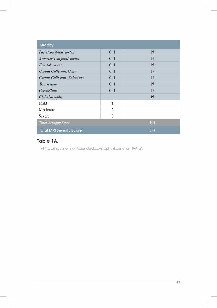
In 1994, Loes et al. was the first to develop an MRI scoring system for leukodystrophy for evaluation of adrenoleukodystrophy (ALD) (Loes et al., 1994a). This was based on findings from a previous study that had characterized the major neuroanatomic location of the disease ( Jensen et al., 1990). Based on the knowledge of the disease location and the presence or absence of focal atrophy, they created a MR severity score (0 to 34) for each patient scan, Table 1A. In this study they retrospectively reviewed 175 MRIs from 85 patients with laboratory confirmed ALD. Most of the MRI exam-inations were scored by only one radiologist, and only a minority, 20%, were reviewed by three radiologists and given a consensus score. The same year this was published, they used this MRI scoring system to evaluate the short term effect of bone marrow

39
transplantation and found it useful for therapeutic evaluation (Loes et al., 1994b).
Five years later the same group developed a similar MRI scoring system for globoid cell leukodystrophy (Krabbe disease) to distinguish between early and late onset disease (Loes et al., 1999).
They modified the scoring system developed for ALD according to the knowledge of the major areas involved in Krabbe disease, Table 1B (Barone et al., 1996; Demaerel et al., 1990). Again they retrospectively reviewed 34 MRI examinations from 22 pa-tients that had been considered for hematopoietic stem cell transplantation (HSCT). Five of those had undergone a successful HSCT. The MRIs were analyzed by one neuroradiologist in at least two separate reading sessions. They concluded that Krabbe disease had characteristic brain MRI findings correlating with the age of clinical onset (Loes et al., 1999).
In 2009, a research group developed a MRI scoring system for metachromatic leuko- dystrophy, (MLD), that was analogous to that used for ALD , with a few exceptions, Table 1C (Eichler et al., 2009). Brain involvement in MLD had previously been well described (Faerber et al., 1999; Kim et al., 1997; van der Voorn et al., 2005). The MRI scoring system therefore was adjusted for prior knowledge of major neuroanatom-ical involvement in MLD. The study design was a retrospective review of 34 MRI examinations in 28 patients with laboratory verified MLD. The scoring system was initially developed and tested by reviewing 10 MRI examinations from patients with MLD at a collaborating center. This was then applied on their 34 MRI examinations which were reviewed by one neurologist and one neuroradiologist with expertise in leukodystrophies. The final score was determined by consensus between the two readers (Eichler et al., 2009).
To differentiate between less defined, heterogeneous lesions and the more well- defined, homogeneous lesions, they applied the terms faint (1) and dense (2) for signal intensities that were T2-hyperintense, Figure 2. From their results, they categorized brain MRI scores into three groups: mild, moderate and severe. Based on this categorization, initial scan results could be compared with findings in patients where longitudinal MRI examinations were used to follow different disease stages. This study also described similarities and differences between MLD, ALD and Krabbe disease, from an MRI scoring point of view. In their conclusion, they stated that MLD had a characteristic image pattern that did not appear to be different among the different age groups or subtypes (Eichler et al., 2009).
These three articles evaluated MRI examinations that had been performed at many different institutions with a variety of images units. All MRI images included T1-weighted sagital and T2-weighted axial imaging planes that were evaluated and used for scoring in ALD and Krabbe disease. The MLD patients were also examined with T2-FLAIR, and the images used for scoring in MLD were axial T2-FLAIR and axial T2-FSE.

40 Christina Sundal • Hereditary diffuse leukoencephalopathy with spheroids
Based on the three MRI scoring systems for ALD, Krabbe disease and MLD, we created a scoring system for HDLS. However, the scoring system deviated from the three prior disease groups, since the MRI pattern for different stages of HDLS was not known. Until 2012 there were only 17 reports on MRI findings in HDLS (Baba et al., 2006; Boisse et al., 2010; Browne et al., 2003; Freeman et al., 2009; Hancock et al., 2003; Itoh et al., 2006; Keegan et al., 2008; Maillart et al., 2009; Mateen et al., 2010; Mayer et al., 2007; Mendes et al., 2010; Moro-de-Casillas et al., 2004; Sundal et al., 2010; Swerdlow et al., 2009; van der Knaap et al., 2000; Van Gerpen et al., 2008; Yamashita and Yamamoto, 2002). However, these reports were on single cases, exam-ined at different time points and thus at variable disease stages. Longitudinal studies had not been performed. Since the evolution of the different stages of HDLS was unknown, a scoring system was created that evaluated the most commonly reported pathological neuroanatomical locations in HDLS, in addition to other commonly reported regions for ALD, Krabbe disease and MLD, Paper IV. We reviewed 20 MRI examinations from 15 neuropathologically-confirmed and CSF1R gene mutation verified HDLS patients. Axial T2-FLAIR and axial T2-FSE sequences were used for scoring. A neuroradiologist (DB) from the Mayo Clinic with expertise in HDLS, and the author were the readers.
Before starting the evaluation, the MRI scoring system was agreed on together with the head of the HDLS consortium (ZW) at the Mayo Clinic. We read the MRI examinations blindly and did not know either the clinical state of the patient at the time of the examination, or what kind of mutations (missense, codon deletion, or in-frame deletion) the patient had. We examined all MRI examinations together and differences in inter-rater score were immediately resolved between us to achieve a consensus of a final score. We re-evaluated all the MRI examinations three months later, following the same procedure, to ensure we could reproduce our scoring system.
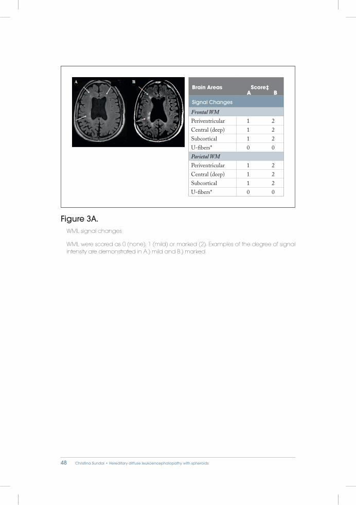
WML were scored as 0 (none), 1 (mild) or marked (2) in order to differentiate the variability in signal intensity (SI) and to improve inter-rater reliability. Examples of the degree of signal intensity are demonstrated in Figure 3.
Cerebral cortex atrophy was similarly scored, 0 (none), 1 (mild) and 2 (marked), in order to define a pattern of recognition for atrophy in HDLS. Examples of the degree of atrophy are demonstrated in Figure 4. We only scored the atrophy for corpus callo-sum (all compartments), cerebellum, and brainstem with 0 (absent) or 1 (present) to make it easier to evaluate and increase the inter-rater reliability. A total severity score (0-57) was calculated for each MRI examination. We also subdivided the total score to analyze the different components, total WML score and total atrophy score, Paper IV.

41

42 Christina Sundal • Hereditary diffuse leukoencephalopathy with spheroids
Brain Areas Score‡ Maximum Score per area
Signal Changes
Parietooccipital white matter (WM) 3†Periventricular 0 1Central (deep) 0 1Subcortical 0 1Anterior Temporal WM 3†Periventricular 0 1Central (deep) 0 1Subcortical 0 1Frontal WM 3†Periventricular 0 1Central (deep) 0 1Subcortical 0 1Visual pathway WM 3†Optic radiation 0 1Lateral geniculate body 0 1Meyer’s loop 0 1Auditory pathway WM 4†Medial geniculate body 0 1Brachium to the inferior colliculus 0 1Lateral lemniscus 0 1Pons (Trapezoid body) 0 1Corpus callosum 3†Genu 0 1Body 0 1Splenium 0 1Frontopontine corticospinal projection fibers 2†Internal capsule 0 1Brain stem 0 1Cerebellum WM 0 1 1†Thalamus (anterior part) 0 1 1†Basal ganglia 0 1 1†
43
Atrophy
Parietooccipital cortex 0 1 1†Anterior Temporal cortex 0 1 1†Frontal cortex 0 1 1†Corpus Callosum, Genu 0 1 1†Corpus Callosum, Splenium 0 1 1† Brain stem 0 1 1†Cerebellum 0 1 1†Global atrophy 3†Mild 1Moderate 2Severe 3Total Atrophy Score 10†
Total MRI Severity Score 34†
Table 1A. MRI scoring system for Adrenoleukodystrophy (Loes et al., 1994a).

44 Christina Sundal • Hereditary diffuse leukoencephalopathy with spheroids
Brain Areas Score‡ Maximum Score per area
Signal Changes
Parietooccipital white matter (WM) 3†Periventricular 0 1Central (deep) 0 1Subcortical 0 1Anterior Temporal WM 3†Periventricular 0 1Central (deep) 0 1Subcortical 0 1Frontal WM 3†Periventricular 0 1Central (deep) 0 1Subcortical 0 1Visual pathway WM 4†Optic radiation 0 1Lateral geniculate body 0 1Meyer’s loop 0 1Optic tract 0 1Pyramidal system WM 3†Corona radiata 0 1Internal capsule 0 1Brain stem 0 1Corpus callosum 3†Genu 0 1Body 0 1Splenium 0 1Frontopontine corticospinal projection fibers 2†Internal capsule 0 1Brain stem 0 1Cerebellum 0 1 2†Cerebellar WM 0 1Dentate nucleii 0 1

45
Thalamus (anterior part) 0 1 1†Basal ganglia 0 1 1†
Atrophy
Parietooccipital cortex 0 1 1†Anterior Temporal cortex 0 1 1†Frontal cortex 0 1 1†Corpus Callosum, Splenium 0 1 1†Corpus Callosum, Genu 0 1 1†Cerebellum 0 1 1†Global atrophy 1†Mild 0 1Moderate 0 1Severe 0 1Total Atrophy Score 7†
Total MRI Severity Score 32†
Table 1B. MRI scoring system for Krabbe (Globoid cell leukodystrophy) disease.
Reproduced with permission from the Journals via Copyright Clearance Centers (Loes et al., 1999).

46 Christina Sundal • Hereditary diffuse leukoencephalopathy with spheroids
Brain Areas Score‡ Maximum Score per area
Signal Changes
Parietooccipital white matter (WM) 6†Periventricular 0 1 2Central (deep) 0 1 2U-fibers 0 1 2Temporal WM 6†Periventricular 0 1 2Central (deep) 0 1 2U-fibers 0 1 2Frontal WM 6†Periventricular 0 1 2Central (deep) 0 1 2Subcortical 0 1 2Corpus callosum WM 4†Genu 0 1 2Splenium 0 1 2Projection fibers WM 6†Internal capsule, posterior limb 0 1 2Internal capsule, anterior limb 0 1 2Midline pons 0 1 2Cerebellum WM 0 1 1†Thalamus (anterior part) 0 1 1†Basal ganglia 0 1 1†
Atrophy
Cerebellum 0 1 1†Global atrophy 2†Ventricular enlargement or inner widening 0 1
Inner or outer CSF space widening 0 1 2Total Atrophy Score 3†
Total MRI Severity Score 34†

47
Table 1C. MRI scoring system for metachromatic leukodystrophy disease.
Reproduced with permission from the Journals via Copyright Clearance Centers (Eichler et al., 2009).
Figure 2. Scoring system in MLD.
Picture A and B demonstrate the differences between faint (1 point, thin arrow A.), and dense (2 points, thin arrow B.). Preservation of the U-fibers are shown in both A.) and B.) (0 points, thick arrow). Measurement of inner atrophy in the third ventricle C.). Reused with permission from the publisher (Eichler et al., 2009).
48 Christina Sundal • Hereditary diffuse leukoencephalopathy with spheroids
Figure 3A. WML signal changes
WML were scored as 0 (none), 1 (mild) or marked (2). Examples of the degree of signal intensity are demonstrated in A.) mild and B.) marked.
Brain Areas Score‡
Signal Changes
Frontal WMPeriventricular 1 2Central (deep) 1 2Subcortical 1 2U-fibers* 0 0Parietal WMPeriventricular 1 2Central (deep) 1 2Subcortical 1 2U-fibers* 0 0
A B

49
Figure 3B. Example of the use of the scoring table
WML signal changes
The most severely affected case with generalized WML that were scored as marked (2) (arrows). Lower pictures arrows show U-fiber involvement.
Signal Changes
Frontal WMPeriventricular 2Central (deep) 2Subcortical 2U-fibers* 0Parietal WMPeriventricular 2Central (deep) 2Subcortical 2U-fibers* 1Temporal WMPeriventricular 2Central (deep) 2Subcortical 2U-fibers* 0Occipital WMPeriventricular 1Central (deep) 1Subcortical 1U-fibers* 0

50 Christina Sundal • Hereditary diffuse leukoencephalopathy with spheroids
Figure 3C. WML signal changes
WMLs were scored as 0 (none), 1 (mild) or marked (2) Examples of the degree of signal intensity affecting corpus callosum are demonstrated in A.) mild in splenium and B.) marked in splenium.
Signal Changes
Genu 0 0Body 0 0Splenium 1 2
A B

51
Figure 4. Atrophy score
Cerebral cortex atrophy was similarly scored as; 0 (none), 1 (mild) and 2 (marked), in order to define a pattern of recognition for atrophy in HDLS. Examples of the degree of atrophy are demonstrated in A.) mild atrophy and B.) marked atrophy.
Atrophy
Cerebral cortexFrontal 1 2Parietal 1 2Temporal 0 0Occipital 0 0Central 1 1
A B

52 Christina Sundal • Hereditary diffuse leukoencephalopathy with spheroids
Diffusion weighted imaging (DWI)The brain is a complex organ consisting of many different structures such as cell membranes, myelin, axons, microtubules, fibers, and macromolecules. These structures will interfere with the movement of water and result in different degrees of restriction. In diffusion-weighted (DW) MRI, the measured signal, for each voxel in the image, is affected by the range and direction of the water diffusion and can thereby give information of the tissue architecture.
A conventional DW-image is acquired by sampling three image planes, each with a different diffusion sensitive gradient direction (GRE-Dir.), which are used to re-construct an average DWI map. Besides these 3 GRE-Dir. images there is also a fourth image sampled without the use of any diffusion sensitive gradient (B0). The 3 GRE-Dir and the B0-image are used to calculate the apparent diffusion coefficient (ADC)-map.
DWI is highly sensitive to changes in tissue structures and can detect an acute cerebral infarction as a hyper intense signal within minutes after an ischemic insult (Moseley et al., 1990; Sorensen et al., 1999). This pattern is thought to be due to cellular swell-ing from cytotoxic edema caused by failure of the Na+/K+ membrane pump causing entrapment of interstitial fluid. In conjugation with the DWI, the ADC map has to be analyzed to verify restriction so the DWI signal change is not a spurious effect of a T2 shine through phenomena (Sorensen et al., 1999). Increased signal on DWI and decreased signal on ADC is the verification of restricted diffusion.
Conventional DWI is useful in the assessment of many WM disorders, besides acute ischemic stroke, since it is a fast technique that is very sensitive for the detection of WM changes on average DWI and ADC maps (Horsfield and Jones, 2002).
Diffusion Tensor Imaging (DTI)Diffusion tensor imaging (DTI) differs from DWI in that it is measured with six or more different GRE-Dir. and at least one B0 image. From these images a more detailed information of water diffusion directionality/anisotropy can be calculated, the Diffusion Tensor (D). D is a diagonally symmetric 3x3 matrix with 6 independ-ent tensor components. The Tensor (D) allows for the description of mobility along each axis and the standard deviation of mobility along three principal directions to characterize the anisotropy. The diffusion tensor is symmetric and in theory it is possible to obtain all components by sampling only 6 non collinear and coplanar diffusion directions, plus 1 non-diffusion weighted (B0), to determine D. However, by adding more diffusion directions, as well as non-diffusion weighted (B0) images, the data will become less biased and more uniform, but result in a longer scan time. From these measurements, the tensor eigenvalues (λ1, λ2, λ3) can be extracted and used to calculate mean diffusivity (MD, ADC), fractional anisotropy (FA), and axial and radial diffusivity (Basser et al., 1994).

53
The Diffusion Tensor (D) parameters most commonly presented are:Eigenvalues: represent the 3 orthogonal diffusivity values of D (λ1, λ2, λ3).
MD: represents the average of the 3 eigenvalues for D.
Axial diffusivity: represents the diffusion along the principal direction (λ1); along the fibers.
Radial diffusivity: which most often is given as an average value of the two diffusion directions perpendicular (λ2+ λ3)/2) to the principal direction (λ1).
FA: gives an estimate, between 0 and 1, of what proportion of D that is due to ani-sotropic diffusion.
DTI has the advantage of being able to indirectly evaluate WM integrity by measuring the water diffusion and water directionality in three dimensions (Le Bihan et al., 2001). This non-invasive observation in vivo provides extensive insights into the structure and geometric organization of the tissue. The structural components of the brain, which restrict the Brownian motion of water, yield different degrees of anisotropy in different regions of the brain. In contrast to conventional MRI, DTI measures physical prop-erties. A change in the physical properties due to a disease process can give distinct “microscopic” information with a high sensitivity for lesion detection, and a more precise neuroanatomical localization of these lesions and their impact on surrounding structures. Compared to conventional MRI, this higher sensitivity for detection of pathological structures may allow for earlier decision-making and better tailoring of interventional procedures in the clinical setting. Ultimately, this can lead to a better correlation with clinical scales and give opportunities to follow the effect of treatments.
Magnetic resonance spectroscopy (MRS)MRS is a non-invasive MR technique that can measure the molecular composition of tissue and determine the relative metabolite concentrations, in contrast to conventional MRI which gives morphological information (Gujar et al., 2005). A great advantage of MRS is that the detection of metabolite changes often precedes structural abnor-malities, and thus MRS can demonstrate abnormalities before they become apparent on MRI. When lesions are found, MRS can give important metabolic information that may help characterize the pathology and may allow for evaluation of response to treatment. For example, in WM disorders metabolic changes in “normal appear-ing white matter” (NAWM) can provide valuable insights into the pathology of the disease evolution; progression or regression (Sajja et al., 2009).
For most clinical studies MRS uses the signal information from hydrogen nuclei (protons), as in conventional MRI. However, MRS can also be used to study other nuclei such as phosphorous-31 and carbon-13. Hydrogen MRS (1H-MRS) has a high sensitivity and is the most abundant and widely used nucleus for clinical studies.

54 Christina Sundal • Hereditary diffuse leukoencephalopathy with spheroids
The position and chemical bond of the hydrogen nuclei in the molecule changes the local magnetic field and thereby changes the spin frequency of the hydrogen nuclei of a specific molecule. The difference in resonance frequency is denoted chemical shift and is often expressed in parts per million (ppm), which do not alter for different magnetic field strengths. The MRS measurement results in a spectrum that is characterized at the frequency axis (x-axis) where each metabolite is positioned according to their specific frequency due to the aforementioned chemical shift. The peak amplitude, which represents the concentration of a given metabolite, is given on the y-axis.
MRS TechniquesAnatomical MR images are used to guide the placement of the volume of interest (VOI) where the spectrum will be acquired. Spectrum acquisition can be done as single voxel spectroscopy (SVS) or multi-voxel imaging (MRSI) using long and/or short echo times (TE). The choice of TE is dependent on the clinical question, e.g. which metabolites that will be studied (Gujar et al., 2005).
For SVS the VOI is defined by the intersection of three consecutively selected orthogonal slices and the most commonly used technique is denoted PRESS (Point-ed REsolved SpectroScopy). More specifically the VOI is obtained by 90o pulse successively followed by two 180o pulses, given at the same time using different field gradient. The signal in the selected VOI is a spin echo (Lenkinski, 2010).
One complication in MRS is that the water concentration is 10,000 times higher than the concentration of metabolites of clinical interest and hence will obscure their signal. Therefore, the signal from water has to be suppressed which is usually achieved by the chemical shift-selective water suppression (Gujar et al., 2005).
MRSI is a multivoxel technique to obtain many voxels concurrently within a slice and thus gives information of the spatial distribution of the metabolite in a single measurement.
It is essential for high quality MRS to obtain a homogeneous magnetic field. How-ever, there are many factors that can influence and cause artifacts, such as motion, lipids, water, and chemical shift displacements due to susceptibility inhomogeneity (Alger, 2010).
Commonly detected compounds by MRS in the brain The use of a short TE (20-40ms) is important to detect metabolites with short relax-ation times such as myo-inositol, amino acids and glutamate while long TE (140-280 ms) is mainly used when it is primarily the major compounds such as NAA, Cho, Cr, Lactate and lipids that are of interest. Several of the other metabolites, such as myo-Inositol will not be visualized unless a shorter TE (20-40ms) is used. Short TE has a higher SNR and less signal loss due to T2 relaxation than longer TEs. On the other hand, with a TE of about 140 ms it is possible to separate Lactate and lipids,

55
since the lactate will invert below the baseline while the lipids remain above. The clinical significance of individual metabolites will depend on the disease process and what aspects of metabolism that a specific metabolite is affecting (Lenkinski, 2010). The most common and generally accepted understanding of the function of individual metabolites can be described as:
Choline (Cho):Cho is a marker of cellular membrane turnover. An increase in Cho peak occurs when there is an active breakdown of myelin due to infarction, inflammation or tumors (Matthews et al., 1991).
Creatinine (Cr):Cr is a marker of the brain’s energy metabolism. Many studies use the Cr peak as an internal standard, as it is believed to be at a stable level throughout the brain paren-chyma and does not change with pathology. However, it may decrease in some diseases with tissue destruction (Hanefeld et al., 1993).
Myo-Inositol (mI):mI is a sugar compound that is mainly synthesized in glial cells. It is a glial marker and found almost exclusively in astrocytes. It is thought to function as an osmoregulator in primary astrocytes which may help protect cells that are exposed to hyperosmotic stress (Isaacks et al., 1994). Increase in mI can be found in glia proliferation (gliosis) or due to increase in glial cell size. This is found in inflammatory processes and, as such, mI is a surrogate marker for brain inflammation. It is also a breakdown product of myelin (Brand et al., 1993).
Lactate (Lac):Lac, a product of anaerobic glycolysis, is below or at the level of detectability in the normal brain. Elevated Lac is found in acute ischemic tissue and when there are active inflammations with cells, especially macrophages (Lopez-Villegas et al., 1995).
N-Acetylaspartate (NAA):NAA is synthesized in the mitochondria of neurons and transported to cytoplasm and axons. Its peak is the highest in normal brain spectroscopy. NAA concentration is found to be approximately equal in the white and grey matter, suggesting that it is a marker of axonal integrity. Reduction of NAA is found in many WM disorders such as leukodystrophies and MS.
NAA is thus regarded as a functional neuronal marker, but it has also been suggested to be a brain osmolyte with possible reversible changes (Baslow, 2003). However, the precise function of NAA is not yet known (Lenkinski, 2010).

56 Christina Sundal • Hereditary diffuse leukoencephalopathy with spheroids
7.4 CEREBROSPINAL FLUID
Cerebrospinal fluid (CSF) investigationAs a diagnostic procedure we performed CSF analysis. A lumbar puncture was done in the L4-L5 interspace. Samples were stored in polypropylene tubes, frozen at -80o C until analysis. The samples were analyzed at the Clinical Neurochemistry Laboratory at the Sahlgrenska University Hospital, Mölndal, Sweden. Biochemical analysis was performed by Dr. Henrik Zetterberg.
CSF BiomarkersThe CSF is in indirect contact with the brain through the extracellular spaces and reflects biochemical changes in the brain. The CSF can therefore be used as a bio-marker (Blennow et al., 2010). A biomarker can serve as an objective measurement of a biological or pathological process that can be used to evaluate disease risk or prognosis, to guide a clinical diagnosis, or to monitor potential therapeutic interventions. CSF biochemical changes might be early indicators of a disease, and hence increase the knowledge about the characteristic pathological mechanism involved in the disease. CSF biomarkers can be divided into two different types: basic or specific biomarkers (Blennow et al., 2010).
Basic BiomarkersBasic biomarkers identify compounds that may coexist in many different disorders.
The brain is protected from various diseases, such as inflammation, infection, and pathogenic molecules in the blood by the blood brain barrier (BBB). This barrier is formed by capillaries that have restricted permeability and serve to protect and control the milieu for neurons.
The standard biomarker for the function of the BBB is the CSF: serum albumin ra-tio. If this ratio is increased, it indicates that the BBB is damaged. Many conditions can increase this ratio such as infections (neuroborreliosis, bacterial meningitis and encephalitis) and inflammation such as Guilliain-Barre, cerebrovascular disorders including vascular dementia and brain tumors.
Alzheimer´s disease (AD) per se has a normal CSF:albumin ratio which is increased in dementia with concomitant cerebrovascular pathology. This ratio can then be used to exclude various other disorders with damage to the brain. A normal ratio can be used to distinguish among other diseases, AD alone, in addition to other markers.
If the central nervous system (CNS) is attacked by a chronic inflammation such as MS or an infection like neuroborreliosis, meningitis or encephalitis, the immune response will produces antibodies. This is named intrathecal immunoglobulin production, and can be measured by two processes:

57
1. IgG and IgM will give a quantitative measurement of the immune response
2. Oligoclonal bands in the CSF will give a qualitative measurement of the immune response
These two measurements are valuable biomarkers to exclude chronic inflammation and infectious disorders in the CSF. Intrathecal immunoglobulin production is normally not present in AD alone. Another important biomarker of infectious disorders is the CSF cell count. Cells of the monophagic system will increase with inflammation and leukocytes will increase in infectious disorders (Blennow et al., 2010).
Specific biomarkersSpecific biomarkers reflect the central pathogenic process of a disease and thus can serve as hallmarks of the studied disorder (Blennow et al., 2010). The following specific biomarkers are widely used:
CSF- Dementia biomarkersCSF amyloid-β (Aβ): Reflects the amyloid, neurofibrillary tangle pathology and the axonal degeneration found in many neurodegenerative disorders where AD is the most central. The Aβ1–42 variant is the main biomarker for Aβ metabolism and plaque formation in the brain.
However, Aβ is also produced during normal cell metabolism, but Aβ1–42 is mark-edly reduced in AD, approximately 50% compared to aged matched controls. Aβ is also reduced in other dementia conditions like dementia with lewy bodies (LBD), frontotemporal dementia (FTD) and others. The reduction is mainly due to aggre-gation of Aβ into plaques, decreasing the availability of Aβ to diffuse into the CSF (Blennow et al., 2010).
CSF Total tau (t-tau): High levels are suggested to reflect the intensity of neuronal degeneration, as well as axonal degeneration and damage in the brain. Increased levels are found in acute brain disorders such as stroke, brain trauma, AD, and other dementia disorders. Creutzfeldt-Jakob disease has been found to have the highest t-tau level. It is suggested that the main increase is t-tau comes from neurofibrillary tangles containing neurons (Blennow et al., 2010).
CSF Phosphorylated tau (p-tau): Reflects both the formation of neurofibrillary tan-gles and the phosphorylated level of tau in the brain. High levels of p-tau have, up to date, only been found in AD and prodromal AD, and can be used to differentiate AD from other types of dementia such as FTD and LBD (Blennow et al., 2010).
Combining the CSF biomarkers of CSF-tau, p-tau and Aβ1–42 achieves high diag-nostic accuracy for identifying AD, prodromal AD, and for differentiating AD from other neurodegenerative disorders (Blennow et al., 2010; Mattsson et al., 2009).

58 Christina Sundal • Hereditary diffuse leukoencephalopathy with spheroids
CSF-Neuronal markers CSF-Neurofilaments: Belonging to the intermediary filament family, neurofilaments (NF) are the main protein constituent of the axonal cytoskeleton. It maintains axonal size and is involved in axonal transport. It is mainly expressed in large subcortical, myelinated axons. Of the three NF subunits, the NF light, (NFL) is the most widely measured and used as a marker of axonal damage. Increased NFL is found in MS, traumatic brain and spinal cord injury, atypical PD, subcortical dementia, ALS, CNS infections and CNS vasculitis (Blennow et al., 2010).
CSF-Tau: Is mainly expressed in cortical axons and is a microtubule associated pro-tein. The increased level in different disorders is previously described.
Both NF and Tau are suggested to reflect the ongoing rate of axonal damage and loss (Blennow et al., 2010).
CSF-Astrocyte markerGlial fibrillary acidic protein (GFAP): Is found in the astrocytes where it is the main intermediate filament of the cytoskeleton. Increased level of GFAP is believed to re-flect astrocyte damage and loss and is found in high levels in Alexander disease but also elevated in other conditions such as astrocytomas, stroke, head trauma, inflammatory CSN disorders (MS), neuromyelitis optica (NMO) and other conditions associated with gliosis (Blennow et al., 2010; Liem and Messing, 2009).
ETICS / ETHICAL ISSUESAll subjects, their proxies and relatives gave informed and written consent. The studies were approved by the ethics committee at the University of Gothenburg (study I and V) and the Mayo Clinic Institutional Review Board (study II, III and IV).
Because genetic testing raises important ethical issues and questions we maintain a good and open dialogue with the original Swedish HDLS family. Our collaborators, under the leadership of Dr. Wszolek at the Mayo Clinic in Jacksonville, Florida, have the same good relationships and protocols that we have.

59

60 Christina Sundal • Hereditary diffuse leukoencephalopathy with spheroids
"The total number of identified HDLS cases
amounted to 15 among 75 individuals in four
generations..."

61
STUDY SETTINGS AND RESULTS
8.1 PAPER I
BackgroundThe original Swedish family was reported in 1984 and consisted of 71 individuals. Sev-enteen were suspected to be affected by HDLS. The age of symptom onset was found to be from 8 years to 60 years. The mean disease duration was 12 years. The development of HDLS was highly variable with three different courses; 1/3 died within three years, 1/3 died after ten years, and 1/3 after more than 30 years.
Subjects and study settingsWe updated the original Swedish family by genealogical and longitudinal clinical ob-servations. Forty family members were neurologically examined, five were interviewed by telephone, and one case from the original publication that was clinically diagnosed with HDLS was neuropathologically investigated. MRI and CSF examinations were performed in two newly identified HDLS cases. One of those cases was also autopsied.
Main resultsThirty-eight individuals were healthy and two new cases developed HDLS during the survey. All five interviewed by telephone were healthy. One case (Case 3:19) who was clinically diagnosed with HDLS in 1984 had only small vessel disease and no axonal spheroids on autopsy. Three cases that had been diagnosed with HDLS in 1984 were further unconfirmed; two from the clinical examination, and one from the telephone interview. The 8 year-old in the 1984 publication did not have any symptoms when we investigated him at age 42 years, thus supporting the implication that this is an adult-onset disease. The total number of identified HDLS cases amounted to 15 among 75 individuals in four generations, including the two new ones, Cases 1 and 2. The clinical course was different in these, with sub-acute and more chronic variants respectively. CSF analysis indicated signs of neurodegeneration without inflamma-tion. The MRI showed WML that were symmetrical, bifrontoparietal and involved periventricular, deep and subcortical regions, but did not affect the U-fibers. The genu and splenium portions of the corpus callosum were affected by WML. In Case 1, HDLS was autopsy-verified as being similar to the previously reported four family cases (Axelsson et al., 1984). There is still an open question whether the primary event in HDLS is axonal or primary myelin. The detailed symptomatology in our two new cases was divergent, supporting both a sub-acute and more chronic course.
BACKGROUNDS

62 Christina Sundal • Hereditary diffuse leukoencephalopathy with spheroids
8.2 PAPER II
BackgroundAn international HDLS consortium was established in 2005 led by Dr. Zbigniew Wszolek, M.D., headquartered at the Mayo Clinic, in Jacksonville, Florida. Dr. Wszolek became interested in HDLS after reading the 1984 publication (Axelsson et al., 1984). He decided to start to collect HDLS cases and, via lectures, congress presentations, and publications, spread information about HDLS and encouraged col-leagues to collaborate. This lead to a steady increase in collaborators and HDLS cases collected both retrospectively and prospectively. All cases confirmed by brain autopsy or biopsy were enrolled into a HDLS study with the goal of finding the causative gene. To ensure cases demonstrated pathological features of HDLS, consistent with the original Swedish family, they were all examined by Dr. Dennis Dickson, M.D., a neuropathologist at the Mayo Clinic in Florida. In this growing HDLS collection, it became evident that they had all been misdiagnosed with other more common neurological conditions.
Subjects and study settingsWe retrospectively reviewed the medical records from three small kindreds from the Mayo Clinic HDLS collection. The inheritance patterns in these cases were not obvi-ous and could easily be misclassified as a sporadic disorder. Our goal was to illustrate that familial clustering of HDLS is not always obvious because of phenotypic heter-ogeneity. The four patients had been identified and followed-up in Norway, Germany, and the US. We reviewed the clinical, neuroimaging and neuropathological findings.
Main resultsThe mean age of symptom onset was 44 years (range, 36-52 years), mean disease dura-tion was six years (range, 3-11), and the mean age at death was 48 years (range, 40-63 years). Neuropathological findings were consistent with those found in the original Swedish HDLS family with demyelination, abundant axonal spheroids and preser-vation of the deep grey nuclei structures. The clinical symptoms progressed in parallel with personality changes, executive dysfunction, memory decline and eventually a multifocal encephalopathy. MRI demonstrated WML with a frontal predominance with involvement of both corpus callosum and corticospinal tracts. Treatments with both intravenous (I.V.) and oral (p.o.) steroids, subcutaneous interferons, I.V plasma-phoresis and L-dopa were attempted for some of the cases but without any benefit. All four cases had been misdiagnosed as having more common neurological disorders.

63
8.3 PAPER III
BackgroundFrom the collected Mayo Clinic HDLS cases we decided to do a genetic investigation on those that fulfilled the criteria of: 1) complete clinical information, 2) neuropatho-logically verified, 3) MRI and 4) available blood samples.
To ensure cases demonstrated pathological features of HDLS, consistent with the original Swedish family, they were all examined by Dr. Dennis Dickson, M.D., a neuropathologist at the Mayo Clinic in Florida.
Subjects and study settings14 kindreds from US, Germany, Norway and Scotland were collected. The largest family, VA, was selected for genome-wide linkage study.
Main resultsThe Mayo Clinic HDLS team had been collecting HDLS cases and conducting genetic analysis on some of their cases since 2005. When I arrived at the Mayo Clinic genetic laboratory, Dr. Rademakers lab, in February 2011, one of the fourteen HDLS families (VA-family) had been selected for investigation with a genome wide non-parametric linkage study in order to apply a model-free analysis. The results indicated that a locus on chromosome 5 had a LOD score of greater than 2.5. This family was then studied with parametric linkage analysis and subsequent a significant linkage was identified on chromosome 5q34. The LOD-score was even higher when the parametric linkage analysis was used, further strengthening the data.
No other loci reached significance. The candidate region was further narrowed by obligate recombinants to genetic distance of 30.3 at the 5q34, containing 233 possible genes. Whole exome sequencing was selected as the next method because Sanger sequencing analysis of all genes in the region would have been more time consuming and costly. A targeted next-generation sequencing strategy on chromosome 5 might have been an option but is also expensive, especially when the goal was to analyze a small set of patients. Whole-exome sequencing is widely used and optimized, so even though all of this information was not needed, this was still the method of choice for the Mayo Clinic cases.
A search for shared heterozygous variants between the two patients of the selected family were performed and filtered against all known variants (SNP). Consequently, 2 different mutations in the 5-hydroxytryptamine (serotonin) receptor 4 (HTR4) gene and in the colony stimulating factor 1 receptor (CSF1R) gene were found. These both segregated with the disease in the extended family and were not present in 660 controls. Exon sequencing of HTR4 and CSF1R in the 13 probands from the other neuropathological-verified HDLS families was then performed. This identified

64 Christina Sundal • Hereditary diffuse leukoencephalopathy with spheroids
heterozygous CSF1R mutations in all 13 probands, but no mutations in the HTR4 gene. Segregation analysis confirmed transmission of the CSF1R mutation and co-segrega-tion with the phenotype in multiple affected individuals from all the HDLS families.
Segregation analysis was confirmed by Sanger sequencing of the relevant exon in all DNA samples available for each family.
Screening of the original Swedish HDLS family In parallel with the genetic work on the Mayo Clinic HDLS collection, the blood samples were received from the original Swedish HDLS family. By the time that I had purified the DNA from these samples of whole blood, the CSF1R mutation was identified in the Mayo Clinic HDLS families. It was therefore decided to start screening these Swedish samples for CSF1R mutations on chromosome 5.
All 22 exons of the CSF1R gene from the two affected Swedish patients, together with one known CSF1R mutation carrier from the Mayo Clinic HDLS collection (proband of MD family = positive control) and one control sample (negative control), were analyzed by PCR and sequencing, Figure 1.
In brief, PCR reactions contained 50ng of DNA template in a 25 μl volume containing 20 pmol of each primer, 0.2mM dNTP’s, 1 unit of Taq polymerase (Qiagen). The ep-pendorf thermal cyclers and a 60-50 touchdown protocol were used for amplification. The first cycle used 60oC and 50oC in the last cycles, which makes it difficult for prim-ers to anneal to anything but a perfect matched sequence. Lower temperatures allow annealing to go faster giving more PCR product. The temperatures can be predicted but usually these are tested in the lab to establish the optimal PCR conditions. The touchdown PCR with the 10oC range of annealing temperatures prevents formation of nonspecific PCR product and eliminates the need to optimize each primer set to a specific temperature. The resulting PCR products were visualized on 2% agarose gel to ensure correct fragment size prior to proceeding with the sequencing reaction. All amplified samples were then purified using Multiscreen (Millipore) technology to remove surplus reagents from the reaction and sequenced in both directions using M13 sequencing primers and Big Dye Terminator v3.1 Cycle Sequencing chemistry (Applied Biosystems) following the manufacturers protocol. Sequencing reactions were purified using the Montage system (Millipore) and analyzed on an ABI3730 Genetic Analyzer (Applied Biosystems). Sequencher software was used to analyze the sequence.
No mutations were identified in any of the CSF1R exons from the two patients from the original Swedish family or the negative control. A CSF1R mutation was detected in the positive control case from the HDLS MD-family. The whole sequencing procedure was consequently reinvestigated both manually and in automatic mode, which confirmed the first result.
In the meantime, all of the 14 HDLS families from the Mayo Clinic collections were confirmed to have the newly identified CSF1R gene mutation. It was decided that

65
the Swedish family should not be included in the CSF1R study because only one sample was autopsy-confirmed HDLS and the only paraffin-embedded tissue samples for the other four affected individuals were very old. Multiple individuals had also handled these samples and it was not possible to extract any DNA from them. The Rademakers laboratory team did not feel comfortable reporting that there was locus heterogeneity with only one neuropathological verified Swedish sample. Since there were only two Swedish HDLS cases that could be genetically investigated, and only one of them fulfilled the inclusion criteria for the study, it was decided not to mention the Swedish family in the Nature Genetics paper because one could not be certain of the result. It was realized that the analysis on the Swedish samples needed further investigation and more blood samples in order to confirm or rule out a mutation in the CSF1R gene. To expedite this, more blood from the Swedish samples for cDNA analysis were requested from Dr. Oluf Andersen. In the meantime, the mutation discovery, paper III, was completed (Rademakers et al., 2012a).
The work on the Swedish cases continued. In order to ascertain whether other types of CSF1R mutations could be involved, haplotype sharing between the two affected individuals was looked for to determine if a set of DNA sequences were inherited together in the CSF1R region on chromosome 5. PCR procedures, as described above, were applied with the exception that the sense primer of the marker pair 5’ was adjust-ed with a fluorescent FAM- label. This is the most commonly used fluorescent dye for labeling oligonucleotides. Using this fluorescently labeled amplification sample could be visualized using the ABI3730 Genetic Analyzer, run on the Genemapper software (Applied Biosystems, Foster City, CA, USA). Genotypes were assigned for each marker in the region and the haplotypes were assessed for shared genotypes. This result was negative, further excluding CSF1R as the causative gene in this family. Complete or partial CSF1R gene deletions were also excluded by PCR and fluorescent primers covering the whole gene.
When the new blood samples arrived from Sweden it was necessary to exclude the possibility of heterogeneity between the genomic DNA sequences and those associ-ated with transcription. This was investigated with cDNA transcript analysis. RNA (PaxGene Blood RNA kit) was extracted and cDNA was synthesized using reverse transcription. The cDNA was amplified and sequenced following specific protocols and cDNA was synthesized using PaxGene RNA as template. Reverse transcription used a 1:1 mix of random hexamers, oligo (dT) primers and the SuperScript III system (Invitrogen, Carlsbad, CA, USA). Following the manufacturer’s protocol, the reactions were cycled for 10 minutes at 25oC, then 50 minutes at 50oC, 5 minutes at 85oC, and finally cooled to 4oC. PCR analysis using the cDNA template only showed the expected size fragments upon agarose gel-electrophoresis. PCR product sequencing did not show any mutations. The result demonstrated no heterogeneity, further excluding mutations in the CSF1R gene.
Since Nasu-Hakola disease (NHD) has been found to be implicated in CSF1R signa-ling and has a phenotype very similar to HDLS exempted bone cysts, it was necessary
66 Christina Sundal • Hereditary diffuse leukoencephalopathy with spheroids
to rule out these genes for the Swedish samples. PCR and sequencing protocol as previously described was used to study TYROBP and TREM2 that are causative for NHD, as well as FLT3 and KIT, members of the same tyrosine kinase family as CSF1R. However, since no mutations in these genes were identified the cause of the disease in the Swedish HDLS family still remains elusive.
14 different CSF1R mutations were identified in the 14 Mayo Clinic families. One kindred had a de novo mutation. All mutations affected the intracellular tyrosine kinase domain of the CSF1R gene encoded by exons 12-22. We found 10 missense mutations, one single codon deletion, and three splice site mutations that lead to an in-frame deletion of exon 13 (NO) and 18 (CA2, FL2). There were differences in the age of onset, disease duration and age of death among the cases, even within the same family. This suggests unidentified genetic or environmental factors influencing the penetrance. Initial symptoms and evolving clinical features also demonstrated substantial variations within and across families.
8.4 PAPER IV
BackgroundSince our group identified the CSF1R gene mutation as causative for the Mayo Clinic HDLS collection the MRI patterns of these patients was investigated.
Subjects and study settings20 MRIs from 15 patients in nine families with neuropathologically verified HDLS and CSF1R mutation carriers were reviewed. Sagital T1-weighted images, axial T1 and T2 -weighted images, and axial fluid attenuated inversion recovery (FLAIR) im-ages were obtained in all patients except one, who had a proton density measurement series. Together with Dr. Broderick, a neuroradiologist, the images were reviewed blindly, twice, three months apart, and differences in interpretation resolved between the two observers. The images were assessed for WML, atrophy and grey matter involvement. A severity score (0-57) was calculated for each MRI based on a point system modified from previous leukodystrophies scoring systems. Since the pattern and evolution of MRI abnormality in HDLS is unknown, we designed the scoring system to evaluate all cerebral regions with respect to abnormal signal intensity in the WM and atrophy.
Main results93% (14/15 patients) demonstrated localized WML, and one had generalized WML with slight involvement of the U-fibers. All patients had bilateral, but asymmetric lesions. WML with frontal predominance involving deep and subcortical regions, were found in all cases. 93% (14/15) had periventricular involvement, while 73% (11/15)

67
had involvement of the corpus callosum. Typically, brain atrophy was present in the same region as the WML. Central atrophy was invariably also present. Corticospinal tract involvement occurred late in the disease course. There was no significant grey matter pathology, no brainstem pathology, and enhancement was absent. Cerebellar pathology was minimal. Indicators of rapid disease progression were; age less than 45 years, female sex, more than 15 points on the severity score, WML extending beyond the frontal region, and a deletion type for the mutation.
8.5 PAPER V
BackgroundBecause the temporal evolution of the WML in HDLS is unknown advanced neu-roimaging was selected to provide insights into the pathogenesis.
Subjects and study settingsOne recently identified HDLS case from the original Swedish family was investigated with repeated DWI/DTI and MRS examination during a 16 month longitudinal study.
Main resultsIn 5 consecutive MRI examinations we found changes in 3 temporal stages of HDLS evolution:
Stage 1: The initiation of the disease seems to start in the anterior, periventricular WM where lesions remain prominent throughout the study period. However, when these lesions first develop on MR imaging there is already an insidious spread of the disease that involves the WM of the centrum semiovale and the body of corpus callosum, the earliest stage. There were clear abnormalities on MRS and DTI also in this region which, however, did not show any signal abnormalities on conventional MRI until much later, when the patient was too affected by his disease to allow for additional MRS and DTI examinations. At this stage, the patient had frontal lobe syndromes with total loss of insight and impaired initiative. He had slight increased tone in the lower extremities, decreased deep sensibility in the feet bilaterally, slight hyperreflexia, but extensor plantar responses were normal. There was occulomotor impersistence on saccade testing.
Stage 2: As the disease progressed beyond the frontal regions new areas of signal abnormalities began to develop in the periventricular posterior-WM. On the first examination, the conventional MRI showed NAWM, but the eigenvalues demon-strated already clear changes with increased axial diffusivity which on subsequent examinations, when T2 signal abnormalities began to develop, showed a marked decrease, while radial diffusivity was normal on all three examinations. The patient demonstrated at this stage further decline in executive function and had apparently

68 Christina Sundal • Hereditary diffuse leukoencephalopathy with spheroids
no feelings or emotions. He now needed moderate assistance in activity of daily living (ADL). There was increased gain of vestibulo-ocular reflex (VOR) and pathological primitive reflexes. The extensor plantar responses were bilaterally positive and he had moderate general tetrarigidity (gegenhalten), more on the right side.
Stage 3: This later stage was found in the center of the primary anterior WM ab-normalities, behind the expanding high signal rim. Here, the eigenvalues showed marked increase in both axial and radial diffusivity already on the first examination. These changes then progressed markedly on each subsequent examination indicating general and progressive tissue destruction with complete loss of WM integrity and disappearance of diffusion anisotropy in the end. On MRS, myo-Inositol was slightly increased initially and increased further on the first follow-up, but dropped slightly on the third when tissue destruction was most pronounced. Choline, Creatine and NAA all showed a progressive and marked decrease over time, reflecting progressive tissue destruction. The loss of all metabolic function in the end was further emphasized by the disappearance of the lactate peak on the last examination; glucose metabolism had ceased. The patient now had dramatically changed behaviors. These included: disquiet, constant roaming, complete initiative loss, greedy eating, ignoring his children, severe amnesia, and almost no contact with reality. The patient also demonstrated reduced facial expression, hesitant responses to questions (answered inappropriately with; yes /no), pronounced frontal lobe syndrome with severe ideomotor apraxia, bizarre movements, and incontinence for urine and feces. Neuropsychiatric tests could not be carried out owing to impersistence.
Five months after the last MRI the patient roamed less but developed a tottering gait, compulsive movements and was constantly shutting cabinet doors. Intermittently, the gait demonstrated small steps accompanied by tripping and freezing. There was also a massive startle movement. He had severe visual disturbance (guides with hands), probable right-sided hemianopia. There was no tremor.
From study V we conclude that these quantitative techniques, when combined, reflect the different stages of the HDLS pathology and can be used to monitor disease evolu-tion, particularly in the context of future treatment trials. We could not draw any firm conclusion whether the primary event is neurodegenerative or demyelinating. Our finding of a peripheral rim of T2/DWI-a signal that showed restriction on MD/ADC, but without contrast enhancement, and with centrifugal migration from the anterior ventricular horns, might be pathognomonic for HDLS of the original Swedish type.

69

70 Christina Sundal • Hereditary diffuse leukoencephalopathy with spheroids
"The inheritance is autosomal dominant with variable penetrance, but sporadic cases also exist."

71
Our studies confirm that HDLS is an adult-onset, invariably lethal, brain WM dis-order. The inheritance is autosomal dominant with variable penetrance, but sporadic cases also exist. The clinical symptoms are characterized by a constellation of fea-tures, including personality changes, cognitive dysfunction and motor impairments that extend across multiple neurological domains over the course of the disease. The neuropathological hallmarks of HDLS are brain WM degeneration with the presence of axonal spheroids.
Since the original Swedish publication in 1984 (Axelsson et al., 1984), there have been more than 30 articles describing over 70 cases with HDLS. It has been classified as a rare neurodegenerative disorder. However, we retrieved 14 families collected at the Mayo Clinic, and the disease has a world-wide distribution with case reports origi-nating from Australia, Canada, France, Germany, Ireland, Israel, Japan, Netherland, Norway, Portugal, Scotland, and the US. In addition some of the US cases are dece-dents from European countries such as Poland, Germany and Italy (Rademakers et al., 2012a; Sundal et al., 2012c; Wider et al., 2009). Furthermore, it is our experience that most cases had erroneous psychiatric or neurological diagnoses before they were classified as HDLS. Therefore, while the disease may not be that rare, no prevalence data are available.
Our study clearly demonstrates that HDLS is a heterogeneous disorder with variable phenotypes, especially in the early disease stages. In the Swedish cases, Case 1 had a sub-acute evolution of multifocal encephalopathy and Case 2 had a more chronic course of severe frontal lobe symptoms that were later followed by multifocal neurological signs.
The Mayo Clinic HDLS collection seems to include at least three phenotypic pres-entations. The first is dominated by a predominantly frontal lobe presentation; the second by extrapyramidal presentation, and the third by pyramidal signs. However, the most common initial presentations in 16 of the 24 cases were frontal lobe signs with personality changes, memory problems, loss of insights and dysexecutive symptoms such as apathy and emotional blunting. This indicates that cognitive impairment is an early clinical feature of HDLS and possibly an anticipatory symptom, as supported by previous reports (Axelsson et al., 1984; Wider et al., 2009).
Two Mayo Clinic patients with initial pyramidal signs soon developed cognitive impairments with marked executive dysfunctions and short-term memory deficits. Extrapyramidal features with bradykinesia and reduced strides were found in two cases and these also had early reduced performance on neuropsychological testing for executive dysfunctions.
DISCUSSION

72 Christina Sundal • Hereditary diffuse leukoencephalopathy with spheroids
With the progression of the disease, all cases developed severe multi-focal symptoms from the following domains; frontal, pyramidal, extrapyramidal, parietal, and occipital lobes. Finally, all cases reached a multisystem encephalopathic stage. The severity and degree of these combined symptoms were divergent, dividing HDLS into at least two symptomatological courses with an evolution of either a sub-acute or a more chronic combination of these symptoms. Pseudobulbar palsies, such as dysarthria, dysphonia, and dysphagia, were present in all patients at the advanced stage of the disease. The evolution of the disease over time was variable and the existence of sub-acute and more chronic courses of HDLS was confirmed.
Interestingly, in Swedish Case 2, the gait impairment seemed to evolve through stages of paratonia, “gegenhalten”, spasticity, startle reactions and extensor rigidity. At the first visit, which was10 months after symptom onset, the patient had slight paratonia in the lower extremities. Six months later, gegenhalten had developed and he demonstrated severe roaming similar to akathisia in the constant walking around. However, the gait pattern was normal. This roaming behavior was also described in one of the Mayo Clinic cases. Additionally, four of the Mayo Clinic cases developed shuffling gait, and in some cases, this was further deteriorated by initiation failure, hesitation on turns, and poor balance with retropulsion. These features were also not-ed as intermittent signs in the Swedish Case 2 on the last examination that was 36 months after symptom onset, when muscle tone had changed to rigidity. Two Mayo Clinic cases with extrapyramidal features, such as tremor of resting type and rigidity, had gait impairment that seemed to be a combination of a frontal gait disorder and a parkinsonian gait. As the pathophysiology behind this dysfunction is not clear, one study has hypothesized the involvement of the basal ganglio-thalamocortical loop (Iseki et al., 2010). Extrapyramidal features in HDLS have been analyzed in a separate paper and are suggested to be caused by disturbances in a complex network composed of the basal ganglia, thalamus, and cerebral cortex that are interconnected through tracts embedded in the WM (Sundal et al. submitted 2013).
Until 2011 the causative gene(s) for HDLS were unknown, but the CSF1R gene mutation responsible for the Mayo Clinic cases was identified. All 14 families had dif-ferent mutations encoded by exons 12-22 in the intracellular tyrosine kinase domain of CSF1R. There were ten missense mutations and one single-codon deletion. In addition three mutations were splice-site mutations, leading to the in-frame deletion of exon 13 (NO-family) or exon 18 (CA2-, and FL2- families) (Rademakers et al., 2012a). CS-F1R is a cell surface receptor involved in the proliferation, differentiation, function and survival of mononuclear phagocytic cells, including the microglia of the central nervous system (Wang et al., 2012). This mutation was not found in the original Swedish family when all of the 22 exons contained in the CSF1R gene were sequenced. We also ruled out the possibility of exon deletions and haplotype sharing among the two Swedish patients, further excluding CSF1R as the causative gene in the Swedish family.
For the CSF1R mutation carriers we demonstrate in paper IV that the different mutations present with different phenotypes; mutation of the deletion type had an

73
earlier age of symptom onset and shorter disease duration than that of the amino acid point mutation type (Sundal et al., 2012d). Consistent with previous publications, we found WML to be predominantly frontally or frontoparietal, with involvement of periventricular-, deep-, and subcortical regions. There was also degeneration in the corpus callosum, pyramidal tracts, and associated cortical atrophy (Wider et al., 2009). However, there was considerable interfamilial and intrafamilial variability, in line with other HDLS reports (Axelsson et al., 1984; Van Gerpen et al., 2008). This variability points to additional genes or environmental factors. Notwithstanding this variability, we observed systematic differences in the MRI pattern among the Swedish and the Mayo Clinic cases. In contrast to our Swedish cases, study I and V, who had symmetrical WML starting in the periventricular region and spread out to, but did not involve the U-fibers, the WML in the Mayo Clinic HDLS cases were more asymmetrical. In some cases these were localized deeper in the WM regions rather than in the periventricular regions. The case with the most severe disease stage had slight U-fiber involvement, which was not found in the Swedish cases. The corpus callosum also seemed to be involved differently, which depended on the type of CSF1R mutation. In contrast, the body of the corpus callosum was unaffected in the Swedish family. These findings suggest that there may be different pathological mechanisms involved in the initiation of the disease process in HDLS between the Swedish and Mayo Clinic cases. I addition to the negative findings concerning the CSF1R mutations in the Swedish family, there may be contribution from epigenetic or environmental factors.
Neuroimaging used for diagnostic clues and to follow the evolution of HDLS
In paper IV we describe the most typical MRI patterns of the Mayo Clinic HDLS cases, carriers of the CSF1R gene mutation, and developed a scoring system. The knowledge of this pattern recognition may assist in diagnosing HDLS. The scoring system can also assist in following the progression of the disease, predict the prognosis, and evaluate future clinical trials.
The scoring system has a point range from 0-57; it also includes regions that are not characteristically affected by HDLS, such as the basal ganglia, thalamus, and cere-bellum. With the increased knowledge obtained from our study, we should be able to create a more narrow scoring system that is adjusted to the major abnormalities found on the MRIs of HDLS patients. Subsequently, we have modified the original scoring system and have suggested a new, easier to use, scoring system, which is more in line with our HDLS findings, shown in Appendix Table 1.
The cases used for the scoring study had been examined in different centers with dif-ferent MRI equipment, which in itself creates certain limitations. Even if the magnets had identical field strength and used the same protocols, there are differences between MRI manufacturers, with regard to image appearance, which can result in variability in the interpretation (Reig et al., 2009). Another important factor for optimal outcome

74 Christina Sundal • Hereditary diffuse leukoencephalopathy with spheroids
is to include patients who are at the same disease stage. When including patients at variable disease stages, as we did in our study, the result of the evaluation will be more diverse, and it will be more difficult to establish the natural course of the disease.
Ideally, this new scoring system for a qualitative assessment would be used in a pro-spective study using a standardized MRI protocol with the same makes of hardware and software, the same magnetic field strength, gradient system, and pulse sequences. Moreover, the patients should be grouped according to their disease stage. Instead of scoring the WML according to their signal intensity, as was done here and in MLD (Eichler et al., 2009), the Scheltens visual rating scale could have been employed. This is validated and has a good inter-rater and intra-rater reliability (Scheltens et al., 1993; Scheltens et al., 1998). However, the decision was taken not to use this as it was developed for dementia and focuses specifically on the deep WM implying an uncertainty with regard to the coverage of the anatomical distribution in HDLS. Nevertheless, it would have been interesting to apply the Scheltens scoring scale, in addition to the one that developed here, and compare the results.
To increase the value of the scoring system, it could have been tested internally for inter-rater and intra-rater reliability by measuring the Kappa statistics before starting. There are also software tools for semi-automated and automated volume measure-ments, which would have increased objectivity and reproducibility. This would allow for the possibility of defining automated morphologic criteria, such as contour and shape patterns, to further increase the reliability of the study (Melhem et al., 2002). However, these quantitative automated methods require validation against the qual-itative methods by manual outlining of the lesions (Maillard et al., 2008).
Primary demyelination vs neuroaxonal degenerationMyelin damage is extensive and potentially the primary feature of HDLS. However it may be secondary to axonal damage. The abundance of axonal spheroids in HDLS, which is the hallmark of the disease, was suggested to support axonal injury as the primary cause of HDLS in the original paper (Axelsson et al., 1984).
The following discussion is based on the results from our I and V papers:
The most pronounced lesions on DWI, which were found in the anterior periventricu-lar regions, initially showed a thick, peripheral rim of high T2 signaling that expanded peripherally to become thinner when reaching the U-fibers, which appeared unaf-fected, Figure 5. The posterior WM changes showed a similar pattern of centrifugal progression of high signal rims on T2, but there was no T2 signal abnormality in this region during the first examination. Simultaneously, there was also a progression of DWI-a signal abnormalities in regions where the WM had initially appeared normal. When comparing DWI-a and MD/ADC maps for subsequent examinations, the areas of highest signaling on DWI-a maps showed restriction on MD. When the peripheral high signal rim reached the subcortical region and became thinner, the DWI-a signal dropped, and the MD signal subsequently increased. There was a pro-

75
gressive signal drop on DWI-a and an increasing MD signal located centrally, behind the rim. FA was already markedly reduced in this anterior central region at the first examination and continued to decrease for each subsequent examination. MD was normal on the first examination but increased markedly on the next two examinations. The same changes were seen in the posterior WM but to a lesser degree.
The DTI eigenvalues showed an increase in axonal diffusivity in the center of the lesion in the anterior-WM that progressed markedly for each subsequent examination. Radial diffusivity also showed a similar evolution with initially increased values that progressed for each examination. These values for axial and radial diffusivity indi-cate a more or less complete structural loss of the tissue in this region. With clinical disease progression, lesions also developed in the posterior periventricular WM. It is reasonable to assume that the lesions observed at 18 and 22 months in the posterior periventricular WM were more active and represented a relatively early development of WM tissue destruction. The high signal areas in this posterior region initially showed a marked decrease in axial diffusivity while the radial component remained unchanged. This would indicate that axonal damage is more prominent in earlier stages of the WM damage associated with HDLS.
On MRS, there was a severe reduction in NAA, a functional neuronal marker, in the center of the anterior-WM at the first examination. This dropped further on subse-quent examinations, probably indicating a very early stage of the pathological process as we detected a reduction in the NAA in the NAWM in the centrum semiovale.
Figure 5. Case 2 – The MD and DWI-a images at 18 months after symptom onset demonstrating the progressive changes with initially bulky high signal on DWI-a with restriction on MD to subsequent thinner, more peripheral bands adjacent to cortex and U-fibers. When the disease process reached the periphery of the white matter on the last examination the high signal bands on DWI-a dropped with subsequent increase in signal on MD to indicate less restriction.

76 Christina Sundal • Hereditary diffuse leukoencephalopathy with spheroids
In parallel with the first MRI study, 10 months after symptom onset in case 2, the CSF examination showed that NFL level was four times as high as the normal reference value. Twelve months later, the NFL had increased to almost 14 times the normal amount, demonstrating axonal damage (Sundal et al., 2012a).
We were able to re-examine the histopathological specimens from Case 1, Figure 6, 7, and 8. We observed that the pathological process had no predilection for specific neuron populations or specific axons. Instead, all information pointed to a process that indiscriminately proceeded centrifugally from the tissue surrounding the lateral ventricles and spread out to the inner surface of the U-fibers. Histologically, we confirmed an edematous border between the severely affected central layer and the U-fibers with no exudation of albumin. Therefore, the edema was probably cytotoxic rather than vasogenic. We reexamined histological sections from this area and noted that the axonal spheroids were almost exclusively found in the edematous border of the lesion at the inner layer of the U-fibers, Figure 6. The WM between this border and the ventricular wall was severely damaged with heavy gliosis, macrophages, and activated microglia. In this central region virtually no axons or myelin sheaths were detected by light microscopy. On the other hand, the brain tissue peripheral to the edematous border, including the cortex, looked normal, except for a minor degree of gliosis, Figure 6. We believe that this edematous region corresponds to the low diffu-sion rim demonstrated by our MR studies. Apparently, this expanding rim is the core of the pathological process. This seemingly destructive process spreads centrifugally up to the inner border of the U-fibers. We also applied the immunocytochemical marker for amyloid precursor protein (APP) which was not used in paper I of this thesis. APP is normally transported from the neuronal cell body through the axon by fast axonal transport and will not reach immunocytochemically detectable levels in a normal homeostatic state (Rodrigues et al., 2012; Sherriff et al., 1994a). Axonal accumulation of APP is a marker for antegrade axonal transport blockage and is also considered to behave like an acute phase reactant in the neuron. This was first demon-strated in traumatic diffuse axonal injury (DAI), followed by MS and other acute demyelinating disorders. It is also seen in a wide range of neurodegenerative disorders such as Alzheimer’s disease, Parkinsonism-dementia of Guam, Creutzfeldt-Jakob disease, and in virus-associated neurodegenerative conditions such as HIV-dementia, malaria, in human T-cell lymphotropic virus 1-associated myelopathy, and in stroke (Coleman, 2005; Rodrigues et al., 2012; Sherriff et al., 1994b).
We found APP expression in spheroids and in a few axons of different sizes, Figure 7 and 8, which indicates that the antegrade axonal transport is affected in HDLS. This is in accordance with the electron microscopic findings, demonstrating that neurofilaments and other organelles are accumulated in the spheroids. APP was also demonstrated in the Mayo Clinic HDLS cases in study II, III and IV, as well as in other HDLS cases (Baba et al., 2006; Lin et al., 2010).
We also observed that the number of APP-expressing spheroids was much greater than the number of neurofilament proteins (NFP)-expressing ones, Figure 7.

77
In general, more small spheroids were detected with the APP staining, which seems to indicate that it is more sensitive or is a marker of an earlier stage in the axonal pathology. In some places there were thin sheaths of myelin embedded around the spheroids. It remains to be investigated whether the transport failure is the primary pathological mechanism or a secondary event.
Figure 6. a.) Loss of myelin staining in the white matter, except for a subcortical band (Luxol fast blue – cresyl violet. C: cortex. W: white matter. Bar: 500 µm).
b.) Same area: Severe gliosis in the white matter (Anti-GFAP. C: cortex. W: white mat-ter. Bar: 500 µm).
c.) Same area: APP-positive spheroids (ar-rows) at the border between myelinated and demyelinated white matter (Anti-APP. C: cortex. W: white matter. Bar: 500 µm).

78 Christina Sundal • Hereditary diffuse leukoencephalopathy with spheroids
Figure 8. In addition to spheroids, a few APP-positive axons (arrows) were found in the edema-tous border of the white matter lesions (Anti-APP. Bar: 70 µm).
The following arguments support primary axonal damage:
a. Many spheroids
b. MRS demonstrates reduction of NAA in early phase in the NAWM of centrum semiovale
c. A thin layer of myelin embedding some of the spheroids
Combining the results from advanced MRI studies, CSF analyses and neuropathology suggest that HDLS is most likely to be a primary neuroaxonal degeneration rather than a demyelinating disorder with secondary axonal loss.
Figure 7. a.) APP-positive spheroids of different sizes (arrows) at the border between myelinated and demyelinated white matter (Anti-APP. Bar: 150 µm).
b.) Neurofilament-positive spheroids at the border between myelinated and demyeli-nated white matter (Anti-NFP. Bar: 150 µm).

79
What is the primarily insult that leads to axonal degeneration and the formation of spheroids?
In order to try to answer this question, we reinvestigate the MRI examinations done on Case 1 six months after symptom onset, Figure 9. On these images there is a marked restriction on the ADC map in the anterior corpus callosum that likely rep-resents stage 2 in the disease process as described in Case 2. Although based on only two cases, we propose the preliminary conclusion that the edematous border found in the histological reexamination of Case 1 represents the same pathological process as the expanding rim of restricted diffusion found by DWI/DTI in Case 2. These find-ings suggest that the origin of the axonal damage may be related to a periventricular gradient of a locally acting noxious factor, the nature of which remains to be deter-mined. In this aspect it is interesting to draw some hypothetical parallels to Alexander disease. It has been suggested that a toxic factor in Alexander disease is related to the accumulation of mutated GFAP which seems to be deleterious to the astrocytes (Liem and Messing, 2009). However, Alexander disease, as well as many neurodegenerative disorders are associated with the accumulation of aggregated proteins such as in SCAs (1-3, 6-8, 17) PD/ Huntington disease; familial encephalitis; or Alzheimer’s disease (AD) causing either nuclear, cytosolic, endoplasmic reticulum or extracellular protein deposits (Rubinsztein, 2006; Taylor et al., 2002). Intriguing, in Giant Axonal Neuropathy, a mouse model has demonstrated a defect in the ubiquitin-proteasome system (UPS) that cause abnormal accumulations of cytoskeletal-associated proteins resulting in the axonopathy (Liem and Messing, 2009; Yang et al., 2007). Since HDLS histology does not demonstrate any extracellular or cytoplasmic protein inclusions, we suggest that the hypothetical detrimental effect in the progressive rim might lie in subcellular processes of reversible rearrangement to non-native protein conformation, increasing the risk of unstable aggregation during the second phase (Burke et al., 2013). These aspects remain to be investigated regarding HDLS.
Figure 9. Case 1 - In the anterior region of corpus callo-sum the T2-FLAIR signal (9B) was rather homog-enous but on DWI-a focus of markedly high-er signal was seen (9A) which showed marked restriction on the ADC map (9C).

80 Christina Sundal • Hereditary diffuse leukoencephalopathy with spheroids
What are the implications of the genetic results?The identification of the CSF1R gene mutation as the causative gene for HDLS in the Mayo Clinic cases is a unique discovery. This finding was unexpected because the CSF1R gene encodes a tyrosine kinase transmembrane receptor expressed on mon-onuclear phagocytes which implies a more systemic function of the gene. CSF1R is a cell-surface receptor for the ligands CSF-1 and interleukin 34 (IL-34), Figure 10. Binding by either of these ligands to CSF1R induces dimerization, auto-phosphoryla-tion of several tyrosine residues and also activates other proteins that play an essential role in the regulation of survival, proliferation, and differentiation of hematopoietic precursor cells (Otero et al., 2009; Zelante and Ricciardi-Castagnoli, 2012). Receptor binding also promotes the release of proinflammatory chemokines that might play an important role in both innate immunity and in inflammatory processes (Otero et al., 2009; Zelante and Ricciardi-Castagnoli, 2012). In the brain, the CSF1R is mainly expressed in microglia. Therefore, the CSF1R gene mutation establishes HDLS as a microgliopathy (Rademakers et al., 2012a). However, it is well known that activated microglia are a consistent microscopic finding in HDLS pathogenesis, and are also found in MS, AD, PD, Stroke, NHD, MLD, Krabbe disease, the cerebral form of ALD among other neurodegenerative and neuroinflammatory conditions ( Jonsson et al., 2013; Lassmann et al., 2012; Neumann et al., 2009; Raymond et al., 2011). Re-cently, it has been implied that microglia play a crucial role in balancing the function of the brain (Aguzzi et al., 2013). As cells of the myeloid lineage, microglia may be the first to react to disrupted homeostasis such as membrane degradation of lipids and other cellular injuries. The genetic finding clearly demonstrates that microglia are extensively involved in HDLS neurodegeneration, without an obvious immune component. They probably act through the innate immunity by releasing toxic factors such as cytokines, increasing oxidative stress and ultimately triggering a toxic cycle of events that leads to neuronal cell death (Czeh et al., 2011; Levesque et al., 2010).
The CSF1R findings also imply a relationship with NHD. Exempting its autosomal recessive inheritance and bone involvement, the neurological phenotype is very similar to HDLS. NHD is caused by a mutation in DAP12-TREM2 protein complex, and a link between the CSF1R and the DAP12 signaling pathways was recently demonstrated (Otero et al., 2009). However, while the exact mechanism remains to be elucidated, it probably consists of a complex network of both signal inhibition and signal activation that tightly regulate the innate immune responses. Figure 11 illustrates this proposed mechanism of interaction between the CSF1R and DAP12-TREM2 protein complex-es. Interestingly, four homozygous, and one heterozygous loss of function mutation in the TREM2 gene were found in early-onset dementia cases without bone cysts in five different families (Chouery et al., 2008; Guerreiro et al., 2012; Montalbetti et al., 2005). In addition, a missense mutation in the TREM2 gene was also found to increase the risk of Alzheimer’s disease ( Jonsson et al., 2013). These data shed light on the microglia and its involvement in neurodegeneration. Further studies on CSF1R may thus provide new insights into the pathogenesis of neurodegeneration.

81
Figure 10. Colony stimulating factor 1 receptor (CSF1R), a tyrosine kinase transmembrane receptor, is a microglia cell-surface receptor for the ligands colony stimulating factor 1 (CSF-1) and interleukin 34 (IL-34). Binding of either ligand results in CSF1R activation and promotes mi-croglia development in the brain.

82 Christina Sundal • Hereditary diffuse leukoencephalopathy with spheroids
Figure 11. Proposed mechanism of interaction between the CSF1R and DAP12-TREM2 protein complexes in a mononuclear phagocytic cell. Binding by CSF-1 to CSF1R induces dimerization and autophos-phorylation of several tyrosine residues and phosphorylation of many other proteins such as the phosphatase Shp-1, and the kinases Src, PLC , PI3K, Akt and Erk in the cytoplasma. Activation of these proteins plays an essential role in the regulation of survival, proliferation, and differentiation of myeloid cells such as the microglia in the brain. Receptor binding also promotes the release of proinflammatory chemokines and plays an important role in innate immunity and in inflam-matory processes (Otero et al., 2009; Rademakers et al., 2012a; Wang et al., 2012). In the case of a mutation in the CSF1R in microglia the autophosphorylation may be compromised and impair microglia development. This will ultimately lead to neurodegeneration. Nasu-Hakola disease (NHD) is caused by mutation in DAP12-TREM2 protein complex. It was recently demonstrated that there is a link among CSF1R and the DAP12 signaling pathway which may go through the tyrosine kinase Pyk2 and Syk which activate nuclear translocation of catenin. The hypothesis pre-sented here is that when there is a partial loss of the CSF1R-DAP12 signaling pathway it may only affect microglia and result in neurodegeneration without bone cysts. If there is a complete loss of this signaling pathway both microglia and bone marrow derived macrophages are affected and bone cysts will be formed as in NHD. The exact signaling mechanism between the CSF1R and the DAP12-TREM2 complex is not known, but probably consists of a complexity of both signal inhibition and signal activation that are tightly regulated. Abbreviations: Akt; Protein kinase B or Akt (PKB/Akt), CSF-1; Colony stimulating factor 1, CSF1R; Col-ony stimulating factor 1 receptor, DAP12; DNAX activating protein of 12 kDa, Erk; Extracellular signal regulated kinase, PI3K; Phosphatidylinositol 3-kinase, PLC ; phospholipase C , Pyk2; Proline-rich tyrosine kinase 2, SHP-1; Src-homology-2-domain-containing protein tyrosine phosphatase 1, Syk; Spleen tyrosine kinase, TLR; Toll like receptor, TREM2; Triggering receptor expressed on myeloid cells.

83
What is the relationship between myelin, glial cells and axons?Insights into the mechanism behind myelin damage have demonstrated that the dis-tinction between primary demyelinating disorders and primary axonal degeneration with secondary demyelination remains unclear (Raymond et al., 2011). Some of the previously described myelin disorders are not primarily myelin disorders but glial or systemic disorders. Alexander disease is a demyelinating disorder found to be caused by a mutation in GFAP which is an astrocyte protein (Gorospe, 1993). Extensive demyelination is found in VWM where a gene mutation in one of the five subunits of the eukaryotic translation initiation factor 2B, eIF2B, leads to aberrant protein folding. Interestingly, the gene expression is systemic (Schiffmann et al., 1993). Extensive WM disorders are also evident in some primary neuronal disorders such as infantile gangliosidosis and neuronal lipofucinosis, in some syndromes of neurodegeneration with brain iron accumulation (NBIA), and in GAN (Tazir et al., 2009; van der Knaap et al., 2005). Leukoencephalopathy with brainstem and spinal cord involvement and lactate elevation (LBSL), which is probably a primary axonal degenerating disease demonstrated by MRS also has WM degeneration (Steenweg et al., 2011). It is thus evident that injury to myelin does not arise in isolation. There must be an in-teraction between neuron, axon, myelin, oligodendrocytes and astrocytes that need further exploration. Intriguingly, Canavan disease has mutation in an enzyme that is produced in the oligodendrocytes, and transported to the neuron to degrade NAA. When mutated there is accumulation of NAA and secondary neuronal destruction, demonstrating an important interplay between the neuron and the oligodendrocytes (Raymond et al., 2011).
A new classification system of the leukodystrophies has been suggested to be reserved for diseases that primarily affect the myelin or myelinating cells such as ALD, MLD, Krabbe, PMD, and Canavan disease (Raymond et al., 2011). As HDLS is, in our view, a primary neuroaxonal degenerative disease, it will not fit into Powers classification system (Powers, 2004). With the continuous discoveries in scientific research, new diseases are being identified and genetically clarified. It is thus clear that the present classification system of leukodystrophies is not perfect and that a future classification should move from a pathological to a genetic basis. It is suggested here that Power’s fourth criterion, a primary destruction of myelin or myelinating cells, should be deleted (Powers, 2004). By taking this approach, the focus can be on delineating the molecular mechanism involved in demyelination and start developing the unmet clinical need for neuroprotective therapies.
During the last three decades, the areas of inherited WM disorders have expanded, albeit mostly for diseases starting in infancy or childhood that have an autosomal recessive or X-linked inheritance pattern. However, we have started to identify com-mon WM disorders in children such as ALD, MLD, Krabbe disease, and VWM with adult-onset (http://www.ncbi.nlm.nih.gov/projects/GeneTests; Raymond et al., 2011). This demonstrates that there may be a high rate of misdiagnosis in adults with leukodystrophies. Adult-onset leukodystrophies with an autosomal dominant pattern

84 Christina Sundal • Hereditary diffuse leukoencephalopathy with spheroids
such as HDLS are still rare, which is probably reflective of the underdiagnosis and misdiagnosis of this group. The increased awareness of pattern recognition discovered by MRI techniques of some of these disorders such as ADLD, Alexander disease, and now also HDLS may assist in the diagnosis (Schiffmann and van der Knaap, 2009).
Even with today’s increased understanding of the nosology and etiology of many of the leukoencephalopathies, there are still a high number of unclassified WM disorders in both children and adults that need further exploration. As some leukoencephalopa-thies such as the leukodystrophies (ALD, MLD, Krabbe disease) and the neuronal storage disorders are treatable, it is crucial to clarify the diagnostic spectrum (Ray-mond et al., 2011). A delay in the diagnosis can have a harmful effect by extending the time to treatment. Nevertheless, by increasing the number of new genetically proven cases, we will gain an increased understanding of the pathophysiology involved in myelin and myelination. This will undoubtedly lead to the development of future trials of curative treatments.
The potential of monogenic/ mendelian disordersStudying monogenic disorders such as HDLS have the advantages of more inter-pretable genetics that might give novel insights into the understanding of common complex traits by reproducing the pathways involved in normal and pathological conditions. We have started to define the monogenic forms of more common complex disorders such as AD, frontotemporal lobar degeneration (FTLD), and PD. These may have the potential to serve as models of neurodegeneration, and thus improve our understanding of disease biology (Bateman et al., 2012; Rademakers et al., 2012b; Sundal et al., 2012b). Many characterized monogenic disorders demonstrate genetic and allelic heterogeneity, as well as variable phenotypes, pointing to additional mo-lecular factors that influence the expression of the disease. This is the case in VWM (Schiffmann et al., 1993), spino-cerebellar ataxias (SCA) (Fujioka et al., 2013), and Alexander disease (Raymond et al., 2011) among others. Many of these disorders may demonstrate more complex phenotypes. These may relate to modifier genes or other genetic factors, which cause differences in the penetrance, expressivity, and severity of a disease. By elucidating molecular mechanisms involved in the disease pathways therapeutic targets can be exploited (Brinkman et al., 2006; Dietz, 2010).
Neuropsychiatric disorders that present with cognitive decline, mental retardation, and executive dysfunction are especially suitable to study in monogenic disorders as these phenotypes are human-specific and thus hard to create and validate in animal models (Brinkman et al., 2006; Vissers et al., 2010). A mutation that changes the activity of the gene product, presents an ideal opportunity for the development of a drug target, either by the use of agonistic or antagonistic physiological property. Ex-amples of drugs that have been developed by using the known biological property of a monogenic disorders include the following; statins (familial hypercholesterolemia); Amlodipine/Nifedipine (Timothy syndrome); Olanzapine/Risperidone/Quetiapine (myoclonus dystonia syndrome); Clopidogrel (congenital bleeding); Sulfanylureas

85
(neonatal diabetes); and Hydrochlorothiazides (Gitelman syndrome) among others (Brinkman et al., 2006).
These examples suggest that one aspect of HDLS research could be to gain increased understanding in the disease biology that might be found in other neurodegenerative disorders with similar phenotype or histology. This might give novel insights into the mechanism of neurodegeneration and future treatment options.
Axonal damage in MSAxonal spheroids and axonal loss are increasingly accepted as the cause of progressive disease and accumulated disability in MS (Stadelmann, 2011). Trapp et al. demon-strated that axonal pathology was a consistent and characteristic feature of MS le-sions. It is related to demyelination (Trapp et al., 1998), and also evident in NAWM (Franklin et al., 2012). However, it is still disputed as to whether the neuronal loss in MS is a primary event or a secondary process of damage to the axons. Nevertheless, the etiology of MS, both the early, acute damage and the slow, chronic damage, remains unknown and there is no treatment available for the progressive stage (Connick et al., 2012). The similarity between HDLS and progressive MS extends beyond this. Intriguingly, both primary progressive MS (PPMS) and the conversion from relaps-ing-remitting to progressive MS occur in an age window of 35-50 years, the same typical age of disease onset in HDLS (Confavreux and Vukusic, 2006). This may be partly explained by an age-dependent loss of trophic factors that accelerate the disease process for both disorders, or, alternatively a threshold of axonal damaged exceeds the capacity for functional compensation in the CNS (Lassmann et al., 2012).
Differential diagnosis to HDLSThe differential diagnoses of HDLS can be divided into cases that, at least in the initial stage(s), are similar from a clinical point of view, those that have a similar pattern on the MRI and those that have both clinical and MRI similarities.
Differential diagnostic spectrum of HDLS is dependent on its initial manifestation. There are no symptoms that are pathogenomic for HDLS. Clinicians should be sus-picious of cases where the adult individual is formerly healthy, but develops cognitive, memory, and personality impairments with a progressive course and WML on MRI. In addition, some of the HDLS cases have a more subacute multifocal symptomatol-ogy with a combination of frontal lobe symptoms, pyramidal, extrapyramidal, parietal, and visual signs. The differential diagnosis is not relevant in the setting of acute onset stoke-like symptoms. Potential differential diagnosis to HDLS is described in the Appendix.

86 Christina Sundal • Hereditary diffuse leukoencephalopathy with spheroids
MisdiagnosisIn paper II, III and IV we demonstrate that HDLS has been an under recognized and misdiagnosed disease entity. These misdiagnoses were mostly AD, PPMS, atypical PD, and CADASIL. We strongly encourage colleges to utilize diagnostic criteria and in suspected cases with cognitive impairment a MRI is the first line imaging to be used in the diagnostic workup. In cases where conventional MRI does not provide enough diagnostic clues, advanced neuroimaging modalities such as DWI, DTI and MRS should be applied, as they can detect pathology before it is evident on a MRI.
What is the relationship of HDLS to POLD?To answer this question we reviewed articles describing hereditary adult-onset WM disorders dating back to 1936. It was evident that POLD is an unspecific term used to describe cases with histopathological detected pigments in the degenerative WM (Belec et al., 1988; Calandriello et al., 1992; Constantinidis and Wisniewski, 1991; Knopman et al., 1996; Moller et al., 2003; Peiffer, 1959; Tunon et al., 1988; Wider et al., 2009). These pigments could be either iron or lipofuscin, or both. However, the quantity of these pigments was variable and in some cases only low (Moller et al., 2003). These findings are not specific enough to be used as hallmarks of the disease and probably reflects breakdown of cellular components. Lipofuscin is a non-degradable product of lipids (Siegel et al., 2006). Importantly, both pigments are also present in the aging brain, further supporting the uncharacteristic nature of these finding (Ellison et al., 2013; Siegel et al., 2006). Pigments were also present in the original Swedish HDLS family (Case III:18, III:19, III:20), and later reports have found combination of both HDLS and POLD features in some kindreds. The term orthochromatic in POLD is actually obsolete because it was used when the tissue staining did not demonstrate metachromasia. In the authors opinion POLD is probably a description of variant forms or stages of HDLS and should therefore not be considered to be a distinct disease entity. Rather, POLD and HDLS are interchangeable terms. It is therefore proposed to use the name HDLS. In support of this view is the crucial contribution by Axelsson et al. (Axelsson et al., 1984) in describing the diagnostic criterion for axonal spheroids. This is the first time that spheroids are described in detail in the CNS. Even though, axonal pathology had been described previously, the peculiar axonal changes and spheroid formation embedded in the demyelinating WM was a new contribution to the mechanism of neurodegeneration, which they suggested was due to axonal transport defects. The comprehensive and meticulous clinical descriptions, together with the extensive pedigree indicating dominant heredity were also unique. These findings justify the term HDLS for this well characterized entity. Interestingly, the centrifugal spread of the demyelinating process, as described in the Swedish Case 2, was also described in the 1936 publication by Van Bogaert and Nyssen (Van Bogaert, 1936), it seems highly probable that this report is a description of HDLS.

87
The discovery of the CSF1R gene mutation as causative for the HDLS Mayo Clinic collection, albeit negative in our original Swedish family, does not necessarily imply that they are different disorders. The neuropathology is identical in both, strength-ening the fact that they belong to the same disease entity. Genetic heterogeneity has become increasingly apparent in many distinct hereditary entities, such as VWM, which is caused by mutation in 1 of 5 genes, and NHD which is caused by mutation in 1 of 2 genes. While we wait for the gene mutation in the original Swedish family, we propose that this family is termed HDLS type 1 (HDLS-1) and the CSF1R gene mutation cases are termed HDLS type 2 (HDLS-2). It is also possible that future HDLS families will have other gene mutations, given the increasing number of cases reported, even those with the CSF1R mutation (Kinoshita et al., 2012; Kleinfeld et al., 2013; Mitsui et al., 2012). HDLS has a heterogeneous phenotype and it is therefore likely that additional causative or modifying genes might modulate the severity and phenotypic expression of the disease.

88 Christina Sundal • Hereditary diffuse leukoencephalopathy with spheroids
"...HDLS is a distinct disease entity that has
neuropathological hallmarks with demyelination, spheroids, and a characteristic pattern on neuroimaging facilitating
its recognition."

89
This study has clearly demonstrated that there is an urgent need to renew the clas-sification system of leukoencephalopathies, with emphasis on the specific subgroups leukodystrophies and neuroaxonal degenerations, as new pathophysiological mech-anisms are being discovered. Advances have revealed a complex interaction between the glia cells and the axons. Distinction between primary myelin disorder and primary axonal disorders are thus diminishing. Physicians therefore need to adhere to genetic classifications as new molecularly defined disease entities evolve. From reviewing of the literature it is obvious that POLD is not a distinct entity but a historical term used to describe progressive WM disorders that demonstrated pigment cells of un-characterized amount.
What is evident is that HDLS is a distinct disease entity that has neuropathological hallmarks with demyelination, spheroids, and a characteristic pattern on neuroimag-ing facilitating its recognition. The clinical characteristics dividing it into at least two phenotypes, but the combination of distinct clinical features from multifocal affections at a later stage, and MRI WML in a characteristic frontoparietal distribution, gives diagnostic clues of HDLS.
By reviewing the MRI from the Swedish and the Mayo Clinic HDLS cases there were differences in the distribution in the WML. In the Mayo Clinic cases there was more asymmetry, atrophy, and patchy distribution, whereas the Swedish cases had symmetrical centrifugally spreading of the WML from the periventricular region. However, the clinical data does not allow definite conclusions concerning possible differences in the phenotype between the Mayo Clinic and the Swedish cases.
Consequent to the discovery of the CSF1R gene mutation, the use of the terms HDLS type 1 for the original Swedish cases that do not harbor this mutation and HDLS type 2 for the CSF1R carriers are suggested. Genetic testing can now clarify the diagnostic work-up in HDLS type 2, which can be highly challenging in the initial stage of the disease because symptoms may imitate many more common neurological disorders. Additionally, a known CSF1R gene mutation will make it possible to develop future treatments. Since HDLS is a monogenic disease with severe neurodegeneration, elu-cidation of the genetic pathway can ultimately increase our understanding of the more complex disorders. This is clearly demonstrated by the interaction between CSF1R and the DAP12-TREM2 protein complex where a mutation in the TREM2 is a risk factor for AD ( Jonsson et al., 2013). Studies conducted on HDLS therefore have the potential to expand our knowledge on neurodegeneration, and increase our understanding of more complex disorders through the identification of additional causative or modifying genes.
CONCLUSION

90 Christina Sundal • Hereditary diffuse leukoencephalopathy with spheroids
Results from MRS, CSF, and histopathology indicate that HDLS type 1 is probably a primary neuroaxonal degeneration. The relationship between the neuroaxonal disorder and the CSF1R mutation was discussed but remains to be clarified.
The major question remaining is the genetic cause of the disease in our Swedish family. For the HDLS type 2 the major questions are related to how the CSF1R mutation results in neurodegeneration and whether the phenomenon of the rim of restricted diffusion is a basic pathogenic feature in both HDLS type 1 and 2.
Elucidating the molecular mechanism of HDLS may provide novel insights into neurodegeneration.

91
92 Christina Sundal • Hereditary diffuse leukoencephalopathy with spheroids
ACKNOWLEDGEMENTS
This research is the result of collaboration, encouragements, faith and help from many people around the world to whom I would like to express my deepest gratitude.
Dr. Oluf Andersen, during this research study you have become one of my closest friends and mentor both professionally and personally. You have given me endless support, help and wisdom and I do not think that I can ever thank you enough. This thesis would never been possible without you.
Dr. Zbigniew K. Wszolek, my Mayo Clinic HDLS mentor; You have been fantastic to me! Teaching me everything from research, article writing and reviewing, clinical skills, sending me to courses, congresses and lectures. Opening up my interest in neurodegeneration and genetics. You always wanting to lift me up to a higher level, I am in deep gratitude to you and admire your great accomplishments in life
Dr. Anne Börjesson-Hanson, Thank you for your great support and valuable advice, and for helping me out whenever needed. It is always a pleasure to listen to your insightful thoughts and comments.
Dr. Thomas Linden, Thank you for helping finalizing this research project, and for positive feedback and support.
Dr. Matti Vittanen, Your insightful thoughts and comments are always greatly appreciated.
Dr. Sven Ekholm, You have really stimulated my interest in neuroimaging. You have always tried your best to explain all of the technical parts and correlate the findings to the clinical setting. Even if it is still hard for me, I am grateful for your enthusiasm trying to teach me.
Dr. Elinor Ben-Menachem, You have always supported me, tried your best to advise me, and taught me a lot about epilepsy. You mean a lot to me.
Dr. Henrik Zetterberg, You are always shining, helping and spreading your enthusiasm for research. Hope for a great collaboration with you in the future.
Dr. Claes Nordborg, Thank you for helping and teaching me pathology. For spending extra time with me looking in the microscope and answering all my inquiries. You have lifted our study to new levels.
Dr. Maria Ljungberg, Thank you for your advanced knowledge in neuroimaging, and your kind patience to teach me this fascinating discipline. The DWI, DTI and MRS study could not have been performed or analyzed without you.

93
Dr. Lars Jönsson, Your extensive neuroimaging knowledge is admirable!
Dr. Dennis W. Dickson, It is by no doubt, you rule in neuropathology. Your lectures on Fridays and Mondays are just outstanding. Thank you for all help with our manuscripts.
Dr. Daniel Broderick, It was a pleasure working with you on the MRI imaging and scoring system.
Dr. Rosa Rademakers, Thank you for letting me be part of your lab and for opening the window into molecular genetics.
Dr. Jan Aasly, Thank you for always giving me positive feedback, encouragements and support.
Dr. John Noseworthy, Thank you very much for inviting me to the Mayo Clinic Rochester. I am more than grateful for have been given the opportunity to present my HDLS collaborative research to the Norwegian Royal Family. My one year stay at your Mayo Clinic was fantastic and I really hope to continue future collaboration.
Dr. Jay A. Van Gerpen, It was a great experience being with you in the clinic. Thank you for all your help and advice on our manuscripts and for teaching me a lot about tremor.
Dr. Ryan Uitti, It was a pleasure following you in the clinic. Thank you for sharing your enormous knowledge on movement disorders with me.
Dr. Neill Graff-Radford, Thank you for teaching me valuable clinical skills.
Dr. Shinsuke Fujioka, you are always happy to help, assist and support. I am grateful for getting to know you and hope we keep in touch.
Dr. Owen A. Ross, Thank you for your great enthusiasm and help in the fields of genetics and neurodegeneration. I very much appreciate your comments and feedbacks.
My research collaborators who made it possible to study HDLS cases during my year at the Mayo Clinic; Dr. Christian Wider, Dr. Elisabeth Shuster, Dr. Keith A. Josephs, Dr. Salvatore Spina , Dr. Bernardino Ghetti, Dr. Sigrun Roeber, Dr. Alex Tselis, Dr. Russell H. Swerdlow, Dr. Bradley B. Miller, Dr. Michael G. Heckman and all my other research partners that I have not mentioned.
The lab workers in Dr. Rademakers lab; Matt Baker, Thank You for always helping and answering all my questions! Alexandra Nicholson for all help and advice on manuscripts, and all the others in the lab.
The Sahlgrenska Academy, University of Gothenburg, I am grateful for having been accepted as a PhD student and hope for future advanced translational research.
The Sahlgrenska University Hospital and The Sahlgrenska Academy, University of Gothenburg, Thank you for giving me the ALF-ST position which made it possible to focus on this research.
Dr. Anna Karlsson, Thank you for teaching me the basic of research at your PhD courses.

94 Christina Sundal • Hereditary diffuse leukoencephalopathy with spheroids
Kelly Viola, for your always happiness to help editing my papers and this thesis.
To all my colleges and friends that have supported me at the Dept. of Neurology, Sahlgrenska University Hopsital, especially Pontus Wasling, Radu Constantinescu, Thordis Gudmundsdottir. The colleges at the Dept. of Neuropsychiatry Helene Svensson, Benny Fhager and all the others at the SU/S and SU/M.
My Ahlden family, Birgitta, Gunnar, Frida och Jonas for all support and help.
Annika and Kristian Samuelsson, Thank you very much for your endless hospitality, friendship and outstanding help with the layout of this thesis.
My Dear Friend Lene Marie Seim
My dad, Atle Sundal, You are very special to me; no matter how crazy my ideas have been; You are always supporting and helping me, and have an incredible patience. I could not have had a better dad than you and you mean the world to me!
My beloved brothers; Andreas and Mathias; Thank you for always being there for me. I care a lot for you, and even though I am always busy working, I hope to spend more time with you.
Antonina and Juliana for your happiness and love; You mean much to me and my family!
My everything; Adam, I love you more than anything else. Many hours have been sacrificed working on this research instead of being with you; I hope you forgive me when you get older. An important advice I would like to give you is: never take a no for an answer; turn around and find a new path to walk on.
Mattias, My wonderful, loving and supportive life partner; I could not be without you. We have both been working like crazy: Now it is time to spend more time together and experience the passion of life.

95

96 Christina Sundal • Hereditary diffuse leukoencephalopathy with spheroids
Aguzzi, A., Barres, B.A., Bennett, M.L., 2013. Microglia: scapegoat, saboteur, or something else? Science 339, 156-161.
Alexander, W.S., 1949. Progressive fibrinoid degeneration of fibrillary astrocytes associated with mental retardation in a hydrocephalic infant. Brain 72, 373-381, 373 pl.
Alger, J.R., 2010. Quantitative proton magnetic resonance spectroscopy and spectroscopic imaging of the brain: a didactic review. Top Magn Reson Imaging 21, 115-128.
Alzheimer, A., 1910. Beitrfige zur Kenntnis der pathologischen Neuroglia und ihrer Beziehung zu den Abbauvorgfingen im Nervengewebe. Fischer, Jena.
Anzil, A.P., Gessaga, E., 1972. Late-life cavitating dystrophy of the cerebral and cerebellar white matter. A form of sudanophil leucodystrophy. Eur Neurol 7, 79-94.
Austin, J., McAfee, D., Armstrong, D., O’Rourke, M., Shearer, L., Bachhawat, B., 1964. Low sulfatase activities in metachromatic leukodystrophy (MLD). A controlled study of enzymes in 9 living and 4 autopsied patients with MLD. Trans Am Neurol Assoc 89, 147-150.
Axelsson, R., Roytta, M., Sourander, P., Akesson, H.O., Andersen, O., 1984. Hereditary diffuse leucoen-cephalopathy with spheroids. Acta Psychiatr Scand Suppl 314, 1-65.
Baba, Y., Ghetti, B., Baker, M.C., Uitti, R.J., Hutton, M.L., Yamaguchi, K., Bird, T., Lin, W., DeLucia, M.W., Dickson, D.W., Wszolek, Z.K., 2006. Hereditary diffuse leukoencephalopathy with spheroids: clinical, pathologic and genetic studies of a new kindred. Acta Neuropathol (Berl) 111, 300-311.
Barkhof, F., Verrips, A., Wesseling, P., van Der Knaap, M.S., van Engelen, B.G., Gabreels, F.J., Keyser, A., Wevers, R.A., Valk, J., 2000. Cerebrotendinous xanthomatosis: the spectrum of imaging findings and the correlation with neuropathologic findings. Radiology 217, 869-876.
Baron, M., 2001. The search for complex disease genes: fault by linkage or fault by association? Mol Psychiatry 6, 143-149.
Barone, R., Bruhl, K., Stoeter, P., Fiumara, A., Pavone, L., Beck, M., 1996. Clinical and neuroradiological findings in classic infantile and late-onset globoid-cell leukodystrophy (Krabbe disease). Am J Med Genet 63, 209-217.
Barrett, J.H., Dawn Teare, M.T., 2010. Linkage Analysis. Humana Press.
Baslow, M.H., 2003. N-acetylaspartate in the vertebrate brain: metabolism and function. Neurochem Res 28, 941-953.
Basser, P.J., Mattiello, J., LeBihan, D., 1994. Estimation of the effective self-diffusion tensor from the NMR spin echo. J Magn Reson B 103, 247-254.
Bateman, R.J., Xiong, C., Benzinger, T.L., Fagan, A.M., Goate, A., Fox, N.C., Marcus, D.S., Cairns, N.J., Xie, X., Blazey, T.M., Holtzman, D.M., Santacruz, A., Buckles, V., Oliver, A., Moulder, K., Aisen, P.S., Ghetti, B., Klunk, W.E., McDade, E., Martins, R.N., Masters, C.L., Mayeux, R., Ringman, J.M., Rossor, M.N., Schofield, P.R., Sperling, R.A., Salloway, S., Morris, J.C., Dominantly Inherited Alzheimer, N., 2012. Clinical and biomarker changes in dominantly inherited Alzheimer’s disease. N Engl J Med 367, 795-804.
REFERENCES

97
Belec, L., Gray, F., Louarn, F., Gherardi, R., Morelot, D., Destee, A., Poirier, J., Castaigne, P., 1988. [Pigmentary orthochromatic leukodystrophy. Van Bogaert and Nyssen disease]. Rev Neurol (Paris) 144, 347-357.
Berkhoff, M., Weis, J., Schroth, G., Sturzenegger, M., 2001. Extensive white-matter changes in case of adult polyglucosan body disease. Neuroradiology 43, 234-236.
Bernard, G., Vanderver, A., 1993. Pol III-Related Leukodystrophies. In: Pagon, R.A., Bird, T.D., Dolan, C.R., Stephens, K., Adam, M.P. (Eds.), GeneReviews, Seattle (WA).
Bersano, A., Lanfranconi, S., Valcarenghi, C., Bresolin, N., Micieli, G., Baron, P., 2012. Neurological features of Fabry disease: clinical, pathophysiological aspects and therapy. Acta Neurol Scand 126, 77-97.
Bianchin, M.M., Martin, K.C., de Souza, A.C., de Oliveira, M.A., Rieder, C.R., 2010. Nasu-Hakola disease and primary microglial dysfunction. Nat Rev Neurol 6, 2 p following 523.
Blennow, K., Hampel, H., Weiner, M., Zetterberg, H., 2010. Cerebrospinal fluid and plasma biomarkers in Alzheimer disease. Nat Rev Neurol 6, 131-144.
Boisse, L., Islam, O., Woulfe, J., Ludwin, S.K., Brunet, D.G., 2010. Neurological picture. Hereditary diffuse leukoencephalopathy with neuroaxonal spheroids: novel imaging findings. J Neurol Neurosurg Psychiatry 81, 313-314.
Bolandzadeh, N., Davis, J.C., Tam, R., Handy, T.C., Liu-Ambrose, T., 2012. The association between cognitive function and white matter lesion location in older adults: a systematic review. BMC Neurol 12, 126.
Bomont, P., Cavalier, L., Blondeau, F., Ben Hamida, C., Belal, S., Tazir, M., Demir, E., Topaloglu, H., Korinthen-berg, R., Tuysuz, B., Landrieu, P., Hentati, F., Koenig, M., 2000. The gene encoding gigaxonin, a new member of the cytoskeletal BTB/kelch repeat family, is mutated in giant axonal neuropathy. Nat Genet 26, 370-374.
Brand, A., Richter-Landsberg, C., Leibfritz, D., 1993. Multinuclear NMR studies on the energy metabo-lism of glial and neuronal cells. Dev Neurosci 15, 289-298.
Brinkman, R.R., Dube, M.P., Rouleau, G.A., Orr, A.C., Samuels, M.E., 2006. Human monogenic disorders - a source of novel drug targets. Nat Rev Genet 7, 249-260.
Browne, L., Sweeney, B.J., Farrell, M.A., 2003. Late-onset neuroaxonal leucoencephalopathy with spheroids and vascular amyloid. Eur Neurol 50, 85-90.
Brunberg, J.A., Jacquemont, S., Hagerman, R.J., Berry-Kravis, E.M., Grigsby, J., Leehey, M.A., Tassone, F., Brown, W.T., Greco, C.M., Hagerman, P.J., 2002. Fragile X premutation carriers: characteristic MR imaging findings of adult male patients with progressive cerebellar and cognitive dysfunction. AJNR Am J Neuroradiol 23, 1757-1766.
Burke, K.A., Yates, E.A., Legleiter, J., 2013. Biophysical insights into how surfaces, including lipid membranes, modulate protein aggregation related to neurodegeneration. Front Neurol 4, 17.
Calandriello, L., Matteucci, C., Bertini, E., Medolago Albani, L., Antonelli, A., Manfredi, M., Palladini, G., 1992. Biopsy diagnosis of a case of adult onset orthochromatic leukodystrophy. Clinical and brain biopsy findings. Ital J Neurol Sci 13, 787-792.
Canavan, M.M., 1931. Scilder’s encephalitis periaxialis diffusa. Arch Neurol Psychiatr. 25, 299-308.
Charcot, J.M., 1877. Lectures on the diseases of the nervous system. Delivered at la Salpêtrière. The New Sydenham Society, London.
Choi, M., Scholl, U.I., Ji, W., Liu, T., Tikhonova, I.R., Zumbo, P., Nayir, A., Bakkaloglu, A., Ozen, S., Sanjad, S., Nelson-Williams, C., Farhi, A., Mane, S., Lifton, R.P., 2009. Genetic diagnosis by whole exome capture and massively parallel DNA sequencing. Proc Natl Acad Sci U S A 106, 19096-19101.

98 Christina Sundal • Hereditary diffuse leukoencephalopathy with spheroids
Chouery, E., Delague, V., Bergougnoux, A., Koussa, S., Serre, J.L., Megarbane, A., 2008. Mutations in TREM2 lead to pure early-onset dementia without bone cysts. Hum Mutat 29, E194-204.
Coffeen, C.M., McKenna, C.E., Koeppen, A.H., Plaster, N.M., Maragakis, N., Mihalopoulos, J., Schwankhaus, J.D., Flanigan, K.M., Gregg, R.G., Ptacek, L.J., Fu, Y.H., 2000. Genetic localization of an autosomal dominant leukodystrophy mimicking chronic progressive multiple sclerosis to chromosome 5q31. Hum Mol Genet 9, 787-793.
Coleman, M., 2005. Axon degeneration mechanisms: commonality amid diversity. Nat Rev Neurosci 6, 889-898.
Confavreux, C., Vukusic, S., 2006. Age at disability milestones in multiple sclerosis. Brain 129, 595-605.
Connick, P., Kolappan, M., Crawley, C., Webber, D.J., Patani, R., Michell, A.W., Du, M.Q., Luan, S.L., Altmann, D.R., Thompson, A.J., Compston, A., Scott, M.A., Miller, D.H., Chandran, S., 2012. Autologous mesenchymal stem cells for the treatment of secondary progressive multiple sclerosis: an open-label phase 2a proof-of-concept study. Lancet Neurol 11, 150-156.
Constantinidis, J., Wisniewski, T.M., 1991. The dominant form of the pigmentary orthochromatic leukodystrophy. Acta Neuropathol 82, 483-487.
Cooper-Knock, J., Kirby, J., Ferraiuolo, L., Heath, P.R., Rattray, M., Shaw, P.J., 2012. Gene expression profiling in human neurodegenerative disease. Nat Rev Neurol 8, 518-530.
Cooper, D., N., , Krawczak, M., Antonorakis, S., E. , 1995. The nature and mechanisms of human gene mutation. . McGraw-Hill, New York
Cotton, R.G., 2002. Communicating “mutation:” Modern meanings and connotations. Hum Mutat 19, 2-3.
Cowen, D., Olmstead, E.V., 1963. Infantile neuroaxonal dystrophy. J Neuropathol Exp Neurol 22, 175-236.
Cruz-Sanchez, F.F., Martos, J.A., Rives, A., Ribalta, T., Ferrer, I., Cardesa, A., 1991. Adult type of leukodystrophy. Krabbe’s disease? Clin Neurol Neurosurg 93, 217-222.
Czeh, M., Gressens, P., Kaindl, A.M., 2011. The yin and yang of microglia. Dev Neurosci 33, 199-209.
Dawn Teare, M., Barrett, J.H., 2005. Genetic linkage studies. Lancet 366, 1036-1044.
Debette, S., Markus, H.S., 2010. The clinical importance of white matter hyperintensities on brain magnetic resonance imaging: systematic review and meta-analysis. BMJ 341, c3666.
Degnan, A.J., Levy, L.M., 2013. Neuroimaging of Rapidly Progressive Dementias, Part 2: Prion, Inflamma-tory, Neoplastic, and Other Etiologies. AJNR Am J Neuroradiol.
Deisenhammer, E., Jellinger, K., 1976. [Cavitating neutral fat leukodystrophy with recurrent course]. Neuropadiatrie 7, 111-121.
Demaerel, P., Wilms, G., Verdru, P., Carton, H., Baert, A.L., 1990. MR findings in globoid cell leucodystro-phy. Neuroradiology 32, 520-522.
den Dunnen, J.T., Antonarakis, S.E., 2000. Mutation nomenclature extensions and suggestions to describe complex mutations: a discussion. Hum Mutat 15, 7-12.
Dietz, H.C., 2010. New therapeutic approaches to mendelian disorders. N Engl J Med 363, 852-863.
Eichler, F., Grodd, W., Grant, E., Sessa, M., Biffi, A., Bley, A., Kohlschuetter, A., Loes, D.J., Kraege-loh-Mann, I., 2009. Metachromatic leukodystrophy: a scoring system for brain MR imaging observations. AJNR Am J Neuroradiol 30, 1893-1897.
Eicke, W.-J., 1962. Polycystische umwandlung desmarklagers mit progredientem verlauf. Arch Psychiatr Nervenkr 203, 599-602.

99
Eldridge, R., Anayiotos, C.P., Schlesinger, S., Cowen, D., Bever, C., Patronas, N., McFarland, H., 1984. Hereditary adult-onset leukodystrophy simulating chronic progressive multiple sclerosis. N Engl J Med 311, 948-953.
Elgar, G., Vavouri, T., 2008. Tuning in to the signals: noncoding sequence conservation in vertebrate genomes. Trends Genet 24, 344-352.
Ellison, D., Love, S., Chimelli, L., Harding, B.N., Lowe, J.S., Vinters, H.V., Brandner, S., Yong, W.H., 2013. A reference text of CNS pathology Neuropathology. Elsevier Mosby, Italy.
Faerber, E.N., Melvin, J., Smergel, E.M., 1999. MRI appearances of metachromatic leukodystrophy. Pediatr Radiol 29, 669-672.
Fanconi, A., Prader, A., Isler, W., Luthy, F., Siebenmann, R., 1963. Morbus Addison mit Hirnsklerose im Kindesalter – Ein hereditares Syndrom mit X-chromosomaler Vererbung? . Helv Paediat Acta 18, 480–501.
Farina, L., Pareyson, D., Minati, L., Ceccherini, I., Chiapparini, L., Romano, S., Gambaro, P., Fancellu, R., Savoiardo, M., 2008. Can MR imaging diagnose adult-onset Alexander disease? AJNR Am J Neuroradiol 29, 1190-1196.
Ferraiuolo, L., Kirby, J., Grierson, A.J., Sendtner, M., Shaw, P.J., 2011. Molecular pathways of motor neuron injury in amyotrophic lateral sclerosis. Nat Rev Neurol 7, 616-630.
Ferraro, A., 1927. Familial form of encephalitis periaxialis diffusa. . J. Nerv. Ment. Dis. 66, 329-354.
Filippi, M., Agosta, F., Barkhof, F., Dubois, B., Fox, N.C., Frisoni, G.B., Jack, C.R., Johannsen, P., Miller, B.L., Nestor, P.J., Scheltens, P., Sorbi, S., Teipel, S., Thompson, P.M., Wahlund, L.O., 2012. EFNS task force: the use of neuroimaging in the diagnosis of dementia. Eur J Neurol 19, e131-140, 1487-1501.
Finsterer, J., Zarrouk Mahjoub, S., 2012. Leukoencephalopathies in mitochondrial disorders: clinical and MRI findings. J Neuroimaging 22, e1-11.
Fluharty, A.L., 1993. Arylsulfatase A Deficiency. In: Pagon, R.A., Bird, T.D., Dolan, C.R., Stephens, K., Adam, M.P. (Eds.), GeneReviews, Seattle (WA).
Franklin, R.J., ffrench-Constant, C., Edgar, J.M., Smith, K.J., 2012. Neuroprotection and repair in multiple sclerosis. Nat Rev Neurol 8, 624-634.
Freeman, S.H., Hyman, B.T., Sims, K.B., Hedley-Whyte, E.T., Vossough, A., Frosch, M.P., Schmahmann, J.D., 2009. Adult onset leukodystrophy with neuroaxonal spheroids: clinical, neuroimaging and neuropatho-logic observations. Brain Pathol 19, 39-47.
Fujioka, S., Sundal, C., Wszolek, Z.K., 2013. Autosomal dominant cerebellar ataxia type III: a review of the phenotypic and genotypic characteristics. Orphanet J Rare Dis 8, 14.
Garbern, J.Y., Hobson, G.M., 1993. PLP1-Related Disorders. In: Pagon, R.A., Bird, T.D., Dolan, C.R., Stephens, K., Adam, M.P. (Eds.), GeneReviews, Seattle (WA).
Gautier, J.C., Gray, F., Awada, A., Escourolle, R., 1984. [Cavitary orthochromatic leukodystrophy in the adult. Oligodendroglial proliferation and inclusions]. Rev Neurol (Paris) 140, 493-501.
Girard, P.F., Tommasi, M., Rochet, M., Boucher, M., 1968. [Leukoencephalopathy with large bilateral symmetrical cavitation. Post-traumatic decortication syndrome]. Presse Med 76, 163-166.
Globus, J.H., Strauss, I. , 1928. Progressive degenerative subcortical encephalopathy (Schilder’s disease). Arch Neurol Psychiatr. 20, 1190-1228.
Gorospe, J.R., 1993. Alexander Disease. In: Pagon, R.A., Bird, T.D., Dolan, C.R., Stephens, K., Adam, M.P. (Eds.), GeneReviews, Seattle (WA).

100 Christina Sundal • Hereditary diffuse leukoencephalopathy with spheroids
Graveleau, P., Gray, F., Plas, J., Graveleau, J., Brion, S., 1985. [Cavitary orthochromatic leukodystrophy with oligodendroglial changes. A sporadic adult case]. Rev Neurol (Paris) 141, 713-718.
Greenfield, J., . G., , Graham, D.I., Lantos, P., 2002. Greenfield’s Neuropathology. Hodder Arnold.
Greenfield, J.G., 1933. A Form of Progressive Cerebral Sclerosis in Infants associated with Primary Degeneration of the Interfascicular Glia. Proc. R. Soc. Med. 26, 690.
Guerreiro, R.J., Lohmann, E., Bras, J.M., Gibbs, J.R., Rohrer, J.D., Gurunlian, N., Dursun, B., Bilgic, B., Hanagasi, H., Gurvit, H., Emre, M., Singleton, A., Hardy, J., 2012. Using Exome Sequencing to Reveal Mutations in TREM2 Presenting as a Frontotemporal Dementia-like Syndrome Without Bone Involve-ment. Arch Neurol, 1-7.
Gujar, S.K., Maheshwari, S., Bjorkman-Burtscher, I., Sundgren, P.C., 2005. Magnetic resonance spectroscopy. J Neuroophthalmol 25, 217-226.
Haberfeld, W., Spieler, F., 1910. Zur diffusen Hirn-Ruckenmarksklerose im Kindesalter. Dt Z Nervheilk 40, 436–463.
Hakola, H.P., Jarvi, O.H., Sourander, P., 1970. Osteodysplasia polycystica hereditaria combined with sclerosing leucoencephalopathy, a new entity of the dementia praesenilis group. Acta Neurol Scand 46, Suppl 43:79+.
Hallervorden, J., Spatz, H., 1922. Eigenartige Erkrankung im extrapyramidalen System mit besonderer Beteiligung des Globus pallidus und der Substantia nigra.: Ein Beitrag zu den Beziehungen zwischen diesen beiden Zentren. Z Ges Neurol Psychiat, 254–302.
Hancock, N., Poon, M., Taylor, B., McLean, C., 2003. Hereditary diffuse leucoencephalopathy with spheroids. J Neurol Neurosurg Psychiatry 74, 1345-1347.
Hanefeld, F., Holzbach, U., Kruse, B., Wilichowski, E., Christen, H.J., Frahm, J., 1993. Diffuse white matter disease in three children: an encephalopathy with unique features on magnetic resonance imaging and proton magnetic resonance spectroscopy. Neuropediatrics 24, 244-248.
Herve, D., Chabriat, H., Rigal, M., Dalloz, M.A., Kawkabani Marchini, A., De Lepeleire, J., Fontaine, B., Ceuterick-de Groote, C., Alili, N., Mine, M., Delaforge, A., Bousser, M.G., Guichard, J.P., Martin, J.J., Gray, F., Tournier-Lasserve, E., 2012. A novel hereditary extensive vascular leukoencephalopathy mapping to chromosome 20q13. Neurology 79, 2283-2287.
Horsfield, M.A., Jones, D.K., 2002. Applications of diffusion-weighted and diffusion tensor MRI to white matter diseases - a review. NMR Biomed 15, 570-577.
Howard, R.S., Greenwood, R., Gawler, J., Scaravilli, F., Marsden, C.D., Harding, A.E., 1993. A familial disorder associated with palatal myoclonus, other brainstem signs, tetraparesis, ataxia and Rosenthal fibre formation. J Neurol Neurosurg Psychiatry 56, 977-981.
http://www.hgvs.org/mutnomen/recs.html#general.
http://www.ncbi.nlm.nih.gov/projects/GeneTests.
Hyden, H., 1962. The neuron and its glia--a biochemical and functional unit. Endeavour 144-155.
Isaacks, R.E., Bender, A.S., Kim, C.Y., Prieto, N.M., Norenberg, M.D., 1994. Osmotic regulation of myo-inositol uptake in primary astrocyte cultures. Neurochem Res 19, 331-338.
Iseki, K., Hanakawa, T., Hashikawa, K., Tomimoto, H., Nankaku, M., Yamauchi, H., Hallett, M., Fukuyama, H., 2010. Gait disturbance associated with white matter changes: a gait analysis and blood flow study. Neuroimage 49, 1659-1666.
Itoh, K., Shiga, K., Shimizu, K., Muranishi, M., Nakagawa, M., Fushiki, S., 2006. Autosomal dominant

101
leukodystrophy with axonal spheroids and pigmented glia: clinical and neuropathological characteristics. Acta Neuropathol 111, 39-45.
Jensen, M.E., Sawyer, R.W., Braun, I.F., Rizzo, W.B., 1990. MR imaging appearance of childhood adrenoleukodystrophy with auditory, visual, and motor pathway involvement. Radiographics 10, 53-66.
Jonsson, T., Stefansson, H., Steinberg, S., Jonsdottir, I., Jonsson, P.V., Snaedal, J., Bjornsson, S., Huttenlocher, J., Levey, A.I., Lah, J.J., Rujescu, D., Hampel, H., Giegling, I., Andreassen, O.A., Engedal, K., Ulstein, I., Djurovic, S., Ibrahim-Verbaas, C., Hofman, A., Ikram, M.A., van Duijn, C.M., Thorsteinsdottir, U., Kong, A., Stefansson, K., 2013. Variant of TREM2 associated with the risk of Alzheimer’s disease. N Engl J Med 368, 107-116.
Järvi, O.H., Lauttamus, L.L., Solonen, K.A., 1964. Membranous reticulin dysplasia of bones. Probably a new disease entity., Proceedings of the 14th Scandinavian Congress of Pathology and Microbiology. Oslo: Universitetforlaget, Oslo, Norway, p. 51.
Kaul, R., Gao, G.P., Aloya, M., Balamurugan, K., Petrosky, A., Michals, K., Matalon, R., 1994. Canavan disease: mutations among Jewish and non-Jewish patients. Am J Hum Genet 55, 34-41.
Kaul, R., Gao, G.P., Balamurugan, K., Matalon, R., 1993. Cloning of the human aspartoacylase cDNA and a common missense mutation in Canavan disease. Nat Genet 5, 118-123.
Keegan, B.M., Giannini, C., Parisi, J.E., Lucchinetti, C.F., Boeve, B.F., Josephs, K.A., 2008. Sporadic adult-onset leukoencephalopathy with neuroaxonal spheroids mimicking cerebral MS. Neurology 70, 1128-1133.
Kim, K.W., MacFall, J.R., Payne, M.E., 2008. Classification of white matter lesions on magnetic resonance imaging in elderly persons. Biol Psychiatry 64, 273-280.
Kim, T.S., Kim, I.O., Kim, W.S., Choi, Y.S., Lee, J.Y., Kim, O.W., Yeon, K.M., Kim, K.J., Hwang, Y.S., 1997. MR of childhood metachromatic leukodystrophy. AJNR Am J Neuroradiol 18, 733-738.
Kinoshita, M., Yoshida, K., Oyanagi, K., Hashimoto, T., Ikeda, S., 2012. Hereditary diffuse leukoencepha-lopathy with axonal spheroids caused by R782H mutation in CSF1R: case report. J Neurol Sci 318, 115-118.
Kleinfeld, K., Mobley, B., Hedera, P., Wegner, A., Sriram, S., Pawate, S., 2013. Adult-onset leukoencepha-lopathy with neuroaxonal spheroids and pigmented glia: report of five cases and a new mutation. J Neurol 260, 558-571.
Knopman, D., Sung, J.H., Davis, D., 1996. Progressive familial leukodystrophy of late onset. Neurology 46, 429-434.
Krabbe, K., 1916. A new familial infantil form of diffuse brain sclerosis. Brain 39, 74-119.
Kristjansdottir, R., Uvebrant, P., Hagberg, B., Kyllerman, M., Wiklund, L.M., Blennow, G., Flodmark, O., Gustavsson, L., Ekholm, S., Mansson, J.E., 1996. Disorders of the cerebral white matter in children. The spectrum of lesions. Neuropediatrics 27, 295-298.
Kuhlenbaumer, G., Timmerman, V., Bomont, P., 1993. Giant Axonal Neuropathy. In: Pagon, R.A., Bird, T.D., Dolan, C.R., Stephens, K., Adam, M.P. (Eds.), GeneReviews, Seattle (WA).
Labauge, P., Horzinski, L., Ayrignac, X., Blanc, P., Vukusic, S., Rodriguez, D., Mauguiere, F., Peter, L., Goizet, C., Bouhour, F., Denier, C., Confavreux, C., Obadia, M., Blanc, F., de Seze, J., Fogli, A., Boesp-flug-Tanguy, O., 2009. Natural history of adult-onset eIF2B-related disorders: a multi-centric survey of 16 cases. Brain 132, 2161-2169.
Lassmann, H., van Horssen, J., Mahad, D., 2012. Progressive multiple sclerosis: pathology and pathogenesis. Nat Rev Neurol 8, 647-656.

102 Christina Sundal • Hereditary diffuse leukoencephalopathy with spheroids
Le Bihan, D., Mangin, J.F., Poupon, C., Clark, C.A., Pappata, S., Molko, N., Chabriat, H., 2001. Diffusion tensor imaging: concepts and applications. J Magn Reson Imaging 13, 534-546.
Leegwater, P.A., Vermeulen, G., Konst, A.A., Naidu, S., Mulders, J., Visser, A., Kersbergen, P., Mobach, D., Fonds, D., van Berkel, C.G., Lemmers, R.J., Frants, R.R., Oudejans, C.B., Schutgens, R.B., Pronk, J.C., van der Knaap, M.S., 2001. Subunits of the translation initiation factor eIF2B are mutant in leukoencephalopa-thy with vanishing white matter. Nat Genet 29, 383-388.
Lenkinski, R.E., 2010. MR Imaging in white matter diseases of the brain and spinal cord. Springer, Germany.
Levesque, S., Wilson, B., Gregoria, V., Thorpe, L.B., Dallas, S., Polikov, V.S., Hong, J.S., Block, M.L., 2010. Reactive microgliosis: extracellular micro-calpain and microglia-mediated dopaminergic neurotoxicity. Brain 133, 808-821.
Liem, R.K., Messing, A., 2009. Dysfunctions of neuronal and glial intermediate filaments in disease. J Clin Invest 119, 1814-1824.
Lin, W.L., Wszolek, Z.K., Dickson, D.W., 2010. Hereditary diffuse leukoencephalopathy with spheroids: ultrastructural and immunoelectron microscopic studies. Int J Clin Exp Pathol 3, 665-674.
Loes, D.J., Hite, S., Moser, H., Stillman, A.E., Shapiro, E., Lockman, L., Latchaw, R.E., Krivit, W., 1994a. Adrenoleukodystrophy: a scoring method for brain MR observations. AJNR Am J Neuroradiol 15, 1761-1766.
Loes, D.J., Peters, C., Krivit, W., 1999. Globoid cell leukodystrophy: distinguishing early-onset from late-onset disease using a brain MR imaging scoring method. AJNR Am J Neuroradiol 20, 316-323.
Loes, D.J., Stillman, A.E., Hite, S., Shapiro, E., Lockman, L., Latchaw, R.E., Moser, H., Krivit, W., 1994b. Childhood cerebral form of adrenoleukodystrophy: short-term effect of bone marrow transplantation on brain MR observations. AJNR Am J Neuroradiol 15, 1767-1771.
Lopez-Villegas, D., Lenkinski, R.E., Wehrli, S.L., Ho, W.Z., Douglas, S.D., 1995. Lactate production by human monocytes/macrophages determined by proton MR spectroscopy. Magn Reson Med 34, 32-38.
Maillard, P., Delcroix, N., Crivello, F., Dufouil, C., Gicquel, S., Joliot, M., Tzourio-Mazoyer, N., Alperovitch, A., Tzourio, C., Mazoyer, B., 2008. An automated procedure for the assessment of white matter hyperinten-sities by multispectral (T1, T2, PD) MRI and an evaluation of its between-centre reproducibility based on two large community databases. Neuroradiology 50, 31-42.
Maillart, E., Rousseau, A., Galanaud, D., Gray, F., Polivka, M., Labauge, P., Hauw, J.J., Lyon-Caen, O., Gout, O., Sedel, F., 2009. Rapid onset frontal leukodystrophy with decreased diffusion coefficient and neuroaxonal spheroids. J Neurol 256, 1649-1654.
Marcao, A.M., Wiest, R., Schindler, K., Wiesmann, U., Weis, J., Schroth, G., Miranda, M.C., Sturzenegger, M., Gieselmann, V., 2005. Adult onset metachromatic leukodystrophy without electroclinical peripheral nervous system involvement: a new mutation in the ARSA gene. Arch Neurol 62, 309-313.
Marotti, J.D., Tobias, S., Fratkin, J.D., Powers, J.M., Rhodes, C.H., 2004. Adult onset leukodystrophy with neuroaxonal spheroids and pigmented glia: report of a family, historical perspective, and review of the literature. Acta Neuropathol 107, 481-488.
Matalon, R., Michals-Matalon, K., 1993. Canavan Disease. In: Pagon, R.A., Bird, T.D., Dolan, C.R., Stephens, K., Adam, M.P. (Eds.), GeneReviews, Seattle (WA).
Matalon, R., Michals, K., Sebesta, D., Deanching, M., Gashkoff, P., Casanova, J., 1988. Aspartoacylase deficiency and N-acetylaspartic aciduria in patients with Canavan disease. Am J Med Genet 29, 463-471.
Mateen, F.J., Keegan, B.M., Krecke, K., Parisi, J.E., Trenerry, M.R., Pittock, S.J., 2010. Sporadic leucodystro-

103
phy with neuroaxonal spheroids: persistence of DWI changes and neurocognitive profiles: a case study. J Neurol Neurosurg Psychiatry 81, 619-622.
Matsuyama, H., Watanabe, I., Mihm, M.C., Richardson, E.P., Jr., 1978. Dermatoleukodystrophy with neuroaxonal spheroids. Arch Neurol 35, 329-336.
Matthews, P.M., Francis, G., Antel, J., Arnold, D.L., 1991. Proton magnetic resonance spectroscopy for met-abolic characterization of plaques in multiple sclerosis. Neurology 41, 1251-1256.
Mattsson, N., Zetterberg, H., Hansson, O., Andreasen, N., Parnetti, L., Jonsson, M., Herukka, S.K., van der Flier, W.M., Blankenstein, M.A., Ewers, M., Rich, K., Kaiser, E., Verbeek, M., Tsolaki, M., Mulugeta, E., Rosen, E., Aarsland, D., Visser, P.J., Schroder, J., Marcusson, J., de Leon, M., Hampel, H., Scheltens, P., Pirttila, T., Wallin, A., Jonhagen, M.E., Minthon, L., Winblad, B., Blennow, K., 2009. CSF biomarkers and incipient Alzheimer disease in patients with mild cognitive impairment. JAMA 302, 385-393.
Mayer, B., Oelschlaeger, C., Keyvani, K., Niederstadt, T., 2007. Two cases of LENAS: diagnosis by MRI and biopsy. J Neurol 254, 1453-1454.
Melberg, A., Hallberg, L., Kalimo, H., Raininko, R., 2006. MR characteristics and neuropathology in adult-onset autosomal dominant leukodystrophy with autonomic symptoms. AJNR Am J Neuroradiol 27, 904-911.
Melhem, E.R., Mori, S., Mukundan, G., Kraut, M.A., Pomper, M.G., van Zijl, P.C., 2002. Diffusion tensor MR imaging of the brain and white matter tractography. AJR Am J Roentgenol 178, 3-16.
Mendes, A., Pinto, M., Vieira, S., Castro, L., Carpenter, S., 2010. Adult-onset leukodystrophy with axonal spheroids. J Neurol Sci 297, 40-45.
Merzbacher, L., 1909. Gesetzmaessigkeiten in der Vererbung und Verbreitung verschiedener hereditaer-fa-miliaerer Erkrankungen. Arch. Rass. Ges. Biol 6, 172-198.
Metzker, M.L., 2010. Sequencing technologies - the next generation. Nat Rev Genet 11, 31-46.
Mitsui, J., Matsukawa, T., Ishiura, H., Higasa, K., Yoshimura, J., Saito, T.L., Ahsan, B., Takahashi, Y., Goto, J., Iwata, A., Niimi, Y., Riku, Y., Goto, Y., Mano, K., Yoshida, M., Morishita, S., Tsuji, S., 2012. CSF1R mutations identified in three families with autosomal dominantly inherited leukoencephalopathy. Am J Med Genet B Neuropsychiatr Genet 159B, 951-957.
Moller, J.C., Sunkeler, I.H., Oertel, W.H., Mennel, H.D., 2003. [Pigmented form of orthochromatic leukodystrophy]. Nervenarzt 74, 1127-1133.
Montalbetti, L., Ratti, M.T., Greco, B., Aprile, C., Moglia, A., Soragna, D., 2005. Neuropsychological tests and functional nuclear neuroimaging provide evidence of subclinical impairment in Nasu-Hakola disease heterozygotes. Funct Neurol 20, 71-75.
Moro-de-Casillas, M.L., Cohen, M.L., Riley, D.E., 2004. Leucoencephalopathy with neuroaxonal spheroids (LENAS) presenting as the cerebellar subtype of multiple system atrophy. J Neurol Neurosurg Psychiatry 75, 1070-1072.
Moseley, M.E., Cohen, Y., Kucharczyk, J., Mintorovitch, J., Asgari, H.S., Wendland, M.F., Tsuruda, J., Norman, D., 1990. Diffusion-weighted MR imaging of anisotropic water diffusion in cat central nervous system. Radiology 176, 439-445.
Mosser, J., Douar, A.M., Sarde, C.O., Kioschis, P., Feil, R., Moser, H., Poustka, A.M., Mandel, J.L., Aubourg, P., 1993. Putative X-linked adrenoleukodystrophy gene shares unexpected homology with ABC transporters. Nature 361, 726-730.
Murray, A.D., 2012. Imaging approaches for dementia. AJNR Am J Neuroradiol 33, 1836-1844.

104 Christina Sundal • Hereditary diffuse leukoencephalopathy with spheroids
Nasu, T., Tsukahara, Y., Terayama, K., 1973. A lipid metabolic disease-”membranous lipodystrophy”-an autopsy case demonstrating numerous peculiar membrane-structures composed of compound lipid in bone and bone marrow and various adipose tissues. Acta Pathol Jpn 23, 539-558.
Nasu, T., Tsukahara, Y., Terayama, K., Mamiya, N., 1970. An autopsy case of “membranous lipodystrophy” with myeloosteopathy of long bones and leucodystrophy of the brain (in Japanese). . Tokyobyouri-syuudankaikiroku, 10-13.
Neumann, H., Kotter, M.R., Franklin, R.J., 2009. Debris clearance by microglia: an essential link between degeneration and regeneration. Brain 132, 288-295.
Nissl, F., 1910. Nervensystem. Urban & Schwarzenberg, Berlin.
Nissl., 1920. Z. blatt. f. Neurol.u. Psych. 19, 438.
Otero, K., Turnbull, I.R., Poliani, P.L., Vermi, W., Cerutti, E., Aoshi, T., Tassi, I., Takai, T., Stanley, S.L., Miller, M., Shaw, A.S., Colonna, M., 2009. Macrophage colony-stimulating factor induces the proliferation and survival of macrophages via a pathway involving DAP12 and beta-catenin. Nat Immunol 10, 734-743.
Outteryck, O., de Seze, J., Stojkovic, T., Cuisset, J.M., Dobbelaere, D., Delalande, S., Lacour, A., Cabaret, M., Lepoutre, A.C., Deramecourt, V., Zephir, H., Fowler, B., Vermersch, P., 2012. Methionine synthase deficiency: a rare cause of adult-onset leukoencephalopathy. Neurology 79, 386-388.
Padiath, Q.S., Saigoh, K., Schiffmann, R., Asahara, H., Yamada, T., Koeppen, A., Hogan, K., Ptacek, L.J., Fu, Y.H., 2006. Lamin B1 duplications cause autosomal dominant leukodystrophy. Nat Genet 38, 1114-1123.
Paloneva, J., Autti, T., Hakola, P., Haltia, M.J., 1993. Polycystic Lipomembranous Osteodysplasia with Sclerosing Leukoencephalopathy (PLOSL). In: Pagon, R.A., Bird, T.D., Dolan, C.R., Stephens, K., Adam, M.P. (Eds.), GeneReviews, Seattle (WA).
Peiffer, J., 1959. [On non-metachromatic leukodystrophy]. Arch Psychiatr Nervenkr Z Gesamte Neurol Psychiatr 199, 417-436.
Peiffer, J., 1970. Leucodystrophies. In: Vinken, P., J,, Bruyn, G., W, (Eds.), Handbook of clinical neurology. North-Holland Publishing company, Amsterdam, pp. 105-119.
Peiffer, J., Schlote, W., Bischoff, A., Boltshauser, E., Muller, G., 1977. Generalized giant axonal neuropathy: a filament-forming disease of neuronal, endothelial, glial, and schwann cells in a patient without kinky hair. Acta Neuropathol 40, 213-218.
Pelizaeus, F., 1885. Ueber eine eigentumliche Form spastischer Lahmung mit Cerebralerscheinungen auf hereditarer Grundlage (multiple Sklerose). . Arch. Psychiat. Nervenkr. 16, 698.
Polman, C.H., Reingold, S.C., Banwell, B., Clanet, M., Cohen, J.A., Filippi, M., Fujihara, K., Havrdova, E., Hutchinson, M., Kappos, L., Lublin, F.D., Montalban, X., O’Connor, P., Sandberg-Wollheim, M., Thompson, A.J., Waubant, E., Weinshenker, B., Wolinsky, J.S., 2011. Diagnostic criteria for multiple sclerosis: 2010 revisions to the McDonald criteria. Ann Neurol 69, 292-302.
Polten, A., Fluharty, A.L., Fluharty, C.B., Kappler, J., von Figura, K., Gieselmann, V., 1991. Molecular basis of different forms of metachromatic leukodystrophy. N Engl J Med 324, 18-22.
Poser, C.M., 1957. Diffuse-disseminated sclerosis in the adult. J Neuropathol Exp Neurol 16, 61-78.
Poser, C.M., 1961. Leukodystrophy and the concept of dysmyelination. Arch Neurol 4, 323-332.
Powers, J., M.,, 2004. Leukodystrophies: overview and classification. Elsevier / Academic press, London.
Prophet, E.B., Mills, B., Arrington. J. B., Sobin, L.H., 1992. Laboratory methods in histotechnology. In: Pathology, T.A.R.o. (Ed.), Washington, D.C.

105
Pujol, J., Lopez-Sala, A., Sebastian-Galles, N., Deus, J., Cardoner, N., Soriano-Mas, C., Moreno, A., Sans, A., 2004. Delayed myelination in children with developmental delay detected by volumetric MRI. Neuroimage 22, 897-903.
Pulst, S.M., 1999. Genetic linkage analysis. Arch Neurol 56, 667-672.
Rademakers, R., Baker, M., Nicholson, A.M., Rutherford, N.J., Finch, N., Soto-Ortolaza, A., Lash, J., Wider, C., Wojtas, A., DeJesus-Hernandez, M., Adamson, J., Kouri, N., Sundal, C., Shuster, E.A., Aasly, J., MacKenzie, J., Roeber, S., Kretzschmar, H.A., Boeve, B.F., Knopman, D.S., Petersen, R.C., Cairns, N.J., Ghetti, B., Spina, S., Garbern, J., Tselis, A.C., Uitti, R., Das, P., Van Gerpen, J.A., Meschia, J.F., Levy, S., Broderick, D.F., Graff-Radford, N., Ross, O.A., Miller, B.B., Swerdlow, R.H., Dickson, D.W., Wszolek, Z.K., 2012a. Mutations in the colony stimulating factor 1 receptor (CSF1R) gene cause hereditary diffuse leukoencephalopathy with spheroids. Nat Genet 44, 200-205.
Rademakers, R., Neumann, M., Mackenzie, I.R., 2012b. Advances in understanding the molecular basis of frontotemporal dementia. Nat Rev Neurol 8, 423-434.
Raymond, G.V., Eichler, F., Fatemi, A., Naidu, S., 2011. Leukodystropies. Mac Keith Press, London.
Reig, S., Sanchez-Gonzalez, J., Arango, C., Castro, J., Gonzalez-Pinto, A., Ortuno, F., Crespo-Facorro, B., Bargallo, N., Desco, M., 2009. Assessment of the increase in variability when combining volumetric data from different scanners. Hum Brain Mapp 30, 355-368.
Rodrigues, E.M., Weissmiller, A.M., Goldstein, L.S., 2012. Enhanced beta-secretase processing alters APP axonal transport and leads to axonal defects. Hum Mol Genet 21, 4587-4601.
Rohrer, J.D., Warren, J.D., 2011. Phenotypic signatures of genetic frontotemporal dementia. Curr Opin Neurol 24, 542-549.
Rosenthal, W., 1898. Über eine eigenthümliche, mit syringomyelie complicirte geschwulst des rückenmarks. Bietr Pathol Anat.
Rubinsztein, D.C., 2006. The roles of intracellular protein-degradation pathways in neurodegeneration. Nature 443, 780-786.
Sajja, B.R., Wolinsky, J.S., Narayana, P.A., 2009. Proton magnetic resonance spectroscopy in multiple sclerosis. Neuroimaging Clin N Am 19, 45-58.
Scheltens, P., Barkhof, F., Leys, D., Pruvo, J.P., Nauta, J.J., Vermersch, P., Steinling, M., Valk, J., 1993. A sem-iquantative rating scale for the assessment of signal hyperintensities on magnetic resonance imaging. J Neurol Sci 114, 7-12.
Scheltens, P., Erkinjunti, T., Leys, D., Wahlund, L.O., Inzitari, D., del Ser, T., Pasquier, F., Barkhof, F., Mantyla, R., Bowler, J., Wallin, A., Ghika, J., Fazekas, F., Pantoni, L., 1998. White matter changes on CT and MRI: an overview of visual rating scales. European Task Force on Age-Related White Matter Changes. Eur Neurol 39, 80-89.
Scheper, G.C., van der Klok, T., van Andel, R.J., van Berkel, C.G., Sissler, M., Smet, J., Muravina, T.I., Serkov, S.V., Uziel, G., Bugiani, M., Schiffmann, R., Krageloh-Mann, I., Smeitink, J.A., Florentz, C., Van Coster, R., Pronk, J.C., van der Knaap, M.S., 2007. Mitochondrial aspartyl-tRNA synthetase deficiency causes leukoencephalopathy with brain stem and spinal cord involvement and lactate elevation. Nat Genet 39, 534-539.
Scherer, K., 2003. Images in clinical medicine. Neurologic manifestations of vitamin B12 deficiency. N Engl J Med 348, 2208.
Schiffmann, R., Fogli, A., Van der Knaap, M.S., Boespflug-Tanguy, O., 1993. Childhood Ataxia with Central Nervous System Hypomyelination/Vanishing White Matter. In: Pagon, R.A., Bird, T.D., Dolan, C.R., Stephens, K., Adam, M.P. (Eds.), GeneReviews, Seattle (WA).

106 Christina Sundal • Hereditary diffuse leukoencephalopathy with spheroids
Schiffmann, R., Moller, J.R., Trapp, B.D., Shih, H.H., Farrer, R.G., Katz, D.A., Alger, J.R., Parker, C.C., Hauer, P.E., Kaneski, C.R., et al., 1994. Childhood ataxia with diffuse central nervous system hypomyelina-tion. Ann Neurol 35, 331-340.
Schiffmann, R., van der Knaap, M.S., 2009. Invited article: an MRI-based approach to the diagnosis of white matter disorders. Neurology 72, 750-759.
Schneider, S.A., Bhatia, K.P., 2012. Excess iron harms the brain: the syndromes of neurodegeneration with brain iron accumulation (NBIA). J Neural Transm.
Schuster, J., Sundblom, J., Thuresson, A.C., Hassin-Baer, S., Klopstock, T., Dichgans, M., Cohen, O.S., Raininko, R., Melberg, A., Dahl, N., 2011. Genomic duplications mediate overexpression of lamin B1 in adult-onset autosomal dominant leukodystrophy (ADLD) with autonomic symptoms. Neurogenetics 12, 65-72.
Schwankhaus, J.D., Parisi, J.E., Gulledge, W.R., Chin, L., Currier, R.D., 1995. Hereditary adult-onset Alexander’s disease with palatal myoclonus, spastic paraparesis, and cerebellar ataxia. Neurology 45, 2266-2271.
Seitelberger, F., 1957. [Histochemistry & classification of Pelizaeus-Merzbacher disease]. Wien Z Nervenheilkd Grenzgeb 14, 74-83.
Seitelberger, F., Gross, H., 1957. [Late infantile form of Hallervorden-Spatz disease. II. Histochemical findings; discussion on nosology]. Dtsch Z Nervenheilkd 176, 104-125.
Shendure, J., Lieberman Aiden, E., 2012. The expanding scope of DNA sequencing. Nat Biotechnol 30, 1084-1094.
Sherriff, F.E., Bridges, L.R., Gentleman, S.M., Sivaloganathan, S., Wilson, S., 1994a. Markers of axonal injury in post mortem human brain. Acta Neuropathol 88, 433-439.
Sherriff, F.E., Bridges, L.R., Sivaloganathan, S., 1994b. Early detection of axonal injury after human head trauma using immunocytochemistry for beta-amyloid precursor protein. Acta Neuropathol 87, 55-62.
Siegel, G., Albers, R.W., Brady, S.T., Price, D.L., 2006. Basic Neurochemestry, molecular, cellular and medical aspects, 7 ed. Elsevier Academic Press, Canada.
Sorbi, S., Hort, J., Erkinjuntti, T., Fladby, T., Gainotti, G., Gurvit, H., Nacmias, B., Pasquier, F., Popescu, B.O., Rektorova, I., Religa, D., Rusina, R., Rossor, M., Schmidt, R., Stefanova, E., Warren, J.D., Scheltens, P., Dementia, E.S.P.o., Cognitive, N., 2012. EFNS-ENS Guidelines on the diagnosis and management of disorders associated with dementia. Eur J Neurol 19, 1159-1179.
Sorensen, A.G., Wu, O., Copen, W.A., Davis, T.L., Gonzalez, R.G., Koroshetz, W.J., Reese, T.G., Rosen, B.R., Wedeen, V.J., Weisskoff, R.M., 1999. Human acute cerebral ischemia: detection of changes in water diffusion anisotropy by using MR imaging. Radiology 212, 785-792.
Spielmeyer, W., 1923. Der anatomische Befund bei einem zweiten Fall von Pelizaeus-Merbacherscher Krankheit. Z. Gesamte Neurol Psychiatr 32, 202-203.
Stadelmann, C., 2011. Multiple sclerosis as a neurodegenerative disease: pathology, mechanisms and therapeutic implications. Curr Opin Neurol 24, 224-229.
Steenweg, M.E., Pouwels, P.J., Wolf, N.I., van Wieringen, W.N., Barkhof, F., van der Knaap, M.S., 2011. Leucoencephalopathy with brainstem and spinal cord involvement and high lactate: quantitative magnetic resonance imaging. Brain 134, 3333-3341.
Steenweg, M.E., Vanderver, A., Blaser, S., Bizzi, A., de Koning, T.J., Mancini, G.M., van Wieringen, W.N., Barkhof, F., Wolf, N.I., van der Knaap, M.S., 2010. Magnetic resonance imaging pattern recognition in hypomyelinating disorders. Brain 133, 2971-2982.

107
Steinberg, S.J., Moser, A.B., Raymond, G.V., 1993. X-Linked Adrenoleukodystrophy. In: Pagon, R.A., Bird, T.D., Dolan, C.R., Stephens, K., Adam, M.P. (Eds.), GeneReviews, Seattle (WA).
Sundal, C., Ekholm, S., Andersen, O., 2010. White matter disorders with autosomal dominant heredity: a review with personal clinical case studies and their MRI findings. Acta Neurol Scand 121, 328-337.
Sundal, C., Ekholm, S., Nordborg, C., Jonsson, L., Borjesson-Hanson, A., Linden, T., Zetterberg, H., Viitanen, M., Andersen, O., 2012a. Update of the original HDLS kindred: divergent clinical courses. Acta Neurol Scand 126, 67-75.
Sundal, C., Fujioka, S., Uitti, R.J., Wszolek, Z.K., 2012b. Autosomal dominant Parkinson’s disease. Parkinsonism Relat Disord 18 Suppl 1, S7-10.
Sundal, C., Lash, J., Aasly, J., Oygarden, S., Roeber, S., Kretzschman, H., Garbern, J.Y., Tselis, A., Rademak-ers, R., Dickson, D.W., Broderick, D., Wszolek, Z.K., 2012c. Hereditary diffuse leukoencephalopathy with axonal spheroids (HDLS): a misdiagnosed disease entity. J Neurol Sci 314, 130-137.
Sundal, C., Van Gerpen, J.A., Nicholson, A.M., Wider, C., Shuster, E.A., Aasly, J., Spina, S., Ghetti, B., Roeber, S., Garbern, J., Borjesson-Hanson, A., Tselis, A., Swerdlow, R.H., Miller, B.B., Fujioka, S., Heckman, M.G., Uitti, R.J., Josephs, K.A., Baker, M., Andersen, O., Rademakers, R., Dickson, D.W., Broderick, D., Wszolek, Z.K., 2012d. MRI characteristics and scoring in HDLS due to CSF1R gene mutations. Neurology 79, 566-574.
Sundblom, J., Melberg, A., Kalimo, H., Smits, A., Raininko, R., 2009. MR imaging characteristics and neuropathology of the spinal cord in adult-onset autosomal dominant leukodystrophy with autonomic symptoms. AJNR Am J Neuroradiol 30, 328-335.
Swerdlow, R.H., Miller, B.B., Lopes, M.B., Mandell, J.W., Wooten, G.F., Damgaard, P., Manning, C., Fowler, M., Brashear, H.R., 2009. Autosomal dominant subcortical gliosis presenting as frontotemporal dementia. Neurology 72, 260-267.
Tan, C.S., Koralnik, I.J., 2010. Progressive multifocal leukoencephalopathy and other disorders caused by JC virus: clinical features and pathogenesis. Lancet Neurol 9, 425-437.
Taylor, J.P., Hardy, J., Fischbeck, K.H., 2002. Toxic proteins in neurodegenerative disease. Science 296, 1991-1995.
Tazir, M., Nouioua, S., Magy, L., Huehne, K., Assami, S., Urtizberea, A., Grid, D., Hamadouche, T., Rautenstrauss, B., Vallat, J.M., 2009. Phenotypic variability in giant axonal neuropathy. Neuromuscul Disord 19, 270-274.
Terayama, K., 1961. Two cases of cystic bone disease showing
peculiar features. Nippon Seikeigeka Gakkai Zasshi 35, 626.
Tikka, S., Mykkanen, K., Ruchoux, M.M., Bergholm, R., Junna, M., Poyhonen, M., Yki-Jarvinen, H., Joutel, A., Viitanen, M., Baumann, M., Kalimo, H., 2009. Congruence between NOTCH3 mutations and GOM in 131 CADASIL patients. Brain 132, 933-939.
Trapp, B.D., Peterson, J., Ransohoff, R.M., Rudick, R., Mork, S., Bo, L., 1998. Axonal transection in the lesions of multiple sclerosis. N Engl J Med 338, 278-285.
Tullberg, M., Hultin, L., Ekholm, S., Mansson, J.E., Fredman, P., Wikkelso, C., 2002. White matter changes in normal pressure hydrocephalus and Binswanger disease: specificity, predictive value and correlations to axonal degeneration and demyelination. Acta Neurol Scand 105, 417-426.
Tunon, T., Ferrer, I., Gallego, J., Delgado, G., Villanueva, J.A., Martinez-Penuela, J.M., 1988. Leucodystro-phy with pigmented glial and scavenger cells (pigmentary type of orthochromatic leucodystrophy). Neuropathol Appl Neurobiol 14, 337-344.

108 Christina Sundal • Hereditary diffuse leukoencephalopathy with spheroids
Uluc, K., Baskan, O., Yildirim, K.A., Ozsahin, S., Koseoglu, M., Isak, B., Scheper, G.C., Gunal, D.I., van der Knaap, M.S., 2008. Leukoencephalopathy with brain stem and spinal cord involvement and high lactate: a genetically proven case with distinct MRI findings. J Neurol Sci 273, 118-122.
Van Bogaert, L., Bertrand, I., 1949. Sur une idiotie familiale avec degerescence sponglieuse de neuraxe (note preliminarie). . Acta Neurol Belg 49, 572-587.
Van Bogaert, L., Nyssen, R.,, 1936. Le type tardif de la leukodystrophie progressive familiale. Rev Neurol (Paris) 65, 21-45.
van der Knaap, M., Valk, J., Barkhof, F., 2005. Magnetic resonance of myelination and myelin disorders. Birkhäuser.
van der Knaap, M.S., Naidu, S., Kleinschmidt-Demasters, B.K., Kamphorst, W., Weinstein, H.C., 2000. Autosomal dominant diffuse leukoencephalopathy with neuroaxonal spheroids. Neurology 54, 463-468.
Van der Knaap, M.S., Scheper, G.C., 1993. Leukoencephalopathy with Brain Stem and Spinal Cord Involvement and Lactate Elevation. In: Pagon, R.A., Bird, T.D., Dolan, C.R., Stephens, K., Adam, M.P. (Eds.), GeneReviews, Seattle (WA).
van der Knaap, M.S., Valk, J., de Neeling, N., Nauta, J.J., 1991. Pattern recognition in magnetic resonance imaging of white matter disorders in children and young adults. Neuroradiology 33, 478-493.
van der Knaap, M.S., van der Voorn, P., Barkhof, F., Van Coster, R., Krageloh-Mann, I., Feigenbaum, A., Blaser, S., Vles, J.S., Rieckmann, P., Pouwels, P.J., 2003. A new leukoencephalopathy with brainstem and spinal cord involvement and high lactate. Ann Neurol 53, 252-258.
van der Voorn, J.P., Pouwels, P.J., Kamphorst, W., Powers, J.M., Lammens, M., Barkhof, F., van der Knaap, M.S., 2005. Histopathologic correlates of radial stripes on MR images in lysosomal storage disorders. AJNR Am J Neuroradiol 26, 442-446.
Van Gerpen, J.A., Wider, C., Broderick, D.F., Dickson, D.W., Brown, L.A., Wszolek, Z.K., 2008. Insights into the dynamics of hereditary diffuse leukoencephalopathy with axonal spheroids. Neurology 71, 925-929.
Vanderver, A., Tonduti, D., Bernard, G., Lai, J., Rossi, C., Carosso, G., Quezado, M., Wong, K., Schiffmann, R., 2013. More than hypomyelination in Pol-III disorder. J Neuropathol Exp Neurol 72, 67-75.
Wang, Y., Szretter, K.J., Vermi, W., Gilfillan, S., Rossini, C., Cella, M., Barrow, A.D., Diamond, M.S., Colonna, M., 2012. IL-34 is a tissue-restricted ligand of CSF1R required for the development of Langerhans cells and microglia. Nat Immunol 13, 753-760.
Watanabe, I., Muller, J., 1967. Cavitating “diffuse sclerosis”. J Neuropathol Exp Neurol 26, 437-455.
Watson, R., Blamire, A.M., Colloby, S.J., Wood, J.S., Barber, R., He, J., O’Brien, J.T., 2012. Characterizing dementia with Lewy bodies by means of diffusion tensor imaging. Neurology 79, 906-914.
Wenger, D.A., 1993. Krabbe Disease. In: Pagon, R.A., Bird, T.D., Dolan, C.R., Stephens, K., Adam, M.P. (Eds.), GeneReviews, Seattle (WA).
Whitwell, J.L., Weigand, S.D., Boeve, B.F., Senjem, M.L., Gunter, J.L., DeJesus-Hernandez, M., Ruther-ford, N.J., Baker, M., Knopman, D.S., Wszolek, Z.K., Parisi, J.E., Dickson, D.W., Petersen, R.C., Rademak-ers, R., Jack, C.R., Jr., Josephs, K.A., 2012. Neuroimaging signatures of frontotemporal dementia genetics: C9ORF72, tau, progranulin and sporadics. Brain 135, 794-806.
Wider, C., Van Gerpen, J.A., DeArmond, S., Shuster, E.A., Dickson, D.W., Wszolek, Z.K., 2009. Leukoencephalopathy with spheroids (HDLS) and pigmentary leukodystrophy (POLD): a single entity? Neurology 72, 1953-1959.
Virchow R., 1854. Über das ausgebreitete Vorkommen einer dem Nervenmark analogen Substanz in den

109
tierischen Geweben. Virchows Arch. Pathol. Anat. 6: 562–72. 6, 562–572.
Vissers, L.E., de Ligt, J., Gilissen, C., Janssen, I., Steehouwer, M., de Vries, P., van Lier, B., Arts, P., Wieskamp, N., del Rosario, M., van Bon, B.W., Hoischen, A., de Vries, B.B., Brunner, H.G., Veltman, J.A., 2010. A de novo paradigm for mental retardation. Nat Genet 42, 1109-1112.
Witte, F., 1921. Ueber pathologische Abbovorgange im Zentralnervensystem. Munch. med. Wschr. 68, 69-82.
Wortmann, S.B., Kremer, B.H., Graham, A., Willemsen, M.A., Loupatty, F.J., Hogg, S.L., Engelke, U.F., Kluijtmans, L.A., Wanders, R.J., Illsinger, S., Wilcken, B., Cruysberg, J.R., Das, A.M., Morava, E., Wevers, R.A., 2010. 3-Methylglutaconic aciduria type I redefined: a syndrome with late-onset leukoencephalopathy. Neurology 75, 1079-1083.
Yamashita, M., Yamamoto, T., 2002. Neuroaxonal leukoencephalopathy with axonal spheroids. Eur Neurol 48, 20-25.
Yang, Y., Allen, E., Ding, J., Wang, W., 2007. Giant axonal neuropathy. Cell Mol Life Sci 64, 601-609.
Young, I.R., Hall, A.S., Pallis, C.A., Legg, N.J., Bydder, G.M., Steiner, R.E., 1981. Nuclear magnetic resonance imaging of the brain in multiple sclerosis. Lancet 2, 1063-1066.
Yousry, T.A., Pelletier, D., Cadavid, D., Gass, A., Richert, N.D., Radue, E.W., Filippi, M., 2012. Magnetic resonance imaging pattern in natalizumab-associated progressive multifocal leukoencephalopathy. Ann Neurol 72, 779-787.
Zelante, T., Ricciardi-Castagnoli, P., 2012. The yin-yang nature of CSF1R-binding cytokines. Nat Immunol 13, 717-719.
Zeman, W., Demyer, W., Falls, H.F., 1964. Pelizaeus-Merzbacher Disease. A Study in Nosology. J Neuropathol Exp Neurol 23, 334-354.
Zittel, S., Nickel, M., Wolf, N.I., Uyanik, G., Glaser, D., Ganos, C., Gerloff, C., Munchau, A., Kohlschutter, A., 2012. “Pelizaeus-Merzbacher-like disease” presenting as complicated hereditary spastic paraplegia. J Neurol 259, 2498-2500.

110 Christina Sundal • Hereditary diffuse leukoencephalopathy with spheroids
Appendix: Table 1. Proposed MRI scoring system for Hereditary diffuse leukoencephalopathy with spheroids (HDLS)
APPENDIX
Brain Areas Score‡ Maximum Score per area
Signal Changes
Frontal WM 7†Periventricular 0 1 2Central (deep) 0 1 2Subcortical 0 1 2U-fibers 0 1 2Parietal WM 7†Periventricular 0 1 2Central (deep) 0 1 2Subcortical 0 1 2U-fibers 0 1Temporal WM 7†Periventricular 0 1 2Central (deep) 0 1 2Subcortical 0 1 2U-fibers 0 1Occipital WM 7†Periventricular 0 1 2Central (deep) 0 1 2Subcortical 0 1 2U-fibers 0 1

111
Corpus callosum WM 6†Genu 0 1 2Body 0 1 2Splenium 0 1 2Projection fibers 2†Internal capsule posterior limb 0 1Midline corticospinal tract in pons 0 1
Total WM Score 36†
Atrophy
Cerebral cortex 8†Frontal 0 1 2Parietal 0 1 2Temporal 0 1 2Occipital 0 1 2
Central 0 1 2 2†Corpus callosum 0 1 1†
Total atrophy score 11†
Total MRI Severity Score 47†
Abbreviations: WM= white matter,a 0, none; 1, mild; 2, markedb Indicate the score of 0 (absent/normal) or 1 (present/abnormal)c Maximum score per areaIf a specific region has unilateral involvement the score of 0.5 is given

112 Christina Sundal • Hereditary diffuse leukoencephalopathy with spheroids
Differential diagnosis to HDLSThe following disorders may have overlapping symptoms with HDLS, especially in the early stages. The focus on the differential diagnosis is on disorders with an adult-onset and a progressive course. Many of these differential diagnoses have a known gene mutation. They are ranked in decreasing likelihood as potential differ-ential diagnosis of HDLS:
Clinical similaritiesA difficult differential diagnosis is the Frontotemporal lobar degeneration (FTLD) syndrome. This encompasses a group of disorders with both frontal lobe affection in combination with pyramidal and/ or extrapyramidal symptoms such as the FTD-ALS phenotype or the FTD-Parkinsonian phenotype (Rademakers et al., 2012b; Rohrer and Warren, 2011). However, the MRI can help differentiate these disorders from HDLS where the cerebral atrophy, anterior and/or temporal, is the main patholog-ical finding in FTLD. The WM lesions are also less pronounced than in HDLS (Whitwell et al., 2012).
Episodic memory problems are characteristic for Early-onset Alzheimer disease (EOAD); however, executive dysfunction and personality changes may be the present-ing feature. Extrapyramidal signs may develop later in the disease course together with language disturbances, seizures, and urinary incontinence. In the early stage of EOAD MRI may be normal or show cortical atrophy in the medial temporal lobe including hippocampus. However, WML can be present but it is much less pronounced than those of HDLS (Filippi et al., 2012; Sorbi et al., 2012). CSF biomarker can differen-tiate between the two (Blennow et al., 2010).
The fluctuating cognitive/attention impairment together with recurrent visual halluci-nations are two of the three core features in Lewy Body Dementia (LBD). These signs have not been seen in HDLS. However, executive dysfunction, personality changes, and extrapyramidal features could dominate the symptomatology in the early stages and make the clinical picture difficult to dissect from HDLS, but MRI are different, and DTI in LBD have demonstrated early changes in parieto-occipital WM tracts (Sorbi et al., 2012; Watson et al., 2012).
The adult cerebral form of X-linked Adrenoleukodystrophy (ALD) might start with insidious cognitive impairments and dementia symptoms with progression to neuro-logical deficits similar to the childhood type. A frontal predominance of the WML may occur particularly in adults. However, a striking difference from HDLS is that the WML are contrast enhancing. The corpus callosum is most often initially affected in its posterior splenium, and if the frontal WML is involved, the genu is usually affected too. Cortocospinal tracts are involved from the cranial, midbrain to medulla. The adrenomyeloneuropathy phenotype presents with slowly progressive spastic para-paresis, reduced distal sensation and incontinence. It is thus clearly clinically different from HDLS. Approximately 20% will develop progressive cerebral abnormalities with

113
severe cognitive and memory impairments, but MRI is as described above differently from that seen in HDLS (Raymond et al., 2011).
Recent studies have recognized the adult form of Alexander disease which has a very different clinical and MRI presentation than the infantile and childhood form. The clinical symptoms are highly variable but bulbar/pseudobulbar, ataxia and spasticity are common, in addition to myriad other symptoms. However palatal myoclonus should lead the diagnostic to test for GFAP in CSF and the GFAP gene mutation (Raymond et al., 2011). The lack of the frontal lobe syndrome should make the dis-tinction to HDLS clear. MRI is also different with brainstem and spinal cord atrophy (Farina et al., 2008).
The adult form of Vanishing white matter (VWM) may start with cognitive decline and later progresses to the development of neurological signs, but most cases reported have initial symptoms with either spastic paraparesis or cerebellar ataxia. The charac-teristic clinical evolution is stress-induced deterioration with minor trauma or infec-tions that separate it from HDLS. Seizures, and in females, ovarian failure are often common signs. MRI in adults demonstrates diffusely abnormal T2 signal intensities in supratentorial WM. On FLAIR, cystic breakdown of the WM can be seen, which is common and is not found in HDLS. U-fibers may be relatively unaffected. Cerebral atrophy is invariably present, as well as cerebellar involvements. The corpus callosum is usually affected and the brainstem may be involved. No pathological signal has been seen in the grey matter (Labauge et al., 2009; Schiffmann et al., 1993).
The adult onset type of Krabbe disease has a variable clinical course but the majority of affected individuals will present with pyramidal signs that later develop into spastic paraparesis or tetraparesis. Other common signs are peripheral neuropathy, which is different from HDLS. The MRIs show T2-hyperintensities with predominance in the posterior part of the WM, along the pyramidal tracts, optic radiation, occipital deep WM, and posterior part of centrum semiovale. A diagnosis can be made by genetic tests or by measuring the GALC enzyme activity in leukocytes (Raymond et al., 2011; Wenger, 1993).
Adult onset autosomal dominant leukodystrophy (ADLD) may be clinically similar to some features of HDLS with cognitive impairments, pyramidal and cerebellar signs. However, the early autonomic dysfunction is strikingly divergent from HDLS. MRI show extensive bilateral diffuse T2 hyperintensities in subcortical and deep cerebral WM with no or less severe changes of the periventricular regions. This periventricular normal rim is not found in HDLS which initially demonstrates severe periventricular WML. The whole length of the corticospinal tracts and the middle cerebellar pe-duncles are affected, seemingly similar to HDLS. However, medulla oblongata and spinal cord (SC) atrophy and diffuse signal intensities of SC are different (Sundblom et al., 2009).
Progressive multifocal leukoencephalopathy (PML) is usually found in immunocom-promised individuals with lytic infections of the glia cells; however, it has also been

114 Christina Sundal • Hereditary diffuse leukoencephalopathy with spheroids
recently demonstrated in patients without apparent immunosuppression. Its start is relatively abrupt and often involves cognitive impairments, dementia, and personality changes in association with polyfocal neurological symptoms and signs, including seizures. Classic PML cases on MRI differ distinctly from HDLS, showing multiple WML in subcortical regions, in the cerebellum peduncles, in the basal ganglia, the thalamus and often show contrast enhancement (Tan and Koralnik, 2010). A retro-spective study of MRI pattern in natalizumab-treated MS patients commonly showed some presence of contrast enhancement and the lesions had a subcortical location. Small, early lesions in presymptomatic patients were detected in the cortical–subcor-tical regions which may be a hallmark and should raise suspicion to prompt further testing of PML. Specific MRI criteria to diagnose PML lesions were suggested by Yousry et al. (Yousry et al., 2012) and differ from HDLS.
Prion diseases such as Creutzfeldt-Jakob Disease (CJD), and Gerstmann-Straus-sler-Scheinker (GSS) syndrome have rapid neurological deterioration and could in the initial stages be a potential differential diagnoses for HDLS. CJD has typical sub-acute progressive dementia, myoclonus, and ataxia. The initial presentation may vary, but prodromal symptoms with fatigue, malaise, eating disturbances, and sleepiness are often helpful to guide the diagnoses toward CJD. Later in the disease course there will be global cognitive and psychiatric symptoms mixed with pyramidal, extrapyramidal, and cerebellar signs. A less frequent symptom is occulomotor disturbances, which have been found early in HDLS. The new variant (nv) CJD has prominent neu-ropsychiatric symptoms, ataxia, pyramidal signs, extrapyramidal signs, and primitive reflexes simulating HDLS. Diagnostic criteria are described and clues for CJD are: EEG findings with a periodic sharp wave complexes (PSWC) function; CSF with pleocytosis, mild protein elevation, and 14-3-3 protein; MRI with hyperintensities in the putamen and caudate nucleus, and DWI showing diffuse hyperintensities in the cortex of the frontal, temporal, and occipital regions. The GSS usually present with truncal and limb ataxia, as well as extrapyramidal signs. It later progresses to cognitive impairments and dementia. Common for both of the prion diseases is that the MRIs are distinctly different from HDLS (Degnan and Levy, 2013).
Cognitive dysfunction is common in primary progressive MS (PPMS), albeit the pattern is typically frontal-subcortical with decreased attention, memory problems, reduced speed of mental processing, and deficit in set-shift task which is usually milder than seen in HDLS. The initial presentation is dominated by central para-paresis which is not a presenting feature in HDLS. Additional symptoms, such as internuclear ophthalmoplegia and optic neuritis are typical of MS, and not described in HDLS. MRI with right angle lesions is characteristic for MS and not present in HDLS. Although some cases of HDLS have been misdiagnosed as MS, it does not fulfill the diagnostic criteria for MS (Polman et al., 2011).

115
Disorders with cognitive impairments, but clinical features and MRIs completely different from HDLS
Some rare Lysosomal storage disorders such as Gaucher’s disease, Niemann-Pick disease, cerebrotendinosis xanthomatosis, and polysaccharidosis may have cognitive decline and dementia as clinical symptoms later in the course of the disease, as well as, other neurological signs and symptoms. Both the clinical pictures and MRIs are dissimilar from HDLS (van der Knaap et al., 2005).
MRI similaritiesWML are often found in individuals with cerebrovascular disease. The prevalence of WML in the general population is found to be 11-21 % for individuals age 64, and 94% at age 82 (Debette and Markus, 2010). Small vessel disease often shows WML in periventricular and deep regions, accompanied by subcortical lacunes and micro- bleeds, which are not found in HDLS. The term subcortical vascular dementia is often used to categorize the insidious onset of cognitive dysfunction, with additional symptoms of acute neurological deterioration due to stroke-like episodes.
Cerebral Autosomal Dominant Arteriopathy with Subcortical Infarcts and Leuko-encephalopathy (CADASIL) might initially start with a frontal lobe syndrome and WML. Nevertheless, additional stroke-like clinical signs and ischemic lesions in the basal ganglia, along with the characteristic hyperintensities on T2-weighted MRI in the anterior temporal lobe differentiates it from HDLS (Tikka et al., 2009).
Recently, a new autosomal dominant vascular leukoencephalopathy was mapped to chromosome 20q13. This disease is characterized by symmetrical and periventricular WML, as well as deep and subcortical WML. In addition, WML were found in the anterior temporal lobe, deep grey nuclei, brainstem, and mesencephalon, which are strikingly different from HDLS. Interestingly, lacunar infarcts and micro-/mac-ro-bleeds were absent even though the neuropathology demonstrated a cerebral arteriolopathy that affected small preterminal arterioles. The clinical symptoms were not always present, and if so, were more stroke-like, opposed to HDLS (Herve et al., 2012).
Disorders with WML but clinical features completely different from HDLS
Mitochondrial disorders (Finsterer and Zarrouk Mahjoub, 2012), Leukoenceph-alopathy with brainstem and spinal cord involvement and high lactate (LBSL) (Finsterer and Zarrouk Mahjoub, 2012), Deficiency of vitamin B12 (Scherer, 2003), Inherited diseases of cobalamin (cbl) intracellular metabolism (Outteryck et al., 2012), Methionine synthase deficiency, 3-methylglutaconic aciduria type 1 (Wort-mann et al., 2010), Fabrys disease (Bersano et al., 2012), Adult onset polyglucosan body disease (Berkhoff et al., 2001), Human Immunodeficiency Virus Dementia

116 Christina Sundal • Hereditary diffuse leukoencephalopathy with spheroids
(HIV-D) (Murray, 2012), Cerebrotendinous xanthomatosis (CTX) (Barkhof et al., 2000), Fragil X syndrome (Brunberg et al., 2002), Giant axonal neuropathy (GAN) (Tazir et al., 2009), Pelizaeus-Merzbacher disease (PMD) (Zittel et al., 2012), and Normal Pressure Hydrocephalus (NPH) (Tullberg et al., 2002).
Both Clinic and MRI similaritiesThe clinical course of Nasu-Hakola disease (NHD) (Polycystic lipomembranous os-teodysplasia with sclerosing leukoencephalopathy) usually evolves through stages with pain/tenderness of feet/wrist, pathological fractures, insidious personality changes, and later a more fulminate last stage of frontal lobe syndromes, motor impairments, dementia, and eventual progression to a vegetative state and death. The age of neuro-logical symptoms is similar to HDLS but the radiological demonstration of polycystic osseous lesions and fractures are easily differentiated from HDLS. However, there have been reports of cases that do not have cystic bone pathology, and these cases could be difficult to differentiate clinically from HDLS. The MRI has a predilection of WML in the frontal lobes, but is otherwise more diffuse. The U-fibers are also partially affected. Bilateral calcification and atrophy of the basal ganglia are common. These MRI findings differentiate it from HDLS (Paloneva et al., 1993).
Metachromatic leukodystrophy (MLD) has an adult type that may at first mimic HDLS by initial signs of executive dysfunction, personality changes and memory problems. With progression, a peripheral neuropathy is evident in most cases, strik-ingly different from HDLS, but there are a few cases with absence of peripheral involvements (Marcao et al., 2005). Additional signs are pyramidal, incontinence and seizures. Similar to HDLS, the MRI images of MLD have T2-hyperintensities that start periventricularly, and in adults, often is frontally predominant. The corpus callo-sum is involved early and there is no contrast enhancement. Distinct from HDLS, the subcortical WM seems to be involved later in the disease, there is a spread of WML into the cerebellar regions, and there are characteristic low density tigroid stripes in the abnormal WM, representing myelin breakdown products (Fluharty, 1993).

117
