hereditary non-spherocytic haemolytic anaemia with post
TRANSCRIPT
J. med. Genet. (1968). 5, 292.
Hereditary Non-spherocytic Haemolytic Anaemia withPost-splenectomy Inclusion Bodies and Pigmenturia
Caused by an Unstable Haemoglobin SantaAna-1388 (F4) Leucine--ProlineR. W. OPFELL, P. A. LORKIN, and H. LEHMANN
From Santa Ana Medical Arts Building, Santa Ana, California, U.S.A.: and Medical Research Council AbnormalHaemoglobin Research Unit, University Department of Biochemistry, Cambridge
There have now been described a number of con-genital non-spherocytic haemolytic anaemias, severalof which are caused by one of a number of unstablehaemoglobins. In one group the instability of thehaemoglobin molecule arises from a loss of hydro-phobic contact which helps to bind the haem to theglobin by Van der Waals forces. One example inhaemoglobin K6on is the replacement of ,B98 valineby methionine (Carrell, Lehmann, and Hutchison,1966). The 98th residue of the 146 of the ,B chainis the fifth of a chain of six amino acids (p94-99)which link the helices F (P85-93) and G (p100-1 17)of the (B chain, and its helical notation is thereforeFG5; it is in direct hydrophobic contact with thehaem group (Perutz et al., 1968). Another exampleis haemoglobin Sydney (Carrell et al., 1967). Herea side chain of valine at (67 (E 11), which is anotherdirect haem contact, is replaced by the shorter sidechain of alanine. Such an apparently small changein the Van der Waals forces which keep the haem inits globin pocket suffices to loosen the haem-globincombination and to make the haemoglobin unstable.Another type of instability is caused by the inser-
tion of the imino acid proline in the middle of ahelically arranged sequence of amino acids in thehaemoglobin molecule. The resulting distortion ofthe tertiary structure of the molecule interferes withits stability, and two such unstable haemoglobins arehaemoglobin Genova where the B helix is inter-rupted by a substitution of ,B28 (B1O) leucine byproline (Sansone, Carrell, and Lehmann, 1967), andhaemoglobin Bibba where the same substitution inposition a 136 (H19) has been demonstrated byKleihauer et al. (1968).
Received August 28, 1968.
Present InvestigationsWe want to describe an unstable haemoglobin which is
a third example for both types of unstable haemoglobin.It shows a neutral mutation at a haem contact resultingin a loss of hydrophobic forces binding the globin to thehaem, and this mutation is a substitution of leucine byproline in a helical region of the haemoglobin molecule.
Case ReportsCase 1. The propositus was born in 1926. Anaemia
was noted when he was in hospital for supposed typhoidfever in 1927. Splenomegaly and anaemia were notedat that time. Hb '40%', RBC 2-7 M/cu. mm. Widaltest positive 1:40. Laboratory reported marked ani-socytosis and slight polychromasia. Urine analysis wassaid to reveal an amber urine. However, the patient'smother stated that the urine became black around thattime, or was noticed to be black around that time. Hewas in hospital again in 1929. Jaundice and spleno-megaly were noted, and the urine was reported to bedark brown. RBC were 3-9 M/cu. mm. Spleno-megaly and jaundice were variable after that but becamepersistent after the age of 12. He was rejected by theU.S. Air Force for enlistment in 1943 because of jaun-dice and splenomegaly. The spleen progressively in-creased in size and in 1947 was at the iliac crest. Hb atthat time was 90 g./100 ml.; RBC 3-5 M/cu. mm.Marked anisocytosis and basophilic stippling were noted.Icterus index 22, osmotic fragility normal, and plateletswere reduced-15,000/cu. mm. on one count, and 20,000on another. Urine analysis revealed dark brown urine.In 1948 at the age of 22 years, splenectomy was per-formed. The spleen weighed 1920 g. Six months aftersplenectomy, Hb was 12 g./100 ml. and the RBC 4-4M/cu. mm. In 1953 Hb was 13 4/100 ml., RBC 3-27 M,reticulocytes 5-6%, haematocrit 49.5%, Coombs testnegative, and serum bilirubin 2-4 mg./100 ml. He wasfirst seen by oneofus (R.W.O.) in 1959. He was found tohave mild icterus and a splenectomy scar but no otherabnormalities. Hb 14 g./100 ml. and haematocrit 44%.
2921%1%11
Unstable Hb Santa Ana-,B88
The urine was coffee-coloured. On the Wright's stain ofthe peripheral blood smear there was marked anisocytosis,polychromasia, slight macrocytosis, numerous Howell-Jolly bodies, occasional Pappenheimer bodies, and baso-philic stippling. Reticulocytes were 1000 but almostevery red cell had a variable-sized inclusion, many ofthem large and centrally located. In October 1960, Hbfell to 9-5 g./100 ml. during an episode of acute pneu-monia. He recovered from this spontaneously. Thehaematological values returned to his usual levels spon-taneously, and Hb has remained in the 12 to 14 g./100ml. range since that time. He is otherwise in excellenthealth.
Case 2. A daughter of the propositus was born in1952. Her mother reports that dark urine was notedwithin several months after birth. Anaemia, jaundice,and splenomegaly became evident at about 18 months ofaae. She was first seen in 1959 at which time the spleenwas enlarged to five fingers below the costal margin.Haematocrit was 3300. The peripheral blood smear re-vealed marked anisocytosis, polychromasia, and baso-philic stippling, but there were no Howell-Jolly bodies orPappenheimer bodies. Reticulocytes numbered 20/,o0and no inclusion or Heinz bodies were seen on thoroughreview of new methylene blue, crystal violet, or Nileblue sulphate stains. In October 1962, her spleen,weighing 552 g., was removed. Biopsy of the liver re-vealed golden pigment in the hepatic cells. There werenegative stains for homogentisic acid, negative buffaloblack stain for Hb, negative PAS stain for lipochromes,negative Schmorl's stain for adrenochromes, and nega-tive tests for bilirubin. Intra-erythrocytic inclusionbodies were first seen on the fourth day, increasing to18? on the eighth day and 58?0/ three months later, atwhich time Hb was 12 2 g./100 ml., haematocrit 37%,/and serum bilirubin 0-6 mg./100 ml. Haematologicalvalues have remained stable since that time, and she hasshown normal growth and development.Case 3. A son of the propositus was born in 1957.
Urine was noted to be black within two months of birth,and jaundice and anaemia became evident at 18 monthsof age When first seen in 1959, the spleen was threefingers below the costal margin. Hb was 8-1 g./100 ml.,haematocrit 30%, reticulocytes 250'. The red cellmorphology was identical to his sister's (Case 2), and nointra-erythrocytic inclusions were seen. In 1965, aspleen weighing 414 g. was removed. Intra-erythro-cytic inclusions were noted on the fourth day and in-creased gradually thereafter. The haematocrit rose to38?>, reticulocyte count fell to 3%, and approximately80%" of the red cells contained intra-erythrocytic inclu-sions.
Case 4. A daughter of the propositus. She wasthoroughly examined. She had no jaundice or spleno-megaly. Haematocrit 38%'. Red cell morphology wasnormal. Reticulocyte stain revealed no intra-erythro-cytic inclusions.Case 5. A son of the propositus was bom in December
1967. Peripheral blood smear suggests that this child isaffected.
Investigations of the other relatives were as follows:Father ofthe propositus: haematocrit 48% ; no enlarge-
ment of the spleen, no jaundice, red cell morphologynormal; reticulocyte stain revealed no abnormalities.Mother of the propositus: haematocrit 46?° ; no
jaundice; no splenomegaly; no abnormalities on Wright'sstain or supravital stain of peripheral blood smear.
Brother of the propositus: haematocrit 50?O; nojaundice, no splenomegaly.
Wife of the propositus and mother of Cases 2, 3, 4, and5: no jaundice, no splenomegaly; haematocrit 40%)°;peripheral blood smear was not remarkable.
Investigation of the Haemoglobin
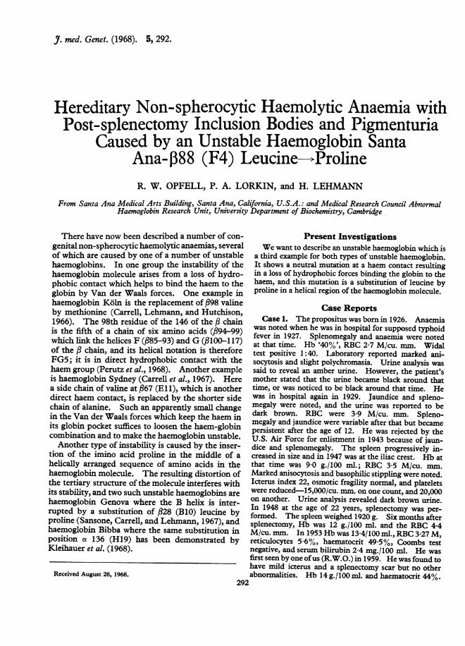
On inc4Abating the blood of the propositus at50° C. for one hour according to Dacie et al. (1964) acopius precipitate was formed indicating that thecause of his Heinz body anaemia was an unstablehaemoglobin. When the haemolysate was ex-amined by paper electrophoresis at pH 9-9, anadditional band to those of Hb A and Hb A2 wasseen moving behind the Hb A and faster than Hb A2(Fig. 1). On starch gel electrophoresis, accordingto Poulik (1957), a second additional band wasnoted in the position of free a chains as is oftennoted when fi chain unstable haemoglobins are ex-amined by that method (Huehns and Shooter, 1965).The haemoglobin was isolated by repeated paperelectrophoresis using Tris buffer pH 8 9 (Cradock-Watson, Fenton, and Lehmann, 1959) until it wasfree from Hb A and Hb A2. It was then noted thatthere was a discrepancy between the determinationof the cyan-methaemoglobin and of the proteinconcentration by refractometry. When untreatedhaemolysates were adjusted to a 5% concentrationof cyanmethaemoglobin, the protein concentrationwas 5-6%. After separation and purification byelectrophoresis of the Hb A and of the abnormalhaemoglobin the protein content of the 500 (cyan-methaemoglobin) solution Hb A was 5 1 0% but thatof the abnormal fraction was 9 8%, indicating that ithad only two haem groups per molecule of haemo-globin. A similar gross impairment of haem-binding has been reported for Hb Gun Hill inwhich five amino acid residues are deleted in the :chain (Bradley, Wohl, and Rieder, 1967), and im-pairment of haem binding is considered a generalfeature of unstable haemoglobins (Carrell et al.,1967), the loss ofhaem contributing to precipitationof Heinz bodies (Jacob et al., 1968).For the examination of the amino acid sequence
of the abnormal haemoglobin the purified solutionwas incubated at 500 C. A whitish precipitateindicating a low haem content appeared and wasthen washed repeatedly withpH 7 phosphate buffer
293
Opfell, Lorkin, and Lehmann
'-A2
-Snto Ai#
-A
,,.-- B.
FIG. 1. Paper electrophoresis (TRIS buffer, pH 8-9) of the haemoglobin solution of the propositus with Hb A and Hb A2 as control.Hb Santa Ana is seen as an additional band in the case of the propositus.
of 500 C. temperature. The globin was amino-ethylated (Jones, 1964) and then purified by gelfiltration on Sephadex G25 and isolated by freeze-drying. Part of the haemoglobin was separatedinto its a and ,B chains according to Clegg, Naughton,and Weatherall (1966). For this the precipitatedunstable haemoglobin was dissolved at a concentra-tion of about 5% w/v, in a solution containing 1%pyridine, 0-05M /3-mercaptoethanol in 8M urea,pH 8-8, and incubated at room temperature for 4hours. This treatment was intended to reduce anyintermolecular disulphide bonds which might haveformed during the heat treatment of the haemo-globin. Globin was then precipitated by addingthe solution dropwise to 1-5% v/v concentratedhydrochloric acid in acetone at -200 C., washedwith acetone at - 200 C., and dried under nitrogen.When chromatography on a column of carboxy-methyl cellulose in 8M urea was then carried outaccording to Clegg et al. (1966), much of the proteinfailed to bind to the column, nevertheless approxi-mately 50% of the theoretical yields were obtainedin some separations.The aminoethylated (AE) globin and the AE a
and ,B chains were digested with trypsin, and 'finger-prints' (peptide chromatograms) were preparedwhich were stained with ninhydrin and specific re-
agents for histidine, arginine, tyrosine, tryptophan,
and sulphur-containing amino acids (methionineand f-aminoethylcysteine). The methods usedhave recently been summarized by Sick et al.(1967) and by Beale (1967).
Part of the globin molecule, the so-called corecomprising residues 93-140 of the a chain and 83-120 of the ,B chain, remains insoluble or onlypartly soluble after tryptic digestion. Amino-ethylation converts the cysteines at position 104 of'the a chain and positions 93 and 112 of the P chaininto ,B-aminoethylcysteine, which as a basic aminoacid is then acted upon by trypsin. On trypticdigestion of the AE globin, part of the a chain core,,and the whole of the ,B chain core, are renderedsoluble. Six extra peptides are found on the finger-print of the AE globin, aTpXI (a93-99); aTpXlIa(alOO-104); PTpX (,83-95); JTpXI (fl96-104);,TpXIIa (fl105-112), f3TpXIIb (,B113-120), andtraces of,TpXa (,B83-93).
In the fingerprint of AE globin of the unstablehaemoglobin all the peptide spots gave the ex-pected staining reactions and were in their usualpositions except for the spot representing flTpX,which had a normal electrophoretic mobility, but alower chromatographic mobility than usual. Whenthe electrophoretic separation is carried out atpH 3 5, ,BTpX has the same electrophoretic mobilityas PTpI (p1-8). In such fingerprints of AE globin
294
42.1
Unstable Hb Santa Ana-,B88
......
'.;~~~~~~~~~~~~~~~~~~~~~~~~~~~~~~~~~~~~~~~...'.
'_. .....
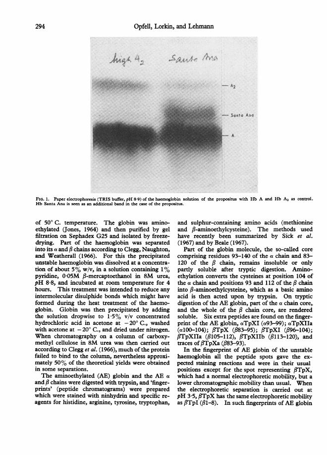
FIG. 2. Comparison of Hb A and Hb Santa Ana. Fingerprint of a mixture of AE globin A and AE globin Santa Ana-pH 6-4. TwoIITpX spots can be seen, of which the upper corresponds to I3ATpX, and the lower to ITpX of Santa Ana. The spot marked with anasterisk represents ,ATpXa (residues ,83-93).
from Hb A, f3TpX is located above /TpI. Theabnormal fTpX was however located just below.fTpI.
Table I lists the chromatographic mobility offlTpX relative to fTpIV, for the unstable haemo-globin, Hb A, and two abnormal haemoglobinswhich do not have amino acid substitutions in thispeptide. Fig. 2 shows the fingerprint of a mixtureof AE Hb A and of the AE unstable haemoglobin.Two ,BTpX spots, both staining for histidine andfl-aminoethylcysteine, are present; the upper cor-responding to PATpX and the lower to the abnormal,BTpX. This clearly shows that the unstablehaemoglobin differs from Hb A in residues 83-95 ofthe fi chain. The fingerprint of the tryptic digest ofthe AE a chain did not show any abnormalities.
Table II shows the amino acid composition of theabnormal flTpX isolated from preparative finger-prints of the AE globin and AE fi chain. In thefirst case the peptide was contaminated withaTpI-II (residues 1-11 of the a chain) and thefigures are corrected for contamination. The
TABLE ICHROMATOGRAPHIC MOBILITIES OF
BTpX RELATIVE TO OTpIV
Haemoglobin Santa Ana A K-Ibadan Koln
0 073 0-25 0-31 0-220-073 0-31 0-22 0-260 047 0-25 0-28 0-220-115 0-24 0-24 0-21
Average 0-077 0 26 0-26 0-23
second set of figures is the amino acid compositionof the pure peptide from the isolated AE ,B chain.In the abnormal peptide there was only one residueof leucine instead of two and one residue of proline,which is normally absent from P3ATpX. The un-stable haemoglobin therefore differs from HbA by asubstitution of proline for leucine in ,BTpX.There was also an abnormality in the amounts of
threonine and glutamic acid found. Normallythey should be in the ratio of 2: 1, but approximatelyequal amounts were found. This may indicate apartial substitution of threonine by glutamine,which would be converted into glutamic acid on
TABLE IIAMINO ACID COMPOSITION OF ABNORMAL
)3TpX
,u Moles No. of ResiduesAminoAcid AE AEOI AE AEO AEaATpXGlobin* Chain Globin ChainA
Asp 0 033 0-024 1-0 1-0 1Thr 0-052 0-038 1-6 1-6 2Ser 0-034 0-023 1.1 1-0 1Glu 0 049 0-038 1-5 1-6 1Pro 0 033 0-018 1-0 0 7 0Gly 0-041 0-025 1-3 1-0 1Ala 0-031 0 033 1-0 1-3 1Leu 0 034 0-024 1.1 1-0 2Phe 0-026 0-018 0-8 0 7 11 residue 0-032 0-024
* Corrected for contamination with aTpI-II.Note: AE IATpX contains 3 basic amino acid residues: Lys, His,
AE Cys. The presence of histidine and aminoethylcysteine in theabnormal j6TpX was indicated by positive staining reactions, and asthe peptide was a tryptic peptide (not staining for arginine) itscarboxy terminal amino acid was lysine.
295
Opfell, Lorkin, and Lehmann
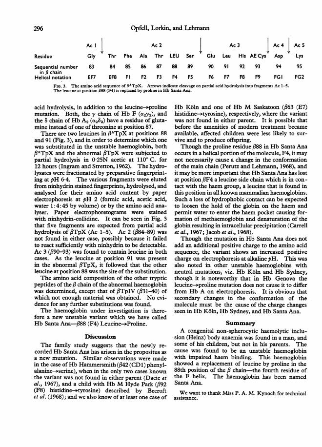
Ac I Ac2 Ac 3 Ac4 Ac5
Residue Gly Thr Phe Ala Thr LEU Ser Glu Leu His AE Cys Asp Lys
Sequential number 83in , chain
Helical notation EF7
84 85 86 87 88 89
EF8 Fl F2 F3 F4 F5
90 91 92 93
F6 F7 F8 F9
FIG. 3. The amino acid sequence of IATpX. Arrows indicate cleavage on partial acid hydrolysis into fragments Ac 1-5.The leucine at position 988 (F4) is replaced by proline in Hb Santa Ana.
acid hydrolysis, in addition to the leucine--prolinemutation. Both, the y chain of Hb F (a2y2), andthe 8 chain of Hb A2 (a282) have a residue of gluta-mine instead of one of threonine at position 87.There are two leucines in PATpX at positions 88
and 91 (Fig. 3), and in order to determine which onewas substituted in the unstable haemoglobin, bothPATpX and the abnormal ,TpX were subjected topartial hydrolysis in 0-25N acetic at 1100 C. for12 hours (Ingram and Stretton, 1962). The hydro-lysates were fractionated by preparative fingerprint-ing at pH 6-4. The various fragments were elutedfrom ninhydrin stained fingerprints, hydrolysed, andanalysed for their amino acid content by paperelectrophoresis at pH 2 (formic acid, acetic acid,water 1:4:45 by volume) or by the amino acid ana-
lyser. Paper electrophoretograms were stainedwith ninhydrin-collidine. It can be seen in Fig. 3that five fragments are expected from partial acidhydrolysis of ,TpX (Ac 1-5). Ac 2 (p84-89) was
not found in either case, possibly because it failedto react sufficiently with ninhydrin to be detectable.Ac 3 (f90-93) was found to contain leucine in bothcases. As the leucine at position 91 was presentin the abnormal flTpX, it followed that the otherleucine at position 88 was the site of the substitution.The amino acid composition of the other tryptic
peptides of the , chain of the abnormal haemoglobinwas determined, except that of fTpIV (,B31-40) ofwhich not enough material was obtained. No evi-dence for any further substitutions was found.The haemoglobin under investigation is there-
fore a new unstable variant which we have calledHb Santa Ana-f88 (F4) Leucine-4Proline.
DiscussionThe family study suggests that the newly re-
corded Hb Santa Ana has arisen in the propositus asa new mutation. Similar observations were madein the case ofHb Hammersmith(f42 (CD1) phenyl-alanine-->serine), when in the only two cases knownthe variant was not found in either parent (Dacie et
al., 1967), and a child with Hb M Hyde Park (,B92(F8) histidine-*tyrosine) described by Becroftet al. (1968); and we also know of at least one case of
Hb Koin and one of Hb M Saskatoon (,B63 (E7)histidine-->tyrosine), respectively, where the variantwas not found in either parent. It is possible thatbefore the amenities of modern treatment becameavailable, affected children were less likely to sur-vive and to produce offspring.Though the proline residue ,B88 in Hb Santa Ana
occurs in a helical portion of the molecule, F4, it maynot necessarily cause a change in the conformationof the main chain (Perutz and Lehmann, 1968), andit may be more important that Hb Santa Ana has lostat position fF4 a leucine side chain which is in con-tact with the haem group, a leucine that is found inthis position in all known mammalian haemoglobins.Such a loss of hydrophobic contact can be expectedto loosen the hold of the globin on the haem andpermit water to enter the haem pocket causing for-mation of methaemoglobin and denaturation of theglobin resulting in intracellular precipitation (Carrellet al., 1967; Jacob et al., 1968).Though the mutation in Hb Santa Ana does not
add an additional positive charge to the amino acidsequence, the variant shows an increased positivecharge on electrophoresis at alkaline pH. This wasalso noted in other unstable haemoglobins withneutral mutations, viz. Hb K6ln and Hb Sydney,though it is noteworthy that in Hb Genova theleucine-*proline mutation does not cause it to differfrom Hb A on electrophoresis. It is obvious thatsecondary changes in the conformation of themolecule must be the cause of the charge changesseen in Hb Ko1n, Hb Sydney, and Hb Santa Ana.
SummaryA congenital non-spherocytic haemolytic inclu-
sion (Heinz) body anaemia was found in a man, andsome of his children, but not in his parents. Thecause was found to be an unstable haemoglobinwith impaired haem binding. This haemoglobinshowed a replacement of leucine by proline in the88th position of the chain-the fourth residue ofthe F helix. The haemoglobin has been namedSanta Ana.
We want to thank Miss P. A. M. Kynoch for technicalassistance.
94 95
FG I FG2
296
Unstable Hb Santa Ana-fl88REFERENCES
Beale, D. (1967). A partial amino sequence for sheep haemoglobinA. Biochem. 7., 103, 129.
Becroft, D. M. O., Douglas, R., Carrell, R. W., and Lehmann, H.(1968). Haemoglobin M Hyde Park; a hereditary methaemo-globinaemia in a Caucasian. N. Z. med. J7., 67, 72.
Bradley, T. B., Wohl, R. C., and Rieder, R. F. (1967). HemoglobinGun Hill: deletion of five amino acid residues and impairedheme-globin binding. Science, 157, 1581.
Carrell, R. W., Lehmann, H., and Hutchison, H. E. (1966). Haemo-globin Koln (1-98 valine-smethionine): an unstable proteincausing inclusion body anaemia. Nature (Lond.), 210, 915.
, , Lorkin, P. A., Raik, E., and Hunter, E. (1967). Haemo-globin Sydney: 167 (El 1) valine- .alanine: an emerging pattern ofunstable haemoglobins. ibid., 215, 626.
Clegg, J. B., Naughton, M. A., and Weatherall, D. J. (1966).Abnormal human haemoglobins. Separation and characterizationof the a and chains by chromatography, and the determination of
two new variants, Hb Chesapeake and Hb J (Bangkok). J. molec.Biol., 19, 91.
Cradock-Watson, J. E., Fenton, J. C. B., and Lehmann, H. (1959).TRIS buffer for the demonstration of haemoglobin A2 by paperelectrophoresis. J. clin. Path., 12, 372.
Dacie, J. V., Grimes, A. J., Meisler, A., Steingold, L., Hensted, E.H., Beaven, G. H., and White, J. C. (1964). Hereditary Heinz-body anaemia. A report of studies on five patients with mildanaemia. Brit. J. Haemat., 10, 388.
, Shinton, N. K., Gaffney, P. J., Carrell, R. W., and Lehmann,H. (1967). Haemoglobin Hammersmith (1342 (CD1) Phe- Ser).Nature (Lond.), 216, 663.
Huehns, E. R., and Shooter, E. M. (1965). Human haemoglobins.J. med. Genet., 2, 48.
Ingram, V. M., and Stretton, A. 0. W. (1962). Human haemo-globin A2. II. The chemistry of some peptides peculiar tohaemoglobin A2. Biochem. biophys. Acta (Amst.), 63, 20.
Jacob, H. S., Brain, M. C., Dacie, J. V., Carrell, R. W., andLehmann, H. (1968). Abnormal haem binding and globin SHgroup blockade in unstable haemoglobins. Nature (Lond.), 218,1214.
Jones, R. T. (1964). Structural studies of aminoethylated hemo-globins by automatic peptide chromatography. Cold Spr. Harb.Symp. quant. Biol., 29, 297.
Kleihauer, E. F., Reynolds, C. A., Dozy, A. M., Wilson, J. B.,Moores, R. R., Berenson, M. P., Wright, C. S., and Huisman,T. H. J. (1968). HemoglobinBibba or a2136Pro 32, an unstable a chainabnormal hemoglobin. Biochim. biophys. Acta (Amst.), 154, 220.
Perutz, M. F., and Lehmann, H. (1968). Molecular pathology ofhuman haemoglobin. Nature (Lond.), 219, 902.-, Muirhead, H., Cox, J. M., and Goaman, L. C. G. (1968).
Three-dimensional Fourier synthesis of horse oxyhaemaglobin at
2-8 A resolution: the atomic model. ibid., 219, 131.Poulik, M. D. (1957). Starch gel electrophrosis in a discontinuous
system of buffers. ibid., 180, 1477.Sansone, G., Carrell, R. W., and Lehmann, H. (1967). Haemo-
globin Genova: 128 (B10) leucine-proline. ibid., 214, 877.Sick, K., Beale, D., Irvine, D., Lehman, H., Goodall, P. T., andMacDougall, S. (1967). Haemoglobin Geopenhagen and Haemo-globin Jcamhri(Ige. Two new 3-chain variants of haemoglobin A.Biochim. biophys. Acta (Amst.), 140, 231.
297