hh-asoct in secondary pediatric glaucoma
TRANSCRIPT

11/13/2019
1
HH-ASOCT in SECONDARY PEDIATRIC GLAUCOMADina El-Fayoumi, MD
Assistant professor of ophthalmology
Evolution of OCT
Time-domain prototype in 1991.
Limited by low-image resolution, the details of the angle structures could not be clearly visualized.
The use of spectral-domain OCT for anterior segment imaging was first described in 2001.
The introduction HH-SDOCT is considered a breakthrough specially in pediatric ophthalmology.

11/13/2019
2
HH-OCT HELPED US DIFFERENTATAITE NORMAL ACA FROM PCG
Can HH-ASOCT HELP US IN THE DIAGNOSIS OF SECONDARY PEDIATRIC GLAUCOMA?
11/13/2019
3
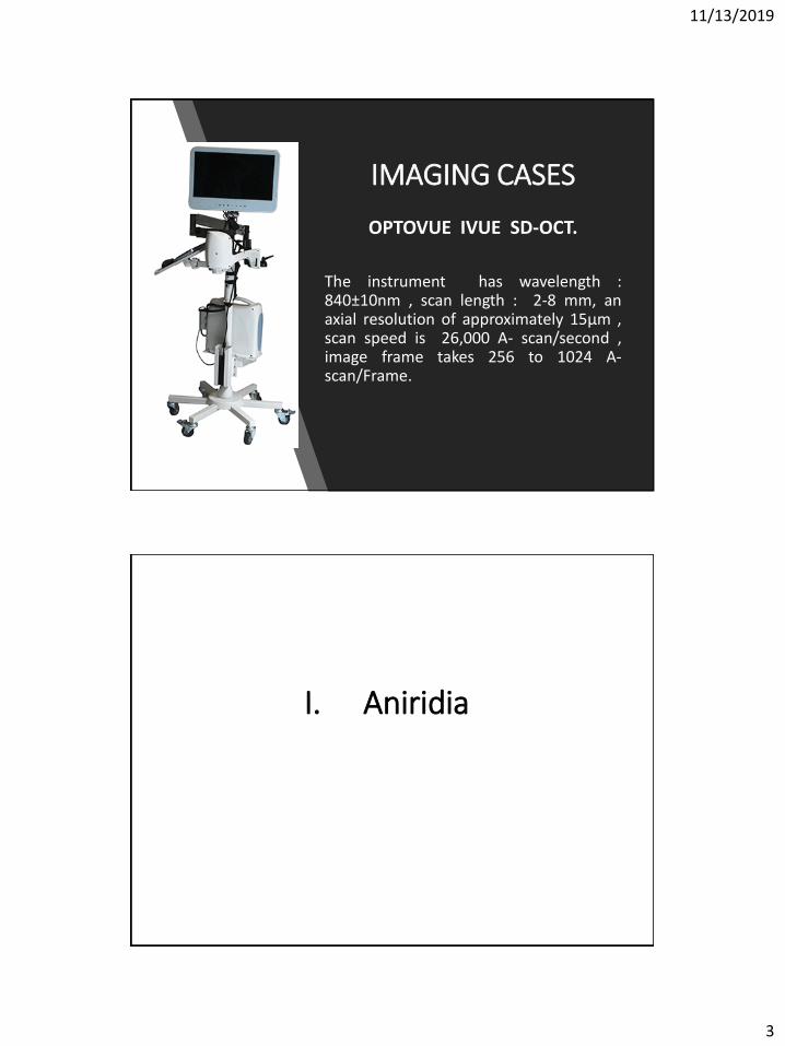
IMAGING CASES
OPTOVUE IVUE SD-OCT.
The instrument has wavelength :840±10nm , scan length : 2-8 mm, anaxial resolution of approximately 15µm ,scan speed is 26,000 A- scan/second ,image frame takes 256 to 1024 A-scan/Frame.
I. Aniridia

11/13/2019
4
GLAUCOMA IN ANIRIDIA
▪ Glaucoma occurs in earlyadulthood.
▪ Occurs in infants and toddlers▪ Incidence range 6% to 75%
Aniridia• HISTOPATHOLOGY
▪ All cases had an iris stump
▪ The TM and ciliary processes: visible posterior to the stump.
▪ If glaucoma develops:
➢ irregular strands from the iris stroma attaching it to the angle wall.
➢ These attachments become thicker, move forward, causing obscuration of the TM, SS and the CB.
➢ The iris stump tilt and the angle gradually closed.

11/13/2019
5
ANIRIDIAOD NASAL AND TEMPORAL ANGLE
II. Anterior segment dysgenesis (ASD)

11/13/2019
6
Anterior segment dysgenesis (ASD)
• A spectrum of developmental anomalies
• Resulting from abnormalities of neural crest migration and differentiation during embryologic development.
• INCLUDE:
• 1-Axenfeld-Rieger anomaly /syndrome
• 2-Peters anomaly
• 3-Posterior keratoconus
• 4-Iridoschisis.
1-AXENFELD-REIGER ANOMALY/SYNDROME
• An autosomal dominant disorder
• A spectrum of anomalies.
• Ranging from isolated bilateral ocular defects to a fully manifested systemic disorder.
• There is at least a 50% risk of developing glaucoma.
11/13/2019
7
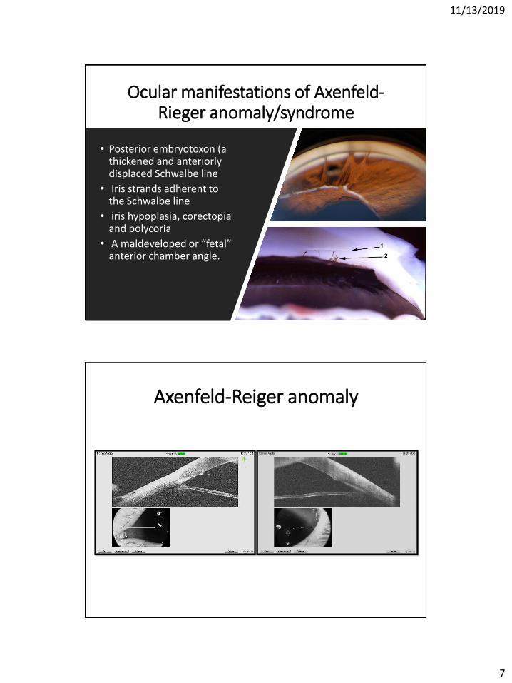
Ocular manifestations of Axenfeld-Rieger anomaly/syndrome
• Posterior embryotoxon (a thickened and anteriorly displaced Schwalbe line
• Iris strands adherent to the Schwalbe line
• iris hypoplasia, corectopiaand polycoria
• A maldeveloped or “fetal” anterior chamber angle.
Axenfeld-Reiger anomaly

11/13/2019
8
2-PETERS’ ANOMALY
• Most common gene mutations include PAX6 and FOXC1
• Classified into two subtypes: Type 1, and Type 2.
• Glaucoma occurs in up to 90% of the cases.
Types of Peters’ anomaly
• 80% of cases present bilaterally
• Central and paracentral corneal opacification.
• The cornea is avascular.
• Iris strands extend from the collarette
• Systemic abnormalities are not present.
• Cases are commonly bilateral
• Denser corneal opacification
• Juxtaposition of the lens
• iris strands may or may not be present.
• The posterior stroma and Descemetmembrane is classically malformed.
• Systemic abnormalities are more common.
Type 1 Type 2

11/13/2019
9
Types of Peters’ anomaly
Peters’ AnomalyCase 1
11/13/2019
10
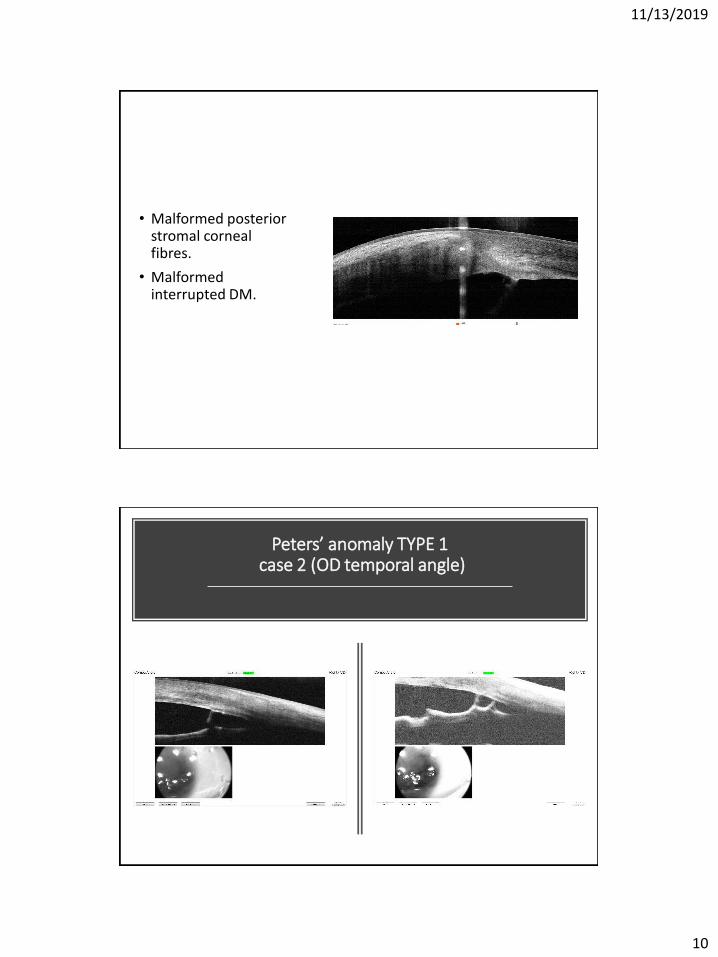
• Malformed posterior stromal corneal fibres.
• Malformed interrupted DM.
Peters’ anomaly TYPE 1case 2 (OD temporal angle)
11/13/2019
11
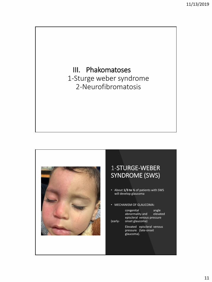
III. Phakomatoses1-Sturge weber syndrome
2-Neurofibromatosis
1-STURGE-WEBER SYNDROME (SWS)
• About 1/3 to ½ of patients with SWS will develop glaucoma
• MECHANISM OF GLAUCOMA:
congenital angle abnormality and elevated episcleral venous pressure
(early- onset glaucoma)
Elevated episcleral venous pressure (late-onset glaucoma).

11/13/2019
12
Histopathological and Goniosopicfindings in SWS
• In infancy and childhood:
Clinical and histopathological features of ACA are similar to PCG.
• On gonioscopy:
❑ The angle structures indistinct, with a high iris insertion.
❑ An anteriorly displaced iris root
❑ Poorly developed scleral spur.
❑SWS patients with early-onset glaucoma present with typical signs of congenital glaucoma.
Sturge-Weber syndrome (SWS)case 1 (OD)
PI
PAS

11/13/2019
13
Sturge-Weber syndrome (SWS)case 1 (OD) temporal and nasal angle
STURGE-WEBER SYNDROMEcase 2

11/13/2019
14
2-Neurofibromatosis
• Glaucoma occurs if the eyelids have neurofibromas.
• The mechanism:
• Infiltration of the angle with neurofibromatous tissue
• Angle closure caused by nodular thickening of the ciliary body and choroid
• Failure of the anterior chamber to develop.
NeurofibromatosisOD nasal and temporal angle

11/13/2019
15
NeurofibromatosisOS nasal and temporal angle (normal)
Is it useful to image the ACA using AS-OCTin secondary pediatric glaucoma?
Non-contact.
Requires no anesthesia.
Helps in confirming the diagnosis.
Follow up the possible changes (postoperative).
Aids in the choice of the proper surgical method.

11/13/2019
16