hi g energy gy applications - istituto nazionale di fisica...
TRANSCRIPT

01-1
1
3D Silicon Detectors for High Energyns
, Ber
gen
25-0g gy
Physics and Medical Applicationsed
ical
App
licat
io
Ci i D Viá Th U i i f M h UK
etec
tors
and
Me Cinzia Da Viá, The University of Manchester, UK
nche
ster
-UK
. De
!3D silicon technology
!Applications to HEP: ATLASFP and ATLAS Upgrades
nive
rsity
of M
an
pp pgand Insertable B-Layer
!Applications to Medicine and Biology and more
aD
aV
iá, t
he U
!Applications to Medicine and Biology and more
!Summary and Perspectives
Cin
zia

01-1
1 3D 3D Silicon detectors detectors
ns, B
erge
n 25
-0ed
ical
App
licat
ioet
ecto
rs a
nd M
e
3D silicon detectors were proposed in 1995
nche
ster
-UK
. De 3D silicon detectors were proposed in 1995
by S. Parker, and active edges in 1997 by C. Kenney.
nive
rsity
of M
an Combine traditional VLSI processing andMEMS (Micro Electro Mechanical Systems)technology.
1. NIMA 395 (1997) 328 2. IEEE Trans Nucl Sci 46 (1999) 12243. IEEE Trans Nucl Sci 48 (2001) 189
aD
aV
iá, t
he U
Electrodes are processed inside the detectorbulk instead of being implanted on the Wafer's surface.
( )4. IEEE Trans Nucl Sci 48 (2001) 1629 5. IEEE Trans Nucl Sci 48 (2001) 2405 6. Proc. SPIE 4784 (2002)3657. CERN Courier, Vol 43, Jan 2003, pp 23-268. NIM A 509 (2003) 86-919 NIMA 524 (2004) 236-244
Cin
zia
The edge is an electrode! Dead volume at the Edge < 5 microns! Essential for
9. NIMA 524 (2004) 236-24410. NIM A 549 (2005) 12211. NIM A 560 (2006) 12712. NIM A 565 (2006) 27213. IEEE TNS 53 (2006) 1676

01-1
1 3D versus planar detectors (not to scale)3D versus planar detectors (not to scale)
ns, B
erge
n 25
-0
p+
particle
n+p+
3D PLANAR
n+
~ 0.5-1 mmActive edge
edic
al A
pplic
atio
p
--------
0 !m
np
50 !m
----
---- i
n
50 !m
etec
tors
and
Me
Collectingn+
++++
++++
--
++
300
++++--
++++++
microcracks,
nche
ster
-UK
. De
p n
Collecting electrode
chips induce surfaceleakage current
MEDICI simulation of a 3D structure
nive
rsity
of M
an
!DEPLETION VOLTAGES < 10 V 70 V
3D planarp n of a 3D structure
aD
aV
iá, t
he U !EDGE SENSITIVITY < 5 !m 500 !m
!CHARGE 1 MIP (300 mm) 24000e- 24000e-
!CAPACITANCE 30-50f ~20fF !COLLECTION DISTANCE 50 !m 300 !m
Cin
zia ! !
!SPEED 1-2ns 10-20 nsn nDrift lines parallel to the surface

01-1
1
A t t
Key processing steps (25Key processing steps (25--3232))ns
, Ber
gen
25-0 Aspect ratio:
D:d = 11:111-- etching the etching the 22--filling themfilling themelectrodes with electrodes with dopantsdopants
edic
al A
pplic
atio
Step 1-3 oxidize and
Step 9-13 dope and fill p+
Dd
etec
tors
and
Me
WAFER BONDING (mechanical stability)Si-OH + HO-Si -> Si-O-Si + H2O
oxidize and fusion bond wafer
and fill p+
electrodes LOW PRESSURECHEMICAL VAPOR DEPOSITION(Electrodes filling with conformal doped polysilicon
nche
ster
-UK
. De
Step 4-6 pattern and etch p+ window contacts
Step 14-17 etch n+ window contacts and electrodes
conformal doped polysilicon SiH4 at ~620C)2P2O5 +5 Si-> 4P + 5 SiO22B2O3 +3Si -> 4 B +3 SiO2
Both electrodes appear on both surfaces
nive
rsity
of M
an29
0!m
contacts
Step 18-23 dope and fill n+
p
aD
aV
iá, t
he U
DEEP REACTIVEION ETCHING (STS) (electrodes definition)
Step 7-8 etch p+ electrodes
and fill n+
electrodes
n
Cin
zia (electrodes definition)
Bosh processSiF4 (gas) +C4F8 (teflon)
Step 24-25 deposit and pattern Aluminum
METAL DEPOSITIONShorting electrodes of the same type with Al for strip electronics readoutor deposit metal for bump-bonding

01-1
1 Active edge processing Active edge processing –– the large area the large area
coverage solutioncoverage solutionns
, Ber
gen
25-0 coverage solutioncoverage solution
A TRENCH IS ETCHED AND DOPED TO TERMINATE THE E-FIELD LINES
edic
al A
pplic
atio
AFTER THE FULL PROCESS IS COMPLETED THE MATERIAL SURROUNDING
etec
tors
and
Me MATERIAL SURROUNDING
THE DETECTORS IS ETCHED AWAY AND THE SUPPORTWAFER REMOVED : NO SAWING NEEDED!!! (NO CHIPS NO CRACKS)
nche
ster
-UK
. De (NO CHIPS, NO CRACKS)
Natural developement " PLANAR+3D = planar/3D
nive
rsity
of M
an
3D ti d
atu a de e ope e t 3 p a a /3PLANAR DETECTOR + DOPANT DIFFUSED IN FROM DEEP ETCHED EDGE THEN FILLED WITH POLYSILICON (C. Kenney 1997)
aD
aV
iá, t
he U
E-fieldp + + Al
3D active edge TOTEM detectors3x4cm2 512 !strips
Cin
zia
n ++ Al
n ++ Al

01-1
1 Active edge and electrode response of 3D sensors
ns, B
erge
n 25
-0
X
Fabricated at Stanford, J. Hasi (Manchester PhD thesis)
edic
al A
pplic
atio
etec
tors
and
Me
nche
ster
-UK
. De
nive
rsity
of M
an
5 !m
aD
aV
iá, t
he U
Electrodes ~ 1.8% of total area
Cin
zia
X-ray micro-beam scan, in 2 µm steps, of a 3D, n bulk and edges, 181 µm thick sensor. The left electrodes are p-type Electrode response

01-1
1 Improving the aspect ratio (D/d) in thick wafersImproving the aspect ratio (D/d) in thick wafers
""improving ximproving x ray detection efficiency ray detection efficiency ns
, Ber
gen
25-0 ""improving ximproving x--ray detection efficiency ray detection efficiency
>Original production D/d=12:1 etching time = 5!m/min D=121 !m>Present production D/d=19:1 etching time = 5 m/min
edic
al A
pplic
atio >Present production D/d=19:1 etching time = 5!m/min
D=180 mm – 240 mm>Double side etching D/d=25:1 etching time = 1.5!m/min
D=525 mm inter electrode spacing = 25 !m J H P D T 2004
etec
tors
and
Me
LOW ASPECT RATIO CAN HAVE OTHER USES FOR MEDICINE AND MORE... laterScintillatorOr neutron converter
nche
ster
-UK
. De
nive
rsity
of M
an
Optimised aspect ratio forGamma detection
Si
aD
aV
iá, t
he U Si
Cin
zia
25 !mC
trench Optimised aspect ration for gamma detection

01-1
1 Other structures already fabricated at STANFORD
(C. Kenney, J. Hasi) to improve speed and detection propertiesns
, Ber
gen
25-0
( y, ) p p p ped
ical
App
licat
ioet
ecto
rs a
nd M
enc
hest
er-U
K. D
e
3D Parallel trenches 3D coaxial layout
nive
rsity
of M
an C. Da Via et al., “Dual readout – strip/pixel systems”,NIM A594, pp. 7-12 (2008).
aD
aV
iá, t
he U Dual readout:
Improved spatial resolutionWith the same material budget
Cin
zia

01-1
1 3D for IBL : 2 designs with equivalent
El t i l f ns
, Ber
gen
25-0 Electrical performance
3DC
edic
al A
pplic
atio 3DConsortium
etec
tors
and
Me
nche
ster
-UK
. De
Double Column DesignAgreed
Full 3D with active edges
nive
rsity
of M
an baseline
aD
aV
iá, t
he U
Cin
zia
01-1
1
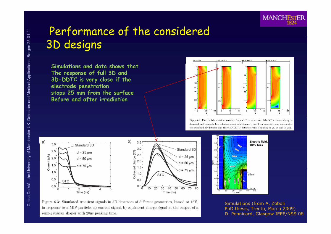
Performance of the considered ns
, Ber
gen
25-0
3D designsed
ical
App
licat
io Simulations and data shows thatThe response of full 3D and 3D-DDTC is very close if theelectrode penetration
h
etec
tors
and
Me stops 25 mm from the surface
Before and after irradiation
nche
ster
-UK
. De
nive
rsity
of M
ana
Da
Viá
, the
UC
inzi
a
Simulations (from A. ZoboliPhD thesis, Trento, March 2009)D. Pennicard, Glasgow IEEE/NSS 08

01-1
1
Where could 3D Si be applied?ns
, Ber
gen
25-0
pped
ical
App
licat
io
3D features:
Active edges
Medical
!Micro structures: endoscopy, dosimetry
etec
tors
and
Me
gLow voltageHigh speedShape adaptation
py, y!Large area imagers (mammography, synchrotron)!Focal planes (diffracted x-rays)
nche
ster
-UK
. De
!Spectroscopy!Edge-on scanned imaging (synchrotron mammography)
nive
rsity
of M
an HEP
!Forward Physics!IBL
!PET (embedded crystals) – photo-multiplication
aD
aV
iá, t
he U !IBL
!Pixel Upgrades !TOF-PET (above + speed)
Cin
zia

01-1
1 THE DISCOVERY POTENTIAL OF THE LHC CAN BE ENHANCED
BY INCREASING ITS LUMINOSITYLint=3000 fb -1
ns, B
erge
n 25
-0
Phase-0 : 15 months: 2013 to spring 2014Ph 1 12 th ti 2017?
intBy 2030
2x10¹⁶ncm-2
edic
al A
pplic
atio
Phase 2
Phase-1 : 12 months: entire 2017?Phase-2 : 18 months: end of 2020-early 2021?
L~5×1034cm-2s-1
etec
tors
and
Me
L 1 2×1034cm-2s-1
Lint=300 fb -1
5x10¹⁵ncm-2
nche
ster
-UK
. De
min
osity
Phase 1
L~1-2×1034cm-2s-1
FLUENCES
nive
rsity
of M
an
tegr
ated
lum Phase 1
L~1×1032 cm-2s-1
Lint=1fb -1 EXPECTED ATINNERMOST LAYER
aD
aV
iá, t
he U In
t
Phase 0 1x10¹⁵ncm-2
C. Da Vià Nov. 2010
Cin
zia
Data from Steve MyersCERN sLHC 23rd June 2010.
2010 2015 2020

01-1
1 Atlas IBL – timescale 2013!!!!!
ns, B
erge
n 25
-0ed
ical
App
licat
io • Baseline layout decided– 14 Staves, “reverse turbine”
• Beam pipe red ction:
etec
tors
and
Me • Beam-pipe reduction:
– Inner R: 29 " 25 mm
Very tight clearance:
nche
ster
-UK
. De Very tight clearance:
– “Hermetic” to straight tracks in ! (1.8º overlap)
– No overlap in Z: minimize gap
nive
rsity
of M
an between sensor active area.
Layout parameters:IBL l 9 i R
aD
aV
iá, t
he U – IBL envelope: 9 mm in R
– 14 staves.– <R> = 33 mm.– Z = 60 cm (active length)
Cin
zia Z 60 cm (active length).– " = 2.5 coverage.

01-1
1 IBL Detectors Specifications
ns, B
erge
n 25
-0
• FE-I4 compatible layout x1 x2 MultiChip18FE-I3
edic
al A
pplic
atio
•<450 !m inactive edge•< 200 mW/cm2 at 5 . 1015 neq/cm2
(after annealing)
FE-I4160
etec
tors
and
Me (after annealing)
•1000 V of bias voltage (safely) •applicable after 5 . 1015 neq/cm2
S/T > 2 after 5 1015 neq/cm2
FE-I4A
nche
ster
-UK
. De S/T 2 after 5 . 10 neq/cm
FE I3 FE I4
nive
rsity
of M
an FE-I3 FE-I4Pixel Size [!m2] 50×400 50×250Pixel Array 18×160 80×336
Chip Size [mm2] 7.6×10.8 20.2×19.0TDAC
250 !m
aD
aV
iá, t
he U Active Fraction 74 % 89 %
Analog Current [!A/pix] 26 10
Digital Current [!A/pix] 17 10A l V lt [V] 6
Am
p2
discri
50 !msynthezised digital region (1/4th )
Cin
zia Analog Voltage [V] 1.6 1.4
Digital Voltage [V] 2 1.2pseudo-LVDS out [Mb/s] 40 160
Preamp FDAC Config Logic

01-1
1 Vertex detectors challenges
ns, B
erge
n 25
-0
Precise vertex determination
Important role in pattern recognition/ track
edic
al A
pplic
atio
p p greconstruction 200 pileup events/bc at 5x1034cm-2s-1
K I400 collisions in the inner trackerAbdel Abdesselam June 2010
etec
tors
and
Me Key Issues:
#Material budget –less multiple scattering, better primary vertex resolution
Thin/small beam-pipe
Abdel Abdesselam, June 2010
nche
ster
-UK
. De p p
Ultra-light detectorsMany channels to reduce occupancyHigh data rates IBL 1.5%X0
nive
rsity
of M
an High data rates –
•High-precision detectors very close to IP# Ultra radiation hard detectors
0
aD
aV
iá, t
he U Radiation hardness up to
2X10¹⁶ 1MeV neutron/cm2 at innermostlayers at 3000 fb-1 5X10¹⁵ncm-2 at 300 fb-1
Cin
zia
Signal/threshold
StripsATLAS Radiation Taskforce [ATL-GEN-2005-01]

01-1
1
Radiation Induced Bulk Damage in Siliconns
, Ber
gen
25-0
From RD48/ROSE
Primary Knock on Atom
edic
al A
pplic
atio Primary Knock on Atom
Displacement threshold in Si:Frenkel pair E~25eV
etec
tors
and
Me p
Defect cluster E~5keV
Vacancy
nche
ster
-UK
. De
V,I MIGRATE UNTIL THEY MEETVan Lint 1980
Interstitial
nive
rsity
of M
an
Ec
V,I MIGRATE UNTIL THEY MEETIMPURITIES AND DOPANTS TOFORM STABLE DEFECTS Effect on sensors
aD
aV
iá, t
he U
Ei V2(-/0)+Vn Ec-0.40eVV2(=/-)+Vn Ec-0.22eVVO- Ec - 0.17eVV6
V2O
CHARGED DEFECTS==>NEFF, VBIAS
DEEP TRAPS, RECOMBINATION CENTERS ==>CHARGE LOSS
Cin
zia
EvCIOI
(0/+) EV+0.36eV
V2O
DEEP TRAPS, GENERATION CENTERS==>LEAKAGE CURRENT

01-1
1
Silicon sensors macroscopic parameters changes observed up to 1x10¹⁵ n 1MeV/cm2 `
150
200
icro
ns)
Electrons
T = -20 oCns
, Ber
gen
25-0
50
100
150
Fl 1015 t -2fect
ive
Dri
ft L
engt
h (m
i
Holes
STANDARD 300!m n-type SILICON at 1015 n/cm210 years of operation at L=1034 cm-2s-1 at R=4 cm
edic
al A
pplic
atio
e- and h+ TRAPPING SHORT #trap affects signal formation
SPACE CHARGE INCREASE -ve Neff (1013/cm3) ~ VFD (5000V)~$
00 1 2 3 4
Fluence = 1015 protons cm 2
Eff
Electric Field ( Volt/micron )
etec
tors
and
Me eff ( FD ( )
TYPE INVERSION-double OXYGEN HELPS!junction
REVERSE ANNEALING INCREASE OF -ve Neff after irradiationLOW T STORAGE HELPS
6
8
10
[1012
cm-3
]
400
500
600
300 !m
)
Carbon-enriched (P503)Standard (P51)
O-diffusion 24 hours (P52)O-diffusion 48 hours (P54)O-diffusion 72 hours (P56)
Carbonated
Standard
nche
ster
-UK
. De LOW T STORAGE HELPS
LEACKAGE CURRENT prop to $ (I/V ~5x10-17 $) LOW T HELPS
0 1 2 3 4 50
2
4|Nef
f|
100
200
300
Vde
p [V
] (3
Oxygenated
nive
rsity
of M
an 0 1 2 3 4 5$24 GeV/c proton [1014 cm-2]
Needs to tackle all those issues:
!For Neff and Reverse annealing" Oxygen
aD
aV
iá, t
he U
!For Neff and Reverse annealing" Oxygenand operational conditions
!For trapping" device engineering
!New materials for low leakage current andL i
Cin
zia
Time [y]Lots of pioneering work from RD48/RoseNow continued by RD50
Low noise

01-1
1
The effect of trapping
CB
ns, B
erge
n 25
-0 The effect of trapping
The carriers move less " less signal since the signal is formed when charges
VB
edic
al A
pplic
atio
g g gmove
etec
tors
and
Me
nche
ster
-UK
. De
nive
rsity
of M
ana
Da
Viá
, the
UC
inzi
a
Trapping has been measured for electrons and holes by G. Kramberger (Ljiubliana) NIMA 481 (2002) 100

01-1
1 Effective drift length due to trapping
Lns
, Ber
gen
25-0 Leff = vdrift x #trap
edic
al A
pplic
atio
200
) o
etec
tors
and
Me
100
150
engt
h (m
icro
ns)
Electrons
T = -20 oC
e-e- mobility 3 times bigger!
nche
ster
-UK
. De
50
Fluence = 1015 protons cm-2
Eff
ectiv
e D
rift
LHoles
For max signal:
nive
rsity
of M
an 00 1 2 3 4
Electric Field ( Volt/micron )
h+
!Collect e-
!W k t V
aD
aV
iá, t
he U
Ottaviani Canali et al
h+ !Work at VdriftSaturated-> e-field >2V/!m
Cin
zia
Trapping times from Kramberger et al. NIMA 481 (2002) 100 Simulations CDV and S.Watts NIM A 501(2003) 138 (Vertex 2001)
Ottaviani, Canali et al.

01-1
1 3D detectors Radiation hardness 8.81x10¹⁵n/cm2
1.73x10¹⁶p/cm2ns
, Ber
gen
25-0
80
100
120
ncy
[%]
2E120
1602E NI7.55e142.00e158.81e15
tude
[mV]
2E9000e-
Vb~130V 45%
Irradiation and measurements performed in PragueC. Da Viá, T. Slaviceck, V. Linhart, P. Bem, S. Parker, S. Pospisil, S. Watts (process J. Hasi, C. Kenney)
2E
edic
al A
pplic
atio
0
20
40
60
Sign
al e
ffici
en
C. Da Viá July 070
40
80
Sign
al A
mpl
it
C. Da Viá July 07 threshold
2E
400 !m50 !m
n IR Laser
etec
tors
and
Me 0
0 2 1015 4 1015 6 1015 8 1015 1 1016
Fluence [n/cm2]
100
120
%]
120
140
1603E-NI7.55e142.00e158.81e15[m
V]
00 50 100 150 200
Bias Voltage [V]
51%
p
n
103!mVfd ~20V
Oscilloscope
bias
nche
ster
-UK
. De
IR
20
40
60
80
Sign
al e
ffici
ency
[%
3E
40
60
80
100
120 8.81e15
Sign
al A
mpl
itude
[
3E10200e-
Vb~112V
51%3E
400 !m50 !m
nive
rsity
of M
an
120
0
20
0 2 1015 4 1015 6 1015 8 1015 1 1016
S
Fluence [n/cm2]
C. Da Viá July 07
140
0
20
0 50 100 150 200Bias Voltage [V]
C. Da Viá July 07 thresholdpn
71 !mVfd ~8V
aD
aV
iá, t
he U
60
80
100
120
effic
ienc
y [%
]
4E
60
80
100
120
1404E NI7.55e142.00e15 8.81e15
Am
plitu
de [m
V]
4E13200e-
Vb~94V
66%
4E
400 !m50 !m
Cin
zia
0
20
40
0 2 1015 4 1015 6 1015 8 1015 1 1016
Sign
al e
Fluence [n/cm2]
C. Da Viá et al. July 070
20
40
0 50 100 150 200
Sign
al A
Bias Voltage [V]
C.DaVia July 07
66%
thresholdp
n
Vfd ~5V

01-1
1 Signal efficiency
d i l h[9] C. Da Via et al.”, (NIMA-D-08-00587)[10] G. Kramberger at al., Nucl. Instr. Meths. A 554 (2005) 212-219[11] G. Kramberger, Workshop on Defect Analysis in Silicon Det, Hamburg, August2006 http://wwwiexp desy de/seminare/defect analysis workshop august 2006 html
ns, B
erge
n 25
-0 and signal charge 2006. http://wwwiexp.desy.de/seminare/defect.analysis.workshop.august.2006.html[12] G. Casse et al., Nucl. Instr. Meths. A (2004) 362-365[14] T. Rohe et al. Nucl. Instr. Meths. A 552 (2005) 232-238[16] F. Lemeilleur et al., Nucl. Instr. Meths. A 360 (1995) 438-444
edic
al A
pplic
atio
100 56!m 3D - 4E [9]7%!m 3D - 3E [9]
25000.0103!m 3D - 2E [9]71 3D 3E [9]
Signal Efficiency (prop 1/L) Signal charge (depends on &'
etec
tors
and
Me
60
80
103!m 3D - 2E [9]75!m epi [11]150!m epi [11]285!m n+n pixels [14]285!m n+p strips[12]300!m p+n strips [16]
ency
[%]
15000.0
20000.0
71!m 3D - 3E [9]56!m 3D - 4E [9]50!m epi [10]75!m epi [11]150!m epi [11]285!m n+p strips[12]285!m n+n pixels [14]
ge [e
- ]
71!m 3D
nche
ster
-UK
. De
20
40
Sign
al E
ffici
e
5000 0
10000.0
Sign
al C
harg
75 m
nive
rsity
of M
an
0
20
0 2 1015 4 1015 6 1015 8 1015 1 1016
Fluence [1MeV Equivalent n/cm2]
C.DaVia; S. Watts Aug08 0.0
5000.0
0 2 1015 4 1015 6 1015 8 1015 1 1016
Fluence [1 MeV equivelent n/cm-2]
C. Da Via S. Watts April 08
75!mepi
aD
aV
iá, t
he U
[ q ] Fluence [1 MeV equivelent n/cm ]
Example at 1016 ncm2 3D wins becauseCollection distance and
Cin
zia
SMIP 3D ~ 80()x (&/L) ~ 2400 x 210/(71-22electrode implant) ~ 10290e-
SMIP planar ~ 80 ((/L) x & ~ 80()~ 80x30 ~ 2400e-distance and substrate thickness aredecoupled

01-1
1 DS 3D from CNM irradiated
Uni-Freibugh with Alibava system (M. Kohler)ns
, Ber
gen
25-0
Before irradiation 2x10¹⁵ncm-2 2x10¹⁶ncm-2
edic
al A
pplic
atio total charge 22000e
2x10 ncm22000e- at 100-150V
2x10 ncm15000e- at 350-380V
etec
tors
and
Me
n-type
nche
ster
-UK
. De
nive
rsity
of M
an
n-type
aD
aV
iá, t
he U
Cin
zia 250 um column overlap, IES= 56 microns
Detectors irradiated at the proton cyclotron Karlsruhe with 25 MeV protonsAnnealing state: ~ 5 days at RT (only p-type detector, 2x10¹⁶ neq/cm2: ~30 days)Noise at 2x10¹⁶is 1000e- at -45 oC -50 oC

01-1
1
3D silicon sensors strategy For the ATLAS IBL in 2013 Sketch of a
CNM
ns, B
erge
n 25
-0
For Fast-Track IBL 3D chose double-side 3D to reduce complexity . f C f
double side3D sensor
edic
al A
pplic
atio They will be fabricated at FBK and CNM and will have 200 microns guard fences and a
thickness of 230 microns. SINTEF and Stanford will optimise Full3D with active edges. Double sided with deep column proved good radiation hard performance with moderate bias voltage (120-150V at 5x10¹⁵ncm-2) and power dissipation of 0 034Wcm-2 at 5x10¹⁵ncm-2 at -
etec
tors
and
Me voltage (120 150V at 5x10 ncm and power dissipation of 0.034Wcm at 5x10 ncm at
10oC
nche
ster
-UK
. De
CNM :6 wafers being completed Feb118 wafers 285 !m thickPre-production of 48 wafers ready by October 118 FE-I4 per wafer common floorplan with FBK
CNM FE-I4 wafersTo be sent to IZM By the beginning of March
nive
rsity
of M
an
8 FE I4 per wafer common floorplan with FBK
FBK
aD
aV
iá, t
he U FBK completed
FE-I4 wafer Currently at IZM
FBK:3 Wafers at IZM for bump-bonding ( March11)24 wafers by May 11 wafers Pre-production of 44 wafers by June11Common floor plan
Cin
zia

01-1
1
ATLASFP – technical proposal 2013 and 2017Forward Detectors = use LHC beam-line as a spectrometerP t n n l ss sults in p t n t j ct h i nt l d p tu
ns, B
erge
n 25
-0 Proton energy loss results in proton trajectory horizontal departureed
ical
App
licat
io
IP1
etec
tors
and
Me
y(m
m)
220m
y(m
m)
420m
nche
ster
-UK
. De
x(mm)x(mm)
V. Avati/Totem
V. Avati/Totem
nive
rsity
of M
ana
Da
Viá
, the
UC
inzi
a
01-1
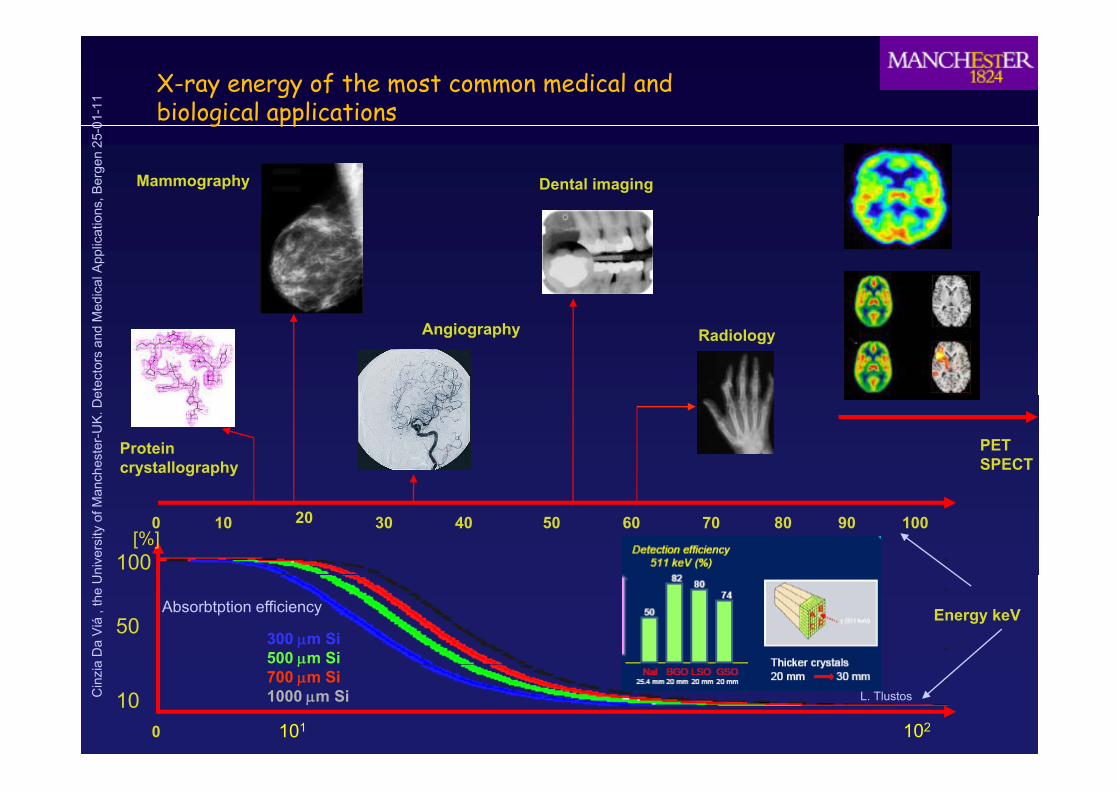
1 X-ray energy of the most common medical and
biological applications ns
, Ber
gen
25-0
Mammography Dental imaging
g pped
ical
App
licat
ioet
ecto
rs a
nd M
e
Angiography Radiology
nche
ster
-UK
. De
Proteincrystallography
PETSPECT
nive
rsity
of M
an
0 10 20 30 40 50 60 70 80 90 100
100 [%]
aD
aV
iá, t
he U
Energy keV50 300 !m Si500 !m Si
Absorbtption efficiency
Cin
zia
0
10101 102
!700 !m Si1000 !m Si L. Tlustos

01-1
1
Detection strategyExample breast cancer:
Micro-calcification ->high contrastHigh spatial resolution
ns, B
erge
n 25
-0gy
Human organ:~water
High spatial resolution
Cysts -> low contrast high detectionEfficiency = low dose
edic
al A
pplic
atio Low energy: Thick silicon or high Z semiconductors
Direct detection: high spatial resolutionX-rays
etec
tors
and
Me g p
S t d d t t
ExampleHybrid detector semiconductor + MEDIPIX
nche
ster
-UK
. De
High energy: crystals +photo detector
Segmented detector
nive
rsity
of M
an High energy: crystals +photo detectoror high Z semiconductors
I di t d t ti hi h d t ti ffi i
X-rays
aD
aV
iá, t
he U Indirect detection : high detection efficiency
but degradation of spatialresolution scattering, diffusion
Precision degradation
Cin
zia g
But high detectionEfficiency " low dose to the patient
01-1
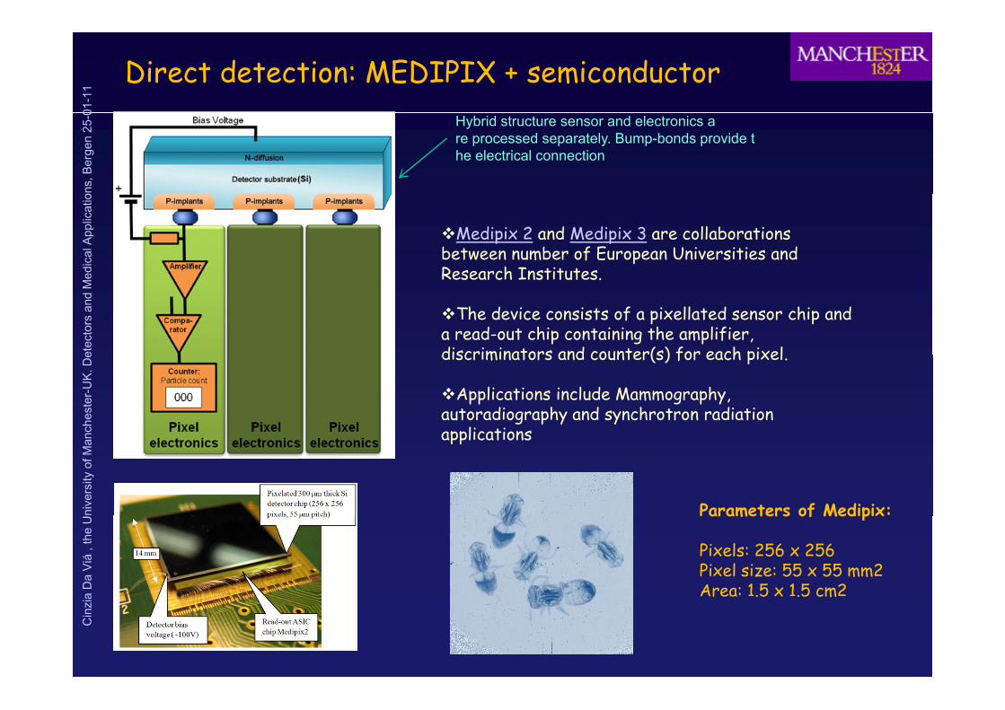
1 Direct detection: MEDIPIX + semiconductor
ns, B
erge
n 25
-0 Hybrid structure sensor and electronics are processed separately. Bump-bonds provide the electrical connection
edic
al A
pplic
atio
!Medipix 2 and Medipix 3 are collaborations between number of European Universities and Research Institutes
etec
tors
and
Me Research Institutes.
!The device consists of a pixellated sensor chip and a read-out chip containing the amplifier, discriminators and counter(s) for each pixel
nche
ster
-UK
. De discriminators and counter(s) for each pixel.
!Applications include Mammography, autoradiography and synchrotron radiation applications
nive
rsity
of M
an
Parameters of Medipix:
applications
aD
aV
iá, t
he U
Parameters of Medipix
Pixels: 256 x 256Pixel size: 55 x 55 mm2Area: 1.5 x 1.5 cm2
Cin
zia Area .5 x .5 cm

01-1
1 Direct detection
Semiconductor Materialsns
, Ber
gen
25-0 Semiconductor Materials
edic
al A
pplic
atio
etec
tors
and
Me
nche
ster
-UK
. De
nive
rsity
of M
ana
Da
Viá
, the
UC
inzi
a

01-1
1 Probability of charge sharing : planar vs 3DProbability of charge sharing : planar vs 3D
3D collects all char e on 1 electrode in most 3D collects all char e on 1 electrode in most ns
, Ber
gen
25-0 3D collects all charge on 1 electrode in most 3D collects all charge on 1 electrode in most
cases cases "" Better Energy resolution Better Energy resolution
edic
al A
pplic
atio
Central electrode
300Fraction of carriers that travel to central Fraction of carriers that travel to central electrode versus start position relative to electrode versus start position relative to
etec
tors
and
Me
1
300
150
[!m]
Y
planarelectrode versus start position relative to electrode versus start position relative to central electrodecentral electrode
3D
nche
ster
-UK
. De
0 8
0.9
ele
ctro
de
0
0-50 -25[!m]
3Dplanar
nive
rsity
of M
an
0.7
0.8
Planar Y=50Planar Y=150Planar Y=250ct
ion
to c
entr
al
0-50 -25[!m]
0
3D
aD
aV
iá, t
he U
0.5
0.6
Planar Y=2503D Y=-12.53D Y=-5Fr
ac
Central electrode
Y
Cin
zia 0.5
-25 -20 -15 -10 -5 0
Distance from central electrode (Microns)
-30
[!m]
Simulations by S. Watts, Brunel

01-1
1
Charge sharing: 241Ans
, Ber
gen
25-0 Charge sharing:
Measurements A. La Rosa/CERN241Am .
edic
al A
pplic
atio planar
etec
tors
and
Me
nche
ster
-UK
. De
nive
rsity
of M
an
3D
aD
aV
iá, t
he U
Cin
zia

01-1
1
Indirect detection needs scintillator+ns
, Ber
gen
25-0
Photomultipliers
edic
al A
pplic
atio
etec
tors
and
Me
nche
ster
-UK
. De
nive
rsity
of M
ana
Da
Viá
, the
U
J ëll B l
Cin
zia Joëlle Barral
Promotion X2001Ecole Polytechnique 1, France

01-1
1
Common Scintillating crystalsns
, Ber
gen
25-0
g yed
ical
App
licat
ioet
ecto
rs a
nd M
enc
hest
er-U
K. D
eni
vers
ity o
f Man
aD
aV
iá, t
he U
Cin
zia

01-1
1 Example PET
ns, B
erge
n 25
-0ed
ical
App
licat
io
Crystals+
etec
tors
and
Me photodetectors
nche
ster
-UK
. De
nive
rsity
of M
ana
Da
Viá
, the
UC
inzi
a
From Del Guerra

01-1
1
ns, B
erge
n 25
-0ed
ical
App
licat
ioet
ecto
rs a
nd M
enc
hest
er-U
K. D
eni
vers
ity o
f Man
aD
aV
iá, t
he U
Cin
zia

01-1
1 Hadron Therapy: in beam PET (TOF-PET)
ns, B
erge
n 25
-0
Need to control during the beam delivery of the energy deposition$Use of ! emitting nuclei from beam projectile reactions in the biologicalmatter
edic
al A
pplic
atio matter
$Use of a dedicated TEP (GSI) to monitor dosimetry and compare withsimulations
Representation of a quality control system for treatment planning. It
etec
tors
and
Me Representation of a quality control system for treatment planning. It
includes a fast beam hodoscope, a PET surrounding the target and anelectromagnetic spectrometer
nche
ster
-UK
. De
From LeDuManchester April
nive
rsity
of M
an Manchester April2008
aD
aV
iá, t
he U
Cin
zia

01-1
1
ns, B
erge
n 25
-0ed
ical
App
licat
ioet
ecto
rs a
nd M
enc
hest
er-U
K. D
eni
vers
ity o
f Man
aD
aV
iá, t
he U
Cin
zia

01-1
1
ns, B
erge
n 25
-0 Promising devices for TOF-PET
Timing resolution for two sintillators in coincidence
~600ps
edic
al A
pplic
atio 600ps
Intrinsic timing measured in FBK SoPm
~60ps Depends on number of photoelectrons
etec
tors
and
Me
TARGET IS 10-20ps!!!!!!
nche
ster
-UK
. De
nive
rsity
of M
ana
Da
Viá
, the
UC
inzi
a
From Del Guerra- April 2009
01-1
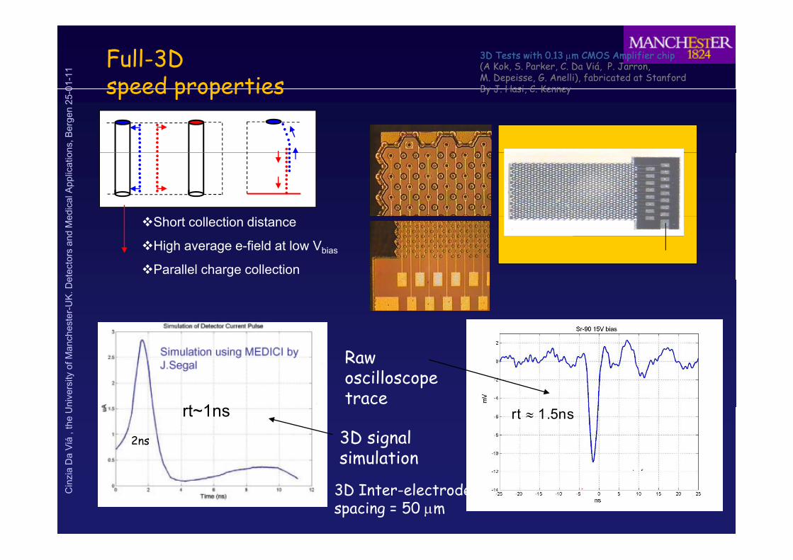
1 Full-3D
speed properties 3D Tests with 0.13 3D Tests with 0.13 !!m CMOS Amplifier chipm CMOS Amplifier chip(A Kok, S. Parker, C. Da Viá, P. Jarron, M. Depeisse, G. Anelli), fabricated at StanfordBy J Hasi C Kenney
ns, B
erge
n 25
-0 speed properties By J. Hasi, C. Kenney
edic
al A
pplic
atio
etec
tors
and
Me !Short collection distance
!High average e-field at low Vbias
!Parallel charge collection
nche
ster
-UK
. De
nive
rsity
of M
an Raw oscilloscope trace
t 1
aD
aV
iá, t
he U
3D signal simulation
2ns
T 300K
rt 1.5ns*rt~1ns
Cin
zia
3D Inter-electrodespacing = 50 !m
5
T 300

01-1
1 Pulse height distribution
20
25number vs. pulse height
Constant Fraction Discrimination
50 umIES
ns, B
erge
n 25
-0
! noise
! T
10
15C
ount
s
67 pulsesanalysed
edic
al A
pplic
atio
Expected noise-induced time-error distribution5
10
Analysis from S. Parker
etec
tors
and
Me
analysis
number vs. time resolution from noise
1000
1200time resolution vs. pulse height
Noise time error
0
nche
ster
-UK
. De y
600
800
t (ps
)
(using constant fraction discrimination) vs pulse height
average dtscatter plot:
155 psbottom plot:
nive
rsity
of M
an
200
400
dt vs. pulse height. bottom plot:134 ps
aD
aV
iá, t
he U
0 5 10 15 20Counts
0
100
200
300
dt (p
s)
dt distribution from ~ noise-free signal
Cin
zia
39
0
2 4 6 8 10 12 14 16
pulse height (mV)
dt distribution from noise-free signaladded repeatedly to separate noisesegments.
01-1
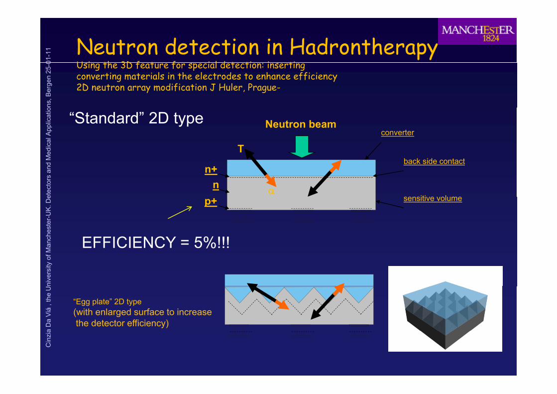
1 Neutron detection in Hadrontherapy
U i th 3D f t f i l d t ti i ti
ns, B
erge
n 25
-0 Using the 3D feature for special detection: insertingconverting materials in the electrodes to enhance efficiency2D neutron array modification J Huler, Prague-
edic
al A
pplic
atio
converter
T
Neutron beam“Standard” 2D type
etec
tors
and
Me
sensiti e ol me+
T
n+n
back side contact
nche
ster
-UK
. De sensitive volumep+
EFFICIENCY = 5%!!!
nive
rsity
of M
an EFFICIENCY = 5%!!!
aD
aV
iá, t
he U
“Egg plate” 2D type(with enlarged surface to increasethe detector efficiency)
Cin
zia

01-1
1 Neutron 3D array modification
bi d t ti d ins
, Ber
gen
25-0 combine detection and conversion
Neutron beam
Prague (J. Huler)
edic
al A
pplic
atio
low n+“Channel” 2D type
Neutron beamback sidecontact grid
Neutron or x-ray
etec
tors
and
Me low n+
p+n
Channel 2D type(maximized filling)
nche
ster
-UK
. De
Converting mediumP and n electrodes
nive
rsity
of M
an
“3D inverse” structure(there are pillars instead of pores)
bottom view
silicon
aD
aV
iá, t
he U silicon
Cin
zia

01-1
1 3D geometry arrays
comparison of cylindrical vs square 10B converterns
, Ber
gen
25-0 - comparison of cylindrical vs. square 10B converter
Measurements made in Prague (J. Huler)
edic
al A
pplic
atio Fixed wall thickness – variance in the converter / cell size
CylinderSquare
etec
tors
and
Me
Square pores, 10B (80% enrichment), walls 5 !m40
Cylinder pores, 10B (80% enrichment), walls 5 !m35
nche
ster
-UK
. De
25
30
35
icie
ncy
[%]
20
25
30
cien
cy [%
]
nive
rsity
of M
an
10
15
20
Det
ectio
n ef
fi
density 1.00 g/cm3density 1.40 g/cm3d it 1 80 / 3 5
10
15
Det
ectio
n ef
fi
density 1.00 g/cm3density 1.40 g/cm3d i 1 80 / 3
aD
aV
iá, t
he U
0
5
0 2 4 6 8 10 12 14 16 18
Pore size [!m]
density 1.80 g/cm3density 2.20 g/cm3
0
5
0 2 4 6 8 10 12 14 16 18
Pore size [!m]
density 1.80 g/cm3density 2.20 g/cm3
Cin
zia
Maximal efficiency: ~36% Maximal efficiency: ~31%

01-1
1 Protein Protein folding 3DX project (MBC)folding 3DX project (MBC)
ns, B
erge
n 25
-0
The Diffraction Pattern of Discrete Bragg Spots The Diffraction Pattern of Discrete Bragg Spots is Captured by the Detectoris Captured by the Detector
edic
al A
pplic
atio
etec
tors
and
Me
nche
ster
-UK
. De
nive
rsity
of M
ana
Da
Viá
, the
UC
inzi
a

01-1
1 Example of reconstructed structure:
Enzyme Active Sitens
, Ber
gen
25-0 Enzyme Active Site (from E. Westbrook)
edic
al A
pplic
atio• Pseudomonas diminuta
phosphotriesterase: This enzyme catalyzes the hydrolysis of
etec
tors
and
Me y y
organophosphoruspesticides and nerve agents. Its crystal structure is being studied
nche
ster
-UK
. De g
by Hazel Holden’s research group at the University of Wisconsin, Madison (see PDB file 1DPM).
nive
rsity
of M
an
)• Purple atoms: zinc• Red: bound water• Yellow: side chains• 1 8 Å resolution map
aD
aV
iá, t
he U• 1.8 Å resolution map,
• Present detectors:• CCDs. Time resolution ms
Cin
zia
• Needs faster ASIC for protein folding

01-1
1 3D pixel detectors x3D pixel detectors x--ray setup ray setup (3DX project)(3DX project)
E Westbrook et al (molecular biology consortium) USAE Westbrook et al (molecular biology consortium) USAns
, Ber
gen
25-0 E. Westbrook et al. (molecular biology consortium) USAE. Westbrook et al. (molecular biology consortium) USA
!High signals!Low noise!Moderate cost!High speed – microsecond
Orthogonal to focal pointNO aberration
edic
al A
pplic
atio
support bar(behind plane of sensors)crystal sample
X-rays
X rays
!High speed microsecondGOOD for Protein folding
etec
tors
and
Me X-rays
nche
ster
-UK
. De
to crystal sample(X ray diffraction source)
silicon sensor
nive
rsity
of M
an
cables
alignment blocks
cables
readout chip
printed circuit board
support bar
aD
aV
iá, t
he U
alignment blocks
a: top view of 3 rows
pp
Bump bond to pixel readout
Cin
zia electronics

01-1
1 Reducing the dead volume in medical imaging:
ti dns
, Ber
gen
25-0
GUARD RINGSinks surface leakage current
active edges
1-2mm depending on segmentation
edic
al A
pplic
atio
E-field p + Aldepe d g o seg e a o
etec
tors
and
Me
n + AlMicrocracks, chips etc.. Staggered microstripDead edge
X ray
nche
ster
-UK
. De
X-rays
Edge-on x-ray imagerExample mammographyOn synchrotron beams
X-ray
nive
rsity
of M
ana
Da
Viá
, the
UC
inzi
a
GapOr poor efficiency
ATLAS microstrips guard rings ~1mm

01-1
1 Further application: micro-dosimetry
ns, B
erge
n 25
-0
Dosimetry Microdosimetry
edic
al A
pplic
atio Integrates energy deposition for all particles
together
Is NOT volume or shape dependent. NO cell level effect
Measures the energy deposition events in a small (cell-size) volume
Is volume and size dependent. Cell level effect (radio-biological effect)
etec
tors
and
Me level effect
NO distinction of type of radiation. Example MOSFET dosimeter, SICEL
(radio biological effect)
Distinction amongst particles, particle counts. Example Rossi chamber (TEPC)
Copyright 1999 Oak Ridge Associated Universities
nche
ster
-UK
. De Copyright 1999, Oak Ridge Associated Universities
nive
rsity
of M
ana
Da
Viá
, the
UC
inzi
a
http://www.siceltech.com

01-1
1
Benchmark Microdosimetersns
, Ber
gen
25-0
TEPC: Tissue Equivalent The HAWK2 Tissue
edic
al A
pplic
atio Proportional Counters
•Real Tissue Equivalent medium •Excellent shape (spherical)
Tissue Equivalent Proportional Counter
etec
tors
and
Me •Excellent shape (spherical)
•Average chord length independent from incident field direction Smaller
Size but
nche
ster
-UK
. De
HOWEVERHOWEVER
Size butRequires gas tank
nive
rsity
of M
an
#Unsuitable for QA or IN-VIVO applications as hadrontheraphy
DeNardo et al. Radiation Protection Dosimetry (2004), Vol. 108, No. 4, pp. 345---352
aD
aV
iá, t
he U
pp p y#poor spatial resolution #SV real micrometric scale needs
continuous gas flowNeeds more suitable true cellular size
Cin
zia continuous gas flow
sensitive volume

01-1
1 Si Microdosimetry (c. da via et al. 2010)
ns, B
erge
n 25
-0
Microdosimetry measures the stochastic energy deposition events at cellular level
!R di Bi l i l Eff ti (RBE) d d li t f
edic
al A
pplic
atio !Radio-Biological Effectiveness (RBE) depends on linear energy transfer
(LET or Lineal Energy) which is different for different radiation type. Average chord length <l> Average chord length <l> independenton radiation direction
etec
tors
and
Me
!!Mixed Field Mixed Field detection ina small sized array of
ll lik l t f ll d fi d
~10!m
~10!m
nche
ster
-UK
. De cell-like elements of well defined
Sensitive volume SVSV is required to precisely determine RBE
SVSV Silicon!dosimeter
Dose distribution d(y)
nive
rsity
of M
an
!Silicon Dose Equivalent can be determined From the lineal EnergySpectra and the tissue equivalent dose D Quality factors QQ determined
Dose distribution d(y)
aD
aV
iá, t
he U dose DTE. . Quality factors QQ determinedExperimentally.
Dsi = DTE SSi/STE
Cin
zia si TE Si TE
Dose equivalent H = Q Dsi
Plot from Rosenfeld et al

01-1
1 SOI planar microdosimetry
ns, B
erge
n 25
-0ed
ical
App
licat
io
cylindrical SV obtained by deep implantation of
etec
tors
and
Me dopants on SOI
CCE~80%. Charge funnels into substrate
nche
ster
-UK
. De
E cellent res lts
nive
rsity
of M
an Excellent resultsproving suitabilityof small SV siliconarrays for micro
aD
aV
iá, t
he U
ydosimetry
Cin
zia
Array of SOI -SV
ComparisonSOI with TEPC

01-1
1
Proposal for microdosimetry using 3D sensor technologyns
, Ber
gen
25-0
!We propose the fabrication of a coaxial silicon microdosimeterwhich uses a combination of micromachining (the same used for MEMS) and
p y g gyed
ical
App
licat
io VLSI.
!3D silicon sensors are an establish technology for High Energy Physicsapplications
etec
tors
and
Me applications
!Readout electrodesand ‘active edges’
p+n+ n
n+
Active Edge electrode
nche
ster
-UK
. De are ‘deep etched’
into the silicon substrate
!To enhance signal to noise and
pn n p+E-fieldelectrode
nive
rsity
of M
an
!To enhance signal to noise and!spatial information we plan to useAn array of cell-like sensitive volumes
!R d h
array
aD
aV
iá, t
he U !Readout scheme
Would depend onapplication
Oxide layer
10 !m
Cin
zia
400 umNot to scale..

01-1
1 Thin 3D sensors fabricated at Thin 3D sensors fabricated at
CNM Barcelona SpainCNM Barcelona Spainns
, Ber
gen
25-0
p+ n+ n+p+ p+
5mmSOI
100um3um
CNM Barcelona, SpainCNM Barcelona, Spain(material from of G. Pellegrini, CNM)
Thin 3D silicon sensors for neutron detection
edic
al A
pplic
atio
LowResistivityn-type
n-type HighResistivity
10um300um
Thin 3D silicon sensors for neutron detectionFabricated at CNM-Barcelona-patentedNuclear Instruments and Methods in Physics Research A 607 (2009) 57–60
etec
tors
and
Me
p d t +Aln+
nche
ster
-UK
. De
300um
Thin membrane
nive
rsity
of M
an 300um
Etched backside
•DC coupled
aD
aV
iá, t
he U •128 channels
•80 um pitch•5um holes•10um thick
Cin
zia •Area=1cm2
•p-n or n-p configuration (p-stop isolation)•Oxide thickness (window) to be decided.

01-1
1
Methodologyns
, Ber
gen
25-0
In-vivo measurements of off-field secondary particles duringhadrontherapy sessions. Use of phantoms For Q determination and
Methodologyed
ical
App
licat
io Geant4 for deep energy deposition
#Monitor the risk of secondary cancer
etec
tors
and
Me #Monitor the risk of secondary cancer
induction outside of the primarybeam in a healthy tissue due to neutronsand other particles
Isotope production duringC theraphy calculatedUsing Geant4. (J. Allison)
nche
ster
-UK
. De p
nive
rsity
of M
an
microdosimeter
Phantom to extract the deposited energy
aD
aV
iá, t
he U
p-beam
p gyIn different positions to be confrontedWith Geant4. simulation
Cin
zia

01-1
1 Other Example of 3D processing:
Micro-Machined Micro-Channel Platens
, Ber
gen
25-0 Micro-Machined Micro-Channel Plate
From D.R. Beaulieu IWORID 2008
edic
al A
pplic
atio
etec
tors
and
Me
nche
ster
-UK
. De
!Fast PhotomultiplierWidely used
!Pores normally coated glas
nive
rsity
of M
an
!Various applications
!Aging at high rates is anIssue
aD
aV
iá, t
he U
!Pores dimension 10!m or less
!Speed depends on pore
Cin
zia
Improvements of gain and lifetime due to novel emission and conduction layers
!Speed depends on pore !dimension

01-1
1 Curved semiconductor detectors
ns, B
erge
n 25
-0 Bernard F. Phlips, Member, IEEE, and Marc ChristophersenPresented at IEEE-NSS 2008, Dresden Germany
D Si G N d SiC l d t i d
edic
al A
pplic
atio •Done on Si , GaN and SiC already tried
•Uses Deep reaction Ion Etching
•Key to technology:
etec
tors
and
Me
•Photo Lithography works: pixels and strips madeusing ‘GrayTone Lithography’ (selects photoresistsdifferently at different depths)
nche
ster
-UK
. De
•Wafer thinning uses standard processing
•Indium bump-bonding still works on curved structure
Can be used on all material that allow DRIE
nive
rsity
of M
an •Can be used on all material that allow DRIE
•Resist spray coating
•Alternatives to CMP to improve flatness
Principle of gray-tone technology: The 3-D resist profile, a) andc), is directly transferred into silicon topography, b) and d).
aD
aV
iá, t
he U
Am-241 photon spectrum taken with a fully depleted curved pixel detector, half-pipe (1.73 keV FWHM at 9 4 V)
Cin
zia 59.54 eV).
Top-view optical micrograph of a pixel array on a curved detector(pixel dimensions 150 x 150 !m).

01-1
1 Thin silicon and 3D interconnect
an alternative to bump-bondingns
, Ber
gen
25-0 an alternative to bump-bonding
Solid-Liquid Inter DiffusionConnection -IZM
Courtesy R. Nisius, HG MoserMunich and IZM
Example of detector module
edic
al A
pplic
atio
Cooling pipeSupport/heat spreader
Co ect oExample of detector module
etec
tors
and
Me Thinned sensor/frame
Multilayer chipFlex-busModule control data link
nche
ster
-UK
. De
50!m thick prototypen-type
f Thi ili i
nive
rsity
of M
an Behaviour after irradiation This silicon requiresNew ROC development
aD
aV
iá, t
he U
Cin
zia

01-1
1 Conclusions and perspectives
ns, B
erge
n 25
-0
!3D technology can be used to fabricate sensors for HEP and Medicine
edic
al A
pplic
atio
!Advantages are: flexibility in tailor shapes and optimise detection
etec
tors
and
Me
!Current Applications include:
!ATLASFP, B-Layer
nche
ster
-UK
. De L F , L y
!Structural Molecular Biology 3DX
nive
rsity
of M
an
!Will be bonded to MEDIPIX2 later this year
!MCP (commercial)
aD
aV
iá, t
he U !MCP (commercial)
!Curved structures (proposed for vertex purposes)..
Cin
zia
And more... It is an emerging exciting technology