high-affinity dna aptamer generation targeting von
TRANSCRIPT

High-affinity DNA aptamer generation targeting von Willebrand fac-tor A1-domain by genetic alphabet expansion SELEX (ExSELEX) us-ing two types of libraries composed of five different bases
Ken-ichiro Matsunaga†,‡,§, Michiko Kimoto†,‡,§, Ichiro Hirao*,†,‡,§
†Institute of Bioengineering and Nanotechnology, 31 Biopolis Way, The Nanos, #04-01, Singapore 138669, ‡TagCyx Biotechnologies, 1-6-126 Suehiro-cho, Tsurumi-ku, Yokohama, Kanagawa 230-0045, Japan, §RIKEN Center for Life Science Technologies, 1-7-22 Suehiro-cho, Tsurumi-ku, Yokohama, Kanagawa 230-0045, Japan.
KEYWORDS: Genetic alphabet expansion, Unnatural base pair, aptamer
ABSTRACT: The novel evolutionary engineering method, ExSELEX (genetic alphabet Expansion for Systematic Evolution of Ligands by EXponential enrichment), provides high-affinity DNA aptamers that specifically bind to target molecules, by introducing an artificial hydrophobic base analogue as a fifth component into DNA aptamers. Here, we present a new-er version of ExSELEX, using a library with completely randomized sequences consisting of five components: four natural bases and one unnatural hydrophobic base, 7-(2-thienyl)imidazo[4,5-b]pyridine (Ds). In contrast to the limited number of Ds-containing sequence combinations in our previous library, the increased complexity of the new randomized library could improve the success rates of high-affinity aptamer generation. To this end, we developed a sequencing method for each clone in the enriched library after several rounds of selection. Using the improved library, we generated a Ds-containing DNA aptamer targeting von Willebrand factor A1-domain (vWF) with significantly higher affinity (KD = 75 pM), relative to those generated by the initial version of ExSELEX, as well as that of the known DNA aptamer consisting of only the natural bases. In addition, the Ds-containing DNA aptamer was stabilized by introducing a mini-hairpin DNA resistant to nucleases, without any loss of affinity (KD = 61 pM). This new version is expected to consistently produce high-affinity DNA aptamers.
INTRODUCTION
DNA aptamers that bind to target molecules and cells are expected to function as an alternative to protein-based anti-bodies with several advantages: their systematic generation in vitro, high-purity and large-scale preparation and easy modification by chemical synthesis, high target specificity, and low immunogenicity in the body.1-12 DNA aptamers are initially generated by an in vitro evolutionary engineering method, SELEX,13,14 using a DNA library with randomized base sequences, and the production of numerous conven-tional and modified DNA aptamers has been reported.1-12,15-19 However, no DNA aptamer has been approved as a therapeu-tic yet, although several DNA aptamers are under evaluation in clinical trials. Only one modified RNA aptamer, peg-aptanib (Macugen), targeting VEGF165, has been used for the treatment of neovascular age-related macular degeneration.20-22
One of the major issues is the insufficient affinities of DNA aptamers to the targets for pharmaceutical purposes. This problem mainly arises from the fact that DNA molecules consist of only the four standard nucleotide components of A, G, C, and T, with similar chemical structures and physical properties. In contrast, proteins comprise twenty different amino acids with a variety of chemical and physical proper-ties. Thus, the increased chemical diversity has the potential to improve both the affinity and specificity, by allowing the
formation of aptamers with higher complexity than that ob-tained with the standard nucleobases. To this end, expansion of the genetic alphabet could address this intrinsic problem of DNA, by introducing artificial extra components as a third base pair with fifth and sixth bases.23-30
Figure 1. Chemical structures of the unnatural Ds–Px and natural A–T and G–C pairs.
By developing genetic alphabet expansion, we have suc-ceeded in generating high-affinity DNA aptamers by Ex-SELEX.26 In the ExSELEX procedure, the highly hydrophobic unnatural base, 7-(2-thienyl)imidazo[4,5-b]pyridine (Ds), is introduced into DNA libraries, which can be accurately am-plified by PCR involving the unnatural base pair between Ds
and its pairing partner, 2-nitro-4-propynylpyrrole (Px) (Fig-ure 1).31-33 The Ds−Px pair exhibits high fidelity as a third base pair in PCR amplification, and thus can be utilized in the SELEX procedure, in which PCR is essential to amplify the isolated DNA library during the selection rounds. By using ExSELEX, we previously generated high-affinity Ds-containing DNA aptamers targeting VEGF165 (KD = ~1 pM) and interferon-γ (KD = ~40 pM).26 Only a few hydrophobic Ds bases significantly increased the aptamer affinities, which were much higher than those of the conventional DNA ap-tamers consisting of the natural bases.
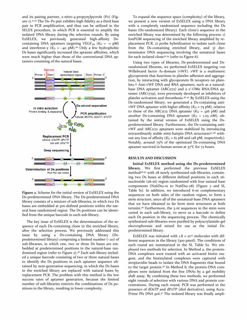
Figure 2. Scheme for the initial version of ExSELEX using the Ds-predetermined DNA library. The Ds-predetermined DNA library consists of a mixture of sub-libraries, in which two Ds bases are embedded at pre-defined positions within the nat-ural base randomized region. The Ds positions can be identi-fied from the unique barcode in each sub-library.
The key issue of ExSELEX is the determination of the se-quence of each Ds-containing clone in the enriched library, after the selection process. We previously addressed this issue by using a Ds-containing DNA library (Ds-predetermined library) comprising a limited number (~20) of sub-libraries, in which one, two or three Ds bases are em-bedded at predetermined positions in the natural-base ran-domized region (refer to Figure 2).26 Each sub-library includ-ed a unique barcode consisting of two or three natural bases to identify the Ds positions in each aptamer sequence ob-tained by next-generation sequencing, in which the Ds bases in the enriched library are replaced with natural bases by replacement PCR. The problem with this method is the low success rates of aptamer generation, because the limited number of sub-libraries restricts the combinations of Ds po-sitions in the library, resulting in lower complexity.
To expand the sequence space (complexity) of the library, we present a new version of ExSELEX using a DNA library with a completely randomized sequence including the Ds bases (Ds-randomized library). Each clone’s sequence in the enriched library was determined by the following process: 1) IonPGM sequencing of the enriched library amplified by re-placement PCR, 2) probe hybridization to isolate each clone from the Ds-containing enriched library, and 3) dye-terminator DNA sequencing involving the unnatural bases for each isolated clone32,34 (refer to Figure 6).
Using two types of libraries, Ds-predetermined and Ds-randomized libraries, we performed ExSELEX targeting von Willebrand factor A1-domain (vWF). vWF is a multimeric glycoprotein that functions in platelet adhesion and aggrega-tion, by interacting with glycoprotein Ib receptors on plate-lets.35 Anti-vWF DNA and RNA aptamers, such as a natural-base DNA aptamer (ARC1172) and a 2′-OMe RNA/DNA ap-tamer (ARC1779), were previously developed as inhibitors of platelet activation and thrombosis.36-42 By ExSELEX using the Ds-randomized library, we generated a Ds-containing anti-vWF DNA aptamer with higher affinity (KD = 75 pM), relative to those of the ARC1172 DNA aptamer (KD = 326 pM) and another Ds-containing DNA aptamer (KD = 1.03 nM), ob-tained by the initial version of ExSELEX using the Ds-predetermined library. Furthermore, the Ds-containing anti-vWF and ARC1172 aptamers were stabilized by introducing extraordinarily stable mini-hairpin DNA structures43-46 with-out any loss of affinity (KD = 61 pM and 128 pM, respectively). Notably, around 75% of the optimized Ds-containing DNA aptamer survived in human serum at 37ºC for 72 hours.
RESULTS AND DISCUSSION
Initial ExSELEX method using the Ds-predetermined library. We first performed the previous ExSELEX method26,47 with 28 newly synthesized sub-libraries, contain-ing two Ds bases at different defined positions in each 26-nucleotide (26-nt) region randomized with four natural base components (N26Ds2-01 to N26Ds2-28) (Figure 2 and SI, Table S1). In addition, we introduced 6-nt complementary sequences on both sides of the random region, to form a stem structure, since all of the unnatural-base DNA aptamers that we have obtained so far form stem structures at both termini.26 Furthermore, the 3-nt sequences in the stem were varied in each sub-library, to serve as a barcode to define each Ds position in the sequencing process. The chemically synthesized sub-libraries were purified by polyacrylamide-gel electrophoresis and mixed for use as the initial Ds-predetermined library.
ExSELEX was initiated with 1.8 × 10¹⁴ molecules with dif-ferent sequences in the library (300 pmol). The conditions of each round are summarized in the SI, Table S2. We em-ployed two methods for selection. In Method a, the protein-DNA complexes were treated with an activated biotin rea-gent, and the biotinylated complexes were captured with streptavidin beads to isolate the DNA fragments that bound to the target protein.26 In Method b, the protein-DNA com-plexes were isolated from the free DNAs by a gel mobility shift assay. By combining these two methods, we performed eight rounds of selection with various DNA and protein con-centrations. During each round, PCR was performed in the presence of dDsTP and dPxTP (diol derivative), using Accu-Prime Pfx DNA pol.32 The isolated library was finally ampli-

fied by PCR (replacement PCR) in the absence of dDsTP and dPxTP, but in the presence of dPa′TP,26,34 to facilitate the replacement of the unnatural bases with natural bases, in which the Ds bases in the library were finally replaced with A or T. We obtained 420,526 sequences. The sequences that appeared in >100 clones are listed in the SI, Table S3, and more than 90% of the sequences converged on a single fami-ly. From the barcodes in the sequences, we assigned two Ds base positions (positions 13 and 22) (SI, Table S3). However, besides these two positions in the main family, other posi-tions, such as 12, 25, and 26, also showed some characteristic base variations (many A and T variants), which might origi-nally correspond to the Ds base position. Thus, further exam-inations were performed using a sequencing method to con-firm the Ds positions in the main family.
Figure 3. Identification of the Ds-base positions in the en-riched DNA library in the initial version of ExSELEX. Through hybridization with a biotinylated probe, a specific clone was captured, and subjected to PCR-amplification in either the presence of dDsTP and dPxTP or the absence of dDsTP and dPxTP (but with dPa′TP). The representative sequencing patterns are shown at the bottom and the others are shown in the SI, Figure S1.
The sequencing method was actually developed for the new version of ExSELEX using a Ds-randomized library, as mentioned later. Based on the sequencing data obtained with IonPGM after replacement PCR, we designed a short DNA probe (5′-biotinylated 25-mer), in which T was chosen as the complementary base opposite Ds, to isolate the clone con-taining Ds bases from the enriched library (Figure 3 and SI, Table S3). The isolated clone was amplified by PCR in the presence of dDsTP and dPxTP for the following sequencing. Dye-terminator sequencing was performed using a BigDye
Terminator v1.1 Cycle Sequencing Kit (Applied Biosystems) with a gel sequencer (ABI 377).32,33 First, we used the Ds-containing strand of the amplified clone for sequencing in the presence or absence of dPa′TP or ddPa′TP (Pa′ = 4-propynylpyrrole-2-carbaldehyde). However, the cycle se-quencing reaction using the Taq DNA pol mutant paused at the first Ds position (SI, Figure S1). Thus, we performed the cycle sequencing using the complementary strand, namely the Px-containing strand, in the presence or absence of dDsTP or ddDsTP. We found that when using the Taq pol mutant provided in the sequencing kit, natural base sub-strates (mainly dATP) were misincorporated opposite Px, but the dideoxy-dye-terminators of the natural bases were not incorporated at the positions. Thus, in the absence of the unnatural base substrates, the sequencing reaction of Px-containing DNA proceeded by the incorporation of the natu-ral base substrate opposite Px, and the unnatural base posi-tions were recognized as a gap. Interestingly, the sequencing peak pattern in the absence of dDsTP was clearer than that in the presence of dDsTP (SI, Figure S1). By comparing the sequencing peak pattern with that of the clone after re-placement PCR, three unnatural base positions (13, 22, and 26) were identified (Figure 3).
Figure 4. Schematic illustration of the secondary structures of the anti-vWF unnatural-base DNA aptamer, Pr-DsDsDs-40, and its variants. The sequence and presumed secondary structure of Pr-DsDsDs-40 are shown on the left, and each variant is schematically represented on the right, with the thermal stabilities of Pre-DsDsDs-40 and Pr-AAA-40. The possible base pairing within the loop region of Pr-DsDsDs-40 is indicated with dotted lines.
The two Ds bases at positions 13 and 22 were initially em-bedded into the sub-library (N26Ds2-01, SI, Table S1), and the other one at position 26 might have been misincorpo-rated into the DNA during PCR in the selection rounds. To examine the contributions of each Ds base in the aptamer to the binding, we chemically synthesized the aptamer (Pr-DsDsDs-40, 40-mer) and its variants, in which the Ds bases were replaced with A in various combinations (Figure 4 and SI, Table S4). Gel-shift assays of each aptamer or variant complex with vWF were performed by native-gel electropho-resis at 4 and 25°C (Figure 5). The results revealed that all three of the Ds bases in the aptamer, especially the two Ds bases at positions 22 and 26, were essential for the tight

binding, and the shifted bands corresponding to the complex of any two Ds → A variants with vWF were not detected on the gel at 25°C. In this ExSELEX experiment, we serendipi-tously obtained the aptamer by the mutation of a natural to the Ds base during the selection process. These results sug-gested that the Ds-predetermined library containing two Ds bases was insufficient for generating high-affinity DNA ap-tamers, and increasing the complexity by using a Ds-randomized library should be necessary for ExSELEX.
Figure 5. Binding analysis of each Pr-DsDsDs-40 aptamer variant by a gel mobility shift assay. Each aptamer variant (100 nM) was incubated with vWF (100 nM) at 37°C for 30 min, and the complexes were separated from the free DNA on native 8% polyacrylamide gels with electrophoresis at either 4°C (upper panel) or 25°C (lower panel). The DNA bands on the gels were stained with SYBR Gold, and their band patterns were detected with a Bio-imaging analyzer (Fuji Film LAS4000).
New version of ExSELEX using the randomized library containing Ds. We prepared the Ds-randomized library containing a 30-nt randomized region flanked by 6-nt com-plementary sequences, to include the stem structures, as well as the sequences of PCR primers (Figure 6 and SI, Table S1). The randomized region was synthesized using mixtures of phosphoramidite reagents consisting of Ds (10%) and the natural bases (22.5% each). In the N30 library synthesized using the ratio (10%) of the Ds amidite, 14, 23, 24, and 18% of the DNA fragments would theoretically contain one, two, three, and four Ds bases, respectively. Our previous results indicated that only two or three Ds bases are sufficient to efficiently increase the affinity of DNA aptamers.
We performed seven rounds of selection using six sets (#1−#6) of the library (300 pmol each, total 1.08 × 10¹⁵ differ-ent molecules) for easier handling, as compared to that of one set of the 1.8-nmol library (Figure 6 and SI, Table S2). During the 4th to 6th rounds in the selection procedure, the known DNA aptamer, ARC1172 (41-mer, SI, Table S3),39 was added as a competitor to accelerate the enrichment, because the complexity was higher than that obtained using the Ds-predetermined library. For the six sets, each enriched library was tested for binding ability by the gel-shift assay (SI, Figure S2) under relatively harsh conditions at 37°C, to select the sets containing the high-affinity DNA aptamers. All of the enriched libraries generated the shifted bands corresponding to the DNA-protein complex, and in two sets (#1 and #4), the
shifted bands were quite clearly observed. Thus, we used these two enriched library sets for the following sequence analysis.
After replacement PCR of these two enriched libraries, the total sequences (#1: 151,495, #4: 180,152) were determined by IonPGM (SI, Tables S5 and S6). Theoretically, some percent-age of the DNA in the initial library contained no Ds bases, but all of the enriched sequences seemed to have Ds bases. The natural-base DNA aptamers might be excluded by the higher-affinity Ds-containing DNA aptamers during the se-lection process.
Figure 6. Scheme for a new version of ExSELEX using the Ds-randomized DNA library. After several rounds of selec-tion, the enriched sequences were analyzed by deep sequenc-ing after replacement PCR. The Ds positions in the enriched sequences were then identified through DNA sequencing analysis of PCR-amplified isolated DNA clones, as shown in Figure 3.
Among the total sequences, more than 83% for #1 and 86% for #4 of the sequences were enriched to each single family, and the sequences of the main families of #1 and #4 were both quite similar, in which we predicted three Ds base
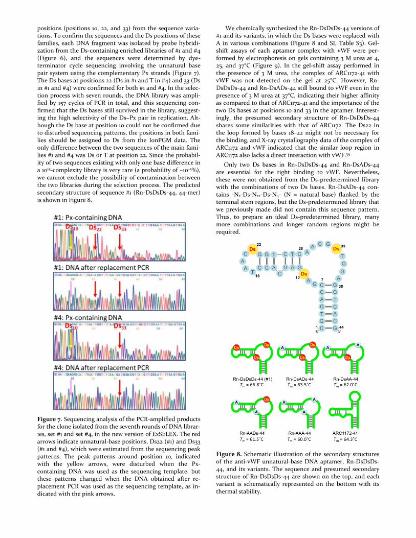
positions (positions 10, 22, and 33) from the sequence varia-tions. To confirm the sequences and the Ds positions of these families, each DNA fragment was isolated by probe hybridi-zation from the Ds-containing enriched libraries of #1 and #4 (Figure 6), and the sequences were determined by dye-terminator cycle sequencing involving the unnatural base pair system using the complementary Px strands (Figure 7). The Ds bases at positions 22 (Ds in #1 and T in #4) and 33 (Ds in #1 and #4) were confirmed for both #1 and #4. In the selec-tion process with seven rounds, the DNA library was ampli-fied by 157 cycles of PCR in total, and this sequencing con-firmed that the Ds bases still survived in the library, suggest-ing the high selectivity of the Ds–Px pair in replication. Alt-hough the Ds base at position 10 could not be confirmed due to disturbed sequencing patterns, the positions in both fami-lies should be assigned to Ds from the IonPGM data. The only difference between the two sequences of the main fami-lies #1 and #4 was Ds or T at position 22. Since the probabil-ity of two sequences existing with only one base difference in a 1015-complexity library is very rare (a probability of ~10-5%), we cannot exclude the possibility of contamination between the two libraries during the selection process. The predicted secondary structure of sequence #1 (Rn-DsDsDs-44, 44-mer) is shown in Figure 8.
Figure 7. Sequencing analysis of the PCR-amplified products for the clone isolated from the seventh rounds of DNA librar-ies, set #1 and set #4, in the new version of ExSELEX. The red arrows indicate unnatural-base positions, Ds22 (#1) and Ds33 (#1 and #4), which were estimated from the sequencing peak patterns. The peak patterns around position 10, indicated with the yellow arrows, were disturbed when the Px-containing DNA was used as the sequencing template, but these patterns changed when the DNA obtained after re-placement PCR was used as the sequencing template, as in-dicated with the pink arrows.
We chemically synthesized the Rn-DsDsDs-44 versions of #1 and its variants, in which the Ds bases were replaced with A in various combinations (Figure 8 and SI, Table S3). Gel-shift assays of each aptamer complex with vWF were per-formed by electrophoresis on gels containing 3 M urea at 4, 25, and 37°C (Figure 9). In the gel-shift assay performed in the presence of 3 M urea, the complex of ARC1172-41 with vWF was not detected on the gel at 25°C. However, Rn-DsDsDs-44 and Rn-DsADs-44 still bound to vWF even in the presence of 3 M urea at 37°C, indicating their higher affinity as compared to that of ARC1172-41 and the importance of the two Ds bases at positions 10 and 33 in the aptamer. Interest-ingly, the presumed secondary structure of Rn-DsDsDs-44 shares some similarities with that of ARC1172. The Ds22 in the loop formed by bases 18–22 might not be necessary for the binding, and X-ray crystallography data of the complex of ARC1172 and vWF indicated that the similar loop region in ARC1172 also lacks a direct interaction with vWF.39
Only two Ds bases in Rn-DsDsDs-44 and Rn-DsADs-44 are essential for the tight binding to vWF. Nevertheless, these were not obtained from the Ds-predetermined library with the combinations of two Ds bases. Rn-DsADs-44 con-tains -N2-Ds-N22-Ds-N4- (N = natural base) flanked by the terminal stem regions, but the Ds-predetermined library that we previously made did not contain this sequence pattern. Thus, to prepare an ideal Ds-predetermined library, many more combinations and longer random regions might be required.
Figure 8. Schematic illustration of the secondary structures of the anti-vWF unnatural-base DNA aptamer, Rn-DsDsDs-44, and its variants. The sequence and presumed secondary structure of Rn-DsDsDs-44 are shown on the top, and each variant is schematically represented on the bottom with its thermal stability.

Figure 9. Binding analysis of each Rn-DsDsDs-44 aptamer variant by a gel mobility shift assay. Each aptamer variant (5ꞌ-biotinylated, 100 nM) was incubated with vWF (100 nM) at 37°C for 30 min, and the complexes were separated from the free DNA on 8% polyacrylamide gels containing 3 M urea with electrophoresis at 4°C (upper panel), 25°C (middle pan-el), or 37°C (lower panel). The DNA bands on the gels were stained with SYBR Gold, and their band patterns were de-tected with a Bio-imaging analyzer (Fuji Film LAS4000).
Characterization of anti-vWF DNA aptamers. The af-finities of the aptamers to vWF were determined by surface plasmon resonance (SPR) using the 5′-biotinylated aptamers (Figure 10), in which a biotin-modified T residue was added to the 5′-terminus of each aptamer for immobilization on the sensor chip. Rn-DsDsDs-44 (KD = 74.9 pM) exhibited the highest affinity, as compared to Pr-DsDsDs-40 (KD = 1.03 nM) and ARC1172-41 (KD = 326 pM). Although the KD value of Pr-DsDsDs-40 was relatively high, its koff value was the smallest among the three aptamers, indicating the slowest off-rate binding to the target. This binding property of Pr-DsDsDs-40 was confirmed by a gel-shift assay in the presence of 3 M urea at different temperatures (4, 25, and 37°C), in which the complex with Pr-DsDsDs-40 exhibited higher stability than that with ARC1172-41 (Figure 11) on the gel at 25°C. Among these three aptamers, the complex of Rn-DsDsDs-44 with vWF showed the highest stability, which was clearly ob-served even on a gel containing 3 M urea at 37°C.
Since the thermal stabilities of the aptamers are also im-portant for diagnostic and therapeutic applications, we measured the Tm values of the aptamers (SI, Figure S3 and Figures 4 and 8). All three aptamers (Tm = 65.7°C for Pr-DsDsDs-40, 66.8°C for Rn-DsDsDs-44, and 64.3°C for ARC1172-41) exhibited high thermal stability. As we previous-
ly observed,26 the Ds→A replacements of the Ds-containing
DNA aptamers reduced their thermal stabilities. The high
stacking ability of the hydrophobic Ds base might stabilize each aptamer’s tertiary structure.
Figure 10. Binding analysis of anti-vWF DNA aptamers by a BIAcore T200 at 37°C, using 0.078 nM to 5 nM vWF. The aptamers were biotinylated at their 5′-termini.
Figure 11. Comparison of the binding affinities of the anti-vWF aptamer, Pr-DsDsDs-40, Rn-DsDsDs-44, and the con-ventional aptamer ARC1172-41, in gel mobility shift assays. Each aptamer variant (5ꞌ-biotinylated, 100 nM) was incubated with vWF (100 nM) at 37°C for 30 min, and the complexes were separated from the free DNA on 8% polyacrylamide gels containing 3 M urea, with electrophoresis at 4°C (upper pan-el), 25°C (middle panel), or 37°C (lower panel). The DNA bands on the gels were stained with SYBR Gold, and their band patterns were detected with a Bio-imaging analyzer (Fuji Film LAS4000).

Stabilization of anti-vWF DNA aptamers by the intro-duction of mini-hairpin DNAs. For pharmaceutical appli-cations, DNA aptamers must be stable at 37°C and resistant to degradation by nucleases. We previously developed a sta-bilization method by introducing extraordinarily stable mini-hairpin DNA sequences containing a GNA loop (N = A, G, C or T)43,44,48 into DNA aptamers.45,46 Rn-DsDsDs-44 and ARC1172-41 were stabilized by this method (Figure 12). First, the mini-hairpin DNA sequence was added to the 3′-termini of both aptamers (Rn-DsDsDs-53mh and ARC1172-50mh). In addition, two A–T pairs in the terminal stem region of Rn-DsDsDs-44 were replaced with G–C pairs, to thermally stabi-lize the stem structure (SI, Table S4). In contrast, the re-placement of A–T with G–C pairs in the terminal stem region of ARC1172-41 reduced the affinity, and thus we left the A–T pairs within the stem (data not shown).
Figure 12. Stabilization of the anti-vWF aptamers, Rn-DsDsDs-44 and ARC1172-41, by introducing mini-hairpin DNAs. The modified regions are indicated by blue lines (mini-hairpin DNA), red lines (A–T replaced with G–C), and brown circles (iT: inverted-dT). Their sequences are shown in SI, Table S4. These variants were characterized by their thermal stabilities (SI, Figures S3 and S4), binding abilities (analyzed with a BIAcore T200, Figures 10 and 13), and nucle-ase resistances in 96% human serum at 37°C (Figure 14).
Next, the internal loops (TAACDs in Rn-DsDsDs-53mh and TTC in ARC1172-50mh) were replaced with the mini-hairpin loop (GAA). The X-ray crystallographic analysis of the ARC1172–vWF complex indicated no direct interaction between the TTC loop and vWF. As for Rn-DsDsDs-53mh, the gel-shift assay of Rn-DsADs-44 (Figure 9) revealed that the Ds base at position 22 in the loop of Rn-DsDsDs-44 did not affect the affinity. Thus, these regions could be replaced with the mini-hairpin DNA, and we finally designed Rn-
DsDs-51mh2 and ARC1172-50mh2, in the same manner as that used for the stabilization of the anti-interferon-γ Ds-containing DNA aptamer.45
The thermal stabilities of these modified aptamers were measured (SI Figure S4 and Figure 12). By introducing the mini-hairpin DNAs, the thermal stabilities of Rn-DsDsDs-53mh (Tm = 75.5°C) and Rn-DsDs-51mh2 (Tm = 76.5°C) signif-icantly increased by around 10°C. Unexpectedly, the thermal stability of ARC1172 (Tm = 61.3°C for ARC1172-50mh and 61.9°C for ARC1172-50mh2) was slightly reduced by introduc-ing the mini-hairpin DNA to the 3′-terminus. These results suggested that the Ds-containing and natural-base DNA ap-tamers have different structural features.
Figure 13. Binding abilities of the optimized anti-vWF DNA aptamers containing mini-hairpin DNAs, assessed using a BIAcore T200 at 37°C with 0.078 nM to 5 nM vWF.
The high-thermal stabilities of Rn-DsDsDs-53mh and Rn-DsDs-51mh2 were also confirmed by gel-shift assays at differ-ent temperatures (Figure 9). Both of the aptamers containing the mini-hairpin DNAs efficiently bound to vWF even at 37°C, and the mobilities corresponding to the free DNAs, especially Rn-DsDs-51mh2, remained unaltered at 4, 25, and 37°C. In contrast, the Ds-containing DNA aptamers without the mini-hairpin DNAs generated different band patterns corresponding to the free DNA on the gels at each tempera-ture. At 4°C, the mobilities of these aptamers without the mini-hairpin DNAs were faster than those of the aptamers with the mini-hairpin DNAs. At 25°C, two bands appeared, and the upper bands may have resulted from the partially denatured structures. At 37°C, they mostly gave only the up-

per bands. Thus, the introduced mini-hairpin DNAs stabi-lized the entire aptamer structures.
Figure 14. Binding specificities of the optimized anti-vWF DNA aptamers with mini-hairpin DNAs to various proteins, measured by SPR. Each DNA aptamer was immobilized on a Sensorchip SA by injecting a 25 nM DNA solution for 1 min, at a flow rate of 5 μl min-1, instead of the 0.5 nM DNA solu-tion for 160 sec for the dissociation constant determinations (see Figures 10 and 13). Each injected protein was used at a 20 nM concentration (vWF A1 domain, human IFN- γ, BSA, hu-man TNF-α, human-α-Thrombin, and human NF-κB(p50)).
The stabilization method maintained the affinity of Rn-DsDsDs-53mh (KD = 61.3 pM), and interestingly, increased the affinity of Rn-DsDs-51mh2 (KD = 182 pM) (Figure 13), as compared to the initial aptamers, Rn-DsDsDs-44 (KD = 74.9 pM) and ARC1172-41 (KD = 326 pM) (Figure 9). In addition, the specificities of both aptamers were also high, and other proteins (human interferon-γ, BSA, human TNF-α, human α-thrombin, and human NF-κB (p50)) clearly did not bind to each aptamer (Figure 14).
We confirmed that the stabilities of these aptamers, espe-cially Rn-DsDs-51mh2, were increased in human serum; 75% of Rn-DsDs-51mh2 and 42% of ARC1172-50mh2 survived after an incubation at 37°C for 72 hours (Figure 15). Without the mini-hairpin DNA sequences, only 30% of Rn-DsDsDs-44 and 19% of ARC1172-41 remained in the human serum after 72 hours. Based on the difference in the stabilities between Rn-DsDs-51mh2 and ARC1172-50mh2, the stabilization method is more effective for Ds-containing DNA aptamers than natu-ral-base DNA aptamers.
We also examined the conventional stabilization method using an inverted T for ARC1172-41. One modification (ARC1172-42iT) was the attachment of the T to ARC1172-41 via the 3′-3′ linkage, and the other (ARC1172-42mh-iT) was de-signed by introducing the mini-hairpin DNA loop (GAA) into the internal loop of ARC1172-42iT (Figure 12). Although the inverted T method also stabilized the aptamer, it was less
effective than the mini-hairpin DNA method (Figure 15). This is because the mini-hairpin DNA stabilizes the entire stem region, by stacking between the 3′-terminal base of the mini-hairpin DNA and the 5′-terminal base of the aptamer.
Figure 15. Nuclease resistance of anti-vWF DNA aptamer derivatives in 96% human serum. Each DNA aptamer (5 µl, Rn-DsDsDs-44, Rn-AAA-44, Rn-DsDsDs-53mh, Rn-DsDs-51mh2, ARC1172-41, ARC1172-42iT, ARC1172-42mh-iT and ARC1172-50mh2; 50µM) was mixed with human serum (120 µl), and the mixture (2 µM DNA in 96% serum) was incubat-ed at 37°C. Aliquots (10 µl) were removed at various time points from 0 to 72 hours, and immediately mixed with 110 µl of denaturing solution (1× TBE containing 10 M urea). Each sample was analyzed by 15% denaturing polyacrylamide gel electrophoresis. DNA stained with SYBR Gold was detected with a bio-imaging analyzer (Fuji Film LAS-4000). To deter-mine the intact fraction, the band intensities were quantified with the Multi Gauge software. The residual amount (%) at each time point was plotted in the upper graphs. Each sam-ple was analyzed twice, and one analysis is displayed.
CONCLUSION
We developed a new version of ExSELEX using a com-pletely randomized DNA library composed of A, G, C, T, and Ds, to efficiently generate high-affinity Ds-containing DNA aptamers. Benner and colleagues also reported the sequence variation analysis for the determination of their unnatural base positions in the aptamers.27,49 To confirm the sequence of each aptamer, each isolated clone must be sequenced. Thus, we developed the method to isolate each clone from the library. By probe hybridization, Ds-containing DNA ap-tamers can be specifically isolated from the enriched library after the selection process, and their sequences can be cor-rectly determined. Using this method, we identified a DNA

aptamer that binds to vWF with a KD = 74.9 pM, indicating the effectiveness of the Ds-randomized library for ExSELEX.
The aptamer contains the terminal stem region, and thus it can be stabilized by the introduction of a mini-hairpin DNA for further applications.45,46 By introducing two mini-hairpin DNA sequences into the internal and terminal re-gions of the aptamer, the optimized aptamer was significant-ly stabilized both thermally and enzymatically, without any loss of the affinity (KD = 61.3 pM). Furthermore, we con-firmed that the stabilization method can be used for natural-base DNA aptamers, as shown by the ARC1172 modifications. The optimized anti-vWF DNA aptamer could be tested for therapeutic purposes.
Since only a few Ds bases are required for tight binding to targets, ExSELEX using Ds-predetermined libraries is also quite promising. However, the complexity of the present Ds-predetermined library was insufficient, and many more sub-libraries should be needed to increase the Ds-containing sequence combinations. In addition, our results indicated the possibility of Ds misincorporation within the library during PCR, and thus the sequencing method that we developed here is important to confirm the Ds positions in the generat-ed DNA aptamers.
There is still some room to improve the ExSELEX method. For example, our sequencing method using the gel or capil-lary sequencer involving the unnatural base pairing is still complicated and time-consuming and often results in a dis-turbed peak pattern, as shown in Figure 7, which makes it difficult to determine the Ds positions correctly. In addition, the increased complexity with five different bases causes another technical problem in the ExSELEX scale limitation (~1015 molecules for selection). When using a library with a 30-nt randomized region, the total number of different se-quence combinations is 9.31 × 10²⁰ (= 530) for five different bases, as compared to 1.15 × 10¹⁸ (= 430) for four different standard bases, reducing the success rates of aptamer gener-ation. Thus, further optimization of the Ds-randomized li-braries is required in terms of the length and the unnatural-base content of the randomized region, as well as the Ds-containing sequencing methods. Efforts to achieve these improvements are now in progress.
METHODS
Reagents and Materials. The selection target, recombinant protein vWF A1 domain (human, amino acids 1,238 to 1,481), was purchased from U-Protein Express. The unnatural nu-cleotide substrates, dDsTP, dPxTP, dPa′TP, ddDsTP, and ddPa′TP, used for PCR were synthesized as described previ-ously.32,34,50 DNA fragments used in this study were either chemically synthesized with an Oligonucleotide Synthesizer nS-8 (Gene Design), using phosphoramidite reagents for the natural, Ds, and Dss50 bases and modifiers (Glen Research), or purchased from Invitrogen or Gene Design. The chemical-ly synthesized DNA fragments were used after purification by denaturing gel electrophoresis, while the biotinylated probes were directly used after desalting purification.
SELEX procedures. The Ds-predetermined library used in this study was prepared by mixing 28 different single-stranded DNA sub-libraries (82-mer, SI, Table S1), each con-taining two Ds bases at defined positions in the 26-nt ran-domized region. Each sub-library consisted of 5′-primer, 5′-
stem (XXXACT), 26-nt randomized, 3′-stem (AGTYYY), and 3′-primer regions, where each combination of the XXX and YYY sequences functions as a barcode to identify the pre-determined Ds positions.
The Ds-randomized DNA library, N30Ds-S6-006, was used for the new version of ExSELEX. Each ExSELEX procedure was performed as described previously,26,47 with some modi-fications. The selection conditions for each round of Ex-SELEX are summarized in the SI, Table S2.
The binding of the DNA libraries to the target protein, vWF, was performed in Binding Buffer, 1× PBS supplemented with 0.005% (wt/vol) Nonidet P-40, at 25°C. To isolate the target-bound DNA from the unbound DNA, the complexes were either captured on streptavidin-coated magnetic beads after the biotinylation of vWF, as described previously, or isolated by native PAGE (8% polyacrylamide gel in 0.5× TBE) in each round. In the separation by native PAGE, DNA frag-ments were eluted in water from the excised gel slices corre-sponding to the shifted band positions. The recovered DNA fragments were subjected to PCR in 1× AccuPrime Pfx Reac-tion mix (Life Technologies), supplemented with 0.1 mM each dNTP (final conc.: 0.4 mM each) and 0.5 mM MgSO4 (final conc.: 1.5 mM), 0.05 mM each dDsTP and dPxTP, 1 µM each primer (Rev019-25 and Fow026-25-LT15 for the pre-determined Ds DNA library; mkP25-006DssTs1 and mkP25-009Ts2-LT15 for the random-Ds DNA library; SI, Table S1), and 0.05 U/µl AccuPrime Pfx DNA polymerase (Life Tech-nologies). PCR was performed as two-step cycling, 12 to 28 cycles of PCR amplification [94°C for 15 sec – 65°C for 3 min 30 sec], after 2 min at 94°C for the initial denaturation step. We used the 5′-primer, mkP25-006DssTs1, in which the 5′-terminus was labeled with the fluorescent Dss base, and the 3′-primer, mkP25-009Ts2-LT15, in which the oligo dT15 was attached to the 5′-terminus via the C12 alkyl linker. Thus, the Ds-strands for the library were labeled with Dss and were shorter than the non-labeled Px-strands, because the exten-sion reaction of the Ds-strands was paused at the position opposite the C12 linker in the Px-strand template in the PCR amplification. The Ds-strands were then separated from the Px-strands on a denaturing 10% polyacrylamide gel for the next round of selection.
Deep DNA sequencing. To determine the enriched se-quences after the 8th round in the initial version of ExSELEX and the 7th round in the new version of ExSELEX (in Set #1 and Set #4), we performed deep sequencing with an IonPGM sequencer system (Life Technologies), after replacement PCR of the DNA fragments recovered from the shifted bands on a gel from the final round of each ExSELEX. To replace the unnatural bases with natural bases in the enriched DNA li-braries, we performed PCR without dDsTP and dPxTP, but in the presence of dPa′TP (final conc., 0.05 mM). Supplementa-tion with dPa′TP facilitates the replacement of the unnatural base with any natural base (mainly A or T) via the Ds–Pa′ pairing followed by the A–Pa′ pairing.47 The PCR products were purified with a silica-membrane PCR purification kit, and then analyzed with the IonPGM sequencing system, ac-cording to the manufacturer’s instructions.
Analysis of sequence data sets obtained by deep DNA sequencing. To analyze the sequence data obtained by deep sequencing, we extracted the specific sequences from the total data, by using the CLC Genomics Workbench software (CLC bio) with the following criteria: 5′-

ACGACCGTTCTCTAATTTTGACGT–[38 bases]– AGGGTC-3′ or 5′-ACCAAATTATTGCGATACAGACCCT–[38 bases]– AACGT-3′ (extracted reads: 420,526 in total) for the Ds-pre-determined library, and 5′-TATATCCGCCATACTTACGTTGTCC–[42 bases]–GTTGGA-3′ or 5′-GCGCGACTTCACTTAAGATTCCAAC–[42 bases]–GGACAA-3′ (extracted reads: 151,495 reads for Set #1; 180,152 reads for Set #4) for the Ds-randomized library. The isolated sequences were processed and clustered with Microsoft Ex-cel, using a custom-made program (SI, Tables S3, S5, and S6).
Determination of Ds positions in isolated DNA frag-ments. To identify the Ds positions in each isolated clone from the enriched DNA libraries, we captured the targeted family sequences from each library, by using a specific hy-bridizing probe (5′-biotin-ACTCCCTCGGTTGTTGGCGAAAGTTG-3′ for the Ds-pre-determined library; 5’-biotin-CGTTGAGACCTGTTAGGTGCTCTTC-3′ for the Ds-randomized library). The DNA library (20 µl, 100 nM in Prob-ing Buffer, 20 mM Tris-HCl, 0.5 M NaCl, 10 mM MgCl2, pH 7.6) was annealed with a biotinylated DNA probe (1 µl, 5 µM in water), by heating at 90°C for 3 min, followed by cooling down by -0.1°C/sec to 55°C, and maintaining it at 55°C for 15 min. The mixture was incubated with Hydrophilic Streptavi-din Magnetic Beads (New England Biolabs) at 55°C for 5 min, and the biotinylated probe and the DNA clones hybridized to the probe were captured. The collected magnetic beads were washed with 150 µl of the probing buffer (pre-warmed at 55°C) five times. The hybridized DNA clones were recovered from the beads by incubating them in 20 µl of water at 75°C for 5 min. The recovered DNA was then subjected to 15-cycle PCR amplification in the presence of either dDsTP and dPxTP (0.05 mM each) or only dPa′TP (0.05 mM, replace-ment PCR), and purified by denaturing PAGE. DNA se-quencing of these PCR-amplified DNAs was performed with BigDye terminator v1.1 (20 µl reactions, 0.15 pmol DNA as template), in the presence of the unnatural substrates (dPa′TP, ddPa′TP, dDsTP, or ddDsTP), as described previous-ly.32,33 The sequencing peak patterns were analyzed on a gel sequencer, ABI377, using the Applied Biosystems Sequencing Analysis Software (ver. 3.2).
Gel mobility shift assays. The binding abilities of the en-riched libraries and each aptamer derivative to vWF were analyzed by gel electrophoresis. Each DNA (final conc. 100 nM) was mixed with vWF (final conc. 100 nM) in Binding Buffer (20 µl solution). After an incubation at 37°C for 30 min, 5 µl of 25% glycerol supplemented with bromophenol blue was added, and the solutions were immediately subject-ed to PAGE (8% polyacrylamide gel containing 5% glycerol with or without 3 M urea in 0.5× TBE) at the indicated tem-perature. The DNA-vWF complexes were separated from the free DNAs. The band patterns on the gels were detected by using a Bio-imaging analyzer, LAS-4000 (Fuji Film) after staining with SYBR Gold.
SPR measurements. The dissociation constants of the ap-tamers were determined by SPR, using a Biacore T200 (GE Healthcare) at 37°C, as described previously.46 The running buffer was 1× PBS supplemented with 0.05% (wt/vol) Nonidet P-40. Each biotinylated DNA was immobilized on a Sensor chip SA (GE Healthcare), and the interaction of the immobi-lized DNA aptamer with vWF was detected by monitoring injections of 0.078 to 5 nM vWF solutions (diluted with the
running buffer) in the Kinetic Injection mode. The condi-tions were flow rate: 100 µl min-1, protein injection time: 150 sec, and dissociation time: 450 sec. Each curve fitting was done with a 1:1 binding model using the BIAevaluation T200 software, version 1.0 (GE Healthcare).
ASSOCIATED CONTENT
Supporting Information.
The Supporting Information is available free of charge on the
ACS Publications website at DOI: 10.1021/jacs.6b05475.
Sequence data obtained from ExSELEX, Binding analysis by the gel-shift assay, Thermal stability profiles of the aptamers, Sequences of DNA fragments, ExSELEX conditions.
This material is available free of charge via the Internet at http://pubs.acs.org.
AUTHOR INFORMATION
Corresponding Author
Notes There is potential competing interest. A patent application describing ideas presented in this article has been filed by TagCyx Biotechnologies. M.K. and I.H. own stock in TagCyx Biotechnologies.
ACKNOWLEDGMENT
We thank Dr. Takashi Yabuki for setting up our clustering method with Excel to analyze the deep sequencing data. This work was supported by a Grant-in-Aid for Scientific Research [KAKENHI 26248043] from the Ministry of Education, Cul-ture, Sports, Science and Technology (I.H.), by grants for projects focused on developing key technologies for discover-ing and manufacturing drugs for next-generation treatment and diagnosis from the Ministry of Economy, Trade, and Industry (I.H.), and by the Japan Science and Technology Agency (JST) Precursory Research for Embryonic Science and Technology (PRESTO) (M.K.).
ABBREVIATIONS
SELEX, Systematic evolution of ligands by exponential en-richment; ExSELEX, genetic alphabet expansion for SELEX; vWF, von Willebrand factor A1-domain; pol., polymerase.
REFERENCES
(1) Zhou, J.; Rossi, J. J. Curr. Top. Med. Chem. 2009, 9, 1144.
(2) Keefe, A. D.; Pai, S.; Ellington, A. Nat. Rev. Drug Discov.
2010, 9, 537.
(3) Zhu, G.; Ye, M.; Donovan, M. J.; Song, E.; Zhao, Z.; Tan,
W. Chem. Commun. 2012, 48, 10472.
(4) Taylor, A. I.; Arangundy-Franklin, S.; Holliger, P. Curr.
Opin. Chem. Biol. 2014, 22, 79.
(5) Wu, X.; Chen, J.; Wu, M.; Zhao, J. X. Theranostics 2015, 5,
322.
(6) Ma, H.; Liu, J.; Ali, M. M.; Mahmood, M. A.; Labanieh, L.;
Lu, M.; Iqbal, S. M.; Zhang, Q.; Zhao, W.; Wan, Y. Chem.
Soc. Rev. 2015, 44, 1240.
(7) Bruno, J. G. Molecules 2015, 20, 6866.
(8) Chen, A.; Yang, S. Biosens Bioelectron. 2015, 71, 230.
(9) Toh, S. Y.; Citartan, M.; Gopinath, S. C.; Tang, T. H.
Biosens Bioelectron. 2015, 64, 392.

(10) Lao, Y. H.; Phua, K. K.; Leong, K. W. ACS Nano 2015, 9,
2235.
(11) Zhou, G.; Wilson, G.; Hebbard, L.; Duan, W.; Liddle, C.;
George, J.; Qiao, L. Oncotarget 2016, 7, 13446.
(12) Parashar, A. J. Clin. Diagn. Res. 2016, 10, BE01.
(13) Tuerk, C.; Gold, L. Science 1990, 249, 505.
(14) Ellington, A. D.; Szostak, J. W. Nature 1990, 346, 818.
(15) Gold, L.; Ayers, D.; Bertino, J.; Bock, C.; Bock, A.; Brody,
E. N.; Carter, J.; Dalby, A. B.; Eaton, B. E.; Fitzwater, T.;
Flather, D.; Forbes, A.; Foreman, T.; Fowler, C.; Gawande,
B.; Goss, M.; Gunn, M.; Gupta, S.; Halladay, D.; Heil, J.;
Heilig, J.; Hicke, B.; Husar, G.; Janjic, N.; Jarvis, T.;
Jennings, S.; Katilius, E.; Keeney, T. R.; Kim, N.; Koch, T.
H.; Kraemer, S.; Kroiss, L.; Le, N.; Levine, D.; Lindsey,
W.; Lollo, B.; Mayfield, W.; Mehan, M.; Mehler, R.;
Nelson, S. K.; Nelson, M.; Nieuwlandt, D.; Nikrad, M.;
Ochsner, U.; Ostroff, R. M.; Otis, M.; Parker, T.;
Pietrasiewicz, S.; Resnicow, D. I.; Rohloff, J.; Sanders, G.;
Sattin, S.; Schneider, D.; Singer, B.; Stanton, M.; Sterkel,
A.; Stewart, A.; Stratford, S.; Vaught, J. D.; Vrkljan, M.;
Walker, J. J.; Watrobka, M.; Waugh, S.; Weiss, A.; Wilcox,
S. K.; Wolfson, A.; Wolk, S. K.; Zhang, C.; Zichi, D. PLoS
One 2010, 5, e15004.
(16) Yatime, L.; Maasch, C.; Hoehlig, K.; Klussmann, S.;
Andersen, G. R.; Vater, A. Nat. Commun. 2015, 6, 6481.
(17) Dougherty, C. A.; Cai, W.; Hong, H. Curr. Top. Med.
Chem. 2015, 15, 1138.
(18) Maier, K. E.; Levy, M. Mol. Ther. Methods Clin. Dev. 2016,
5, 16014.
(19) Tolle, F.; Brandle, G. M.; Matzner, D.; Mayer, G. Angew.
Chem. Int. Ed. Engl. 2015, 54, 10971.
(20) Ruckman, J.; Green, L. S.; Beeson, J.; Waugh, S.; Gillette,
W. L.; Henninger, D. D.; Claesson-Welsh, L.; Janjic, N. J.
Biol. Chem. 1998, 273, 20556.
(21) Ng, E. W. M.; Shima, D. T.; Calias, P.; Cunningham, E. T.;
Guyer, D. R.; Adamis, A. P. Nat. Rev. Drug Discov. 2006,
5, 123.
(22) Drolet, D. W.; Green, L. S.; Gold, L.; Janjic, N. Nucleic
Acid Ther. 2016.
(23) Malyshev, D. A.; Romesberg, F. E. Angew. Chem. Int. Ed.
Engl. 2015, 54, 11930.
(24) Benner, S. A. Curr. Opin. Chem. Biol. 2012, 16, 581.
(25) Hirao, I.; Kimoto, M. Proc. Jpn. Acad. Ser. B, Phys. Biol.
Sci. 2012, 88, 345.
(26) Kimoto, M.; Yamashige, R.; Matsunaga, K.; Yokoyama, S.;
Hirao, I. Nat. Biotechnol. 2013, 31, 453.
(27) Sefah, K.; Yang, Z.; Bradley, K. M.; Hoshika, S.; Jimenez,
E.; Zhang, L.; Zhu, G.; Shanker, S.; Yu, F.; Turek, D.; Tan,
W.; Benner, S. A. Proc. Natl. Acad. Sci. U S A 2014, 111,
1449.
(28) Zhang, L.; Yang, Z.; Sefah, K.; Bradley, K. M.; Hoshika, S.;
Kim, M. J.; Kim, H. J.; Zhu, G.; Jimenez, E.; Cansiz, S.;
Teng, I. T.; Champanhac, C.; McLendon, C.; Liu, C.;
Zhang, W.; Gerloff, D. L.; Huang, Z.; Tan, W.; Benner, S.
A. J. Am. Chem. Soc. 2015, 137, 6734.
(29) Zhang, L.; Yang, Z.; Le Trinh, T.; Teng, I. T.; Wang, S.;
Bradley, K. M.; Hoshika, S.; Wu, Q.; Cansiz, S.; Rowold,
D. J.; McLendon, C.; Kim, M. S.; Wu, Y.; Cui, C.; Liu, Y.;
Hou, W.; Stewart, K.; Wan, S.; Liu, C.; Benner, S. A.; Tan,
W. Angew. Chem. Int. Ed. Engl. 2016, 55, 12372.
(30) Malyshev, D. A.; Dhami, K.; Lavergne, T.; Chen, T.; Dai,
N.; Foster, J. M.; Correa, I. R., Jr.; Romesberg, F. E. Nature
2014, 509, 385.
(31) Kimoto, M.; Kawai, R.; Mitsui, T.; Yokoyama, S.; Hirao, I.
Nucleic Acids Res. 2009, 37, e14.
(32) Yamashige, R.; Kimoto, M.; Takezawa, Y.; Sato, A.;
Mitsui, T.; Yokoyama, S.; Hirao, I. Nucleic Acids Res.
2012, 40, 2793.
(33) Okamoto, I.; Miyatake, Y.; Kimoto, M.; Hirao, I. ACS
Synth. Biol. DOI: 10.1021/acssynbio.5b00253
(34) Hirao, I.; Kimoto, M.; Mitsui, T.; Fujiwara, T.; Kawai, R.;
Sato, A.; Harada, Y.; Yokoyama, S. Nat. Methods 2006, 3,
729.
(35) Sadler, J. E. Annu. Rev. Biochem. 1998, 67, 395.
(36) Gilbert, J. C.; DeFeo-Fraulini, T.; Hutabarat, R. M.;
Horvath, C. J.; Merlino, P. G.; Marsh, H. N.; Healy, J. M.;
Boufakhreddine, S.; Holohan, T. V.; Schaub, R. G.
Circulation 2007, 116, 2678.
(37) Oney, S.; Nimjee, S. M.; Layzer, J.; Que-Gewirth, N.;
Ginsburg, D.; Becker, R. C.; Arepally, G.; Sullenger, B. A.
Oligonucleotides 2007, 17, 265.
(38) Diener, J. L.; Daniel Lagasse, H. A.; Duerschmied, D.;
Merhi, Y.; Tanguay, J. F.; Hutabarat, R.; Gilbert, J.;
Wagner, D. D.; Schaub, R. J. Thromb. Haemost. 2009, 7,
1155.
(39) Huang, R. H.; Fremont, D. H.; Diener, J. L.; Schaub, R. G.;
Sadler, J. E. Structure 2009, 17, 1476.
(40) Bae, O. N. Arch. Pharm. Res. 2012, 35, 1693.
(41) Jilma, B.; Paulinska, P.; Jilma-Stohlawetz, P.; Gilbert, J. C.;
Hutabarat, R.; Knobl, P. Thromb. Haemost. 2010, 104, 563.
(42) Mayr, F. B.; Knobl, P.; Jilma, B.; Siller-Matula, J. M.;
Wagner, P. G.; Schaub, R. G.; Gilbert, J. C.; Jilma-
Stohlawetz, P. Transfusion 2010, 50, 1079.
(43) Hirao, I.; Kawai, G.; Yoshizawa, S.; Nishimura, Y.; Ishido,
Y.; Watanabe, K.; Miura, K. Nucleic Acids Res. 1994, 22,
576.
(44) Yoshizawa, S.; Kawai, G.; Watanabe, K.; Miura, K.; Hirao,
I. Biochemistry 1997, 36, 4761.
(45) Matsunaga, K.; Kimoto, M.; Hanson, C.; Sanford, M.;
Young, H. A.; Hirao, I. Sci. Rep. 2015, 5, 18478.
(46) Kimoto, M.; Nakamura, M.; Hirao, I. Nucleic Acids Res.
2016, 44, 7487.
(47) Kimoto, M.; Matsunaga, K.; Hirao, I. Methods Mol. Biol.
2016, 1380, 47.
(48) Hirao, I.; Nishimura, Y.; Tagawa, Y.; Watanabe, K.; Miura,
K. Nucleic Acids Res. 1992, 20, 3891.
(49) Yang, Z.; Chen, F.; Alvarado, J. B.; Benner, S. A. J. Am.
Chem. Soc. 2011, 133, 15105.
(50) Kimoto, M.; Mitsui, T.; Yokoyama, S.; Hirao, I. J. Am.
Chem. Soc. 2010, 132, 4988.

12
TOC Figure