high diagnostic value of fdg-pet/ct in endometrial cancer...
TRANSCRIPT

1
High diagnostic value of FDG-PET/CT in endometrial cancer:
Systematic review and meta-analysis of the literature.
Vikram Rao Bollineni 1, 2†, Sigmund Ytre-Hauge 2, 3, Oksana Bollineni. Balabay 4, Helga Birgitte
Salvesen 1, 2 and Ingfrid Salvesen Haldorsen 2, 3
1 Department of Obstetrics and Gynecology, Haukeland University Hospital, University of
Bergen, Bergen, Norway.
2 Center for Cancer Biomarkers, Department of Clinical Science, University of Bergen, Bergen,
Norway.
3 Department of Radiology, Haukeland University Hospital, University of Bergen, Bergen,
Norway.
4 Department of Quantitative Economics, School of Business and Economics, Maastricht
University, Maastricht, the Netherlands.
Running title: FDG-PET/CT in endometrial cancer.
† Corresponding author
Dr. Vikram rao Bollineni, M.D, PhD.
Department of Obstetrics and Gynecology, Haukeland University Hospital, Center for Cancer
Biomarkers, Department of Clinical Science, University of Bergen, Bergen, Norway.
Phone: +31 65 2397771
E-mail: [email protected]; [email protected]
Journal of Nuclear Medicine, published on January 28, 2016 as doi:10.2967/jnumed.115.170597by on July 27, 2018. For personal use only. jnm.snmjournals.org Downloaded from

2
Conflict of Interest Statement
The author’s state that the research presented in this manuscript is free of conflicts of interest.
by on July 27, 2018. For personal use only. jnm.snmjournals.org Downloaded from

3
Aim: To evaluate the diagnostic performance of fluorodeoxyglucose positron emission
tomography/computed tomography (FDG-PET/CT) for the preoperative assessment of lymph
node metastases (LNM) in endometrial cancer patients and for the assessment of endometrial
cancer recurrence (ECR) after primary surgical treatment.
Methods: A comprehensive search was performed on Pubmed/MEDLINE databases for studies
reporting the diagnostic performance of FDG-PET/CT for assessment of LNM and ECR
published up to August 15th 2015. Twenty one studies (13 for LNM and 8 for ECR) were
included in the systematic review and meta-analysis. Pooled estimates of sensitivity, specificity,
positive likelihood ratio (PLR), negative likelihood ratio (NLR) and diagnostic odds ratio (DOR)
of the FDG-PET/CT were calculated along with 95% confidence intervals (CI). A summary
receiver operating characteristics curve (SROC) was constructed and the area under the SROC
curve (AUC) was determined along with Q* index.
Results: The overall pooled sensitivity, specificity, PLR, NLR, DOR and AUC (with 95% CI) of
FDG-PET/CT for detection of LNM were 0.72 (0.63-0.80), 0.94 (0.93- 0.96), 10.9 (7.9-15.1),
0.36 (0.27-0.48), 39.7 (21.4-73.6) and 0.94 (0.85-0.99), respectively; whereas the corresponding
numbers for detection of ECR were 0.95 (0.91-0.98), 0.91 (0.86-0.94), 8.8 (6.0-12.7), 0.08 (0.05-
0.15), 171.7 (67.9-434.3) and 0.97 (0.95-0.98), respectively. The overall diagnostic accuracy (Q*
index) in LNM and ECR were 0.88 and 0.93, respectively.
Conclusions: FDG-PET/CT has an excellent diagnostic performance for detecting LNM
preoperatively and disease recurrence postoperatively in endometrial cancer patients.
Keywords: Endometrial cancer, FDG, PET and CT.
by on July 27, 2018. For personal use only. jnm.snmjournals.org Downloaded from

4
Endometrial cancer is the most common gynaecological malignancy in the developed countries
(1). The prognosis is traditionally determined by clinical and histopathologic factors i.e. age,
histologic type, grade, stage of disease including assessment of cervical invasion, depth of
myometrial invasion, lymph node spread and distant metastases (2–4). The 5-year overall
survival rate is generally favourable, around 80%. However, pelvic LNM represents the most
common site for extra uterine disease at primary treatment and the 5 year survival rate is around
50% for this patient subgroup (5).
Currently, the final staging of endometrial cancer is based on histopathologic findings at primary
surgery, which includes abdominal exploration, peritoneal cytology washing from the pelvis,
hysterectomy, bilateral salpingo-oophorectomy and lymphadenectomy in selected patients
presumed to have high risk of disease spread (6–8). Routine systemic pelvic lymphadenectomy
for early stage endometrial cancer disease, although not well defined as surgical technique,
improves detection of LNM, but the procedure showed no survival benefit in two randomized
clinical trials (9,10). Valid preoperative identification of patients with LNM who may benefit
from lymphadenectomy is thus essential if futile surgical staging and unnecessary postoperative
staging related complications are to be minimized. If a non-invasive imaging technique could
accurately preclude lymph node metastases preoperatively, lymphadenectomy procedures,
currently with unproven clinical benefit for survival, could be safely circumvented. Hence, the
development of non-invasive imaging methods enabling more accurate preoperative staging of
endometrial cancer may facilitate better tailored surgical decision making based on selection of
appropriate risk groups for LNM.
Conventional diagnostic imaging by transvaginal ultrasound (TVU), magnetic resonance imaging
(MRI) and computed tomography (CT) provide detailed anatomical information, while functional
by on July 27, 2018. For personal use only. jnm.snmjournals.org Downloaded from

5
or metabolic tumor characteristics may remain undetected. However, vigorous debate has
challenged the use of anatomic assessments solely relying on tumor morphological information,
not taking into account functional tumor characteristics that may prove highly relevant for the
clinical phenotype (11–13). In this regard, to better understand the tumor microenvironment,
metabolic positron emission tomography (PET) tracers such as fluorodeoxyglucose (FDG), in
combination with CT can overcome the limitations of morphological imaging alone, since
functional changes possible to detect by FDG-PET/CT often precede morphological changes
detectable by conventional MRI or CT (14,15).
FDG-PET/CT has long been used successfully for evaluation of several malignancies including
endometrial cancer (15,16) (Fig.1). Based on a systematic review, we here report diagnostic
indices of FDG-PET/CT for preoperative prediction of lymph node metastases and for detection
of disease recurrence after surgery with curative intent in endometrial cancer patients.
MATERIALS AND METHODS
Search Strategy
Because the study was not conducted on patients, no informed consent or ethical committee
approval was needed. To identify all relevant publications we performed systematic searches in
the bibliographic databases PUBMED.com from inception to August 17, 2015. Search terms
included controlled terms from Mesh in PUBMED.com using the following search terms ‘FDG
PET’ in combination with ‘Endometrial neoplasms’. The references of the identified articles were
also searched for relevant publications.
Selection Process
by on July 27, 2018. For personal use only. jnm.snmjournals.org Downloaded from
6
One physician (VRB) and one statistician (OBB) reviewed each published article independently
to determine the eligibility for inclusion in the meta-analysis, and to extract information
regarding clinical patient data and PET/CT characteristics. From the studies selected, data on first
author, year of publication, number of patients included, study design (prospective or
retrospective), patient age (mean/median), results from surgical International Federation of
Gynecology and Obstetrics (FIGO) staging, percentage with nodal metastases, percentage with
endometrioid subtype, FDG-PET/CT technical characteristics and numbers for diagnostic
performance of FDG-PET/CT (i.e. true negatives, false negatives, true positives, false positives,
positive predictive value and negative predictive value) were extracted and recorded. Any
differences were resolved by consensus.
PET/CT studies that met the following criteria were included. First, studies which reported the
diagnostic performance of FDG-PET/CT in detecting lymph node metastases preoperatively
and/or disease recurrence in endometrial cancer patients after primary surgery. Secondly, clinical
studies which included at least 10 patients. Third, studies which applied FDG as a tracer on
dedicated device and published after peer review. Studies on animals or in vitro studies, studies
not available in full text or not written in English and non-original articles (e.g. reviews,
editorials, letters, legal cases, interviews, case reports) were not evaluated systematically in this
review.
Statistical Analysis
We performed standard methods recommended for meta-analysis of diagnostic test evaluations
(17). Statistical analyses were carried out using Meta-Disc 1.4 software (18). We computed
pooled measures for the following test indices of each study: sensitivity, specificity, positive
by on July 27, 2018. For personal use only. jnm.snmjournals.org Downloaded from

7
likelihood ratio (PLR), negative likelihood ratio (NLR) and diagnostic odds ratio (DOR). Further
the summary receiver operating characteristics curve (SROC) was constructed and the area under
the SROC curve (AUC) was determined.
A random effects model was used for statistical pooling of the data. Pooled data were presented
with 95% confidence intervals (CI). The CI for diagnostic indices are exact, i.e they are based on
the binomial distribution and hence are asymmetric. The I-square index was used to test for
heterogeneity between studies. The AUC was calculated to measure the overall diagnostic
performance of FDG-PET/CT in detecting the LNM and endometrial cancer recurrence. The
sensitivity and specificity for the single test threshold identified for each study were used to plot
the SROC curve along with Q* index representing an overall measure of the test’s discriminatory
power.
RESULTS
Literature Search Results
The literature search yielded a total of 58 references in PUBMED.com. In addition, three relevant
recent articles on LNM in endometrial cancer and one in ECR that we were aware of, were
included in our database. The flow chart of the search and selection process is presented in Fig 2.
Out of a total of 62 articles, only 21 were eligible according to the criteria. Tables 1 and 2
summarize details for the included endometrial cancer studies of LNM and ECR by FDG-
PET/CT imaging, respectively.
Preoperative Detection of Lymph Node Metastases
by on July 27, 2018. For personal use only. jnm.snmjournals.org Downloaded from

8
In our meta-analysis of LNM, 13 studies were included, comprising a total of 861 endometrial
cancer patients. The overall pooled diagnostic indices of preoperative FDG-PET/CT for detecting
LNM are calculated on the patient basis. The pooled sensitivity and specificity values are 0.72
(95% CI: 0.63 to 0.80) (Fig. 3A) and 0.94 (95% CI: 0.93 to 0.96) (Fig. 3B), respectively. The
pooled PLR is 10.9 (95% CI: 7.9 to 15.1), the pooled NLR is 0.36 (95% CI: 0.27 to 0.48), and the
DOR is 39.7 (95% CI: 21.4 to 73.6).
The SROC representing a global summary score for the test performance yielded an AUC of 0.94
and a Q* value of 0.88 (Fig. 3C), indicating a relatively high level of overall accuracy.
Detection of Endometrial Cancer Recurrence
In the present meta-analysis for ECR, 8 studies comprising 378 patients have been included. The
pooled sensitivity is 0.95 (95% CI: 0.91 to 0.98) (Fig. 4A), while the pooled specificity is 0.91
(95% CI: 0.86 to 0.94) (Fig. 4B). The pooled PLR is 8.8 (95% CI: 6.0 to 12.7), NLR 0.08 (95%
CI: 0.05 to 0.15) and the DOR 171.7 (95% CI: 67.9 to 434.3). The SROC curve for the FDG-
PET/CT in the detection of endometrial cancer recurrence yields an AUC and Q* values of 0.97
and 0.93 (Fig. 4C), respectively, suggesting that the level of overall accuracy is high.
DISCUSSION
Lymphadenectomy is currently commonly applied for lymph node staging in endometrial
carcinoma as part of the surgical FIGO staging systems. However, non-invasive accurate lymph
node staging in endometrial cancer by preoperative imaging seems advantageous compared to the
more invasive nature of surgical lymph node staging, also with an unproven survival benefit from
the procedure (9,10). Similar to other tumors, endometrial cancer has an increased tumor glucose
metabolism and glycolysis rate which makes it suitable for FDG-PET/CT imaging (19–21). The
by on July 27, 2018. For personal use only. jnm.snmjournals.org Downloaded from

9
present meta-analysis yields very high diagnostic performances of FDG-PET/CT for diagnosing
LNM preoperatively- High diagnostic accuracy was also demonstrated for the procedure
detecting endometrial cancer recurrence after primary surgical treatment. This clearly supports a
role of FDG-PET/CT to enable more accurately tailored primary surgical endometrial cancer
treatment and subsequent patient care.
The pooled sensitivity for preoperative detection of lymph node metastases by FDG-PET/CT in
this meta-analysis was 72%, highlighting that as much as about 1/4 of the metastatic lymph nodes
are still missed by FDG-PET/CT. One possible explanation for this finding is that FDG avidity
relies on the presence of sufficient number of malignant cells exhibiting increased glucose
metabolism. Furthermore, the spatial resolution of PET/CT is not good enough to reliably detect
small tumors or micro metastatic disease. There is no documented threshold for lymph node size
allowing PET/CT to correctly identify metastatic lymph nodes in endometrial cancer, although
one study, reported node based sensitivities of 17% (4/24) for nodes ≤4 mm, 67% (14/24) for
nodes measuring 5-9 mm, and 93% (14/15) for nodes ≥ 10 mm (22). Similar figures with node-
based sensitivities of 13%, 67% and 100% in metastatic lymph nodes of ≤4 mm, 5-9 mm and ≥10
mm, respectively, in endometrial cancer was reported in another study (23). It should, however,
be kept in mind, that although this meta-analysis found the overall sensitivity of FDG-PET/CT to
be moderate for the detection of LNM in endometrial cancer, it compares favourably with the
reported sensitivities for LNM detection by conventional MRI and CT (24).
A very high pooled specificity of 0.94 for metastatic lymph node detection by FDG-PET/CT was
found in this study, and it may be argued that this specificity is sufficiently high to safely omit a
major surgical procedure in patients with low risk based on results from preoperative endometrial
biopsy and preoperative imaging, reducing operative and post-surgical complications and costs
by on July 27, 2018. For personal use only. jnm.snmjournals.org Downloaded from

10
(25). Furthermore, the present meta-analysis, showed that FDG-PET/CT has a very high PLR
(10.9), pinpointing that FDG-PET/CT findings suggesting metastatic lymph nodes are very likely
to be confirmed at surgical staging. The high diagnostic performance of FDG-PET/CT for
detecting endometrial cancer lymph node metastases is also justified by high AUC of 0.94 in this
meta-analysis. Interestingly, Kang et al. (26), reported almost identical figures for the diagnostic
performance of FDG-PET/CT for detecting LNM in cervical cancer, with reported sensitivity of
0.73 (95% CI 0.53 to 0.87) and specificity of 0.93 (95% CI 0.86 to 0.97). Thus, FDG-PET/CT
seems to be equally feasible in endometrial and cervical cancer for lymph node staging, and
FDG-PET/CT may be particularly justified in endometrial- and cervical cancer patients with high
risk for disease spread, in order to identify metastatic lymph nodes preoperatively.
Several recent studies in endometrial cancer have demonstrated that preoperative primary tumor
metabolic parameters have been associated with the presence of LNM. In a prospective study,
Antonsen et al. (27) found significantly higher SUVmax values in patients with LNM compared
to those with no LNM (P=0.04). Additionally, they found that SUVmax was significantly higher
in patients with high FIGO stage, myometrial invasion and cervical invasion. Furthermore,
Crivellaro et al. (28) found strong association between the presence of LNM and metabolic tumor
volume in endometrial cancer. Recently, we demonstrated that the preoperative metabolic tumor
volume cut-off value of 30 ml yielded sensitivity and specificity of 85% and 76% for LNM,
respectively, suggesting that the metabolic tumor volume is a promising marker for LNM (25). In
this regard, preoperative FDG-PET/CT imaging of primary endometrial carcinomas may provide
an adequate tool for prognostication and LNM detection that facilitate personalized patient care.
However, additional prospective studies are required to define optimal cut-off values for
predicting LNM based on FDG-PET/CT metabolic parameters. Earlier studies describe measures
by on July 27, 2018. For personal use only. jnm.snmjournals.org Downloaded from

11
for SUV values from a single region of interest, which does not represent the overall tumor
profile. Therefore, advanced techniques like the whole tumor voxel-by-voxel analysis may be a
preferable approach to reduce operator dependence and capture more relevant and comprehensive
measures for tumor microenvironment and heterogeneity.
The pooled sensitivity and specificity of FDG-PET/CT for the detection ECR were 0.95 and 0.91,
respectively with AUC in ROC analysis of 0.97 (95% CI: 0.95 to 0.99), all supporting high level
of overall diagnostic accuracy. Again, similar FDG-PET/CT diagnostic performance indices
were reported for detecting recurrent uterine cervical carcinomas with reported pooled sensitivity,
specificity and AUC of 0.92 (95% CI 0.91 to 0.94), 0.84 (95% CI 0.74 to 0.91) and 0.95,
respectively (29). Thus, FDG-PET/CT seem to perform equally well in the diagnosis of
endometrial and cervical cancer recurrences, supporting a promising role of FDG-PET/CT as
diagnostic tool for patients with suspected recurrence.
The findings in this study regarding FDG-PET/CT and ECR must, however, be interpreted with
care, considering that the studies report lack of histological confirmation of all putative
metastases based on FDG-PET/CT, and they report variable follow-up of the cases considered
non-metastatic based on FDG-PET/CT. Thus, some of the cases classified as correctly staged for
ECR by FDG-PET/CT, may have been erroneously classified. This limitation shared by most
published studies including the studies on cervical cancer recurrence, is however hard to
circumvent, as it seems unethical to perform biopsies of all suspected metastatic lesions in
patients due to risk of complications. Furthermore, frequent FDG PET/CT follow-up scans is
both very expensive and implies unwanted radiation exposure for the patients.
by on July 27, 2018. For personal use only. jnm.snmjournals.org Downloaded from

12
As both LNM and endometrial cancer recurrence studies exhibit inter study heterogeneity, the
SROC curve should be asymmetric (Supplementary Fig.1 for both symmetric and asymmetric
SROCs). Since all possible curves with the same true odds ratio and different degrees of
heterogeneity would pass through the same point on the anti-diagonal, the heterogeneity does not
affect the Q* estimate, but rather the shape of the curve and its standard errors. Walter et al. (30)
notes that the AUC standard errors calculated under the homogeneity assumption provide a good
approximation for heterogeneous studies. The approximation may be poor for extremely high
DOR values (higher than 20), as is the case in both meta-analyses presented here (37.5 and 171.7
for LNM and recurrence, respectively). However, the bias in the homogeneity-based standard
errors is mostly positive, and hence conservative, i.e. can be overestimated, but rarely
underestimated. The supplementary Fig. 1 illustrates that the confidence intervals of the
asymmetric SROC are much narrower compared to those of the symmetric SROC, while the
difference between the AUC estimates is negligible.
This meta-analysis has several limitations. First, positive result publication bias is a major
concern, since non-significant or unfavourable study results tend to be discarded. However, we
evaluated publication bias in our meta-analysis using funnel plot asymmetry, finding the funnel
plots to be symmetric for both sensitivity and specificity pooling, implying no large bias in our
study. Second, the current meta-analysis did not include region by region or node by node
evaluation since this was not reported in most studies; however, this could have provided
additional information. Third, not all included studies had a prospective study design. Fourth, the
gold standard for confirmation of LNM or ECR, being histopathological examination from
biopsies, was not obtained from all the lesions reported in the studies. However, clinical follow-
up data and results from renewed diagnostic imaging were recorded and clinically putative lymph
by on July 27, 2018. For personal use only. jnm.snmjournals.org Downloaded from

13
nodes metastases or endometrial cancer recurrence was used as gold standard when histologic
confirmation was missing.
CONCLUSION
Overall FDG-PET/CT demonstrated high diagnostic performance in identifying lymph node
metastases preoperatively and in detecting recurrence after endometrial carcinoma surgery with
curative intent. Larger prospective studies are needed to validate this high diagnostic performance
of FDG-PET/CT in endometrial cancer and further assess patient subgroups with particular
clinical benefit from applying this advanced imaging procedure.
ACKNOWLEDGMENTS
Writing this paper was supported by funding from the Norwegian Cancer Society.
by on July 27, 2018. For personal use only. jnm.snmjournals.org Downloaded from

14
REFERENCES
1. Amant F, Moerman P, Neven P, Timmerman D, Van Limbergen E, Vergote I. Endometrial
cancer. Lancet. 366(9484):491-505.
2. Dewdney SB, Jiao Z, Roma AA, et al. The prognostic significance of lymphovascular
space invasion in laparoscopic versus abdominal hysterectomy for endometrioid
endometrial cancer. Eur J Gynaecol Oncol. 2014;35(1):7-10.
3. Larson DM, Connor GP, Broste SK, Krawisz BR, Johnson KK. Prognostic significance of
gross myometrial invasion with endometrial cancer. Obstet Gynecol. 1996;88(3):394-398.
4. Uharcek P. Prognostic factors in endometrial carcinoma. J Obstet Gynaecol Res.
2008;34(5):776-783.
5. Lewin SN, Herzog TJ, Barrena Medel NI, et al. Comparative performance of the 2009
international federation of gynecology and obstetrics’ staging system for uterine corpus
cancer. Obstet Gynecol. 2010;116(5):1141-1149.
6. Boronow RC, Morrow CP, Creasman WT, et al. Surgical staging in endometrial cancer:
clinical-pathologic findings of a prospective study. Obstet Gynecol. 1984;63(6):825-832.
7. Pecorelli S. Revised FIGO staging for carcinoma of the vulva, cervix, and endometrium.
Int J Gynaecol Obstet. 2009;105(2):103-104.
8. Rungruang B, Olawaiye AB. Comprehensive surgical staging for endometrial cancer. Rev
Obstet Gynecol. 2012;5(1):28-34.
9. Benedetti Panici P, Basile S, Maneschi F, et al. Systematic pelvic lymphadenectomy vs. no
lymphadenectomy in early-stage endometrial carcinoma: randomized clinical trial. J Natl
by on July 27, 2018. For personal use only. jnm.snmjournals.org Downloaded from

15
Cancer Inst. 2008;100(23):1707-1716.
10. Kitchener H, Swart AMC, Qian Q, Amos C, Parmar MKB. Efficacy of systematic pelvic
lymphadenectomy in endometrial cancer (MRC ASTEC trial): a randomised study. Lancet.
2009;373(9658):125-136.
11. Faria S, Sagebiel T, Devine C, Lal C, Balachandran A, Bhosale P. Imaging in endometrial
carcinoma. Indian J Radiol Imaging. 2015;25(2):137.
12. Ben-Shachar I, Vitellas KM CD. The role of MRI in the conservative management of
endometrial cancer. - PubMed - NCBI. Gynecol Oncol. 2004;93(1):233-237.
13. Connor JP, Andrews JI, Anderson B BR. Computed tomography in endometrial
carcinoma. - PubMed - NCBI. Obstet Gynecol. 2000;95(5):692-696.
14. Amit A, Schink J, Reiss A LL. PET/CT in gynecologic cancer: present applications and
future prospects--a clinician’s perspective. - PubMed - NCBI. Obs Gynecol Clin North Am.
2011;38(1):1-21.
15. Tsujikawa T, Tsuchida T, Yoshida Y, Kurokawa T, Kiyono Y, Okazawa H KH. Role of
PET/CT in gynecological tumors based on the revised FIGO staging classification. Clin
Nucl Med. 2011;36(9).
16. Nakamura K, Kodama J, Okumura Y, Hongo A, Kanazawa S HY. The SUVmax of 18F-
FDG PET correlates with histological grade in endometrial cancer. - PubMed - NCBI. Int J
Gynecol cancer. 2010;20(1):110-115.
17. Devillé WL, Buntinx F, Bouter LM, et al. Conducting systematic reviews of diagnostic
studies: didactic guidelines. BMC Med Res Methodol. 2002;2:9.
by on July 27, 2018. For personal use only. jnm.snmjournals.org Downloaded from
16
18. Zamora J, Abraira V, Muriel A, Khan K CA. Meta-DiSc: a software for meta-analysis of
test accuracy data. - PubMed - NCBI. BMC Med Res Methodol. 2006;12(6):31.
19. Kitajima K, Kita M, Suzuki K, Senda M, Nakamoto Y, Sugimura K. Prognostic
significance of SUVmax (maximum standardized uptake value) measured by [18F]FDG
PET/CT in endometrial cancer. Eur J Nucl Med Mol Imaging. 2012;39(5):840-845.
20. Malgorzata Walentowicz-Sadlecka, Bogdan Malkowski, Pawel Walentowicz, Pawel
Sadlecki, Andrzej Marszalek TP and MG. The Preoperative Maximum Standardized
Uptake Value Measured by 18F-FDG PET/CT as an Independent Prognostic Factor of
Overall Survival in Endometrial Cancer Patients. Biomed Res Int. 2014;2014:7.
21. Chung HH, Cheon GJ, Kim HS, Kim JW, Park N-H, Song YS. Preoperative PET/CT
standardized FDG uptake values of pelvic lymph nodes as a significant prognostic factor in
patients with endometrial cancer. Eur J Nucl Med Mol Imaging. 2014;41(9):1793-1799.
22. Kitajima K, Murakami K, Yamasaki E, et al. Accuracy of 18F-FDG PET/CT in detecting
pelvic and paraaortic lymph node metastasis in patients with endometrial cancer. AJR Am J
Roentgenol. 2008;190(6):1652-1658.
23. Kitajima K, Murakami K, Yamasaki E, Kaji Y, Sugimura K. Accuracy of integrated FDG-
PET/contrast-enhanced CT in detecting pelvic and paraaortic lymph node metastasis in
patients with uterine cancer. Eur Radiol. 2009;19(6):1529-1536.
24. Salvesen HB, Haldorsen IS, Trovik J. Markers for individualised therapy in endometrial
carcinoma. Lancet Oncol. 2012;13(8):e353-e361.
25. Husby JA, Reitan BC, Biermann M, et al. Metabolic Tumor Volume on 18F-FDG PET/CT
by on July 27, 2018. For personal use only. jnm.snmjournals.org Downloaded from

17
Improves Preoperative Identification of High-Risk Endometrial Carcinoma Patients. J
Nucl Med. 2015;56(8):1191-1198.
26. Kang S, Kim S-K, Chung D-C, et al. Diagnostic value of (18)F-FDG PET for evaluation of
paraaortic nodal metastasis in patients with cervical carcinoma: a metaanalysis. J Nucl
Med. 2010;51(3):360-367.
27. Antonsen SL, Loft A, Fisker R, et al. SUVmax of 18FDG PET/CT as a predictor of high-
risk endometrial cancer patients. Gynecol Oncol. 2013;129(2):298-303.
28. Crivellaro C, Signorelli M, Guerra L, et al. Tailoring systematic lymphadenectomy in
high-risk clinical early stage endometrial cancer: the role of 18F-FDG PET/CT. Gynecol
Oncol. 2013;130(2):306-311.
29. Ding XP, Feng L ML. Diagnosis of recurrent uterine cervical cancer: PET versus PET/CT:
a systematic review and meta-analysis. Arch Gynecol Obs. 2014;290(4):741-747.
30. Walter SD. Properties of the summary receiver operating characteristic (SROC) curve for
diagnostic test data. Stat Med. 2002;21(9):1237-1256.
31. Nakamura K, Hongo A, Kodama J, Hiramatsu Y. The measurement of SUVmax of the
primary tumor is predictive of prognosis for patients with endometrial cancer. Gynecol
Oncol. 2011;123(1):82-87.
32. Nogami Y, Banno K, Irie H, et al. The efficacy of preoperative positron emission
tomography-computed tomography (PET-CT) for detection of lymph node metastasis in
cervical and endometrial cancer: clinical and pathological factors influencing it. Jpn J Clin
Oncol. 2015;45(1):26-34.
by on July 27, 2018. For personal use only. jnm.snmjournals.org Downloaded from

18
33. Inubashiri E, Hata K, Kanenishi K, et al. Positron emission tomography with the glucose
analog [F]-fluoro-2-deoxy-D-glucose for evaluating pelvic lymph node metastasis in
uterine corpus cancer: comparison with CT and MRI findings. J Obstet Gynaecol Res.
2009;35(1):26-34.
34. Signorelli M, Guerra L, Buda A, et al. Role of the integrated FDG PET/CT in the surgical
management of patients with high risk clinical early stage endometrial cancer: detection of
pelvic nodal metastases. Gynecol Oncol. 2009;115(2):231-235.
35. Boonya-ussadorn T, Choi WH, Hyun J, Kim SH, Chung SK, Yoo IR. 18F-FDG PET/CT
findings in endometrial cancer patients: the correlation between SUVmax and
clinicopathologic features. J Med Assoc Thai. 2014;97 Suppl 2:S115-S122.
36. Kitajima K, Suenaga Y, Ueno Y, et al. Value of fusion of PET and MRI for staging of
endometrial cancer: comparison with 18F-FDG contrast-enhanced PET/CT and dynamic
contrast-enhanced pelvic MRI. Eur J Radiol. 2013;82(10):1672-1676.
37. Suga T, Nakamoto Y, Saga T, et al. Clinical value of FDG-PET for preoperative
evaluation of endometrial cancer. Ann Nucl Med. 2011;25(4):269-275.
38. Picchio M, Mangili G, Samanes Gajate AM, et al. High-grade endometrial cancer: value of
[(18)F]FDG PET/CT in preoperative staging. Nucl Med Commun. 2010;31(6):506-512.
39. Horowitz NS, Dehdashti F, Herzog TJ, et al. Prospective evaluation of FDG-PET for
detecting pelvic and para-aortic lymph node metastasis in uterine corpus cancer. Gynecol
Oncol. 2004;95(3):546-551.
40. Ryu S-Y, Kim K, Kim Y, et al. Detection of recurrence by 18F-FDG PET in patients with
by on July 27, 2018. For personal use only. jnm.snmjournals.org Downloaded from

19
endometrial cancer showing no evidence of disease. J Korean Med Sci. 2010;25(7):1029-
1033.
41. Sharma P, Kumar R, Singh H, et al. Carcinoma endometrium: role of 18-FDG PET/CT for
detection of suspected recurrence. Clin Nucl Med. 2012;37(7):649-655.
42. Ozcan Kara P, Kara T, Kaya B, Kara Gedik G, Sari O. The value of FDG-PET/CT in the
post-treatment evaluation of endometrial carcinoma: a comparison of PET/CT findings
with conventional imaging and CA 125 as a tumour marker. Rev Esp Med Nucl Imagen
Mol. 2012;31(5):257-260.
43. Chung HH, Kang WJ, Kim JW, et al. The clinical impact of [(18)F]FDG PET/CT for the
management of recurrent endometrial cancer: correlation with clinical and histological
findings. Eur J Nucl Med Mol Imaging. 2008;35(6):1081-1088.
44. Kitajima K, Murakami K, Yamasaki E, et al. Performance of FDG-PET/CT in the
diagnosis of recurrent endometrial cancer. Ann Nucl Med. 2008;22(2):103-109.
45. Park J-Y, Kim EN, Kim D-Y, et al. Clinical impact of positron emission tomography or
positron emission tomography/computed tomography in the posttherapy surveillance of
endometrial carcinoma: evaluation of 88 patients. Int J Gynecol Cancer. 18(6):1332-1338.
46. Saga T, Higashi T, Ishimori T, et al. Clinical value of FDG-PET in the follow up of post-
operative patients with endometrial cancer. Ann Nucl Med. 2003;17(3):197-203.
47. Sironi S, Picchio M, Landoni C, et al. Post-therapy surveillance of patients with uterine
cancers: value of integrated FDG PET/CT in the detection of recurrence. Eur J Nucl Med
Mol Imaging. 2007;34(4):472-479.
by on July 27, 2018. For personal use only. jnm.snmjournals.org Downloaded from
20
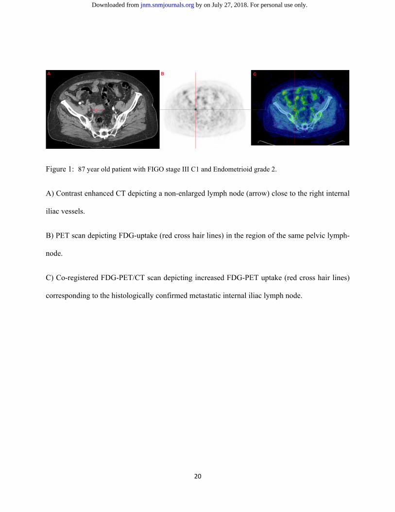
Figure 1: 87 year old patient with FIGO stage III C1 and Endometrioid grade 2.
A) Contrast enhanced CT depicting a non-enlarged lymph node (arrow) close to the right internal
iliac vessels.
B) PET scan depicting FDG-uptake (red cross hair lines) in the region of the same pelvic lymph-
node.
C) Co-registered FDG-PET/CT scan depicting increased FDG-PET uptake (red cross hair lines)
corresponding to the histologically confirmed metastatic internal iliac lymph node.
by on July 27, 2018. For personal use only. jnm.snmjournals.org Downloaded from

21
Figure 2: Flow chart of selection process of eligible studies
by on July 27, 2018. For personal use only. jnm.snmjournals.org Downloaded from

22
Figure 3: LNM: (3A) Forest plot of sensitivity pooling; (3B) Forest plot of specificity pooling;
(3C) SROC curve.
Individual study estimates of sensitivity and specificity of FDG-PET/CT for identifying LNM in
endometrial cancer. In the detection of LNM the FDG-PET/CT have a moderate sensitivity of
0.72, high specificity of 0.94 and AUC of 0.94 demonstrating good diagnostic performance.
by on July 27, 2018. For personal use only. jnm.snmjournals.org Downloaded from

23
Figure 4: ECR: (4A) Forest plot of sensitivity pooling; (4B) Forest plot of specificity pooling;
(4C) SROC curve.
Individual study estimates of sensitivity and specificity of FDG-PET/CT for identifying disease
recurrence in endometrial cancer. In the detection of ECR the FDG-PET/CT have a high
sensitivity of 0.95, high specificity of 0.91 and AUC of 0.97 demonstrating excellent diagnostic
performance.
by on July 27, 2018. For personal use only. jnm.snmjournals.org Downloaded from

24
Table 1: Summary of clinical studies on LNM in endometrial cancer
Study type (reference)
No. of Patients
Patient age (median)
FIGO stage
Endo-metrioid (%)
Purpose
Results
Conclusion(s)
P (27)
236 65 IA-IVB 79%
Evaluate the value of FDG-PET/CT SUVmax as a tool in the preoperative work-up of EC patients particularly focus on MI, CI, FIGO stage, risk stratification and LNM.
Patient based sensitivity (75%), specificity (93%), PPV (60%), NPV (96%) and accuracy (90%) for LNM. High risk EC patients (high FIGO, MI>50%, CI compared to no CI, LNM compared to no LNM, showed significantly higher SUVmax values compared with low risk tumors.
FDG-PET/CT SUVmax is a promising biomarker to distinguish between high and low risk EC, indirectly determine the tumor aggressiveness.
P (25)
129 67 IA-IVB 76%
Determine the diagnostic value of preoperative FDG-PET/CT for staging of EC and to relate FDG-PET/CT parameters to clinicopathological tumor characteristics.
Sensitivity (85%), specificity (92%), PPV (65%), NPV (98%) and accuracy (91%) in detecting LNM.FDG-PET parameters (SUVmax, MTV and TLG were significantly related to deep MI, LNM and high histological grade.
Preoperative FDG-PET/CT is a valuable tool in detecting LNM in EC. FDG-PET parameters are also associated with tumor aggressiveness and aid preoperative identification of high risk patients.
(31) 106 61 IA-IVB 88% Determine the clinical value of FDG-PET/CT in determining the pelvic LNM in EC.
Patient based sensitivity (97%), specificity (69%), PPV (75%), NPV (96%), and accuracy (93%), respectively for LNM.
Preoperative FDG-PET/CT is a valuable tool in detecting LNM in EC.
P (28)
76 63 IA-IVB 87%
Evaluate the role of FDG-PET/CT parameter as a predictor of LNM in EC
Positive correlation between LNM and SUVmax (P=0.003), MTV (P=0.007), and TLG (P=0.003) of the primary tumor. Patient based sensitivity (79%); specificity (98%), accuracy (95%), PPV(92%) and NPV (95%) for LNM
FDG-PET/CT parameters have a potential to predict LNM in EC patients
R (32)
53 58 IA-IVB 83% Determine the accuracy of FDG-PET/CT for LNM in EC.
Patient based sensitivity (50%), specificity (94%), PPV (40%) and NPV (96%), respectively for LNM.
High specificity and NPV may be useful in selecting patients who may benefit from lymphadenectomy, minimizing surgical complications.
P (33)
46 56 I-IV 63% The clinical value of FDG-PET/CT in determining the pelvic LNM in EC.
The sensitivity (50%) and specificity (92%) of FDG-PET for detecting pelvic LNM.
FDG-PET might increase the accuracy for detecting LNM and reduce the false positive results in preoperative EC patients.
by on July 27, 2018. For personal use only. jnm
.snmjournals.org
Dow
nloaded from

25
P (22)
40 56 IA-IIIC 92%
Evaluate the accuracy of FDG-PET/CT to detect LNM in EC.
Patient based sensitivity (50%), specificity (86%) and accuracy (77%) of LNM.
FDG-PET/CT is a valuable tool to detect LNM prior to treatment.
P (34)
37 61 IA-IVB 83%
Determine the accuracy of FDG-PET/CT to detect LNM in high risk EC
Patient based sensitivity (78%), specificity (100%), PPV (100%), NPV (93%), and accuracy (94%) for LNM.
FDG-PET/CT is an accurate procedure for preoperative evaluation of pelvic LNM.
R (35)
33 54 N/A 94%
Evaluate the diagnostic sensitivity of FDG-PET/CT compared to MRI alone in EC patients and also to evaluate the correlation between FDG-PET/CT SUVmax and clinicopathological tumor characteristics.
Patient based sensitivity (80%), specificity (96%) and accuracy (94%) for LNM. Positive correlation between SUVmax of the primary tumor and lesion size (P=0.001).
The diagnostic sensitivity of FDG-PET/CT is superior to CT or MRI alone in detecting both primary tumor and LN.
R (36)
30 62 IA-IIIC 90% Evaluate the accuracy of FDG-PET/CT and PET/MR in assessment of LNM in EC.
Patient based sensitivity (100%), specificity (96%) and accuracy (97%) for detecting LNM for both PET/MR and PET/CT. However, accuracy of PET/MR is superior to PET/CT in tumor staging (80% vs 60%, P<0.04).
Integrated FDG-PET/MR is superior to PET or MRI alone. It is a valuable tool in detecting primary tumor and nodal staging in EC patients.
R (37)
30 56 IA-IVB 63% Evaluate the clinical usefulness of FDG-PET/CT for preoperative evaluation in EC.
Patient based sensitivity (100%) and specificity (100%) for detecting LNM.
FDG-PET/CT demonstrated high diagnostic performance in EC patients preoperatively.
R (38)
26 61 IA-IVB 53% Determine the clinical value of FDG-PET/CT in the primary staging of high risk EC patients.
Patient based sensitivity (57%), specificity (100%), PPV (100%), NPV (86%) and accuracy (88%), respectively for revealing lymph node involvement. Whereas for detecting distant metastases sensitivity (100%), specificity (96%), PPV (87%), NPV (100%) and accuracy (97%) respectively.
FDG-PET/CT is a valuable tool to detect distant metastases in the abdomen and extra-abdominal regions with high diagnostic performance.
P (39)
19 66 IA-IVB 75% Determine the sensitivity and specificity of preoperative FDG-PET in detecting LNM in EC.
Patient based sensitivity (67%) and specificity (94%), respectively for predicting LNM disease preoperatively in endometrial cancer.
Preoperative FDG-PET may be helpful with safe omission of lymphadenectomy in selected patients.
by on July 27, 2018. For personal use only. jnm
.snmjournals.org
Dow
nloaded from

26
Abbreviations: CI: Cervical invasion; EC: Endometrial cancer; FIGO: International federation of gynecology and obstetrics; FDG: Fluorodeoxyglucose; LNM: Lymph node metastases; MRI: Magnetic resonance imaging; MTV: Metabolic tumor volume; MI: Myometrial invasion; NED: No evidence of disease; NPV: Negative predictive value; P: prospective; PPV: Positive predictive value; PET/CT: Positron emission tomography/Computed tomography; R: retrospective; SUVmax: Maximum standardized uptake value; TLG: Tumor lesion glycolysis.
by on July 27, 2018. For personal use only. jnm
.snmjournals.org
Dow
nloaded from

27
Table 2: Summary of clinical studies on endometrial cancer recurrence in endometrial cancer
Study Type
(reference)
No. of Patients
Patients age (Median)
FIGO stage
Endometrioid (%)
Purpose
Results
Conclusion(s)
R (40)
127 52
IA-IVB
86%
Feasibility of FDG-PET/CT for post therapy surveillance in EC patients who showed NED.
The sensitivity (100%), specificity (88%), PPV (59%) and NPV (100%) of FDG-PET for detecting the recurrence in EC showing NED.
FDG-PET could effectively detect early recurrences in patients with EC showing NED after primary treatment.
R (41)
101 56
IA-IVB
n/a
Evaluate the accuracy of FDG-PET/CT for the identification of suspected EC recurrence.
Sensitivity (89%), specificity (93%), PPV (94%), NPV (88%), and accuracy (91%) of FDG-PET/CT respectively.
FDG-PET/CT has a high diagnostic yield in detecting recurrent EC.
R (42)
31
61
IA-IVB
n/a
Determine the value of post-treatment FDG-PET/CT compared to conventional imaging and CA-125 in the EC patients.
The overall sensitivity (100%), specificity (96%) and accuracy (97%) for PET/CT imaging. Whereas for conventional imaging the corresponding values were 46%, 87% and 74% respectively.
Post-treatment FDG-PET/CT is a more clinically useful modality than conventional imaging in the evaluation of suspected EC recurrence.
R (43)
31 53
IA-IVB
88.5%
Evaluate accuracy of PET/CT for the identification of suspected EC recurrence after treatment.
The overall patient based sensitivity (100%); specificity (84%), PPV (100%), NPV (97%) and accuracy (92%).) A significantly better PFS was observed in patients with negative PET/CT result than those with positive PET/CT scan (P<0.015).
FDG-PET/CT demonstrated high diagnostic indices in detecting ECR.
R (44)
30 59
IA-IVB
90%
Evaluate the diagnostic accuracy of FDG-PET/CT compared to PET alone, in the diagnosis of suspected EC recurrence.
Overall patient based sensitivity (93%), specificity (93%) and accuracy (93%) for PET/CT in detecting EC recurrence. Whereas with PET alone the corresponding values were 80%, 80% and 80% respectively.
Diagnostic accuracy of FDG-PET/CT is superior to PET alone in detecting localization of sites of recurrence during follow-up.
by on July 27, 2018. For personal use only. jnm
.snmjournals.org
Dow
nloaded from

28
Abbreviations: CI: Cervix invasion; DFS: Disease free survival; EC: Endometrial cancer; FIGO: International federation of gynecology and obstetrics; FDG: Fluorodeoxyglucose; LNM: Lymph node metastases; MRI: Magnetic resonance imaging; NED: No evidence of disease; NPV: Negative predictive value; P: prospective; PPV: Positive predictive value; PET/CT: Positron emission tomography/Computed tomography; R: retrospective; SUVmax: Maximum standardized uptake value.
R (45)
24 52
IA-IVB
81%
Evaluate the clinical impact of post-treatment FDG-PET/CT in the surveillance of EC patients.
The overall sensitivity (100%), specificity (94%), PPV (96%), NPV (95%) and accuracy (100%) of PET/CT imaging in detecting recurrence.
FDG-PET/CT is highly effective in determining true recurrence in patients with suspected recurrence. FDG-PET/CT carries high impact on clinical decisions in majority of patients.
R (46)
21 62
IA-IVB
67%
Clinical utility of post-operative FDG-PET in EC recurrence.
Sensitivity (100%), specificity (88%) and accuracy (93%) of FDG-PET/CT. Whereas, sensitivity (84%), specificity (85%) and accuracy (85%) of combined conventional imaging (CT/MRI).
FDG-PET is superior to combined conventional imaging (CT/MRI) in detecting tumor recurrence.
P (47)
13 59
IB-IIIB
n/a
Determine the diagnostic accuracy of prospective PET/CT in the detection of EC recurrence.
The overall sensitivity (93%), specificity (100%), PPV (100%), NPV (92%) and accuracy (90%) of PET/CT imaging in detecting EC recurrence.
FDG-PET/CT carries high diagnostic accuracy in determine the EC recurrence.
by on July 27, 2018. For personal use only. jnm
.snmjournals.org
Dow
nloaded from

Doi: 10.2967/jnumed.115.170597Published online: January 28, 2016.J Nucl Med. Vikram rao Bollineni, Sigmund Ytre-Hauge, Oksana Bollineni-Balabay, Helga Salvesen and Ingfrid Haldorsen meta-analysis of the literature.High diagnostic value of FDG-PET/CT in endometrial cancer: Systematic review and
http://jnm.snmjournals.org/content/early/2016/01/27/jnumed.115.170597This article and updated information are available at:
http://jnm.snmjournals.org/site/subscriptions/online.xhtml
Information about subscriptions to JNM can be found at:
http://jnm.snmjournals.org/site/misc/permission.xhtmlInformation about reproducing figures, tables, or other portions of this article can be found online at:
and the final, published version.proofreading, and author review. This process may lead to differences between the accepted version of the manuscript
ahead of print area, they will be prepared for print and online publication, which includes copyediting, typesetting,JNMcopyedited, nor have they appeared in a print or online issue of the journal. Once the accepted manuscripts appear in the
. They have not beenJNM ahead of print articles have been peer reviewed and accepted for publication in JNM
(Print ISSN: 0161-5505, Online ISSN: 2159-662X)1850 Samuel Morse Drive, Reston, VA 20190.SNMMI | Society of Nuclear Medicine and Molecular Imaging
is published monthly.The Journal of Nuclear Medicine
© Copyright 2016 SNMMI; all rights reserved.
by on July 27, 2018. For personal use only. jnm.snmjournals.org Downloaded from