high quality image magniflcation using cross …ceur-ws.org/vol-446/p444.pdf · high quality image...
TRANSCRIPT

High Quality Image Magnificationusing Cross-Scale Self-Similarity
Andre Gooßen1, Arne Ehlers1, Thomas Pralow2, Rolf-Rainer Grigat1
1Vision Systems, Hamburg University of Technology, D-21079 Hamburg2General X-Ray, Philips Medical Systems, D-22335 Hamburg
Abstract. In medical imaging there is a frequent need to magnify acertain region of interest (ROI) of an image. However many modalitiessuffer from severe noise and traditional upscaling methods produce poorenlargement results. We present an approach based on cross-scale simi-larity of an image extendable to sequences using time domain informationwithout explicit motion compensation. It combines noise suppression andupscaling and hence yields superior image quality compared to standardzooming algorithms. We evaluated our algorithm using artificial andclinical fluoroscopy acquisitions.
1 Introduction
Image magnification is the process of computing a high-resolved image from anobserved low-resolution image. In the field of medical X-ray image processing theobservation is provided by the X-ray detector. Because of the limited resolutionof image sensors and the influence of noise during image acquisition, especiallyin X-ray fluoroscopy, the observation is a degraded version of the captured scene.Using traditional upscaling techniques [1, 2] such as cubic interpolation, Lanczos,etc. that incorporate neighboring pixel to interpolate the high-resolved imagetend to smooth structures and do not perform well in the presence of noise. Therecently proposed non-local means filter [3] besides denoising shows potential forimage magnification [4].
2 Methods and materials
In this section we first briefly introduce the problem of image zooming that hasto be solved and derive the technique of incorporating self-similarity from thegeneral non-local means filtering approach [3].
2.1 Image zooming problem
For the image zooming problem we assume a simple degradation model in whichthe relation between the observation and the high-resolution image is given by
u = Hv + η (1)
High quality image magnification 445
with u denoting the low resolution observation and v is the high-resolved image,that should be reconstructed. η is additive white Gaussian noise with zero meanand variance σ2. Besides we assume that the resolution of the image v is z-timeshigher than the resolution of the observation with z ∈ N being a positive integer.
The operator H is a composition of blurring and downsampling and has tobe inverted when solving the ill-posed problem of reconstructing v from u.
2.2 Non-local means filtering
Let u : Ω → R be an image with Ω ⊂ R2 and let N ru (x) ⊂ Ω denote a quadratic
neighborhood of size R×R, R = 2r+1 within u around the center x ∈ Ω. Thenthe weighting function
w(x,y) = e−1
h2 ‖(N ru(x)−N r
u(y))‖2 (2)
maps similarity of neighborhoods around x and y to the interval [0, 1]. Theparameter h controls the steepness of this mapping function. Exploiting thismeasure it is possible to average information according to its similarity:
v(x) =1
W (x)
∫
Ω
w(x, y) · u(y)dy, x, y ∈ Ω . (3)
The similarity weights are normalized by W (x) =∫
Ωw(x, y)dy. The result v(x)
contains a filtered version of u(x) based on similarity of neighborhoods in u.With N denoting the number of pixels in each dimension, i.e. |Ω| = N2,
this method has a complexity of O(N4 ·R2
). The algorithm can be simplified
by reducing the search space for similar neighborhoods to N su(x) of size S × S,
S = 2s + 1 which corresponds to updating the weights w(x, y) to
w(x, y) = w(x, y) · σ (‖x− y‖∞) , σ (‖x− y‖∞) =
1 ‖x− y‖∞ < s
0 else. (4)
2.3 Image zooming using self-similarity
The algorithm introduced in this section allows to up-scale a single low-resolutionimage by a factor z ∈ N. A method similar to the Non-local means filter is usedwhich works on neighborhoods of different scales exploiting cross-scale similarity[4] within an image.
Let u denote the image to zoom and v denote the result of the magnification.The initial estimation v′ is computed by upscaling u with a Lanczos interpolation.Then for a certain position x we compare the neighboring pixels on a z-spacedgrid, i.e. y = x + (m · z, n · z)T with n, m ∈ Z. Hence we derive the weights by
w(x,y) = e−1
h2 ‖(N rv′ (x)−N r
v′ (y))‖2 . (5)
Now instead of replacing the pixel at x with samples from v′ we extract theinformation from the corresponding pixel of the low resolution image u. Inverting

446 Gooßen et al.
the decimation, we insert the averaged pixel only at x avoiding the blurring effectof interpolating methods. Hence we derive the high-resolved magnified image by
v(x) =1
W (x)
∑
Ω
w(x, y) · u(
1zy
)dy, x, y ∈ Ω . (6)
This can be enhanced by not only using the image itself but also the re-cent frames within the sequence of acquisitions. In [5] we propose a temporalextension of the NLM filter based on a multi-scale motion estimation
v t[x] =1
W [x]
t−τ∑
t=t
∑
Ω
wt[x, y] · ut
[1zy
], W [x] =
t−τ∑
t=t
∑
Ω
wt[x, y] (7)
with t ∈ N denoting the index of a certain frame within the sequence history andτ determining the number of frames to consider for filtering the current frameat t = t.
For the motion estimation we decompose the image into a Gaussian pyramidrepresentation and compute the sum of squared difference (SSD) for each leveli. Thus the estimated position of the block center within ut for a given blockN b
u t(x) of size B ×B, B = 2b + 1 is calculated by
yi = argmaxy∈Ω
∥∥∥N bGi(u t)
(x)−N bGi(ut)
(y)∥∥∥ , i = 0, 1, · · · , n (8)
‖yi − yi+1‖ < B, yn+1 = 0 (9)
with Gi(·) denoting the Gaussian level i and G0(u) = u.The restriction of higher level estimations yi to the vicinity of the lower level
estimation introduces a natural regularization compared to matching only fullresolution blocks and prevents outliers. However the proposed approach does notdepend on accurate motion detection. A rough estimation is sufficient to selectthe correct neighborhood and even outliers do not spoil the algorithm as thecontained neighborhoods will have very low weights and hence will not influencethe reconstructed value.
To achieve best results v′ can be assigned the result v and the method maybe iterated l times. Experiments have shown that l = 2 iterations are usuallysufficient and further loops do not yield significant enhancements. Incorporatingthe simplification shown in Equation (4) the total complexity of the proposedmethod is O
(N2 · S2 ·R2 · τ · l).
2.4 Test set
To evaluate the proposed method we used clinical fluoroscopy sequences con-taining continuous navigation frames acquired at low X-ray doses and singleexposures at higher doses of the same anatomy. Thus we were able to compareimpression of sharpness and noise with traditional upsampling as well as thehigh dose image serving as ground truth. In addition we processed a sequencecontaining an artificial rotating phantom and evaluated the noise suppressioncapability and resolution of our method.
High quality image magnification 447
3 Results
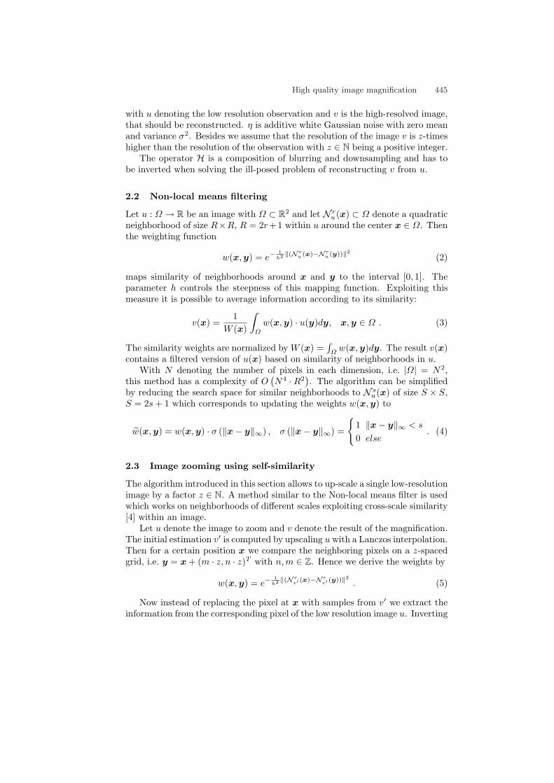
We visualize the results for two test sequences. The first one is a frame takenfrom a clinical angiography. Fig. 1 shows a part of the cranium magnified bytraditional Lanczos interpolation compared to the proposed method.
As one can see, Lanczos interpolation significantly suffers from the inherentnoise in low dose frames. The proposed method achieves a noise impression thatis comparable to a high dose exposure and preserves sharpness but is not ableto reconstruct all details contained in the high dose exposure from multiple lowdose frames. The difference image between both methods contains only noiseand the histogram shows an almost ideal Gaussian distribution.
(a) High dose, Lanczos (b) Low dose, Lanczos
(c) Low dose, our method (d) Difference image and histogram
Fig. 1. Magnifying a cranial angiography: (a) Single frame acquired with high X-raydose enlarged with Lanczos interpolation, (b) same anatomy acquired with low doseenlarged with Lanczos filter and (c) from τ = 10 frames with the proposed magnificationmethod, (d) contains the difference between Lanczos filtered and proposed image.

448 Gooßen et al.
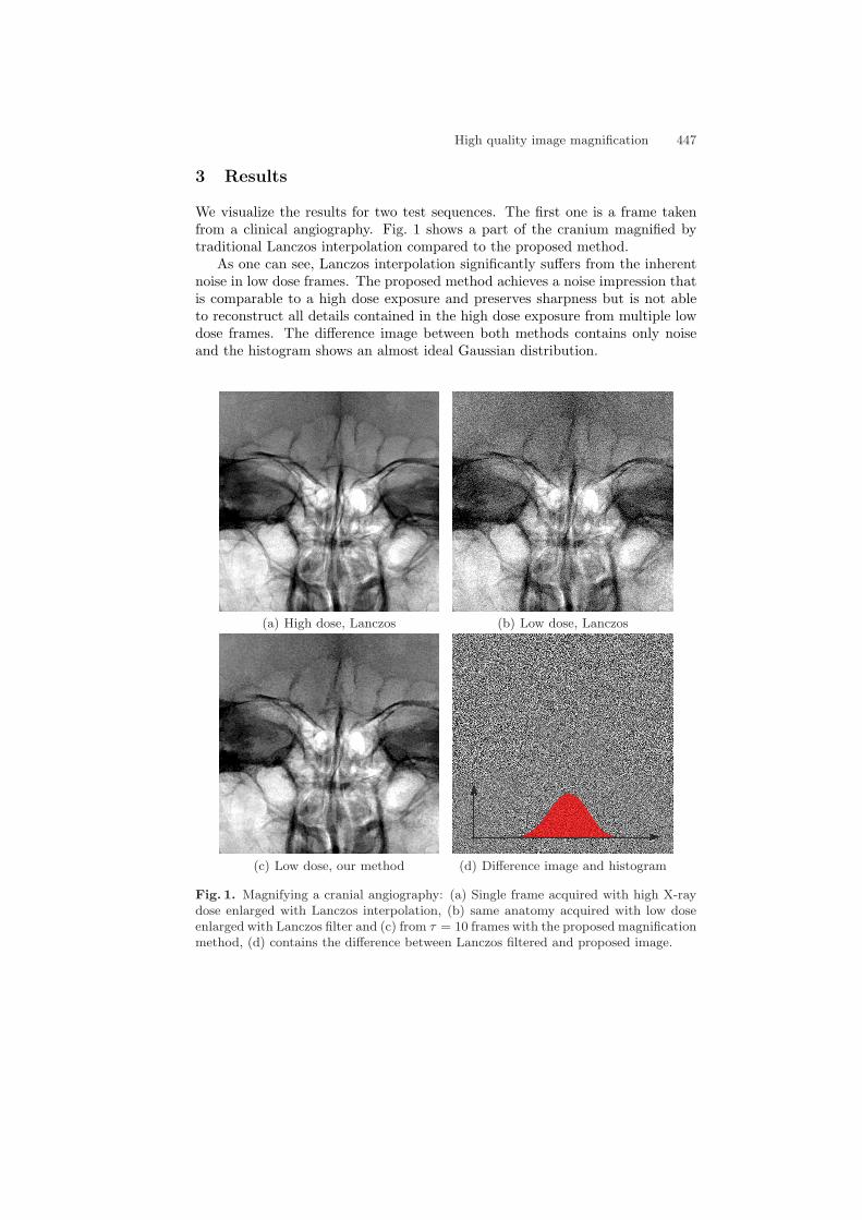
Fig. 2. Comparison of magnification techniques: The part marked by the red rectangleis upsampled for enhanced visibility. The complete detail view is magnified using(a) Lanczos interpolation and (b) the proposed method. (c) and (d) contain contrastenhanced detail views respectively.
(a) Lanczos (b) proposed (c) Lanczos (d) proposed
The second sequence (Fig. 2) has been chosen to evaluate the spatial reso-lution. The Siemens star contained in this sequence is corrupted by noise andLanczos as well as other interpolation methods even amplify this noise upon mag-nification. The proposed method though slightly degrading contrast achievessuperior magnification and even reconstructs the corrupted pattern. Noise isheavily reduced and lines are better recognizable.
4 Discussion
In direct comparison to simple interpolation the proposed magnification showssuperior quality. Evaluation of difference images indicates that no relevant struc-ture is lost due to the filtering operation. Hence it is possible to achieve enhancedmagnification of noisy image data without tampering the diagnostic information.As a rule of thumb the X-ray dose of a single high dose exposure corresponds to30 seconds of navigation at low dose. As this technique might render some ofthe exposures unnecessary, we reduce the overall dose for patients and staff.
The high complexity yet prevents the algorithm from being executed inter-actively, however we have successfully implemented a real-time similarity baseddenoising [5] and as computational power grows it will be possible to do thesame for the proposed method.
References
1. Keys R. Cubic convolution interpolation for digital image processing. IEEE TransASSP. 1981;29(6):1153–60.
2. Turkowski K. Filters for common resampling tasks. Apple Computer; 1990.3. Buades A, Coll B, Morel JM. A non-local algorithm for image denoising. Proc IEEE
CVPR. 2005;2:60–5.4. Ebrahimi M, Vrscay ER. Examining the role of rcale in the context of the non-local-
means filter. LNCS. 2008;5112:170–81.5. Gooßen A, Pralow T, Grigat RR. Medical X-ray image enhancement by intra-image
and inter-image similarity. Proc SPIE. 2009.