highlights from the koch institute public galleries insights · pdf filehighlights from the...
TRANSCRIPT
insights
Highlights from the Koch Institute Public GalleriesSP
RIN
G/S
UM
ME
R 2
017
insights
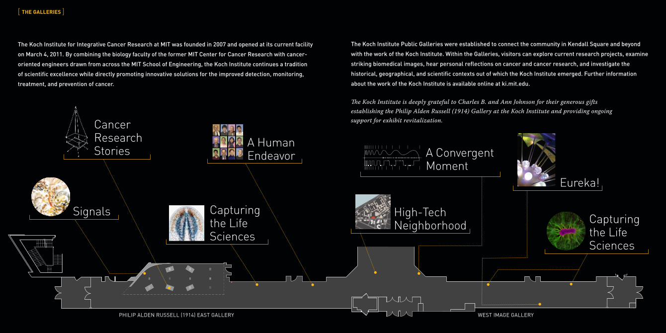
The Koch Institute for Integrative Cancer Research at MIT was founded in 2007 and opened at its current facility
on March 4, 2011. By combining the biology faculty of the former MIT Center for Cancer Research with cancer-
oriented engineers drawn from across the MIT School of Engineering, the Koch Institute continues a tradition
of scientific excellence while directly promoting innovative solutions for the improved detection, monitoring,
treatment, and prevention of cancer.
[ THE GALLERIES ]
PHILIP ALDEN RUSSELL (1914) EAST GALLERY WEST IMAGE GALLERY
Cancer Research Stories
Capturing the Life Sciences
Signals High-Tech Neighborhood Capturing
the Life Sciences
Eureka!
A Convergent Moment
A Human Endeavor
The Koch Institute Public Galleries were established to connect the community in Kendall Square and beyond
with the work of the Koch Institute. Within the Galleries, visitors can explore current research projects, examine
striking biomedical images, hear personal reflections on cancer and cancer research, and investigate the
historical, geographical, and scientific contexts out of which the Koch Institute emerged. Further information
about the work of the Koch Institute is available online at ki.mit.edu.
The Koch Institute is deeply grateful to Charles B. and Ann Johnson for their generous gifts establishing the Philip Alden Russell (1914) Gallery at the Koch Institute and providing ongoing support for exhibit revitalization.
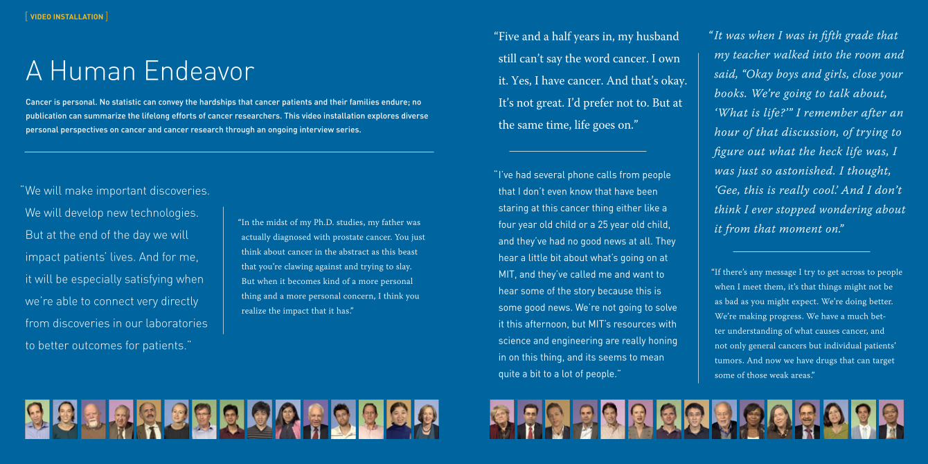
A Human EndeavorCancer is personal. No statistic can convey the hardships that cancer patients and their families endure; no
publication can summarize the lifelong efforts of cancer researchers. This video installation explores diverse
personal perspectives on cancer and cancer research through an ongoing interview series.
“ Five and a half years in, my husband
still can’t say the word cancer. I own
it. Yes, I have cancer. And that’s okay.
It’s not great. I’d prefer not to. But at
the same time, life goes on.”
“ It was when I was in fifth grade that my teacher walked into the room and said, “Okay boys and girls, close your books. We’re going to talk about, ‘What is life?’” I remember after an hour of that discussion, of trying to figure out what the heck life was, I was just so astonished. I thought, ‘Gee, this is really cool.’ And I don’t think I ever stopped wondering about it from that moment on.”
“ We will make important discoveries.
We will develop new technologies.
But at the end of the day we will
impact patients’ lives. And for me,
it will be especially satisfying when
we’re able to connect very directly
from discoveries in our laboratories
to better outcomes for patients.”
“ I’ve had several phone calls from people
that I don’t even know that have been
staring at this cancer thing either like a
four year old child or a 25 year old child,
and they’ve had no good news at all. They
hear a little bit about what’s going on at
MIT, and they’ve called me and want to
hear some of the story because this is
some good news. We’re not going to solve
it this afternoon, but MIT’s resources with
science and engineering are really honing
in on this thing, and its seems to mean
quite a bit to a lot of people.”
“ If there’s any message I try to get across to people
when I meet them, it’s that things might not be
as bad as you might expect. We’re doing better.
We’re making progress. We have a much bet-
ter understanding of what causes cancer, and
not only general cancers but individual patients’
tumors. And now we have drugs that can target
some of those weak areas.”
[ VIDEO INSTALLATION ]
“ In the midst of my Ph.D. studies, my father was
actually diagnosed with prostate cancer. You just
think about cancer in the abstract as this beast
that you’re clawing against and trying to slay.
But when it becomes kind of a more personal
thing and a more personal concern, I think you
realize the impact that it has.”
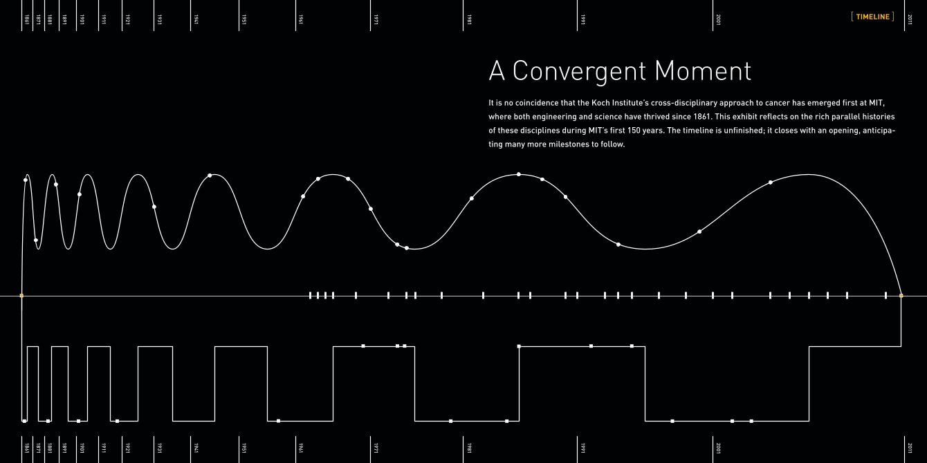
A Convergent MomentIt is no coincidence that the Koch Institute’s cross-disciplinary approach to cancer has emerged first at MIT,
where both engineering and science have thrived since 1861. This exhibit reflects on the rich parallel histories
of these disciplines during MIT’s first 150 years. The timeline is unfinished; it closes with an opening, anticipa-
ting many more milestones to follow.
[ TIMELINE ]
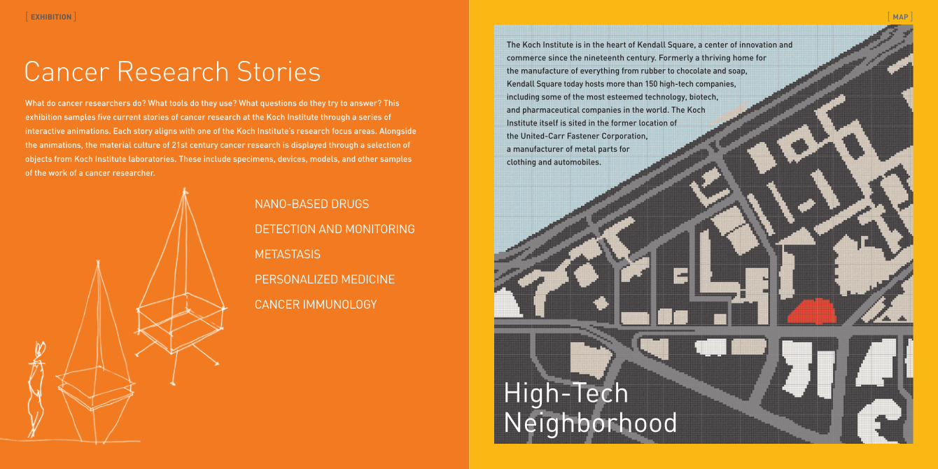
The Koch Institute is in the heart of Kendall Square, a center of innovation and
commerce since the nineteenth century. Formerly a thriving home for
the manufacture of everything from rubber to chocolate and soap,
Kendall Square today hosts more than 150 high-tech companies,
including some of the most esteemed technology, biotech,
and pharmaceutical companies in the world. The Koch
Institute itself is sited in the former location of
the United-Carr Fastener Corporation,
a manufacturer of metal parts for
clothing and automobiles.
Cancer Research Stories
High-Tech Neighborhood
What do cancer researchers do? What tools do they use? What questions do they try to answer? This
exhibition samples five current stories of cancer research at the Koch Institute through a series of
interactive animations. Each story aligns with one of the Koch Institute’s research focus areas. Alongside
the animations, the material culture of 21st century cancer research is displayed through a selection of
objects from Koch Institute laboratories. These include specimens, devices, models, and other samples
of the work of a cancer researcher.
[ EXHIBITION ] [ MAP ]
NANO-BASED DRUGS
DETECTION AND MONITORING
METASTASIS
PERSONALIZED MEDICINE
CANCER IMMUNOLOGY
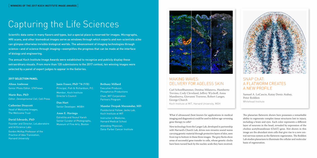
MAKING WAVES: DELIVERY FOR AGELESS SKINCarl Schoellhammer, Denitsa Milanova, Humberto Trevino, Cody Cleveland, Jeffrey Wyckoff, Anna Mandinova, Giovanni Traverso, Robert Langer, George Church Koch Institute at MIT, Harvard University, MGH
SNAP ChAT: A FLATWORM CREATES A NEW PROFILESamuel A. LoCascio, Kutay Deniz Atabay, Peter ReddienWhitehead Institute
Capturing the Life Sciences
What if ultrasound (best known for applications in medical imaging and diagnostics) could be used to deliver age-reversing gene therapy to cells?
New technology from the Langer Lab, developed in partnership with Harvard’s Church Lab, drives non-invasive sound waves carrying genetic material through protective layers of skin, seen from top to bottom in these three images. The grey flecks show areas of successful gene transfer to cells, whose genetic clocks have been turned back by the nucleic acids they have received.
The planarian flatworm shown here possesses a remarkable ability to regenerate complex tissue structures lost to injury, including a brain and eyes. Each color represents a different layer of neurons in the head, revealed by expression of the choline acetyltransferase (ChAT) gene. Not shown in this image are the abundant stem cells that give rise to a new cen-tral nervous system as the flatworm regenerates. The Reddien Lab studies planarians to illuminate the cellular and molecular basis of regeneration.
[ WINNERS OF THE 2017 KOCH INSTITUTE IMAGE AWARDS ]
Scientific data come in many flavors and types, but a special place is reserved for images. Micrographs,
MRI scans, and other biomedical images serve as windows through which experts and non-scientists alike
can glimpse otherwise invisible biological worlds. The advancement of imaging technologies through
science—and of science through imaging—exemplifies the progress that can be made at the interface
of biology and engineering.
The annual Koch Institute Image Awards were established to recognize and publicly display these
extraordinary visuals. From more than 120 submissions to the 2017 contest, ten winning images were
selected by a panel of expert judges to appear in the Galleries.
2017 SELECTION PANEL
Alissa AmbroseSenior Photo Editor, STATnews
Marie Bao, PhDEditor, Developmental Cell, Cell Press
Catherine DraycottHead of Wellcome Images, The Wellcome Trust
David Edwards, PhDFounder and Director, LeLaboratoire and ArtScience Labs
Gordon McKay Professor of the Practice of Idea Translation, Harvard University
Janis Fraser, PhD ’76 (VII) Principal, Fish & Richardson, P.C.
Member, Koch Institute Director’s Council
Dan HartSenior Developer, WGBH
Anne E. HavingaEstrellita and Yousuf Karsh Senior Curator of Photographs, Museum of Fine Arts, Boston
Bethany MillardExecutive Producer, Phosphorus Productions
Chair, MIT Corporation Partners Program
Mandar Deepak Muzumdar, MDPostdoctoral Fellow, Jacks Lab, Koch Institute at MIT
Instructor in Medicine, Harvard Medical School
Attending Physician, Dana-Farber Cancer Institute
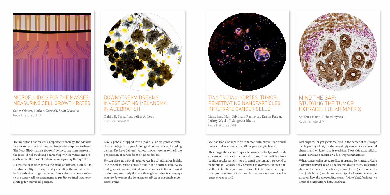
MICROFLUIDICS FOR THE MASSES: MEASURING CELL GROWTH RATESSelim Olcum, Nathan Cermak, Scott ManalisKoch Institute at MIT
DOWNSTREAM DREAMS: INVESTIGATING MELANOMA IN A ZEBRAFISHDahlia E. Perez, Jacqueline A. LeesKoch Institute at MIT
MIND THE GAP: STUDYING THE TUMOR EXTRACELLULAR MATRIXSteffen Rickelt, Richard HynesKoch Institute at MIT
TINY TROJAN HORSES: TUMOR-PENETRATING NANOPARTICLES INFILTRATE CANCER CELLS Liangliang Hao, Srivatsan Raghavan, Emilia Pulver, Jeffrey Wyckoff, Sangeeta BhatiaKoch Institute at MIT
To understand cancer cells’ response to therapy, the Manalis Lab measures how their masses change while exposed to drugs. The fluid-filled channels (bottom) connect tiny mass sensors in the form of hollow diving boards (top) whose vibrations pre-cisely reveal the mass of individual cells passing through them.
As treated cells flow across the array of sensors, each cell is weighed multiple times, thereby revealing the rate at which individual cells change their mass. Researchers are now starting to use tumor cell measurements to predict optimal treatment strategy for individual patients.
Like a pebble dropped into a pond, a single genetic muta-tion can trigger a ripple of biological consequences, including cancer. The Lees Lab uses various model systems to track the progression of cancer from origin to disease.
Here, a close-up view of melanocytes in zebrafish gives insight into the organization of these cells in their normal state. Next, biologists will mutate a single gene, a known initiator of uveal melanoma, and study the cells throughout zebrafish develop-ment to determine the downstream effects of this single muta-tional event.
Although the brightly colored cells in the center of this image catch your eye first, it’s the seemingly neutral tissue around them that the Hynes Lab is studying. Does this extracellular matrix serve as a barrier or a doorway to metastasis?
When cancer cells spread to distant organs, they must navigate a complex network of cells and proteins to get there. This image shows colon cancer metastases (brown clusters) surrounded by liver (light brown) and immune cells (pink). Researchers seek to discover how the surrounding matrix (white/blue) facilitates or limits the interactions between them.
You can lead a nanoparticle to tumor cells, but you can’t make them shrink—at least not until the particle gets inside.
This image shows biocompatible nanoparticles (yellow) inside clusters of pancreatic cancer cells (pink). The particles’ two-peptide uptake system—one to target the tumor, the second to penetrate it—was specially designed to overcome known diff-iculties in treating pancreatic cancer, but the Bhatia Lab hopes to expand the use of this modular delivery system for other cancer types as well.

SHAPE SHIFTERS: CANCER CELLS IN MOTION Claudia Schafer, Frank GertlerKoch Institute at MIT
PUSHING BOUNDARIES: OVARIAN CANCER HIDES IN PLAIN SIGHTErik C. Dreaden, Yi Wen Kwong, Michael Yaffe, Paula T. HammondKoch Institute at MIT
HASHTAG NO FILTER: VISUALIZING BREAST CANCER CONVERSATIONSEric Clarke, Richard Arnett, Jane BurnsRoyal College of Surgeons in Ireland, Wellcome Images
BODY OF KNOWLEDGE: SELF-ORGANIZING BRAIN CELLSCollin Edington, Iris Lee, Linda GriffithMIT Department of Biological Engineering and Koch Institute at MIT
These metastatic lung cancer cells are showing their true nature as they wander around the dish. Images taken ten minutes apart over the course of 16 hours are stacked atop each other to create a composite image. Researchers in the Gertler Lab are studying how different levels of proteins expressed by the cells affect their shape and motion.
Take a look—can you trace the pathways? Are the cells moving slowly or quickly? Do they change shape or stay round? How do they compare to each other?
Persistence is key. Here, an ovarian tumor clings to the abdomi-nal wall, slowly breaking through the tissue boundaries that block its metastatic spread. The tissues here were stained with a mol-ecule that binds to cells that are rapidly growing and multiplying (white). Just as tenacious as these proliferating cells, however, are the researchers who study it. The Hammond and Yaffe Labs are working together to better understand and exploit the genetic weaknesses of these tumors in various disease models, and soon will test their response to experimental treatments, unlocking new avenues for investigation and intervention.
Eight weeks. 92,915 tweets. One hashtag. This image literally visualizes conversations around breast cancer, and the network of connected cancer patients and their loved ones, patient advo-cates, oncologists and other health care professionals, as well as cancer researchers. #breastcancer joins these diverse stake-holders together in one conversation and puts everyone on the same page, erasing societal boundaries to share knowledge and support in real time.
This image appears in the Koch Institute Public Galleries as part of a partnership between the Koch Institute and Wellcome Images.
Imagine a uniform field of neural stem cells sitting on gel-like matrix. Slowly, they begin to differentiate, grouping and clustering together until they have self-assembled into a mini-organ—a brain!
The neurons (green) and astrocytes (red) seen here are part of the Griffith Lab’s “Human on a Chip” project. Many diff-erent “mini-organs” are linked together in a bioreactor plat-form, allowing researchers to study the interactions of multiple organs and the crosstalk between them in an in vitro setting, and to accelerate the development of novel disease treatments.
KOCH INSTITUTE PUBLIC GALLERIES 500 Main Street, Cambridge, MA
[INFO]web ki-galleries.mit.edu
email kigalleries mit.edu
[HOURS]8am–6pm, Mon–Thu
8am–4pm, Fri
Admission is free
[OTHER VISITOR ATTRACTIONS AT MIT]MIT Museum
MIT List Visual Arts Center and public art tour
Maihaugan Gallery, MIT Libraries
MIT Media Lab
Ray and Maria Stata Center
Corridor Lab in Strobe Alley, MIT Edgerton Center
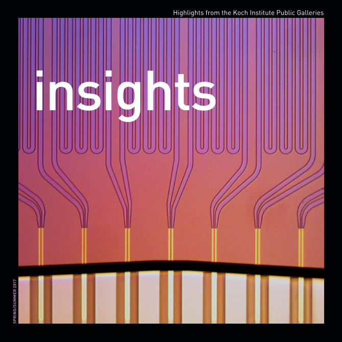
cover artMicrofluidics for the Masses Selim Olcum, Nick Calistri, Scott Manalis (Manalis Lab, Koch Institute at MIT)
gallery design Biber Architects and P
entagram / videography AM
PS – M
IT / design Hecht/H
orton Partners