highly stretchable self-healing poly(n,n-dimethylacrylamide) hydrogels · 2014-08-16 · highly...
TRANSCRIPT

European Polymer Journal 59 (2014) 113–121
Contents lists available at ScienceDirect
European Polymer Journal
journal homepage: www.elsevier .com/locate /europol j
Highly stretchable self-healing poly(N,N-dimethylacrylamide)hydrogels
http://dx.doi.org/10.1016/j.eurpolymj.2014.07.0220014-3057/� 2014 Elsevier Ltd. All rights reserved.
⇑ Corresponding author. Tel.: +90 212 2853156; fax: +90 212 2856386.E-mail address: [email protected] (O. Okay).
1 Present address: Aksaray University, Vocational School of HealthServices, Aksaray, Turkey.
Melek Pamuk Algi 1, Oguz Okay ⇑Istanbul Technical University, Department of Chemistry, 34469 Istanbul, Turkey
a r t i c l e i n f o a b s t r a c t
Article history:Received 16 May 2014Received in revised form 16 July 2014Accepted 19 July 2014Available online 30 July 2014
Keywords:HydrogelsSelf-healingHydrophobic associationsMechanical properties
Poly(N,N-dimethylacrylamide) (PDMA) is a very useful hydrophilic biocompatible polymerwith associative properties. Although there is a growing interest in PDMA hydrogels, theygenerally exhibit rather low mechanical strength and very low stretchability due to thelack of an efficient energy dissipation mechanism in the chemically crosslinked gel net-work. Highly stretchable PDMA hydrogels with self-healing properties are of great interestin tissue engineering and for biomedical applications. A promising strategy to design syn-thetic hydrogels with self-heal ability is to substitute the covalently crosslinked polymerchains by supramolecular ones. Here, we describe preparation of self-healing PDMA hydro-gels by micellar copolymerization of N,N-dimethylacrylamide with 2 mol% stearyl methac-rylate in aqueous sodium dodecyl sulfate–NaCl solutions. The supramolecular PDMAhydrogels formed via hydrophobic interactions in micellar solutions can be compressedup to about 100% strain without any permanent failure, while during elongation, they rup-ture when stretched to 4200% strain. The hydrogels soften with increasing strain and exhi-bit liquid-like response (tand > 1) at high strains, while they turn back to the initial gelstate, if the force is removed. Loading and unloading mechanical cycles show a significanthysteresis and perfect superposition of the successive loading curves demonstrating dam-age done during loading is recoverable in nature. Tensile testing experiments performedusing virgin and healed gel samples show that a healing time of 20 min suffices to recoverall the initial mechanical properties of PDMA hydrogels.
� 2014 Elsevier Ltd. All rights reserved.
1. Introduction
Poly(N,N-dimethylacrylamide) (PDMA) is a very usefulhydrophilic biocompatible polymer with associative prop-erties [1–3]. Linear polymers [4], hydrogels [5–7], inter-penetrating polymer networks [8], and blends [9] basedon PDMA find numerous applications in molecular biology[10], DNA sequencing [11–13], medical and pharmaceuti-cal fields including contact lenses and in drug delivery
[4,14,15]. However, PDMA hydrogels generally exhibitrather low mechanical strength due to the lack of an effi-cient energy dissipation mechanism in the chemicallycrosslinked PDMA network. In the past decade, hybridPDMA hydrogels with improved mechanical propertieshave been prepared using silica or clay nanoparticlesacting as dynamic multifunctional crosslinkers [16–18].Haraguchi et al. demonstrated self-healing behavior ofhybrid PDMA hydrogels prepared using Laponite claynanoparticles [19]. The hydrogels sustain up to about1500% strain and the damage created in the gels could behealed at 50 �C within 10 h. Mechanically strong hydrogelsbased on PDMA and hyaluronic acid were also prepared bya two-step photo-crosslinking process [20]. Recently, Hao

114 M.P. Algi, O. Okay / European Polymer Journal 59 (2014) 113–121
and Weiss prepared PDMA hydrogels by free-radical copo-lymerization of N,N-dimethylacrylamide (DMA) and 2-(N-ethylperfluorooctane sulfonamido) ethyl acrylate (FOSA) indioxane with varying FOSA concentration [21]. The hydro-gels formed via hydrophobic interactions between FOSAunits exhibit a modulus of 130–190 kPa and elongationat break of 1000–1600%. Since the elastic modulus of thesegels decreases with increasing temperature [21], it is likelythat the gels have self-healing ability, which could beinduced by heating. In contrast to these improvements,highly stretchable PDMA hydrogels with the ability toself-heal autonomously at room temperature have notbeen prepared before. Such hydrogels are of great interestin tissue engineering and for biomedical applications.
The unique properties of PDMA are mainly due to theexistence of both hydrogen bonding and hydrophobicinteractions in its aqueous solutions and hydrogels[22,23]. For instance, strong hydrogen bonding interactionsexist between PDMA and silica nanoparticles in aqueoussolutions leading to hydrogels with reinforced properties[16,17]. Different from PDMA, polyacrylamide (PAAm)shows weak interactions with these particles, since it issimultaneously hydrogen-bonding donor and acceptor[17]. PDMA hydrogels also exhibit reentrant phenomenonin aqueous solutions of acetone, dioxane, propanol ort-butanol, i.e., the hydrogels first collapse then reswell ifa particular external parameter such as organic solventconcentration is continuously varied [24,25]. Since thisphenomenon is not observable in PAAm hydrogels [25],hydrophobic interactions between the methyl groups ofPDMA network and the organic solvent are responsiblefor the reentrant transition behavior of the hydrogels.Thus, we conclude that PDMA generates much strongerreversible molecular interactions as compared to PAAm,making it a good candidate for the preparation of supramo-lecular polymer gels.
A promising strategy to design synthetic hydrogels withself-heal ability is to substitute the covalently crosslinkedpolymer chains by supramolecular ones. In the past fewyears, different reversible molecular interactions havebeen used to generate self-healing hydrogels, includinghydrogen bonding [19,26–29], electrostatic interactions[30–32], molecular recognition [33–35], metal coordina-tion [36,37], p–p stacking [38], dynamic chemical bonds[39–42], and molecular diffusion [43,44]. Recently, we pre-sented a simple strategy for the production of self-healinghydrogels via hydrophobic interactions in micellar solu-tions [45–50]. Large hydrophobes such as stearyl methac-rylate (C18) could be copolymerized with the hydrophilicmonomer acrylamide (AAm) in aqueous sodium dodecylsulfate (SDS) solutions. This was achieved by the additionof salt (NaCl) into the reaction solution. Salt leads to micel-lar growth and hence, solubilization of large hydrophobeswithin the grown wormlike SDS micelles [45,46]. Incorpo-ration of hydrophobic sequences within the hydrophilicPAAm chains via micellar polymerization generatesdynamic hydrophobic associations between the hydropho-bic domains of polymer chains and surfactant micelles act-ing as physical crosslinks of the resulting hydrogels.
We propose that a stronger self-healing and a bettermechanical performance in such hydrogels could be
achieved by replacing PAAm with PDMA backbone thatwould mediate stronger hydrophobic associations andhydrogen bonding across a rupture in the hydrogel. Here,we prepared physical PDMA hydrogels by the micellarcopolymerization of N,N-dimethylacrylamide (DMA) withthe hydrophobic monomer C18 in aqueous SDS-NaCl solu-tions. The gels obtained in the presence of 2 mol % C18exhibit very high stretchability (about 43 timed its originallength) and autonomous self-healing within 20 min, as evi-denced by mechanical measurements.
2. Experimental
2.1. Materials
N,N-dimethylacrylamide (DMA, Sigma), sodium dode-cylsulfate (SDS, Merck), ammonium persulfate (APS,Sigma), N,N,N0,N0-tetramethylethylenediamine (TEMED,Sigma), and NaCl (Merck) were used as received. Commer-cially available stearyl methacrylate (C18, Sigma) consistsof 65% n-octadecyl methacrylate and 35% n-hexadecylmethacrylate.
2.2. Hydrogel preparation
Hydrogels were prepared by the micellar copolymeriza-tion of DMA with 2 mol% C18 at 50 �C for 16 h in the pres-ence of an APS (3.5 mM) – TEMED (0.25 v/v %) redoxinitiator system. SDS and NaCl concentrations were set to7 w/v % (0.24 M) and 0.5 M, respectively. The aggregationnumber of SDS micelles in this solution is 200 nm [45],as compared to 60 for the minimum spherical SDS micelle.The growth of the micelles provides complete solubiliza-tion of the hydrophobe C18, [45,47] and thus permits itscopolymerization with DMA in aqueous media. The totalmonomer concentration was varied while C18 content ofthe monomer mixture was fixed at 2 mol%. The gel prepa-ration procedure was the same as in our previous studies[45]. Shortly, SDS (0.7 g) was dissolved in 8.14 mL aqueoussolution of NaCl (0.2925 g) at 35 �C to obtain a transparentsolution. Then, hydrophobic monomer C18 (0.0949 g) wasdissolved in this SDS-NaCl solution under stirring for 2 h at35 �C. After addition and dissolving DMA (1.46 mL) for 1 h,TEMED (25 lL) was added into the solution. Finally, 0.1 mLof APS stock solution (0.8 g APS/10 mL distilled water) wasadded to initiate the reaction. A portion of this solutionwas transferred between the plates of the rheometer to fol-low the reaction by oscillatory small-strain shear measure-ments. For the determination of the gel fraction and for themechanical measurements, the remaining part of the solu-tion was transferred into several plastic syringes of 4.8 mminternal diameters and the polymerization was conductedfor 16 h at 50 �C.
2.3. Gel fraction and swelling measurements
Cylindrical hydrogel samples (diameter 4.8 mm, lengthabout 2 cm) were immersed in a large excess of water at24 �C for at least 15 days by replacing water every secondor third day to extract any soluble species. The mass m of

M.P. Algi, O. Okay / European Polymer Journal 59 (2014) 113–121 115
the gel samples was monitored as a function of swellingtime by weighing the samples. The relative weight swell-ing ratio mrel of gels was calculated as mrel = m/m0, wherem0 is the initial mass of the gel sample. Then, the equilib-rium swollen gel samples were taken out of water andfreeze dried. The gel fraction Wg (mass of water-insolublepolymer / initial mass of the monomer) was calculatedfrom the masses of dry, extracted polymer network andfrom the comonomer feed.
2.4. Rheological experiments
Gelation reactions were carried out at 50 �C within therheometer (Gemini 150 Rheometer system, Bohlin Instru-ments) equipped with a cone-and-plate geometry with acone angle of 4� and a diameter of 40 mm. The instrumentwas equipped with a Peltier device for temperature con-trol. During all rheological measurements, a solvent trapwas used to minimize the evaporation. An angular fre-quency x of 6.3 rad s�1 and a deformation amplitude co
of 0.01 were selected to ensure that the oscillatory defor-mation is within the linear regime. After a reaction timeof 2 h, the dynamic moduli of the reaction solutionsapproached limiting values. Then, frequency-sweep testsat co = 0.01 were carried out at 25 �C over the frequencyrange 0.06 to 410 rad s�1. The gels formed within the rhe-ometer were also subjected to strain-sweep tests (both upand down) at x = 6.3 rad s�1 for co ranging from 0.001 to10.
2.5. Mechanical tests
The measurements were performed in a thermostatedroom at 24 �C on cylindrical hydrogel samples of 4.8 mmdiameter. The uniaxial compression measurements wereperformed on a Zwick Roell test machine using a 500 Nload cell. The hydrogel sample of 5.3 ± 0.7 mm lengthwas placed between the plates of the instrument. Beforethe test, an initial compressive contact to 0.004 ± 0.003 Nwas applied to ensure a complete contact between thegel and the plates. The tests were conducted at a nominalstrain rate of 0.0156 s�1 (5 mm/min). Load anddisplacement data were collected during the experiment.Compressive stres was presented by its nominal rnom andtrue values rtrue (=k rnom), which are the forces per cross-sectional area of the undeformed and deformed gelspecimen, respectively, while the strain is given by k, thedeformation ratio (deformed length/initial length).Compressive modulus was calculated from the slope ofstress–strain curves between 5% and 15% compressions.Cyclic compression tests were conducted at a constantcrosshead speed of 5 mm/min to a maximum compressionratio, followed by retraction to zero force and awaitingtime of 7 min, until the next cycle of compression. Forreproducibility, at least five samples were measured foreach gel and the results were averaged.
The uniaxial elongation measurements were performedon a Zwick Roell test machine using a 10 N load cell underthe following conditions: Nominal strain rate = 0.0833 s�1
(50 mm/min), sample length between jaws = 10 ± 0.5 mm.The tensile modulus was calculated from the slope of
stress–strain curves between elongations of 5% and 15%.Samples were held on the test machine between clampsaltered with anti-slip tape (Tesa, 25 � 15 mm) togetherwith wood strips to better grip the slippery gel samples.Cyclic elongation tests were conducted at a constant cross-head speed of 50 mm min�1 to a maximum elongationratio, followed by retraction to zero force and a waitingtime of 7 min, until the next cycle of elongation. For repro-ducibility, at least five samples were measured for each geland the results were averaged.
The self-healing efficiency of the gel samples was deter-mined by tensile testing experiments performed using vir-gin and healed cylindrical gel samples of 5 mm in diameterand 6 cm in length. The samples were cut in the middleand then, the two halfs were merged together within aplastic syringe (of the same diameter as the gel sample)at 24 �C by slightly pressing the piston plunger. The healingtime was varied from 2 to 30 min, and each experimentwas carried out starting from a virgin sample. The tensilemodulus E, the fracture stress rf, and the elongation ratioat break kf of the healed gel samples were recorded.
3. Results and discussion
PDMA hydrogels were prepared by the micellar copoly-merization of DMA with 2 mol% C18 at 50 �C in aqueousSDS–NaCl solutions. Since the hydrogels formed at orbelow 10 w/v % DMA were too weak, the hydrogel pre-pared at 15 w/v % DMA will be presented and used for dis-cussion. We first conducted the micellar polymerization ofDMA with and without the hydrophobic comonomer C18by real-time rheological measurements. In Fig. 1A, theelastic modulus G0, viscous modulus G00, and the loss factortand (=G00/G0) of the reaction solutions at 50 �C are shownas a function of the reaction time t. With or without C18,the general trend is a rapid increase of the dynamic modulifollowed by a plateau regime where the moduli slightlyincrease. The addition of the hydrophobe C18 into thepolymerization system increases both the elastic andviscous, energy dissipating properties of the resultingphysical gel system.
Fig. 1B and C show frequency dependencies of G0, G00,and tand after a reaction time of 2 h for the systems with-out and with 2 mol % C18, respectively. PDMA solutionformed in the absence of C18 exhibits a liquid-likeresponse typical for a semi-dilute polymer solution, i.e.,G00 exceeds G0 at low frequencies while there is a crossoverbetween G0 and G00 at x = 0.5 s�1. Incorporation of 2 mol%C18 into the PDMA backbone shifts the crossover fre-quency outside of the experimental window indicating for-mation of hydrophobic associations between the blocks ofstearyl groups in the semi-dilute PDMA solution. Since thehydrodynamic correlation length of SDS micelles contain-ing C18 is about 3 nm in the gelation solution [45], thephysical gel system can be considered as a network ofPDMA chains formed by reversible nanoparticles (mixedmicelles) occupying an unswollen volume fraction of about0.08.
To determine the mechanical performance of the phys-ical gels formed via hydrophobic interactions, cylindrical

ω / rad.s-1
10-1 100 101 102
tan δ
10-1
100
ω / rad.s-1
10-1 100 101 102
t / min0 20 40 60
G', G'' / Pa
100
101
102
103
104
with C18
without C18with C18without C18
A B C
Fig. 1. (A): Elastic moduli G0 (filled symbols), the viscous moduli G00 (open symbols), and the loss factor tand (lines) during the DMA polymerization at 50 �Cwithout and with 2 mol% C18 shown as a function of the reaction time t. x = 6.3 rad s�1 (B and C): G’ (filled symbols), G00 (open symbols) and tand (lines) ofthe reaction system at 25 �C without (B) and with 2 mol% C18 (C) shown as a function of angular frequency x. Reaction time = 2 h. co = 0.01.
116 M.P. Algi, O. Okay / European Polymer Journal 59 (2014) 113–121
gel samples after a reaction time of 16 h were subjected touniaxial compression and tensile tests. The Young’s modu-lus of the hydrogel was found to be 11 ± 2 kPa both duringcompression and elongation. The compressive stress–strain curves of 15 gel samples are shown in Fig. 2A, wherethe nominal rnom and true rtrue (=k rnom) stresses are plot-ted against the deformation ratio k. The gel samples did notbreak even at a strain of about 100% compression andtherefore, the nominal stress rnom increases continuouslywith increasing strain. However, the correspondingrtrue � k plots pass through maxima indicating the onsetof failure in the gel specimen (gray curves in the figure).The fracture nominal stress and stretch kf at failure, calcu-lated from the maxima in rtrue � k plots, were2.4 ± 0.2 MPa and 0.04 (96% compression), respectively.However, successive compression tests conducted on thesame gel sample shows that this failure is recoverable innature. This is illustrated in the inset to Fig. 2A wheretwo successive test results are given. A good superpositionof the curves indicates that the damage in the gel is
σtrue / kPa
0
20
40
60
80
100
λ
σnom / kPa
0
2000
4000
6000
8000
0.0 0.1 0.2 0.3 0.4
0.1 0.2 0.30
30
60
90
A
Fig. 2. (A): Typical stress–strain curves of PDMA hydrogel under compression as(gray dashed curves) on the deformation ratio k. Results of 15 separate tests aresamples. The inset shows rtrue � k curves of 2 successive tests conducted on thPDMA hydrogel under elongation. (For interpretation of the references to colouarticle.)
self-healed upon unloading. The results thus reveal thatthe gel can be compressed up to about 100% strain withoutany permanent failure. Fig. 2B shows tensile stress–straindata of PDMA hydrogel. The gel ruptures when stretchedto 43 ± 4 times its original length (4200 ± 400% elonga-tion). We have to mention that PAAm hydrogels formedunder identical conditions exhibit elongation at break ofabout 2000% [45,47]. This suggests that the hydrogenbonding and hydrophobic interactions between the DMAunits of the network chains additionally contribute to themechanical properties of gels.
The large strain properties of the hydrogels were inves-tigated by uniaxial cyclic mechanical tests. Successiveloading – unloading compression (k < 1) and tensile cycles(k > 1) of the gels are shown in Fig. 3A. The tests were car-ried out with increasing maximum strain and with a wait-ing time of 7 min between cycles. For clarity, loading andunloading curves of successive cycles are presented withdifferent colors (dark red and blue) and line types (solidand dashed). In all cases, the loading curve of the
λ10 20 30 40 50
σnom / kPa
0
5
10
15
20
25
B
the dependences of nominal rnom (blue solid curves) and true stresses rtrue
shown in the figure. Red circles represent the points of failure in the gele same gel sample up to 99.99% compression. (B): Stress–strain curve ofr in this figure legend, the reader is referred to the web version of this
λ
σtrue / kPa
10-1
100
101
102
0.2 0.4 0.6 0.8 1.0 2 4 6 8 10 12
101
102A
γo
10-3 10-2 10-1 100 101
G', G'' / Pa
1000
2000
3000
4000
tan δ
10-1
100B
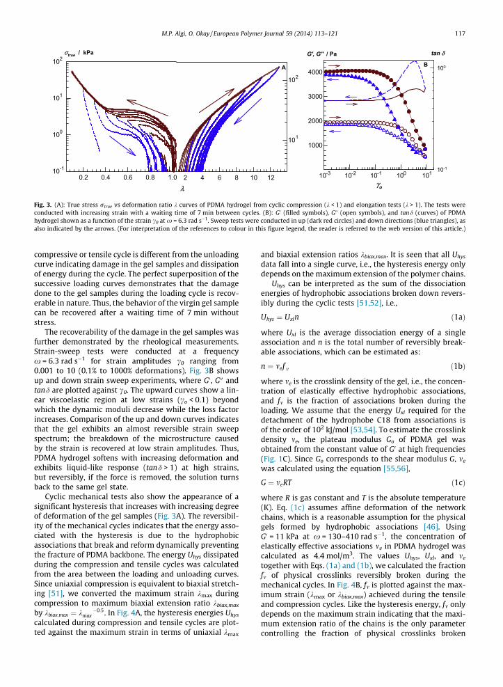
Fig. 3. (A): True stress rtrue vs deformation ratio k curves of PDMA hydrogel from cyclic compression (k < 1) and elongation tests (k > 1). The tests wereconducted with increasing strain with a waiting time of 7 min between cycles. (B): G0 (filled symbols), G00 (open symbols), and tand (curves) of PDMAhydrogel shown as a function of the strain c0 at x = 6.3 rad s�1. Sweep tests were conducted in up (dark red circles) and down directions (blue triangles), asalso indicated by the arrows. (For interpretation of the references to colour in this figure legend, the reader is referred to the web version of this article.)
M.P. Algi, O. Okay / European Polymer Journal 59 (2014) 113–121 117
compressive or tensile cycle is different from the unloadingcurve indicating damage in the gel samples and dissipationof energy during the cycle. The perfect superposition of thesuccessive loading curves demonstrates that the damagedone to the gel samples during the loading cycle is recov-erable in nature. Thus, the behavior of the virgin gel samplecan be recovered after a waiting time of 7 min withoutstress.
The recoverability of the damage in the gel samples wasfurther demonstrated by the rheological measurements.Strain-sweep tests were conducted at a frequencyx = 6.3 rad s�1 for strain amplitudes c0 ranging from0.001 to 10 (0.1% to 1000% deformations). Fig. 3B showsup and down strain sweep experiments, where G0, G00 andtand are plotted against c0. The upward curves show a lin-ear viscoelastic region at low strains (co < 0.1) beyondwhich the dynamic moduli decrease while the loss factorincreases. Comparison of the up and down curves indicatesthat the gel exhibits an almost reversible strain sweepspectrum; the breakdown of the microstructure causedby the strain is recovered at low strain amplitudes. Thus,PDMA hydrogel softens with increasing deformation andexhibits liquid-like response (tand > 1) at high strains,but reversibly, if the force is removed, the solution turnsback to the same gel state.
Cyclic mechanical tests also show the appearance of asignificant hysteresis that increases with increasing degreeof deformation of the gel samples (Fig. 3A). The reversibil-ity of the mechanical cycles indicates that the energy asso-ciated with the hysteresis is due to the hydrophobicassociations that break and reform dynamically preventingthe fracture of PDMA backbone. The energy Uhys dissipatedduring the compression and tensile cycles was calculatedfrom the area between the loading and unloading curves.Since uniaxial compression is equivalent to biaxial stretch-ing [51], we converted the maximum strain kmax duringcompression to maximum biaxial extension ratio kbiax,max
by kbiax;max ¼ k �0:5max . In Fig. 4A, the hysteresis energies Uhys
calculated during compression and tensile cycles are plot-ted against the maximum strain in terms of uniaxial kmax
and biaxial extension ratios kbiax,max. It is seen that all Uhys
data fall into a single curve, i.e., the hysteresis energy onlydepends on the maximum extension of the polymer chains.
Uhys can be interpreted as the sum of the dissociationenergies of hydrophobic associations broken down revers-ibly during the cyclic tests [51,52], i.e.,
Uhys ¼ Uxln ð1aÞ
where Uxl is the average dissociation energy of a singleassociation and n is the total number of reversibly break-able associations, which can be estimated as:
n ¼ mef m ð1bÞ
where me is the crosslink density of the gel, i.e., the concen-tration of elastically effective hydrophobic associations,and fm is the fraction of associations broken during theloading. We assume that the energy Uxl required for thedetachment of the hydrophobe C18 from associations isof the order of 102 kJ/mol [53,54]. To estimate the crosslinkdensity me, the plateau modulus Go of PDMA gel wasobtained from the constant value of G0 at high frequencies(Fig. 1C). Since Go corresponds to the shear modulus G, me
was calculated using the equation [55,56],
G ¼ meRT ð1cÞ
where R is gas constant and T is the absolute temperature(K). Eq. (1c) assumes affine deformation of the networkchains, which is a reasonable assumption for the physicalgels formed by hydrophobic associations [46]. UsingG0 = 11 kPa at x = 130–410 rad s�1, the concentration ofelastically effective associations me in PDMA hydrogel wascalculated as 4.4 mol/m3. The values Uhys, Uxl, and me
together with Eqs. (1a) and (1b), we calculated the fractionfm of physical crosslinks reversibly broken during themechanical cycles. In Fig. 4B, fm is plotted against the max-imum strain (kmax or kbiax,max) achieved during the tensileand compression cycles. Like the hysteresis energy, fm onlydepends on the maximum strain indicating that the maxi-mum extension ratio of the chains is the only parametercontrolling the fraction of physical crosslinks broken

λmax , λbiax,max
100 101
Uhys / kJ.m-3
10-3
10-2
10-1
100
101
102
103
λmax , λbiax,max
100 101
fv
10-6
10-5
10-4
10-3
10-2
10-1
100
uniaxial biaxial
A B
Fig. 4. Hysteresis energy Uhys (A) and the fraction fm of dissociated crosslinks (B) during the loading/unloading compression (open symbols) and elongationcycles (filled symbols) of PDMA hydrogel shown as a function of the maximum strain kmax or kbiax,max.
118 M.P. Algi, O. Okay / European Polymer Journal 59 (2014) 113–121
during the loading. Thus, both compression and elongationhave the same effect on the physical crosslinks of the pres-ent hydrogels. However, when fm is plotted against themaximum value of the first strain invariant J1,max, whichrepresents a general strain state of gels, the data do not fallinto a single curve (Fig. S1). This behavior is similar to thatof double network hydrogels during their first compressionand tensile cycles, as reported by Webber et al. [51]. Fig. 4Balso shows that fm varies between 10�6 and 100 indicatingthat up to all of the physical crosslinks dissociate underforce, but reversibly, if the force is removed they reformagain.
The results reveal reversible disengagements of thehydrophobic units from the associations under an externalforce and thus point out the ability of PDMA hydrogel toself-heal upon damage. This was indeed observed experi-mentally. The images in Fig. 5A were taken from a gel sam-ple before and after elongation up to k = 10. After a waiting
Fig. 5. (A): Photographs of a gel sample before and after stretching to an elongati(B): Photographs of two gel samples. One of the samples was colored with a dye ftogether for 10 min, they merge into a single piece. (For interpretation of the reversion of this article.)
time of 10 min, the gel returns to its initial length indicat-ing the recovery of the original shape. Moreover, when thefracture surfaces of a ruptured PDMA gel sample arepressed together, the two pieces merge into a single piece(Fig. 5B). The joint reformed withstands very large exten-sion ratios before its fracture.
To quantify the healing efficiency, tensile testing exper-iments were performed using virgin and healed cylindricalgel samples. The samples were cut in the middle and then,the two halfs were merged together at 24 �C for varioushealing times. In Fig. 6A, stress–strain curves of the virginand healed gel samples are shown for different healingtimes. In Fig. 6B, the tensile modulus E, the fracture stressrf, and the elongation ratio at break kf of the healed gelsamples are plotted against the healing time. The horizon-tal solid lines represent the characteristics of the virgin gelwith standard deviations indicated by the dashed lines.The modulus E is recovered within 2 min indicating the
on ratio of 10. After a waiting time of 10 min, it recovers its original length.or clarity. After cutting into two pieces and pressing the fractured surfacesferences to colour in this figure legend, the reader is referred to the web

λ10 20 30 40
σnom / kPa
0
5
10
15
20
25
virgin sample
20min
15min
10 min
5 min
2min
A B
Healing time / min
E / kPa
0
2
4
6
8
10
12
σf / kPa
0
5
10
15
20
25
0 5 10 15 20 5 10 15 20 5 10 15 20
λf
0
10
20
30
40
λfσf
Fig. 6. (A): Stress–strain curves of virgin and healed gel samples. Healing times are indicated. Temperature = 24 �C. (B): Tensile modulus E, tensile fracturestress rf, and elongation ratio kf at break of healed gels shown as a function of the healing time. The solid lines represent the behavior of virgin gel sampleand the dashed lines are the standard deviations.
M.P. Algi, O. Okay / European Polymer Journal 59 (2014) 113–121 119
occurrence of a very rapid and autonomous self-healingprocess in PDMA hydrogel. After 10 min, the fracture stressof the healed gel is 15 kPa which is 60% of the fracturestress of the virgin sample. A healing time of 20 min suf-fices to recover all the initial mechanical properties ofthe physical hydrogel.
Experiments were also carried out to optimize the for-mation conditions of PDMA hydrogels. Decreasing the ini-tial DMA concentration Co in the feed from 15% to 10%produced weak hydrogels which were not suitable forthe mechanical tests. Increasing Co from 15% to 20%increased the fracture stress to 70 ± 5 kPa while the elon-gation ratio at break decreased to 34 ± 2 (Fig. S2). Simulta-neously, the self-healing efficiency drastically decreased.For instance, after a healing time of 1 h, the fracture stressof the healed gel was 10 kPa, which is 14% of the virginsample (Fig. S2). Moreover, increasing SDS concentrationat the gel preparation from 7% to 8.5% did not improvethe healing ability of the gels formed at Co = 20% (Fig. S3).Thus, Co = 15% and SDS = 7% was found to be the optimumsynthesis condition of PDMA hydrogel with a very highstretchability and complete self-healing at roomtemperature.
We have to mention that, all the features of PDMAhydrogels described above are related to their states justafter their preparation, i.e., those containing surfactantmicelles. The solubility tests showed that the hydrogelsare insoluble in water with a gel fraction Wg equals tounity. This reveals that the monomers in the feed are com-pletely converted into a water insoluble, physically cross-linked polymer network. The swelling kinetics of PDMAhydrogel formed at Co = 15% exhibited typical behavior ofgels containing surfactants [45–47]. The gel immersed inwater initially behaves like an ionic gel due to the presenceof SDS counterions inside the gel network and thus exhib-its a large swelling ratio mrel (Fig. 7A). However, as SDS isprogressively extracted, this osmotic effect disappearsand the gel gradually converts into a nonionic gel havingan equilibrium swelling ratio mrel of 4.1 ± 0.2 in water.
Indeed, SDS concentration in the external solutions rapidlydropped below the detection limit of the methylene bluemethod (0.20 mg/L) after 10 d [45,57].
The internal dynamics of PDMA hydrogels significantlychanged after equilibrium swelling in water, i.e., afterextraction of surfactant micelles. Fig. 7B compares themechanical spectra of the hydrogels before and after swell-ing in water. The swollen gel exhibits nearly time-indepen-dent elastic modulus and a loss factor below 0.1corresponding solid-like behavior. It is obvious that thisweak-to-strong gel transformation of PDMA hydrogel uponswelling is responsible for its insolubility in water. Sincethe extraction of SDS from the gel network increases thelifetime of hydrophobic associations (Fig. 7B), the gel with-out SDS remains stable in water. Indeed, immersion of thehydrogels in 7% SDS solution instead of water resulted intheir solubilization due to the weakening of the hydropho-bic interactions and disruption of the associations [3]. Thegels were also soluble in aqueous DMA solutions contain-ing more than 50% DMA. Since DMA is a good solvent forC18 due to its hydrophobicity, it solubilizes C18 units ofthe network chains and thus, enables dissociation of thephysical crosslinks of PDMA hydrogels.
Fig. 7C compares tensile stress–strain data of PDMAhydrogel before and after swelling in water. Highly stretch-able PDMA gel becomes a brittle one after swelling inwater and ruptures at a stretch of kf = 5 ± 0.1. Cyclicmechanical tests conducted on swollen hydrogels revealedappearance of irreversibility cycles and a significantdecrease in the hysteresis (Fig. S4), indicating decreasingnumber of reversibly breakable associations. Indeed, noself-healing was observed in swollen gel samples. Thus,the hydrophobic interaction without micelles is too strongto reversibly associate. Surfactant micelles within thesupramolecular network are needed to create self-healingin PDMA hydrogels. The weakening of hydrophobic inter-actions due to the presence of surfactant moleculesincreases the mobility of the hydrophobic associationsinside the physical network so that self-healing of the

ω / rad.s-1
10-1 100 101 102
G' / Pa
103
104
tan δ
10-2
10-1
100
after swelling
BA C
after preparation
Swelling time / days
0 3 6 9 12 15 18 21
mrel
2
4
6
8
λ10 20 30 40 50
σnom / kPa
0
5
10
15
20
25after swelling
after preparation
Fig. 7. (A): Relative weight swelling ratio mrel of PDMA gels plotted against the swelling time in water. C0 = 15%. (B): G0 (symbols) and tand (lines) of PDMAgels after preparation (gray) and after equilibrium swelling in water (black) shown as a function of angular frequency x. Temperature = 25 �C. co = 0.01. (C):(C): Stress–strain curves of the hydrogels under elongation.
120 M.P. Algi, O. Okay / European Polymer Journal 59 (2014) 113–121
damaged gel samples occurs within a short period of time.We have to mention that, in our previous study, we inves-tigated the effect of SDS concentration on the self-healingcharacteristics of physical gels based on hydrophobicallymodified PAAm [47]. The highest self-healing efficiencywas observed at the preparation state of the hydrogels con-taining 7% SDS while it gradually decreased as the SDS con-tent is decreased. Thus, the unique properties of PDMAhydrogels such as very high stretchability and autonomicself-healing only exist if the hydrogels contain surfactantmicelles. Without surfactant, they exhibit low stretchabil-ity and lose their ability to self-heal due to the increasedlifetime of hydrophobic associations. Further work is inprogress using hydrophobically modified polyelectrolyteswith oppositely charged surfactants to stabilize the self-healing properties of hydrogels in aqueous environment.
4. Conclusions
PDMA hydrogels formed via hydrophobic interactionsin SDS solution exhibit very high stretchability (about4200% elongation) and autonomous self-healing at roomtemperature. They are also able to sustain large compres-sions without any permanent failure. The extraordinarymechanical properties of the present hydrogels as com-pared to the self-healing PAAm hydrogels formed underidentical conditions suggest that the DMA units of the net-work chains additionally contribute to their mechanicalproperties. Loading–unloading compression and tensilecycles show perfect superposition of the successive loadingcurves demonstrating that the damage done to the gelsamples during the loading cycle is recoverable in nature.The recoverability of the damage in the gel samples wasalso demonstrated by the rheological measurements.PDMA hydrogel softens with increasing deformation andexhibits liquid-like response (tand > 1) at high strains,while the solution turns back to the same gel state if theforce is removed. The hysteresis energies calculated fromthe mechanical cycles indicate that up to all of the physicalcrosslinks dissociate under force, but reversibly, if the forceis removed they reform again. Self-healing tests show that
a healing time of 20 min suffices to recover all the initialmechanical properties of PDMA hydrogels. The results alsoshow that the hydrophobic interaction without micelles istoo strong to reversibly associate and, the key factor lead-ing to the self-healing behavior of PDMA hydrogels is theweakening of strong hydrophobic interactions due to thepresence of surfactant molecules.
Acknowledgments
MPA acknowledges the financial support from the Sci-entific and Technical Research Council of Turkey (TUBITAK)for a postdoctoral scholarship. This work was supported byTUBITAK, TBAG–109T646. OO thanks the Turkish Academyof Sciences (TUBA) for the partial support.
Appendix A. Supplementary material
Supplementary data associated with this article can befound, in the online version, at http://dx.doi.org/10.1016/j.eurpolymj.2014.07.022.
References
[1] Uemura Y, McNulty J, Macdonald PM. Associative behavior anddiffusion coefficients of hydrophobically modified poly(N, N-dimethylacrylamides). Macromolecules 1995;28:4150–8.
[2] Peppas NA, editor. Hydrogels in medicine and pharmacy. NewYork: Wiley; 1987.
[3] Relogio P, Martinho JMG, Farinha JPS. Effect of surfactant on theintra- and intermolecular association of hydrophobically modifiedpoly(N, N-dimethylacrylamide). Macromolecules 2005;38:10799–811.
[4] Mullarney MP, Seery TAP, Weiss RA. Drug diffusion inhydrophobically modified N, N-dimethylacrylamide hydrogels.Polymer 2006;47:3845–55.
[5] Gundogan N, Okay O, Oppermann W. Swelling, elasticity and spatialinhomogeneity of poly(N, N-dimethylacrylamide) hydrogels formedat various polymer concentrations. Macromol Chem Phys2004;205:814–23.
[6] Caykara T, Akcakaya I. Synthesis and network structure of ionicpoly(N, N-dimethylacrylamide-co-acrylamide) hydrogels: comparisonof swelling degree with theory. Eur Polym J 2006;42:1437–45.
[7] Kuru EA, Orakdogen N, Okay O. Preparation of homogeneouspolyacrylamide hydrogels by free-radical crosslinkingcopolymerization. Eur Polym J 2007;43:2913–21.

M.P. Algi, O. Okay / European Polymer Journal 59 (2014) 113–121 121
[8] Aoki T, Kawashima M, Katono H, Sanui K, Ogata N, Okano T, et al.Temperature-responsive interpenetrating polymer networksconstructed with poly(acrylic acid) and poly(N, N-dimethylacrylamide). Macromolecules 1994;27:947–52.
[9] Meaurio E, Cesteros LC, Katime I. FTIR study of hydrogen bonding ofblends of poly(mono n-alkyl itaconates) with poly(N, N-dimethylacrylamide) and poly(ethyloxazoline). Macromolecules1997;30:4567–73.
[10] Allen R, Maurer RH, editors. Electrophoresis and isoelectric focusingin polyacrylamide gels: advances of methods and theories,biochemical and clinical applications. Berlin: Walter De Gruyter;1974.
[11] Heiger DN, Cohen AS, Karger BL. Separation of DNA restrictionfragments by high performance capillary electrophoresis with lowand zero crosslinked polyacrylamide using continuous and pulsedelectric fields. J Chromatogr 1990;516:33–48.
[12] Ren J, Ulvik A, Refsum H, Ueland PM. Applications of short-chainpolydimethylacrylamide as sieving medium for the electrophoreticseparation of DNA fragments and mutation analysis in uncoatedcapillaries. Anal Biochem 1999;276:188–94.
[13] Song L, Liang D, Chen Z, Fang D, Chu B. DNA sequencing by capillaryelectrophoresis using mixtures of polyacrylamide and poly(N, N-dimethylacrylamide). J Chromatogr A 2001;915:231–9.
[14] Kataoka K, Miyazaki H, Okano T, Sakurai Y. Sensitive glucose-induced change of the lower critical solution temperature of poly[N,N-(dimethylacrylamide)-co-3-(acrylamido)-phenylboronic acid] inphysiological saline. Macromolecules 1994;27:1061–2.
[15] Liu SQ, Tong YW, Yang Y-Y. Incorporation and in vitro release ofdoxorubicin in thermally sensitive micelles made from poly(N-isopropylacrylamide-co-N, N-dimethylacrylamide)-b-poly(d, l-lactide-co-glycolide) with varying compositions. Biomaterials2005;26:5064–74.
[16] Lin W-C, Fan W, Marcellan A, Hourdet D, Creton C. Large strain andfracture properties of poly(dimethylacrylamide)/silica hybridhydrogels. Macromolecules 2010;43:2554–63.
[17] Rose S, Marcellan A, Hourdet D, Narita T. Dynamics of hybridpoly(acrylamide-co-N, N-dimethylacrylamide) hydrogels containingsilica nanoparticles studied by dynamic light scattering.Macromolecules 2013;46:5329–36.
[18] Haraguchi K, Farnworth R, Ohbayashi A, Takehisa T. Compositionaleffects on mechanical properties of nanocomposite hydrogelscomposed of poly(N, N-dimethylacrylamide) and clay.Macromolecules 2003;36:5732–41.
[19] Haraguchi K, Uyama K, Tanimoto H. Self-healing in nanocompositehydrogels. Macromol Rapid Commun 2011;32:1253–8.
[20] Weng L, Gouldstone A, Wu Y, Chen W. Mechanically strong doublenetwork photocrosslinked hydrogels from N,N-dimethylacrylamideand glycidyl methacrylated hyaluronan. Biomaterials 2008;29:2153–63.
[21] Hao J, Weiss RA. Viscoelastic and mechanical behavior ofhydrophobically modified hydrogels. Macromolecules 2011;44:9390–8.
[22] Zhang Z, Tomlinson MR, Golestanian R, Geoghegan M. The interfacialbehaviour of single poly(N, N-dimethylacrylamide) chains as afunction of pH. Nanotechnology 2008;19:035505.
[23] Doherty EAS, Berglund KD, Buchholz BA, Kourkine IV, PrzybycienTM, Tilton RD, et al. Critical factors for high-performance physicallyadsorbed (dynamic) polymeric wall coatings for capillaryelectrophoresis of DNA. Electrophoresis 2002;23:2766–76.
[24] Pagonis K, Bokias G. Upper critical solution temperature—typecononsolvency of poly(N, N-dimethylacrylamide) in water—organicsolvent mixtures. Polymer 2004;45:2149–53.
[25] Orakdogen N, Okay O. Reentrant conformation transition in poly(N,N-dimethylacrylamide) hydrogels in water-organic solventmixtures. Polymer 2006;47:561–8.
[26] Phadke A, Zhang C, Arman B, Hsu C-C, Mashelkar RA, Lele AK, et al.Rapid self-healing hydrogels. PNAS 2012;109:4383–8.
[27] Zhang H, Xia H, Zhao Y. Poly(vinyl alcohol) Hydrogel CanAutonomously Self-Heal. ACS Macro Lett 2012;1:1233–6.
[28] Cui J, del Campo A. Multivalent H-bonds for self-healing hydrogels.Chem Commun 2012;48:9302–4.
[29] Liu J, Song G, He C, Wang H. Self-healing in tough graphene oxidecomposite hydrogels. Macromol Rapid Commun 2013;34:1002–7.
[30] Sun J-Y, Zhao X, Illeperuma WRK, Chaudhuri O, Oh KH, Money DJ,et al. Highly stretchable and tough hydrogels. Nature2012;489:133–6.
[31] South AB, Lyon LA. Autonomic self-healing of hydrogel thin films.Angew Chem Int Ed 2010;49:767–71.
[32] Wang Q, Mynar JL, Yoshida M, Lee E, Lee M, Okura K, et al. High-water-content mouldable hydrogels by mixing clay and a dendriticmolecular binder. Nature 2010;463:339–43.
[33] Appel EA, Biedermann F, Rauwald U, Jones ST, Zayed JM, SchermanOA. Supramolecular cross-linked networks via host�guestcomplexation with cucurbit[8]uril. J Am Chem Soc2010;132:14251–60.
[34] Cheryl TS, Foo WP, Lee JS, Mulyasasmita W, Parisi-Amon A,Heilshorn SC. Two-component protein-engineered physicalhydrogels for cell encapsulation. PNAS 2009;106:22067–72.
[35] Skrzeszewska PJ, Sprakel J, Wolf FA, Fokkink R, Stuart MAC, van deGucht J. Fracture and self-healing in a well-defined self-assembledpolymer network. Macromolecules 2010;43:3542–8.
[36] Holten-Andersen N, Harrington MJ, Birkedal H, Lee BP, MessersmithPB, Lee KYC, et al. PH-induced metal-ligand cross-links inspired bymussel yield self-healing polymer networks with near-covalentelastic moduli. PNAS 2011;108:2651–5.
[37] Shafiq Z, Cui J, Pastor-Perez L, San Miguel V, Gropeanu RA, Serrano C,del Campo A. Bioinspired underwater bonding and debonding ondemand. Angew Chem 2012;124:4408–11.
[38] Xu Y, Wu Q, Sun Y, Bai H, Shi G. Three-dimensional self-assembly ofgraphene oxide and DNA into multifunctional hydrogels. ACS Nano2010;4:7358–62.
[39] Liu F, Li F, Deng G, Chen Y, Zhang B, Zhang J, et al. Rheological imagesof dynamic covalent polymer networks and mechanisms behindmechanical and self-healing properties. Macromolecules2012;45:1636–45.
[40] Deng G, Tang C, Li F, Jiang H, Chen Y. Covalent cross-linked polymergels with reversible sol�gel transition and self-healing properties.Macromolecules 2010;43:1191–4.
[41] Zhang Y, Tao L, Li S, Wei Y. Synthesis of multiresponsive anddynamic chitosan-based hydrogels for controlled release of bioactivemolecules. Biomacromolecules 2011;12:2894–901.
[42] He L, Fullenkamp DE, Rivera JG, Messersmith PB. PH responsive self-healing hydrogels formed by boronate–catechol complexation.Chem Commun 2011;47:7497–9.
[43] Froimowicz P, Klinger D, Landfester K. Photoreactive nanoparticlesas nanometric building blocks for the generation of self-healinghydrogel thin films. Chem Eur J 2011;17:12465–75.
[44] Quint SB, Pacholski C. Extraordinary long range order in self-healingnon-close packed 2D arrays. Soft Matter 2011;7:3735–8.
[45] Tuncaboylu DC, Sari M, Oppermann W, Okay O. Tough and self-healing hydrogels formed via hydrophobic interactions.Macromolecules 2011;44:4997–5005.
[46] Tuncaboylu DC, Sahin M, Argun A, Oppermann W, Okay O. Dynamicsand large strain behavior of self-healing hydrogels with and withoutsurfactants. Macromolecules 2012;45:1991–2000.
[47] Tuncaboylu DC, Argun A, Sahin M, Sari M, Okay O. Structureoptimization of self-healing hydrogels formed via hydrophobicinteractions. Polymer 2012;53:5513–22.
[48] Akay G, Hassan-Raeisi A, Tuncaboylu DC, Orakdogen N,Abdurrahmanoglu S, Oppermann W, et al. Self-healing hydrogelsformed in catanionic surfactant solutions. Soft Matter2013;9:2254–61.
[49] Gulyuz U, Okay O. Self-healing polyacrylic acid hydrogels. SoftMatter 2013;9:10287–93.
[50] Argun A, Algi MP, Tuncaboylu DC, Okay O. Surfactant-inducedhealing of tough hydrogels formed via hydrophobic interactions.Colloid Polym Sci 2014;292:511–7.
[51] Webber RE, Creton C, Brown HR, Gong JP. Large strain hysteresis andMullins effect of tough double-network hydrogels. Macromolecules2007;40:2919–27.
[52] Lake GJ, Thomas AG. The strength of highly elastic materials. Proc RSoc London A 1967;300:108–19.
[53] Annable T, Buscall R, Ettelaie R, Whittlestone D. The rheology ofsolutions of associating polymers: comparison of experimentalbehavior with transient network theory. J Rheol 1993;37:695–726.
[54] Ng WK, Tam KC, Jenkins RD. Lifetime and network relaxation time ofa HEUR-C20 associative polymer system. J Rheol 2000;44:137–48.
[55] Flory PJ. Principles of polymer chemistry. Ithaca, NY: CornellUniversity Press; 1953.
[56] Treloar LRG. The physics of rubber elasticity. Oxford: UniversityPress; 1975.
[57] ISO 7875-1. Water quality. Determination of surfactants. Part 1:Determination of anionic surfactants by measurement of themethylene blue index (MBAS). USO/TC 147, 1996.