hippocampal activity mediates the relationship between ... · hippocampal activity mediates the...
TRANSCRIPT

Neuropsychologia 75 (2015) 617–625
Contents lists available at ScienceDirect
Neuropsychologia
http://d0028-39
n CorrE-m
journal homepage: www.elsevier.com/locate/neuropsychologia
Hippocampal activity mediates the relationship between circadianactivity rhythms and memory in older adults
Stephanie M. Sherman a,n, Jeanette A. Mumford b, David M. Schnyer a,c
a Department of Psychology, The University of Texas at Austin, 108 E. Dean Keeton A8000, Austin, TX 78712, USAb Center for Investigating Healthy Minds at the Waisman Center, University of Wisconsin-Madison, 1500 Highland Avenue, Suite S119, Madison, WI 53705,USAc The Institute for Neuroscience, The University of Texas at Austin, 1 University Station, C7000, Austin, TX 78712, USA
a r t i c l e i n f o
Article history:Received 24 March 2015Received in revised form17 July 2015Accepted 18 July 2015Available online 20 July 2015
Keywords:Associative memoryCircadian activity rhythmsSleepHippocampus
x.doi.org/10.1016/j.neuropsychologia.2015.07.032/& 2015 Elsevier Ltd. All rights reserved.
esponding author.ail address: [email protected] (S.M. Sherm
a b s t r a c t
Older adults experience parallel changes in sleep, circadian rhythms, and episodic memory. These pro-cesses appear to be linked such that disruptions in sleep contribute to deficits in memory. Although morevariability in circadian patterns is a common feature of aging and predicts pathology, little is knownabout how alterations in circadian activity rhythms within older adults influence new episodic learning.Following 10 days of recording sleep-wake patterns using actigraphy, healthy older adults underwentfMRI while performing an associative memory task. The results revealed better associative memory wasrelated to more consistent circadian activity rhythms, independent of total sleep time, sleep efficiency,and level of physical activity. Moreover, hippocampal activity during successful memory retrieval eventswas positively correlated with associative memory accuracy and circadian activity rhythm (CAR) con-sistency. We demonstrated that the link between consistent rhythms and associative memory perfor-mance was mediated by hippocampal activity. These findings provide novel insight into how the cir-cadian rhythm of sleep-wake cycles are associated with memory in older adults and encourage furtherexamination of circadian activity rhythms as a biomarker of cognitive functioning.
& 2015 Elsevier Ltd. All rights reserved.
1. Introduction
Sleep and circadian rhythms change across the lifespan. Com-pared to young adults, older adults have shorter, less efficient, andmore disrupted sleep (Ohayon et al., 2004) along with decreases inslow wave sleep (for review see: Fogel et al., 2012) and sleepspindle density (Martin et al., 2013; for review see: De Gennaroand Ferrara, 2003). The circadian rhythms of sleep-wake cyclesbecome inconsistent, shift earlier in time, and reduce in amplitudewith increased age (Huang et al., 2002; Neikrug and Ancoli-Israel,2010). In animal models, the neural structure that mediates cir-cadian rhythms, the suprachiasmatic nucleus (SCN), degeneratesin older adults and is hypothesized to contribute to age-relateddisruptions in circadian rhythms (Farajnia et al., 2012). In parallelto the changes in sleep and circadian rhythms, older individualsdemonstrate significant changes in cognitive functioning, includ-ing episodic memory performance (Naveh-Benjamin et al., 2004).Research has begun to link changes in sleep and cognition bysuggesting that co-occurring sleep problems may uniquely
20
an).
contribute to cognitive deficits. Negative correlations have beenobserved in older adults between cognitive performance and dis-rupted sleep on tests of global cognitive impairment (Jelicic et al.,2002; Blackwell et al., 2006; Carvalho-Bos et al., 2007), workingmemory (Haimov et al., 2008; Nebes et al., 2009; Lim et al., 2012),mental speed (Oosterman et al., 2009), memory encoding (Manderet al., 2013a), memory retrieval (Westerberg et al., 2010), andmemory consolidation (Wilson et al., 2012; Mander et al., 2013b;Sonni and Spencer, 2015).
Beyond the contribution of disrupted sleep, disturbances in thecircadian rhythm of the rest-activity cycle are notably linked toage-related changes in cognitive functioning (Lim et al., 2012).More variability in the rest-activity cycle, known as the circadianactivity rhythm (CAR), has been related to greater dementia se-verity (Gehrman et al., 2005) and higher mortality rates (Tranahet al., 2010). In longitudinal studies, older women who had morevariable CARs were more likely to show cognitive decline 5 yearslater (Walsh et al., 2014), or in some cases, neurocognitive dis-orders such as mild cognitive impairment (MCI) or dementia(Tranah et al., 2011). Since these studies included broad and ofteninsensitive measures of cognition in very old women (80s) withhealth problems (Scullin and Bliwise, 2015) little is known about
S.M. Sherman et al. / Neuropsychologia 75 (2015) 617–625618
whether disrupted CARs in healthy older individuals contribute tolower memory function.
Neuroimaging studies across the lifespan have targeted thehippocampus to understand how sleep quantity and physiologycontribute to learning and memory. Work in young adults hasdemonstrated that sleep is essential to hippocampal-dependentlearning (Yoo et al., 2007; Marshall and Born, 2007; Van Der Werfet al., 2009; Nguyen et al., 2013; for review see: Abel et al., 2013).In older individuals more hippocampal activity during memorytasks has been associated with greater total sleep time (Joneliset al., 2012), higher sleep spindle density (Mander et al., 2013a),and more slow wave activity (Van Der Werf et al., 2009) which inturn led to better episodic learning. Although disrupted CARs are acommon feature of aging and can be predictive of pathology(Tranah et al., 2011) it is unknown how CAR disruptions influencenew episodic learning and whether the hippocampus plays anessential role in this relationship.
The purpose of this study was to investigate whether moreconsistent CARs (lower variability in the CAR) was related to betterassociative memory performance. Associative memory was chosenbecause it is highly sensitive to memory declines in older in-dividuals (Dennis et al., 2008) and reliably recruits the hippo-campus, a structure that shows consistent age-related changes(Giovanello et al., 2009). Furthermore, we examined whetherhippocampal activity during successful associative memory re-trieval mediates the relationship between CAR consistency andmemory in older adults. We hypothesized that more consistentCARs would be associated with better memory performance as aresult of greater hippocampal activity.
2. Materials and methods
2.1. Participants
Interested 60–80 year old Austin community members com-pleted a self-reported health screening and a neuropsychologicaltest battery prior to entrance into our research study. The healthscreening excluded those who reported a medical history of heartconditions or vascular disease, including hypertension and ele-vated body mass index (BMI greater than 30). The cardiovascularexclusion criteria was included because of previous work illus-trating that beginning at middle age, cardiovascular risk factors areassociated with significant neurological changes (Salat et al., 2012;Haley, 2014). Additionally, adults were excluded for any history ofneurological injury or disorder, diabetes, current psychiatric dis-order, major depression within the past five years, current sleepdisorder or a score greater than 8 on Pittsburg Sleep Quality Index(PSQI; Fichtenberg et al., 2001), cancer in the last three years, orcurrent medications that affect the central nervous system. Allolder adults included in the study were within 1 SD of normalperformance on composite scores in the cognitive domains ofmemory, vocabulary, and executive function.
Forty-one participants (29 females, mean age¼66.875.1, agerange 60–78 years) were included in our final analyses from 45eligible participants. Four participants were dropped due to in-complete actigraph data, sleep logs, and/or memory task data. Allparticipants provided written informed consent that was ap-proved by the Institutional Review Board at the University of Texasat Austin.
2.2. Procedure
2.2.1. OverviewParticipants took part in two experimental testing sessions that
were separated by a minimum of 10 days. Both experimental
sessions occurred in the morning so older adults would performduring their optimal time of day (May et al., 1993). In the baselinesession, participants were fitted with wrist actigraphs (Mo-tionlogger Actigraphs; Ambulatory Monitoring, Inc., Ardsley, NY)to record objective measures of their sleep-wake patterns undernormal environmental conditions. To obtain baseline measures ofsleep quality, vigilance, and attention, participants completed thePittsburg Sleep Quality Index (PSQI) and the Psychomotor Vigi-lance Task (PVT). For the next 10 days, participants wore the ac-tigraph continuously except during situations where it could getwet or damaged. In addition, participants filled out daily, onlinesleep logs based on the Consensus Sleep Diary (Carney et al.,2012), usually in the morning after waking. Following the re-cording period, participants returned to the laboratory and com-pleted the PSQI, Insomnia Severity Index (ISI), the PVT, and un-derwent structural and functional magnetic resonance imagingwhile performing a word-pair associates task (mean time of test-ing session: 10 am, SD: 30 min, range: 8:30–11:00 am).
2.2.2 ActigraphyAfter the 10 day recording period, participants returned the
actigraphs and data were downloaded from each device using theambulatory monitoring software (Action 4; http://www.ambulatory-monitoring.com/action4.html). Actigraph recordings werecollected in 1-min epochs in the proportion integration mode(PIM). The PIM captures the frequency and intensity of movementby calculating counts from the area under the receiver operatingcharacteristic curve analysis (Tranah et al., 2010). This channel waschosen because the activity levels have been shown to best cor-respond to measures from the gold standard of sleep monitoring,polysomnography (PSG; Blackwell et al., 2011b).
Following the procedures described in previous work (Black-well et al., 2011a, 2011b) we used the actigraphy and sleep log datato create valid profiles of each participant's sleep-wake patterns bydefining intervals that designate when the participant was in bedtrying to sleep each night (in-bed intervals). These procedureshave been validated against PSG in older individuals (Blackwellet al., 2011a). Although some participants wore the actigraph forlonger than 10 days, analyses only included data from the 10 daysprior to the MRI testing session. The times when the participantremoved the actigraph were not included in the analysis (Black-well, et al., 2011b). No participant took the watch off for more than10% of the recording period (M¼5.5 h, range 0.73–19.95 h). Weapplied the University of California, San Diego (UCSD) SleepScoring algorithm to the data collected in the PIM channel. Thisalgorithm codes each minute of data as sleep or wake based on theactivity levels at that minute as well as the surrounding minutes ofrecording. Within each in-bed interval, the number of minutesscored as sleep by the algorithm was summed to calculate ameasure of total sleep time for each night. Nightly sleep efficiencywas calculated by dividing the total sleep time by the number ofminutes within the in-bed interval (Blackwell et al., 2011b). Totalsleep time and sleep efficiency across the 10 day period wereaveraged within each participant.
Central to the research question, the continuous collection ofactigraph data allowed us to measure aspects of circadian activityrhythms (CARs) to effectively quantify the timing and consistencyof the rest-activity cycle (Ancoli-Israel et al., 2002; Savard et al.,2009; Tranah et al., 2010; Witkowski et al., 2015; McKenna et al.,2014). CARs were computed by inputting the minute-by-minutePIM activity (from the 10 day actigraph recording) into a five-parameter extended cosine model (Martin et al., 2000). The ex-tended cosine model characterizes the square-like shape of humanactivity data (Marler et al., 2006; Tranah et al., 2010) better thantraditional cosinor methods that include underlying assumptionsabout the activity shape that are imprecise (Van Someren et al.
S.M. Sherman et al. / Neuropsychologia 75 (2015) 617–625 619
1997; Dowling et al., 2005) and too simplistic (Calogiuri et al.,2011). Parameters were estimated from the model using a non-linear least squares approach to data fitting. The parameters ofinterest included the mean (mesor), peak (acrophase), and am-plitude of the activity rhythm. The mean, known as the mesor, isthe average activity level of the modeled curve. The peak, calledthe acrophase, represents the time of day when the highest levelof activity occurs. The amplitude is the difference between themaximum and minimum values of the activity curve. In addition,we investigated the time of day when activity moved from belowthe mean level to above the mean level on the modeled curvecalled up-mesor. These parameters were computed for each in-dividual to create the best fitting activity curve. After we createdthe activity curve using the individually estimated shape, timing,and amplitude parameters, the F-statistic was computed to assesshow well the activity data fit the curve – referred to as “CARconsistency”. More consistent activity rhythms indicate lessvariability in the rest-activity pattern. If the timing, amplitude, andshape of activity are consistent across days, then the calculatedparameters will closely match the actual data. This translates to abetter fit of the curve (smaller standard error) and a more con-sistent CAR. More details about these measures can be found inprevious publications (Tranah et al., 2010; Liu et al., 2013). Themeasures from this model have been shown to correlate with corebody temperature and reliably match the circadian period of PSGdata (Pollak et al., 2001; for review see: Ancoli-Israel et al., 2003).
2.2.3. Psychomotor Vigilance task (PVT)Immediately before the scan session, participants completed a
high-signal load computerized reaction time (RT) task that mea-sures sustained attention and vigilance. The PVT has been em-ployed in hundreds of studies to measure sustained attentionbecause it is reliable, valid, and highly sensitive to changes in sleep(for review see: Lim and Dinges, 2008). Participants attended to afixation cross at the center of a computer screen. At random in-tervals, a millisecond timer appeared at the screen center (2–10 sinter-trial intervals). Participants were instructed to press a buttonthe instant they detected the start of the timer. The button pressstopped the timer and displayed the reaction time for 1 s. The PVTwas 10 min in length and required sustained attention in order todetect the onset of the timer. Response lapses (i.e. RTs4500 ms),and false starts (i.e. RTso100 ms) were removed before comput-ing summary statistics. Mean, median, and the standard deviationof RTs were calculated as well as the mean of the fastest andslowest 10% of the trials.
2.2.4. Image acquisitionFollowing the 10 day recording period, participants underwent
structural and functional magnetic resonance imaging (MRI) on a3T Siemens Skyra MRI scanner with a 32-channel phase arrayhead coil at the University of Texas at Austin Imaging ResearchCenter. Two high-resolution T1-weighted MPRAGE scans(TR¼2.53, TE¼3.37, flip angle¼7°, 1 mm slice thickness, 176 slices,FOV¼256�256 mm2) were collected for anatomical coregistra-tion with other datasets. The T1 scans were motion corrected andaveraged to optimize the signal and contrast for analysis. Func-tional EPI images were collected using a GRAPPA reconstructedparallel image sequence with an acceleration factor of 2 and a sliceorientation to reduce artifact (approximately 20° off the AC-PCplane, TR¼2000 ms, TE¼30 ms, flip angle¼90°). Thirty-five in-terleaved axial slices with voxel size 2.5�2.5�3 mm3 and 10%gap were collected for the best whole-head coverage. Head motionwas minimized with foam inserts.
2.2.5. Word-pair associates taskThe stimuli from the word-pair associates task were taken from
a previously normed list of word pairs (Giovanello et al., 2009).The task consisted of a study phase immediately followed by a testphase. Participants were in the MRI for the duration of the task butfMRI data was only collected during the test phase. We collectedfMRI data during retrieval based on previous work illustrating thatthe hippocampus is involved in associative memory retrievalduring the word-pair associates task (Giovanello et al., 2004, 2009;Giovanello and Schacter, 2012). In the study phase, a series of twounrelated nouns were presented on a high-resolution, back pro-jection system viewed by the participant via a mirror mounted onthe head coil. Responses were collected with an MR compatibleoptical transmission device that was held in one hand. Participantswere instructed to mentally create a sentence that incorporatedthe two words at their own pace. They were asked to press abutton to indicate that they completed the sentence formation.They were informed that they would use these sentences to re-member the word pairs for a later memory test. During the testphase, participants viewed 3 different types of word pairs-Intact(pair of words that were previously seen together), Rearranged(pair of words that were previously seen but not together), or New(pair of novel words). They were instructed to press one of 3 but-tons to indicate which pair-type was presented. After each pair-type judgment, participants rated the confidence of their response(1-guess, 2–25% sure, 3–75% sure, 4-sure). An odd/even classifi-cation task (Stark and Squire, 2001) was randomly interspersed inbetween the pair-type judgments as an active control task. Parti-cipants had 5 s to respond to the pair-type judgment, 5 s to re-spond to the confidence rating, and 2 s to respond to the activecontrol task. A fixation cross was presented during the inter-sti-mulus interval, which varied from 2 to 10 s. They alternated be-tween study and test 3 times (3 runs). Each participant completeda practice run of the study and test phase outside of the MRI todemonstrate to the experimenter that they understood theinstructions.
2.2.6. Behavioral data analysisAccuracy on the associative memory task was assessed by
calculating an “associative recognition accuracy” score (pR) bytaking the percent of hits for the intact pair trials and subtractingthe percent of the rearranged trials that were falsely identified asintact pairs (associative errors; de Chastelaine et al., 2011). Allparticipants performed at greater than chance on associative re-cognition accuracy (pR; hits minus associative errors40; t(40)¼19.45, po .0001 – range .27–.97).
2.2.7. fMRI data analysisThe imaging analyses included 36 participants because five
participants did not have complete fMRI data due to participantdiscomfort, safety concerns, or they did not follow task instruc-tions. Functional MRI data were analyzed using tools from thesoftware package FSL (http://fsl.fmrib.ox.ac.uk/fsl/fslwiki/). Imageswere motion-corrected, spatially smoothed using a 6 mm Gaussianfilter, and high pass filtered at 100 s to remove low frequency driftcomponents. Framewise displacement (fd) values from the motioncorrection processes were examined across each run to ensure thatparticipants were not moving excessively. No participants had amean fd value greater than 0.36 mm (M¼ .17, SD¼ .06), which iswell below the threshold of what is problematic (Siegel et al.,2013).
The canonical hemodynamic response and its temporal deri-vative were modeled for all events. Correct trials were modeled foreach memory condition (intact, rearranged, new). Mean reactiontimes (RT) for all memory trials within each run and participantwere applied as a duration variable. The same procedures werecompleted for correct control trials and answered confidencetrials. Nuisance regressors were added to the model for incorrect

S.M. Sherman et al. / Neuropsychologia 75 (2015) 617–625620
memory trials, no response memory trials, no response and in-correct control trials, and no response confidence trials. To accountfor RT related effects, we included RT regressors for the memory,control, and confidence trials. The modulation column for all threeRT regressors included the individual trial reaction times aftermean-centering within each regressor. All events were enteredinto a first level analysis for each separate run of each participant.Multiple runs within a subject (no less than 2) were combined in asecond level analysis using a fixed effects model (1 participant had2 runs and all other participants had 3 runs) and individual sub-jects were registered to the MNI standard brain template. FLAME(FMRIB's Local Analysis of Mixed Effects) in FSL was used to createrandom effects group level maps of our critical contrast, successfulassociative memory retrieval (correct memory for intact vs. con-trol). All group level comparisons are cluster-corrected for multi-ple comparisons where clusters were determined by z42.3 and po .05 threshold.
Fig. 1. More consistent circadian activity rhythms (F-statistic) were related tobetter associative recognition accuracy (pR).
Fig. 2. Example actigraph activity levels from two participants are plotted across a4-day period. The top panel displays data from a participant with low circadianactivity rhythm consistency and the bottom panel shows data from a participantwith high circadian activity rhythm consistency. Both participants received ap-proximately 8 h of sleep on average across the recording period.
3. Results
3.1. Behavioral results
3.1.1. The association between sleep, circadian activity rhythms(CARs), and associative memory.
A Shapiro–Wilks Normality Test ensured that CAR consistencyas measured by the F-statistic values were normally distributed(W¼ .97, p¼ .39) and could be examined using linear statisticalmodels. Age was not correlated with associative recognition ac-curacy (pR; r¼� .14, p¼ .38) nor CAR consistency, r¼ .03, p¼ .83.More consistent CARs were correlated with better pR, r¼ .33, CI[.02, .58], p¼ .04 (see Fig. 1). Associative recognition accuracy (pR)was not related to the timing of the activity rhythm as indexed byacrophase (r¼-.27, p¼ .09) and up-mesor (r¼� .29, p¼ .07). Insummary, more consistent activity rhythms were associated withbetter memory performance. Fig. 2
The average physical activity as measured by the circadian ac-tivity mesor was also not related to pR, r¼� .03, p¼ .85. In addi-tion, mesor was not a significant covariate in the linear regressionusing CAR consistency to predict pR, β¼� .10, p¼ .53. Similar tomesor, the amplitude of the rhythm was not related to pR(r¼� .13, p¼ .42) and it was also not a significant covariate in themodel (β¼� .12, p¼ .45).
Total sleep time the night before testing, average total sleeptime, and sleep efficiency across the 10 day period were also notrelated to pR (r¼� .2, p¼ .20; r¼ .04, p¼ .82; r¼� .09, p¼ .57, re-spectively), nor were the global score from the PSQI, ISI, and theGeriatric Depression Scale (Yesavage et al., 1983; r¼ .05, p¼ .74;r¼ .02, p¼ .85, r¼ .08, p¼ .63, respectively). These findings suggestthat physical activity, sleep calculated from the actigraph, and self-reported sleep and depression measures from survey instrumentswere not correlated with associative memory performance.
3.1.2. The association between sleep, circadian activity rhythms,memory, and sustained attention
The outcome measures from the PVT following the actigraphrecording period were not correlated with pR (mean RT: r¼� .27,p¼ .08; fastest 10% RT: r¼� .17, p¼ .28; slowest 10% RT: r¼� .23,p¼ .14) or CAR consistency (mean RT: r¼� .02, p¼ .90; fastest 10%RT: r¼� .09, p¼ .58; slowest 10% RT: r¼� .01, p¼ .93). None of thePVT measures were significant covariates in the linear regressionusing CAR consistency to predict pR (mean RT: β¼� .27, p¼ .08;fastest 10% RT: β¼� .14, p¼ .35; slowest 10% RT: β¼� .24, p¼ .12).In line with previous work, lower variability in reaction times onthe PVT (measured by the standard deviation) was associated withgreater average total sleep time, r¼� .34 CI[� .59, � .04], p¼ .03.
Therefore sustained attention was correlated with sleep quantitybut not CAR consistency. Vigilant attention did not account for orcontribute to the relationship between memory function and CARconsistency.
3.2. fMRI results
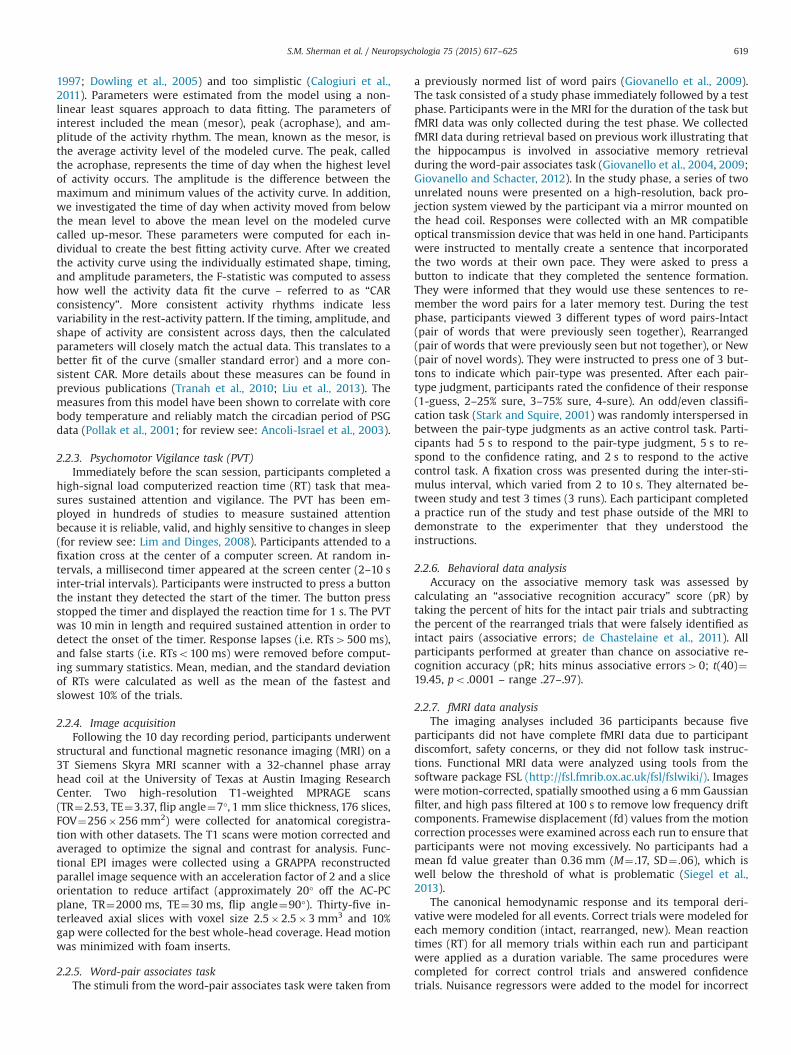
3.2.1. Whole brain analysisWhole brain contrast maps for successful associative memory
retrieval (correct memory for intact vs. control task) revealed anetwork of brain regions previously implicated in episodic mem-ory retrieval (Rugg and Vilberg, 2013), including the left hippo-campus and left inferior frontal gyrus (see Table 1 and Fig. 3). Thiswas the primary contrast of interest since there were not enoughassociative errors to reliably examine the contrast representingassociative recognition accuracy (correct memory for the intactcondition minus associative errors).
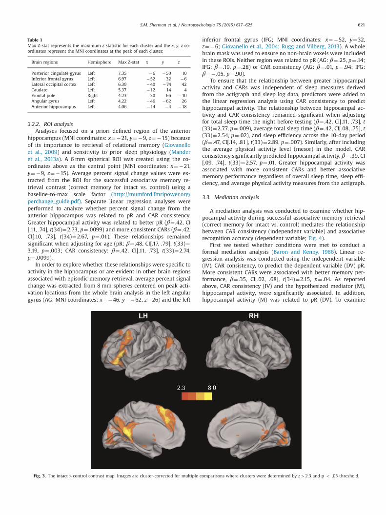
Table 1Max Z-stat represents the maximum z statistic for each cluster and the x, y, z co-ordinates represent the MNI coordinates at the peak of each cluster.
Brain regions Hemisphere Max Z-stat x y z
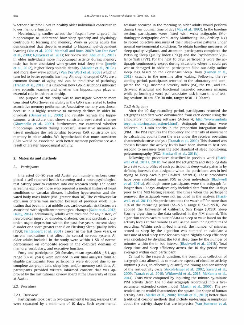
Posterior cingulate gyrus Left 7.35 �6 �50 10Inferior frontal gyrus Left 6.97 �52 32 �6Lateral occipital cortex Left 6.39 �40 �74 42Caudate Left 5.37 �12 14 4Frontal pole Right 4.23 30 66 �10Angular gyrus Left 4.22 �46 �62 26Anterior hippocampus Left 4.06 �14 �4 �18
S.M. Sherman et al. / Neuropsychologia 75 (2015) 617–625 621
3.2.2. ROI analysisAnalyses focused on a priori defined region of the anterior
hippocampus (MNI coordinates: x¼�21, y¼�9, z¼�15) becauseof its importance to retrieval of relational memory (Giovanelloet al., 2009) and sensitivity to prior sleep physiology (Manderet al., 2013a). A 6 mm spherical ROI was created using the co-ordinates above as the central point (MNI coordinates: x¼�21,y¼�9, z¼�15). Average percent signal change values were ex-tracted from the ROI for the successful associative memory re-trieval contrast (correct memory for intact vs. control) using abaseline-to-max scale factor (http://mumford.fmripower.org/perchange_guide.pdf). Separate linear regression analyses wereperformed to analyze whether percent signal change from theanterior hippocampus was related to pR and CAR consistency.Greater hippocampal activity was related to better pR (β¼ .42, CI[.11, .74], t(34)¼2.73, p¼ .0099) and more consistent CARs (β¼ .42,CI[.10, .73], t(34)¼2.67, p¼ .01). These relationships remainedsignificant when adjusting for age (pR: β¼ .48, CI[.17, .79], t(33)¼3.19, p¼ .003; CAR consistency: β¼ .42, CI[.11, .73], t(33)¼2.74,p¼ .0099).
In order to explore whether these relationships were specific toactivity in the hippocampus or are evident in other brain regionsassociated with episodic memory retrieval, average percent signalchange was extracted from 8 mm spheres centered on peak acti-vation locations from the whole brain analysis in the left angulargyrus (AG; MNI coordinates: x¼�46, y¼�62, z¼26) and the left
Fig. 3. The intact4control contrast map. Images are cluster-corrected for multiple c
inferior frontal gyrus (IFG; MNI coordinates: x¼�52, y¼32,z¼�6; Giovanello et al., 2004; Rugg and Vilberg, 2013). A wholebrain mask was used to ensure no non-brain voxels were includedin these ROIs. Neither region was related to pR (AG: β¼ .25, p¼ .14;IFG: β¼ .19, p¼ .28) or CAR consistency (AG: β¼ .01, p¼ .94; IFG:β¼� .05, p¼ .90).
To ensure that the relationship between greater hippocampalactivity and CARs was independent of sleep measures derivedfrom the actigraph and sleep log data, predictors were added tothe linear regression analysis using CAR consistency to predicthippocampal activity. The relationship between hippocampal ac-tivity and CAR consistency remained significant when adjustingfor total sleep time the night before testing (β¼ .42, CI[.11, .73], t(33)¼2.77, p¼ .009), average total sleep time (β¼ .42, CI[.08, .75], t(33)¼2.54, p¼ .02), and sleep efficiency across the 10-day period(β¼ .47, CI[.14, .81], t(33)¼2.89, p¼ .007). Similarly, after includingthe average physical activity level (mesor) in the model, CARconsistency significantly predicted hippocampal activity, β¼ .39, CI[.09, .74], t(33)¼2.57, p¼ .01. Greater hippocampal activity wasassociated with more consistent CARs and better associativememory performance regardless of overall sleep time, sleep effi-ciency, and average physical activity measures from the actigraph.
3.3. Mediation analysis
A mediation analysis was conducted to examine whether hip-pocampal activity during successful associative memory retrieval(correct memory for intact vs. control) mediates the relationshipbetween CAR consistency (independent variable) and associativerecognition accuracy (dependent variable; Fig. 4).
First we tested whether conditions were met to conduct aformal mediation analysis (Baron and Kenny, 1986). Linear re-gression analysis was conducted using the independent variable(IV), CAR consistency, to predict the dependent variable (DV) pR.More consistent CARs were associated with better memory per-formance, β¼ .35, CI[.02, .68], t(34)¼2.15, p¼ .04. As reportedabove, CAR consistency (IV) and the hypothesized mediator (M),hippocampal activity, were significantly associated. In addition,hippocampal activity (M) was related to pR (DV). To examine
omparisons where clusters were determined by z42.3 and p o .05 threshold.
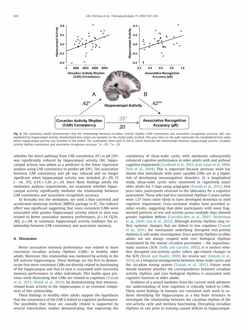
Fig. 4. The mediation model demonstrates that the relationship between circadian activity rhythm (CAR) consistency and associative recognition accuracy (pR) wasmeditated by hippocampal activity. Standardized beta values are included on the model paths in black. The gray value on the path represents the standardized beta valuebefore hippocampal activity was included in the model. The scatterplots show path A and B, which illustrate the relationships between hippocampal activity, circadianactivity rhythm consistency, and associative recognition accuracy. *po .05, **po .01.
S.M. Sherman et al. / Neuropsychologia 75 (2015) 617–625622
whether the direct pathway from CAR consistency (IV) to pR (DV)was significantly reduced by hippocampal activity (M), hippo-campal activity was added as a predictor to the linear regressionanalysis using CAR consistency to predict pR (DV). The associationbetween CAR consistency and pR was reduced and no longersignificant when hippocampal activity was included, β¼ .20, CI[� .14, .55], t(33)¼1.20, p¼ .24. Since these findings satisfy themediation analysis requirements, we examined whether hippo-campal activity significantly mediates the relationship betweenCAR consistency and associative recognition accuracy.
To formally test the mediation, we used a bias-corrected andaccelerated bootstrap method (MBESS package in R). The indirecteffect was significant suggesting that more consistent CARs wereassociated with greater hippocampal activity which in turn wasrelated to better associative memory performance, β¼ .14 CI[.01,.40], p¼ .04. In summary, hippocampal activity mediates the re-lationship between CAR consistency and associative memory.
4. Discussion
Better associative memory performance was related to moreconsistent circadian activity rhythms (CARs) in healthy olderadults. Moreover, this relationship was mediated by activity in theleft anterior hippocampus. These findings are the first to demon-strate that more consistent CARs are directly related to functioningof the hippocampus and that in turn is associated with successfulmemory performance in older individuals. This builds upon pre-vious work illustrating that CARs are related to cognition (Tranahet al., 2011; Walsh et al., 2014), by demonstrating that memory-related brain activity in the hippocampus is an essential compo-nent of this relationship.
These findings in healthy older adults support the hypothesisthat the consistency of the CAR is linked to cognitive performance.The possibility that these are causally related is supported byseveral intervention studies demonstrating that improving the
consistency of sleep-wake cycles with melatonin subsequentlyenhanced cognitive performance in older adults with and withoutcognitive impairment (Cardinali et al., 2012: Jean-Louis et al., 1998;Peck et al., 2004). This is important because previous work hasshown that individuals with more variable CARs are at a higherrisk of developing neurocognitive disorders. In a longitudinalstudy, sleep-wake cycles were monitored in cognitively intactolder adults for 3 days using actigraphy (Tranah et al., 2011). Fiveyears later, participants returned to the laboratory for a cognitiveassessment. Those who had less consistent rhythms 5 years earlierwere 1.57 times more likely to have developed dementia or mildcognitive impairment. Cross-sectional studies have provided si-milar results, suggesting that older participants with more frag-mented patterns of rest and activity across multiple days showedgreater cognitive deficits (Carvalho-Bos et al., 2007; Oostermanet al., 2009; Lim et al., 2012). Although activity rhythms may re-flect systemic changes that are linked to low cognition (Tranahet al., 2011) the mechanism underlying disrupted rest-activityrhythms is still under investigation. Since activity rhythms in olderadults are not always coupled with core biological rhythmsmaintained by the master circadian pacemaker – the suprachias-matic nucleus (SCN; Duffy and Czeisler, 2002), it is unclear whe-ther disrupted rest-activity cycles reflect age-related changes inthe SCN (Monk and Kupfer, 2000; for review see: Zelinski et al.,2014), or a temporal misalignment between sleep-wake cycles andthe circadian timing system (Tranah et al., 2011). Future workshould examine whether the correspondence between circadianactivity rhythms and core biological rhythms is associated withcognitive function in older adults.
Evidence of a neural mediator from the current work advancesour understanding of how cognition is critically linked to CARs.These novel findings in humans are consistent with work in ro-dents identifying the hippocampus as a key brain structure toinvestigate the relationship between the circadian rhythm of therest-activity cycle and memory functioning. Disrupting circadianrhythms in rats prior to training caused deficits in hippocampal-

S.M. Sherman et al. / Neuropsychologia 75 (2015) 617–625 623
dependent learning (Craig and McDonald, 2008). In this study,fragmented rhythms were induced in the rats by shifting the light/dark schedule. After two weeks of shifted rest-activity cycles, ac-quisition and retention of the platform location on the water mazetask was impaired. The results were specific to hippocampal-de-pendent learning since performance on the hippocampal-in-dependent learning fear-conditioning task was unaffected by thechange in circadian rhythms. Learning on other tasks that involvethe hippocampus such as discriminating a novel object from afamiliar one was similarly disturbed following altered circadianrhythms in hamsters (Ruby et al., 2008). Furthermore, circadiandisruptions following training impaired memory retrieval (Zelinskiet al., 2013). Rats were trained on a spatial navigation task and avisual discrimination task. Following training, photoperiod shiftswere used to disturb circadian rhythms. Photoperiod shifts sig-nificantly decreased retention on the spatial navigation task butnot on the visual discrimination task, indicating that changes incircadian rhythms negatively impact hippocampal-dependentlearning and memory. Prior research in animals has established astrong linkage between circadian rhythms and hippocampal-de-pendent learning and while the current study cannot definitivelyconclude that circadian activity rhythms in older adults are se-lectively associated with hippocampal-dependent memory theresults here are certainly consistent with the animal findings.
4.1. Potential biochemical mechanisms of circadian influences onhippocampal functioning
Previous work has offered potential biochemical mechanismsto understand the relationship between alterations in circadianrhythms and functioning of the hippocampus. First, hippocampalfunction has been linked to changes in cortisol (Lupien et al.,1998), a hormone that follows a distinct circadian rhythm and isregulated by the suprachiasmatic nucleus – the main circadianpacemaker (Bailey and Heitkemper, 2001). Evidence has shownthat high cortisol levels impair hippocampal-dependent memories(Li et al., 2006) and decrease hippocampal volume (Lupien et al.,1998). Additionally, aging is associated with increases in cortisolconcentration, which may be linked to age-related cognitive de-cline (Lupien et al., 1994). Stress also contributes to elevated cor-tisol levels and similarly results in cognitive deficits (for reviewsee: Lupien, et al., 2009). Therefore previous work suggests it ispossible that increased cortisol levels in older individuals thatoccur in aging have a disruptive effect on hippocampal-dependentlearning. This connection may have important implications formanaging changes in memory functioning during the agingprocess.
Another potential contributor to the relationship between cir-cadian rhythms and hippocampal functioning lies with the ex-pression of circadian clock genes. The clock genes are associatedwith the regulation of circadian rhythms (Brancaccio et al., 2014)and are expressed in the suprachiasmatic nucleus as well as sev-eral other brain regions, including the hippocampus (Kyriacou andHastings, 2010). Animal work illustrates that mice without clockgenes, Period1 or Period2, show impaired hippocampal-depen-dent learning (Jilg et al., 2010; Wang et al., 2009). In addition, oldage is accompanied by significant alterations in CLOCK gene ex-pression within the mouse hippocampus, which is thought tocontribute to age-related disruptions in circadian rhythms (Wyseand Coogan, 2010). Future research could focus on better under-standing the relationships between changes in cortisol and/orclock gene expression, alterations in circadian activity rhythms,and hippocampal-dependent memory in humans.
4.2. Circadian activity rhythm disruptions as early indicator of
cognitive impairment
It is possible that individual variations in CARs in older adultsmay reflect systemic changes associated with low cognition (Tra-nah et al., 2011; Scullin and Bliwise, 2015). Since disrupted circa-dian rhythms, which naturally occur with increased age (Huanget al., 2002; Neikrug and Ancoli-Israel, 2010), are also related topoor memory function, monitoring the consistency of CARs overtime may be an important predictor of age-related cognitive de-cline. Recent longitudinal work supports this assertion by de-monstrating that CARs predict deficits in executive function5-years in the future (Walsh et al., 2014). Sleep-wake patternswere measured for 3 days in women without dementia and5-years later, the same participants underwent cognitive assess-ments. Those who had significant alterations in CARs 5-years priorexhibited lower cognitive performance, especially executive func-tion performance. Although this study along with the current workfound that associations between CARs and cognition were in-dependent of sleep measures derived from the actigraph (Walshet al., 2014), it is unknown whether these associations are in-dependent of other physiological measures of sleep, such as slowwave sleep or slow wave activity. This is especially importantbecause hippocampal activity has been linked to slow wave sleep(Van Der Werf et al., 2009). Future work should examine sleepphysiology in relation to CAR consistency and investigate whetherthese measures are independently associated with memory per-formance. Finally, it would be important to implement a carefullongitudinal study focused on how changes in CARs in healthyolder adults influence the association between hippocampalfunction and cognition, potentially revealing a predictor of cog-nitive decline that could be implemented for early diagnosis ofneurocognitive disorders.
5. Conclusion
Our results illustrate a direct link between variations in circa-dian activity rhythms (CARs) and hippocampal function, whichcollectively relate to memory performance. Additionally thesefindings make an important link between the human and animalwork by demonstrating that changes in the circadian rhythm ofthe rest-activity cycle leads to hippocampal dysfunction and con-tributes to memory deficits. Although aging is generally accom-panied by changes in cognitive function (for review see: Drag andBieliauskas, 2010), it is possible that the amount of disruption insleep-wake cycles may differentiate cognitively healthy olderadults from those who will later develop neurocognitive disorders(Tranah et al., 2011). Carefully designed longitudinal studiesshould examine whether changes in CARs identify systemicchanges that detect early signs of neurocognitive disorders. Inconclusion, these results provide novel insight into how the cir-cadian rhythm of sleep-wake cycles are associated with memoryperformance and encourage further investigation of circadian ac-tivity rhythms as a biomarker of later life cognitive functioning.
Disclosure statement
The authors declare no competing financial interests.
Acknowledgments
This research was funded by Army Grant W911NF-07-2-0023via The Center for Strategic and Innovative Technologies at UT-
S.M. Sherman et al. / Neuropsychologia 75 (2015) 617–625624
Austin and Chief of Staff of the Army - Grant to West Point'sNetwork Science Center.
References
Abel, T., Havekes, R., Saletin, J.M., Walker, M.P., 2013. Sleep, plasticity and memoryfrommolecules to whole-brain networks. Curr. Biol. 23 (17), 774–788. http://dx.doi.org/10.1016/j.cub.2013.07.025.
Ancoli-Israel, S., Cole, R., Alessi, C., Chambers, M., Moorcroft, W., Pollak, P., 2003. Therole of actigraphy in the study of sleep and circadian rhythms. Sleep 26,342–392.
Ancoli-Israel, S., Martin, J.L., Kripke, D.F., Marler, M.R., Klauber, M.R., 2002. Effect oflight treatment on sleep and circadian rhythms in demented nursing homepatients. J. Am. Geriatr. Soc. 50, 282–289.
Bailey, S.L., Heitkemper, M.M., 2001. Circadian rhythmicity of cortisol and bodytemperature: morningness-eveningness effects. Chronobiol. Int. 18, 249–261.
Baron, R.M., Kenny, D.A., 1986. The moderator–mediator variable distinction insocial psychological research: conceptual, strategic, and statistical considera-tions. J. Personal. Soc. Psychol. 51, 1173.
Blackwell, T., Ancoli-Israel, S., Redline, S., Stone, K., 2011a. Factors that may influ-ence the classification of sleep-wake by wrist actigraphy: the MrOS SleepStudy. J. Clin. Sleep Med. 7, 357.
Blackwell, T., Yaffe, K., Ancoli-israel, S., Redline, S., Ensrud, K.E., Stefanick, M.L.,Laffan, A., Stone, K.L., 2011b. Association of sleep characteristics and cognitionin older community-dwelling men: the MrOS sleep study. Sleep 34, 1347–1356.
Blackwell, T., Yaffe, K., Ancoli-Israel, S., Schneider, J.L., Cauley, J.A., Hillier, T.A., Fink,H.A., 2006. Poor sleep is associated with impaired cognitive function in olderwomen: the study of osteoporotic fractures. J. Gerontol. Ser.: Biol. Sci. Med. Sci.61, 405–410.
Brancaccio, M., Enoki, R., Mazuski, C.N., Jones, J., Evans, J.A., Azzi, A., 2014. Network-mediated encoding of circadian time: the suprachiasmatic nucleus (SCN) fromgenes to neurons to circuits, and back. J. Neurosci. 34, 15192–15199.
Calogiuri, G., Weydahl, A., Carandente, F., 2011. Methodological issues for studyingthe rest-activity cycle and sleep disturbances: a chronobiological approachusing actigraphy data. Biol. Res. Nurs. 15 (1), 5–12.
Cardinali, D.P., Vigo, D.E., Olivar, N., Vidal, M.F., Furio, A.M., Brusco, L.I., 2012.Therapeutic application of melatonin in mild cognitive impairment. Am. J.Neurodegener. Dis. 1 (3), 280–291.
Carney, C.E., Buysse, D.J., Ancoli-Israel, S., Edinger, J.D., Krystal, A.D., Lichstein, K.L.,Morin, C.M., 2012. The consensus sleep diary: standardizing prospective sleepself-monitoring. Sleep 35, 287.
Carvalho-Bos, S.S., Riemersma-van der Lek, R.F., Waterhouse, J., Reilly, T., VanSomeren, E.J.W., 2007. Strong association of the rest-activity rhythm with well-being in demented elderly women. Am. J. Geriatr. Psychiatry 15, 92–100.
Craig, L.A., McDonald, R.J., 2008. Chronic disruption of circadian rhythms impairshippocampal memory in the rat. Brain Res. Bull. 76 (1), 141–151.
de Chastelaine, M., Wang, T.H., Minton, B., Muftuler, L.T., Rugg, M.D., 2011. Theeffects of age, memory performance, and callosal integrity on the neural cor-relates of successful associative encoding. Cereb. Cortex 21, 2166–2176.
De Gennaro, L., Ferrara, M., 2003. Sleep spindles: an overview. Sleep Med. Rev. 7 (5),423–440.
Dennis, N.A., Hayes, S.M., Prince, S.E., Madden, D.J., Huettel, S.A., Cabeza, R., 2008.Effects of aging on the neural correlates of successful item and source memoryencoding. J. Exp. Psychol.: Learn. Mem. Cognit. 34, 791–808.
Dowling, G.A., Hubbard, E.M., Mastick, J., Luxenberg, J.S., Burr, R.L., Van Someren, E.J.W., 2005. Effect of morning bright light treatment for rest-activity disruption ininstitutionalized patients with severe Alzheimer's disease. Int. Psychogeriatr.17, 221–236.
Drag, L.L., Bieliauskas, L.A., 2010. Contemporary review 2009: cognitive aging. J.Geriatr. Psychiatry Neurol. 23 (2), 75–93.
Duffy, J.F., Czeisler, C.A., 2002. Age-related change in the relationship betweencircadian period, circadian phase, and diurnal preference in humans. Neurosci.Lett. 318 (3), 117–120.
Farajnia, S., Michel, S., Deboer, T., Tjebbe vanderLeest, H., Houben, T., Rohling, J.H.,Meijer, J.H., 2012. Evidence for neuronal desynchrony in the aged suprachias-matic nucleus clock. J. Neurosci. 32 (17), 5891–5899.
Fichtenberg, N.L., Putnam, S.H., Mann, N.R., Zafonte, R.D., Millard, A.E., 2001. In-somnia screening in postacute traumatic brain injury: utility and validity of thePittsburgh Sleep Quality Index. Am. J. Phys. Med. Rehabil. 80, 339–345.
Fogel S, Martin N, Lafortune M, Barakat M, Debas K, Laventure S, Latreille V, GagnonJ-F, Doyon J and Carrier J (2012) NREM sleep oscillations and brain plasticity inaging. Front. Neur. 3:176. doi: 10.3389/fneur.2012.00176.
Gehrman, P.R., Marler, M.R., Martin, J.L., Shochat, T., Corey-Bloom, J., Ancoli-Israel,S., 2005. The relationship between dementia severity and rest/activity circadianrhythms. Neuropsychiatr. Dis. Treat. 1 (2), 155.
Giovanello, K.S., Schacter, D.L., 2012. Reduced specificity of hippocampal and pos-terior ventrolateral prefrontal activity during relational retrieval in normalaging. J. Cognit. Neurosci. 24 (1), 159–170.
Giovanello, K., Schnyer, D.M., Verfaellie, M., 2004. A critical role for the anteriorhippocampus in relational memory: evidence from an fMRI study comparingassociative and item recognition. Hippocampus 14 (1), 5–8. http://dx.doi.org/10.1002/hipo.10182.
Giovanello, K.S., Schnyer, D.M., Verfaellie, M., 2009. Distinct hippocampal regions
make unique contributions to relational memory. Hippocampus 19, 111–117.Haimov, I., Hanuka, E., Horowitz, Y., 2008. Chronic insomnia and cognitive func-
tioning among older adults. Behav. Sleep Med. 6 (1), 32–54.Haley, A.P., 2014. Vascular functions and brain integrity in midlife: effects of obesity
and metabolic syndrome. Adv. Vasc. Med. 13, 1–7. http://dx.doi.org/10.1016/j.neuroscience.2012.11.016.
Huang, Y.L., Liu, R.Y., Wang, Q.S., Van Someren, E.J.W., Xu, H., Zhou, J.N., 2002. Age-associated difference in circadian sleep-wake and rest-activity rhythms. Phy-siol. Behav. 76, 597–603.
Jean-Louis, G., Gizycki, von, H., Zizi, F., 1998. Melatonin effects on sleep, mood, andcognition in elderly with mild cognitive impairment. J. Pineal Res. 25 (3),177–183.
Jelicic, M., Bosma, H., R.W., Ponds, Van Boxtel, M.P., Houx, P.J., Jolles, J., 2002. Sub-jective sleep problems in later life as predictors of cognitive decline. Reportfrom the Maastricht Ageing Study (MAAS). Int. J. Geriatr. Psychiatry 17, 73–77.
Jilg, A., Lesny, S., Peruzki, N., Schwegler, H., Selbach, O., Dehghani, F., Stehle, J.H.,2010. Temporal dynamics of mouse hippocampal clock gene expression supportmemory processing. Hippocampus 20, 377–388.
Jonelis, M.B., Drummond, S.P.A., Salamat, J.S., McKenna, B.S., Ancoli-Israel, S., Bondi,M.W., 2012. Age-related influences of prior sleep on brain activation duringverbal encoding. Front. Neurol. 3, 1–8.
Kyriacou, C.P., Hastings, M.H., 2010. Circadian clocks: genes, sleep, and cognition.Trends Cognit. Sci. 14, 259–267.
Li, G., Cherrier, M.M., Tsuang, D.W., Petrie, E.C., Colasurdo, E.A., Craft, S., Schellen-berg, G.D., Peskind, E.R., Raskind, M.A., Wilkinson, C.W., 2006. Salivary cortisoland memory function in human aging. Neurobiol. Aging 27, 1705–1714.
Lim, A.S.P., Yu, L., Costa, M.D., Leurgans, S.E., Buchman, A.S., Bennett, D.A., Saper, C.B., 2012. Increased fragmentation of rest-activity patterns is associated with acharacteristic pattern of cognitive impairment in older individuals. Sleep 35,633.
Lim, J., Dinges, D.F., 2008. Sleep deprivation and vigilant attention. Ann. N.Y. Acad.Sci. 1129, 305–322.
Liu, L., Rissling, M., Neikrug, A., Fiorentino, L., Natarajan, L., Faierman, M., Sadler, G.R., Dimsdale, J.E., Mills, P.J., Parker, B.A., Ancoli-Israel, S., 2013. Fatigue andcircadian activity rhythms in breast cancer patients before and after che-motherapy: a controlled study. Fatigue: Biomed. Health Behav. 1, 12–26.
Lupien, S.J., de Leon, M., de Santi, S., Convit, A., Tarshish, C., Nair, N.P.V., Thakur, M.,McEwen, B.S., Hauger, R.L., Meaney, M.J., 1998. Cortisol levels during humanaging predict hippocampal atrophy and memory deficits. Nat. Neurosci. 1,69–73.
Lupien, S.J., Lecours, A.R., Lussier, I., Schwartz, G., Nair, N., Meaney, M.J., 1994. Basalcortisol-levels and cognitive deficits in human aging. J. Neurosci. 14,2893–2903.
Lupien, S.J., McEwen, B.S., Gunnar, M.R., Heim, C., 2009. Effects of stress throughoutthe lifespan on the brain, behaviour and cognition. Nat. Rev. Neurosci. 10,434–445.
Mander, B. A., Rao, V., Lu, B., Saletin, J. M., Ancoli-Israel, S., Jagust, W. J., & Walker, M.P. (2013a). Impaired prefrontal sleep spindle regulation of hippocampal-de-pendent learning in older adults. Cerebral Cortex, bht188.
Mander, B.A., Rao, V., Lu, B., Saletin, J.M., Lindquist, J.R., Ancoli-Israel, S., Walker, M.P., 2013b. Prefrontal atrophy, disrupted NREM slow waves and impaired hip-pocampal-dependent memory in aging. Nat. Neurosci. 16 (3), 357–364.
Marler, M.R., Gehrman, P., Martin, J.L., Ancoli-Israel, S., 2006. The sigmoidallytransformed cosine curve: a mathematical model for circadian rhythms withsymmetric non-sinusoidal shapes. Stat. Med. 25, 3893–3904.
Marshall, L., Born, J., 2007. The contribution of sleep to hippocampus-dependentmemory consolidation. Trends Cognit. Sci. 11 (10), 442–450. http://dx.doi.org/10.1016/j.tics.2007.09.001.
Martin, J., Marler, M.R., Shochat, T., Ancoli-Israel, S., 2000. Circadian rhythms ofagitation in institutionalized patients with Alzheimer's disease. Chronobiol. Int.17, 405–418.
Martin, N., Lafortune, M., Godbout, J., Barakat, M., Robillard, R., Poirier, G., Carrier, J.,2013. Topography of age-related changes in sleep spindles. Neurobiol. Aging 34(2), 468–476.
May, C.P., Hasher, L., Stoltzfus, E.R., 1993. Optimal time of day and the magnitude ofage differences in memory. Psychol. Sci. 4, 326–330.
McKenna, B.S., Drummond, S.P.A., Eyler, L.T., 2014. Associations between circadianactivity rhythms and functional brain abnormalities among euthymic bipolarpatients_ A preliminary study. J. Affect. Disord. 164, 101–106.
Monk, T.H., Kupfer, D.J., 2000. Circadian rhythms in healthy aging–effects down-stream from the pacemaker. Chronobiol. Int. 17 (3), 355–368.
Naveh-Benjamin, M., Guez, J., Kilb, A., Reedy, S., 2004. The associative memorydeficit of older adults: further support using face-name associations. Psychol.Aging 19, 541–546.
Nebes, R.D., Buysse, D.J., Halligan, E.M., Houck, P.R., Monk, T.H., 2009. Self-reportedsleep quality predicts poor cognitive performance in healthy older adults. J.Gerontol. Ser. B: Psychol. Sci. Soc. Sci. 64B, 180–187.
Neikrug, A.B., Ancoli-Israel, S., 2010. Sleep disorders in the older adult – a mini-review. Gerontology 56, 181–189.
Nguyen, N.D., Tucker, M.A., Stickgold, R., Wamsley, E.J., 2013. Overnight sleep en-hances hippocampus-dependent aspects of spatial memory. Sleep 36 (7),1051–1057. http://dx.doi.org/10.5665/sleep.2808 2013.
Ohayon, M.M., Carskadon, M.A., Guilleminault, C., Vitiello, M.V., 2004. Meta-ana-lysis of quantitative sleep parameters from childhood to old age in healthyindividuals: developing normative sleep values across the human lifespan.Sleep 27, 1255–1273.
S.M. Sherman et al. / Neuropsychologia 75 (2015) 617–625 625
Oosterman, J.M., Van Someren, E.J.W., Vogels, R.L.C., van Harten, B., Scherder, E.J.A.,2009. Fragmentation of the rest-activity rhythm correlates with age-relatedcognitive deficits. J. Sleep Res. 18, 129–135.
Peck, J.S., LeGoff, D.B., Ahmed, I., Goebert, D., 2004. Cognitive effects of exogenousmelatonin administration in elderly persons: a pilot study. Am. J. Geriatr. Psy-chiatry 12 (4), 432–436.
Pollak, C.P., Tryon, W.W., Nagaraja, H., Dzwonczyket, R., 2001. How accurately doeswrist actigraphy identify the states of sleep and wakefulness? Sleep 24,957–965.
Ruby, N.F., Hwang, C.E., Wessells, C., Fernandez, F., Zhang, P., Sapolsky, R., Heller, H.C., 2008. Hippocampal-dependent learning requires a functional circadiansystem. Proc. Natl. Acad. Sci. USA 105, 15593–15598.
Rugg, M. D., & Vilberg, K. L. (2013). Brain networks underlying episodic memoryretrieval. Current opinion in neurobiology, 23(2), 255-260.
Salat, D. H., Williams, V. J., Leritz, E. C., Schnyer, D. M., Rudolph, J. L., Lipsitz, L. A., &Milberg, W. P. (2012). Inter-individual variation in blood pressure is associatedwith regional white matter integrity in generally healthy older adults.Neuro-image, 59(1), 181-192.
Savard, J., Liu, L., Natarajan, L., Rissling, M.B., Neikrug, A.B., He, F., Dimsdale, J.E.,Mills, P.J., Parker, B.A., Sadler, G.R., Ancoli-Israel, S., 2009. Breast cancer patientshave progressively impaired sleep-wake activity rhythms during chemother-apy. Sleep 32, 1155.
Scullin, M.K., Bliwise, D.L., 2015. Sleep, cognition, and normal aging: integrating ahalf century of multidisciplinary research. Perspect. Psychol. Sci. 10 (1), 97–137.http://dx.doi.org/10.1177/1745691614556680.
Siegel, J.S., Power, J.D., Dubis, J.W., Vogel, A.C., Church, J.A., Schlaggar, B.L., Petersen,S.E., 2013. Statistical improvements in functional magnetic resonance imaginganalyses produced by censoring high-motion data points. Hum. Brain Mapp. 35,1981–1996.
Sonni, A., Spencer, R.M., 2015. Sleep protects memories from interference in olderadults. Neurobiol. Aging 36, 2272–2281.
Stark, C.E.L., Squire, L.R., 2001. When zero is not zero: the problem of ambiguousbaseline conditions in fMRI. Proc. Natl. Acad. Sci. 98, 12760–12766.
Tranah, G.J., Blackwell, T., Ancoli-Israel, S., Paudel, M.L., Ensrud, K.E., Cauley, J.A.,Redline, S., Hillier, T.A., Cummings, R.S., Stone, K.L., 2010. Circadian activityrhythms and mortality: the study of osteoporotic fractures. J. Am. Geriatr. Soc.58, 282–291.
Tranah, G.J., Blackwell, T., Stone, K.L., Ancoli-Israel, S., Paudel, M.L., Ensrud, K.E.,Redline, S., Hillier, T.A., Cummings, S.R., Yaffe, K., 2011. Circadian activityrhythms and risk of incident dementia and mild cognitive impairment in older
women. Ann. Neurol. 70, 722–732.Van Der Werf, Y.D., Altena, E., Schoonheim, M.M., Sanz-Arigita, E.J., Vis, J.C., De Rijke,
W., Van Someren, E.J.W., 2009. Sleep benefits subsequent hippocampal func-tioning. Nat. Neurosci. 12, 122–123.
Van Someren, E.J., Kessler, A., Mirmiran, M., Swaab, D.F., 1997. Indirect bright lightimproves circadian rest-activity rhytm disturbances in demented patients. Biol.Psychiatry 41, 955–963.
Walsh, C.M., Blackwell, T., Tranah, G.J., Stone, K.L., Ancoli-Israel, S., Redline, S.,Paudel, M., Kramer, J.H., Yaffe, K., 2014. Weaker circadian activity rhythms areassociated with poorer executive function in older women. Sleep 37,2009–2016.
Wang, L.M.C., Dragich, J.M., Kudo, T., Odom, I.H., Welsh, D.K., O’Dell, T.J., Colwell, C.S., 2009. Expression of the circadian clock gene Period2in the hippocampus:possible implications for synaptic plasticity and learned behaviour. Asn Neuro.1, 139–152.
Westerberg, C.E., Lundgren, E.M., Florczak, S.M., Mesulam, M.M., Weintraub, S., Zee,P.C., Paller, K.A., 2010. Sleep influences the severity of memory disruption inamnestic mild cognitive impairment: results from sleep self-assessment andcontinuous activity monitoring. Alzheimer Dis. Assoc. Disord. 24, 325–333.
Wilson, J.K., Baran, B., Pace-Schott, E.F., Ivry, R.B., Spencer, R.M., 2012. Sleep mod-ulates word-pair learning but not motor sequence learning in healthy olderadults. Neurobiol. Aging 33 (5), 991–1000.
Witkowski, S., Trujillo, L. T., Sherman, S. M., Carter, P., Matthews, M. D., & Schnyer, D.M. (2015). An examination of the association between chronic sleep restrictionand electrocortical arousal in college students. Clinical Neurophysiology, 126(3),549-557.
Wyse, C.A., Coogan, A.N., 2010. Impact of aging on diurnal expression patterns ofCLOCK and BMAL1 in the mouse brain. Brain Res. 1337, 21–31.
Yesavage, J.A., Brink, T.L., Rose, T.L., Lum, O., Huang, V., Adey, M., Leirer, V.O., 1983.Development and validation of a geriatric depression screening scale: a pre-liminary report. J. Psychiatr. Res. 17, 37–49.
Yoo, S.S., Hu, P.T., Gujar, N., Jolesz, F.A., Walker, M.P., 2007. A deficit in the ability toform new human memories without sleep. Nat. Neurosci. 10, 385–392.
Zelinski, E.L., Deibel, S.H., McDonald, R.J., 2014. The trouble with circadian clockdysfunction: multiple deleterious effects on the brain and body. Neurosci.Biobehav. Rev. 40, 80–101.
Zelinski, E.L., Hong, N.S., McDonald, R.J., 2013. Persistent impairments in hippo-campal function following a brief series of photoperiod shifts in rats. Anim.Cognit. 17, 127–141.