histogenetic classification of neoplasms · naevus spilus (congenital)–up to 10cm in diam....
TRANSCRIPT

General Pathology
Histogenetic Classification
of Neoplasms
Neuroectodermal,
Mixed, Germ Cell Neoplasms,
and Mesothelioma
Jaroslava Dušková
Inst. Pathol. ,1st Faculty of Medicine
Charles University, Prague
Jaroslava Duško
vá

Definitons &
Terminology
Behaviour
Macroscopy
Histopathology
– architecture
– cytology
Symptoms &
diagnosis
Complications
Pathological diagnosis
of these neoplasms
typing
grading
staging
prediction
prognostication
Neuroectodermal, Mixed, Germ Cell Neoplasms, and
Mesothelioma – table of contents
Jaroslava Duško
vá

NEOPLASIA – classification
HISTOGENETIC
mesenchymal epithelial neuroectodermal mixed germ cell (& teratoma,
choriocarcinoma)
mesothelioma
Jaroslava Duško
vá

Neuroectodermal Tumours -derived from:
ganglion cells gangliocytoma, neuroblastoma
glial & Schwann cells gliomas, neurilemmoma
glioblastoma, neurog. sarcoma,
mixed (ganglion and glial cells) ganglioglioma, ganglioneuroma
melanocytes pigmented nevimelanoma
CNS & PNS
located
Jaroslava Duško
vá

WHO – CNS – 2016 – revised 4th edition
117 contributors from
20 countries revised
the 2007 classification.
Three-day Consensus
conference by a
Working group of 35
neuropathologists.
Molecular parameters
incorporated into the
classificationJarosla
va Dušková

WHO Classifications
of Tumours of the CNS
1979, 1993, 2000, 2007,
- 134 nosology units
revised 4th classification 2016
- 154 nosology units
Jaroslava Duško
vá
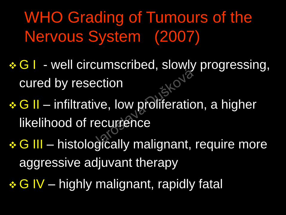
WHO Grading of Tumours of the
Nervous System (2007)
G I - well circumscribed, slowly progressing,
cured by resection
G II – infiltrative, low proliferation, a higher
likelihood of recurrence
G III – histologically malignant, require more
aggressive adjuvant therapy
G IV – highly malignant, rapidly fatal
Jaroslava Duško
vá

WHO - CNS - 2016
154 nosology units
(WHO 2007 134 nosology units)
Jaroslava Duško
vá

WHO Classification of Tumours of the Central Nervous System 2016 – selection 1/6
Gliomas
diffuse astrocytic and oligodendroglial – GII –IV
other astrocytic –e.g. pilocytic, subependymal GI-III
ependymal tumours GI-III
other gliomas -angiocentric, astroblastoma GI-III
tumours of the choroidal plexus –papilloma, atypical
papilloma, papillocarcinoma GI-III
(cont.)
Jaroslava Duško
vá
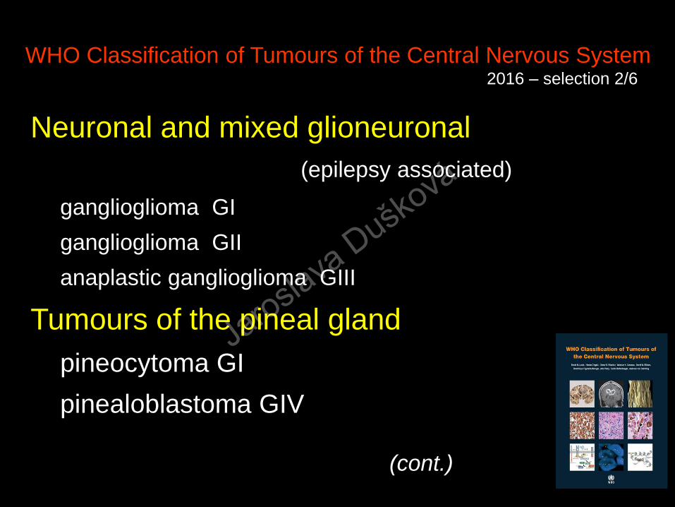
WHO Classification of Tumours of the Central Nervous System 2016 – selection 2/6
Neuronal and mixed glioneuronal
(epilepsy associated)
ganglioglioma GI
ganglioglioma GII
anaplastic ganglioglioma GIII
Tumours of the pineal gland
pineocytoma GI
pinealoblastoma GIV
(cont.)
Jaroslava Duško
vá

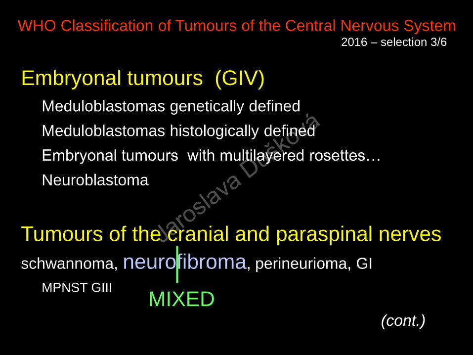
WHO Classification of Tumours of the Central Nervous System 2016 – selection 3/6
Embryonal tumours (GIV)
Meduloblastomas genetically defined (WNT, SHH)
Meduloblastomas histologically defined
Embryonal tumours with multilayered rosettes…
Neuroblastoma
Tumours of the cranial and paraspinal nerves schwannoma, neurofibroma, perineurioma, GI
MPNST GIII
(cont.)
Jaroslava Duško
vá

WHO Classification of Tumours of the Central Nervous System 2016 –selection 4/6
Meningeal tumours
meningioma GI, atypical GII, anaplastic GIII
Mesenchymal non-meningothelial tumours
Solitary fibrous tumour GI-III
Hemangioblastoma, hemangioma,
hemangioendothelioma, angiosarcoma, Kaposi sarcoma,
Ewing/PNET, lipoma, liposarcoma, leiomyoma,
rhabdomyoma, chondroma, osteoma, and sarcomas GI-III
(cont.)
Jaroslava Duško
vá
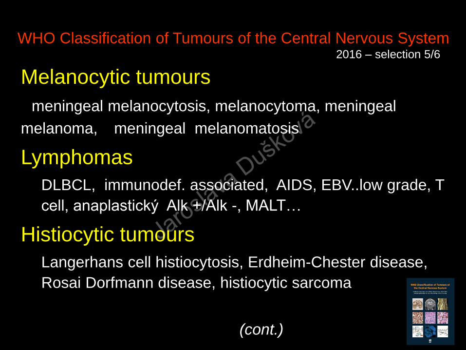
WHO Classification of Tumours of the Central Nervous System 2016 – selection 5/6
Melanocytic tumours
meningeal melanocytosis, melanocytoma, meningeal
melanoma, meningeal melanomatosis
Lymphomas
DLBCL, immunodef. associated, AIDS, EBV..low grade, T
cell, anaplastický Alk +/Alk -, MALT…
Histiocytic tumours
Langerhans cell histiocytosis, Erdheim-Chester disease,
Rosai Dorfmann disease, histiocytic sarcoma
(cont.)
Jaroslava Duško
vá

WHO Classification of Tumours of the Central Nervous System 2016 – selection 6/6
Germ cell tumours
germinoma
embryonal carcinoma
yolc sac tumour
choriocarcinoma
teratoma (mature, immature)
Tumours of the sellar region
craniopharyngeoma
pituicytoma
spindle cell oncocytoma
granular cell tumnour of the sellar region
METASTATIC TUMOURS
Jaroslava Duško
vá

Neuroectodermal Tumours -derived from:
ganglion cells gangliocytoma, neuroblastoma
glial & Schwann cells gliomas, neurilemmoma
glioblastoma, neurog. sarcoma,
mixed (ganglion and glial cells) ganglioglioma, ganglioneuroma
melanocytes pigmented nevimelanoma
CNS & PNS
located
Jaroslava Duško
vá

Gangliocytoma
Def.:
well differentiated slowly growing neuroepithelial tumour composed of neoplastic mature ganglion cells
Age /sex – no special predisposition (diagnosed mostly in childhood – young adults)
Incidence: RARE
Histogenesis: most probably highly differentiated remnants of embryonal neuroblasts
Jaroslava Duško
vá

Brigham RAD
Gangliocytoma
man 51
long history of
headaches
Jaroslava Duško
vá

Neuroectodermal Tumours -derived from:
ganglion cells gangliocytoma, neurocytoma, neuroblastoma
glial & Schwann cells gliomas, neurilemmoma
glioblastoma, neurog. sarcoma,
mixed (ganglion and glial cells) ganglioglioma, ganglioneuroma
melanocytes pigmented nevi
melanoma
CNS & PNS
located
Jaroslava Duško
vá
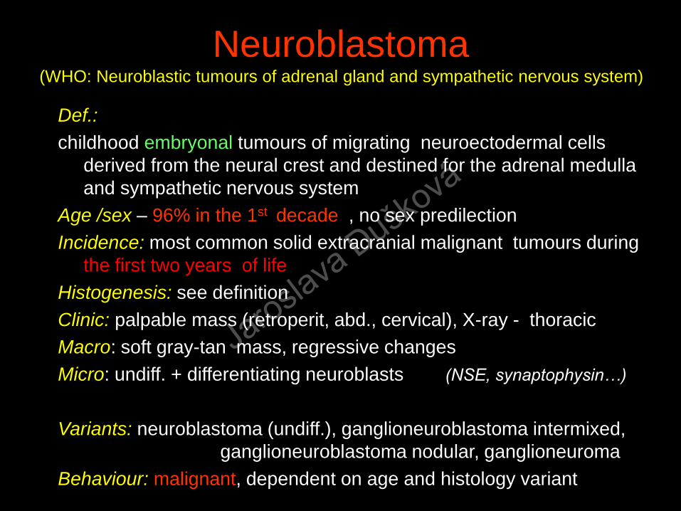
Neuroblastoma (WHO: Neuroblastic tumours of adrenal gland and sympathetic nervous system)
Def.:
childhood embryonal tumours of migrating neuroectodermal cells
derived from the neural crest and destined for the adrenal medulla
and sympathetic nervous system
Age /sex – 96% in the 1st decade , no sex predilection
Incidence: most common solid extracranial malignant tumours during
the first two years of life
Histogenesis: see definition
Clinic: palpable mass (retroperit, abd., cervical), X-ray - thoracic
Macro: soft gray-tan mass, regressive changes
Micro: undiff. + differentiating neuroblasts (NSE, synaptophysin…)
Variants: neuroblastoma (undiff.), ganglioneuroblastoma intermixed,
ganglioneuroblastoma nodular, ganglioneuroma
Behaviour: malignant, dependent on age and histology variant
Jaroslava Duško
vá

Neuroblastoma
Jaroslava Duško
vá

Jaroslava Duško
vá

Neuroectodermal Tumours -derived from:
ganglion cells gangliocytoma, neuroblastoma
glial & Schwann cells gliomas, neurilemmoma
glioblastoma, neurog. sarcoma,
mixed (ganglion and glial cells) ganglioglioma, ganglioneuroma
melanocytes pigmented nevimelanoma
CNS & PNS
located
Jaroslava Duško
vá

Ganglioneuroma
Jaroslava Duško
vá

Paraganglioma
Def.:
benign neuroendocrine neoplasm arising
in specialised neural crest cells
associated with autonomic ganglia
Macro: encapsulated , solid
Micro: uniform cells forming compact nests
(Zellballen)
Biology: benign
Jaroslava Duško
vá
2016
Jaroslava Duško
vá

Carotid
Body
Tumour
Chief cells :
chromogranin A+
Sustentacular
cells : GFAP +Jaroslava Duško
vá

Phaeochromocytoma
Def.:
tumour of chromaffin cells (mostly) benign
(intraadrenal paraganglioma)
Macro: whittish, solid, regressive changes
Micro: solid alveolar (Zellballen)
Behaviour: benign (15% bilateral, 10% in
children,10% malignant)PASS score – Phaeochromocytoma of the Adrenal Gland Scoring Scale
GAPP score – Grrading System for Adrenal Pheochromocytoma and
Paraganglioma
MEN II and von Hippel-Lindau disease component
Jaroslava Duško
vá

Phaeochromocytoma
B 3125/12– 640 g, PASS score 6
M 8700/1 D 441
Jaroslava Duško
vá

Pheochromocytoma
Jaroslava Duško
vá

WHO Classification of Tumours of the Central Nervous System 2016 – selection 3/6
Embryonal tumours (GIV)
Meduloblastomas genetically defined (WNT, SHH)
Meduloblastomas histologically defined
Embryonal tumours with multilayered rosettes…
Neuroblastoma
Tumours of the cranial and paraspinal nerves schwannoma, neurofibroma, perineurioma, GI
MPNST GIII
(cont.)
Jaroslava Duško
vá

Neurilemmoma (WHO Schwannoma)Def.:
a usually encapsulated benign tumour
composed of differentiated neoplastic Schwann cells
Age /sex – all ages, peak 4-6th decade,
no sex predilection
Incidence: common solid head, neck, extremities, INTRACRANIAL intramedullary
Histogenesis: see definition
Clinic: periph.- palpable asymptomatic mass, intraspinal – pain, intracranial – cerebellopontine lesion symptoms – hearing, tinnitus, facial paresthesias
Macro: white, soft – firm, encapsulated, (+nerve)
Micro: elongated Schwann cells , palisading, Verocay bodies
Variants: Antoni A, B, biphasic, cellular, pigmented, plexiform
Behaviour: slowly growing, benign, malignant transformation rare
Jaroslava Duško
vá

Neurilemmoma
Jaroslava Duško
vá

Neurilemmoma
Jaroslava Duško
vá

Neurilemmoma - cellular (A)
Jaroslava Duško
vá

Neurilemmoma (B)
Jaroslava Duško
vá

Neurilemmoma subcutaneum (actin negative)
Jaroslava Duško
vá

Neurilemmoma subcutaneum S100 positive
Jaroslava Duško
vá
WHO Classification of Tumours of the Central Nervous System 2016 – selection 3/6
Embryonal tumours (GIV)
Meduloblastomas genetically defined
Meduloblastomas histologically defined
Embryonal tumours with multilayered rosettes…
Neuroblastoma
Tumours of the cranial and paraspinal nerves
schwannoma, neurofibroma, perineurioma, GI
MPNST GIII
(cont.)
MIXED
Jaroslava Duško
vá

Jaroslava Duško
vá

Jaroslava Duško
vá

Neuroectodermal Tumours -derived from:
ganglion cells gangliocytoma, neuroblastoma
glial & Schwann cells gliomas, neurilemmoma
glioblastoma, neurog. sarcoma,
mixed (ganglion and glial cells) ganglioglioma, ganglioneuroma
melanocytes pigmented nevimelanoma
CNS & PNS
located
Jaroslava Duško
vá

WHO Classification of Tumours of the Central Nervous System 2016 – selection 5/6
Melanocytic tumours
meningeal melanocytosis, melanocytoma, meningeal
melanoma, meningeal melanomatosis
Lymphomas
DLBCL, immunodef. associated, AIDS, EBV..low grade, T
cell, anaplastický Alk +/Alk -, MALT…
Histiocytic tumours
Langerhans cell histiocytosis, Erdheim-Chester disease,
Rosai Dorfmann disease, histiocytic sarcoma
(cont.)
Jaroslava Duško
vá

WHO Classification of Tumours of the Central Nervous System 2016 – selection 5/6
Melanocytic tumours
meningeal melanocytosis, melanocytoma,
meningeal melanoma, meningeal
melanomatosis
- rare on the meninges or in the eye, common on the SKIN
WHO 2006,
new edition
planed for 2018
Jaroslava Duško
vá

Melanocytes – Melanophores- Melanophages
Jaroslava Duško
vá
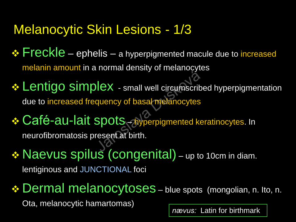
Melanocytic Skin Lesions - 1/3
Freckle – ephelis – a hyperpigmented macule due to increased
melanin amount in a normal density of melanocytes
Lentigo simplex - small well circumscribed hyperpigmentation
due to increased frequency of basal melanocytes
Café-au-lait spots – hyperpigmented keratinocytes. In
neurofibromatosis present at birth.
Naevus spilus (congenital) – up to 10cm in diam.
lentiginous and JUNCTIONAL foci
Dermal melanocytoses – blue spots (mongolian, n. Ito, n.
Ota, melanocytic hamartomas)nævus: Latin for birthmark
Jaroslava Duško
vá

Melanocytic Skin Lesions - 2/3
Acquired melanocytic nevi– junctional
– dermal
– compound
dysplastic
– deep dermal (blue) nevus
Congenital melanocytic nevus
Jaroslava Duško
vá

Naevus congenitalis
Jaroslava Duško
vá

Naevi naevocellulares (N. verrucosus)
Jaroslava Duško
vá

Naevus intradermalis naevocellularis
Jaroslava Duško
vá

Naevus intradermalis naevocellularis
Jaroslava Duško
vá

Naevus intradermalis naevocellularis
Jaroslava Duško
vá
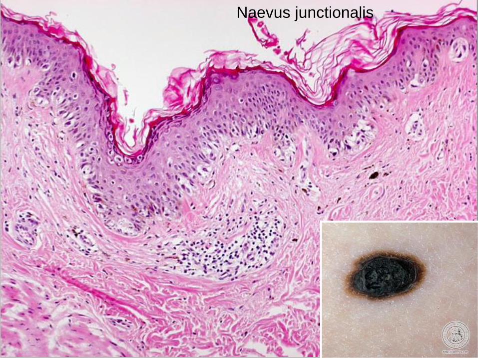
Naevus junctionalis
Jaroslava Duško
vá

Halo naevus
Jaroslava Duško
vá

Melanocytic Skin Lesions – 3/3
Malignant melanoma M 8720/3– Superficially spreading M 8743/3
– Nodular M 8721/3
– Lentigo maligna M 8742/2
– Acral-lentiginous M 8744/3
– Desmoplastic M 8745/3
– Melanoma arising from blue naevus M 8780/3
– Melanoma arising in a giant congenital naevus M 8761/3
– Melanoma of childhood
– Naevoid melanoma M 8720/3
– Persistent melanoma M 8720/3
Jaroslava Duško
vá

Malignant Melanoma
World incidence:
2000 133000
2008 197000 increase by 5% / year
Jaroslava Duško
vá

12th position
worldwide
Jaroslava Duško
vá

Melanoma
Jaroslava Duško
vá

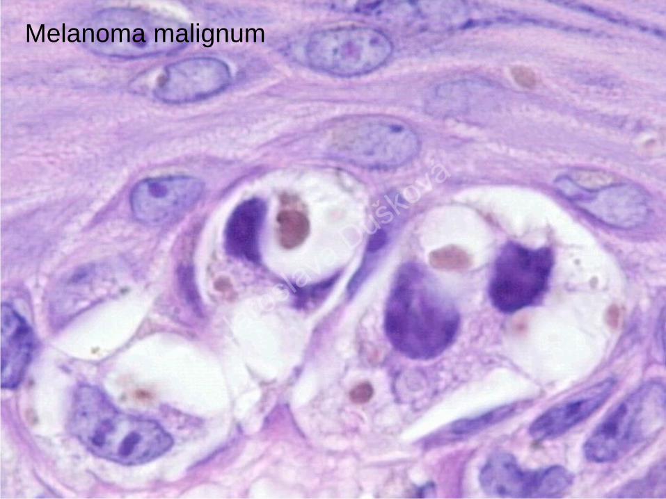
Melanoma malignum
Jaroslava Duško
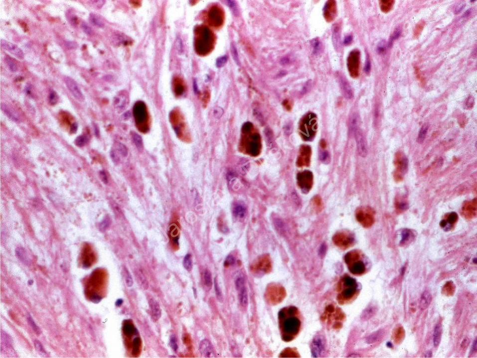
vá
Melanoma malignum
Jaroslava Duško
vá
Jaroslava Duško
vá

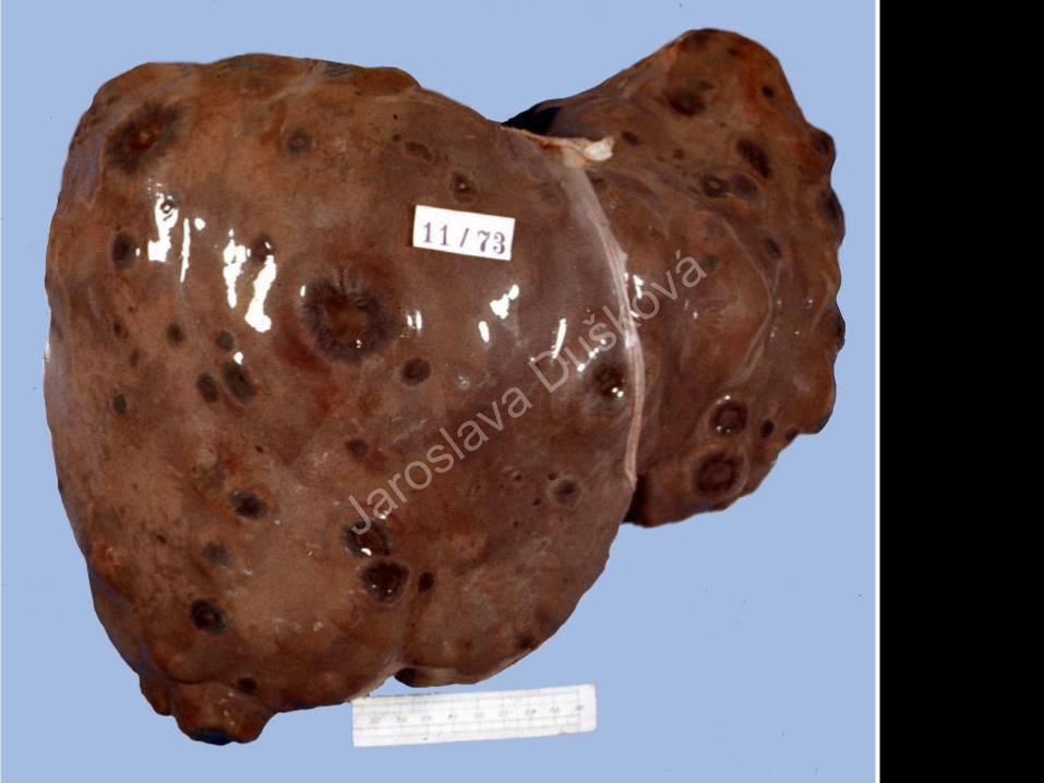
Metastases
melanomatis
maligni ad
cerebrum
47% of
patients
have
mutation
BRAF and
half of these
are stage IV
Jaroslava Duško
vá
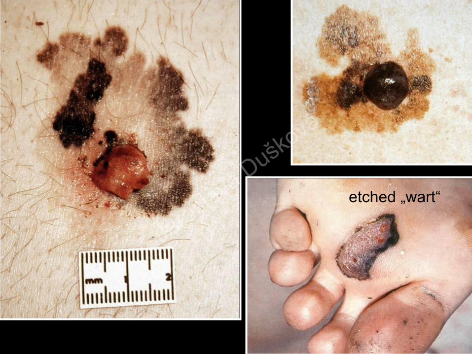
etched „wart“
Jaroslava Duško
vá

Melanoma malignum Mycosis ungualis
Hutchinson´s sign
Jaroslava Duško
vá

Melanoma malignum Haematoma subunguale
Jaroslava Duško
vá

Jaroslava Duško
vá
Jaroslava Duško
vá
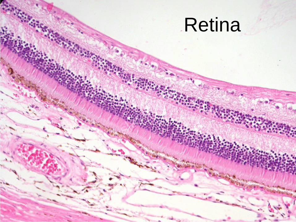
Retina
Jaroslava Duško
vá

Melanoma retinae
Jaroslava Duško
vá

Melanoma retinae
Jaroslava Duško
vá
Histogenetic Classification of tumors
continued…
mixed tumours
germ cell tumours
mesotheliomaJaroslava Duško
vá

Mixed TumoursDef.:
Tumours (benign or malignant)
composed of two or more
different cell lines that are
normally present in the place
of tumour origin
Jaroslava Duško
vá
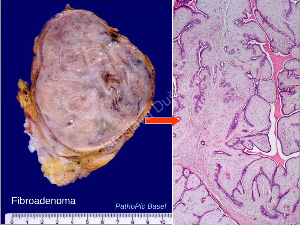
FibroadenomaPathoPic Basel
Jaroslava Duško
vá

Neurofibroma Def.:
a well demarcated intraneural or diffusely infiltrative extraneural tumour
consisting of a mixture of cell types including Schwann cells, perineurial-
like cells, and fibroblasts. Multiple in Nerofibromatosis 1.
Age /sex – all ages, no sex predilection
Incidence: common
Clinic: palpable asymptomatic mass, mostly cutaneous nodule (s) ass. with
café-au-lait spots
Macro: white, firm, circumscribed
Micro: elongated Schwann cells , fibroblasts, Wagner-Meissner-like tactile
corpuscles
Variants: atypical, cellular, plexiform, may be pigmented
Behaviour: slowly growing, benign, malignant transformation rare – mostly
in plexiform variants
Jaroslava Duško
vá

NeurofibromaJarosla
va Dušková

Histogenetic Classification of tumors
continued…
mixed tumours
germ cell tumours
mesotheliomaJaroslava Duško
vá
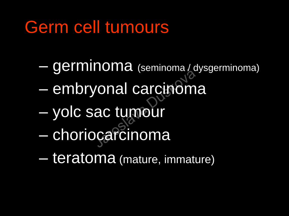
Germ cell tumours
– germinoma (seminoma / dysgerminoma)
– embryonal carcinoma
– yolc sac tumour
– choriocarcinoma
– teratoma (mature, immature)
Jaroslava Duško
vá

Seminoma testis
Jaroslava Duško
vá

Seminoma testis B 12 667 / 11
PLAP
38 yo
Jaroslava Duško
vá
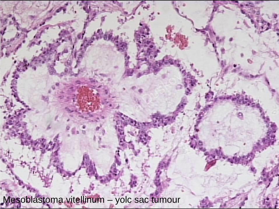
Mesoblastoma vitellinum – yolc sac tumour
Jaroslava Duško
vá
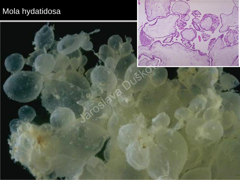
Mola hydatidosa
Jaroslava Duško
vá

Choriocarcinoma
Jaroslava Duško
vá

Teratomas
Def.:
Tumours (benign or malignant)
composed of two or more
different cell lines that are NOT
normally present in the place of
tumour origin
Jaroslava Duško
vá
Teratoma
coetaneous –
differentiated -cystic
embryonal –
nondifferentiated -
solid
Jaroslava Duško
vá

Teratoma maturum ovarii PathoPic Basel
Jaroslava Duško
vá

Cystis dermoides
Jaroslava Duško
vá

Teratoma malignum
Jaroslava Duško
vá

„Other“ Tumours
mixed tumours
germ cell tumours
mesotheliomaJaroslava Duško
vá

Mesothelioma
Def.: a neoplasm derived from the coelom
cavity epithelium – mesothelium
benign - circumscribed (papillary, adenomatoid)
malignant – circumscribed, diffuseJarosla
va Dušková

Mesothelioma
Jaroslava Duško
vá

Asbestosis
Jaroslava Duško
vá

Asbestosis
Jaroslava Duško
vá

Fibrous pleural plaque – marker of possible asbestosis!
Hyalinosis pleurae
Jaroslava Duško
vá

Hyalinosis pleuraeMesothelioma
Jaroslava Duško
vá
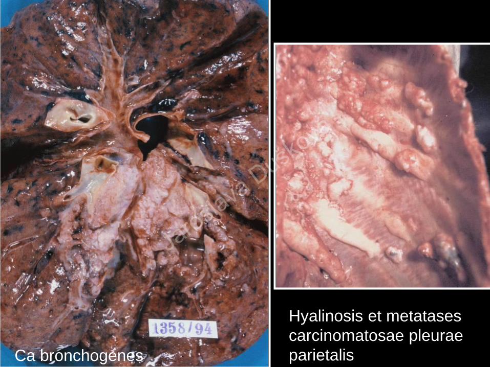
Ca bronchogenes
Hyalinosis et metatases
carcinomatosae pleurae
parietalis
Jaroslava Duško
vá

carcinoma
Mesothelioma
Mesothelioma
Jaroslava Duško
vá

Jaroslava Duško
vá