hormonal asynchrony and embryonic development… · hormonal asynchrony and embryonic...
TRANSCRIPT
HORMONAL ASYNCHRONY AND EMBRYONIC DEVELOPMENT(a)
R. R. Maurer(b) and S. E. Echternkamp (b)
U. S. Department of Agriculture (c) Roman L. Hruska Lt. S. Meat Animal Research Center
Clay Center, NE 68933
Abstract
Luteinizing hormone (LH), progesterone and estradiol profiles in peripheral blood serum were compared among parous and nonparous females with normal, abnormal or no e~ryonic development. Ho~nal profiles between parous and nonparous females of the same e~ryonic status did not differ and the data were combined. Estrous cycle length was longer (Pe.05) in parous (22.3k.4 days) than nonparous females (21.05.4 days). Females with normal developing embryos had a higher serum Progesterone concentration at Days 3 and 6 and a lower ratio of estradiol to progesterone than did females with abnormal embryonic development. Females with a normal embryo had higher (Pc.05) preovulatory LH peaks than females with abnormal development or no recovery of an oocyte or embryo (34.3k4.7, 11.826.8 and 13.3k2.5 nglml, respectively). The interval from onset of estrus to LX peak was 8.9*2.1, 13.7t3.7 and 13.5+ 6.2 hr for females with normal, abnormal or no recovery of an embryo. The lower LH peak, the longer interval from onset of estrus to LH peak, and lower progesterone concentration in peripheral blood serum in females with abnormal embryos or no recovery indicated that these females had a hormonal asynchrony. The hormonal asynchrony may produce an undesirable uterine environment for male and female gametes or embryos which resulted in fertilizatfon failure or embryonfc death. In the second experiment, more transferable embryos were obtained when superovulated females received prostaglandin Fna (PGFsa) intravenously rather than intramuscularly. A~inis~r~ng PGFsa intravenously rather than intramuscularly may have caused the demfse of the corpus luteum sooner and thereby produced a more normal uterine environment which allowed more embryos to develop normally.
(4 The authors are grateful to Betty Petitjean and Bfll McKay for technical assistance, to Gary Peterson, Walt Green, John Siebrandt, Gabe Johnson, Becky Gerlach, Doug Weich~n and Dave Jansen for the care, breeding and bleeding of cattle, and to Cheryl Yates for stenographic work. Cooperation of the Nebraska AgrScultural Experdment Station, University of Nebraska, Lincoln is acknowledged.
(b) Science and Education Administration, Agricultural Research.
(c) Mention of a trade name, proprietary product, or specific equipment does not constitute a guarantee or warranty by the U. S. Department of Agriculture and does not imply its approval to the exclusion of other products that may also be suitable.
JANUARY 1982VOL.17NO.l 11
THERIOGENOLOGY
Introduction
Infertility and embryonic mortality account for 30 to 40 percent reduction in reproductive efficiency in cattle. Maurer and Chenault (1) have shown in parous females that embryonic mortal jty (32%) accounted for 100 percent of the reduction in reproductive efficiency, whereas in nonpa~us females, infertility (17%) and e~ryonic mortality (17%) equally contributed to the losses that occurred by Day 18 of gestation.
Christenson et al. (2) reported that 20% of the beef heifers had unfertilized oocytes. Ovulation was reported to occur between 26 and 36 hr after the onset of estrus in 95% of the heifers. The remaining 5% ovulated after 36 hr. The interval from estrus to the LH peak was 2.8+ 0.8 hr. However, the interval from the onset of estrus to LH peak was 2.7 hr longer in females ovulating between 30 and 36 hr than in those ovulating before 30 hr. Also, the interval from the LH peak to ovulation was 1 hr longer in those females ovulating later. Swanson and Hafs (3) found in Holstein heifers that the LH peak occurred 3 hr befow onset of estrus, but varied among heifers from 8 hr before to 8 hr after the onset of estrus. The interval from estrus to ovulation ranged from 16 to 42 hr. Henricks et al. (4) reported the LH peak to occur between 3 to 6 hr after the onset of estrus in nonlactating Holstein cows. All of the above reports indicated a wide range in the intervals between onset of estrus and the LH peak, and between estrus and ovulation.
In superovulated females, embryos collected vary in develo~~tal stages within donor, indicating differences in ovulation time fertilization time and possible developmental difficulties (5f. The superovulatory treatment itself may adversely affect development of the embryos. Therefore, to achieve high pregnancy rates after embryo transfer, embryo quality must be high.
The objectives of this study were (1) to determine if differences in intervals between estrus and luteinizing hormone (LH) peak affect subsequent embryonic development in parous and nonparous females and (2) to dete~ine if a ~lationship exists between estradiol and progesterone concentrations in peripheral blood serum and embryo survival. In a second experiment the route of administration of prostaglandin Fad given to follicle stimulatinghormone (FSH) primed females and subsequent embryonic development was compared.
Materials and Methods
Twenty-four Limousin x Hereford and Limousin x Angus crossbred cows and 24 three-way crossbred heifers were divided equally into three groups of 16 (8 heifers and 8 cows . Each group was given intramuscularly 25 mg pros~glandin F2a (Lutalyse to synchronize iestrus and then observed for 1 estrous behavior. Each group of 16 animals iras either placed in individual stalls or small holding pens for two weeks, Eighteen days after the synchronized estrus, the 16 animals were placed in two adjacent pens with eight females per pen and observed continuously for estrous behavior. In the first group, onset of estrus was determined by homosexual behavior and the females were artificially inseminated with frozen semen from a high fertility bull 12 and 24 hr after the onset of estrus. In the second and third groups, onset of estrus was dete~ined
12 JANUARY 1982 VOL. 17 NO. 1
by actual mating with was mated once to two estrus or mating a 50 Thereafter, a jugular
THERIOGENOLOGY
Hereford x Red Poll crossbred bulls. Each female different bulls. Immediately after the onset of ml blood sample was drawn via venipuncture. cannula was placed in the females in groups one and __
two and the females were housed In individual stalls or small pens. Venipuncture was used to draw blood samples from the females in the third group and these females were housed in small pens. All females were fed a diet of 50% corn silage and 50% haylage ad libitun.
Catheters
Tygon micro bore tubing (1.27 mn inner diameter and 2.29 nvn outer diameter) was cut Tn 100 cm lengths and filled with a 7% tri- dodecylmethyl ammonium chloride heparin solution for at least two minutes. Each catheter was flushed and allowed to dry for several days. The catheters were sterilized in a Roccal-D solution and placed into the external jugular vein via a 11 gauge needle. The catheters were passed 20 to 30 cm into the vein and attached via silicone cement to a small piece of a larger bore tygon tubing which was sutured to the outer skin. The catheters were covered by tag cement and elastic adhesive tape. Each catheter was fitted with a female luer-lok which was stoppered with a solid plug. After drawing a blood sample, the catheters were filled with heparinized saline (20 units/ml) containing 1% benzyl alcohol. The plugs were placed in 70% ethyl alcohol to maintain sterility while the blood sample was collected.
Blood Collection
A 50 ml blood sample was collected imnediately after the onset of estrus, 2 and 4 hours later and every four hours thereafter until at least 40 hr after the onset of estrus. Thereafter, a 25 ml sample was collected every 12 hr until slaughter.
Embryo Recovery
The females were slaughtered at 8 to 10 or 13 to 16 days after the onset of estrus and reproductive tracts collected. The uterine horn ipsilateral to the corpus luteum was flushed with 30 ml physiological saline and the flushings searched for an embryo or oocyte. If none were found, the flushing procedure was repeated until an embryo or oocyte was found or until the tract had been flushed five times. At Days 8 to 10, fertilization rate and viability were based on the recove
r of a
blastocyst (Day 8 to 9) or hatched blastocyst (Day 9 to 10 with a well formed blastocoele and inner cell mass. At Days 13 to 16, viability was based on a round to oblong blastocyst with an embryonic disc. Blastocysts containing mostly dark, fragile, necrotic tissue were considered degenerate or degenerating.
Assay of Gonadotropins and Steroids
CH
Serum LH concentrations were determined by the double antibody radioinnnunoassay for bovine LH described by Niswender et al. (6) and modified by Echternkamp (7). The measurable range of the LH assay was
JANUARY 1982 VOL. 17 NO. 1 13
THERIOGENOLOGY
from 0.2 to 176 ng LH/ml of serum. Interassay coefficient of variation was 8.7%. Serum LH concentrations are expressed as ng of NIH-LH-B8/ml of peripheral serum.
Progesterone
Serum progesterone was extracted using 0.2 ml serum, 0.5 ml 5% NaCL, 0.05 ml 1N NaOH and 2.5 ml heptane. Separation of phases was facilitated by freezing and the solvent phase was decanted into 12 x 75 ml glass disposable culture tubes. Samples were dried individually under a stream of N2 in a heater block (40°C). This procedure was repeated twice. Extraction recovery was determined by incubatin Progesterone 1,2,6,7-3H(N); New England Nuclear B
*P4 (NET-381 with a serum pool and
sample recoveries ranged from 90 to 95%. Dried extracts were assayed for progesterone (Pk) by radioimmunoassay using a specific antiserum prepared against progesterone-lla-bovine serum albumin (Miles-Yeda Ltd., Israel). Both the antiserum and 3H-progesterone were dissolved in gel phosphate buffered saline at a concentration sufficient to bind 40 to 60% of the tritiated progesterone. Separation of free and antibody bound progesterone was accomplished using 0.1 ml dextran charcoal (62.5 mg dextran T-70 and 625 mg Norit A charcoal in 50 ml gel phosphate buffered saline) and refrfgerated at 5°C for 15 min. After centrifugation at 800 x g (4°C) for 5 min, 0.5 ml of the supernatant was added to 5 ml of scintillation fluid for subsequent counting. The lower limit of sensitivity of the assay for progesterone was 10 pg per tube. The intra- and interassay coefficient of variation was 5.4 and 6.7X, respectively.
Estradiol
Estrogens were extracted with 7 ml benzene from 3 to 4 ml serum to which 1500 CPM of *E2 (NET-517 Estradiol, [2,4,6,17,16,17-3H(N)I; New England Nuclear) was added to adjust for recovery losses. The serum was extracted twice and each extract dried under nitrogen in a block heater (40°C). To the dried extract 500 ~1 of gel phosphate buffered saline was added. To account for recovery of estrogen extraction, 100 ~1 of the gel phosphate buffered saline was added to 5 ml scintillation fluid and counted. The remaining extracted sample was assayed for estradiol-17B (E2) by radioimnunoassay (8) using a specific antisera provided by Dr. Norman Mason of Eli Lilly Company. Cross-reactivity of the antiserum to various steroids has been reported by Kesler et al. (9). Both the antiserum and 3H-estradiol were dissolved in gel phosphate buffered saline at a concentration sufficient to bind 40 to 60% of the tritiated estradiol. Separation of free and antibody bound estradiol was accomplished using 0.1 ml dextran charcoal (same as for progesterone) and refrigerated at 5°C for 15 min. After centrifugation at 800 x g (4°C) for 5 min, 0.5 ml of the supernatant was added to 5 ml of scintillation fluid for subsequent counting. The samples were corrected for extraction losses. The sensitivity of this assay for 4 ml of serum was 2 pg. The intra- and interassay coefficient of variation was 3.5 and 9.9%, respectively.
In a second experiment 31 Red Poll and 32 Angus females 3 years of age were superovulated with follicle stimulating hormone (FSH). The first FSH injection was given intramuscularly on Days 10 to 12 of the estrous cycle and subsequent injections of FSH and prostaglandin Fsa were
JANUARY 1982 VOL. 17 NO. 1
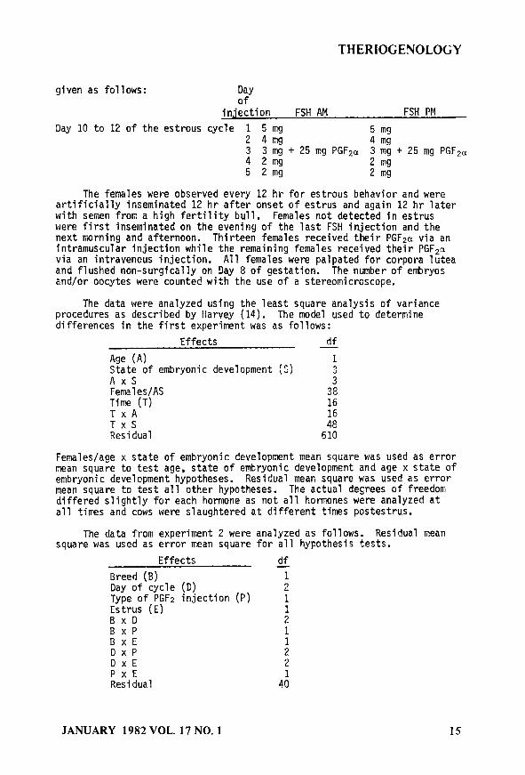
THERIOGENOLOGY
given as follows: Day of
injection FSH AM FSH PM
Day 10 to 12 of the estrous cycle 1 5 mg 5 mg 2 4mg 4 mg 3 3 mg + 25 mg PGF~c~ 3 mg + 25 mg PGFpu 4 2mg 2 mg 5 2mg 2 mg
The females were observed every 12 hr for estrous behavior and were artificially inseminated 12 hr after onset of estrus and again 12 hr later with semen from a high fertility bull, Females not detected in estrus were first inseminated on the evening of the last FSH injection and the next morning and afternoon. Thirteen females received their PGF2a via an intramuscular injection while the remaining females received their PGFza via an intravenous injection. All females were palpated for corpora lutea and flushed non-surgically on Day 8 of gestation. The number of etiryos and/or oocytes were counted with the use of a stereomicroscope.
The data were analyzed using the least square analysis of variance procedures as described by Harvey (14). The model used to determine differences in the first experiment was as follows:
Effects df -
Age (A) 1 State of embryonic development (S) 3 AxS Females/AS 3: Time (T) 16 TxA TxS :8" Residual 610
Females/age x state of embryonic development mean square was used as error mean square to test age, state of embryonic development and age x state of embryonic development hypotheses. Residual mean square was used as error mean square to test all other hypotheses. The actual degrees of freedom differed slightly for each hormone as not all hormones were analyzed at all times and cows were slaughtered at different times postestrus.
The data from experiment 2 were analyzed as follows. Residual mean square was used as error mean square for all hypothesis tests.
Effects af Breed (B) 1 Day of cycle (0) 2 Type of PGF2 injection (P) Estrus (E) : BxD 2 BxP 1 BxE DxP : DxE 2 PxE Residual 4:
JANUARY 1982 VOL. 17 NO. 1 15
THERIOCENOLOGY
Results and Discussion
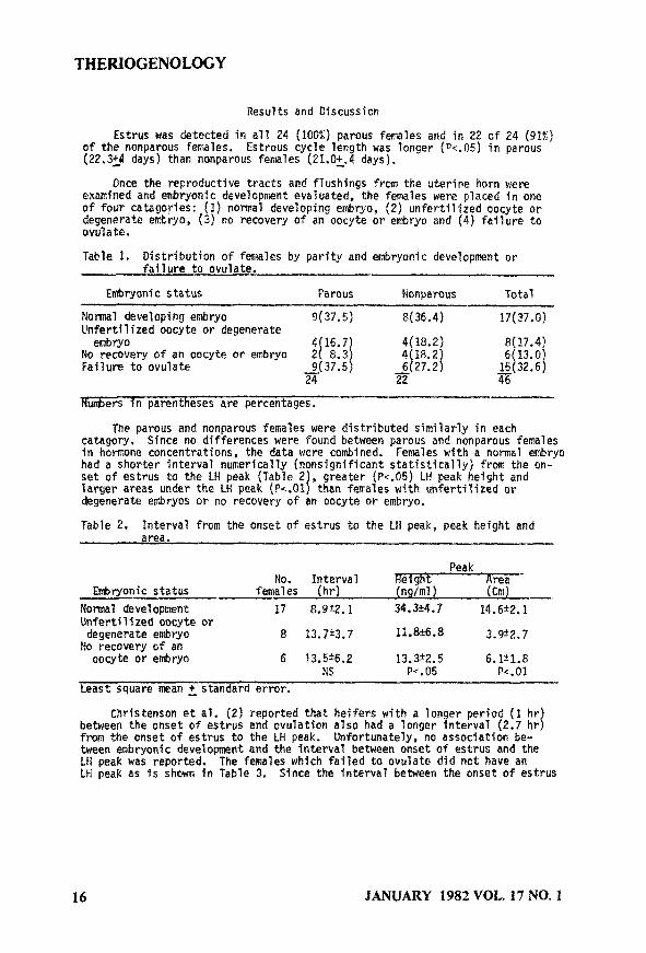
Estrus was detected in all 24 (100%) parous females and in 22 of 24 (91%) of the nonparous females. Estrous cycle length was longer (Pc.05) in parous (22.324 days) than nonparous females (21.054. days).
Once the reproductive tracts and flushings from the uterine horn were examined and embryonic development evaluated, the females were ptaced in one of four catagories: (1) normal developing embryo, (2) unfertilized oocyte or degenerate embryo, (3) no recovery of an oocyte or embryo and (4) failure to ovulate.
Table 1. Distribution of females by parity and embryonic development or failure to ovulate. --
Embryonic status Parous Nonparous Total
Normal developing embryo 9f37.5) Pf36.4) !7(37.0) Unfertilized oocyte or degenerate embryo 4(18.2) 8l17.4)
No recovery of an oocyte or embryo 4(18*2) Failure to ovulate 6t27.2)
-B
Tluabers In parentheses are percentages.
The parous and nonparous females were distributed similarly in each catagory. Since no differences were found between parous and nonparous females in hormone concentrations, the data were combined. Females with a normal embryo had a shorter interval numerically (nonsignificant statistically) from the on- set of estrus to the LH peak (Table 2 , greater (Pc.05) Lti peak height and larger areas under the LH peak (Pc.01 t than fetrales with unfertilized or degenerate embryos or no recovery of an oocyte or embryo.
Table 2. Interval from the onset of estrus to the LH peak, peak height and area.
Peak NO. Interval w
Embryonic status females (hr) (n9~ml) (Cm)
Norma? development 17 8.922.1 34.?*4,? 14.6t2.1 ’ Unfertil$zed oocyte or degenerate embryo 8 13.7t3.7 X.8*6*8 3.9t2.7
No recovery of an oocyte or embryo 6 13.5t6.2 13.3t2.5 6.1f1.8
NS Pe.05 PC.01
Least square mean + standard error.
Christenson et al. (2) reported that heifers with a longer perSod (1 hr between the onset of estrus and ovulation also had a longer interval (2.7 hr 1 from the onset of estrus to the LH peak. Unfortunately, no association be- tween embryonic develo~nt and the interval bet&'een onset of estrus and the I_11 peak was reported. The females which failed to ovulate did not have an LH peak as is shown in Table 3. Since the interval between the onset of estrus
16 JANUARY 1982 VOL. 17 NO. 1
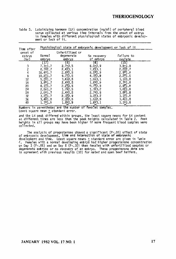
THERIOGENOLOGY
Table 3. Luteinjzing hormone (LH) concentration (ng/ml) of peripheral blood serum collected at various time intervals from the onset of estrus in females with different physiological states of embryonic develop- ment or lack of it.
Time after Physiological state of embryonic development or lack of it
onset of Unfertilized or estrus Normal degenerate Ho recovery Failure to (hr) embryo embryp of embryo ovulate
(17) (8) (6) (15) cl 7.3t1.7 2.7t2.5 6.Ot2.9 3.9t1.8 2 8.m.7 7.273.1 2.22.7
: 16.4Tl.7
2.423.1
10.2T1.7 zz: 8.5r2.9 2.9Tl.8
12 5.2Tl.7 3:6T2:5 4.3T2.9 2.2Tl.8 1.9T3.1 3.1Tl.8
16 3.8T1.7 2.472.5 1.672.9 2.Fl.8 20 8.1T1.7 2.2T2.5 0.G2.9 2.971.8 24 2.DT1.7 1.F2.5 1.3T3.2 3.OTl.8 28 2.071.7 2.472.5 2.7T2.9 3.871.8
:: 3.2T1.7 2.32.9 3.OT3.2 3.Gl.9 3.4T1.8 2.3T2.5 1.5T2.9 3.4Tl.8
40 l.El.9 1.8z2.9 1.673.1 3.1z1.8
Ntiers in parentheses are the number of females samples. Least square mean + standard error.
and the LH peak differed within groups, the least square means for Ltl content at different times are less than the peak heights calculated in Table 2. Peak heights in all groups may have been higher if more frequent blood samples were collected.
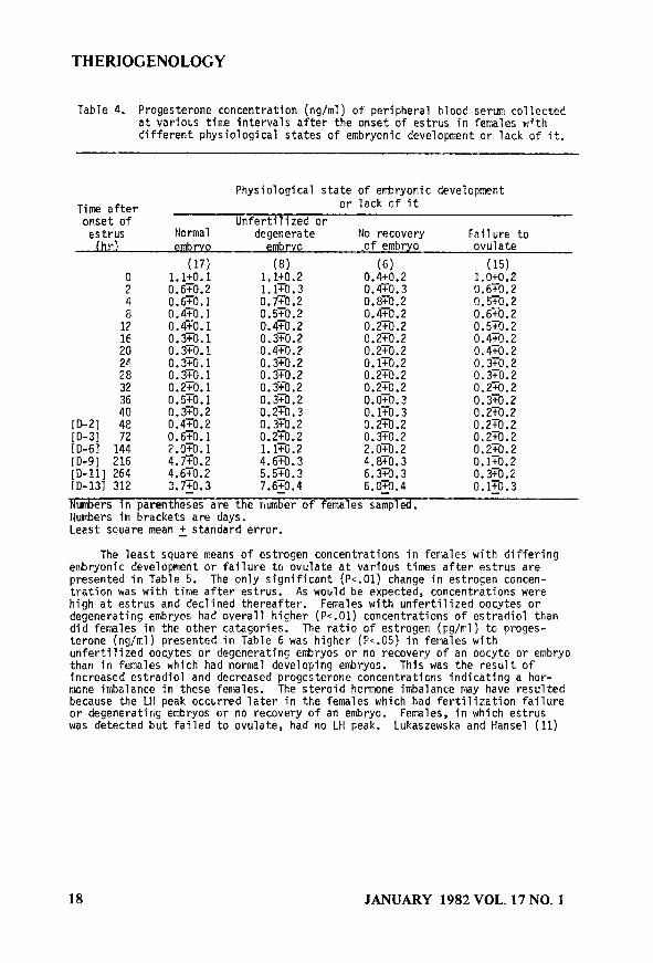
The analysis of progesterone showed a significant (Pc.05) effect of state of embryonic development, time and interaction of state of embryonic development and time. Least square means + standard error are given in Table 4. Females with a normal developing embryo had higher progesterone concentration on Day 3 (Pc.05) and on Day 6 (Pc.10) than females with unfertilized oocytes or degenerate embryos or no recovery of an embryo. These progesterone data are in agreement with previous results (10) for mated and open beef heifers.
JANUARY 1982 VOL. 17 NO. 1 17
THERIOGENOLOGY
Table 4. Progesterone concentration (ng/ml) of peripheral blood serum collected at various time intervals after the onset of estrus in females with different physiological states of embryonic development or lack of it.
Time after onset of estrvs Ihr,
Physiological state of embryonic development or lack of it
Unfertilized or Normal degenerate No recovery Failure to embrvo embrvo .of embryo ovulate
(17) (8) 0 1.1+0.1 1.1to.2 : 0.6TO.2 1.170.3
0.6TO.l o.flo.2 8 0.4TO.l 0.5FO.2
:: D.4TO. 0.370.1 1 0.470.2 0.3TO.2 20 0.3TO.l 0.470.2 24 0.3TG. 1 0.3TO.2 28 0.370.1 0.3TO.2 32 0.2To. 1 0.3yO.2 43: 0.5TO.l 0.3TO.2
0.3TO.2 0.2TO.3 ID-21 4e 0.4TO.2 0.3TO.2 [D-3] 72 0.6’cO.l 0.2TO.2 CD-61 144 2.OTO.l l.lTO.2 ID-91 216 4.flO.2 4.6TO.3 [D-11] 264 4.6TO.2 5.570.3 ID-131 312 3.7zo.3 7.6zO.4
(6) 0.4to.2 0.470.3 0.870.2 0.470.2 0.270.2 0.2TO.2 0.2TO.2 O.lTO.2 0.2TO.2 0.2To.2 0.070.3 0.170.3 0.2To.2 0.3TO.2 2.oEo.2
%E 6&0:4
(15) 1.oto.2 0.6TO.2 0.570.2 0.6TO.2 0.5TO.2 0.470.2 0.4TO.2 0.370.2 0.3TO.2 0.270.2 0.3rO.2 0.2TO.2 0.2TO.2 012TOI2 0.2TO.2 0.170.2 0.3TO.2 O.lTO.3 -
Numbers in parentheses are the number of females sampled. liurrbers in brackets are days. Least square mean + standard error.
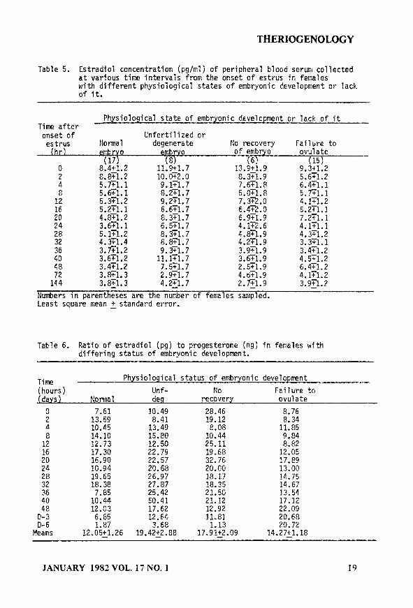
The least square means of estrogen concentrations in females with differing embryonic development or failure to ovulate at various times after estrus are presented in Table 5. The only significant (Pc.01) change in estrogen concen- tration has with time after estrus. As would be expected, concentrations were high at estrus and declined thereafter. Females with unfertilized oocytes or degenerating embryos had overall higher (Pc.01) concentrations of estradiol than did females in the other catagories. The ratio of estrogen (pg/ml) to proges- terone (ng/ml) presented in Table 6 was higher (Pc.05) in females with unfertilized oocytes or degenerating embryos or no recovery of an oocyte or embryo than in females which had normal developing errbryos. This was the result of increased estradiol and decreased progesterone concentrations indicating a hor- mone imbalance in these females. The steroid hormone imbalance may have resulted because the LH peak occurred later in the females which had fertilization failure or degeneratirig embryos or no recovery of an embryo. Females, in which estrus was detected but failed to ovulate, had no LH peak. Lukaszewska and Hansel (11)
18 JANUARY 1982 VOL. 17 NO. 1
THERIOGENOLOGY
Table 5. Lstradiol concentratjon ~pg/ml) of peripheral blood serum collected at various time intervals from the onset of estrus in females with different physiological states of embryonic development or lack of it.
Physiological state of embryonic development or lack of it Time after onset of Unfertilized or estrus Normal degenerate No recovery Failure to (hrl embrv
0 8%!O2
embrvo of embryo ovulate
8:871:2 5.7Tl.l 5.tZl.l 5.3+1.2 5.271.1 4.8Fl.2 3.671.1 5.1T1.2 4.3Tl.4 3.7Tl.2 3.671.2 3.471.2 3.8Tl.3
2
12 16
$04 28 32 36 40 48 72
11% 7 10:&2:0 9.1Tl.7 8.271.7 9.2T1.7 6.6Fl.7 8.3T1.7 6.5151.7 8.3Tl.7 8.8Tl.7 9.371.7 ll.Gl.7 7.5Tl.7 2.9T1.7
17 $Z 9 C3K9 7.671.X 5.oR.a 7.3T2.0 6.4F2.0 6.9Fl.9 4.172.6 4.8X9 4.271.9 3.971.9 3.6Tl.9 2.5T1.9 4.671.9
(15) - 9.3+1.2 5.651.2
%X 4x2 5.2Tl.l 7.2Tl.l 4.1Tl.l 4..371.2 3.3Tl.l 3.4rl.2 4.571.2 6.4Tl.2 4.Gl.2
144 3.8x1.3 4.2Tl.7 2.Gl.9
Numbers in parentheses are the number of feir&les sampled. Least square mean + standard error.
3.971.2 -
Table 6. Ratio of estradiol (pgf to progesterone (ng) in females with differing status of embryonfc development.
Time Physiological status of embryonic development
(hours) Unf- No Failure to jdavs 1 Mornlal des recovery ovulate -
: 13.59 7.61 10.49 a.41 28.46 19.12 8.76 8.34 4 10.45 13.49 a.08 Il.85
1: 14.10 12.73 15.8D 12.50 25.11 10.44 9.84 8.82
:8 24
:2" 36 40 48 c-3 D-6
Means
17.30 22.79 19.68 12.05 16.90 22.57 32.76 17.a9 10.94 20.68 2o.eo 13.00 19.65 26.97 18.17 14.75 18.38 27.87 38.35 14.67 7.85 25.42 21.50 13.54 10.44 50.41 21.12 17.12 12.03 17.62 12.92 22.09 6.a5 12.64 11.81 20.68 1.87 3.68 1.13 20.72
12.05+_1.26 19.4222.88 17.91+2.09 14.27~1.18
JANUARY 1982 VOL. 17 NO. 1 19
THERIOGENOLOGY
measured estradiol and progesterone in pregnant and nonpregnant heifers. They found higher progesterone concentration in plasma of pregnant females between Days 10 and 18 and higher estrogen concentration in plasma of pregnant females between Days 6 and 16 than nonpregnant females. Their progesterone values increased later than what was observed in the present study, however, their females were not observed continuously for estrus and their initial observation could have varied as much as 12 hours later than the present study. Erb et al. (12) reported higher progesterone concentrations in pregnant females than nonpregnant females. They concluded that delayed estrus and delayed preovulatory increase in LH after progesterone had decreased to less than .75 ng/ml were the main cause of subnormal progesterone concentrations. The results of the present study are in agreement with their findings.
In the females which failed to ovulate, the progesterone concentration was above 1 ng/ml only at estrus but declined to 0.5 ng/ml or less for the rest of the sampling period. Estradiol concentrations were above 5 pg/ml for 24 hours after the onset of estrus and declined to less than 5 pg/ml thereafter. Therefore, the estradiol and progesterone concentrations do not appear to be abnormally high or low. Handling may have caused these females to become stressed since these females had been maintained on the range. Moberg and Stoebel (13) reported that in Holstein heifers given cortisol, three of four females failed to have a LH peak and follicular development was arrested.
The results of administering PGFza either intramuscularly or intravenously are given in Table 7. In animals given PGFza IV, 70% were detected in estrus, whereas, only 46% of the females receiving their PGF2a IM were in estrus (X2 = 2.58, ldf, P-.12). No embryos or oocytes were recovered in 8 females given PGF 2a IV while all 13 females receiving PGF2a IW had embryos (X2 = 2.38, ldf, P-.14). The route of administration of PGF2a did not have a significant effect on ovulation rate in the 55 females which had embryos or oocytes (P-.24) but did increase numerically the number of oocytes and embryos recovered (P-.13), as well as, the percentage of recovered morula and blastocysts (P-.13). However, the among animal variability in response to the superovulatory procedure still remained high. With the intravenous injection of PGFza, we obtained on the average 2 more transferable embryos than with IM injections. The embryo quality appeared to be better as evidenced by the increased number of oocytes or embryos (1.4) while the number of transferable embryos increased by 2.1 in females receiving PGF2, intravenously.
Giving the PGFz IV may have produced a large surge in prostaglandin to the corpus luteum and, thereby, hastened its demise. This could have produced a more normal endocrine balance and uterine environment, thereby allowing more embryos to develop normally.
20 JANUARY 1982 VOL. 17 NO. 1
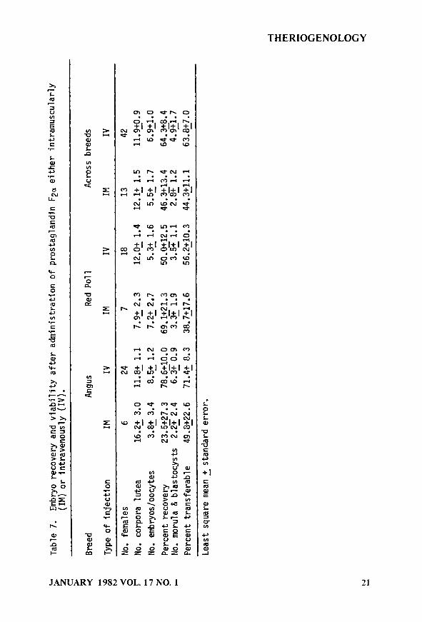
2 Ta
ble
7. Em
bryo
rec
over
y an
d vi
abil
ity
afte
r ad
mini
stra
tion
of
pr
osta
glan
din
F2(x
eith
er in
tram
uscu
larl
y (I
M)
or in
trav
enou
sly
(IV
).
z %
* Br
eed
Angu
s Re
d Po
ll
Acro
ss b
reed
s L E
Type
of
inje
ctio
n IM
IV
IM
IV
IM
IV
3
No.
fem
ales
6
24
7 18
13
42
No.
corp
ora lu
tea
16.2
2 3.
0 11
.85
1.1
7.9
2 2
.3
12.0
2 1
.4
12.1
2 1
.5
11.9
fO.9
Y
l N
o. emb
ryos
jooc
ytes
3.
82 3
.4
8.5+
1.
2 7.
222‘
7 5.
3~ 1.
6 5.
52 1.
7 6.
921.
0
3 Pe
rcen
t re
cove
ry
23.5
+27.3
7;.
$1;.
; 69.1
221.3
50.0
+12.5
46.3
+13.4
64.3
+8.4
L
No.
mor
ula ?I
blas
tocy
sts
2.2
E2.4
.
3.3
+ 1.9
3.5
z 1.1
2.8
z 1.2
4.9
51.7
Perc
ent tr
ansf
erab
le
49.8
z22.
6 71
:4;
8.3
38.7
zl7.
6 56
.221
0.3
44.3
211.
1 63
.827
.0
Leas
t sq
uare
mea
n f
stan
dard
err
or.
THERIOGENOLOGY
REFERENCES
1.
2.
3.
4.
5.
6.
7.
a.
9.
10.
11.
12.
13.
14.
Maurer, R. R., and Chenault, J.R. Fertilization failure and embryonic mortality in parous and nonparous beef cattle. J. Animal Sci. (Submitted).
Christenson, R.K., Echternkamp, S.E., and Laster, D.B. Oestrus, LH, ovulation and fertility in beef heifers. J. Renrod. Fert. 43:543-546 -
Swanson, L.V., and Hafs, H.D. LH and prolactin in blood serum from estrus to ovulation in Holstein heifers. J. P.nimal Sci. 2:1038-1041 (1971).
Henricks, D.M., and Dickey, J.F. Serum luteinizing hormone and plasma progesterone levels during the estrous cycle and early pregnancy in cows. Biology of Reproduction 2:346-351 (1970).
Shea, B.F. Evaluating the bovine embryo. Theriogenology E:31-42 (1981).
Niswender, G.D., Reichert, L.E., Jr., Midgley, A.R., Jr., and Nalbandov, A.V. Radioimmunoassay for bovine and ovine luteinizing hormone. Endocrinology gl166 (1969).
Echternkamp, S.E. Stimulation of estrogen and luteinizin hormone secretion in postpartum beef cows. J. Animal Sci. =521-531 (1978 . 3
Berardinelli, J.G., Anderson, L.L., Ford, J.J., and Christenson, R.K. Luteinizing hormone secretion in gilts after hypophysial stalk transection and estradiol-176. Am. J. Physiol. (Submitted)
Kesler, D.J., Garverick, H.A., Youngquist, R.S., Elmore, R.G., and Biers&al, C.J. Effect of days postpartum and endogenous reproductive hormones on GnRH-induced LH release in dairy cows. J. Animal Sci. g:797-803 (1977).
Henricks. D.M.. Lamond, D.R., Hill, J.R.. and Dickey. J.R. Plasma proqesterone concentrations-before mating-and in early pregnancy-in the beef heiier; J. Animal Sci. 33:450-454 (1971).
Lukaszewska, J., and Hansel, W. Corpus luteum maintenance during early pregnancy in the cow. J. Reprod. Fert. 59:485-493 (1980). -
Erb, R.E., Garverick, H.A., Randel, R.D., Brown, B.L., and Callahan, C.J. Profiles of reproductive hormones associated with fertile and nonfertile inseminations of dairy cows. Thericgenology 2:227-242 (1976).
Moberg, G.P., and Stoebel, D.P. The effect of cortisol on ovulation in the dairy cow. 9th International Congress on Animal Reproduction and AI, Madrid, Spain, Vol. III, p. 103 (1980).
Harvey, W.R. Least-squares analysis of data with unequal subclass numbers. USDA, ARS H-4 (1975).
22 JANUARY 1982 VOL. 17 NO. 1