human anatomy & physiology, sixth edition 5 integumentary system
TRANSCRIPT

Human Anatomy & Physiology, Sixth Edition
5Integumentary System

Functions of the Integumentary System
Chemical & physical barrier Body temperature regulation Cutaneous sensation Synthesis of vitamin D

Skin (Integument)
Figure 5.1

Layers & Cells of the Epidermis:
stratum spinosum
stratum granulosum
stratum corneum
cell membranes filled with cross-linked keratin
dying cells filled with keratin & lipid granules
keratinocytes actively producing keratin filaments
stratum basale
mitotically dividing cells
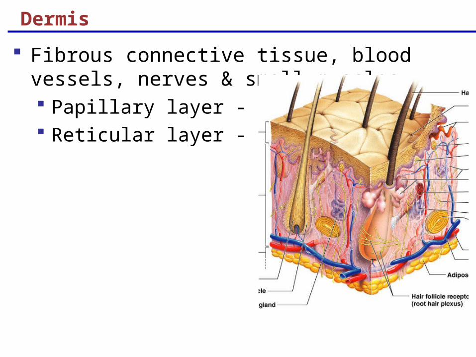
Dermis
Fibrous connective tissue, blood vessels, nerves & small muscles Papillary layer - areolar Reticular layer - reticular

Hypodermis
Subcutaneous layer not technically integument Composed of adipose and areolar connective tissue Thermal insulation & shock absorption

Skin Color
Pigments Melanin – reddish-brown Carotene – yellowish-orange
Hemoglobin

Sweat Glands & Modified Sweat Glands
Eccrine – all over body, empty to pore Apocrine –axillary & anogenital areas, empty to hair
follicle Ceruminous glands – secrete cerumen (ear wax) Mammary glands – secrete milk

Sebaceous Glands
Simple alveolar glands distributed all over body Oily secretion called sebum Secreted to pore or hair follicle Prevents desiccation & waterproofs skin

Keratinized Processes
Stratum corneum Nail Hair Keratinization
soft & hard keratin degree of crosslinking of keratin proteins
Keratin is a long filamentous protein Transglutaminase joins glutamic acid residues on
neighboring keratin proteins creating a keratin aggregate ~ 15 distinct keratin proteins

Keratin Diseases
Epidermolysis bullosa Mutations in keratin proteins such that they can not
be properly crosslinked

Structure of a Nail
Scalelike modification of the epidermis on the distal, dorsal surface of fingers and toes
Figure 5.4

Hair
Bundles of keratinized cells produced in follicles Follicles are epithelial cells deeply invaginated into
the dermal layer Pigmented by melanocytes Sensory nerve endings wraps each follicle Connected to papilary dermis by tiny arrector
muscle

Hair Follicle – Sagittal & Cross Sections
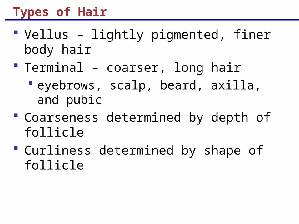
Types of Hair
Vellus – lightly pigmented, finer body hair Terminal – coarser, long hair
eyebrows, scalp, beard, axilla, and pubic Coarseness determined by depth of follicle Curliness determined by shape of follicle

Basal Cell Carcinoma
Most common skin cancer Stratum basale cells proliferate and invade the
dermis and hypodermis Slow growing and non-metastatic Surgical excision effective in
99% of cases

Squamous Cell Carcinoma
Keratinocytes of stratum spinosum Most frequent on scalp, ears, and lower lip Rapid growing and metastatic Early surgical removal followed by irradiation Poor prognosis if metastasized

Melanoma
Cancer of melanocytes Most dangerous type of skin cancer
Highly metastatic Resistant to chemotherapy
Treated by wide surgical excision & immunotherapy Chance of survival is poor if the lesion is over 4 mm
thick

Melanoma
Melanomas are characterized by the ABCD rule A: Asymmetric: halves of the pigmented unequal B: Border: irregular with indentations C: Color: black, brown, tan, sometimes red or blue D: Diameter: larger than 6 mm

Burns
1st -degree – only the epidermis
localized redness, swelling, and pain
2nd -degree – epidermis and upper regions of dermis
Like first degree burns with blistering
3rd -degree – entire thickness of the skin is damaged
Burned area appears gray-white, cherry red, or black; no initial edema or pain

Rule of Nines
Estimates the severity of burns
Burns considered critical if: 2nd degree > 25% of body 3rd degree >10% of body 3rd degree on face, hands, feet

Developmental Aspects of the Integument: Fetal Epidermis develops from ectoderm Dermis and hypodermis develop from mesoderm Lanugo – downy coat of delicate hairs covering the
fetus Vernix caseosa – substance produced by sebaceous
glands that protects the skin of the fetus in the amnion

Developmental Aspects of the Integument: Adolescent to Adult
Skin and hair become oilier and acne may appear Skin shows the effects of cumulative environmental
assaults around age 30 Scaling and dermatitis become more common

Epidermal replacement of cells slows and skin becomes thinner
Skin becomes dry and itchy Subcutaneous fat layer diminishes Decreased elasticity and loss of subcutaneous tissue
leads to wrinkles Decreased numbers of melanocytes and Langerhans’
cells increase skin cancer risk
Developmental Aspects of the Integument: Old Age