human cognition during rem sleep and the activity profile ... · the topography of the quiescent...
TRANSCRIPT

S. Laureys (Ed.)
Progress in Brain Research, Vol. 150
ISSN 0079-6123
Copyright r 2005 Elsevier B.V. All rights reserved
CHAPTER 16
Human cognition during REM sleep and the activityprofile within frontal and parietal cortices: areappraisal of functional neuroimaging data
Pierre Maquet1,�, Perrine Ruby1, Audrey Maudoux1, Genevieve Albouy1,
Virginie Sterpenich1, Thanh Dang-Vu1, Martin Desseilles1, Melanie Boly1,
Fabien Perrin2, Philippe Peigneux1 and Steven Laureys1
1Cyclotron Research Centre, University of Lieege-Sart Tilman, 4000 Liege, Belgium2Neurosciences and Systemes Sensoriels (UMR 5020), Universite Claude Bernard Lyon I, 69007 Lyon, France
Abstract: In this chapter, we aimed at further characterizing the functional neuroanatomy of the humanrapid eye movement (REM) sleep at the population level. We carried out a meta-analysis of a large datasetof positron emission tomography (PET) scans acquired during wakefulness, slow wave sleep and REMsleep, and focused especially on the brain areas in which the activity diminishes during REM sleep. Resultsshow that quiescent regions are confined to the inferior and middle frontal cortex and to the inferiorparietal lobule. Providing a plausible explanation for some of the features of dream reports, these findingsmay help in refining the concepts, which try to account for human cognition during REM sleep. Inparticular, we discuss the significance of these results to explain the alteration in executive processes,episodic memory retrieval and self representation during REM sleep dreaming as well as the incorporationof external stimuli into the dream narrative.
Introduction
During the last decade, functional neuroimaging bypositron emission tomography (PET) and function-al magnetic resonance imaging (fMRI) character-ized a very reproducible functional neuroanatomyof human sleep, which we will briefly summarizebelow.
During slow wave sleep (SWS), as compared towakefulness, the cerebral energy metabolism andblood flow globally decrease (Maquet, 2000). Themost deactivated areas are located in the dorsalpons and mesencephalon, cerebellum, thalami, ba-sal ganglia, basal forebrain/hypothalamus, pre-
frontal cortex, anterior cingulate cortex andprecuneus. As detailed elsewhere (Maquet, 2000),these findings are in keeping with the generation ofnon-rapid eye movement (REM) sleep in mam-mals, whereby the decreased firing in brainstemstructures causes an hyperpolarization of thalamicneurons and triggers a cascade of processes re-sponsible for the generation of various non-REMsleep rhythms (spindles, theta, slow rhythm).
During REM sleep, as compared to wakefulness,significant activations were found in the pontinetegmentum, thalamic nuclei and limbic and par-alimbic areas (e.g., amygdaloid complexes, hippo-campal formation and anterior cingulate cortex).Posterior cortices in temporo-occipital areas arealso activated and their functional interactionsare different in REM sleep than in wakefulness
�Corresponding author. Tel.: +32 43 66 36 87;
Fax:+32 43 66 29 46; E-mail: [email protected]
DOI: 10.1016/S0079-6123(05)50016-5 219

(Braun et al., 1998). In contrast, the dorso-lateralprefrontal cortex, parietal cortex as well as theposterior cingulate cortex and precuneus are theleast active brain regions (Maquet, 1996; Braun etal., 1997). Although early animal studies had al-ready mentioned the high limbic activity duringREM sleep (Lydic et al., 1991), functional ne-uroimaging in humans highlighted the contrast be-tween the activation of limbic, paralimbic andposterior cortical areas, and the relative quiescenceof the associative frontal and parietal cortices. Thepattern of activity in subcortical structures is easilyexplained by the known neurophysiological mech-anisms which generate REM sleep in animals. Incontrast, the distribution of the activity within thecortex remains harder to explain and its origin re-mains speculative.
The particular pattern of cerebral activity ob-served during REM sleep has generated a numberof papers and commentaries emphasizing the linkbetween the organization of human brain functionduring sleep stage and the main characteristics ofdreaming activity (Maquet et al., 1996; Hobsonet al., 1998, 2003; Maquet, 2000). The main rela-tionships between brain structure and dream fea-tures are as follows. First, the perceptual aspects ofdreams would be related to the activation of pos-terior (occipital and temporal) cortices. Accord-ingly, patients with occipito-temporal lesions mayreport a cessation of visual dreams imagery(Solms, 1997). Second, emotional features indreams would be related to the activation of am-ygdalar complexes, orbito-frontal cortex, and an-terior cingulate cortex (Maquet et al., 1996;Maquet and Franck, 1997; Hobson et al., 1998,2003; Maquet, 2000). Third, the activation of me-sio-temporal areas would account for the memorycontent commonly found in dreams. Fourth, therelative hypoactivation of the prefrontal cortexwould explain the alteration in logical reasoning,working memory, episodic memory, and executivefunctions that manifest themselves in dream re-ports from REM sleep awakenings (Maquet et al.,1996; Hobson et al., 1998, 2003; Maquet, 2000).Oddly enough, at this stage, the deactivation of theparietal cortex has received little attention.
Although we globally agree with these interpre-tations of the available PET results, we reasoned
that analyzing a large set of PET data acquiredduring sleep in normal human subjects would im-prove the characterization of the cerebral func-tional organization during sleep, especially REMsleep. In particular, we were interested in betterspecifying the topography of the relatively de-creased activity in frontal and parietal areas, whichare believed to decisively shape human cognitionduring REM sleep. At the outset, we draw the at-tention of the reader on the speculative nature ofthe present paper. Although the hypotheses pre-sented here are based on sound experimentalwork, the functional relationships between the dis-tribution of regional brain activity and dream fea-tures are still to be confirmed experimentally.
Meta-analysis of PET data during human sleep
We ran a meta-analysis on 207 PET scans obtainedin 22 young, male, healthy subjects (age range:18–30 years), during awake resting state (eyesclosed, 58 scans), SWS (66 scans), and REM sleep(83 scans). These data were obtained in the frame-work of two already published experimental proto-cols (Maquet et al., 2000; Peigneux et al., 2004).
Briefly, sleep was monitored by polysomno-graphy during two consecutive nights spent onthe scanner table. Polygraphic recordings includedelectroencephalogram (EEG recorded betweenelectrode pairs C3-A2 and C4-A1), electro-oculo-gram and chin-electromyogram and were scoredusing international criteria (Rechtschaffen andKales, 1968). Only subjects who showed at leasttwo periods of 15min spent in each stage of sleepwere scanned with PET during the third night. PETdata were acquired under polygraphic recording,during wakefulness and sleep, on a Siemens CTI951 R 16/31 scanner in 3D mode. The subject’shead was stabilized by a thermoplastic facemasksecured to the head holder (Truscan imaging, MA),and a venous catheter was inserted into a left an-tebrachial vein. Regional cerebral blood flow(rCBF) was estimated during 12–14 90-s emissionscans using automated, non-arousing, slow intra-venous water (H2
15O) infusion (6mCi/222MBqin 5 cc saline). Data were reconstructed using aHanning filter (cutoff frequency: 0.5 cycle/pixel) and
220

corrected for attenuation and background activity.A transmission scan was acquired to performmeasured attenuation correction.
The analysis was run using the statistical para-metric mapping software (SPM2, WellcomeDepartment of Cognitive Neurology, Institute ofNeurology, London, UK). In short, a general lin-ear model was designed for each individual subjectand tested for the effect of condition (wakefulness,SWS, REM sleep). Global flow adjustment wasperformed by proportional scaling. Areas of sig-nificant changes were determined using linear con-trasts. The contrasts of interest simply estimatedthe main differential effects of conditions, with aspecial emphasis on the least active brain regionsduring REM sleep. A comprehensive characteri-zation of these areas required three contrasts,which looked for the brain areas more active inwakefulness than in REM sleep, in REM sleepthan in SWS, and finally in wakefulness than inSWS. Taking advantage of the large data setavailable, the summary statistics images were thenentered in second-level design matrices testing forrandom effects, which were assessed for each con-trast of interest using a sample t-test. The resultingset of voxel values for each contrast constituted amap of the t-statistic {SPM(T)}, thresholded atpo0.001. Corrections for multiple comparisonswere then performed using the theory of randomgaussian fields, at po0.05 at the voxel level overthe entire brain volume. The random effects anal-ysis allows us for the first time to take into accountthe inter-subjects variability in our analysis. As aconsequence, the reported findings can be thoughtas representative of regional brain activity duringsleep in the general population.
In this paper, we focus on the results pertainingto the least active brain areas during REM sleep.Concerning the most active brain areas duringREM sleep, our data confirmed the metabolicpattern reported earlier (for review see Maquet,2000), which has already been extensively discussed(Maquet et al., 1996; Maquet and Franck, 1997;Hobson et al., 1998, 2003; Maquet, 2000). The re-sults are summarized in Tables 1–3 and in Fig. 1.Among the cortical areas that are relatively lessactive during REM sleep than during quietwakefulness, it appears clearly that neither the
whole frontal lobe nor the entire parietal cortex ishypoactive. In the parietal lobe, the quiescent areaencompasses only the temporo-parietal region andthe inferior lobule of the parietal cortex, well belowthe intra-parietal sulcus (Fig. 1). In the prefrontalcortex, the least active area is located in the inferiorfrontal gyrus. The significantly hypoactive regionspreads to the middle frontal gyrus but does notreach neither the superior frontal gyrus nor themedial aspect of the frontal lobe. The pattern ofactivity in the latter region is remarkable. In themedial prefrontal cortex, the rCBF is significantlydecreased during SWS as compared to wakefulness,but no difference in regional activity is observedbetween REM sleep and wakefulness (Fig. 1, inset).These findings indicate a quite remarkable redistri-bution of the regional activity within the frontaland parietal cortices that probably constrain hu-man cognition during REM sleep in a unique way.
Lateral prefrontal cortex and executive functions
Executive processes coordinate external informa-tion, thoughts, and emotions and organize actionsin relation to internal goals. The selection of motoractions may directly rely on external stimuli, but inother cases it is based on the perceptual context ora whole temporal episode during which the indi-vidual is acting. The set of processes that subtendthese operations may require the integration ofinformation from various sources but referring toa specific, sometime extended, period of time(Baddeley, 2000). Another model, based on infor-mation theory, rather suggests a cascade of nestedlevels of control (Koechlin et al., 2003). Thesevarious executive processes are anatomically seg-regated. The selection of stimulus–response asso-ciation involves the dorsal premotor cortex; thecaudal prefrontal cortex would carry out the con-textual control; and the episodic1 control would besubserved by the rostral and ventral prefrontalcortex.
1In this paragraph, we use the term episodic in the sense that
this control system selects information according to events that
occurred in the past. It does not necessarily refer to the way
information is stored in long-term memory (Koechlin et al.,
2003).
221

The topography of the quiescent prefrontal areasduring REM sleep overlap with the regions thatsupport the contextual and episodic control sys-tems. This finding suggests that if the perceptualcontrol is maintained in this stage of sleep, the con-textual and the episodic controls are probably less
efficient than during wakefulness. This would ex-plain the lack of ‘‘orientational stability’’ observedin dream reports, whereby the dreamer is unable tocoordinate information of a whole episode: the‘‘person, times and place are fused, incongruousand discontinuous’’ (Hobson et al., 2003). Likewise,
Table 1. Frontal and parietal areas where regional cerebral blood flow is decreased during REM sleep as compared to wakefulness
Region x y z Z score p value
Right inferior frontal gyrus 50 52 2 7.24 o 0.001
Right middle frontal gyrus 42 60 14 6.50 o 0.001
Right orbito-frontal cortex 22 32 �26 6.40 o 0.001
Left inferior frontal gyrus �50 40 2 6.42 o 0.001
Left middle frontal gyrus �36 56 0 6.40 o 0.001
Left orbito-frontal cortex �32 54 �14 6.09 o 0.001
Right inferior parietal lobule 64 �48 40 7.32 o 0.001
Left inferior parietal lobule �54 �56 50 6.38 o 0.001
Note: Only one peak voxel is reported for each cortical region. x, y, z: coordinates in the stereotactic MNI (Montreal Neurological Institute) space
(mm); Z score and p (corrected for multiple comparisons at the voxel level over the entire brain volume).
Table 2. Frontal and parietal areas where regional cerebral blood flow is higher during REM sleep than during slow wave sleep
Region x y z Z score p value
Anterior cingulate cortex 2 34 10 6.94 o 0.001
Ventral medial prefrontal cortex 2 46 �6 6.77 o 0.001
Dorsal medial frontal cortex 4 4 60 6.36 o 0.001
Right inferior frontal gyrus 26 10 �10 6.52 o 0.001
Left superior parietal cortex �14 �50 66 6.24 o 0.001
Precuneus �4 �50 66 6.44 o 0.001
Table 3. Frontal and parietal areas where regional cerebral blood flow is higher during wakefulness than during slow wave sleep
Region x y z Z score p value
Anterior cingulate cortex 2 40 8 6.94 o 0.001
Ventral medial frontal cortex �2 30 �14 6.17 o 0.001
Dorsal medial frontal cortex 4 2 60 4.56 0.048
Right inferior frontal gyrus 60 12 0 6.20 o 0.001
Right middle frontal gyrus 40 54 4 6.60 o 0.001
Right orbito-frontal cortex 38 42 �26 6.65 o 0.001
Left middle frontal gyrus �38 44 24 6.61 o 0.001
Left orbito-frontal cortex �34 42 �26 7.58 o 0.001
Right inferior parietal lobule 68 �48 34 7.36 o 0.001
Left inferior parietal lobule �56 �50 42 6.36 o 0.001
Precuneus �6 �76 60 6.14 o 0.001
222
the dreamer would fail to organize his mental rep-resentation toward a well-identified internal goaland is seldom able to ‘‘control the flow of dreamevents’’ (Hobson et al., 2003). Volitional control isnotoriously decreased in dreams. In contrast, the
dreamer’s behavior would be usually adapted to theobjects and locations internally perceived, which areputatively related, as previously mentioned, to theactivity in the posterior (temporal and occipital)cortices, highly active during REM sleep.
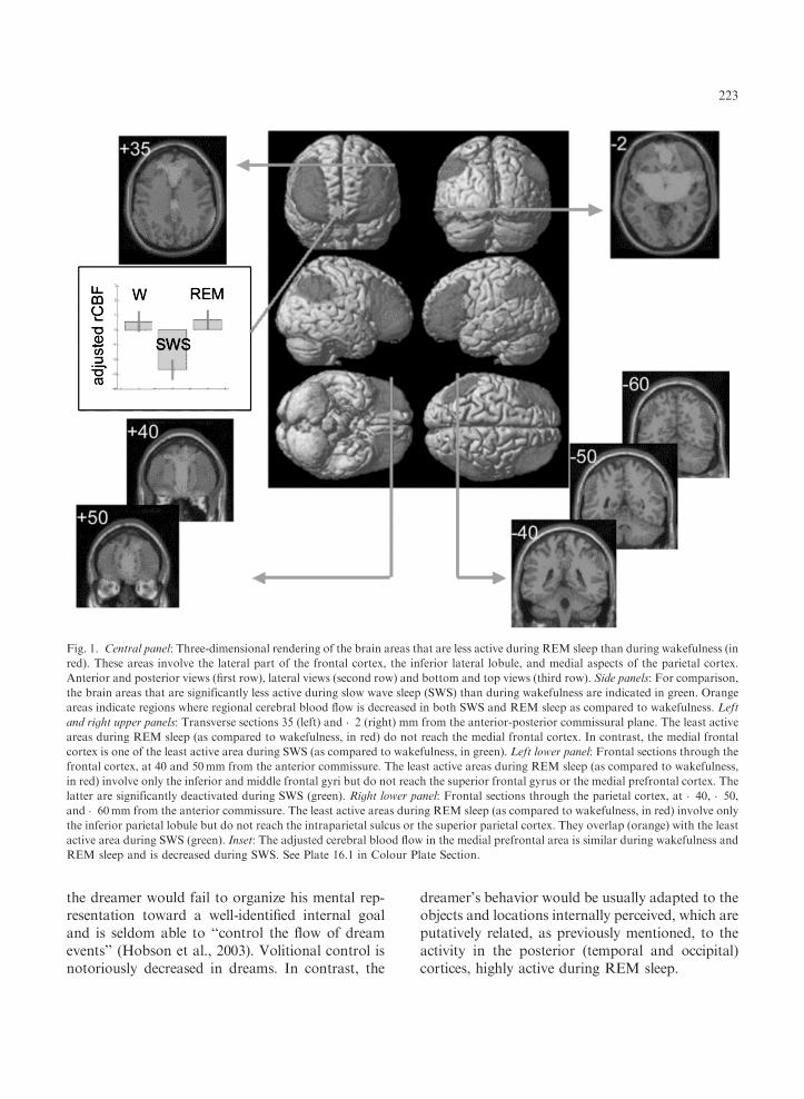
Fig. 1. Central panel: Three-dimensional rendering of the brain areas that are less active during REM sleep than during wakefulness (in
red). These areas involve the lateral part of the frontal cortex, the inferior lateral lobule, and medial aspects of the parietal cortex.
Anterior and posterior views (first row), lateral views (second row) and bottom and top views (third row). Side panels: For comparison,
the brain areas that are significantly less active during slow wave sleep (SWS) than during wakefulness are indicated in green. Orange
areas indicate regions where regional cerebral blood flow is decreased in both SWS and REM sleep as compared to wakefulness. Left
and right upper panels: Transverse sections 35 (left) and �2 (right) mm from the anterior-posterior commissural plane. The least active
areas during REM sleep (as compared to wakefulness, in red) do not reach the medial frontal cortex. In contrast, the medial frontal
cortex is one of the least active area during SWS (as compared to wakefulness, in green). Left lower panel: Frontal sections through the
frontal cortex, at 40 and 50mm from the anterior commissure. The least active areas during REM sleep (as compared to wakefulness,
in red) involve only the inferior and middle frontal gyri but do not reach the superior frontal gyrus or the medial prefrontal cortex. The
latter are significantly deactivated during SWS (green). Right lower panel: Frontal sections through the parietal cortex, at �40, �50,
and �60mm from the anterior commissure. The least active areas during REM sleep (as compared to wakefulness, in red) involve only
the inferior parietal lobule but do not reach the intraparietal sulcus or the superior parietal cortex. They overlap (orange) with the least
active area during SWS (green). Inset: The adjusted cerebral blood flow in the medial prefrontal area is similar during wakefulness and
REM sleep and is decreased during SWS. See Plate 16.1 in Colour Plate Section.
223

Lateral prefrontal cortex and episodic memory
Episodic memory refers to the capacity to encodeand recollect past episodes with their specific in-tegrated details, place and time (Tulving, 2004).Prefrontal cortex is not necessary to store, or toaccess to, episodic memories, since damage tofrontal lobes does not impair episodic memory tothe same extent as lesions to mesio-temporal areas(Henson et al., 1999). However, there are reportsof densely amnesic patients after frontal lobe le-sions. For instance, a remarkable patient presentedwith isolated retrograde amnesia due to a post-traumatic lesion in the right ventral prefrontalcortex (Levine et al., 1998). On the other hand,functional neuroimaging studies consistently re-ported the activation of prefrontal cortex, espe-cially on the right side, during episodic memoryretrieval (Rugg et al., 2002). The usually held viewconsiders that prefrontal areas participate inprocessing information retrieved from episodicmemory, essentially by checking its accuracy andcompleteness. The cognitive processes underpin-ning these frontal activations remain debated andwould represent either a specific retrieval mode,retrieval effort or retrieval success (Rugg et al.,2002). The frontal areas activated during retrievalof episodic information are located in the right andleft anterior prefrontal cortices and in the rightdorso-lateral prefrontal cortex (Rugg et al., 2002).
It is intriguing to observe that the brain areasactivated in functional imaging studies of episodicmemory or this brain-lesioned amnesic patientoverlap with the cortical regions hypoactive duringREM sleep. The relative quiescence of the anteriorprefrontal areas, and to some extent of the dorso-lateral prefrontal cortex would explain that recentwaking life episodes, characterized by their specificlocation, characters, objects, and actions are sel-dom described as such in dream reports (1.7%)(Fosse et al., 2003). In contrast, ‘‘snips’’ of recentwaking activity are frequently (65%) observed indream reports (Fosse et al., 2003). Although thedreamer has access to ‘‘day residues,’’ probablyspontaneously generated by the coordinated activ-ity of the mesio-temporal areas and the posteriorcortices, he would be prevented by the relativehypoactivity in the anterior prefrontal cortex from
tying up the various details of a specific past ep-isode into an identified autobiographical event.
The ventral parieto-frontal system of attention
Recent neuroimaging data studying the neuralcorrelates of human attention have establishedthat two systems exist and interact in the normalhuman brain (Corbetta and Shulman, 2002). Onesystem exerts a top-down control on perception. Itcarries neuronal signals related to the selection ofstimuli and allows for the preparation of goal-di-rected motor responses. This system includes partof the intraparietal sulcus and the superior frontalcortex. The second system is not involved in top-down selection. On the contrary, it is specialized inthe detection of salient, unexpected, behaviorallyrelevant stimuli and reorients the focus of atten-tion. For instance, the activity in this network isenhanced in response to targets occurring at un-expected locations or to low frequency targets, es-pecially when relevant to the task at hand(Corbetta et al., 2000). This system would workas an alerting mechanism when salient stimuli ariseoutside the present focus of processing. The acti-vation of this system would result in an interrup-tion in the current attentional set and in anattentional shift toward the incoming stimulus.As compared to the top-down system, this systemis more ventral, mainly lateralized on the righthemisphere and involves the temporo-parietalcortex and the inferior and middle frontal gyri(Corbetta and Shulman, 2002). Several data sug-gest the activity in the ventral network would bemodulated by the locus coeruleus. For instance,the locus coeruleus is involved in selective atten-tion, especially to salient and unexpected stimuli(Aston-Jones et al., 2000). Furthermore, in themacaque, the inferior parietal cortex is known toreceive heavy projection from the locus coeruleus(Morrison and Foote, 1986).
As far as a precise anatomical localization is al-lowed by PET, the topography of the relativelyquiescent areas during REM sleep is similar to thisventral attentional network. The parietal area,hypoactive during REM sleep extends through theposterior part of the inferior parietal lobuletoward the posterior end of the sylvian fissure. It
224

does not seem to extend to temporal areas. Thehypoactive frontal region is located in the anteriorpart of the inferior frontal gyrus, on the anterioraspect of the region identified in the ventral at-tentional system. Moreover, although bilateral, thehypoactive areas are more extended on the rightside, as is the ventral attention system (Corbettaand Shulman, 2002). Finally, the firing rate ofnoradrenergic locus coeruleus neurons dramatical-ly decreases during REM sleep, depriving the pa-rietal areas from an important positive modulation(Steriade and McCarley, 1990). These considera-tions suggest that the ventral attentional networkis relatively quiescent during REM sleep. If thishypothesis were true, one predicts that the focus ofattention during REM sleep should be less sensi-tive to salient, behaviorally relevant, external stim-uli than during wakefulness. The focus of attentionis difficult to assess experimentally during REMsleep. However, it may transpire in dream reports.One would expect that the dream narrative (ob-tained after awakening from REM sleep) wouldnot be easily modified by external stimulation,even if behaviorally relevant. Some observationssupport this hypothesis. During REM sleep,external stimuli instead of interrupting the flowof the dream storyline, are incorporated into it(Foulkes, 1966). Similarly, responsiveness to ex-ternal auditory stimulation is reduced during sleep,at least in part, due to incorporation of the exter-nal information into ongoing cognitive activity(Burton et al., 1988).
The frontal and parietal areas and mind
representation during REM sleep
The medial frontal areas are active during REMsleep as during wakefulness. In this respect, REMsleep is very different from SWS because the sameregions are relatively less active in SWS, as com-pared to either wakefulness or REM sleep. Theoryof mind refers to the ability to attribute intentions,thoughts and feelings to oneself and to others(Carruthers and Smith, 1996). It is an inductivereasoning allowing interpretation and understand-ing of others’ actions and speech, and prediction oftheir behavior. In comparison to reasoning appliedto physical events or to lower level tasks, theory of
mind was shown to reliably involve medial pre-frontal cortex, temporo-parietal junction especial-ly in the right hemisphere and temporal poles(Fletcher et al., 1995; Brunet et al., 2000, for areview Frith, 2001).
Dreaming usually appears as a multisensorialnarrative involving characters interacting with eachother. These oneiric characters are creditedthoughts, intentions, and emotions by the dream-er himself. Mind representation is thus a key fea-ture of dreaming. We hypothesize that thepersistence of a level of activity in the medial fron-tal areas similar to the activity observed duringwakefulness might participate to mind representa-tion during REM sleep.
However, during REM sleep, the preserved ac-tivity in the medial prefrontal areas (as comparedto wakefulness) contrasts with the low activity inthe inferior parietal cortex. In the right hemi-sphere, this area is a part of the network activatedduring mind representation at wakefulness and isinvolved in the distinction of first versus third per-son perspective in the representation of action,mind and emotion (Ruby and Decety, 2001, 2003,2004; Chaminade and Decety, 2002; Farrer et al.2003). During REM sleep, the low activation ofthe right temporo-parietal junction might be re-lated to a loosening in the distinction between firstand third level perspectives. Accordingly, in dreamreport, the self can participate to the dream actionboth in a first-person (i.e., the self sees and acts)and in a third-person perspective (i.e., the dreamersees the self acting in the dream).
Finally, social emotion such as jealousy, pride,embarrassment, infatuation, sexual love, shame,guilt and pride are often reported in dreaming(Adolphs, 2002; Schwartz and Maquet, 2002). Ne-uropsychological and neuroimaging studies in hu-mans have demonstrated that medial prefrontalcortex and amygdala are consistently involved inbasic and social emotions processing and moregenerally in social cognition (Adolphs, 1999; Phanet al., 2002; Ruby and Decety, 2004). Theamygdala are known to be very active duringREM sleep (Maquet et al., 1996). The preservedactivity in the medial prefrontal cortex might alsoaccount for the high proportion of dreams involv-ing social emotions.
225

Conclusions
Our meta-analysis refines the description of thefunctional neuroanatomy of normal human sleepand provides a tentative framework to explain therelationship between human cognition duringREM sleep and regional patterns of decreased ac-tivity within frontal and parietal areas. In partic-ular, the hypoactivity in frontal and parietal areasduring REM sleep is more precisely characterized.It involves the inferior and middle frontal gyrus aswell as the posterior part of the inferior parietallobule. Interestingly enough, the superior frontalgyrus, the medial frontal areas, the intraparietalsulcus, and the superior parietal cortex are not lessactive in REM sleep than during wakefulness. Wesuggest that this peculiar distribution of regionalbrain activity during REM sleep might correlatewith some features of cognition, as reflected in
dream reports (Fig. 2). Especially, this regionalmetabolic pattern provides new insights on thepossible neural bases of some characteristic dreamfeatures such as self and characters’ mind repre-sentations, the poor episodic recall, the lowerability of external stimuli to break the dream nar-rative and the difficulty to organize one’s oneiricbehavior toward a well identified and persistentgoal.
Acknowledgments
Sleep research presented in this paper was sup-ported by the Fonds National de la RechercheScientifique de Belgique (FNRS), the FondationMedicale Reine Elisabeth, the Research Fund ofULg and the PAI/IAP Interuniversity Pole ofAttraction P5/04. MD, TDV, GV, SL and PM aresupported by FNRS.
References
Adolphs, R. (1999) Social cognition and the human brain.
Trends Cogn. Sci., 3: 469–479.
Adolphs, R. (2002) Neural systems for recognizing emotion.
Curr. Opin. Neurobiol., 12: 169–177.
Aston-Jones, G., Rajkowski, J. and Cohen, J. (2000) Locus
coeruleus and regulation of behavioral flexibility and
attention. Prog. Brain Res., 126: 165–182.
Baddeley, A. (2000) The episodic buffer: a new component of
working memory? Trends Cogn. Sci., 4: 417–423.
Braun, A.R., Balkin, T.J., Wesenten, N.J., Carson, R.E., Varga,
M., Baldwin, P., Selbie, S., Belenky, G. and Herscovitch, P.
(1997) Regional cerebral blood flow throughout the sleep-
wake cycle. An H2(15)O PET study. Brain, 120: 1173–1197.
Braun, A.R., Balkin, T.J., Wesensten, N.J., Gwadry, F.,
Carson, R.E., Varga, M., Baldwin, P., Belenky, G. and
Herscovitch, P. (1998) Dissociated pattern of activity in vis-
ual cortices and their projections during human rapid eye
movement sleep. Science, 279: 91–95.
Brunet, E., Sarfati, Y., Hardy-Bayle, M.C. and Decety, J.
(2000) A PET investigation of the attribution of intentions
with a nonverbal task. Neuroimage, 11: 157–166.
Burton, S.A., Harsh, J.R. and Badia, P. (1988) Cognitive ac-
tivity in sleep and responsiveness to external stimuli. Sleep,
11: 61–68.
Carruthers, P. and Smith, P. (1996) Theories of Theories of
Mind. Cambridge University Press, Cambridge.
Chaminade, T. and Decety, J. (2002) Leader or follower? In-
volvement of the inferior parietal lobule in agency. Neuro-
report, 28: 1975–1978.
Fig. 2. Dream Caused by the Flight of a Bee Around a Pome-
granate One Second Before Awakening, Salvador Dali, 1944.
r Fundacioon Gala-Salvador Dali, c/o Beeldrecht Amsterdam
2005.
226

Corbetta, M., Kincade, J.M., Ollinger, J.M., McAvoy, M.P.
and Shulman, G.L. (2000) Voluntary orienting is dissociated
from target detection in human posterior parietal cortex.
Nat. Neurosci., 3: 292–297.
Corbetta, M. and Shulman, G.L. (2002) Control of goal-di-
rected and stimulus-driven attention in the brain. Nat. Rev.
Neurosci., 3: 201–215.
Farrer, C., Franck, N., Georgieff, N., Frith, C.D., Decety, J.
and Jeannerod, M. (2003) Modulating the experience of
agency: a positron emission tomography study. Neuroimage,
18: 324–333.
Fletcher, P.C., Happe, F., Frith, U., Baker, S.C., Dolan, R.J.,
Frackowiak, R.S. and Frith, C.D. (1995) Other minds in the
brain: a functional imaging study of ‘‘theory of mind’’ in
story comprehension. Cognition, 57: 109–128.
Fosse, M.J., Fosse, R., Hobson, J.A. and Stickgold, R.J. (2003)
Dreaming and episodic memory: a functional dissociation? J.
Cogn. Neurosci., 15: 1–9.
Foulkes, D. (1966) The Psychology of Sleep. Charles Scribner’s
Sons, New York.
Frith, C.D. (2001) Mind blindness and the brain in autism.
Neuron, 20: 969–979.
Henson, R.N., Shallice, T. and Dolan, R.J. (1999) Right pre-
frontal cortex and episodic memory retrieval: a functional MRI
test of the monitoring hypothesis. Brain, 122(Pt 7): 1367–1381.
Hobson, J.A., Pace-Schott, E.F., Stickgold, R. and Kahn, D.
(1998) To dream or not to dream? Relevant data from new
neuroimaging and electrophysiological studies. Curr. Opin.
Neurobiol., 8: 239–244.
Hobson, J., Pace-Schott, E. and Stickgold, R. (2003) Dreaming
and the brain: toward a cognitive neuroscience of conscious
states. In: Pace-Schott E., Solms M., Blagrove M. and
Harnad S. (Eds.), Sleep and Dreaming. Cambridge Univer-
sity Press, Cambridge, pp. 1–50.
Koechlin, E., Ody, C. and Kouneiher, F. (2003) The architec-
ture of cognitive control in the human prefrontal cortex.
Science, 302: 1181–1185.
Levine, B., Black, S.E., Cabeza, R., Sinden, M., McIntosh,
A.R., Toth, J.P., Tulving, E. and Stuss, D.T. (1998) Episodic
memory and the self in a case of isolated retrograde amnesia.
Brain, 121(Pt 10): 1951–1973.
Lydic, R., Baghdoyan, H.A., Hibbard, L., Bonyak, E.V.,
DeJoseph, M.R. and Hawkins, R.A. (1991) Regional brain
glucose metabolism is altered during rapid eye movement sleep
in the cat: a preliminary study. J. Comp. Neurol., 304: 517–529.
Maquet, P. (2000) Functional neuroimaging of normal human
sleep by positron emission tomography. J. Sleep Res., 9:
207–231.
Maquet, P. and Franck, G. (1997) REM sleep and amygdala.
Mol. Psychiatr., 2: 195–196.
Maquet, P., Laureys, S., Peigneux, P., Fuchs, S., Petiau, C.,
Phillips, C., Aerts, J., Del Fiore, G., Degueldre, C.,
Meulemans, T., Luxen, A., Franck, G., Van Der Linden,
M., Smith, C. and Cleeremans, A. (2000) Experience-de-
pendent changes in cerebral activation during human REM
sleep. Nat. Neurosci., 3: 831–836.
Maquet, P., Peters, J., Aerts, J., Delfiore, G., Degueldre, C.,
Luxen, A. and Franck, G. (1996) Functional neuroanatomy
of human rapid-eye-movement sleep and dreaming. Nature,
383: 163–166.
Morrison, J.H. and Foote, S.L. (1986) Noradrenergic and se-
rotoninergic innervation of cortical, thalamic, and tectal vis-
ual structures in Old and New World monkeys. J. Comp.
Neurol., 243: 117–138.
Peigneux, P., Laureys, S., Fuchs, S., Collette, F., Perrin, F.,
Reggers, J., Phillips, C., Degueldre, C., Del Fiore, G., Aerts,
J., Luxen, A. and Maquet, P. (2004) Are spatial memories
strengthened in the human hippocampus during slow wave
sleep? Neuron, 44: 535–545.
Phan, K., Wager, T., Taylor, S. and Liberzon, I. (2002) Func-
tional neuroanatomy of emotion: A meta-analysis of emotion
activation studies in PET and fMRI. Neuroimage, 16:
331–348.
Rechtschaffen, A. and Kales, A.A. (1968) A Manual of Stand-
ardized Terminology, Techniques and Scoring System for
Sleep Stages of Human Subjects. US Department of Health
Education and Welfare, Bethesda.
Ruby, P. and Decety, J. (2001) Effect of subjective perspective
taking during simulation of action: a PET investigation of
agency. Nat. Neurosci., 4: 546–550.
Ruby, P. and Decety, J. (2003) What you believe versus what
you think they believe: A neuroimaging study of conceptual
perspective-taking. Eur. J. Neurosci., 17(11): 2475–2480.
Ruby, P. and Decety, J. (2004) How would you feel versus how
do you think she would feel? A neuroimaging study of per-
spective-taking with social emotions. J. Cogn. Neurosci., 16:
988–999.
Rugg, M.D., Otten, L.J. and Henson, R.N. (2002) The neural
basis of episodic memory: evidence from functional neuroim-
aging. Phil. Trans. R. Soc. Lond. B Biol. Sci., 357:
1097–1110.
Schwartz, S. and Maquet, P. (2002) Sleep imaging and the ne-
uro-psychological assessment of dreams. Trends Cogn. Sci.,
1: 23–30.
Solms, M. (1997) The Neuropsychology of Dreams. Lawrence
Erlbaum Associates Inc., Mahwah.
Steriade, M. and McCarley, R.W. (1990) Brainstem Control of
Wakefulness and Sleep. Plenum Press, New York.
Tulving, E. (2004) Episodic memory: from mind to brain. Rev.
Neurol. (Paris), 160: S9–S23.
227