human-robotics interface for the interaction with...
TRANSCRIPT

Human-Robotics Interface for the Interaction with Cognitive and EmotionalHuman Domains
G.L. Mariottini, D. Prattichizzo, M. De Biasi, C. SnickarsDipartimento di Ingegneria dell’Informazione
Universit̀a di SienaVia Roma 56, 53100 Siena, Italy
Email: {gmariottini,prattichizzo}@dii.unisi.it
A. Rufa†, A. De Capua‡, S. Rossi†Dep. of Neuroscience,†Sects. Neurology,‡Psichiatry
Policlinico “Le Scotte”, Universit̀a di SienaSiena, Italy
Email: {rufa,decapua,rossisimo}@unisi.it
Abstract— For a human-robot interface it is important tohave a good model of how the human subject operates. However,since such a model is difficult to obtain, then the roboticsinterface must observe accurately the subject’s behaviour wheninteracting with him. We present here a new human-robotinterface for active interaction with the cognitive and emo-tional human domains. Since eye movements convey a lot ofinformation about one subject’s cognitive and emotive status,we have designed a new human-robot interface which usesa video-based Eye-Tracker (ET) to observe the subject’s lineof gaze. Since we are also interested in using our interfacefor studying and treating depression, our interface can sendstimulating inputs to the subject using both a TranscranialMagnetic Stimulator (TMS) and a visual stimulus. The latterelicits the subject’s emotions and consists of a set of picturesof facial expressions, which have been shown according to anovel visualization protocol, called Memory-Guided Filtering(MGF). Its effectiveness has been verified by means of manyexperimental results. We also present the application of ourhuman-robot interface for preliminary studies concerning newcognitive rehabilitation strategies in depression.
I. I NTRODUCTION
The increasing interest toward research in Human-RobotInteraction (HRI), led to the development of a great numberof robots of different types (humanoid robots [1], pets [3],medical tools, etc.) in many application areas (entertain-ment [2], elderly assistance and health care [21], rescuerobotics [15], etc.). In the last few years, we have alsowitnessed a growing interest towards robots that could inter-act more and more with humans, e.g., for the rehabilitationof sensorimotor functions. In order to pursue this goal, therobot must be designed to have some degrees of autonomyto monitor the human and to plan rehabilitation actions tocorrect the dysfunctional human behaviour [12].
Beyond sensorimotor system, the research on human-robotics interaction interfaces is extending to other funda-mental aspects of the human being: thecognitive and theemotion systems. These two systems are deeply intertwinedand they have been recently studied for the design ofautonomous robots that must operate in conjunction withpeople [3]. In order to interact with humans the robot devicemust have a good model for how the other operates. How-ever, a conceptual model could not be the most appropriatesolution when dealing with the emotion system, which isresponsible for evaluating and judging events (e.g., good or
TMS coil
ET
TMS
Visual Stimulus(PC monitor)
P
Subject
Fig. 1. Human-Robotics Interaction for cognitive and emotional study:software and hardware architecture.
bad, desirable or undesirable, happiness or sadness, etc.).Then, the robotic device mustobserve accurately the humanbehaviour and then decide its actions.
We present here a new human-robotics interface for activeinteraction with the cognitive and emotional human domains,through a measurement system and astimulating inputsystem.
The measurement of the subject’s behaviour is provided bya video-based Eye-Tracker (ET) (see Fig. 1) that can trackthe pupil and compute the subject’s line of gaze: the eyemovements convey a lot of information about one subject’scognitive and emotive status.
Stimulating inputs are given by both a Visual Stimulusand a magnetic stimulus, generated by the TranscranialMagnetic Stimulator (TMS) toward some subject’s brainareas, through the TMS coil. We use TMS since we are hereparticularly interested in the applicability of our interfacein the study and treatment of depression. In fact, it hasbeen shown that the magnetic field pulses generated by theTMS coil can painlessly and transiently change the moodof depressed patients [17] and change the subjective rate ofhappiness or sadness [16]. Moreover, the coil position couldbe efficiently controlled towards specific brain areas by arobotic manipulator to which the TMS coil is fixed. In thisway, the HRI will be completely automatized.
The visual stimulus, which appears on a PC monitor, has

been designed to stimulate the subject’s emotions: we indeedused a set of pictures representing human facial expressions(neutral, sad and happy), which are known to elicit distinctbrain activations according to neuroimaging studies [10].The facial expressions are shown according to a novelvisual stimulation protocol, calledMemory Guided Filtering(MGF), whose robustness in guiding the subject’s attentiononly toward the most important facial expressive features(lips and eyes) has been also experimentally validated. TheHRI synchronization between ET and TMS is managed inreal-time by the softwareASTIDET which can also activatethe TMS via control signals depending on subject’s eyefixation regions.
The original contributions of our work are the following:
• we present the design and implementation of a newhuman-robotic interface in which ET, TMS and roboticmanipulator have been used to allow the interaction withthe cognitive and emotional human system (for possiblerehabilitation purposes in the treatment of depression);
• ASTIDET software manages the integration and theexact synchronization between ET and TMS;
• subject’s emotions are elicited by means of a visualstimulation strategy using a set of pictures representinghuman facial expressions (neutral, sad and happy),which are presented according to a novel protocol(MGF) whose effectiveness has been observed via manyexperiments;
• preliminary results on normal subjects and a depressedpatient are presented, showing interesting and promisingresults.
The paper is structured as follows: in Sect.II we presentthe hardware and software structure of our human-roboticsinterface. Sect.III describes the visual stimulation protocolMGF and its experimental validation. Finally, in Sect.IV wepresent preliminary experimental results on normal subjectsand depressed patients. In Section.V, we provide some con-cluding remarks highlighting the main contributions of thepaper.
II. T HE HUMAN-ROBOT INTERFACE: HARDWARE AND
SOFTWARE ARCHITECTURE
A. The eye-tracker device (ET)
The study of ocular movements using non invasive visionbased eye-tracker is quite recent and represents an excitingchallenge in both engineering and medicine research fields.Video-based ET allows to compute in real-time, using mod-ern computer vision techniques, the subject’sline of gazeand some other important parameters, such as the pupil di-ameter [5], [6], [13], [25]. From a medical point of view, therecording of eye movements is an objective evidence to bothclinicians and scientists, and is becoming a standard part ofpsychophysical experimentation [7]. It permits, e.g., to inferand partly understand visual and cognitive behaviours [8].
Refer to Fig. 1: a subject is placed in front of a PCmonitor and is asked to look at a visual stimulus (whosecharacteristics will be described in Sect. III). The eye-tracker
(ET) detects in real-time the line of gaze, i.e., the pixelcoordinates of the pointP = (x , y)T where the subject islooking at. We used a robotized pan-tilt ASL Model 504 [13]eye tracker, that can keep the eye in its camera’s field of view.
B. The transcranial magnetic stimulator (TMS)
Transcranial magnetic stimulation is a neuromodulatorytechnique recently employed for cognitive rehabilitation[20].TMS allows the non invasive and painlessly stimulationof the cerebral cortex of the subject via magnetic fieldpulses [23] exploiting the principle of magnetic induction. Infact, a rapidly changing current (activated from an externaltrigger signal) flows from the large capacitor of the TMS(principal unit) to the TMS coils (see Fig. 1), thus producinga magnetic field oriented orthogonally to the plane of thecoil, that passes unimpeded through the skin and bones ofthe subject’s head, inducing an oppositely directed currentinto the brain.
In our experiments we used a Magstim Super Rapidstimulator [14] and an eight-shaped coil (maximum output2.2 Tesla) that permits to precisely direct the magnetic fieldto the brain regions of interest. The automatic positioningof the TMS coil to the head is accomplished by the roboticmanipulator (to which the coil is fixed), also according tobehavioral and clinical information and requirements.
We have chosen to use TMS in our human-robot interfacefor two reasons:i) repetitive TMS (rTMS) delivered on thedorsolateral prefrontal cortex (DLPFC) in depressed patientsimproves their mood (see [17]);ii) rTMS on the left orright DLPFC can change the subjective rate of happiness orsadness [16] in normal subjects. On these basis, we assumethat the permanence of the subjects’ gaze over happy orsad faces could reflect their empathy towards happiness orsadness feelings. We further predict that eventual rTMS-induced gaze changes could be independent by a directinterference of rTMS on voluntary control of eye movements.
Finally, the technique is safe, provided that ad hoc guide-lines are adhered to [24]. Since a more extensive interdis-ciplinary interaction and analysis is needed when dealingwith these advanced topics, this work has been realized incollaboration with neuroscientists. All the subjects werefullyinformed about the aims of the study.
C. ASTIDET: software for Advanced STImuli Design in Eye-Tracking
ASTIDET v.2.0 is a software package we developed tosynchronize the generation of control inputs to the TMS andthe acquisition of data from the ET. Integration and exactsynchronization of ET and TMS is paramount for our human-robot interface.
The visual stimulus is also generated using ASTIDETwhich allows to load a set of pictures (Fig. 2-(A)) anddefine the time instants for picture presentations (Fig. 2-(B)).ASTIDET allows to manage also the control signal propertiesto the TMS, such as frequency, number of magnetic pulsesand starting delay (Fig. 2-(C,D)).

Much effort has been devoted to improve the usabilityof ASTIDET by means of its Graphical User’s Interface(GUI). In fact, due to the interdisciplinarity of our work,we needed to design a software that could be used also bydomain experts (physicians, neuroscientists, etc.) more thanrobotics experts.
We list here the main functionalities of ASTIDET:
• accurate synchronization between the ET data acquisi-tion and the TMS trigger input generation (with separatethreads by MSDN multimedia timers);
• generation of the visual stimuli on the PC monitor(neutral, sad and happy faces);
• storage of theP = (x, y) subject’s line of gazecoordinates (in the PC monitor);
• computation of the gaze fixations.
III. V ISUAL STIMULATION : THE MGF PROTOCOL
Some recent studies have shown that in patients affectedby psychiatric disorders (such as schizophrenia, depressionand neurological disorders [10]), there is an impairmentin emotional processing, specifically emotion recognitioninfacial expressions. It is well known that facial expressionsconvey a wealth of social signals [4]. It has also been shownthat the processing of facial expressions can be impaired bymeans of TMS [9].
Based on the above facts, as introduced in Sect. I, wewant to stimulate the subject’s emotions by means of thevisualization of human facial expressions (neutral, happyandsad) [18].
One of the innovative contributions of this work consists inthe design of a novel visual stimulation protocol, according towhich the facial expressions are shown: theMGF (Memory-Guided Filtering). The peculiarity of MGF is that it doesnot require any teaching phase of the subject since it hasbeen designed to naturally guide his gaze towards those facialfeatures (lips, mouth and eyes) that are known to convey
(A)
(B)(C)
(D)
Fig. 2. ASTIDET Graphical User’s Interface (GUI) and main function-alities. (A) Load a set of pictures; (B) Define the time lags forwhichthe pictures will appear on the PC monitor; (C) Set the TMS triggersignal properties (frequency and number of pulses); (D) Gaze-contingentproperties.
0 T1 T2
(N) (H) (S)
t (secs)
Fig. 3. The Memory Guided Filtering (MGF) protocol.
(N) (H) (S)
Fig. 4. MGF protocol. (N): during the neutral expression thesubjectmemorizes many features of the face; (H)-(S): the subject’s gaze moves fromfrom (H) to (S) images many times in order to memorize the expressions.
the expression [11]. The MGF protocol first shows a neutralfacial expression (N) for a given time lengthT1 (Fig. 3), sothat the subject can naturally memorize all the characteristicsof the viewed face (hairs, mouth, eyes, nose, etc.). Then, fort > T1, MGF shows at the same time instant a pair of happy(H) and sad (S) facial expressions (Fig. 3), on the left andright side of the screen, respectively. Note that we randomlychanged the faces and also their position from an experimentto another (in order to eliminate any kind of bias in the gazemovement).
MGF has a natural explanation: since the only changes be-tween (N) and the pair (H)-(S), occur in the facial expressionfeatures (eyes, lips and mouth) afterT1 then, the subject willonly focus his attention to these features after the previousexploration of (N). Fig. 4 shows the effectiveness of the MGFprotocol in a real experiment. We have shown here the gazeof a subject exploring the picture during the MGF protocol.For t < T1 the face (N) is presented ) and the subject’s gazeexplores the face focusing on many facial features (eyes,nose and ears) and not only on those conveying the facialexpression (see Fig. 4(N). When the happy (H) and sad (S)faces are presented (Fig. 4(H)(S)), the subject spontaneouslylooks only at those facial features encoding the expression.This behaviour has been observed in all the experimentsperformed on many subjects and reveals the effectivenessof MGF protocol.
As shown in Fig. 4, when the happy (H) and sad (S) facesare presented, then the subject does not observe exclusivelythe (H) or (S), but alternatively looks at the two picturesfor an exploration phase whose duration isTe. After Te,as previously described, we argue that the permanence ona given face may represent a reflection of the subject’sempathy toward that face and then, accordingly to the setupin Fig. 1, a trigger signal will be send to the TMS which

t (secs)0 T1 T2 T3 T4
Fig. 5. The Memory Guided Filtering 2 (MGF2) protocol.
will magnetically stimulate the subject’s brain.However, we observed via 150 experiments on 30 subjects,
that the durationTe strongly depends on the subject. Thisalso reflects in an unpredictable (i.e., with high-variability)numbern of transitions between the two images.
A. MGF2
In order to reduce the variability of the number of tran-sitionsn, we have designed the MGF2 protocol. It consistsin showing the MGF protocol two times consecutively, asshown in Fig. 5, and in stimulating via TMS only when thepair of happy and sad images is presented for the secondtime, i.e., during the last periodT4.
In this way, duringT2, the subject can freely explore (H)-(S) thus memorizing the viewed expressions. The first neutralexpression (N) is then shown for a short periodT3 (duringwhich the subjects will unconsciously elaborate previouslygathered visual information), finally followed by the (H)-(S)stimulus (for a timeT4). Our idea is that, due to the visualstimulus inT2, the subject will modify the exploration duringT4, thus reducing the numbern of switching between (H) and(S), thus reducingTe. Our hypothesis has been confirmed byexperimental evidence, as shown in Fig. 6), where we showthe numbern of transitions (mean and standard deviation)obtained with the two protocols (MGF and MGF2) on acontrol group of 30 subjects. We have noticed thatn stronglydecreases in MGF2.
1 20
1
2
3
4
5
6
7
8
9
10
MGF MGF2
Fig. 6. Number of transitionsn between (H) and (S) faces(mean and standard deviations) obtained experimentally us-ing the MGF and MGF2 protocols. Over 150 experimentshave been executed using 30 subjects.
Finally, note that the data acquired from the eye-trackerhave been analyzed with a novel dedicated MATLAB soft-ware realized in our labs. It removes eye-blinks, extracts
Fig. 7. The MATLAB interface aims at computing andvisualizing the regions of attentions (boxes), creating moviesof the experiments and many others.
and visualizes regions of fixation and creates movies of theexperiments (Fig. 7).
IV. EXPERIMENTAL RESULTS
The experimental setup we used to study the influence ofTMS in visual attention brain mechanisms towards happy andsad faces is that described in Sect. II. The visual stimulationhas been realized using the MGF2 protocol (Sect. III).
Two different experiments have been designed. In the firstexperiment, we studied the gaze behaviour of30 subjects. Inparticular, we analyzed the exploration and used the fixationtimes. These were paramount to estimate the parametersT1,T2, T3 andT4 for the MGF2 protocol. In the second and thirdexperiment, we used the above parameters and studied theeffects on the gaze behaviour under repetitive TMS (rTMS)in normal and depressed patients, respectively. Due to thenovelty of our research, we decided in these first experimentsto keep fixed the TMS coil over the left and right DLPFC(Dorso-Lateral Pre-Frontal Cortex) area, without using therobotic manipulator.
A. Gaze behaviour
When the neutral face (N) is shown for a periodT1, anexploration phase is initially performed by the subject. Weexperimentally observed interesting peculiarities aboutthisphase, that can be roughly divided in:
• Studying: the subject’s gaze scouts the face (N) observ-ing all the features for a timeT exp
1;
• Fixation: the subjects starts to fixate some facial featuresof particular interest for a period greater or equal thanT
fix1
.
We visually stimulated30 subjects using12 different faces(for a total of 150 experiments) and we obtained that theaverage exploration time isT exp
1=4.9 s, while the fixation
time is Tfix1
=0.9 s. Consequently, the other periods havebeen chosen asT2 =10 s,T3 =3 s andT4 =10 s.

Fig. 8. Experimental session. A subject is placed in front ofthe PC display and stimulated using the MGF2 protocol.rTMS has been delivered on the dorso-lateral pre-frontalcortex according to the time instantsT1,...,4.
t (secs)0T
exp1
T2 =10 T3 =3 T4 =10(GC)
Fig. 9. Experimental results. The Memory Guided Filtering2 protocol.
B. rTMS effects on visual attention
We here present some preliminary experiments obtainedon 10 subjects. We adopted the MGF2 protocol with the timeperiods chosen as in Sect. IV-A and implemented thegaze-contingent algorithm [7] in ASTIDET in order to automati-cally switch toT2 (Fig. 9) when a fixation was detected afterT
exp1
. The use of gaze-contingent reduced, in many cases, thelength of the experiment. Each subject has been stimulated inT4 with rTMS (20 pulses at 10 Hz) after 5 s. We stimulatedthe dorsolateral pre-frontal cortex (DLPFC) [17] 1)left and2) right. Other two control conditions were carried out: 3)no rTMS stimulation and 4)sham rTMS. Sham stimulationconsists in positioning the coil perpendicularly to the head(thus no magnetic field is induced). However, in sham(differently to 3), the subject continues to hear the typicalstimulation noise from TMS, which in some circumstancesis per se able to change cognitive strategies [22].
Fig. 10(a) represents thex, y eye-coordinates motion forthe sham stimulation delivered at 22 s. The highlighted boxgroups the whole duration of the stimulation. As can beseen, the eye scouts from one face to the other (spike inthe x-coordinates). Instead, in Fig. 10(b), the subject hasbeen stimulated using rTMS on DLPFC (right) at 23 s. Ascan be seen, the eye remains on the face he was pointingat the moment of rTMS (i.e., the happy in nearly 80% ofcases) and continues to explore it by means of physiologicalmicrosaccades.
This interesting evidence, that has been observed inall
0 5 10 15 20 25 300
100
200
300
400
500
600
700
800
900
sec
x (pixels)y (pixels)ROI (pixels)
(a)
0 5 10 15 20 25 300
100
200
300
400
500
600
700
800
900
sec
x (pixels)y (pixels)ROI (pixels)
(b)
Fig. 10. Experimental results. (a) Sham stimulation for2 s.(gray square) (b) rTMS on DLPC right (gray square)2 s.
the experiments, suggests thatactive rTMS delivered on theDLPFC strongly reduces the number of transitions betweenthe two presented faces. There was no carry over effect afterthe end of stimulation. On the other hand, the number oftransitions was not affected by sham rTMS, suggesting thatthe interference on the gaze control was not due to unspecificalerting effects which are known to affect other types ofcognitive tasks as memory [19]. Moreover, it is unlikelythat this effect was due to an interference on brain areascontrolling eye movements: indeed, even during the rTMS-induced gaze permanence on the face, finalized explorativemicro-saccades and vertical scanning eye movements onspecific regions of interest (ROIs) of the face expression (i.e.,eyes, nose, mouth) were preserved in the majority of subjects.
C. HRI application in depression
As described in the Introduction, our human-robot inter-face aims at acting on the cognitive and emotional humansystem. Thus, according to Sect. II-B, we present here some
7 17 20 25 27 30
200
300
400
500
600
700
800
x y ROI
(H)
(S) (S
TMS S
(a)
Fig. 11. Experimental results on depressed patient. At timet = 23 sec.the subject was looking at the happy face (H) (correspondingto the left sideof the monitor). During the transcranial stimulation the depressed patientwas deviated toward sad face (S) (corresponding to the rightside of themonitor).
experiments obtained on a voluntary patient, A.C., 19 yearsold, who is affected by major depression. We have used theMGF2 protocol with T1 = 7 sec and without the gaze-contingent (this has been proven to be a reasonable timefor a subject to memorize all facial features). We also usedthe same stimulation protocol than in Sect. IV-B. First ofall, we have been impressed by the fact that the patient wasalways looking at sad face features (S), with minor attentionto happiness (H)1. Second, and most important, we havenoticed in all the experiments that, if the patient was notlooking at (S) at the moment of stimulation then, duringrTMS, his gaze was deviated towards (S) (e.g., see gray boxin Fig. 11). Since the patient preferred to look always at thesad face (S), this experiment shows that our human-robotinterface really acts of the cognitive and emotive behaviour.This can be addressed as an interesting results for futureapplicability of our interface to the rehabilitation in cognitiveand emotional domains.
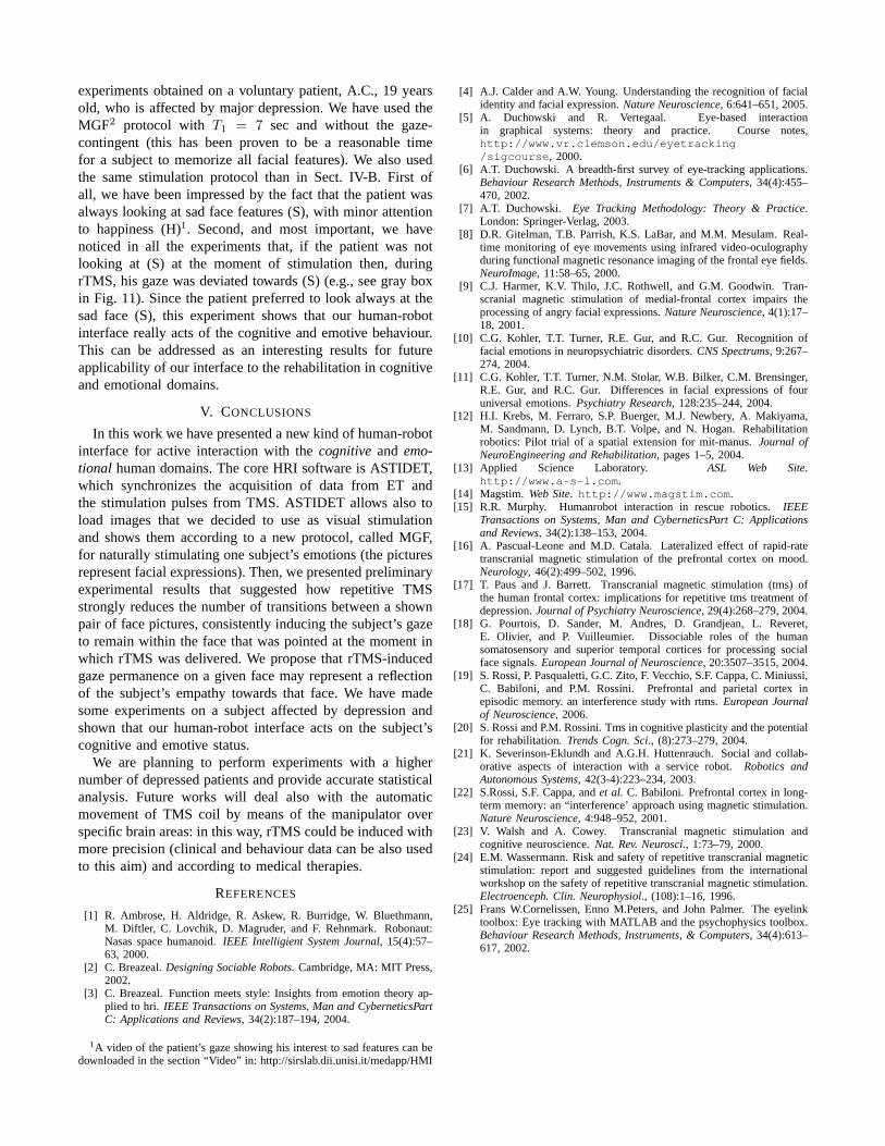
V. CONCLUSIONS
In this work we have presented a new kind of human-robotinterface for active interaction with thecognitive and emo-tional human domains. The core HRI software is ASTIDET,which synchronizes the acquisition of data from ET andthe stimulation pulses from TMS. ASTIDET allows also toload images that we decided to use as visual stimulationand shows them according to a new protocol, called MGF,for naturally stimulating one subject’s emotions (the picturesrepresent facial expressions). Then, we presented preliminaryexperimental results that suggested how repetitive TMSstrongly reduces the number of transitions between a shownpair of face pictures, consistently inducing the subject’sgazeto remain within the face that was pointed at the moment inwhich rTMS was delivered. We propose that rTMS-inducedgaze permanence on a given face may represent a reflectionof the subject’s empathy towards that face. We have madesome experiments on a subject affected by depression andshown that our human-robot interface acts on the subject’scognitive and emotive status.
We are planning to perform experiments with a highernumber of depressed patients and provide accurate statisticalanalysis. Future works will deal also with the automaticmovement of TMS coil by means of the manipulator overspecific brain areas: in this way, rTMS could be induced withmore precision (clinical and behaviour data can be also usedto this aim) and according to medical therapies.
REFERENCES
[1] R. Ambrose, H. Aldridge, R. Askew, R. Burridge, W. Bluethmann,M. Diftler, C. Lovchik, D. Magruder, and F. Rehnmark. Robonaut:Nasas space humanoid.IEEE Intelligient System Journal, 15(4):57–63, 2000.
[2] C. Breazeal.Designing Sociable Robots. Cambridge, MA: MIT Press,2002.
[3] C. Breazeal. Function meets style: Insights from emotion theory ap-plied to hri. IEEE Transactions on Systems, Man and CyberneticsPartC: Applications and Reviews, 34(2):187–194, 2004.
1A video of the patient’s gaze showing his interest to sad features can bedownloaded in the section “Video” in: http://sirslab.dii.unisi.it/medapp/HMI
[4] A.J. Calder and A.W. Young. Understanding the recognition of facialidentity and facial expression.Nature Neuroscience, 6:641–651, 2005.
[5] A. Duchowski and R. Vertegaal. Eye-based interactionin graphical systems: theory and practice. Course notes,http://www.vr.clemson.edu/eyetracking/sigcourse, 2000.
[6] A.T. Duchowski. A breadth-first survey of eye-tracking applications.Behaviour Research Methods, Instruments & Computers, 34(4):455–470, 2002.
[7] A.T. Duchowski. Eye Tracking Methodology: Theory & Practice.London: Springer-Verlag, 2003.
[8] D.R. Gitelman, T.B. Parrish, K.S. LaBar, and M.M. Mesulam.Real-time monitoring of eye movements using infrared video-oculographyduring functional magnetic resonance imaging of the frontal eye fields.NeuroImage, 11:58–65, 2000.
[9] C.J. Harmer, K.V. Thilo, J.C. Rothwell, and G.M. Goodwin.Tran-scranial magnetic stimulation of medial-frontal cortex impairs theprocessing of angry facial expressions.Nature Neuroscience, 4(1):17–18, 2001.
[10] C.G. Kohler, T.T. Turner, R.E. Gur, and R.C. Gur. Recognition offacial emotions in neuropsychiatric disorders.CNS Spectrums, 9:267–274, 2004.
[11] C.G. Kohler, T.T. Turner, N.M. Stolar, W.B. Bilker, C.M. Brensinger,R.E. Gur, and R.C. Gur. Differences in facial expressions offouruniversal emotions.Psychiatry Research, 128:235–244, 2004.
[12] H.I. Krebs, M. Ferraro, S.P. Buerger, M.J. Newbery, A. Makiyama,M. Sandmann, D. Lynch, B.T. Volpe, and N. Hogan. Rehabilitationrobotics: Pilot trial of a spatial extension for mit-manus.Journal ofNeuroEngineering and Rehabilitation, pages 1–5, 2004.
[13] Applied Science Laboratory. ASL Web Site.http://www.a-s-l.com.
[14] Magstim. Web Site. http://www.magstim.com.[15] R.R. Murphy. Humanrobot interaction in rescue robotics. IEEE
Transactions on Systems, Man and CyberneticsPart C: Applicationsand Reviews, 34(2):138–153, 2004.
[16] A. Pascual-Leone and M.D. Catala. Lateralized effect of rapid-ratetranscranial magnetic stimulation of the prefrontal cortex on mood.Neurology, 46(2):499–502, 1996.
[17] T. Paus and J. Barrett. Transcranial magnetic stimulation (tms) ofthe human frontal cortex: implications for repetitive tms treatment ofdepression.Journal of Psychiatry Neuroscience, 29(4):268–279, 2004.
[18] G. Pourtois, D. Sander, M. Andres, D. Grandjean, L. Reveret,E. Olivier, and P. Vuilleumier. Dissociable roles of the humansomatosensory and superior temporal cortices for processingsocialface signals.European Journal of Neuroscience, 20:3507–3515, 2004.
[19] S. Rossi, P. Pasqualetti, G.C. Zito, F. Vecchio, S.F. Cappa, C. Miniussi,C. Babiloni, and P.M. Rossini. Prefrontal and parietal cortex inepisodic memory. an interference study with rtms.European Journalof Neuroscience, 2006.
[20] S. Rossi and P.M. Rossini. Tms in cognitive plasticity and the potentialfor rehabilitation.Trends Cogn. Sci., (8):273–279, 2004.
[21] K. Severinson-Eklundh and A.G.H. Huttenrauch. Socialand collab-orative aspects of interaction with a service robot.Robotics andAutonomous Systems, 42(3-4):223–234, 2003.
[22] S.Rossi, S.F. Cappa, andet al. C. Babiloni. Prefrontal cortex in long-term memory: an “interference’ approach using magnetic stimulation.Nature Neuroscience, 4:948–952, 2001.
[23] V. Walsh and A. Cowey. Transcranial magnetic stimulationandcognitive neuroscience.Nat. Rev. Neurosci., 1:73–79, 2000.
[24] E.M. Wassermann. Risk and safety of repetitive transcranial magneticstimulation: report and suggested guidelines from the internationalworkshop on the safety of repetitive transcranial magnetic stimulation.Electroenceph. Clin. Neurophysiol., (108):1–16, 1996.
[25] Frans W.Cornelissen, Enno M.Peters, and John Palmer. The eyelinktoolbox: Eye tracking with MATLAB and the psychophysics toolbox.Behaviour Research Methods, Instruments, & Computers, 34(4):613–617, 2002.