hyaluronic acid-recombinant gelatin … · 320. european cells and materials vol. 24 2012 (pages...
TRANSCRIPT
320 www.ecmjournal.org
A Tuin et al. Hyaluronic acid-recombinant gelatin scaffoldsEuropean Cells and Materials Vol. 24 2012 (pages 320-330) DOI: 10.22203/eCM.v024a23 ISSN 1473-2262
Abstract
An array of different types of hyaluronic acid (HA)- and collagen-based products is available for filling soft-tissue defects. A major drawback of the current soft-tissue fillers is their inability to induce cell infiltration and new tissue formation. Our aim is to develop novel biodegradable injectable gels which induce soft tissue regeneration, initially resulting in integration and finally replacement of the gel with new autologous tissue. Two reference gels of pure HA, monophasic HA-1 and micronised HA-2, were used. Furthermore, both gels were mixed with recombinant gelatin (RG) resulting in HA-1+RG and HA-2+RG. All gels were subcutaneously injected on the back of rats and explanted after 4 weeks. Addition of RG to HA-1 resulted in stroma formation (neovascularisation and ECM deposition) which was restricted to the outer rim of the HA-1+RG gel. In contrast, addition of RG to HA-2 induced stroma formation throughout the gel. The RG component of the gel was degraded by macrophages and giant cells and subsequently replaced by new vascularised tissue. Immunohistochemical staining showed that the extracellular matrix components collagen I and III were deposited throughout the gel. In conclusion, this study shows the proof of principle that addition of RG to HA-2 results in a novel injectable gel capable of inducing soft tissue regeneration. In this gel HA has a scaffold function whereas the RG component induces new tissue formation, resulting in proper vascularisation and integration of the HA-2+RG gel with the autologous tissue.
Keywords: Tissue regeneration; hydrogel; scaffold; hyaluronic acid; recombinant gelatin.
*Address for correspondence:M.C. HarmsenUniversity Medical Centre GroningenHanzeplein 1, 9713 GZ GroningenThe Netherlands
Tel: +31503614776Fax: +31503619911
E-mail: [email protected]
Introduction
Defects or loss of the soft tissue below the skin may be the result of surgery, deep burns, trauma and ageing. Their common denominator is that regeneration of the hypodermal tissue does not occur. In clinical practice, the transplantation of autologous adipose tissue for use as filling replacement tissue has shown promising results. Adipose tissue transplants also appear to rejuvenate the skin. Meier et al. (2009) reported an average volume retention of 32 % at 16 months after injection. However, the degree of resorption of the transplanted fat over time is unpredictable and uncontrollable (Meier et al., 2009; Kaufman et al., 2007). Therefore, the development of acellular therapies by use of smart biomaterials as fillers is warranted. These smart materials will serve to replace and regenerate lost soft tissue. They should be biocompatible, non-toxic, biodegradable and stimulate their replacement by autologous tissue. At present a number of extracellular matrix-inspired protein and polysaccharide-based materials exist, that all are semi-permanent fillers at best. However, none of these fillers induces tissue regeneration. The most frequently used compounds for semi-permanent soft-tissue fillers are the biodegradable polymers hyaluronic acid (HA) and collagen both of which are also natural skin components. Skin extracellular matrix (ECM) is composed of two main classes of molecules; polysaccharides, also called glycosaminoglycans (GAGs), and proteins like elastin, collagen III and collagen I – the latter being the principal collagen of the skin. The most prominent GAG, HA, is found in many tissues throughout the body with the highest concentrations in connective tissue and the skin. HA is an unbranched non-sulphated polysaccharide, comprising approximately 10,000 repeat units of the disaccharide D-glucuronic acid and D-N-acetylglucosamine. Due to its high water-binding capacity (Johl and Burgett, 2006; Sutherland, 1998), HA has important functions in the body. These include maintenance of extracellular space, facilitation of extracellular transport of ions and nutrients, control of tissue hydration in the skin, lubrication in the synovial fluid of joints, as well as binding of interleukins and growth factors (Ramsden and Rider, 1992; Fernandez-Botran et al., 1999). In the skin, HA is produced intracellularly at the cell membrane of fibroblasts by HA-synthases (Weigel et al., 1997; Itano and Kimata, 2002) and extruded directly into the ECM. Degradation of HA occurs by oxygen free radicals (Agren et al., 1997; Deguine et al., 1998), but primarily through hydrolysis by hyaluronidases (Laurent and Fraser, 1992). Degradation products of HA are physiologic regulators. HA chains of 4-25 disaccharides are pro-angiogenic (West et al., 1985). Because HA is chemically and structurally
HYALURONIC ACID-RECOMBINANT GELATIN GELS AS A SCAFFOLDFOR SOFT TISSUE REGENERATION
A Tuin1, J Zandstra1, SG Kluijtmans2, JB Bouwstra2, MC Harmsen1* and MJA Van Luyn1
1University Medical Centre Groningen, Groningen, The Netherlands2Fujifilm Manufacturing Europe BV, Tilburg, The Netherlands

321 www.ecmjournal.org
A Tuin et al. Hyaluronic acid-recombinant gelatin scaffolds
identical, regardless of its origin, allergic reactions are rare and skin tests before injection are not necessary (Beasley et al., 2009). This led to the use of HA in several tissue engineering areas, such as dermal regeneration, ophthalmic surgery, drug delivery and aesthetic surgery. To date, the HA-based filler Restylane is the most frequently used filler for small cosmetic soft-tissue defects. A major drawback though of HA and collagen based fillers, and in fact of all semi-permanent fillers, is the lack of a tissue regeneration, i.e. the filling material is only degraded and not replaced by autologous tissue. Typically, the subcutaneous longevity in the human of collagen- and HA-based semi-permanent fillers has been reported to vary between 2-3 months (collagen filler Zyderm I) and 6-12 months (HA filler Restylane). Fernández-Cossío showed that 8 months after subcutaneous injection of Restylane in rats hardly any cellular infiltration was observed and no connective tissue formation inside the gel, and thus no integration with the surroundings had occurred (Fernandez-Cossio and Castano-Oreja, 2006). Between 6-12 months, the HA will be enzymatically degraded and the original defect emerges again and repeated injections are needed. In previous research, we showed that, in rats, subcutaneously injected microspheres (MS), prepared from recombinant gelatin (RG), were degraded by a tissue reaction called the foreign body reaction (FBR) (Tuin et al., 2010). Fibroblasts and macrophages infiltrated in-between the RG-based MS. Fibroblasts deposited ECM, e.g. collagen I and III, in-between the MS and macrophages, some of which locally fused to giant cells, simultaneously degraded the MS. We therefore surmise that RG is capable of initiating collagen deposition and inducing tissue regeneration. In contrast to naturally-derived gelatins, the production of recombinant gelatins (RGs) is well-defined and controllable (Werten et al., 2001). Due to their production process, naturally derived gelatins are mixtures of molecules of different molecular weights and amino acid sequences, which may result in batch-to-batch differences. The advantages of using RG are that it is well-defined in terms of composition, MW and isoelectric point – to mention but a few. Furthermore, its large scale production, purification, and purity are standardisation to good manufacturing practice (GMP) requirements. This guarantees minimal to near absent batch-to-batch variation. Manipulations to the amino acid sequence of RGs are easily achieved through genetic mutation and cloning techniques, and directly influence the characteristics of the RG. Recombinant DNA technology thus allows for on demand RG design. We hypothesised that addition of RG to HA gels not only provides temporary volume restoration, exerted by HA which serves as a scaffold, but also leads to integration of the gel with the surrounding tissue and finally to tissue regeneration. In this study, we aimed to demonstrate the proof of principle for this hypothesis. We used two types of pure HA gels, i.e. a monophasic HA-1 gel and a micronised HA-2 gel. Both HA gels were mixed with RG, resulting in 4 different gels. The RG we used has an amino acid sequence consisting of multiple repeats of a human collagen I fragment that contains an RGD triplet in its sequence.
The 4 gels were subcutaneously injected on the back of rats. After 4 weeks, the tissue response to all gels was evaluated and soft tissue regeneration was examined by immunohistological assessment of the FBR, i.e. assessing neovascularisation, the presence of macrophages, giant cells and fibroblasts, and the deposition of new ECM in the gels.
Materials and Methods
Preparation of gelatin/HA gelsIn order to ensure a low bio-burden all gels and solutions were prepared in a laminar flow cabinet. Solutions were filtered through a 0.2 µm filter and used glassware was autoclaved. HA-1 and HA-2 are crosslinked pure HA gels. HA-2 was obtained by crosslinking a 10 % (w/v) HA solution (hyaluronic acid salt from Streptococcus equi sp. Fluka #1351058, Mw 1.6 x 10⁶ Da) in 1 % (w/v) sodium hydroxide by 1,4-butanediol diglycidyl ether (BDDE; Fluka, Buchs, Switzerland). The amount of BDDE was 0.5 µmol/g HA. The gel was allowed to crosslink for 24 h at room temperature. After neutralising with 1 M hydrochloric acid (Merck, Darmstadt, Germany) and diluted with saline sodium phosphate buffer pH 7.4 (phosphate-buffered saline (PBS) with Tween 20, pH 7.4, tablets, Fluka) to 2.6 % (w/v), the resulting gel was dialysed against saline sodium phosphate buffer for 48 h. Finally, the gel was mashed using a microniser (Ultra-turrax T25; IKA, Staufen im Breisgau, Germany). Pure HA-1 was obtained by an adapted crosslinking step which crosslinked the -OH / -COOH groups. HA-1 was used without further micronising. The recombinant gelatin (RG) was produced in the yeast Pichia pastoris, using methods described in patents EP0926543 en EP1014176. The RG has an amino acid sequence of 821 amino acids (Mw = 51 kDa) and a pI of 10.0, and is derived from the human collagen I sequence. Basically the RG sequence consists of a repeated human collagen I fragment that contains an RGD sequence. After production, the RG was purified from host cell proteins and salts using multiple chromatography and diafiltration steps. Use of RG composed microspheres has been described earlier by (Tuin et al., 2010). In order to obtain HA-1+RG, a pure RG gel was prepared in advance by crosslinking RG using N-(3-dimethylaminopropyl)-N’-ethylcarbodiimide hydrochloride (EDC; Sigma Aldrich, Zwijndrecht, Netherlands). EDC was chosen because it is highly soluble in water and forms zero-length crosslinks between amines and carboxylic acid groups of the amino acids in RG. This crosslinking procedure has the advantage that no chemical structure is added to form the crosslink, and its reactivity under relatively mild conditions. EDC itself is converted into its urea derivative upon forming the crosslink, which is readily water soluble and non-cytotoxic. The RG gel was prepared starting from a 10 % (w/v) RG solution and crosslinked by 2.1 mmol EDC /g RG at pH 4.7. The gel was allowed to crosslink for 24 h and subsequently mashed by micronising until the particles were small enough to pass

322 www.ecmjournal.org
A Tuin et al. Hyaluronic acid-recombinant gelatin scaffolds
through a 25 G needle (particle size measured in methanol 200-250 µm, Mastersizer, MS2000; Malvern, Malvern, UK). In order to remove non-reacted EDC and urea, the mashed RG gel was dialysed against phosphate buffer for 24 h – resulting in an RG gel with respectively 2.0 % (w/v) RG. The HA-1+RG mixed gel was then obtained by homogeneously mixing the RG gel with the HA-1 in a volume ratio of 3:7 using a spatula. The HA-2+RG gel was prepared by first preparing a 10 % (w/v) HA gel with the same amount of BDDE as for pure HA-2. After 24 h, the pH of the HA-2 gel was lowered by addition of 2.25 mL 1 M HCl / 10 g HA gel. Then, 2.56 g of a 20 % (w/v) RG solution (pH 7.4), to which 2 mL 50 % (w/v) EDC / 10 g was added, was prepared and added directly after mixing to 12.25 g of HA-2 gel. The mixed gel was homogenised using the Ultra-turrax. Subsequently, the pH was lowered by addition of 0.2 mL 5 % (v/v) sulphuric acid (Sigma Aldrich) to initiate EDC crosslinking. Crosslinking proceeded for 30 min at 40 ºC. The resulting gel, with a HA-2/RG volume ratio of 7:3, was diluted with phosphate buffer, pH 7.4, to 40 mL and then dialysed against phosphate buffer, pH 7.4, for 3 d. Finally, all gels were transferred into syringes and frozen samples were sterilised by gamma radiation (7 kGy), while being kept frozen. The volume ratio of the mixed gels, the resulting final HA and RG concentrations and the mass ratio of each gel are summarised in Table 1. Mass ratios were determined by weighing the HA and RG volumes before mixing them. The preparation method of each gel is schematically depicted in Fig. 1.
Rheological characterisationGel samples were characterised by rheological measurements on an Anton Paar Rheoplus MCR 301 rheometer, using a plate to plate geometry (PP20) at 20 ºC. The plates used were 40 mm and the gap size used was 1 mm. The storage modulus (G’), loss modulus (G”) and complex viscosity (h*) were determined at angular frequencies between 0.05 and 100 rad/s. The samples did
not show much frequency dependence up to 100 rad/s. Hence, the low frequency values at 0.05 rad/s were chosen as being representative (Table 2).
MTS cytotoxicity assayCytotoxicity of the gels was assessed by an MTS assay. Briefly, PK-84 cells (5,000 cells per well) were seeded in a 96-well plate. After 24 h the medium was replaced by medium extracts of the gels (n = 3 wells per gel extract). Extracts were prepared by shaking the gels in cell culture medium for 24 h at 37 °C. Then, 24 h after addition of the extracts, the mitochondrial substrate MTS [3-(4,5-dimethylthiazol-2-yl)-5-(3-carboxymethoxyphenyl)-2-(4-sulfophenyl)-2H-tetrazolium] was added. Formation of the soluble formazan product was measured at 490 nm on a spectrophotometer. Results showed that cell viability and morphology were not influenced by the gel extracts (data not shown).
AnimalsAnimal experiments were carried out with male AO rats (Harlan Nederland, Horst, the Netherlands) weighing 250 ±10 g. The animals were housed under standard laboratory conditions; a regular light dark cycle, and laboratory chow and acidified water ad libitum. All animal experiments were approved by the Local Committee on Animal Experimentation.
In vivo experimental setupAnimals were put under isoflurane/N2O/O2 anaesthesia and their backs were shaved and disinfected. Subsequently, 100 mL of each of the four gels was subcutaneously injected with a 27 G needle at a different location on the back of the rat. The gels and the surrounding tissue were explanted under general anaesthesia after 4 weeks (n = 3 rats) and thereafter rats were killed by cervical dislocation.
HistochemistryAfter explantation, explants were fixed in 2 % glutaraldehyde solution prior to embedding in plastic,
Gel HA/RG volume ratio % HA % RG HA/RG mass ratioHA-1 n.a. 2.7 - 0HA-1+RG 7:3 1.9 0.6 3.2HA-2 n.a. 2.5 - 0
Sample Storage Modulus[Pa]
Loss Modulus[Pa]
Complex Viscosity[Pa·s]
HA-1 1230 370 25800HA-1+RG1 370 122 7800
HA-2 273 30 4500HA-2 + RG 169 17 3400
Restylane (Lot 9424) 274 74 5690
Table 1. The HA/RG volume ratio, final overall HA and RG concentrations (%) and HA/RG mass ratio of the gels.
Table 2. Rheological properties of HA-1, HA-2, HA-1+RG, HA-2+RG and Restylane measured at 0.05 rad/s.
323 www.ecmjournal.org
A Tuin et al. Hyaluronic acid-recombinant gelatin scaffolds
as described previously (Tuin et al., 2010). Sections of 2 mm were stained with toluidine blue and analysed by light microscopy. For the measurement of collagen deposition, 2 mm sections were stained with a picrosirius solution (0.1 % (w/v) sirius-red in saturated aqueous picric acid solution for 24 h at 50 °C). Subsequently, the sections were washed in running tap-water for 5 min, counterstained with Mayer’s haematoxylin, dehydrated in graded ethanol, cleared in xylene and mounted in resinous medium (Cerri and Sasso-Cerri, 2003).
ImmunohistochemistryAfter explantation, explants were embedded in Tissue Tek® O.C.T. Compound and frozen immediately in liquid nitrogen and stored at -80 °C until preparation of cryosections. Sections of 5 mm were cut at -25 °C and fixed
with acetone. Thereafter, sections were pre-incubated with 10 % (v/v) rabbit serum in PBS and subsequently incubated with antibodies against CD68 (Serotec Ltd, Oxford, UK), for the detection of macrophages and giant cells; and collagen IV (cat.no. ab6311-100, Abcam, Cambridge, UK) for the detection of blood vessels. To determine which types of collagen were deposited in the gels, staining for collagen I (cat.no. 1310-01, Southern Biotech, Birmingham, UK) and collagen III (cat.no. ab6310-100, Abcam, Cambridge, UK) were performed. After incubation with the first antibody, endogenous peroxidase activity was blocked by H2O2 treatment which was followed by incubation with appropriate secondary antibodies (DAKO, Glostrup, Denmark). Slides were then stained with 3-amino-9-ethylcarbazole (AEC), counterstained with haematoxylin, and embedded in Kaiser’s glycerine.
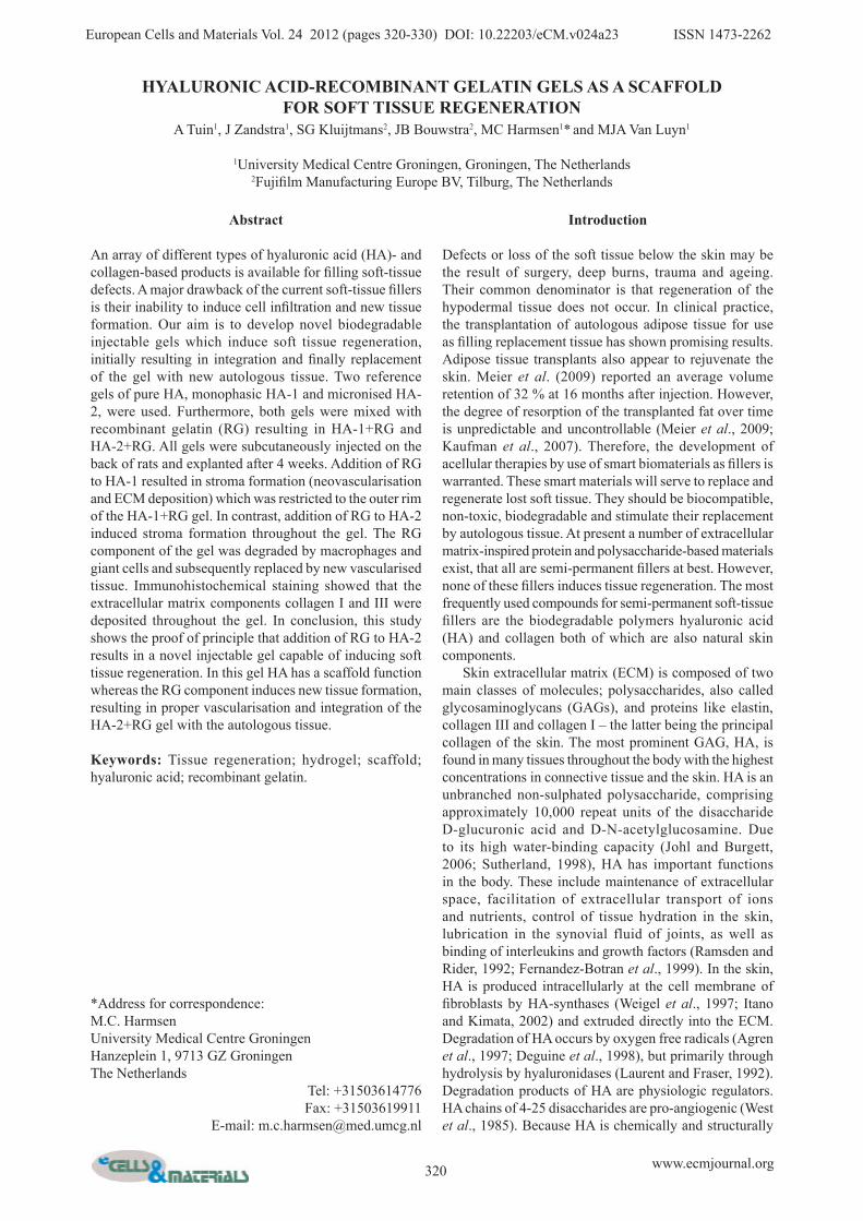
Fig. 1. Schematic representation of the preparation of the gels. The blue container in the upper row represents the HA-1 gel. The blue container in the second row is also representing the HA-1 gel and the yellow container represents the RG gel. The RG is mixed into HA-1 by manual mixing resulting in a HA-1 gel with RG particles dispersed through the gel. HA-1+RG is therefore represented by a blue container with yellow particles in it. The light blue container in the third row represents HA-2 which is crosslinked by BDDE and subsequently micronised, resulting in a gel consisting of many small HA-2 particles. The fourth row represents the preparation of HA-2+RG. HA-2 (represented by a light blue container) is crosslinked by BDDE and subsequently mixed with the RG solution and EDC and crosslinked while micronising the mixture with the thurax. The green box represents the HA-2+RG gel, a mixture of HA-2 (light blue) and RG (yellow).
324 www.ecmjournal.org
A Tuin et al. Hyaluronic acid-recombinant gelatin scaffolds
QuantificationStaining for collagen I, III and IV and for CD68 were measured using computerised morphometry (Leica Qwin 2.8 Software). Staining inside the gel area of explant sections was quantified as the percentage stained area of the total gel surface area. The quantification was performed at a magnification of x 12.5. Staining in the tissue surrounding the gel was excluded from the measurements. The nature of the FBR was examined by double-blind scoring of random slides of each gel by two independent experienced investigators. Scoring of the FBR parameters followed a robust protocol that has been in use for more than two decades in laboratories that examine the tissue response to implanted foreign bodies. Briefly, this semi-quantitative method scores the extent of vascularisation and scores the level of infiltrated fibroblasts, macrophages, giant
cells and lymphocytes. We used a modified scoring system from Luttikhuizen et al. (2004), who ranked histological parameters from 0 to 5, only we used intervals of 0.5: 0 = no vascularisation, fibroblasts, macrophages, giant cells or lymphocytes; 1 = low degree of vascularisation, sporadic presence of fibroblasts, macrophages, giant cells or lymphocytes; 2 = moderate degree of vascularisation, presence of some fibroblasts, macrophages, giant cells or lymphocytes; 3 = high degree of vascularisation, large numbers of fibroblasts, macrophages, giant cells or lymphocytes; 4 = very high degree of vascularisation, very large numbers of fibroblasts, macrophages, giant cells or lymphocytes; 5 = extensive vascularisation, extremely large numbers of fibroblasts, macrophages, giant cells or lymphocytes.
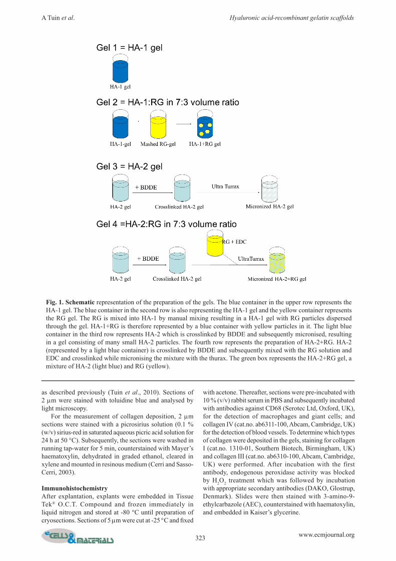
Fig. 2. A) Surface area of and B) tissue ingrowth in cross sections of the HA-1, HA-1+RG, HA-2 and HA-2+RG gels. The percentage of the surface area of the gel in which tissue ingrowth is present was calculated by dividing the surface area with ingrowth (area between the dashed lines in the large micrographs in Fig. 2C) by the total surface are of the gel (area within the outer dashed line). Each bar represents the average of at least n = 3 samples. Significance is denoted in the graph. C) Histological overview of toluidine blue stained cross sections of HA-1, HA-1+RG, HA-2 and HA-2+RG gels 4 weeks after subcutaneous implantation. In the 1.25x magnification micrographs, the rim of gel is indicated by the outer dashed line. The interface of the HA-1 and HA-2 gels with the surrounding tissue is visible in more detail in the 20x magnification micrographs on the right of the 1.25x micrographs. The high magnification micrographs to the right of the lower magnification micrographs of HA-1+RG and HA-2+RG show the details of the cellular infiltration, formation of new tissue (blood vessels and stroma) and degradation of material by giant cells. In the low and higher magnification micrographs, surrounding tissue is located below the lowest dashed line and is denoted by SR. Other indicated structures are: b = blood vessel, GC = giant cell, s = stroma, H = hyaluronic acid.
325 www.ecmjournal.org
A Tuin et al. Hyaluronic acid-recombinant gelatin scaffolds
Statistical analysisAll data were expressed as the mean ± SD and analysed using one-way ANOVA followed by Bonferroni’s post hoc test. Differences are considered significant at p < 0.05.
Results
Physical characteristics of the gelsThe physical characteristics of a gel depend on many factors such as the composition of the gel, the degree of crosslinking, the type of crosslinker which is used, the particle size of the gel after crosslinking, etc. These physical characteristics of the gels determine the injectability as well as their biological function in vivo. Therefore, two HA gels which differ in particle size, i.e. monophasic HA-1 and micronised HA-2 (Materials
and Methods section and Fig. 1) were studied. Differences are reflected in their rheological properties as summarised in Table 2. As expected the storage modulus (G’) and loss modulus (G”) of HA-1, which is non-micronised, is much higher than that of HA-2, 1230 vs. 273 for G’ and 370 vs. 30 for G” (Table 2). Except for HA-1, the rheological properties of the gel formulations are quite similar to the commercial dermal filler Restylane (Table 2). Addition of RG to HA-1 and HA-2 considerably changed the rheological properties of both gels. The storage moduli decreased from 1230 and 273 Pa to 370 and 169 Pa respectively and the viscous moduli decreased from 370 and 30 Pa to 122 and 17 Pa respectively (Table 2). The lower G’ and G” of HA-2+RG compared to HA-1+RG are caused by physical differences in the nature of both HA types (micronised versus non-micronised) and by the mixing procedure of HA with RG. RG is manually
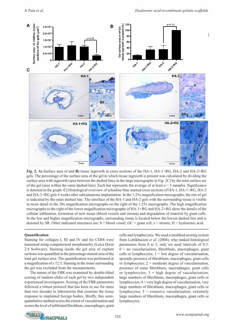
Fig. 3. Immunohistochemical staining and quantification of the staining for A) collagen IV for the detection of vascularisation and B) CD68 to demonstrate the presence of macrophages and giant cells in the HA-1, HA-1+RG, HA-2 and HA-2+RG gels after 4 weeks of subcutaneous implantation in vivo. Cross sections of the gels were stained and only the red staining in the gels (below the dashed line in the pictures) was measured using computerised morphometry. Staining in the surroundings (indicated by SR) was not measured. The inset in the HA-1+RG micrograph in Fig. 3B shows a RG remnant surrounded by macrophages. Remnants of RG stained dark blue with haematoxylin and are denoted by RG.
B
A
326 www.ecmjournal.org
A Tuin et al. Hyaluronic acid-recombinant gelatin scaffolds
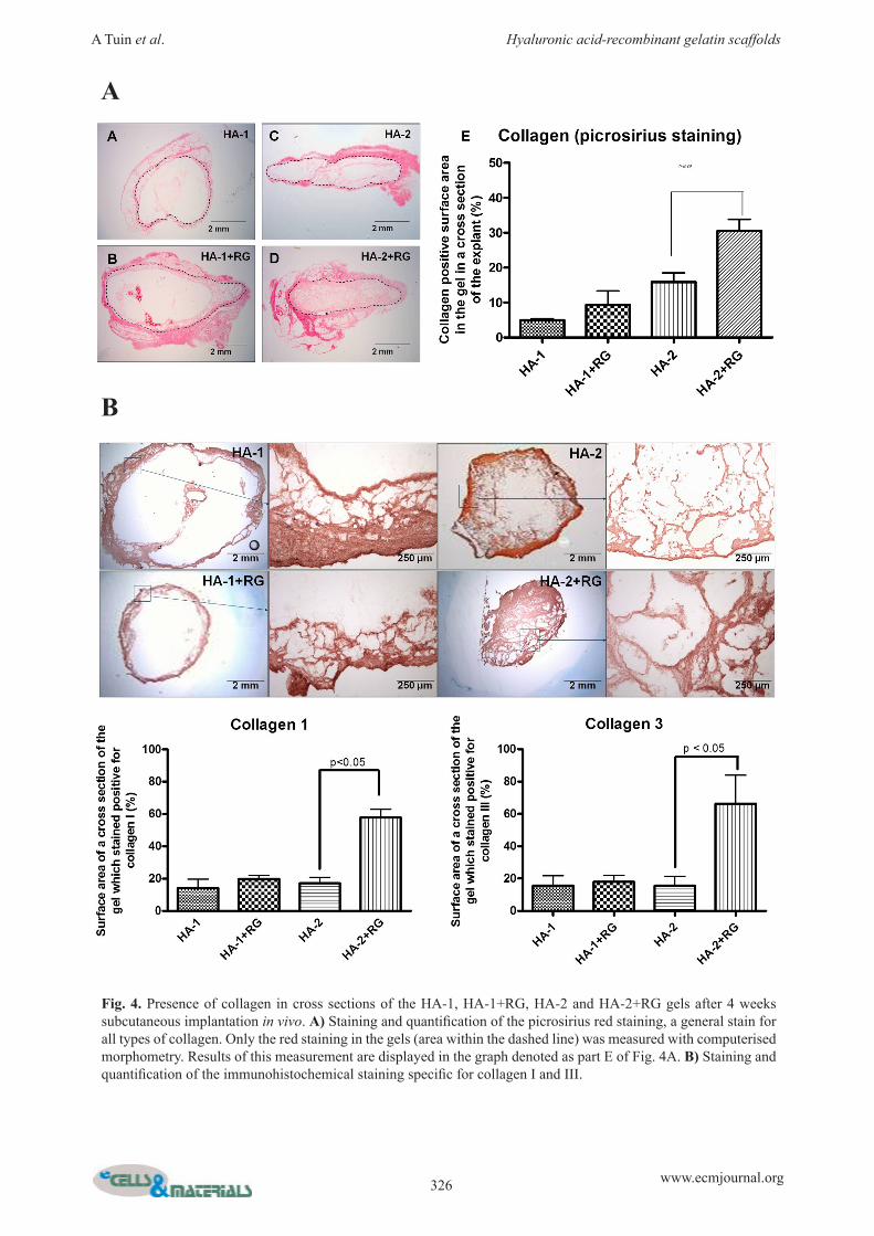
Fig. 4. Presence of collagen in cross sections of the HA-1, HA-1+RG, HA-2 and HA-2+RG gels after 4 weeks subcutaneous implantation in vivo. A) Staining and quantification of the picrosirius red staining, a general stain for all types of collagen. Only the red staining in the gels (area within the dashed line) was measured with computerised morphometry. Results of this measurement are displayed in the graph denoted as part E of Fig. 4A. B) Staining and quantification of the immunohistochemical staining specific for collagen I and III.
A
B
327 www.ecmjournal.org
A Tuin et al. Hyaluronic acid-recombinant gelatin scaffolds
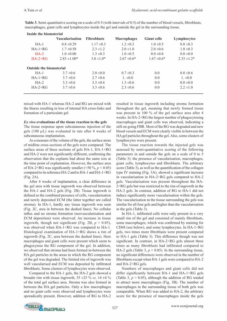
Table 3. Semi-quantitative scoring on a scale of 0-5 (with intervals of 0.5) of the number of blood vessels, fibroblasts, macrophages, giant cells and lymphocytes inside the gel and outside the gel in the surrounding tissue.
Inside the biomaterialVascularisation Fibroblasts Macrophages Giant cells Lymphocytes
HA-1 0.8 ±0.29 1.17 ±0.3 1.2 ±0.3 1.0 ±0.5 0.8 ±0.3HA-1+RG 1.7 ±0.58 2.3 ±1.2 2.0 ±1.0 2.0 ±0.6 1.8 ±0.3
HA-2 1.0 ±0.00 1.3 ±0.3 1.0 ±0.5 0.0 ±0.0 0.0 ±0.0HA-2+RG 2.83 ±1.00* 3.0 ±1.0* 2.67 ±0.6* 1.67 ±0.6* 2.33 ±1.2*
Outside the biomaterialHA-1 3.7 ±0.6 2.0 ±0.0 0.7 ±0.3 0.0 0.8 ±0.6
HA-1+RG 3.7 ±0.6 2.7 ±0.6 1. ±0.0 0.0 1. ±0.0HA-2 3.3 ±0.6 3.3 ±0.6 1.3 ±0.6 0.0 0.0 ±0.0
HA-2+RG 3.7 ±0.6 3.3 ±0.6 2.3 ±0.6 0.0 2.2 ±1.0
mixed with HA-1 whereas HA-2 and RG are mixed with the thurax resulting in loss of internal HA cross-links and formation of a particulate gel.
Ex vivo evaluations of the tissue reaction to the gelsThe tissue response upon subcutaneous injection of the gels (100 mL) was evaluated in rats after 4 weeks of subcutaneous implantation. As a measure of the volume of the gels, the surface areas of midline cross-sections of the gels were compared. The surface areas of these sections of gels HA-1, HA-1+RG and HA-2 were not significantly different, confirming the observation that the explants had about the same size at the time point of explantation. However, the surface area of HA-2+RG was significantly smaller (<50 %, p < 0.05) compared to its reference HA-2 and to HA-1 and HA-1+RG (Fig. 2A). After 4 weeks of implantation, a clear difference in the gel area with tissue ingrowth was observed between the HA-1 and HA-2 gels (Fig. 2B). Tissue ingrowth is defined as the combined presence of cells, vascularisation and newly deposited ECM (the latter together are called stroma). In HA-1, hardly any tissue ingrowth was seen (Fig. 2C, area in between the dashed lines). No cellular influx and no stroma formation (neovascularisation and ECM deposition) were observed. An increase in tissue ingrowth, though not significant (Fig. 2B, p > 0.05), was observed when HA-1+RG was compared to HA-1. Histological examination of HA-1+RG shows a rim of ingrowth (Fig. 2C, area between the dashed lines). Here macrophages and giant cells were present which seem to phagocytose the RG component of the gel. In addition, we observed that stroma had been formed in-between the HA gel particles in the areas in which the RG component of the gel was degraded. The limited rim of ingrowth was well vascularised and ECM was deposited by infiltrated fibroblasts. Some clusters of lymphocytes were observed. Compared to the HA-1 gels, the HA-2 gels showed a broader rim with tissue ingrowth, 35 ±25 % vs. 14 ±4 % of the total gel surface area. Stroma was also formed in between the HA gel particles. Only a few macrophages and no giant cells were observed and lymphocytes were sporadically present. However, addition of RG to HA-2
resulted in tissue ingrowth including stroma formation throughout the gel, meaning that newly formed tissue was present in 100 % of the gel surface area after 4 weeks. In HA-2+RG the largest number of phagocytosing macrophages and giant cells was observed, indicating a still on-going FBR. Most of the RG was degraded and new blood vessels and ECM were clearly visible in between the HA gel particles throughout the gel. Also, some clusters of lymphocytes were present. The tissue reaction towards the injected gels was assessed by semi-quantitative scoring of the following parameters in and outside the gels on a scale of 0 to 5 (Table 3): the presence of vascularisation, macrophages, giant cells, lymphocytes and fibroblasts. The arbitrary score (Table 3), as well as the quantification of the collagen type IV staining (Fig. 3A), showed a significant increase in vascularisation in HA-2+RG gels compared to HA-2 gels. Vascularisation was present throughout the HA-2+RG gels but was restricted to the rim of ingrowth in the HA-2 gels. In contrast, addition of RG to HA-1 did not induce significantly more vascularisation within the gels. The vascularisation in the tissue surrounding the gels was similar for all four gels and higher than the vascularisation in the gels (Table 3). In HA-1, infiltrated cells were only present in a very small rim of the gel and consisted of mainly fibroblasts, some macrophages, which was confirmed by staining for CD68 (see below), and some lymphocytes. In HA-1+RG gels, two times more fibroblasts were present compared to HA-1 gels (Table 3). This difference though was not significant. In contrast, in HA-2+RG gels almost three times as many fibroblasts had infiltrated compared to HA-2 gels (Table 3, p < 0.05). In the surrounding tissue, no significant differences were observed in the number of fibroblasts except when HA-1 gels were compared to HA-2 and HA-2+RG gels. Numbers of macrophages and giant cells did not differ significantly between HA-1 and HA-1+RG gels (Table 3, p > 0.05), although the addition of RG tended to attract more macrophages (Fig. 3B). The number of macrophages in the surrounding tissue of both gels was comparable. When RG was added to HA-2, the arbitrary score for the presence of macrophages inside the gels
328 www.ecmjournal.org
A Tuin et al. Hyaluronic acid-recombinant gelatin scaffolds
increased from 1.0 ±0.5 to 2.7 ±0.6 (Table 3, p < 0.05). Immunohistochemical staining confirmed this and showed that the CD68 positivity in the HA-2+RG gels was three times higher than in the HA-2 gels (Fig. 3B, p < 0.05). The number of macrophages in the surrounding tissue of both gels was comparable. The picrosirius red collagen staining showed that almost no collagen was deposited in HA-1 gels after 4 weeks (Fig. 4A). Addition of RG to HA-1 shows a trend towards more collagen deposition, but this was restricted to the outer rim of the gels (Fig. 4A, graph E, p > 0.05). In contrast, addition of RG to the HA-2 gel significantly increased the amount of deposited collagen in the gels. The collagen was deposited throughout the HA-2+RG gels (Fig. 4A, p < 0.05). Immunohistochemical staining for collagen I and III revealed that in all four gels these collagens were deposited in areas that stained positive in the picrosirius staining. This means minor deposition of both collagens in HA-1 gels, and only at the rim in HA-1+RG and HA-2 gels. Deposition occurred throughout the gel in HA-2+RG gels, resulting in an increase of 40 % for both collagen types compared to the other gels (Fig. 4B, p < 0.05).
Discussion
Our aim was to develop a novel biodegradable gel which not only provides temporary volume restoration but which also induces tissue regeneration. We hypothesised that addition of RG to HA will lead to a gel in which HA will serve as a temporary scaffold that provides volume and in which RG induces integration of the scaffold with the autologous tissue. In this case the gel will function as a semi-permanent dermal filler which will be replaced by new autologous tissue. Our results show that addition of RG to the micronised HA-2 gel met our proof of principle, as tissue ingrowth was seen throughout the HA-2+RG gels after 4 weeks. Vascularisation (Fig. 3A) and ECM deposition (Fig. 2B, 4A and 4B) were present throughout these gels, thereby demonstrating their full integration with the autologous tissue. In contrast, addition of RG to the monophasic HA-1 gel did not meet our proof of principle, i.e. it did lead to tissue ingrowth including stroma formation (vascularisation and ECM deposition), but this only occurred at the outer rim of the gels. The observed integration of the gel in the autologous tissue and replacing the temporary scaffold by autologous tissue is a prerequisite for finally achieving soft tissue regeneration. Here this was initiated by a tissue reaction, called the FBR, against the RG component of the scaffold. The FBR is the primary reaction of the nonspecific immune system towards implanted materials (Luttikhuizen et al., 2006) and is aimed at degrading and/or encapsulating the material. The FBR reaction is characterised by a series of events: influx of cells in a scaffold, neovascularisation in and around the material, possible degradation of the scaffold and deposition of new ECM. HA-based dermal fillers, e.g. Restylane, as such hardly
support tissue ingrowth into the scaffold (Fernandez-Cossio and Castano-Oreja, 2006), and therefore these materials will not or only partly integrate with the autologous tissue and they will not guide to tissue repair. This is confirmed by our results with the HA-1 and HA-2 gels. In the present study we show that HA-1 and HA-2 gels without RG displayed a quiet end-stage FBR at 4 weeks. HA-2 shows only partial integration with the surrounding tissue at the rim of the gels, whereas HA-1 hardly shows any integration with the surrounding tissue. When RG was added to HA-2, a FBR was induced that featured infiltration of significantly more cells into HA-2+RG gels compared to HA-2 gels (denoted by an * in Table 3). In contrast, addition of RG to HA-1 only marginally increased cellular infiltration (Table 3). This increased cellular influx is likely due to the RGD sequences of the RG. RGD sequences promote adhesion (Alvarez-Barreto and Sikavitsas, 2007) and infiltration of cells into scaffolds (Scott et al., 2010). Macrophages and giant cells were present in significantly higher numbers in HA-2+RG compared to HA-2 (Table 3 * = p < 0.05 and Fig. 3B). When HA-1 and HA-1+RG were compared, only a trend towards a higher infiltration of macrophages in HA-1+RG was visible. Histological analysis at 4 weeks revealed that these macrophages and giant cells were located mainly around remnants of RG in the HA-1+RG and HA-2+RG gels (Fig. 3B, RG remnants denoted by RG). This demonstrated that the degradation reaction in the FBR was mainly directed at the RG component of the HA/RG gels. This fits with previous reports that gelatin gels, depending on the degree of crosslinking, are relatively rapidly degraded by the body when placed subcutaneously (Tabata et al., 1999) and crosslinked HA gels are more slowly degraded (Fernandez-Cossio and Castano-Oreja, 2006). Degradation of the RG component of the gels by macrophages and giant cells likely occurred via the same process as we described earlier for the RG MS (Tuin et al., 2010). A prominent cell type involved in ECM deposition is the fibroblast. Of all infiltrated cell types in the gels, the ECM producing fibroblast was the most abundant cell type in the gels after 4 weeks. Especially in the RG containing gels, fibroblasts were present in large numbers, with the largest number and presence throughout the whole gel in HA-2+RG gels (Table 3). As a consequence, ECM, i.e. collagen I and III, was most abundantly deposited in these gels, and in addition it was deposited throughout these gels. HA-1+RG gels did not confirm our hypothesis because stroma formation was restricted to the outer rim of these gels. Our hypothesis that adding RG to HA would lead to more tissue in-growth in the gels and thus to better integration of the gels with the surrounding tissue certainly holds true for HA-2. When RG was added to HA-2, cellular influx (Fig. 2C), vascularisation (Fig. 3A) and collagen deposition (Fig. 4A and 4B) were seen throughout the gels. In contrast, addition of RG to HA-1 did not lead to a significant increase in tissue ingrowth in HA-1+RG gels and consequently no integration with the surrounding tissue was observed. An explanation for this could be the particle size of the HA gels. The HA-1 gel is a massive
329 www.ecmjournal.org
A Tuin et al. Hyaluronic acid-recombinant gelatin scaffolds
monophasic gel making it rather impenetrable for cells or penetrable at a slower rate. No cellular infiltration also means no vascularisation and no ECM deposition in the gel. In contrast, this study shows that the HA-2 gel, which consists of HA particles, facilitates infiltration of cells into the gel. Although both HA formulations were mixed with the same RGD-containing RG gel, significant differences in cellular infiltration and consequently tissue in-growth in both preparations were observed. This means that the characteristics of the HA gel are essential for the induction of optimal stroma formation, i.e. integration of the HA gel with the subcutaneous tissue. Furthermore, we have to realise that occurrence of HA degradation probably also depends on the characteristics of HA. As smaller HA particles, like the particles in the HA-2 gel, provide more surface area compared to bigger particles, especially enzymatic degradation can be faster in these gels (Tezel and Fredrickson, 2008). Volume loss was significant with the HA-2+RG gels. An explanation for this might, on the one hand, be the maturation of the deposited collagens in the remaining HA scaffold. When new collagen matures by crosslinking by lysyl oxidase, the collagen stabilises and the volume of the initially deposited collagen decreases. Deposition of ECM was highest in HA-2+RG gels and, as a consequence, maturation will reduce this gel volume in time. On the other hand, giant cells locally phagocytosed some HA in HA-2+RG gels and this will also result in volume reduction (data not shown). This proof of principle shows that in future HA-RG gels need to be developed with HA-particles that are large enough to avoid quick degradation by enzymes, but small enough to support cellular infiltration. In addition, the crosslinking density of the HA scaffolds needs to be increased to delay enzymatic degradation (Segura et al., 2005), but the injectability of the gel has to be observed carefully, as the degree of crosslinking influences viscosity and thereby the injectability of the gel. Our study shows that mixing of our RG gel with HA-2 leads to a biocompatible and biodegradable gel which supports tissue ingrowth throughout the gel and results in integration of the gel with the autologous tissue. This means that soft-tissue regeneration might be achieved with this novel type of mixed gel. Currently, higher crosslinked gels of HA-2 mixed with different ratios of RG are being examined in long-term animal studies for their suitability as semi-permanent fillers which have long-lasting-effects. In addition, incorporation of growth factors in the gels will be examined in order to obtain delivery vehicles applicable for repair of specific tissues. The second step, i.e. after final selection and/or modification of the HA-RG gel, in the process of developing a soft-tissue filler would be testing our gels in a model of a soft-tissue defect, e.g. full thickness skin wounds in pigs.
Acknowledgements
We wish to confirm that there are no known conflicts of interest associated with this publication and there has been no significant financial support for this work that could have influenced its outcome.
References
Agren UM, Tammi RH, Tammi MI (1997) Reactive oxygen species contribute to epidermal hyaluronan catabolism in human skin organ culture. Free Radic Biol Med 23: 996-1001. Alvarez-Barreto JF, Sikavitsas VI (2007) Improved mesenchymal stem cell seeding on RGD-modified poly(L-lactic acid) scaffolds using flow perfusion. Macromol Biosci 7: 579-588. Beasley KL, Weiss MA, Weiss RA (2009) Hyaluronic acid fillers: a comprehensive review. Facial Plast Surg 25: 86-94. Cerri PS, Sasso-Cerri E (2003) Staining methods applied to glycol methacrylate embedded tissue sections. Micron 34: 365-372. Deguine V, Menasche M, Ferrari P, Fraisse L, Pouliquen Y, Robert L (1998) Free radical depolymerization of hyaluronan by Maillard reaction products: role in liquefaction of aging vitreous. Int J Biol Macromol 22: 17-22. Fernandez-Botran R, Yan J, Justus DE (1999) Binding of interferon gamma by glycosaminoglycans: a strategy for localization and/or inhibition of its activity. Cytokine 11: 313-325. Fernandez-Cossio S, Castano-Oreja MT (2006) Biocompatibility of two novel dermal fillers: histological evaluation of implants of a hyaluronic acid filler and a polyacrylamide filler. Plast Reconstr Surg 117: 1789-1796. Itano N, Kimata K (2002) Mammalian hyaluronan synthases. IUBMB Life 54: 195-199. Johl SS, Burgett RA (2006) Dermal filler agents: a practical review. Curr Opin Ophthalmol 17: 471-479. Kaufman MR, Miller TA, Huang C, Roostaeian J, Wasson KL, Ashley RK, Bradley JP (2007) Autologous fat transfer for facial recontouring: is there science behind the art? Plast Reconstr Surg 119: 2287-2296. Laurent TC, Fraser JR (1992) Hyaluronan. FASEB J 6: 2397-2404. Luttikhuizen DT, Harmsen MC, de Leij LF, van Luyn MJ (2004). Expression of P2 receptors at sites of chronic inflammation. Cell Tissue Res 317: 289-298. Luttikhuizen DT, Harmsen MC, van Luyn MJ (2006) Cellular and molecular dynamics in the foreign body reaction. Tissue Eng 12: 1955-1970. Meier JD, Glasgold RA, Glasgold MJ (2009) Autologous fat grafting: long-term evidence of its efficacy in midfacial rejuvenation. Arch Facial Plast Surg 11: 24-28. Ramsden L, Rider CC (1992) Selective and differential binding of interleukin (IL)-1 alpha, IL-1 beta, IL-2 and IL-6 to glycosaminoglycans. Eur J Immunol 22: 3027-3031. Scott EA, Nichols MD, Kuntz-Willits R, Elbert DL (2010) Modular scaffolds assembled around living cells using poly(ethylene glycol) microspheres with macroporation via a non-cytotoxic porogen. Acta Biomater 6: 29-38. Segura T, Anderson BC, Chung PH, Webber RE, Shull KR, Shea LD (2005) Crosslinked hyaluronic acid hydrogels: a strategy to functionalize and pattern. Biomaterials 26: 359-371.
330 www.ecmjournal.org
A Tuin et al. Hyaluronic acid-recombinant gelatin scaffolds
Sutherland IW (1998) Novel and established applications of microbial polysaccharides. Trends Biotechnol 16: 41-46. Tabata Y, Nagano A, Ikada Y (1999) Biodegradation of hydrogel carrier incorporating fibroblast growth factor. Tissue Eng 5: 127-138. Tezel A, Fredrickson GH (2008) The science of hyaluronic acid dermal fillers. J Cosmet Laser Ther 10: 35-42. Tuin A, Kluijtmans SG, Bouwstra JB, Harmsen MC, van LM (2010) Recombinant gelatin microspheres: novel formulations for tissue repair? Tissue Eng Part A 16: 1811-1821.
Weigel PH, Hascall VC, Tammi M (1997) Hyaluronan synthases. J Biol Chem 272: 13997-14000. Werten MW, Wisselink WH, Jansen-van den Bosch TJ, de Bruin EC, de Wolf FA (2001) Secreted production of a custom-designed, highly hydrophilic gelatin in Pichia pastoris. Protein Eng 14: 447-454. West DC, Hampson IN, Arnold F, Kumar S (1985) Angiogenesis induced by degradation products of hyaluronic acid. Science 228: 1324-1326.