hybridization based molecular markers 1
TRANSCRIPT
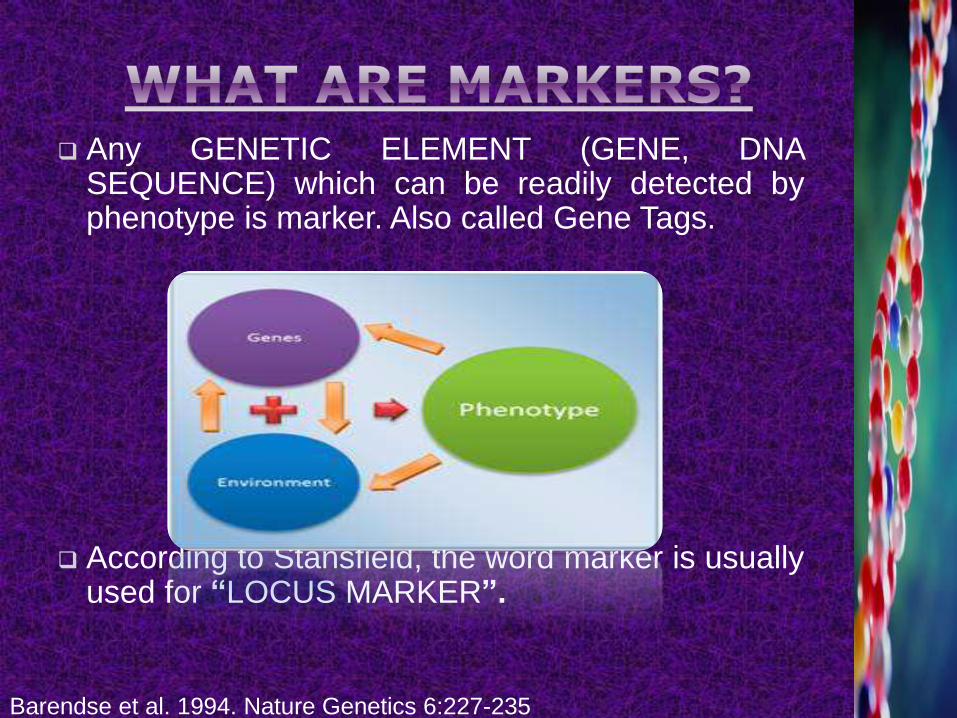
Any GENETIC ELEMENT (GENE, DNASEQUENCE) which can be readily detected byphenotype is marker. Also called Gene Tags.
According to Stansfield, the word marker is usuallyused for “LOCUS MARKER”.
Barendse et al. 1994. Nature Genetics 6:227-235
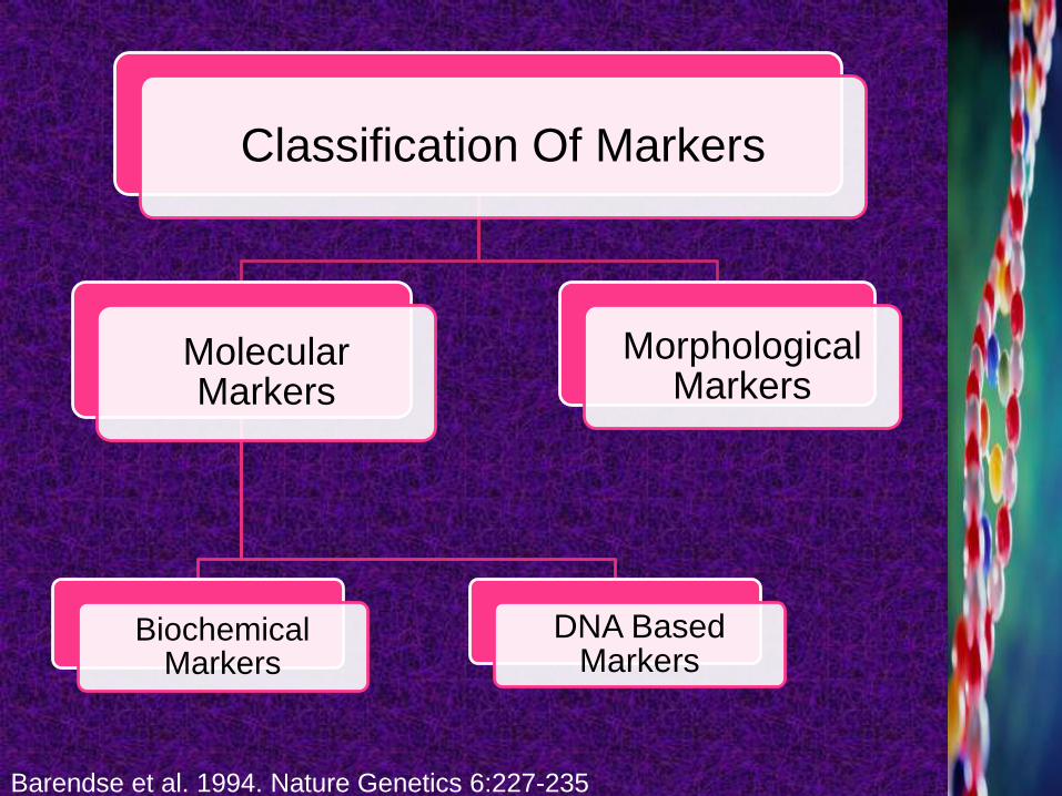
Classification Of Markers
Molecular Markers
Biochemical Markers
DNA Based Markers
Morphological Markers
Barendse et al. 1994. Nature Genetics 6:227-235
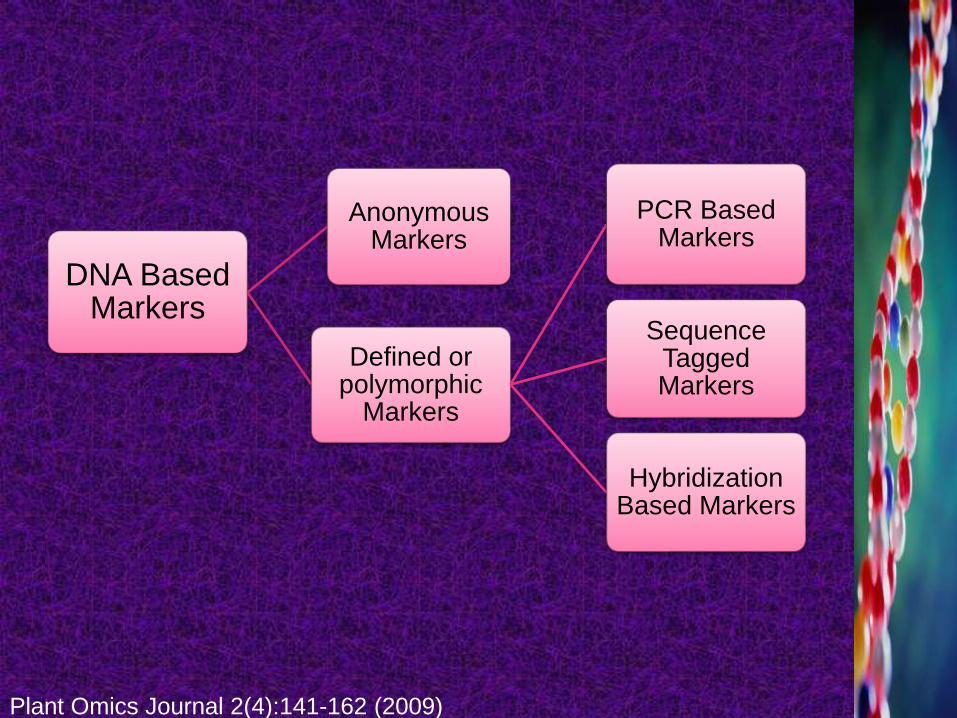
DNA Based Markers
Anonymous Markers
Defined or polymorphic
Markers
PCR Based Markers
Sequence Tagged Markers
Hybridization Based Markers
Plant Omics Journal 2(4):141-162 (2009)
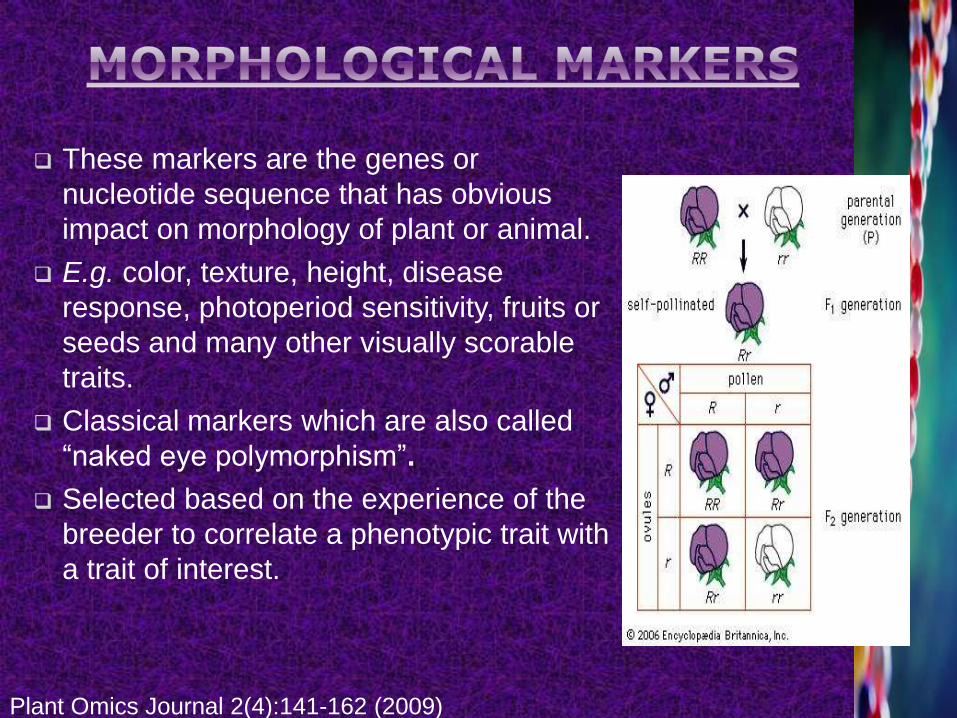
These markers are the genes or
nucleotide sequence that has obvious
impact on morphology of plant or animal.
E.g. color, texture, height, disease
response, photoperiod sensitivity, fruits or
seeds and many other visually scorable
traits.
Classical markers which are also called
“naked eye polymorphism”.
Selected based on the experience of the
breeder to correlate a phenotypic trait with
a trait of interest.
Plant Omics Journal 2(4):141-162 (2009)
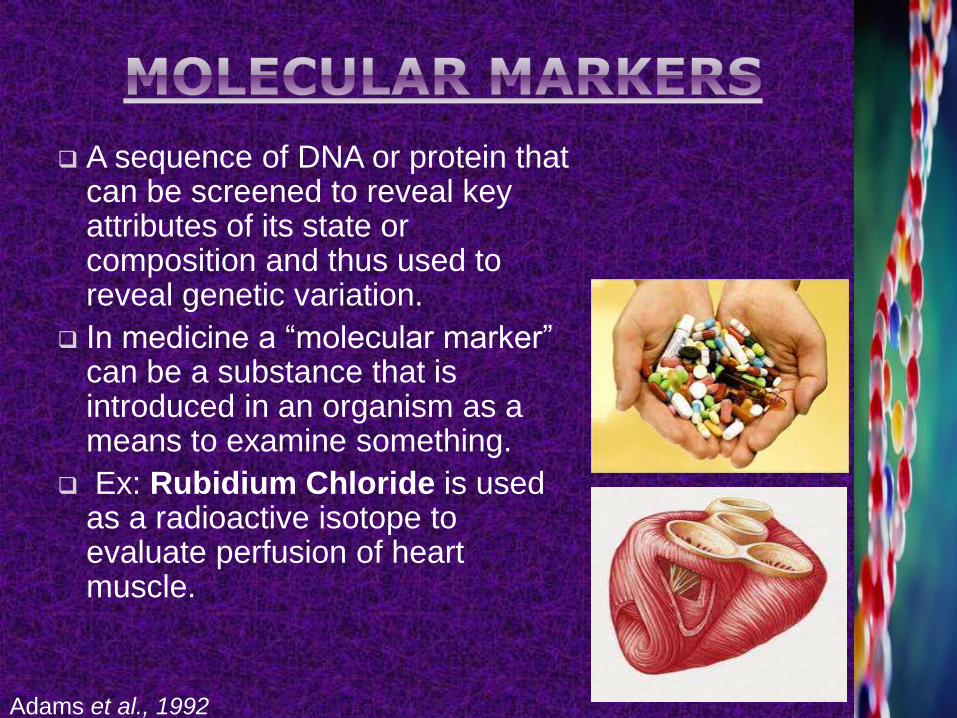
A sequence of DNA or protein that can be screened to reveal key attributes of its state or composition and thus used to reveal genetic variation.
In medicine a “molecular marker” can be a substance that is introduced in an organism as a means to examine something.
Ex: Rubidium Chloride is used as a radioactive isotope to evaluate perfusion of heart muscle.
Adams et al., 1992
Also known as “Genetic Marker”.
Genetic markers are the sequences of DNA which
have been traced to specific location on the
chromosomes and associated with particular
traits.
Molecular Markers are classified as:
1. Protein Based Markers/ Biochemical Markers
2. DNA Based Markers
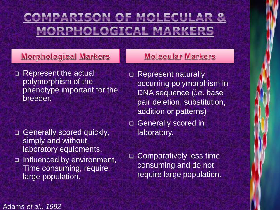
Represent the actual polymorphism of the phenotype important for the breeder.
Generally scored quickly, simply and without laboratory equipments.
Influenced by environment, Time consuming, require large population.
Represent naturally
occurring polymorphism in
DNA sequence (i.e. base
pair deletion, substitution,
addition or patterns)
Generally scored in
laboratory.
Comparatively less time
consuming and do not
require large population.
Adams et al., 1992
These are also called isozymes.
Allozymes = isozymes encoded by the different
alleles of the same gene, which is easily
visualized by activity of gel.
These are the electrophoretic variants of proteins
produced by different alleles at protein coding
genes, it also involve the detection of specific
metabolite usually based on color.
DNA marker = direct reflection of genotype.
“Any unique DNA sequence which can be used in
DNA hybridization, PCR or restriction mapping
experiments to identify that sequence.”
In 1980, variations in the pattern of DNA fragments
were observed generated by restriction enzyme
digestion of genomic DNA could be used as genetic
marker.
DNA-markers allow the breeder to introduce into
their cultivated plant only the gene(s) of interest
from a related species as compare to conventional
breeding.
Wakasugi et al., 1994a,b
Allow eliminating in a few generations ‘undesired’ genome regions.
Segregate as single genes.
Not affected by the environment.
DNA based markers are classified as:
1) Anonymous Markers
2) Defined Markers
Anonymous Markers:
“A cloned random DNA fragment whose function or specific features are not known e.g. Microsatellites and AFLP. These marker type generally measure apparently neutral DNA variations.
Wakasugi et al., 1994a,b
Defined Marker OR Polymorphic marker:
“A defined marker may contain a gene or some other specific features, e.g. restriction sites for cutting restriction enzymes, etc.”
Polymorphism of DNA Marker:
“DNA markers representing polymorphism in the actual base sequence of DNA.”
This can be represented by:
Mutation at restriction site.
Insertion or deletion between restriction sites.
Mutations at single nucleotide.
Changes in number of repeat unit between restriction site or PCR primer sites.
Wakasugi et al., 1994a,b
DNA Polymorphic markers are classified
into three categories:
1) PCR Based Markers
2) Sequence Tagged Sites
3) Hybridization Based Markers
PCR Based Markers:
“PCR based markers involve in vitro amplification
of particular DNA sequences or loci, with the help
of specifically and arbitrarily chosen primers and a
DNA polymerase enzyme. The amplified fragments
are separated electrophoretically and banding
patterns are detected by different methods i.e.
staining and autoradiography.”
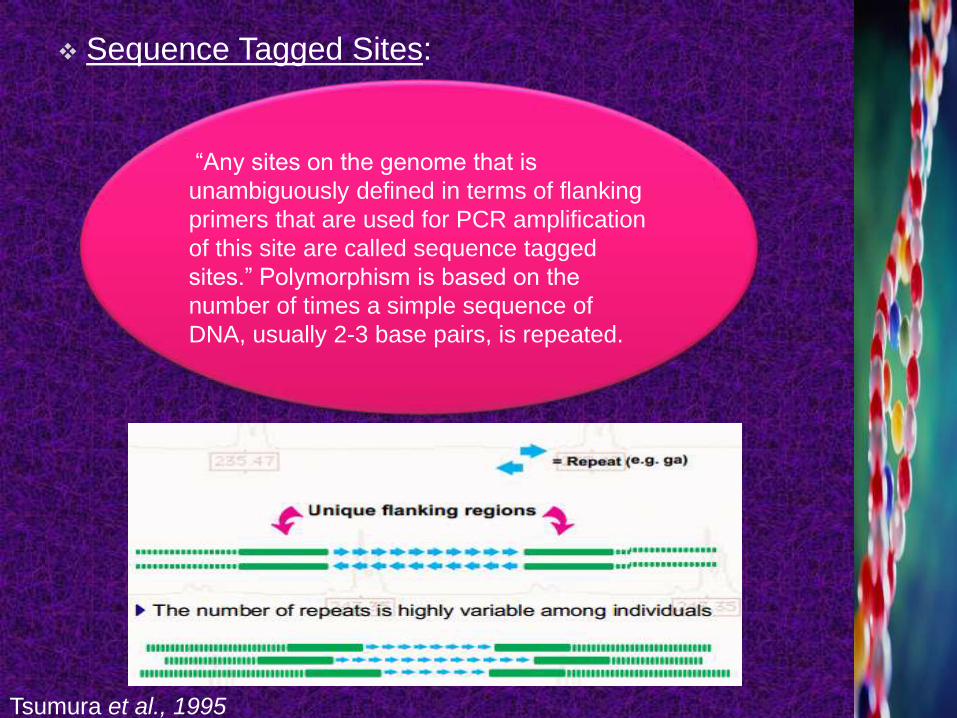
Sequence Tagged Sites:
“Any sites on the genome that is
unambiguously defined in terms of flanking
primers that are used for PCR amplification
of this site are called sequence tagged
sites.” Polymorphism is based on the
number of times a simple sequence of
DNA, usually 2-3 base pairs, is repeated.
Tsumura et al., 1995
1970s --- Genetic marker system based on DNA-DNA Hybridization was developed.
Eukaryotes have large genome so genetic polymorphism can’t easily be studied.
Hybridization technique reveal polymorphism over there.
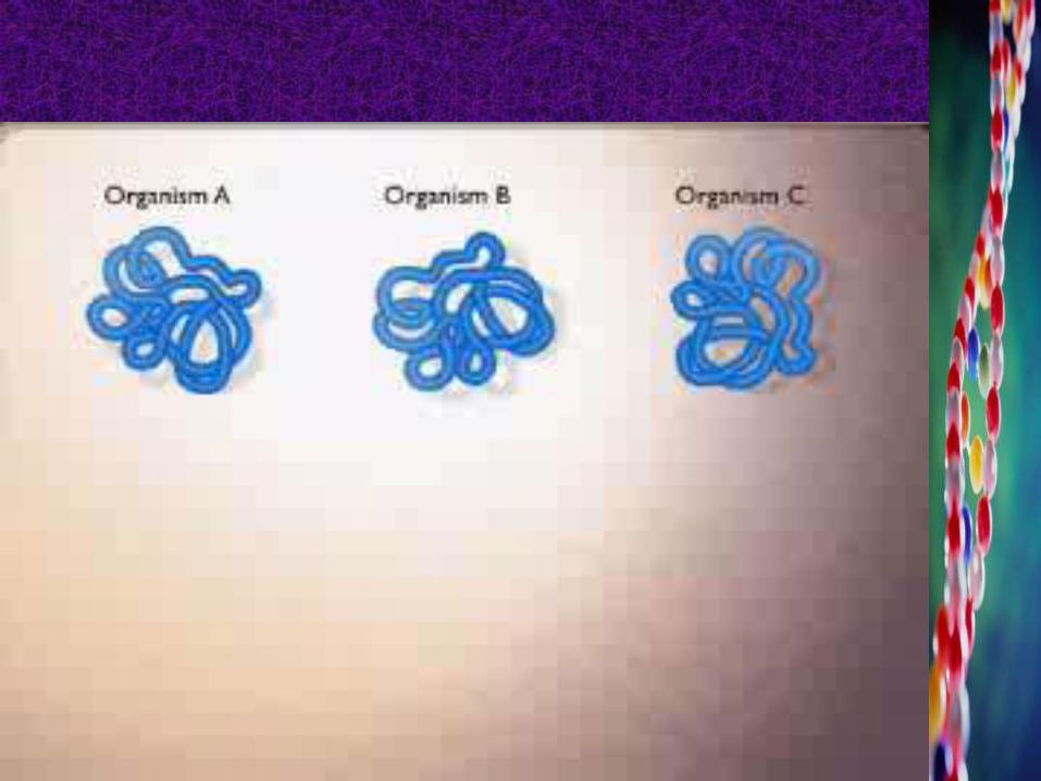
“The variation in the length of DNA fragments produced
by specific Restriction Endonucleases from genomic DNA
s of two or more individuals of a species is called
hybridization and markers produced by this technique are
called hybridization based molecular markers.”
Barendse et al. 1994. Nature Genetics 6:227-235
The type of hybridization based markers
includes
RFLP
Barendse et al. 1994. Nature Genetics 6:227-235
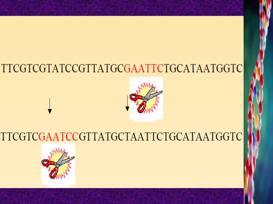
“RFLP is restriction fragment length
polymorphism, which are variations in the
DNA sequence of an individual which may be
detected by Restriction Endonucleases, which
cut the double stranded DNA whenever they
recognize a highly specific oligonucleotide
sequence or a restriction site.”
Amer J Bot 81:1309–1326
“ A molecular method of genetic
analysis that allows individuals
to be identified on the basis of
unique patterns of restriction
enzymes cutting in specific
regions of DNA. It is an
application of Southern
Hybridization Procedure. ”
Amer J Bot 81:1309–1326
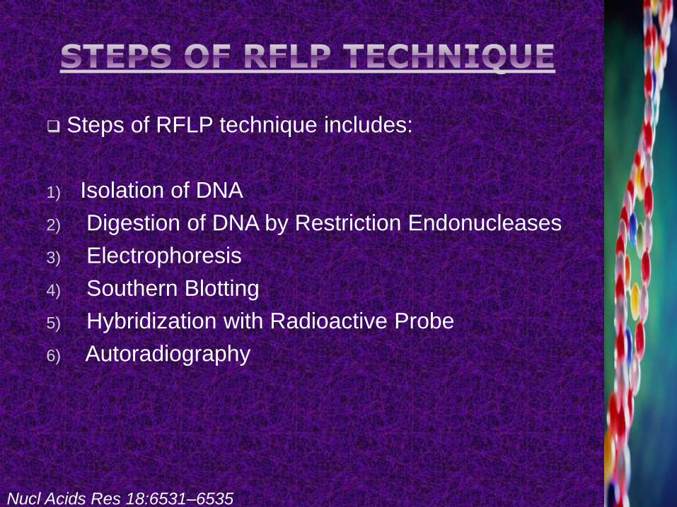
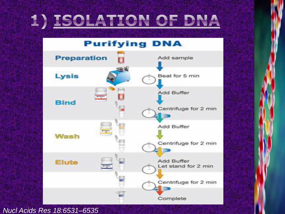
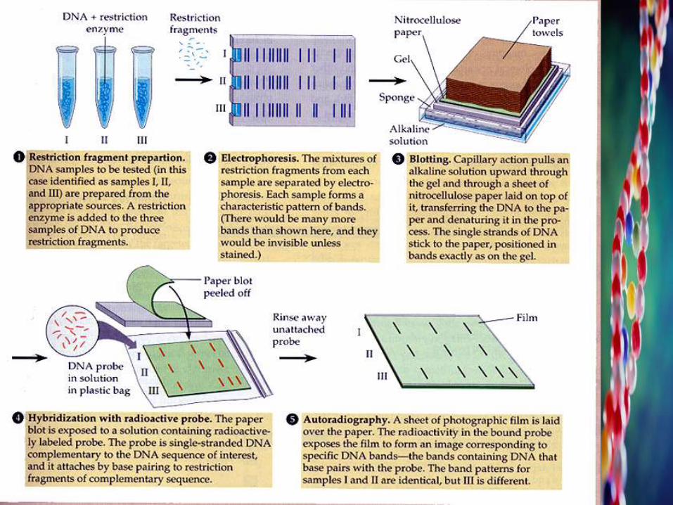
Steps of RFLP technique includes:
1) Isolation of DNA
2) Digestion of DNA by Restriction Endonucleases
3) Electrophoresis
4) Southern Blotting
5) Hybridization with Radioactive Probe
6) Autoradiography
Nucl Acids Res 18:6531–6535
Nucl Acids Res 18:6531–6535
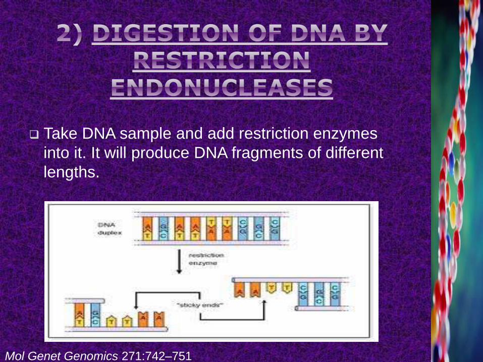
Take DNA sample and add restriction enzymes
into it. It will produce DNA fragments of different
lengths.
Mol Genet Genomics 271:742–751
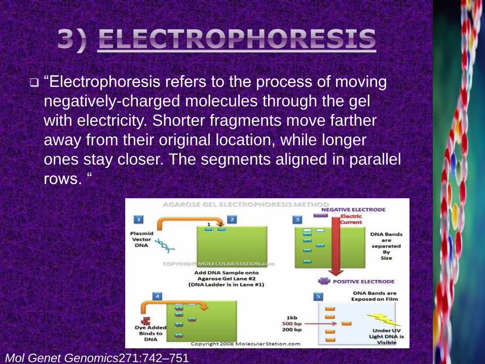
“Electrophoresis refers to the process of moving
negatively-charged molecules through the gel
with electricity. Shorter fragments move farther
away from their original location, while longer
ones stay closer. The segments aligned in parallel
rows. “
Mol Genet Genomics271:742–751
“Southern blotting is a method routinely used in
molecular biology for the detection of specific
DNA sequence in DNA samples. Southern blotting
combines’ transfer of electrophoresis separated
DNA fragments to a filter membrane and
subsequent fragment detection by hybridization
probes. ”
‘Nitrocellulose’ or ‘Nylon’ sheet is placed on top of
the gel.
Pressure is applied through paper towel.
Permanent attachment is done either by ‘Baking
(80ᴼC, 2 Hrs)’ or by ‘UV-exposure’.
Botstein et al.1980
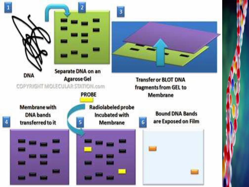
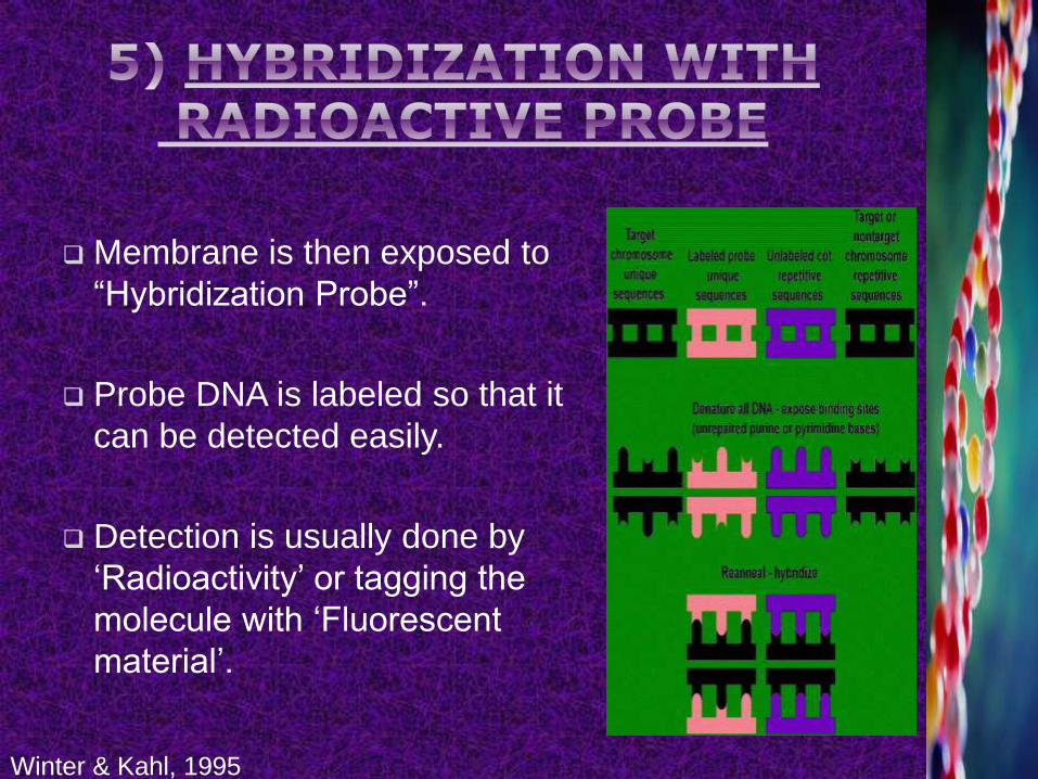
Membrane is then exposed to
“Hybridization Probe”.
Probe DNA is labeled so that it
can be detected easily.
Detection is usually done by
‘Radioactivity’ or tagging the
molecule with ‘Fluorescent
material’.
Winter & Kahl, 1995
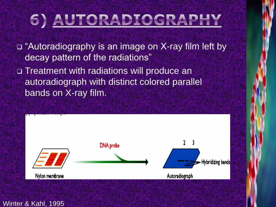
“Autoradiography is an image on X-ray film left by
decay pattern of the radiations”
Treatment with radiations will produce an
autoradiograph with distinct colored parallel
bands on X-ray film.
Winter & Kahl, 1995
They are co- dominant.
Measure variation at the level of DNA sequence,
not protein sequence.
RFLP loci are very large so even very small
segments of chromosomes can be mapped and
also study phylogenetic relationship.
Very reliable for linkage analysis and for detecting
coupling phase of DNA molecules.
Plant Omics Journal 2(4):141-162 (2009)
Requires relatively very large amount of DNA.
Requirement of radioactive probe makes the
analysis expensive and hazardous.
They are not useful for detecting single base
change or point mutations.
It is time consuming, laborious, and expensive.
The level of polymorphism is low.
Plant Omics Journal 2(4):141-162 (2009)