hyperinsulinemic hypoglycemia in children and adolescents ... · hyperinsulinemic hypoglycemia in...
TRANSCRIPT
Hyperinsulinemic hypoglycemia in children and adolescents: Recentadvances in understanding of pathophysiology and management
Maria Gϋemes1,2,3 & Sofia Asim Rahman1& Ritika R. Kapoor4 & Sarah Flanagan5
& Jayne A. L. Houghton5,6&
Shivani Misra7 & Nick Oliver7 & Mehul Tulsidas Dattani1,2 & Pratik Shah1,2
# The Author(s) 2020
AbstractHyperinsulinemic hypoglycemia (HH) is characterized by unregulated insulin release, leading to persistently low bloodglucose concentrations with lack of alternative fuels, which increases the risk of neurological damage in these patients. Itis the most common cause of persistent and recurrent hypoglycemia in the neonatal period. HH may be primary,Congenital HH (CHH), when it is associated with variants in a number of genes implicated in pancreatic developmentand function. Alterations in fifteen genes have been recognized to date, being some of the most recently identifiedmutations in genes HK1, PGM1, PMM2, CACNA1D, FOXA2 and EIF2S3. Alternatively, HH can be secondary whenassociated with syndromes, intra-uterine growth restriction, maternal diabetes, birth asphyxia, following gastrointestinalsurgery, amongst other causes. CHH can be histologically characterized into three groups: diffuse, focal or atypical.Diffuse and focal forms can be determined by scanning using fluorine-18 dihydroxyphenylalanine-positron emissiontomography. Newer and improved isotopes are currently in development to provide increased diagnostic accuracy inidentifying lesions and performing successful surgical resection with the ultimate aim of curing the condition. Rapiddiagnostics and innovative methods of management, including a wider range of treatment options, have resulted in areduction in co-morbidities associated with HH with improved quality of life and long-term outcomes. Potential futuredevelopments in the management of this condition as well as pathways to transition of the care of these highlyvulnerable children into adulthood will also be discussed.
Keywords Hyperinsulinism . Hypoglycemia . Sirolimus . Lanreotide . 18F-DOPA-PET . Transition to adult services
1 Introduction
Glucose is one of the principal energy substrates, providinghalf of the body’s total energy requirements. As the brain canneither synthesize nor store more than a fewminutes supply ofglucose, its function is solely dependent on maintenance ofnormal glucose concentrations in the circulation. An abnor-mally reduced concentration of glucose in the blood is referredto as hypoglycemia. It is a medical emergency and can lead tosymptoms due to neuroglycopenia [1].
In healthy individuals, maintenance of a normal plasmaglucose concentration relies on a tightly controlled balancebetween glucose production (dietary intake, glycogenolysis,gluconeogenesis) and its utilization by the tissues (glycolysis,glycogenesis, conversion to fatty acids). A normal endocrinesystem is essential for integrating and modulating substratemobilization, interconversion, and utilization. In addition,the endocrine system interacts with metabolic pathways thatrely critically on functionally intact enzymes. There are two
* Pratik [email protected]; [email protected]
1 Genetics and Genomic Medicine Programme, UCL Great OrmondStreet Institute of Child Health, Great Ormond Street,London WC1N 3JH, UK
2 Department of Pediatric Endocrinology, Great Ormond StreetHospital for Children, London, UK
3 Endocrinology Service, Hospital Infantil Universitario Niño Jesús,Madrid, Spain
4 Pediatric Diabetes and Endocrinology, King’s College Hospital NHSTrust, Denmark Hill, London, UK
5 Institute of Biomedical and Clinical Science, University of ExeterMedical School, Exeter, UK
6 Royal Devon and Exeter Foundation Trust, Exeter, UK7 Department of Diabetes, Endocrinology and Metabolic Medicine,
Faculty of Medicine, Imperial College Healthcare NHS Trust,London, UK
Reviews in Endocrine and Metabolic Disordershttps://doi.org/10.1007/s11154-020-09548-7

types of metabolic hormones affecting blood glucose concen-trations – an anabolic hormone (insulin), which decreasesblood glucose, and several catabolic hormones (such asglucagon, cortisol and catecholamines) which increase bloodglucose concentrations.
Hyperinsulinemic hypoglycemia (HH) is the commonestcause of persistent hypoglycemia in infants and children [2]and it can be transient –associated to risk factors- or permanent–linked to genetic mutations-. The risk of permanent braininjury in infants with HH continues to be as high as 25–50%due to delays in diagnosis and inadequate treatment. Despiteadvances in genetics, improved modes of investigation, novelmanagement options and abrigding pediatric and adult follow-up in holistic multidisciplinary transition clinics, significantmorbidity and mortality is still a major issue in children andyoung adults with HH [3–5].
The present review has beenwritten using a comprehensiveand up- to -da t e l i t e r a tu re sea r ch on congen i t a lhyperinsulinism/HH including the latest publications avail-able in PubMed (last search in August 2019). It also incorpo-rates clinical and laboratory experience from reference centersfor the diagnosis and management of HH, as well as availabledata from on-going pharmaceutical trials.
2 normal blood glucose and hypoglycemia
2.1 Definition of normal blood glucose
Blood glucose concentrations of normal term neonates appro-priate for gestational age may range between 1.4–6.2 mmol/l(25–112mg/dl) during the first 72 h of life; however after that,healthy children and adults will maintain blood glucose con-centrations between 3.5–5.5 mmol/l (63–99 mg/dl) [6]. It isdifficult to numerically define hypoglycemia given that a sin-gle cut-off value cannot suit all individuals in every situation.Therefore operational thresholds are recommended which in-dicate that in any baby with clinical signs of hypoglycemia,blood glucose levels must be maintained over 2.6 mmol/l(47 mg/dl) except for suspected cases of hyperinsulinemichypoglycemia in which 3.5 mmol/l (63 mg/dl) should be thecut-off point [7]. However, the Pediatric EndocrinologySociety recommends that when a congenital disorder causinghypoglycemia is suspected in a neonate and when confirmedin older infants and children, the aim is to keep plasma glucoseconcentrations over 3.9 mmol/l (70 mg/dl) [7].
2.2 Causes of hypoglycemia
For hypoglycemia to occur, the rate of appearance ofglucose into the plasma space must be less than its rateof utilization [8]. This can be due to defective glucoseproduction, increased glucose utilization, or some
combination of the two. Excessive glucose utilizationdue to hyperinsulinism (exogenous/endogenous) is oneo f t h e commone s t c a u s e s o f hypog l yc em i a .Hypoglycemia can also occur due to deficiencies ofvarious counter regulatory hormones. The causes arecollected in Table 1.
Hereditary disorders caused by deficiency of specific en-zymes involved inmobilization, interconversion, or utilizationof metabolic substrates frequently are associated with hypo-glycemia. These enzymatic defects may involve carbohydrate,amino acid, or fat metabolism and are individually rare; al-most all are inherited as autosomal recessive traits [8].
3 HYPERINSULINEMIC hypoglycemia (HH)
HH is a condition caused by the upregulation of β-cellsecretion of insulin producing a hypoglycemic state.Congenital hyperinsulinism (CHH) is the most commoncause of transient or permanent hypoglycemia and couldpotentially be life threatening causing neurological dam-age. Hence it requires quick and effective treatment andmanagement [8]. This disorder is rare and has an inci-dence of around 1:40,000 births in the general popula-tion [15]. CHH can occur due to genetic mutations andone of the most common causes are defects in the β-cell ATP-sensitive potassium (KATP) channels, known aschannelopathies [8]. KATP channels are comprised oftwo subunits; the inward rectifying Kir6.2 channelsand the sulphonylurea receptor-1, SUR-1, which areencoded for by the KCNJ11 (potassium voltage-gatedchannel subfamily J member 11) and ABCC8 (ATP-binding cassette transporter sub-family C member 8)genes, respectively [16]. Both these subunits are sensi-tive to the ADP/ATP nucleotide ratio and work togetherto promote cell depolarization and eventual insulin se-cretion. Mutations in the KCNJ11/ABCC8 genes areknown to cause defects in biogenesis/trafficking of thesesubunits to the plasma membrane, thus causing HH.
4 Causes of HH
4.1 Transient forms of HH
Transient HH is a poorly defined term that refers to thegroup of patients in whom HH spontaneously resolveswithin a few days to approximately a week. However,the cohort includes children requiring medications up to6 months of life and is usually negative for a knowngenetic etiology for HH [17]. It is associated with intra-uterine growth retardation, erythroblastosis fetalis, peri-natal asphyxia, maternal diabetes mellitus (gestational or
Rev Endocr Metab Disord
insulin dependent) and after the maternal administrationof drugs such as sulphonylureas, and intravenous glu-c o s e i n f u s i o n s du r i n g l a b o r [ 2 ] . Abno rma lneurodevelopment is evident in one third of childrenwith transient forms of HH associated with perinatalrisk factors [5].
4.2 Permanent form of HH
A permanent form of HH, usually congenital (CHH), is wherechildren continue to need medical treatment even after6 months of age. Various genetic causes have been identified,however nearly 40–50% of children still remain geneticallyunidentified [17].
4.2.1 Molecular basis of CHH
To date, at least 15 genes have been identified to be accom-panied with CHH, which include ABCC8, KCNJ11, GLUD1,GCK, HADH, SLC16A1, UCP2, HNF1A, HNF4A, HK1,PGM1, PMM2, FOXA2, CACNA1D and EIF2S3.
Various modes of inheritance are observed. For some pa-tients specific clinical characteristics, such as the presence ofhyperammonemia, can help guide molecular testing; however,for most of the genetic subgroups there is an overlap in phe-notype and as such testing of all the known genes is oftenrequired.
KATP channel genes (ABCC8 and KCNJ11) The pancreatic KATP
channel is a key component of the insulin secretion pathway.
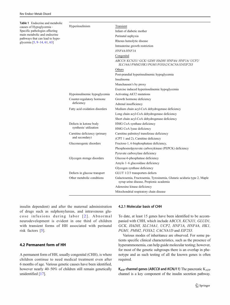
Table 1 Endocrine and metaboliccauses of Hypoglycemia -Specific pathologies affectingmain metabolic and endocrinepathways that can lead to hypo-glycemia [5, 9–14, 61, 63]
Hyperinsulinism Transient
Infant of diabetic mother
Perinatal asphyxia
Rhesus hemolytic disease
Intrauterine growth restriction
HNF4A/HNF1A
Congenital
ABCC8/ KCNJ11/ GCK/ GDH/ HADH/ HNF4A/ HNF1A/ UCP2/SLC16A1/PMM2/HK1/PGM1/FOXA2/CACNA1D/EIF2S3
Others
Post-prandial hyperinsulinemic hypoglycemia
Insulinoma
Munchausen’s by proxy
Exercise induced hyperinsulinemic hypoglycemia
Hypoinsulinemic hypoglycemia Activating AKT2 mutations
Counter-regulatory hormonedeficiency
Growth hormone deficiency
Adrenal insufficiency
Fatty acid oxidation disorders Medium chain acyl-CoA dehydrogenase deficiency
Long chain acyl-CoA dehydrogenase deficiency
Short chain acyl-CoA dehydrogenase deficiency
Defects in ketone bodysynthesis/ utilization
HMG CoA synthase deficiency
HMG CoA lyase deficiency
Carnitine deficiency (primaryand secondary)
Carnitine palmitoyl transferase deficiency
(CPT 1 and 2), Carnitine deficiency
Gluconeogenic disorders Fructose-1, 6-bisphosphatase deficiency,
Phosphoenolpyruvate carboxykinase (PEPCK) deficiency
Pyruvate carboxylase deficiency
Glycogen storage disorders Glucose-6-phosphatase deficiency
Amylo 1–6 glucosidase deficiency
Glycogen synthase deficiency
Defects in glucose transport GLUT 1/2/3 transporters defects
Other metabolic conditions Galactosemia, Fructosemia, Tyrosinemia, Glutaric aciduria type 2, Maplesyrup urine disease, Propionic academia
Adenosine kinase deficiency
Mitochondrial respiratory chain disease
Rev Endocr Metab Disord
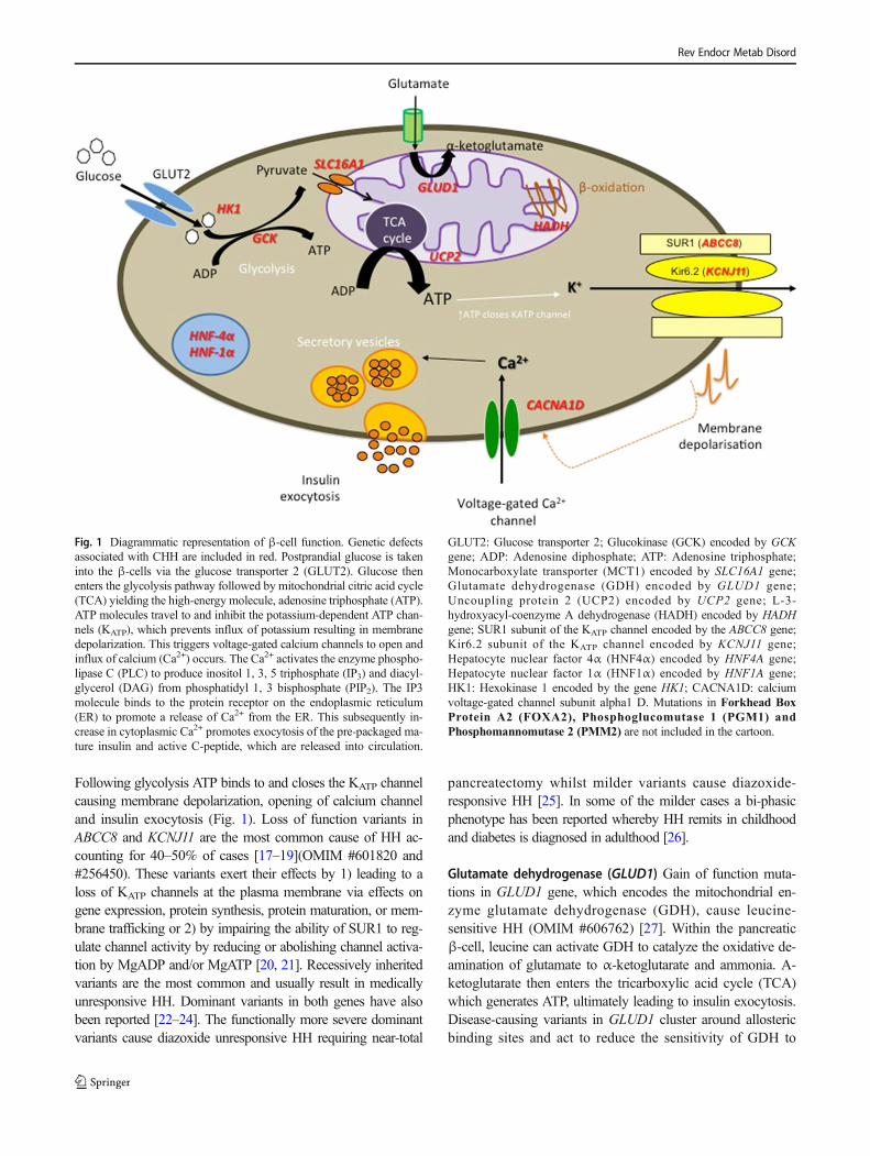
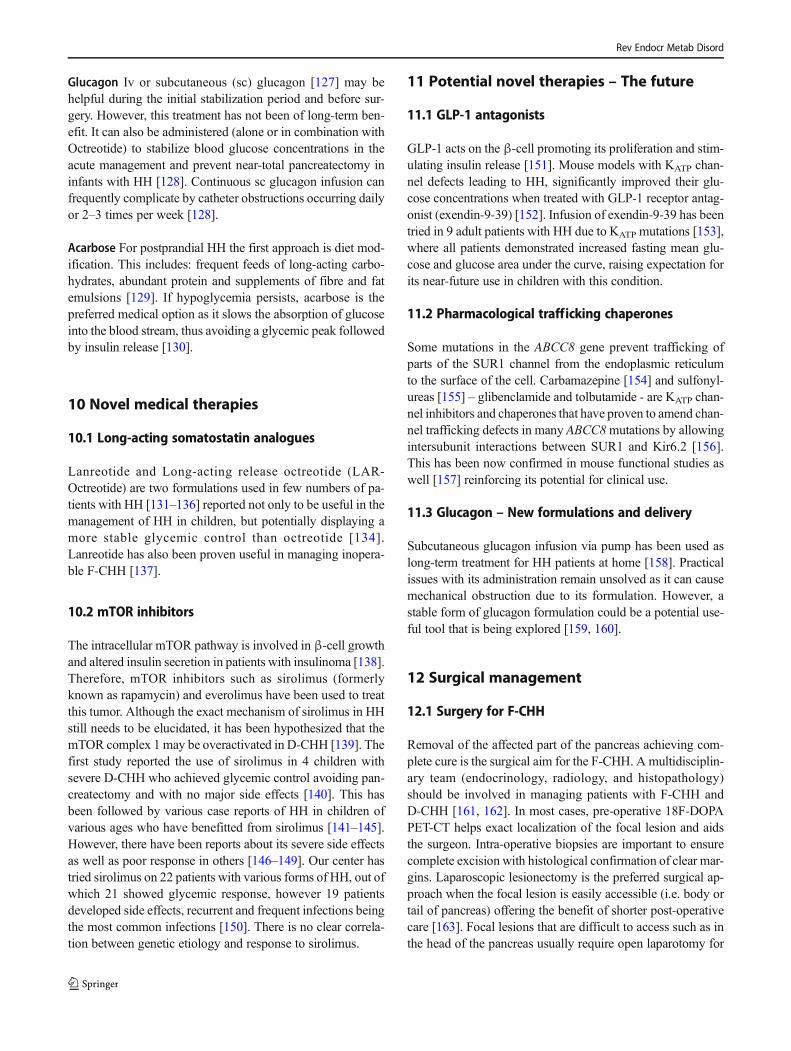
Following glycolysis ATP binds to and closes the KATP channelcausing membrane depolarization, opening of calcium channeland insulin exocytosis (Fig. 1). Loss of function variants inABCC8 and KCNJ11 are the most common cause of HH ac-counting for 40–50% of cases [17–19](OMIM #601820 and#256450). These variants exert their effects by 1) leading to aloss of KATP channels at the plasma membrane via effects ongene expression, protein synthesis, protein maturation, or mem-brane trafficking or 2) by impairing the ability of SUR1 to reg-ulate channel activity by reducing or abolishing channel activa-tion by MgADP and/or MgATP [20, 21]. Recessively inheritedvariants are the most common and usually result in medicallyunresponsive HH. Dominant variants in both genes have alsobeen reported [22–24]. The functionally more severe dominantvariants cause diazoxide unresponsive HH requiring near-total
pancreatectomy whilst milder variants cause diazoxide-responsive HH [25]. In some of the milder cases a bi-phasicphenotype has been reported whereby HH remits in childhoodand diabetes is diagnosed in adulthood [26].
Glutamate dehydrogenase (GLUD1) Gain of function muta-tions in GLUD1 gene, which encodes the mitochondrial en-zyme glutamate dehydrogenase (GDH), cause leucine-sensitive HH (OMIM #606762) [27]. Within the pancreaticβ-cell, leucine can activate GDH to catalyze the oxidative de-amination of glutamate to α-ketoglutarate and ammonia. A-ketoglutarate then enters the tricarboxylic acid cycle (TCA)which generates ATP, ultimately leading to insulin exocytosis.Disease-causing variants in GLUD1 cluster around allostericbinding sites and act to reduce the sensitivity of GDH to
Fig. 1 Diagrammatic representation of β-cell function. Genetic defectsassociated with CHH are included in red. Postprandial glucose is takeninto the β-cells via the glucose transporter 2 (GLUT2). Glucose thenenters the glycolysis pathway followed by mitochondrial citric acid cycle(TCA) yielding the high-energy molecule, adenosine triphosphate (ATP).ATP molecules travel to and inhibit the potassium-dependent ATP chan-nels (KATP), which prevents influx of potassium resulting in membranedepolarization. This triggers voltage-gated calcium channels to open andinflux of calcium (Ca2+) occurs. The Ca2+ activates the enzyme phospho-lipase C (PLC) to produce inositol 1, 3, 5 triphosphate (IP3) and diacyl-glycerol (DAG) from phosphatidyl 1, 3 bisphosphate (PIP2). The IP3molecule binds to the protein receptor on the endoplasmic reticulum(ER) to promote a release of Ca2+ from the ER. This subsequently in-crease in cytoplasmic Ca2+ promotes exocytosis of the pre-packaged ma-ture insulin and active C-peptide, which are released into circulation.
GLUT2: Glucose transporter 2; Glucokinase (GCK) encoded by GCKgene; ADP: Adenosine diphosphate; ATP: Adenosine triphosphate;Monocarboxylate transporter (MCT1) encoded by SLC16A1 gene;Glutamate dehydrogenase (GDH) encoded by GLUD1 gene;Uncoupling protein 2 (UCP2) encoded by UCP2 gene; L-3-hydroxyacyl-coenzyme A dehydrogenase (HADH) encoded by HADHgene; SUR1 subunit of the KATP channel encoded by the ABCC8 gene;Kir6.2 subunit of the KATP channel encoded by KCNJ11 gene;Hepatocyte nuclear factor 4α (HNF4α) encoded by HNF4A gene;Hepatocyte nuclear factor 1α (HNF1α) encoded by HNF1A gene;HK1: Hexokinase 1 encoded by the gene HK1; CACNA1D: calciumvoltage-gated channel subunit alpha1 D. Mutations in Forkhead BoxProtein A2 (FOXA2), Phosphoglucomutase 1 (PGM1) andPhosphomannomutase 2 (PMM2) are not included in the cartoon.
Rev Endocr Metab Disord
inhibition byGTP andATP [28]. This loss of regulation leads toan increase in the activity of GDH, a subsequent increase in theamount of α-ketoglutarate entering the TCA cycle and conse-quently an unregulated insulin secretory response.
Individuals with GLUD1 variants usually present with amilder form of HH that is often diagnosed outside of the neo-natal period and is diazoxide-responsive [27]. In some patientsdietary protein restriction may also be required. A consistentfeature of this disorder is the presence of plasma ammoniumconcentrations raised two to three times the upper limit ofnormal. The presence of persistent hyperammonemia, in mostbut not all patients with GLUD1 variants [29], has led to thissubtype of hyperinsul inism being referred to asHyperinsulinism/Hyperammonemia (HI/HA) syndrome(OMIM #606762). An increased risk of epilepsy has also beenobserved in individuals with disease-causingGLUD1 variants[29].
In the majority of cases GLUD1 variants arise de novo,with no family history of hyperinsulinism. In keeping withtheir dominant nature, 50% of future offspring of affectedindividuals are at risk of inheriting the variant and developingHH.
Glucokinase (GCK) Within the pancreatic β-cell glucokinase(GCK) plays a key role in linking insulin secretion to a glucosechallenge by facilitating the phosphorylation of glucose toglucose-6-phosphate, the first step in glycolysis.Heterozygous gain-of-function variants in GCK cause HHby increasing the affinity of GCK for glucose which then actsto lower the threshold for glucose-stimulated insulin secretion[30] (OMIM #602485).
Individuals with gain-of-function GCK variants will oftenhave a dominant family history of HH. The absence of a fam-ily history should however not preclude testing as de novovariants have been reported [31]. Variability in the severityof HH is also observed both in terms of age at presentation,which can range from birth to adulthood, and treatment re-sponse [31, 32]. Whilst the majority of individuals are suc-cessfully treated with diazoxide, some patients have medicallyunresponsive HH and require near-total pancreatectomy [32].Although these differences in phenotype are likely to correlatewith the functional severity of the variant, phenotypic variabil-ity within families with the same variant has been observedand is likely to be a consequence of genetic background and/or environmental factors [33].
Hydroxyacyl-coenzyme a dehydrogenase (HADH) HADH en-codes 3-Hydroxyacyl-coenzyme A dehydrogenase, anintramitochondrial enzyme that catalyzes the penultimate re-action in the β-oxidation pathway. Loss-of-function variantsin HADH result in loss of interaction between HADH andglutamate dehydrogenase [34, 35]. This in turn leads to anincrease in glutamate dehydrogenase activity, a subsequent
rise in intracellular ATP and upregulated insulin secretion[36].
Patients with HADH disease-causing variants present withdiazoxide-responsive protein-induced HH [36–38](OMIM#609975). The severity in phenotype ranges from mild lateonset hypoglycemia to severe neonatal hypoglycemia. Insome patients there are raised plasma concentrations of 3-hydroxybutyrylcarnitine and urinary 3-hydroxyglutaric acid[36]. Loss- of- function variants in HADH are recessivelyinherited and this is the most common genetic subtype ofHH in consanguineous individuals [39].
Hepatocyte nuclear factors (HNF4A and HNF1A) The hepato-cyte nuclear transcription factors, HNF1A and HNF4A, playcrucial roles in glucose-stimulated insulin secretion as evi-denced by the identification of loss-of- function variants inthese genes in individuals with maturity-onset diabetes ofthe young (MODY)(OMIM #125850), an autosomal domi-nant form of diabetes typically diagnosed before the age of25 years [40]. HNF4A variants were reported to cause a bi-phasic phenotype in individuals presenting with macrosomiaand transient HH during the neonatal period and diabetes inlater life [41]. The duration of HH varies markedly with somepatients treated with intravenous glucose infusion for 1–2 days, yet others require diazoxide treatment for up to11 years [42–44]. HH with Fanconi syndrome, a renal tubulardysfunction, has been reported in at least 8 patients withHNF4AHH, all individuals have the p.R76W variant suggest-ing that this is a mutation specific phenotype [45–48]. A fewcases with HNF1A variants and transitory neonatal hypogly-cemia have also been reported [45, 49].
Whilst a dominant family history of macrosomia, neonatalhypoglycemia and/or young onset diabetes can help to guidegenetic testing for this condition, the absence of affected fam-ily members should not preclude analysis of HNF4A as denovo disease-causing variants have been reported [43].
Solute carrier family 16 member 1 (SLC16A1) SLC16A1 geneencodes the monocarboxylate transporter (MCT1) whichtransports the insulin secretagogues pyruvate and lactate.Under normal physiological conditions SLC16A1 is notexpressed in the β-cell thus preventing insulin from beingsecreted in response to lactate and pyruvate. Rare activatingdominant variants result in the expression of MCT1 in the β-cell leading to pyruvate-stimulated insulin secretion followingexercise [50, 51] (OMIM #610021). For patients withexercise-induced HH, treatment is not usually necessary ashypoglycemic episodes may be prevented by avoiding stren-uous exercise [52].
Uncoupling protein 2 (UCP2) UCP2 gene encodes an innermitochondrial carrier protein UCP2which is widely expressedin tissues, including pancreatic islets [53, 54]. UCP2 can
Rev Endocr Metab Disord
inhibit ATP generation by causing proton leak across the innermitochondrial membrane and negatively regulates glucose-mediated insulin secretion [54]. Inactivating heterozygousmutations in the UCP2 gene can enhance glucose oxidationand increase intracellular ATP synthesis leading to HH [54].UCP2 mutations can present with either transient orprolonged HH [55]. However, in a recent published study,no mutations were detected in the UCP2 gene among 206diazoxide responsive patients [56], suggesting that the roleof UCP2 in HH needs further investigation.
Hexokinase 1 (HK1) HK1 encodes the hexokinase HK1, whichcatalyzes the phosphorylation of glucose to glucose-6-phosphateas substrate for glycolysis. A family with dominant gain-of-function mutation in the HK1 gene has been reported with “idi-opathic hypoglycemia of infancy” [57]. In vitro studies evaluat-ing pancreatic β-cells from CHH patients have shown inappro-priate expression of HK1. These pancreatic tissues showed func-tional KATP channels with inappropriate secretion of insulin atlow plasma glucose concentrations (1 mmol/L) [58].
Phosphoglucomutase 1 (PGM1) PGM1 is involved in glyco-genmetabolism and is responsible for reversible conversion ofglucose-6-phosphate to glucose-1-phosphate. PGM1 gene en-codes the enzyme PGM1 and recessive loss-of-function mu-tations in PGM1 cause hypoglycemia [59].
Children with PGM1mutation presented with postprandialHH and fasting hyperketotic hypoglycemia [59].
Phosphomannomutase 2 (PMM2) Recessively inherited vari-ants in PMM2, which encodes a key enzyme in N-glycosyla-tion, have been identified in individuals with HH and poly-cystic kidney disease. In all individuals a c.-167G > Tpromotor variant was identified that was either homozygousor in trans with a coding variant [60].
The majority of patients with PMM2 variants present withmacrosomia at birth and hypoglycemia in the first year of life.For many, hypoglycemia was the presenting feature and oftenmanifested with seizures. Patients are responsive to treatmentwith diazoxide [60].
Forkhead box protein A2 (FOXA2) Mutations in FOXA2 havebeen reported to cause hypopituitarism, CHH and endoderm-derived organ abnormalities. These children have a uniqueclinical phenotype of hypopituitarism, CHH, dysmorphic fea-tures, and liver, pancreas, heart and gastrointestinal abnormal-ities [61, 62].
Calcium voltage-gated channel subunit alpha1 D (CACNA1D)CACNA1D gene encodes an L-type voltage-gated calciumchannel which is expressed in pancreatic β-cells and regulatesinsulin secretion. Mutations in CACNA1D have been reportedto cause HH, heart defects and severe hypotonia [63].
Eukaryotic translation initiation factor 2 subunit 3 (EIF2S3)Three cases published with variant in EIF2S3 present an un-usual dysregulation of glucose fluctuating between diazoxide-responsive HH and postprandial hyperglycemia, along withlearning difficulties and hypopituitarism [64].
4.3 Other forms
4.3.1 Postprandial forms of HH
In postprandial hyperinsulinemic hypoglycemia (PPHH), hy-poglycemia is induced hours after meal intake due toinappropriate/exaggerated insulin secretion in response tothe meal. The most common cause is “dumping” syndromein infants /chi ldren who have undergone Nissenfundoplication/gastric bypass [65, 66]. Children with PPHHafter Nissen fundoplication have an abnormally exaggeratedsecretion of the insulin secretagogue glucagon-like peptide 1(GLP-1) which may contribute to the exaggerated insulinsurge and resultant hypoglycemia [67].
PPHH is also reported in the insulin autoimmune syndromeleading to development of insulin binding autoantibodies in chil-dren whowere not previously exposed to exogenous insulin [68].
4.3.2 Other causes of HH
Insulinoma is a rare cause of hyperinsulinism and must beconsidered in older children or adolescents presenting withHH [69]. A detailed family history of tumors especially likeinsulinoma, is relevant as it can be part of multiple endocrineneoplasia syndrome type 1 (MEN1).
Munchausen by proxy leading to exogenous administrationof insulin or anti-diabetic drugs such as sulphonylureas canpresent as factitious HH. This has led to misdiagnosis andconsequent pancreatectomy [70].
4.3.3 Syndromes associated with HH
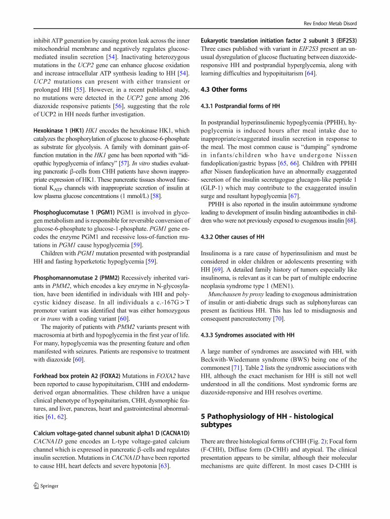
A large number of syndromes are associated with HH, withBeckwith-Wiedemann syndrome (BWS) being one of thecommonest [71]. Table 2 lists the syndromic associations withHH, although the exact mechanism for HH is still not wellunderstood in all the conditions. Most syndromic forms arediazoxide-reponsive and HH resolves overtime.
5 Pathophysiology of HH - histologicalsubtypes
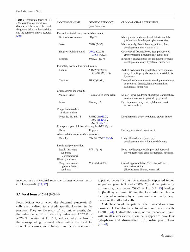
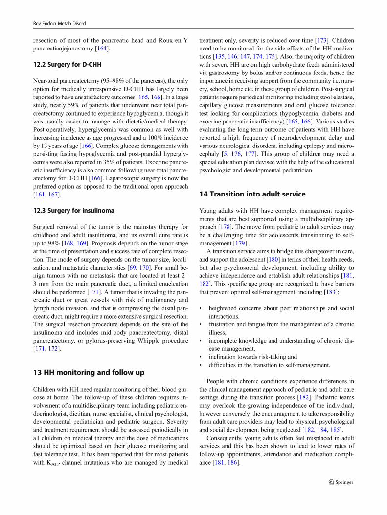
There are three histological forms of CHH (Fig. 2); Focal form(F-CHH), Diffuse form (D-CHH) and atypical. The clinicalpresentation appears to be similar, although their molecularmechanisms are quite different. In most cases D-CHH is
Rev Endocr Metab Disord
inherited in an autosomal recessive manner whereas the F-CHH is sporadic [22, 72].
5.1 Focal form of CHH (F-CHH)
Focal lesions occur when the abnormal pancreatic β-cells are localized to a single specific location in thepancreas. They are the result of two unique events, firstthe inheritance of a paternally inherited ABCC8 orKCNJ11 mutation at 11p15.1, and secondly the loss ofthe corresponding maternal allele within the focal le-sion. This causes an imbalance in the expression of
imprinted genes such as the maternally expressed tumorsuppressor gene H19 and CDKN1C, and the paternallyexpressed growth factor IGF-2, at 11p15.5 [73] leadingto β-cell hyperplasia. Within the focal islet cell lesionthere is adenomatous hyperplasia and abnormally largenuclei in the affected cells.
A duplication of the paternal allele located on chro-mosome 11 has also been found in some patients withF-CHH [74]. Outside the lesion, normal endocrine tissuewith small nuclei exists. These cells appear to have lesscytoplasm and diminished proinsulin production[75–78].
Table 2 Syndromic forms of HH- Various developmental syn-dromes have been described withthe gene/s linked to the conditionand the common clinical features[203]
SYNDROME NAME GENETIC ETIOLOGY
gene (location)
CLINICAL CHARACTERISTICS
Pre- and postnatal overgrowth (Macrosomia)
Beckwith-Wiedemann (11p15) Macroglossia, abdominal wall defects, ear lobepits/ creases, hemihypertrophy, tumor risk
Sotos NSD1 (5q35) Macrocephaly, frontal bossing, pointed chin,developmental delay, tumor risk
Simpson-Golabi-Behmel GPC3 (Xq26),GPC4 (Xp22)
Coarse facial features, broad feet, polydactyly,cryptorchidism, hepatomegaly, tumor risk
Perlman DIS3L2 (2q37) Inverted V-shaped upper lip, prominent forehead,developmental delay, hypotonia, tumor risk
Postnatal growth failure (short stature)
Kabuki KMT2D (12q13),KDM6A (Xp11.3)
Arched eyebrows, long eyelashes, developmentaldelay, fetal finger pads, scoliosis, heart defects,hypotonia
Costello HRAS (11p15) Deep palmar/plantar creases, developmental delaycoarse facial features, heart abnormalities,papillomas, tumor risk
Chromosomal abnormality
Mosaic Turner (Loss of X in some cells) Milder Turner syndrome phenotype (short stature,coarctation of aorta, gonadal dysgenesis)
Patau Trisomy 13 Developmental delay, microphthalmia, heart& neural defects
Congenital disordersof glycosylation
Types 1a, 1b, and 1d PMM2 (16p13.2),MPI (15q24.1),ALG3 (3q27.1)
Developmental delay, hypotonia, growth failure
Contiguous gene deletion affecting the ABCC8 gene
Usher 11 genes Hearing loss, visual impairment
Abnormalities in calcium homoeostasis
Timothy CACNA1C (12p13.33) Long QT syndrome, syndactyly,developmental delay, immune deficiency
Insulin receptor mutation:
Insulin resistancesyndrome(leprechaunism)
INS (19p13) Hypo- and hyperglycemia, pre- and postnatalgrowth restriction, elfin-like features, hirsutism
Other Syndromes:
Congenital centralhypoventilationsyndrome
PHOX2B (4p13) Central hypoventilation, “box-shaped” face,neurocristopathies(Hirschsprung disease, tumor risk)
Rev Endocr Metab Disord
Patients who have a heterozygous paternally inherited mu-tation in ABCC8 or KCNJ11 could have F-CHH, which ac-counts for 30–40% of all CHH cases [79]. F-CHH is con-firmed by a fluorine-18 dihydroxyphenylalanine-positronemission tomography (18F-DOPA-PET) scan, which canshow the presence of a focal lesion and determine its locationwith diagnostic accuracy [80].
5.2 Diffuse form of CHH (D-CHH)
The D-CHH form occurs when all the islet cells in the pan-creas are abnormal [77, 81]. Patients with a homozygous re-cessive or a compound heterozygote mutation in ABCC8 orKCNJ11 present with D-CHH. These patients are usuallymedically unresponsive, and this histological form accountsfor 60–70% of all CHH cases. Most islets throughout theentire pancreas are affected with the presence of largehyperchromatic nuclei [75–78, 81].
5.3 Atypical forms of HH
Histologically atypical forms of CHH are categorized whenthe pancreatic morphology does not fit into the F-CHH or D-CHH types and are a mosaic pattern of the two [73, 82]. Theislets can either be enlarged or shrunken. Some cases havebeen cured with a lesionectomy whilst others also requiremedical management. However, to date only one patient hasbeen described with a ABCC8 nonsense mutation (Q54X)causing this histological form of CHH [82]. Studies haveshown that the heterogeneous expression of β-cells HK1 inthe atypical CHHmay be causing the abnormal insulin release[83, 84].
6 Clinical presentation of HH
HH usually manifests in infancy or early childhood, althoughsome patients can present during adolescence or adulthood.Signs and symptoms of hypoglycemia are non-specific duringthe neonatal period (poor feeding, jitteriness, irritability), chal-lenging its diagnosis. At later ages, symptoms can be easier torecognize and can be classified as adrenergic (hunger, pallor,tachycardia, tremor, diaphoresis) and neuroglycopenic (tired-ness, blurred vision, confusion, coma and even death).
Risk factors for HH need to be looked for in the perinatalhistory. These include maternal intrapartum administration ofhypoglycemic agents, stressful delivery, large/low birthweight, and neonatal polycythemia or jaundice, among others.Examination of the baby will search for macrosomia/leanness,careful phenotypic characterization to identify syndromes,features of cardiomyopathy (due to glycogen storage inHH), hepatomegaly present in metabolic conditions (alsopresent in HH), and midline abnormalities (including genita-lia) to exclude hypothalamic-pituitary deficits. Data in thefamily history could suggest a genetic inheritance, thereforeanamnesis must enquire for consanguinity, cases of diabetesmellitus, hypoglycemia, seizures and unexplained deaths [85].
7 Diagnostic investigations of HH
Any child requiring intravenous glucose load greater than8 mg/kg/min (normal requirement is 4-6 mg/kg/min) to avoidhypoglycemia, can essentially be labeled as HH [86]. Thetiming of hypoglycemic events with regard to meals will giveinsight into the intrinsic mechanism. HH typically manifestsduring brief periods of fasting, however certain types can bemanifest soon after meal ingestion (protein induced HH) or
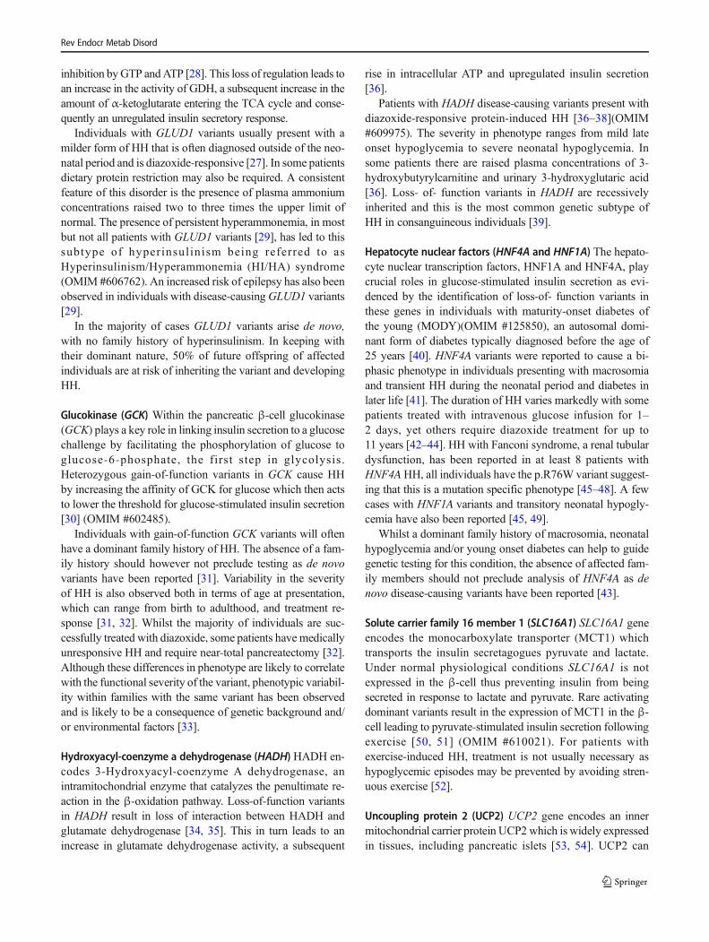
Fig. 2 Diffuse and focal form ofHH with 18F-DOPA-PET-CTimages. A – Diagrammatic repre-sentation of diffuse form of CHHand B – 18F-DOPA-PET imageof diffuse form of CHH. C –Diagrammatic representation offocal form of CHH (showing dif-ferent types of focal lesions) andD – 18F-DOPA-PET-CT image offocal lesion in the head of pan-creas. SUV – Standardized uptakevalue.
Rev Endocr Metab Disord

hours after meal ingestion (postprandial HH). Should there bea clear link between physical activity and occurrence of hypo-glycemia, then exercise induced HH should be excluded.
7.1 Biochemistry
Biochemical interpretation will only be possible if the criticalsample is taken adequately at the time of hypoglycemia(≤3.0 mmol/l), ideally in a controlled fasting setting. HH willdemonstrate detectable C-peptide or insulin in the face of hy-poglycemia, with simultaneously low/undetectable alternativesubstrates (ketones and fatty acids) [86, 87]. Of note, the con-centration of insulin does not parallel the degree of severity ofthe condition [88, 89]. Table 3 defines the diagnostic criteriafor HH. If a particular mechanism is suspected to trigger HH(exercise, protein, carbohydrate etc) specific stimulation testswill be carried out:
7.1.1 Protein load test
In this test leucine or a combination of aminoacids is admin-istered enterally. Evidence of raised insulin concentrations atthe time of hypoglycemia will confirm the clinical suspicionof protein sensitive HH [29].
7.1.2 Oral glucose tolerance test / mixed meal test
Enteral administration of a mixed meal or oral glucose loadfollowed by hypoglycemia with a detectable insulin within 2–5 h, indicates incretin mediation leading to postprandial HH.In adults the prefered test is the standardized hyperglucidic
breakfast [91], which has not yet been standardized inchildren.
7.1.3 Exercise test and pyruvate load
Biochemical evidence of HH during these tests indicate alactate/pyruvate mechanism triggering exercise induced HH[92].
7.1.4 Fructose load test
Individuals with ketotic hypoglycemia after fructose ingestionshould undertake a fructose load test to identify hereditaryfructose intolerance and fructose-1,6-diphosphatasedeficiency, and this can be followed by genetic testing.
7.1.5 The role of real-time continuous glucose monitoring(CGM)
CGM measures interstitial glucose concentrations indicatingthe direction, magnitude, frequency, duration and causes offluctuations in these. There is now a study on its accuracy inchildren with HH [93], as this can significantly improve thequality of life of these patients.
7.2 Imaging
7.2.1 Recent advances in diagnostic imaging of HH
As is not possible to differentiate between F-CHH and D-CHH based on clinical presentation and biochemical features,
Table 3 Diagnostic criteria forHH - The cut-off values for eachanalyte to aid in the diagnosis ofHH. IGFBP-1: Insulin growthfactor binding protein-1. HI/HA:Hyperinsulinemic hypoglycemiahyperammonemia syndrome.HADH: short chain L-3-hydroxyacil-CoA dehydrogenase.Im: intramuscular. Iv: intrave-nous. Sc: subcutaneous *[90, 200,203]¥
Serum analyte Result in patients with HH
Blood glucose < 3.0 mmol/l (54 mg/dl) and:
Insulin Detectable
C-peptide Detectable (≥0.5 ng/mL¥)
Free fatty acids Low or suppressed (<1.5 mmol/l* or < 1.7 mmol/l¥)
Ketone bodies Low or suppressed (3-β-hydroxybutyrate <2 mmol/l* or < 1.8 mmol/l¥)
IGFBP-1 Low (≤110 ng/mL¥) as insulin negatively regulates IGFBP-1 expression
Ammonia Normal. Can be raised in HI/HA syndrome
Hydroxybutyrylcarnitine Normal. Raised in HH due to HADH mutation
Cortisol, Growthhormone
Raised. Generally Cortisol >20 μg/dL [500 nmol/L]; growth hormone>7 ng/mL - younger children might have poor counter-regulatory response
Amino acids and urineorganic acids
Normal. Leucine, isoleucine and valine may be suppressed in HH
Proinsulin >20 pmol/l
Additional information when diagnosis of HH uncertain:
Glucose infusion rate >8 mg/kg/min to achieve euglycemia
Im or iv glucagonadministration or
sc octreotide administration
>1.5 mmol/L or 27 mg/dl (Positive glycemic response)
Rev Endocr Metab Disord
pre-operative differentiation of both subtypes is critically im-portant. Conventional radiological imaging such as MagneticResonance Imaging (MRI) and Computerized Tomography(CT) scans fail to localize the focal lesions. Genetic analysisfor mutations inABCC8/KCNJ11, with combination of recent-ly described Fluorine 18 L-3, 4- dihydroxyphenyalanine pos-itron emission tomography (18F-DOPA-PET-CT) scanning,allows differentiation between F-CHH and D-CHH with ahigh sensitivity (88%) and specificity (94%) [94–98], withan accuracy of 100% [99–102]. The principle of 18F-DOPA-PET analysis is based on the selective uptake of L-DOPA by β-cells and its conversion into dopamine byDOPA decarboxylase enzyme, which is expressed in the pan-creatic β-cells. Thus, imaging with 18F-DOPA-PET-CTshould be performed in all patients who are thought to haveF-CHH. Some patients have been described with atypical his-tological forms, which are either atypical focal, atypical dif-fuse or ectopic β-cell hyperplasia and do not show classicalfeatures of F-CHH or D-CHH [103]. Demonstration of in-creased activity of DOPA decarboxylase by 18F-DOPA-PETin combination with an enhanced CT imaging can successful-ly differentiate diffuse and focalβ-cell hyperplasia. Therefore,this technique has radically changed the surgical approach topatients with medically unresponsive HH [102, 104].
A dosimetry study of 18F-DOPA derived from the PET-CTimages in ten infants (median age 4.84 weeks) with HH sug-gested that a modest radiation dose (0.30 ± 0.04 mSv/MBq)was adequate [105]. However, there have been recent reportsof its inaccuracy in precisely detecting focal lesions [106,107]. Glucagon-like-peptide-1 (GLP-1) receptor analogs arethe latest agents being used in the detection of insulinomas inadults [108, 109]. Similarly, this isotope is being investigatedin children to detect focal lesions.
7.2.2 Imaging for insulinoma
Most insulinomas are benign (islet-cell tumors) and usuallyvery small (<2 cm) making it difficult to localize with thecurrent imaging techniques. Various non-invasive techniqueshave been used for the detection of insulinomas e.g.,transabdominal ultrasonography, spiral CT, MRI, 111In-pentetreotide imaging, and 18F-l-dihydroxyphenylalaninePET. In difficult cases, invasive procedures like endoscopicultrasonography or selective arterial calcium stimulation testwith hepatic venous sampling have also been used [110].
A systematic review and meta-analysis of (68)Ga-DOTATATE compared with octreotide and conventionalimaging documented a high sensitivity (90.9%) and speci-ficity (90.6%) of (68)Ga-DOTATATE in detecting neuro-endocrine tumors [111]. Various Gallium based isotopeshave also been used to detect insulinoma in adults [108,112].
8 Management of HH
Maintaining normoglycemia (blood glucose >3.5 mmol/l) isparamount to avoid hypoglycemic brain injury in view of thehypoketotic nature of HH [113]. This may be achieved byincreasing glucose administration (feeds or intravenous (iv)fluids), stimulating endogenous glucose release (glucagon ad-ministration) or suppressing insulin release from the β-cell(diazoxide, octreotide, nifedipine). Oral feeds with additionalglucose polymer in combination with iv fluids can also beused to maintain normoglycemia. The traditional and newdrugs available for the management of HH are summarizedin Table 4.
9 Current medical management
9.1 Acute management of hypoglycemia
If the oral route is unavailable or glycemia is not improvingdespite oral glucose (glucose gel/glucose containing drinks ortablets), then an iv dextrose bolus (2 ml/kg 10%Glucose)needs to be administered followed by continuous iv glucoseinfusion (>8 mg/kg/min). If there is persistence of hypoglyce-mia, hypoglycemic seizure or inadequate iv access, then anintramuscular or subcutaneous bolus (1 mg) or infusion ofglucagon can be life saving as it causes immediate release ofglycogen stores from the liver leading to a temporary im-provement in blood glucose concentrations. [117].
As high iv glucose concentration is usually needed forweeks, insertion of a central venous access supports in man-aging glycemia safely.
9.2 Long-term pharmacological agents
Medical therapies can be tried to wean the infant off iv supportand achieve a near-normal feeding pattern. Surgical therapy isconsidered when trial of medical therapies is unsuccessful orin case of established F-CHH.
HH drug responsiveness is defined by: 1) normal feedingfrequency and volume, 2) fasting capability adequate for agewhilst maintaining euglycemia, 3) low/suppressed serum in-sulin level at the end of the fast, and 4) appropriate generationof ketone bodies and fatty acids by the end of the fast [117].
Diazoxide The first drug of choice is diazoxide [86], whichrequires a functional KATP channel to bind onto and is henceineffective in D-CHH due to an inactivating KATP mutationand in most cases of F-CHH. Most other forms of HH andprotracted HH secondary to risk factors such as IUGR andperinatal asphyxia respond to treatment with diazoxide. Dueto its fluid retaining properties, diazoxide therapy must beused with caution in infants with HH who are often receiving
Rev Endocr Metab Disord
large volumes of iv/oral fluids to maintain normoglycemia.Fluid restriction prior to starting diazoxide is commonly prac-ticed, along with concomitant use of a thiazide diuretic such aschlorothiazide, which also has a synergistic action over theKATP channels.9.2.1 Medical management of diazoxide unresponsive D-CHH
NifedipineAuthors suggest triyng it before heading to pancre-atectomy [118] as there have been isolated case reports onsuccessful treatment of HH with nifedipine [119–121] butexperience of large HH centers has generally been disappoint-ing [117, 122, 123]. It is sometimes considered as an add-ondrug in partial diazoxide/octreotide resistance, and/or
following partial pancreatectomy [122, 124] but rarely usedas monotherapy.
Octreotide Octreotide is a second-line treatment fordiazoxide-insensitive patients [125, 126]. It may be usedin combination with diazoxide and glucagon in cases withpartial diazoxide response, or often combined with fre-quent feeding which may require a gastrostomy to enablehigh calorie bolus feeding during the day and overnightcontinuous feeds. As octreotide binds to the somatostatinreceptors SSTR-2 and SSTR-5, prolonged use may devel-op into drug desensitizing caused by internalisation of thereceptors [122].
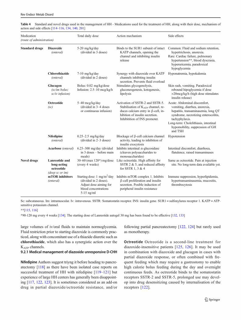
Table 4 Standard and novel drugs used in the management of HH - Medications used for the treatment of HH, along with their dose, mechanism ofaction and side effects [114–116, 134, 140, 201]
Medication(route of administration)
Total daily dose Action mechanism Side effects
Standard drugs Diazoxide(enteral)
5-20 mg/kg/day(divided in 3 doses)
Binds to the SUR1 subunit of intactKATP channels, opening thechannel and inhibiting insulinrelease
Common: Fluid and sodium retention,hypertrichosis, anorexia.
Rare: Cardiac failure, pulmonaryhypertension**, blood dyscrasia,hyperuricemia, paradoxicalhypoglycemia
Chlorothiazide(enteral)
7-10 mg/kg/day(divided in 2 doses)
Synergy with diazoxide over KATPchannels inhibiting insulinsecretion. Prevents fluid overload
Hyponatremia, hypokalemia
Glucagon(sc/im bolus;sc/iv infusion)
Bolus: 0.02 mg/kg/doseInfusion: 2.5–10 mcg/kg/h
Stimulates glycogenolysis,gluconeogenesis, ketogenesis,lipolysis
Skin rash, vomiting. Paradoxicalrebound hipoglycemia if dose>20mcg/kg/h (high dose stimulatesinsulin release)
Octreotide(sc)
5–40 mcg/kg/day(divided in 3–4 dosesor continuous infusion)
Activation of SSTR-2 and SSTR-5.Stabilisation of KATP channel, re-duces calcium entry in β-cell, in-hibition of insulin secretion.Inhibitition of INS promoter.
Acute: Abdominal discomfort,vomiting, diarrhea, anorexia,hepatitis, transaminasemia, long QTsyndrome, necrotizing enterocolitis,tachyphylaxis.
Long-term: Cholelithiasis, intestinalhypomobility, suppression of GHand TSH
Nifedipine(enteral)
0.25–2.5 mg/kg/day(divided in 2–3 doses)
Blockage of β-cell calcium channelactivity, leading to inhibition ofinsulin exocytosis
Hypotension
Acarbose (enteral) 6.25–300 mg/day (dividedin 3 doses – before mainmeals)
Inhibits intestinal α-glucosidase(cleaves polysaccharides tomonosaccharides)
Intestinal discomfort, diarrhoea,flatulence, raised transaminases
Novel drugs Lanreotide andlong-actingoctreotide
(deep sc or im)
30–60 (max 120*) mg/dose(every 4 weeks)
Like octreotide. High affinity forSSTR 2 & 5, and reduced affinityfor SSTR 1, 3 & 4
Same as octreotide. Pain at injectionsite. No long-term data available yet.
mTOR inhibitors(enteral)
Starting dose: 1 mg/m2/day(divided in 2 doses).Adjust dose aiming forblood concentrations5-15 ng/ml
Inhibits mTOR complex 1. Inhibitsβ-cell proliferation and insulinsecretion. Posible induction ofperipheral insulin resistance
Immune suppression, hyperlipidemia,hypertransaminasemia, mucositis,thrombocytosis
Sc: subcutaneous. Im: intramuscular. Iv: intravenous. SSTR: Somatostatin receptor. INS: insulin gene. SUR1 = sulfonylurea receptor 1. KATP =ATP-sensitive potassium channel.
**[115, 116]
*90-120 mg every 4 weeks [134]. The starting dose of Lanreotide autogel 30 mg has been found to be effective [132, 133]
Rev Endocr Metab Disord
Glucagon Iv or subcutaneous (sc) glucagon [127] may behelpful during the initial stabilization period and before sur-gery. However, this treatment has not been of long-term ben-efit. It can also be administered (alone or in combination withOctreotide) to stabilize blood glucose concentrations in theacute management and prevent near-total pancreatectomy ininfants with HH [128]. Continuous sc glucagon infusion canfrequently complicate by catheter obstructions occurring dailyor 2–3 times per week [128].
Acarbose For postprandial HH the first approach is diet mod-ification. This includes: frequent feeds of long-acting carbo-hydrates, abundant protein and supplements of fibre and fatemulsions [129]. If hypoglycemia persists, acarbose is thepreferred medical option as it slows the absorption of glucoseinto the blood stream, thus avoiding a glycemic peak followedby insulin release [130].
10 Novel medical therapies
10.1 Long-acting somatostatin analogues
Lanreotide and Long-acting release octreotide (LAR-Octreotide) are two formulations used in few numbers of pa-tients with HH [131–136] reported not only to be useful in themanagement of HH in children, but potentially displaying amore stable glycemic control than octreotide [134].Lanreotide has also been proven useful in managing inopera-ble F-CHH [137].
10.2 mTOR inhibitors
The intracellular mTOR pathway is involved in β-cell growthand altered insulin secretion in patients with insulinoma [138].Therefore, mTOR inhibitors such as sirolimus (formerlyknown as rapamycin) and everolimus have been used to treatthis tumor. Although the exact mechanism of sirolimus in HHstill needs to be elucidated, it has been hypothesized that themTOR complex 1 may be overactivated in D-CHH [139]. Thefirst study reported the use of sirolimus in 4 children withsevere D-CHH who achieved glycemic control avoiding pan-createctomy and with no major side effects [140]. This hasbeen followed by various case reports of HH in children ofvarious ages who have benefitted from sirolimus [141–145].However, there have been reports about its severe side effectsas well as poor response in others [146–149]. Our center hastried sirolimus on 22 patients with various forms of HH, out ofwhich 21 showed glycemic response, however 19 patientsdeveloped side effects, recurrent and frequent infections beingthe most common infections [150]. There is no clear correla-tion between genetic etiology and response to sirolimus.
11 Potential novel therapies – The future
11.1 GLP-1 antagonists
GLP-1 acts on the β-cell promoting its proliferation and stim-ulating insulin release [151]. Mouse models with KATP chan-nel defects leading to HH, significantly improved their glu-cose concentrations when treated with GLP-1 receptor antag-onist (exendin-9-39) [152]. Infusion of exendin-9-39 has beentried in 9 adult patients with HH due to KATP mutations [153],where all patients demonstrated increased fasting mean glu-cose and glucose area under the curve, raising expectation forits near-future use in children with this condition.
11.2 Pharmacological trafficking chaperones
Some mutations in the ABCC8 gene prevent trafficking ofparts of the SUR1 channel from the endoplasmic reticulumto the surface of the cell. Carbamazepine [154] and sulfonyl-ureas [155] – glibenclamide and tolbutamide - are KATP chan-nel inhibitors and chaperones that have proven to amend chan-nel trafficking defects in many ABCC8mutations by allowingintersubunit interactions between SUR1 and Kir6.2 [156].This has been now confirmed in mouse functional studies aswell [157] reinforcing its potential for clinical use.
11.3 Glucagon – New formulations and delivery
Subcutaneous glucagon infusion via pump has been used aslong-term treatment for HH patients at home [158]. Practicalissues with its administration remain unsolved as it can causemechanical obstruction due to its formulation. However, astable form of glucagon formulation could be a potential use-ful tool that is being explored [159, 160].
12 Surgical management
12.1 Surgery for F-CHH
Removal of the affected part of the pancreas achieving com-plete cure is the surgical aim for the F-CHH. A multidisciplin-ary team (endocrinology, radiology, and histopathology)should be involved in managing patients with F-CHH andD-CHH [161, 162]. In most cases, pre-operative 18F-DOPAPET-CT helps exact localization of the focal lesion and aidsthe surgeon. Intra-operative biopsies are important to ensurecomplete excision with histological confirmation of clear mar-gins. Laparoscopic lesionectomy is the preferred surgical ap-proach when the focal lesion is easily accessible (i.e. body ortail of pancreas) offering the benefit of shorter post-operativecare [163]. Focal lesions that are difficult to access such as inthe head of the pancreas usually require open laparotomy for
Rev Endocr Metab Disord
resection of most of the pancreatic head and Roux-en-Ypancreaticojejunostomy [164].
12.2 Surgery for D-CHH
Near-total pancreatectomy (95–98% of the pancreas), the onlyoption for medically unresponsive D-CHH has largely beenreported to have unsatisfactory outcomes [165, 166]. In a largestudy, nearly 59% of patients that underwent near total pan-createctomy continued to experience hypoglycemia, though itwas usually easier to manage with dietetic/medical therapy.Post-operatively, hyperglycemia was common as well withincreasing incidence as age progressed and a 100% incidenceby 13 years of age [166]. Complex glucose derangements withpersisting fasting hypoglycemia and post-prandial hypergly-cemia were also reported in 35% of patients. Exocrine pancre-atic insufficiency is also common following near-total pancre-atectomy for D-CHH [166]. Laparoscopic surgery is now thepreferred option as opposed to the traditional open approach[161, 167].
12.3 Surgery for insulinoma
Surgical removal of the tumor is the mainstay therapy forchildhood and adult insulinoma, and its overall cure rate isup to 98% [168, 169]. Prognosis depends on the tumor stageat the time of presentation and success rate of complete resec-tion. The mode of surgery depends on the tumor size, locali-zation, and metastatic characteristics [69, 170]. For small be-nign tumors with no metastasis that are located at least 2–3 mm from the main pancreatic duct, a limited enucleationshould be performed [171]. A tumor that is invading the pan-creatic duct or great vessels with risk of malignancy andlymph node invasion, and that is compressing the distal pan-creatic duct, might require a more extensive surgical resection.The surgical resection procedure depends on the site of theinsulinoma and includes mid-body pancreatectomy, distalpancreatectomy, or pylorus-preserving Whipple procedure[171, 172].
13 HH monitoring and follow up
Children with HH need regular monitoring of their blood glu-cose at home. The follow-up of these children requires in-volvement of a multidisciplinary team including pediatric en-docrinologist, dietitian, nurse specialist, clinical psychologist,developmental pediatrician and pediatric surgeon. Severityand treatment requirement should be assessed periodically inall children on medical therapy and the dose of medicationsshould be optimized based on their glucose monitoring andfast tolerance test. It has been reported that for most patientswith KATP channel mutations who are managed by medical
treatment only, severity is reduced over time [173]. Childrenneed to be monitored for the side effects of the HH medica-tions [135, 146, 147, 174, 175]. Also, the majority of childrenwith severe HH are on high carbohydrate feeds administeredvia gastrostomy by bolus and/or continuous feeds, hence theimportance in receiving support from the community i.e. nurs-ery, school, home etc. in these group of children. Post-surgicalpatients require periodical monitoring including stool elastase,capillary glucose measurements and oral glucose tolerancetest looking for complications (hypoglycemia, diabetes andexocrine pancreatic insufficiency) [165, 166]. Various studiesevaluating the long-term outcome of patients with HH havereported a high frequency of neurodevelopment delay andvarious neurological disorders, including epilepsy and micro-cephaly [5, 176, 177]. This group of children may need aspecial education plan devised with the help of the educationalpsychologist and developmental pediatrician.
14 Transition into adult service
Young adults with HH have complex management require-ments that are best supported using a multidisciplinary ap-proach [178]. The move from pediatric to adult services maybe a challenging time for adolescents transitioning to self-management [179].
A transition service aims to bridge this changeover in care,and support the adolescent [180] in terms of their health needs,but also psychosocial development, including ability toachieve independence and establish adult relationships [181,182]. This specific age group are recognized to have barriersthat prevent optimal self-management, including [183];
& heightened concerns about peer relationships and socialinteractions,
& frustration and fatigue from the management of a chronicillness,
& incomplete knowledge and understanding of chronic dis-ease management,
& inclination towards risk-taking and& difficulties in the transition to self-management.
People with chronic conditions experience differences inthe clinical management approach of pediatric and adult caresettings during the transition process [182]. Pediatric teamsmay overlook the growing independence of the individual,however conversely, the encouragement to take responsibilityfrom adult care providers may lead to physical, psychologicaland social development being neglected [182, 184, 185].
Consequently, young adults often feel misplaced in adultservices and this has been shown to lead to lower rates offollow-up appointments, attendance and medication compli-ance [181, 186].
Rev Endocr Metab Disord
Discharge to a non-specialist adult team carries with itsome challenges, including lack of awareness about this rarecondition and its complex on-going health-needs. In addition,non-specialist teams may not be best placed to undertake dis-cussions around pre-conception counselling in those with aconfirmed genetic etiology.
14.1 Aims of the transition service
Recognizing the core attributes of this age group, a transitionservice for patients with HH may be established, with thefollowing aims and in line with established NICE guidance[178].
1. Supporting education: Establish and support under-standing of HH. Where genetic causes are identified,patients should be aware of this and have an understand-ing of its inheritance.
2. Empowering self-management: To support handoverof care to the affected individual from the carers [187],by encouraging shared decis ion-making andempowering self-management, with an ultimate aim ofself-management [188].
3. Addressing parental and care-giver concern [189]:Establishing an understanding between the young per-son and their carer of how aspects of self-care might bejointly undertaken and transferred is an important com-ponent of managing distress, expectations and, ultimate-ly, transition [190].
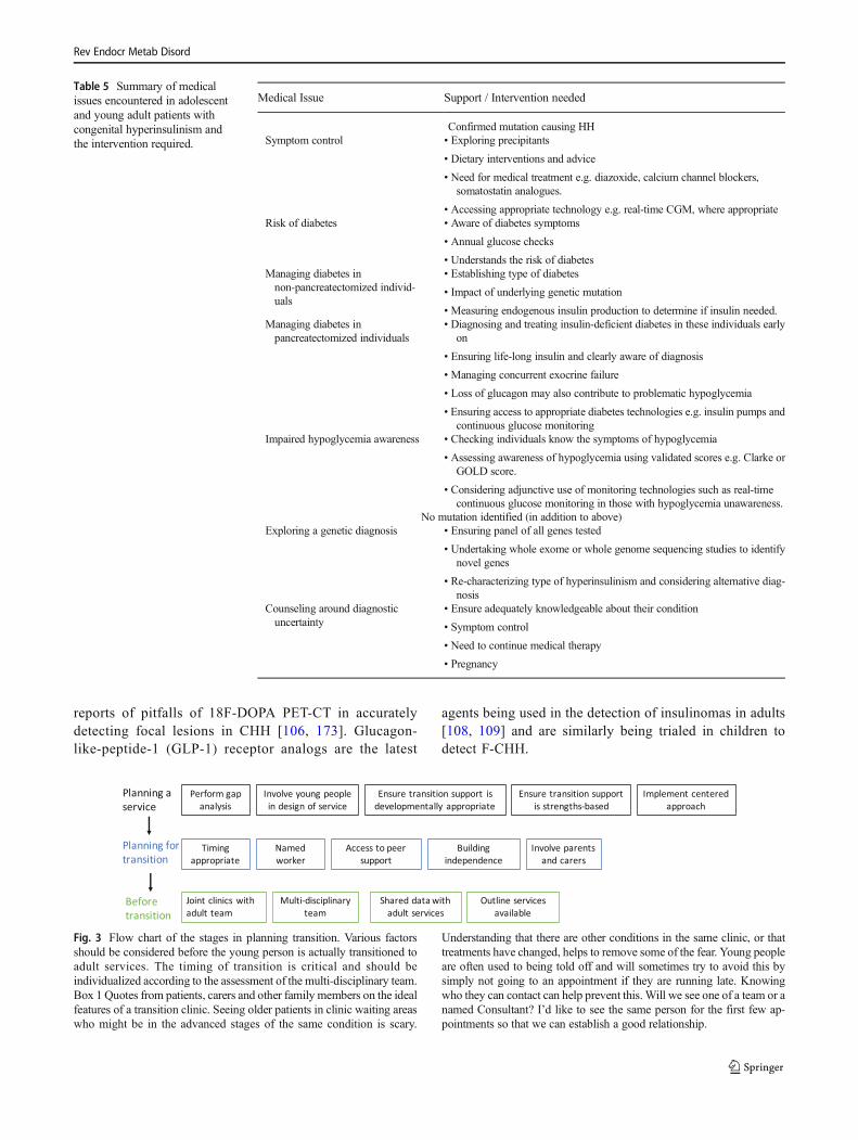
4. Addressing on-going medical issues: Table 5 summa-rizes some of themedical issues that may be addressed ina transition service. The risk and progression to diabetesis variable and appropriate treatments for diabetes maydepend on both the mutation causing HH and the extentof β-cell failure.
5. Dealing with impaired awareness of hypoglycemia[191]: If found in severe cases of HH it can be formallyassessed using the validated Clarke [192] or Gold score[193]. The evidence base for managing impaired aware-ness of hypoglycemia is limited in the context of HH[194] and so, care is needed in managing these individ-uals. CGM could be of use in some cases [191].
6. Providing advice around driving: Supporting individ-uals at high risk of hypoglycemia or impaired awarenessof hypoglycemia to apply for a driving license may beexplored, and fully explaining driving regulations in re-lation to hypoglycemia is important for those starting tolearn to drive [195].
7. Providing support to higher educational institutionsand those in employment [196, 197]: Flexibility maybe required to allow time for testing, or additional breaksfor calorie consumption.
8. Pre-conception genetic counsel ing, whereappropriate: In those with confirmed dominant muta-tions, ensuring adequate counseling around offspring,and accessing genetic counselors when appropriate, isimportant. Insulin modulates Sertoli cell function andyoung hyperinsulinemic patients have been found tohave lower anti-Müllerian hormone and inhibin B se-cretion [198]. This could therefore influence testicularfunction and have a future impact on fertility, which stillneeds to be studied.
9. Continued dietetic advice: On-going advice about die-tary management of hypoglycemia, especially wherethere is protein-induced hypoglycemia, and replacementof calories with other macronutrients may be provided.
10. Alcohol and recreational drug advice [199]: alcoholconsumption can potentiate the risk of hypoglycemiaand also impair awareness of hypoglycemia.Recreational drugs may also mask the adrenergic symp-toms of hypoglycemia.
14.2 Developing the HH transition pathways
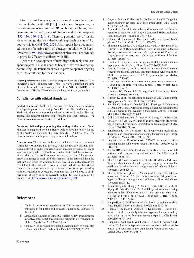
In line with NICE transition service guidelines [178], the op-timal pathway for transitioning individuals with HH must bedeveloped, summarized in Fig. 3. This includes:
– Planning a service: Young people should be involved indesigning the service.
– Planning for transition, which should be appropriatelytimed for the individual.
– Before Transition: young adults should have joint clinicswith the pediatric and adult teams.
15 Concluding remarks and future directions
HH is a challenging condition to treat due to its heterogeneity.Despite diagnostic and therapeutic advances, HH remains animportant cause of morbidity in children, still accounting for26–44% of permanent intellectual disabilities, especially inneonatal-onset patients. The increasing use of NGS targetpanels, combinedwith clinical, biochemical and imaging find-ings allows differentiating the diagnostic management of chil-dren with F-CHH, surgically curable, from those with D-CHH, more conservatively treated with pharmacological andnutritional interventions [200].
There is now more research of use of CGM in childrenwith HH which might help to detect early hypoglycemialeading to prompt management [93]. There have been
Rev Endocr Metab Disord
reports of pitfalls of 18F-DOPA PET-CT in accuratelydetecting focal lesions in CHH [106, 173]. Glucagon-like-peptide-1 (GLP-1) receptor analogs are the latest
agents being used in the detection of insulinomas in adults[108, 109] and are similarly being trialed in children todetect F-CHH.
Table 5 Summary of medicalissues encountered in adolescentand young adult patients withcongenital hyperinsulinism andthe intervention required.
Medical Issue Support / Intervention needed
Confirmed mutation causing HHSymptom control • Exploring precipitants
• Dietary interventions and advice
• Need for medical treatment e.g. diazoxide, calcium channel blockers,somatostatin analogues.
• Accessing appropriate technology e.g. real-time CGM, where appropriateRisk of diabetes • Aware of diabetes symptoms
• Annual glucose checks
• Understands the risk of diabetesManaging diabetes innon-pancreatectomized individ-uals
• Establishing type of diabetes
• Impact of underlying genetic mutation
• Measuring endogenous insulin production to determine if insulin needed.Managing diabetes inpancreatectomized individuals
• Diagnosing and treating insulin-deficient diabetes in these individuals earlyon
• Ensuring life-long insulin and clearly aware of diagnosis
• Managing concurrent exocrine failure
• Loss of glucagon may also contribute to problematic hypoglycemia
• Ensuring access to appropriate diabetes technologies e.g. insulin pumps andcontinuous glucose monitoring
Impaired hypoglycemia awareness • Checking individuals know the symptoms of hypoglycemia
• Assessing awareness of hypoglycemia using validated scores e.g. Clarke orGOLD score.
• Considering adjunctive use of monitoring technologies such as real-timecontinuous glucose monitoring in those with hypoglycemia unawareness.
No mutation identified (in addition to above)Exploring a genetic diagnosis • Ensuring panel of all genes tested
• Undertaking whole exome or whole genome sequencing studies to identifynovel genes
• Re-characterizing type of hyperinsulinism and considering alternative diag-nosis
Counseling around diagnosticuncertainty
• Ensure adequately knowledgeable about their condition
• Symptom control
• Need to continue medical therapy
• Pregnancy
Involve young peoplein design of service
Ensure transition support isdevelopmentally appropriate
Ensure transition supportis strengths-based
Implement centeredapproach
Perform gapanalysis
Planning aservice
Planning fortransition
Namedworker
Access to peersupport
Buildingindependence
Involve parentsand carers
Timingappropriate
Beforetransition
Multi-disciplinaryteam
Shared datawithadult services
Outline servicesavailable
Joint clinics withadult team
Fig. 3 Flow chart of the stages in planning transition. Various factorsshould be considered before the young person is actually transitioned toadult services. The timing of transition is critical and should beindividualized according to the assessment of the multi-disciplinary team.Box 1 Quotes from patients, carers and other family members on the idealfeatures of a transition clinic. Seeing older patients in clinic waiting areaswho might be in the advanced stages of the same condition is scary.
Understanding that there are other conditions in the same clinic, or thattreatments have changed, helps to remove some of the fear. Young peopleare often used to being told off and will sometimes try to avoid this bysimply not going to an appointment if they are running late. Knowingwho they can contact can help prevent this. Will we see one of a team or anamed Consultant? I’d like to see the same person for the first few ap-pointments so that we can establish a good relationship.
Rev Endocr Metab Disord
Over the last few years, numerous medications have beentried in children with HH [201]. For instance long-acting so-matostatin analogues and mTOR inhibitors (sirolimus) havebeen used in various groups of children with varied response[133–136, 140–142, 148]. There is potential use of insulinreceptor antagonists as a therapeutic approach to control hy-poglycemia in CHH [202, 203]. Also, reports have document-ed the use of a stable form of glucagon in adults with hypo-glycemia [159, 160], however more clinical trials are requiredto prove its efficacy in children with HH.
Besides the development of new diagnostic tools and ther-apeutic agents, clinicians need to become involved in creating/potentiating HH transition clinics to provide optimal ongoingcare into adulthood for these patients.
Funding information Nick Oliver is supported by the NIHR BRC atImperial College Healthcare NHS Trust. The views expressed are thoseof the authors and not necessarily those of the NHS, the NIHR or theDepartment of Health. The other authors have no funding to declare.
Compliance with ethical standards
Conflict of interest Nick Oliver has received honoraria for advisoryboard participation or speaking from Dexcom, Roche diabetes, andMedtronic diabetes; support for education from Novo Nordisk andTakeda; and research funding from Dexcom and Roche diabetes. Theother authors have no information to disclose.
Grants and fellowships supporting the writing of the paper SarahFlanagan is supported by a Sir Henry Dale Fellowship jointly fundedby the Wellcome Trust and the Royal Society (105,636/Z/14/Z). Theother authors are not supported by grants or fellowships.
Open Access This article is licensed under a Creative CommonsAttribution 4.0 International License, which permits use, sharing, adap-tation, distribution and reproduction in any medium or format, as long asyou give appropriate credit to the original author(s) and the source, pro-vide a link to the Creative Commons licence, and indicate if changes weremade. The images or other third party material in this article are includedin the article's Creative Commons licence, unless indicated otherwise in acredit line to the material. If material is not included in the article'sCreative Commons licence and your intended use is not permitted bystatutory regulation or exceeds the permitted use, you will need to obtainpermission directly from the copyright holder. To view a copy of thislicence, visit http://creativecommons.org/licenses/by/4.0/.
References
1. Ahrén B. Autonomic regulation of islet hormone secretion–implications for health and disease. Diabetologia. 2000;43(4):393–410.
2. Senniappan S, Shanti B, James C, Hussain K. Hyperinsulinaemichypoglycaemia: genetic mechanisms, diagnosis and management.J Inherit Metab Dis. 2012;35(4):589–601.
3. Chinoy, A. et al. ‘Focal congenital hyperinsulinism as a cause forsudden infant death’. Pediatr Dev Pathol. 2019;22(1):65–69.
4. Guyot A, Moreau F, Eberhard M, Gaulier JM, Paraf F. Congenitalhyperinsulinism revealed by sudden infant death. Ann Pathol.2017;37(5):429–32.
5. Avatapalle HB, et al. Abnormal neurodevelopmental outcomes arecommon in children with transient congenital Hyperinsulinism.Front Endocrinol (Lausanne). 2013;4:60.
6. Güemes M, Rahman SA, Hussain K. What is a normal bloodglucose? Arch Dis Child. 2016;101:569–574.
7. Thornton PS, Stanley CA, de Leon DD, Harris D, HaymondMW,Hussain K, et al. Recommendations from the pediatric EndocrineSociety for evaluation and Management of PersistentHypoglycemia in neonates, infants, and children. J Pediatr.2015;167(2):238–45.
8. Hussain K. Diagnosis and management of hyperinsulinaemichypoglycaemia of infancy. Horm Res. 2008;69(1):2–13.
9. Patel P, Charles L, Corbin J, et al. A unique allosteric insulinreceptor monoclonal antibody that prevents hypoglycemia in theSUR-1-/- mouse model of KATP hyperinsulinism. MAbs.2018;10(5):796–802.
10. Arya VB,MohammedZ, BlankensteinO, de Lonlay P, Hussain K.Hyperinsulinaemic hypoglycaemia. Horm Metab Res.2014;46(3):157–70.
11. Vannucci RC, Vannucci SJ. Hypoglycemic brain injury. SeminNeonatol. 2001;6(2):147–55.
12. Hussain K, et al. An activating mutation of AKT2 and humanhypoglycemia. Science. 2011;334(6055):474.
13. Staufner C, Lindner M, Dionisi-Vici C, Freisinger P, DobbelaereD, Douillard C, et al. Adenosine kinase deficiency: expanding theclinical spectrum and evaluating therapeutic options. J InheritMetab Dis. 2016;39(2):273–83.
14. Gillis D, Krishnamohan A, Yaacov B, Shaag A, Jackman JE,Elpeleg O. TRMT10A dysfunction is associated with abnormali-ties in glucose homeostasis, short stature and microcephaly. J MedGenet. 2014;51(9):581–6.
15. Senniappan S, Arya VB, Hussain K. The molecular mechanisms,diagnosis and management of congenital hyperinsulinism. IndianJ Endocrinol Metab. 2013;17(1):19–30.
16. Inagaki N, et al. Reconstitution of IKATP: an inward rectifiersubunit plus the sulfonylurea receptor. Science. 1995;270(5239):1166–70.
17. Kapoor RR, et al. Clinical and molecular characterisation of 300patients with congenital hyperinsulinism. Eur J Endocrinol.2013;168(4):557–64.
18. Thomas PM, Cote GJ, Wohllk N, Haddad B, Mathew PM, RablW, et al. Mutations in the sulfonylurea receptor gene in familialpersistent hyperinsulinemic hypoglycemia of infancy. Science.1995;268(5209):426–9.
19. Thomas P, Ye Y, Lightner E. Mutation of the pancreatic islet in-ward rectifier Kir6.2 also leads to familial persistenthyperinsulinemic hypoglycemia of infancy. Hum Mol Genet.1996;5(11):1809–12.
20. Taschenberger G, Mougey A, Shen S, Lester LB, LaFranchi S,Shyng SL. Identification of a familial hyperinsulinism-causingmutation in the sulfonylurea receptor 1 that prevents normal traf-ficking and function of KATP channels. J Biol Chem.2002;277(19):17139–46.
21. Huopio H, et al. K(ATP) channels and insulin secretion disorders.Am J Physiol Endocrinol Metab. 2002;283(2):E207–16.
22. Huopio H, Reimann F, Ashfield R, Komulainen J, Lenko HL,Rahier J, et al. Dominantly inherited hyperinsulinism caused bya mutation in the sulfonylurea receptor type 1. J Clin Invest.2000;106(7):897–906.
23. Huopio H, Otonkoski T, Vauhkonen I, Reimann F, Ashcroft FM,Laakso M. A new subtype of autosomal dominant diabetes attrib-utable to a mutation in the gene for sulfonylurea receptor 1.Lancet. 2003;361(9354):301–7.
Rev Endocr Metab Disord
24. Flanagan SE, Kapoor RR, Banerjee I, Hall C, Smith VV, HussainK, et al. Dominantly acting ABCC8 mutations in patients withmedically unresponsive hyperinsulinaemic hypoglycaemia. ClinGenet. 2011;79(6):582–7.
25. Pinney SE, MacMullen C, Becker S, Lin YW, Hanna C, ThorntonP, et al. Clinical characteristics and biochemical mechanisms ofcongenital hyperinsulinism associated with dominant KATP chan-nel mutations. J Clin Invest. 2008;118(8):2877–86.
26. Kapoor RR, et al. Hyperinsulinaemic hypoglycaemia and diabetesmellitus due to dominant ABCC8/KCNJ11 mutations.Diabetologia. 2011;54(10):2575–83.
27. Stanley CA, et al. Hyperinsulinism and hyperammonemia in in-fants with regulatory mutations of the glutamate dehydrogenasegene. N Engl J Med. 1998;338(19):1352–7.
28. Stanley CA, Fang J, Kutyna K, Hsu BY, Ming JE, Glaser B, et al.Molecular basis and characterization of the hyperinsulinism/hyperammonemia syndrome: predominance of mutations in exons11 and 12 of the glutamate dehydrogenase gene. HI/HAContributing Investigators. Diabetes. 2000;49(4):667–73.
29. Kapoor RR, Flanagan SE, Fulton P, Chakrapani A, Chadefaux B,Ben-Omran T, et al. Hyperinsulinism-hyperammonaemia syn-drome: novel mutations in the GLUD1 gene and genotype-phenotype correlations. Eur J Endocrinol. 2009;161(5):731–5.
30. Glaser B, et al. Familial hyperinsulinism caused by an activatingglucokinase mutation. N Engl J Med. 1998;338(4):226–30.
31. Christesen HB, et al. Activating glucokinase (GCK) mutations asa cause of medically responsive congenital hyperinsulinism: prev-alence in children and characterisation of a novel GCK mutation.Eur J Endocrinol. 2008;159(1):27–34.
32. Cuesta-Munoz AL, et al. Severe persistent hyperinsulinemic hy-poglycemia due to a de novo glucokinase mutation. Diabetes.2004;53(8):2164–8.
33. Christesen HB, Jacobsen BB, Odili S, Buettger C, Cuesta-MunozA, Hansen T, et al. The second activating glucokinase mutation(A456V): implications for glucose homeostasis and diabetes ther-apy. Diabetes. 2002;51(4):1240–6.
34. Heslegrave AJ, et al. Leucine-sensitive hyperinsulinaemichypoglycaemia in patients with loss of function mutations in 3-Hydroxyacyl-CoA dehydrogenase. Orphanet J Rare Dis. 2012;7:25.
35. Filling C, Keller B, Hirschberg D, Marschall HU, Jörnvall H,Bennett MJ, et al. Role of short-chain hydroxyacyl CoA dehydro-genases in SCHAD deficiency. Biochem Biophys Res Commun.2008;368(1):6–11.
36. Clayton PT, et al. Hyperinsulinism in short-chain L-3-hydroxyacyl-CoA dehydrogenase deficiency reveals the impor-tance of beta-oxidation in insulin secretion. J Clin Invest.2001;108(3):457–65.
37. Molven A,Matre GE, DuranM,Wanders RJ, RishaugU, NjølstadPR, et al. Familial hyperinsulinemic hypoglycemia caused by adefect in the SCHAD enzyme of mitochondrial fatty acid oxida-tion. Diabetes. 2004;53(1):221–7.
38. Kapoor RR, James C, Flanagan SE, Ellard S, Eaton S, Hussain K.3-Hydroxyacyl-coenzyme a dehydrogenase deficiency andhyperinsulinemic hypoglycemia: characterization of a novel mu-tation and severe dietary protein sensitivity. J Clin EndocrinolMetab. 2009;94(7):2221–5.
39. Flanagan SE, et al. Genome-wide homozygosity analysis revealsHADH mutations as a common cause of diazoxide-responsivehyperinsulinemic-hypoglycemia in consanguineous pedigrees. JClin Endocrinol Metab. 2011;96(3):E498–502.
40. Colclough K, Bellanne-Chantelot C, Saint-Martin C, FlanaganSE, Ellard S. Mutations in the genes encoding the transcriptionfactors hepatocyte nuclear factor 1 alpha and 4 alpha in maturity-onset diabetes of the young and hyperinsulinemic hypoglycemia.Hum Mutat. 2013;34(5):669–85.
41. Pearson ER, Boj SF, Steele AM, Barrett T, Stals K, Shield JP, et al.Macrosomia and hyperinsulinaemic hypoglycaemia in patientswith heterozygous mutations in the HNF4A gene. PLoS Med.2007;4(4):e118.
42. Kapoor RR, Locke J, Colclough K, Wales J, Conn JJ, HattersleyAT, et al. Persistent hyperinsulinemic hypoglycemia and maturity-onset diabetes of the young due to heterozygous HNF4A muta-tions. Diabetes. 2008;57(6):1659–63.
43. Flanagan SE, et al. Diazoxide-responsive hyperinsulinemic hypo-glycemia caused by HNF4A gene mutations. Eur J Endocrinol.2010;162(5):987–92.
44. McGlacken-Byrne SM, et al. The evolving course of HNF4Ahyperinsulinaemic hypoglycaemia–a case series. Diabet Med.2014;31(1):e1–5.
45. Stanescu DE, Hughes N, Kaplan B, Stanley CA, de León DD.Novel presentations of congenital hyperinsulinism due to muta-tions in the MODY genes: HNF1A and HNF4A. J ClinEndocrinol Metab. 2012;97(10):E2026–30.
46. Hamilton AJ, Bingham C, McDonald T, Cook PR, Caswell RC,Weedon MN, et al. The HNF4A R76W mutation causes atypicaldominant Fanconi syndrome in addition to a beta cell phenotype. JMed Genet. 2014;51(3):165–9.
47. Numakura C, Hashimoto Y, Daitsu T, Hayasaka K, Mitsui T,Yorifuji T. Two patients with HNF4A-related congenital hyperin-sulinism and renal tubular dysfunction: a clinical variation whichincludes transient hepatic dysfunction. Diabetes Res Clin Pract.2015;108(3):e53–5.
48. Walsh SB, Unwin R, Kleta R, van't Hoff W, Bass P, Hussain K,et al. Fainting Fanconi syndrome clarified by proxy: a case report.BMC Nephrol. 2017;18(1):230.
49. Rozenkova K, Malikova J, Nessa A, Dusatkova L, Bjørkhaug L,Obermannova B, et al. High incidence of heterozygous ABCC8and HNF1A mutations in Czech patients with congenitalHyperinsulinism. J Clin Endocrinol Metab. 2015;100(12):E1540–9.
50. Meissner T, Otonkoski T, Feneberg R, Beinbrech B, ApostolidouS, Sipilä I, et al. Exercise induced hypoglycaemic hyperinsulin-ism. Arch Dis Child. 2001;84(3):254–7.
51. Otonkoski T, Jiao H, Kaminen-Ahola N, Tapia-Paez I, Ullah MS,Parton LE, et al. Physical exercise-induced hypoglycemia causedby failed silencing of monocarboxylate transporter 1 in pancreaticbeta cells. Am J Hum Genet. 2007;81(3):467–74.
52. Meissner T, Friedmann B, Okun JG, Schwab MA, Otonkoski T,Bauer T, et al. Massive insulin secretion in response to anaerobicexercise in exercise-induced hyperinsulinism. Horm Metab Res.2005;37(11):690–4.
53. Fleury C, et al. Uncoupling protein-2: a novel gene linked toobesity and hyperinsulinemia. Nat Genet. 1997;15(3):269–72.
54. González-Barroso MM, et al. Mutations in UCP2 in congenitalhyperinsulinism reveal a role for regulation of insulin secretion.PLoS One. 2008;3(12):e3850.
55. Ferrara CT, Boodhansingh KE, Paradies E, Fiermonte G,Steinkrauss LJ, Topor LS, et al. Novel hypoglycemia phenotypein congenital Hyperinsulinism due to dominant mutations ofuncoupling protein 2. J Clin Endocrinol Metab. 2017;102(3):942–9.
56. Laver TW, Weedon MN, Caswell R, Hussain K, Ellard S,Flanagan SE. Analysis of large-scale sequencing cohorts doesnot support the role of variants in UCP2 as a cause ofhyperinsulinaemic hypoglycaemia. Hum Mutat. 2017;38(10):1442–4.
57. Pinney SE, Ganapathy K, Bradfield J, Stokes D, Sasson A,Mackiewicz K, et al. Dominant form of congenital hyperinsulin-ism maps to HK1 region on 10q. Horm Res Paediatr. 2013;80(1):18–27.
Rev Endocr Metab Disord
58. Henquin JC, Sempoux C,Marchandise J, Godecharles S, Guiot Y,Nenquin M, et al. Congenital hyperinsulinism caused by hexoki-nase I expression or glucokinase-activating mutation in a subset ofβ-cells. Diabetes. 2013;62(5):1689–96.
59. Tegtmeyer LC, et al. Multiple phenotypes in phosphoglucomutase1 deficiency. N Engl J Med. 2014;370(6):533–42.
60. Cabezas OR, Flanagan SE, Stanescu H, García-Martínez E,Caswell R, Lango-Allen H, et al. Polycystic kidney disease withHyperinsulinemic hypoglycemia caused by a promoter mutationin Phosphomannomutase 2. J AmSoc Nephrol. 2017;28(8):2529–39.
61. Giri D, Vignola ML, Gualtieri A, Scagliotti V, McNamara P, PeakM, et al. Novel FOXA2 mutation causes Hyperinsulinism, hypo-pituitarism with craniofacial and endoderm-derived organ abnor-malities. Hum Mol Genet. 2017;26(22):4315–26.
62. Vajravelu ME, et al. Congenital Hyperinsulinism and hypopituita-rism attributable to a mutation in FOXA2. J Clin EndocrinolMetab. 2018;103(3):1042–7.
63. Flanagan SE, Vairo F, JohnsonMB, Caswell R, Laver TW, LangoAllen H, et al. A CACNA1D mutation in a patient with persistenthyperinsulinaemic hypoglycaemia, heart defects, and severe hy-potonia. Pediatr Diabetes. 2017;18(4):320–3.
64. Gregory LC, Ferreira CB, Young-Baird SK, Williams HJ,Harakalova M, van Haaften G, et al. Impaired EIF2S3 functionassociated with a novel phenotype of X-linked hypopituitarismwith glucose dysregulation. EBioMedicine. 2019;42:470–80.
65. Bufler P, Ehringhaus C, Koletzko S. Dumping syndrome: a com-mon problem following Nissen fundoplication in young children.Pediatr Surg Int. 2001;17(5–6):351–5.
66. Foster-Schubert KE. Hypoglycemia complicating bariatric sur-gery: incidence and mechanisms. Curr Opin EndocrinolDiabetes Obes. 2011;18(2):129–33.
67. Palladino AA, et al. Increased glucagon-like peptide-1 secretionand postprandial hypoglycemia in children after Nissenfundoplication. J Clin Endocrinol Metab. 2009;94(1):39–44.
68. Hirata Y. Insulin autoimmune syndrome. Nihon Rinsho.1973;31(7):2227–31.
69. Shin JJ, Gorden P, Libutti SK. Insulinoma: pathophysiology, lo-calization and management. Future Oncol. 2010;6(2):229–37.
70. Ozon A, Demirbilek H, Ertugrul A, Unal S, Gumruk F, KandemirN. Anemia and neutropenic fever with high dose diazoxide treat-ment in a case with hyperinsulinism due to Munchausen by proxy.J Pediatr Endocrinol Metab. 2010;23(7):719–23.
71. Toda N, Ihara K, Kojima-Ishii K, Ochiai M, Ohkubo K,Kawamoto Y, et al. Hyperinsulinemic hypoglycemia inBeckwith-Wiedemann, Sotos, and kabuki syndromes: a nation-wide survey in Japan. Am J Med Genet A. 2017;173(2):360–7.
72. Henquin JC, Nenquin M, Sempoux C, Guiot Y, Bellanné-Chantelot C, Otonkoski T, et al. In vitro insulin secretion by pan-creatic tissue from infants with diazoxide-resistant congenital hy-perinsulinism deviates from model predictions. J Clin Invest.2011;121(10):3932–42.
73. Sempoux C, et al. Morphological Mosaicism of the pancreaticislets: a novel Anatomopathological form of persistentHyperinsulinemic hypoglycemia of infancy. J Clin EndocrinolMetab. 2011;96(12):3785–93.
74. Damaj L, le Lorch M, Verkarre V, Werl C, Hubert L, Nihoul-Fékété C, et al. Chromosome 11p15 paternal isodisomy in focalforms of neonatal hyperinsulinism. J Clin Endocrinol Metab.2008;93(12):4941–7.
75. Rahier J, Fält K, Müntefering H, Becker K, Gepts W, Falkmer S.The basic structural lesion of persistent neonatal hypoglycaemiawith hyperinsulinism: deficiency of pancreatic D cells or hyperac-tivity of B cells? Diabetologia. 1984;26(4):282–9.
76. Rahier J, Guiot Y, Sempoux C. Persistent hyperinsulinaemichypoglycaemia of infancy: a heterogeneous syndrome unrelated
to nesidioblastosis. Arch Dis Child Fetal Neonatal Ed. 2000;82(2):F108–12.
77. Goossens AGW. Saudubray JM, Bonnefont JP, Nihoul-Fekete,Heitz PU, Klöppel G., diffuse and focal nesidioblastosis. A clini-copathological study of 24 patients with persistent neonatalhyperinsulinemic hypoglycemia. Am J Surg Pathol. 1989;3(9):766–55.
78. Sempoux C, et al. Neonatal hyperinsulinemic hypoglycemia: het-erogeneity of the syndrome and keys for differential diagnosis. JClin Endocrinol Metab. 1998;83(5):1455–61.
79. Rahier J, et al. Partial or near-total pancreatectomy for persistentneonatal hyperinsulinaemic hypoglycaemia: the pathologist's role.Histopathology. 1998;32(1):15–9.
80. Otonkoski T, et al . Noninvasive diagnosis of focalHyperinsulinism of infancy with [18F]-DOPA positron emissiontomography. Diabetes. 2006;55(1):13–8.
81. Sempoux C, Guiot Y, Jaubert F, Rahier J. Focal and diffuse formsof congenital hyperinsulinism: the keys for differential diagnosis.Endocr Pathol. 2004;15(3):241–6.
82. Hussain K, et al. An ABCC8 gene mutation and mosaic uniparen-tal isodisomy resulting in atypical diffuse congenital hyperinsulin-ism. Diabetes. 2008;57(1):259–63.
83. Henquin JC, Sempoux C,Marchandise J, Godecharles S, Guiot Y,Nenquin M, et al. Congenital hyperinsulinism caused by hexoki-nase I expression or glucokinase-activating mutation in a subset ofbeta-cells. Diabetes. 2013;62(5):1689–96.
84. Shi Y, Avatapalle HB, Skae MS, Padidela R, Newbould M, RigbyL, et al. Increased plasma Incretin concentrations identifies a sub-set of patients with persistent congenital Hyperinsulinism withoutKATP Channel gene defects. J Pediatr. 2015;166(1):191–4.
85. Hussain K. Investigations for neonatal hypoglycaemia. ClinBiochem. 2011;44(7):465–6.
86. Aynsley-Green A, Hussain K, Hall J, Saudubray JM, Nihoul-Fékété C, de Lonlay-Debeney P, et al. Practical management ofhyperinsulinism in infancy. Arch Dis Child Fetal Neonatal Ed.2000;82(2):F98–F107.
87. Yorifuji T, Horikawa R, Hasegawa T, Adachi M, Soneda S,Minagawa M, et al. Clinical practice guidelines for congenitalhyperinsulinism. Clin Pediatr Endocrinol. 2017;26(3):127–52.
88. Palladino AA, Bennett MJ, Stanley CA. Hyperinsulinism in in-fancy and childhood: when an insulin level is not always enough.Clin Chem. 2008;54(2):256–63.
89. Al-Otaibi H, et al. Biochemical studies in patients withhyperinsulinaemic hypoglycaemia. Eur J Pediatr. 2013;172(11):1435–40.
90. Ferrara C, et al. Biomarkers of insulin for the diagnosis ofHyperinsulinemic hypoglycemia in infants and children. JPediatr. 2016;168:212–9.
91. Brun JF, Fédou C, Bouix O, Raynaud E, Orsetti A. Evaluation of astandardized hyperglucidic breakfast test in postprandial reactivehypoglycaemia. Diabetologia. 1995;38(4):494–501.
92. Otonkoski T, Kaminen N, Ustinov J, Lapatto R, Meissner T,Mayatepek E, et al. Physical exercise-induced hyperinsulinemichypoglycemia is an autosomal-dominant trait characterized byabnormal pyruvate-induced insulin release. Diabetes.2003;52(1):199–204.
93. Alsaffar H, et al. Continuous flash glucose monitoring in childrenwith congenital Hyperinsulinism; first report on accuracy and pa-tient experience. Int J Pediatr Endocrinol. 2018;2018:3.
94. Meintjes M, Endozo R, Dickson J, Erlandsson K, Hussain K,Townsend C, et al. 18F-DOPA PET and enhanced CT imagingfor congenital hyperinsulinism: initial UK experience from a tech-nologist's perspective. Nucl Med Commun. 2013;34(6):601–8.
95. Lord K, et al. Clinical presentation and management of childrenwith diffuse and focal hyperinsulinism: a review of 223 cases. JClin Endocrinol Metab. 2013;98(11):E1786–9.
Rev Endocr Metab Disord
96. Blomberg BA, et al. The value of radiologic interventions and(18)F-DOPA PET in diagnosing and localizing focal congenitalhyperinsulinism: systematic review and meta-analysis. MolImaging Biol. 2013;15(1):97–105.
97. Ismail D, Kapoor RR, Smith VV, Ashworth M, Blankenstein O,Pierro A, et al. The heterogeneity of focal forms of congenitalhyperinsulinism. J Clin Endocrinol Metab. 2012;97(1):E94–9.
98. Hardy OT, et al. Accuracy of [18F]fluorodopa positron emissiontomography for diagnosing and localizing focal congenital hyper-insulinism. J Clin Endocrinol Metab. 2007;92(12):4706–11.
99. Banerjee I, Avatapalle B, Padidela R, Stevens A, Cosgrove KE,Clayton PE, et al. Integrating genetic and imaging investigationsinto the clinical management of congenital hyperinsulinism. ClinEndocrinol. 2013;78(6):803–13.
100. Ribeiro MJ, Boddaert N, Delzescaux T, Valayannopoulos V,Bellanné-Chantelot C, Jaubert F, et al. Functional imaging of thepancreas: the role of [18F]fluoro-L-DOPA PET in the diagnosis ofhyperinsulinism of infancy. Endocr Dev. 2007;12:55–66.
101. Barthlen W, et al. Evaluation of [18F]fluoro-L-DOPA positronemission tomography-computed tomography for surgery in focalcongenital hyperinsulinism. J Clin Endocrinol Metab. 2008;93(3):869–75.
102. Zani A, et al. The predictive value of preoperative fluorine-18-L-3,4-dihydroxyphenylalanine positron emission tomography-computed tomography scans in children with congenital hyperin-sulinism of infancy. J Pediatr Surg. 2011;46(1):204–8.
103. Capito C, Khen-Dunlop N, Ribeiro MJ, Brunelle F, Aigrain Y,Crétolle C, et al. Value of 18F-fluoro-L-dopa PET in the preoper-ative localization of focal lesions in congenital hyperinsulinism.Radiology. 2009;253(1):216–22.
104. Treglia G, Mirk P, Giordano A, Rufini V. Diagnostic performanceof fluorine-18-dihydroxyphenylalanine positron emission tomog-raphy in diagnosing and localizing the focal form of congenitalhyperinsulinism: a meta-analysis. Pediatr Radiol. 2012;42(11):1372–9.
105. Garg PK, et al. Pancreatic uptake and radiation dosimetry of 6-[18F]fluoro-L-DOPA from PET imaging studies in infants withcongenital hyperinsulinism. PLoS One. 2017;12(11):e0186340.
106. Maines E, Giacomello L, D'Onofrio M, Salgarello M, Gaudino R,Baggio L, et al. Images from. NuclMedMol Imaging. 2017;51(4):362–3.
107. Kühnen P, Matthae R, Arya V, Hauptmann K, Rothe K, WächterS, et al. Occurrence of giant focal forms of congenital hyperinsu-linism with incorrect visualization by (18) F DOPA-PET/CT scan-ning. Clin Endocrinol. 2014;81(6):847–54.
108. Parihar AS, et al. 68Ga DOTA-Exendin PET/CT for detection ofInsulinoma in a patient with persistent Hyperinsulinemic hypogly-cemia. Clin Nucl Med. 2018;43(8):e285–e286.
109. Cuthbertson DJ, Banks M, Khoo B, Antwi K, Christ E, CampbellF, et al. Application of Ga(68) -DOTA-exendin-4 PET/CT to lo-calize an occult insulinoma. Clin Endocrinol. 2016;84(5):789–91.
110. Tuzcu SA, Pekkolay Z, Kılınç F, Tuzcu AK. Ga-DOTATATEPET/CT can be an alternative imaging method in Insulinoma pa-tients. J Nucl Med Technol. 2017;45(3):198–200.
111. Deppen SA, Blume J, Bobbey AJ, Shah C, Graham MM, Lee P,et al. 68Ga-DOTATATE compared with 111In-DTPA-Octreotidea n d c o n v e n t i o n a l i m a g i n g f o r p u lm o n a r y a n dGastroenteropancreatic neuroendocrine tumors: a systematic re-view and meta-analysis. J Nucl Med. 2016;57(6):872–8.
112. Sharma P, et al. Somatostatin receptor based PET/CT imagingwith68Ga-DOTA-Nal3-octreotide for localization of clinically andbiochemically suspected insulinoma. Q J Nucl Med MolImaging. 2016;60(1):69–76.
113. Hussain K, Blankenstein O, de Lonlay P, Christesen HT.Hyperinsulinaemic hypoglycaemia: biochemical basis and the
importance of maintaining normoglycaemia during management.Arch Dis Child. 2007;92(7):568–70.
114. Nebesio TD, Hoover WC, Caldwell RL, Nitu ME, Eugster EA.Development of pulmonary hypertension in an infant treated withdiazoxide. J Pediatr Endocrinol Metab. 2007;20(8):939–44.
115. Timlin MR, Black AB, Delaney HM, Matos RI, Percival CS.Development of pulmonary hypertension during treatment withDiazoxide: a case series and literature review. Pediatr Cardiol.2017;38(6):1247–50.
116. Chen SC, Dastamani A, Pintus D, Yau D, Aftab S, Bath L, et al.Diazoxide‐induced pulmonary hypertension in hyperinsulinaemichypoglycaemia: Recommendations from amulticentre study in theUnited Kingdom. Clin Endocrinol. 2019;91(6):770–775.
117. Arnoux JB, et al. Congenital hyperinsulinism: current trends indiagnosis and therapy. Orphanet J Rare Dis. 2011;6:63.
118. Müller D, Zimmering M, Roehr CC. Should nifedipine be used tocounter low blood sugar levels in children with persistenthyperinsulinaemic hypoglycaemia? Arch Dis Child. 2004;89(1):83–5.
119. Baş F, et al. Successful therapy with calcium channel blocker(nifedipine) in persistent neonatal hyperinsulinemic hypoglycemiaof infancy. J Pediatr Endocrinol Metab. 1999;12(6):873–8.
120. Shanbag P, Pathak A, Vaidya M, Shahid SK. Persistenthyperinsulinemic hypoglycemia of infancy–successful therapywith nifedipine. Indian J Pediatr. 2002;69(3):271–2.
121. Eichmann D, Hufnagel M, Quick P, Santer R. Treatment ofhyperinsulinaemic hypoglycaemia with nifedipine. Eur J Pediatr.1999;158(3):204–6.
122. Welters A, et al. Long-term medical treatment in congenital hy-perinsulinism: a descriptive analysis in a large cohort of patientsfrom different clinical centers. Orphanet J Rare Dis. 2015;10:150.
123. Güemes M, Shah P, Silvera S, Morgan K, Gilbert C, Hinchey L,et al. Assessment of Nifedipine therapy in Hyperinsulinemic hy-poglycemia due to mutations in the ABCC8 gene. J ClinEndocrinol Metab. 2017;102(3):822–30.
124. Durmaz E, Flanagan SE, Parlak M, Ellard S, Akcurin S, Bircan I.A combination of nifedipine and octreotide treatment in anhyperinsulinemic hypoglycemic infant. J Clin Res PediatrEndocrinol. 2014;6(2):119–21.
125. Glaser B, Hirsch HJ, Landau H. Persistent hyperinsulinemic hy-poglycemia of infancy: long-term octreotide treatment withoutpancreatectomy. J Pediatr. 1993;123(4):644–50.
126. Thornton PS, Alter CA, Katz LE, Baker L, Stanley CA. Short- andlong-term use of octreotide in the treatment of congenital hyper-insulinism. J Pediatr. 1993;123(4):637–43.
127. Roženková K, et al. The diagnosis and Management ofHyperinsulinaemic Hypoglycaemia. J Clin Res PediatrEndocrinol. 2015;7(2):86–97.
128. Mohnike K, Blankenstein O, Pfuetzner A, Pötzsch S, Schober E,Steiner S, et al. Long-term non-surgical therapy of severe persis-tent congenital hyperinsulinism with glucagon. Horm Res.2008;70(1):59–64.
129. Brun JF, Fedou C, Mercier J. Postprandial reactive hypoglycemia.Diabetes Metab. 2000;26(5):337–51.
130. Salvatore T, Giugliano D. Pharmacokinetic-pharmacodynamic re-lationships of Acarbose. Clin Pharmacokinet. 1996;30(2):94–106.
131. Le Quan Sang KH, et al. Successful treatment of congenital hy-perinsulinism with long-acting release octreotide. Eur JEndocrinol. 2012;166(2):333–9.
132. Modan-Moses D, Koren I, Mazor-Aronovitch K, Pinhas-HamielO, Landau H. Treatment of congenital hyperinsulinism withlanreotide acetate (Somatuline autogel). J Clin EndocrinolMetab. 2011;96(8):2312–7.
133. Shah P, Rahman SA, McElroy S, Gilbert C, Morgan K, HincheyL, et al. Use of long-acting Somatostatin analogue (Lanreotide) inan adolescent with Diazoxide-responsive congenital
Rev Endocr Metab Disord
Hyperinsulinism and its psychological impact. HormRes Paediatr.2015;84(5):355–60.
134. Kühnen P, et al. Long-term lanreotide treatment in six patientswith congenital hyperinsulinism. Horm Res Paediatr.2012;78(2):106–12.
135. van der Steen I, van Albada M, Mohnike K, Christesen HT,Empting S, Salomon-EstebanezM, et al. Amulticenter experiencewith long-acting Somatostatin analogues in patients with congen-ital Hyperinsulinism. Horm Res Paediatr. 2018;89(2):82–9.
136. Corda H, et al. Treatment with long-acting lanreotide autogel inearly infancy in patients with severe neonatal hyperinsulinism.Orphanet J Rare Dis. 2017;12(1):108.
137. Dastamani A, Güemes M, Pitfield C, Morgan K, Rajab M,Rottenburger C, et al. The use of a long-acting Somatostatin ana-logue (Lanreotide) in three children with focal forms of congenitalHyperinsulinaemic Hypoglycaemia. Horm Res Paediatr.2019;91(1):56–61.
138. Kulke MH, Bergsland EK, Yao JC. Glycemic control in patientswith insulinoma treated with everolimus. N Engl J Med.2009;360(2):195–7.
139. Alexandrescu S, et al. Persistent hyperinsulinemic hypoglycemiaof infancy: constitutive activation of the mTOR pathway withassociated exocrine-islet transdifferentiation and therapeutic im-plications. Int J Clin Exp Pathol. 2010;3(7):691–705.
140. Senniappan S, Alexandrescu S, Tatevian N, Shah P, Arya V,Flanagan S, et al. Sirolimus therapy in infants with severehyperinsulinemic hypoglycemia. N Engl J Med. 2014;370(12):1131–7.
141. Guemes M, et al. Severe Hyperinsulinaemic Hypoglycaemia inBeckwith-Wiedemann syndrome due to paternal UniparentalDisomy of 11p15.5 managed with Sirolimus therapy. Horm ResPaediatr. 2016;85(5):353–7.
142. Shah P, Arya VB, Flanagan SE, Morgan K, Ellard S, SenniappanS, et al . Sirolimus therapy in a patient with severehyperinsulinaemic hypoglycaemia due to a compound heterozy-gous ABCC8 gene mutation. J Pediatr Endocrinol Metab.2015;28(5–6):695–9.
143. Al-Balwi R, et al. Sirolimus in the treatment of three infants withdiffuse congenital hyperinsulinism. J Pediatr Endocrinol Metab.2017;30(9):1013–7.
144. Minute M, et al. Sirolimus therapy in congenital Hyperinsulinism:a successful experience beyond infancy. Pediatrics. 2015;136(5):e1373–6.
145. Méder Ü, et al. Severe Hyperinsulinemic hypoglycemia in a neo-nate: response to Sirolimus therapy. Pediatrics. 2015;136(5):e1369–72.
146. Haliloğlu B, Tüzün H, Flanagan SE, et al. Sirolimus-Inducedhepatitis in two patients with hyperinsulinemic hypoglycemia. JClin Res Pediatr Endocrinol. 2018;10(3):279–283.
147. Dastamani A, GüemesM,Walker J, Shah P, Hussain K. Sirolimusprecipitating diabetes mellitus in a patient with congenitalhyperinsulinaemic hypoglycaemia due to autosomal dominantABCC8 mutation. J Pediatr Endocrinol Metab. 2017;30(11):1219–22.
148. SzymanowskiM, EstebanezMS, Padidela R, Han B,Mosinska K,Stevens A, et al. mTOR inhibitors for the treatment of severecongenital Hyperinsulinism: perspectives on limited therapeuticsuccess. J Clin Endocrinol Metab. 2016;101(12):4719–29.
149. Banerjee I, De Leon D, Dunne MJ. Extreme caution on the use ofsirolimus for the congenital hyperinsulinism in infancy patient.Orphanet J Rare Dis. 2017;12(1):70.
150. Maria G, Antonia D, Michael A, Kate M, Sian E, Sarah FE, et al.Sirolimus: efficacy and complications in children withHyperinsulinemic hypoglycemia: a 5-year follow-up study. JEndocr Soc. 2019;3(4):699–713.
151. McClenaghan NH, Flatt PR, Ball AJ. Actions of glucagon-likepeptide-1 on KATP channel-dependent and -independent effectsof glucose, sulphonylureas and nateglinide. J Endocrinol.2006;190(3):889–96.
152. De León DD, et al. Exendin-(9-39) corrects fasting hypoglycemiain SUR-1−/−mice by lowering cAMP in pancreatic beta-cells andinhibiting insulin secretion. J Biol Chem. 2008;283(38):25786–93.
153. Calabria AC, Li C, Gallagher PR, Stanley CA, de León DD. GLP-1 receptor antagonist exendin-(9-39) elevates fasting blood glu-cose levels in congenital hyperinsulinism owing to inactivatingmutations in the ATP-sensitive K+ channel. Diabetes.2012;61(10):2585–91.
154. Chen PC, Olson EM, Zhou Q, Kryukova Y, Sampson HM,Thomas DY, et al. Carbamazepine as a novel small moleculecorrector of trafficking-impaired ATP-sensitive potassium chan-nels identified in congenital hyperinsulinism. J Biol Chem.2013;288(29):20942–54.
155. Yan F, Lin CW, Weisiger E, Cartier EA, Taschenberger G, ShyngSL. Sulfonylureas correct trafficking defects of ATP-sensitive po-tassium channels caused bymutations in the sulfonylurea receptor.J Biol Chem. 2004;279(12):11096–105.
156. Devaraneni PK, Martin GM, Olson EM, Zhou Q, Shyng SL.Structurally distinct ligands rescue biogenesis defects of theKATP channel complex via a converging mechanism. J BiolChem. 2015;290(12):7980–91.
157. Martin GM, Rex EA, Devaraneni P, Denton JS, BoodhansinghKE, DeLeon D, et al. Pharmacological correction of traffickingdefects in ATP-sensitive potassium channels caused by sulfonyl-urea receptor 1 mutations. J Biol Chem. 2016;291(42):21971–83.
158. Neylon OM, Moran MM, Pellicano A, Nightingale M, O'ConnellMA. Successful subcutaneous glucagon use for persistenthypoglycaemia in congenital hyperinsulinism. J PediatrEndocrinol Metab. 2013;26(11–12):1157–61.
159. Hövelmann U, Bysted BV, Mouritzen U, Macchi F, Lamers D,Kronshage B, et al. Pharmacokinetic and Pharmacodynamic char-acteristics of Dasiglucagon, a novel soluble and stable glucagonanalog. Diabetes Care. 2018;41(3):531–7.
160. Laguna Sanz AJ, Mulla CM, Fowler KM, Cloutier E, GoldfineAB, Newswanger B, et al. Design and clinical evaluation of anovel low-glucose prediction algorithm with mini-dose stable glu-cagon delivery in post-bariatric hypoglycemia. Diabetes TechnolTher. 2018;20(2):127–39.
161. Pierro A, Nah SA. Surgical management of congenital hyperinsu-linism of infancy. Semin Pediatr Surg. 2011;20(1):50–3.
162. Adzick NS, Thornton PS, Stanley CA, Kaye RD, Ruchelli E. Amultidisciplinary approach to the focal form of congenital hyper-insulinism leads to successful treatment by partial pancreatectomy.J Pediatr Surg. 2004;39(3):270–5.
163. Al-Shanafey S. Laparoscopic vs open pancreatectomy for persis-tent hyperinsulinemic hypoglycemia of infancy. J Pediatr Surg.2009;44(5):957–61.
164. Laje P, et al. Pancreatic head resection and roux-en-Ypancreaticojejunostomy for the treatment of the focal form ofcongenital hyperinsulinism. J Pediatr Surg. 2012;47(1):130–5.
165. Beltrand J, Caquard M, Arnoux JB, Laborde K, Velho G, VerkarreV, et al. Glucose metabolism in 105 children and adolescents afterpancreatectomy for congenital hyperinsulinism. Diabetes Care.2012;35(2):198–203.
166. Arya VB, Senniappan S, Demirbilek H, Alam S, Flanagan SE,Ellard S, et al. Pancreatic endocrine and exocrine function in chil-dren following near-total pancreatectomy for diffuse congenitalhyperinsulinism. PLoS One. 2014;9(5):e98054.
167. Shah P, Demirbilek H, Hussain K. Persistent hyperinsulinaemichypoglycaemia in infancy. Semin Pediatr Surg. 2014;23(2):76–82.
Rev Endocr Metab Disord
168. Gouya H, Vignaux O, Augui J, Dousset B, Palazzo L, Louvel A,et al. CT, endoscopic sonography, and a combined protocol forpreoperative evaluation of pancreatic insulinomas. AJR Am JRoentgenol. 2003;181(4):987–92.
169. Iglesias P, Lafuente C, Martín Almendra MÁ, López Guzmán A,Castro JC, Díez JJ. Insulinoma: a multicenter, retrospective anal-ysis of three decades of experience (1983-2014). Endocrinol Nutr.2015;62(7):306–13.
170. Okabayashi T, Shima Y, Sumiyoshi T, Kozuki A, Ito S, Ogawa Y,et al. Diagnosis and management of insulinoma. World JGastroenterol. 2013;19(6):829–37.
171. Antonakis PT, Ashrafian H, Martinez-Isla A. Pancreaticinsulinomas: laparoscopic management. World J GastrointestEndosc. 2015;7(16):1197–207.
172. Ito T, Igarashi H, Jensen RT. Pancreatic neuroendocrine tumors:clinical features, diagnosis and medical treatment: advances. BestPract Res Clin Gastroenterol. 2012;26(6):737–53.
173. Salomon-Estebanez M, Flanagan SE, Ellard S, Rigby L, BowdenL, Mohamed Z, et al. Conservatively treated congenitalHyperinsulinism (CHI) due to K-ATP channel gene mutations:reducing severity over time. Orphanet J Rare Dis. 2016;11(1):163.
174. Sun L, Coy DH. Somatostatin and its analogs. Curr Drug Targets.2016;17(9):529–537.
175. Demirbilek H, Shah P, Arya VB, Hinchey L, Flanagan SE, EllardS, et al. Long-term follow-up of children with congenital hyperin-sulinism on octreotide therapy. J Clin Endocrinol Metab.2014;99(10):3660–7.
176. Lord K, et al. High risk of diabetes and neurobehavioral deficits inindividuals with surgically treated Hyperinsulinism. J ClinEndocrinol Metab. 2015;100(11):4133–9.
177. Ludwig A, et al. Formal neurocognitive testing in 60 patients withcongenital Hyperinsulinism. Horm Res Paediatr. 2018;89(1):1–6.
178. NICE, NG43. Transition from children’s to adults’ services foryoung people using health or social care services. https://www.nice.org.uk/guidance/ng43/resources. Accessed 1 Feb 2016.
179. Lotstein DS, et al. Transition from pediatric to adult care for youthdiagnosed with type 1 diabetes in adolescence. Pediatrics.2013;131(4):e1062–70.
180. Viner R. Transition from paediatric to adult care. Bridging the gapsor passing the buck? Arch Dis Child. 1999;81(3):271–5.
181. Zhou H, Roberts P, Dhaliwal S, Della P. Transitioning adolescentand young adults with chronic disease and/or disabilities frompaediatric to adult care services - an integrative review. J ClinNurs. 2016;25(21–22):3113–30.
182. de Silva PS, Fishman LN. Transition of the patient with IBD frompediatric to adult care-an assessment of current evidence. InflammBowel Dis. 2014;20(8):1458–64.
183. Borus JS, Laffel L. Adherence challenges in the management oftype 1 diabetes in adolescents: prevention and intervention. CurrOpin Pediatr. 2010;22(4):405–11.
184. Valenzuela JM, Buchanan CL, Radcliffe J, Ambrose C, HawkinsLA, Tanney M, et al. Transition to adult services among behavior-ally infected adolescents with HIV–a qualitative study. J PediatrPsychol. 2011;36(2):134–40.
185. Hanna KM, Woodward J. The transition from pediatric to adultdiabetes care services. Clin Nurse Spec. 2013;27(3):132–45.
186. van Staa A, Sattoe JN. Young adults' experiences and satisfactionwith the transfer of care. J Adolesc Health. 2014;55(6):796–803.
187. Barlow J, et al. Self-management approaches for people withchronic conditions: a review. Patient Educ Couns. 2002;48(2):177–87.
188. Acuña Mora M, Sparud-Lundin C, Bratt EL, Moons P. Person-centred transition programme to empower adolescents with con-genital heart disease in the transition to adulthood: a study protocol
for a hybrid randomised controlled trial (STEPSTONES project).BMJ Open. 2017;7(4):e014593.
189. Turkel S, Pao M. Late consequences of chronic pediatric illness.Psychiatr Clin North Am. 2007;30(4):819–35.
190. Peters A, Laffel L, A.D.A.T.W. Group. Diabetes care for emergingadults: recommendations for transition from pediatric to adult di-abetes care systems: a position statement of the American DiabetesAssociation, with representation by the American College ofOsteopathic Family Physicians, the American Academy ofPed ia t r i c s , the Amer ican Assoc ia t ion o f C l in i ca lEndocrinologists, the American Osteopathic Association, theCenters for Disease Control and Prevention, Children withDiabetes, The Endocrine Society, the International Society forPediatric and Adolescent Diabetes, Juvenile Diabetes ResearchFoundation International, the National Diabetes EducationProgram, and the Pediatric Endocrine Society (formerly LawsonWilkins Pediatric Endocrine Society). Diabetes Care.2011;34(11):2477–85.
191. Choudhary P, Amiel SA. Hypoglycaemia in type 1 diabetes: tech-nological treatments, their limitations and the place of psychology.Diabetologia. 2018;61(4):761–9.
192. Clarke WL, Cox DJ, Gonder-Frederick LA, Julian D, Schlundt D,Polonsky W. Reduced awareness of hypoglycemia in adults withIDDM. A prospective study of hypoglycemic frequency and asso-ciated symptoms. Diabetes Care. 1995;18(4):517–22.
193. Gold AE, MacLeod KM, Frier BM. Frequency of severe hypo-glycemia in patients with type I diabetes with impaired awarenessof hypoglycemia. Diabetes Care. 1994;17(7):697–703.
194. Hay WW, et al. Knowledge gaps and research needs for under-standing and treating neonatal hypoglycemia: workshop reportfrom Eunice Kennedy Shriver National Institute of Child Healthand Human Development. J Pediatr. 2009;155(5):612–7.
195. DVLA Assessing fitness to drive – a guide for medical. profes-sionals [Internet]. May 20, 2018.
196. Wallis C. Transition of care in children with chronic disease. BMJ.2007;334(7606):1231–2.
197. Rosenbaum P, Stewart D. Perspectives on transitions: rethinkingservices for children and youth with developmental disabilities.Arch Phys Med Rehabil. 2007;88(8):1080–2.
198. Cannarella R, Arato I, Condorelli RA, et al. Effects of insulin onporcine neonatal sertoli cell responsiveness to FSH In Vitro. J ClinMed. 2019;8(6):809.
199. Garvey KC, Wolpert HA, Laffel LM, Rhodes ET, Wolfsdorf JI,Finkelstein JA. Health care transition in young adults with type 1diabetes: barriers to timely establishment of adult diabetes care.Endocr Pract. 2013;19(6):946–52.
200. Johnson SR, Leo PJ, McInerney-Leo A, Anderson LK, MarshallM, McGown I, et al. Whole-exome sequencing for mutation de-tection in pediatric disorders of insulin secretion: maturity onsetdiabetes of the young and congenital hyperinsulinism. PediatrDiabetes. 2018;19(4):656–62.
201. Shah P, et al. Hyperinsulinaemic hypoglycaemia in children andadults. Lancet Diabetes Endocrinol. 2017;5(9):729–42.
202. Corbin JA, Bhaskar V, Goldfine ID, Issafras H, Bedinger DH, LauA, et al. Inhibition of insulin receptor function by a human, allo-steric monoclonal antibody: a potential new approach for the treat-ment of hyperinsulinemic hypoglycemia. MAbs. 2014;6(1):262–72.
203. Kapoor RR, James C, Hussain K. Hyperinsulinism in develop-mental syndromes. Endocr Dev. 2009;14:95–113.
Publisher’s note Springer Nature remains neutral with regard to jurisdic-tional claims in published maps and institutional affiliations.
Rev Endocr Metab Disord